Embed Size (px)
Citation preview
BioMed CentralBMC Microbiology
ss
Open AcceResearch articleBiofilm development by potentially pathogenic non-pigmented rapidly growing mycobacteriaJaime Esteban*1, Nieves Z Martín-de-Hijas1, Teemu J Kinnari1, Guillermo Ayala2, Ricardo Fernández-Roblas1 and Ignacio Gadea1Address: 1Department of Clinical Microbiology, Fundación Jiménez Díaz-UTE, Madrid, Spain and 2Department of Statistics, University of Valencia, Valencia, Spain
Email: Jaime Esteban* - [email protected]; Nieves Z Martín-de-Hijas - [email protected]; Teemu J Kinnari - [email protected]; Guillermo Ayala - [email protected]; Ricardo Fernández-Roblas - [email protected]; Ignacio Gadea - [email protected]
* Corresponding author
AbstractBackground: A study to evaluate the biofilm-development ability in three different media(Middlebrook 7H9, sterile tap water and PBS-5% glucose) was performed with 19 collection strainsfrom 15 different species on non-pigmented rapidly growing mycobacteria (NPRGM). A microtiterplate assay was developed to evaluate the percentage of covered surface of the microtiter platewells in different days from day 1 to day 69.
Results: All strains were able to develop biofilm in all the tested media. Middlebrook 7H9 showedthe fastest growth, followed by sterile tap water and PBS-5% glucose. A sigmoid growth curve wasdetected in all the strains both in Middlebrook 7H9 and in sterile tap water. A difference could bedetected for Mycobacterium abscessus in tap water, where it showed faster growth than all the otherstrains.
Conclusion: Biofilm development seems to be a property of all the species of NPRGM and itdepends on the nutrients present in the medium. The microtiter plate assay described here is auseful tool to evaluate differences in biofilm development among the different species of rapidlygrowing mycobacteria.
BackgroundNon-pigmented rapidly growing mycobacteria (NPRGM)are among the most commonly isolated species of nontu-berculous mycobacteria in clinical laboratories. Althoughmost of the members of this group have been described asthe cause of human infections, Mycobacterium abscessus,Mycobacterium fortuitum and Mycobacterium chelonae arethe most frequently isolated species in these syndromes[1,2]. Among the broad spectrum of these infections,nosocomial diseases are the most important because they
may have devastating outcomes [1,3]. Many of theseinfections are related to implantable medical devices.
NPRGM are also environmental organisms that can befound in many habitats [4,5]. Several studies have shownthat these organisms can be recovered from differentwater sources, including biofilms present in plumbingsystems. Biofilms are considered important in device-related infections due in part to their increased resistanceto antimicrobials [6,7]. However, despite their impor-
Published: 17 October 2008
BMC Microbiology 2008, 8:184 doi:10.1186/1471-2180-8-184
Received: 8 April 2008Accepted: 17 October 2008
This article is available from: http://www.biomedcentral.com/1471-2180/8/184
© 2008 Esteban et al; licensee BioMed Central Ltd. This is an Open Access article distributed under the terms of the Creative Commons Attribution License (http://creativecommons.org/licenses/by/2.0), which permits unrestricted use, distribution, and reproduction in any medium, provided the original work is properly cited.
Page 1 of 8(page number not for citation purposes)
BMC Microbiology 2008, 8:184 http://www.biomedcentral.com/1471-2180/8/184
tance as human pathogens, there are only a few in vitrostudies about NPRGM. In this study we report the resultsof a series of experiments aimed to evaluate the ability ofdifferent strains of NPRGM (including all the clinicallyrelevant species) to develop biofilm under different nutri-ent conditions, and the relationship of biofilm develop-ment with the presence of sliding motility among thesestrains.
MethodsStrainsThe following strains were used in the experiments: Myco-bacterium fortuitum ATCC 6841T and ATCC 13756, Myco-bacterium chelonae ATCC 19235 and ATCC 35752T,Mycobacterium abscessus DSM 44196T, Mycobacterium per-egrinum ATCC 14467T, Mycobacterium mucogenicum DSM44124, Mycobacterium septicum ATCC 700731T, Mycobacte-rium immunogenum ATCC 700505T, Mycobacterium mager-itense ATCC 700351T, Mycobacterium porcinum ATCC33776T, Mycobacterium senegalense NCTC 10956T, Myco-bacterium elephantis DSM 44368T, Mycobacterium smegma-tis ATCC 607, ATCC 19420T and ATCC 14468,Mycobacterium goodii ATCC 700504T, Mycobacterium alveiATCC 51304T, and Mycobacterium brumae ATCC 51384T.All strains were maintained frozen at -20°C until theexperiments were performed.
Sliding motility testOne colony of each mycobacteria was put in the centre ofa plate of motility medium, consisting in Middlebrook7H9 with 0.3% agar without supplements. The inoculatedmedia were incubated at 37°C in a 5% CO2 atmosphereduring 2 weeks [8]. The diameter of the bacterial growthwas measured at days 4, 8, 12 and 16 using a digital cali-per.
Biofilm development testAfter thawing, mycobacteria were checked for purity, inoc-ulated on Middlebrook 7H9 broth and incubated at 30°Cduring 5 days. These broth cultures were centrifuged at3000 × g, washed with sterile phosphate buffered saline(PBS) and resuspended and calibrated to a 0.5 McFarlandStandard with PBS. Ninety-six well sterile flat-bottom tis-sue-culture treated polystyrene microtiter plates (Costar,USA) were inoculated with 100 μl of the suspension.Plates were incubated at 37°C during 30 minutes. Theinoculum was then removed with a sterile Pasteur pipette.The wells were washed with sterile PBS and 100 μl of thefollowing media were added for all the strains: Middle-brook 7H9, PBS-5% glucose, PBS- 5% glucose-0.5% glyc-erol and filter sterilized tap water. Plates were placed onan orbital shaker (80 rpm) and incubated at ambientroom temperature for 69 days. The media were replacedon days 1, 4, 7, 11, 14, 18, 21, 25, 28, 32, 35, 39, 41, 44,47, 51, 54, 58, 61, 65 and 69. On days 1, 4, 7, 11, 21, 28,
35, 41, 47, 54, 61 and 69, one well was washed with steriledistilled water and stained with basic fuchsine during 30minutes, washed and decoloured for 10 seconds withabsolute ethanol. The stained well was then photo-graphed (3–4 images/well) at low magnification (10×)using a Leitz DM IL inverted microscope (Leica, Germany)with an attached Nikon Coolpix 8400 digital camera(Nikon, Japan). Each strain was tested at least two timesin different experiments.
To check the viability of mycobacteria, randomly selectedwells were analyzed by inoculating the culture mediumonto tryptic soy agar-5% blood agar plates, which wereincubated at room temperature during 3 days. All theexperiment was repeated when a contamination wasdetected.
Data analysisDigital photographs obtained from the stained wells wereanalysed with the Image J software (National Institute ofHealth, Bethesda, MD, USA) to evaluate the surface cov-ered by the biofilm. The proportion of surface covered bybiofilm at each time point was used to construct a growthcurve of the biofilm. The experiment was stopped whengrowth covered 100% of the surface.
Confocal laser scanning microscopy (CLSM)To evaluate the biofilm development, we randomlyselected 4 strains (M. fortuitum ATCC 6841T, M. septicumATCC 700731T, M. smegmatis ATCC 19420 and M. immu-nogenum ATCC 700505T) to be analysed by using confo-cal laser scanning microscopy (CLSM) using the followingprotocol:
One ml of a 0.5 McFarland turbidity inoculum was inoc-ulated onto 6 × 4 well-plates (Nunc, USA) with a polysty-rene plastic disc (Nunc, USA) at the bottom of each well.Plates were then incubated at room temperature using thesame protocol that microtiter plates with Middlebrook7H9 as culture medium. On days 7, 14 and 21, themedium was removed, and the plastic discs were stainedwith the Live/dead© BacLight© viability stain (Invitrogen,Eugene, OR, USA) according to instructions provided bythe manufacturer. Stained discs were then analysed with aLeica DM IRB confocal laser scanning microscope (Leica,Germany). Possible contaminations were also checkedusing the above described protocol.
Statistical analysisGrowth curves were obtained with the percentages of cov-ered surface for each medium and each strain, obtainedfrom the analysis of all the photographs. A linear mixed-effects model was used to make comparisons between thedifferent observed growth curves. If yij denotes the j-thobservation of the i strain then we assumes that:
Page 2 of 8(page number not for citation purposes)
BMC Microbiology 2008, 8:184 http://www.biomedcentral.com/1471-2180/8/184
yij = β1tij + b1tij + εij (1)
where tij is j-th observation time of the i-th strain. Theparameter β1 corresponds with the mean slope meanwhilethe random variable b1 (with normal distribution) corre-sponds with the random slope. It is assumed that b1 has anormal distribution with null mean and standard devia-tion σb1. The error term in the model, εij is assumed inde-pendent and normally distributed (with variance σ2) fordifferent times and different species. Additional details ofthe model used can be found in [9]. The statistical analysiswas performed by using the NLME software package [10]and, particularly, for linear mixed-effect models, it hasbeen used the package [11].
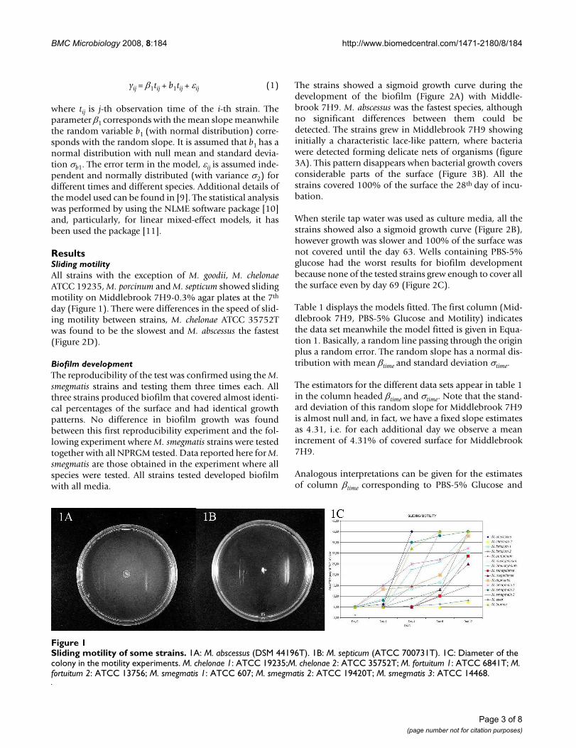
ResultsSliding motilityAll strains with the exception of M. goodii, M. chelonaeATCC 19235, M. porcinum and M. septicum showed slidingmotility on Middlebrook 7H9-0.3% agar plates at the 7th
day (Figure 1). There were differences in the speed of slid-ing motility between strains, M. chelonae ATCC 35752Twas found to be the slowest and M. abscessus the fastest(Figure 2D).
Biofilm developmentThe reproducibility of the test was confirmed using the M.smegmatis strains and testing them three times each. Allthree strains produced biofilm that covered almost identi-cal percentages of the surface and had identical growthpatterns. No difference in biofilm growth was foundbetween this first reproducibility experiment and the fol-lowing experiment where M. smegmatis strains were testedtogether with all NPRGM tested. Data reported here for M.smegmatis are those obtained in the experiment where allspecies were tested. All strains tested developed biofilmwith all media.
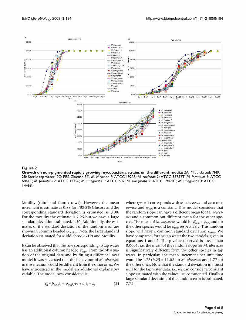
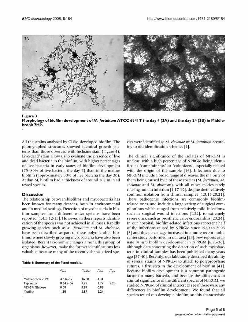
The strains showed a sigmoid growth curve during thedevelopment of the biofilm (Figure 2A) with Middle-brook 7H9. M. abscessus was the fastest species, althoughno significant differences between them could bedetected. The strains grew in Middlebrook 7H9 showinginitially a characteristic lace-like pattern, where bacteriawere detected forming delicate nets of organisms (figure3A). This pattern disappears when bacterial growth coversconsiderable parts of the surface (Figure 3B). All thestrains covered 100% of the surface the 28th day of incu-bation.
When sterile tap water was used as culture media, all thestrains showed also a sigmoid growth curve (Figure 2B),however growth was slower and 100% of the surface wasnot covered until the day 63. Wells containing PBS-5%glucose had the worst results for biofilm developmentbecause none of the tested strains grew enough to cover allthe surface even by day 69 (Figure 2C).
Table 1 displays the models fitted. The first column (Mid-dlebrook 7H9, PBS-5% Glucose and Motility) indicatesthe data set meanwhile the model fitted is given in Equa-tion 1. Basically, a random line passing through the originplus a random error. The random slope has a normal dis-tribution with mean βtime and standard deviation σtime.
The estimators for the different data sets appear in table 1in the column headed βtime and σtime. Note that the stand-ard deviation of this random slope for Middlebrook 7H9is almost null and, in fact, we have a fixed slope estimatesas 4.31, i.e. for each additional day we observe a meanincrement of 4.31% of covered surface for Middlebrook7H9.
Analogous interpretations can be given for the estimatesof column βtime corresponding to PBS-5% Glucose and
Sliding motility of some strainsFigure 1Sliding motility of some strains. 1A: M. abscessus (DSM 44196T). 1B: M. septicum (ATCC 700731T). 1C: Diameter of the colony in the motility experiments. M. chelonae 1: ATCC 19235;M. chelonae 2: ATCC 35752T; M. fortuitum 1: ATCC 6841T; M. fortuitum 2: ATCC 13756; M. smegmatis 1: ATCC 607; M. smegmatis 2: ATCC 19420T; M. smegmatis 3: ATCC 14468.
Page 3 of 8(page number not for citation purposes)
BMC Microbiology 2008, 8:184 http://www.biomedcentral.com/1471-2180/8/184
Motility (third and fourth rows). However, the meanincrement is estimate as 0.88 for PBS-5% Glucose and thecorresponding standard deviation is estimated as 0.08.For the motility the estimate is 2.25 but we have a largestandard deviation estimated, 1.30. Additionally, the esti-mates of the standard deviation of the random error areshown in column headed σresidual. Note the large standarddeviation estimated for Middlebrook 7H9 and Motility.
It can be observed that the row corresponding to tap waterhas an additional column headed ψtype. From the observa-tion of the original data and by fitting a different linearmodel it was suggested that the behaviour of M. abscessusin this medium could be different from the other ones. Wehave introduced in the model an additional explanatoryvariable. The model now considered is:
yij = βtimetij + ψtypetype + b1tij + εij (2)
where type = 1 corresponds with M. abscessus and zero oth-erwise and ψtype is a constant. This model considers thatthe random slope can have a different mean for M. absces-sus and a common but different mean for the other spe-cies. The mean of M. abscessus would be βtime+ ψtype and forthe other species would be βtime respectively. This randomslope will have a common standard deviation σtime. Wehave compared, for the tap water the two models, given inequations 1 and 2. The p-value observed is lesser than0.0001, i.e. the mean of the random slope for M. abscessusis significatively different from the other species in tapwater. In particular, the mean increment per unit timewould be 1.78+9.25 = 11.02 for M. abscessus and 1.77 forthe other ones. Note that the standard deviation is almostnull for the tap water data, i.e. we can consider a constantslope estimated with the values just commented. Finally alarge standard deviation of the random error is estimated,7.79.
Growth on non-pigmented rapidly growing mycobacteria strains on the different media: Figure 2Growth on non-pigmented rapidly growing mycobacteria strains on the different media: 2A: Middlebrook 7H9. 2B: Sterile tap water. 2C: PBS-Glucose 5%. M. chelonae 1: ATCC 19235; M. chelonae 2: ATCC 35752T; M. fortuitum 1: ATCC 6841T; M. fortuitum 2: ATCC 13756; M. smegmatis 1: ATCC 607; M. smegmatis 2: ATCC 19420T; M. smegmatis 3: ATCC 14468.
Page 4 of 8(page number not for citation purposes)
BMC Microbiology 2008, 8:184 http://www.biomedcentral.com/1471-2180/8/184
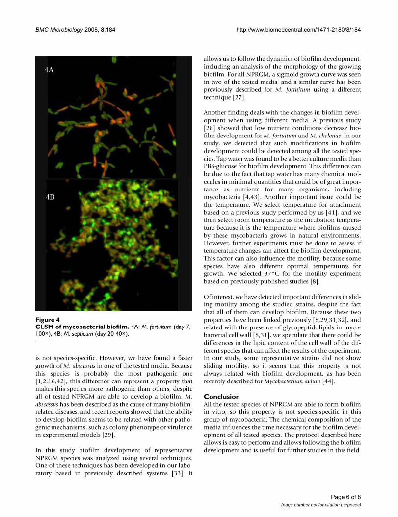
All the strains analysed by CLSM developed biofilm. Thephotographed structures showed identical growth pat-terns than those observed with fuchsine stain (Figure 4).Live/dead© stain allow us to evaluate the presence of liveand dead bacteria in the biofilm, with higher percentagesof live bacteria in early states of biofilm development(75–80% of live bacteria the day 7) than in the maturebiofilm (approximately 50% of live bacteria the day 20).At day 24, biofilm had a thickness of around 20 μm in alltested species.
DiscussionThe relationship between biofilms and mycobacteria hasbeen known for many decades, both in environmentaland in medical settings. Detection of mycobacteria in bio-film samples from different water systems have beenreported [1,4,5,12-15]. However, in these reports identifi-cation of the species was not achieved in all cases. Rapidlygrowing species, such as M. fortuitum and M. chelonae,have been described as part of these polymicrobial bio-films, where slowly growing mycobacteria have also beenisolated. Recent taxonomic changes among this group oforganisms, however, make the former identifications lessvaluable, because many of the recently characterized spe-
cies were identified as M. chelonae or M. fortuitum accord-ing to old identification schemes [1].
The clinical significance of the isolates of NPRGM isunclear, with a high percentage of NPRGM being identi-fied as "contaminants" or "colonizers", especially relatedwith the origin of the sample [16]. Infections due toNPRGM include a broad range of diseases, the majority ofthem being caused by 3 of these species (M. fortuitum, M.chelonae and M. abscessus), with all other species rarelycausing human infection [1,17-19], despite their relativelycommon isolation from clinical samples [1,3,16,20,21].These pathogenic infections are commonly biofilm-related ones, and include a large variety of surgical com-plications which ranged from relatively mild infections,such as surgical wound infections [1,22], to extremelysevere ones, such as prosthetic valve endocarditis [23,24].In our hospital, biofilm-related infections represent halfof the infections caused by NPRGM since 1980 to 2003[3] and this percentage increased in a more recent multi-center study performed in our area [23]. Few reports eval-uate in vitro biofilm development in NPRGM [8,25-36],although data concerning the detection of such mycobac-teria in clinical samples has been published many yearsago [37-40]. Recently, our laboratory described the abilityof several strains of NPRGM to attach to polypropylenesutures, a first step in the development of biofilm [41].Because biofilm development is a common pathogenicfactor for many bacteria, and because the differences inclinical significance of the different species of NPRGM, westudied NPRGM of clinical interest to see if there were anydifferences in biofilm development. We found that allspecies tested can develop a biofilm, so this characteristic
Morphology of biofilm development of M. fortuitum ATCC 6841T the day 4 (3A) and the day 24 (3B) in Middlebrook 7H9Figure 3Morphology of biofilm development of M. fortuitum ATCC 6841T the day 4 (3A) and the day 24 (3B) in Middle-brook 7H9.
Table 1: Summary of the fitted models.
σtime σresidual βtime βtype
Middlebrook 7H9 4.63e-05 16.00 4.31Tap water 8.64 e-06 7.79 1.77 9.25PBS-5% Glucose 0.08 3.89 0.88Motility 1.30 5.87 2.24
Page 5 of 8(page number not for citation purposes)
BMC Microbiology 2008, 8:184 http://www.biomedcentral.com/1471-2180/8/184
is not species-specific. However, we have found a fastergrowth of M. abscessus in one of the tested media. Becausethis species is probably the most pathogenic one[1,2,16,42], this difference can represent a property thatmakes this species more pathogenic than others, despiteall of tested NPRGM are able to develop a biofilm. M.abscessus has been described as the cause of many biofilm-related diseases, and recent reports showed that the abilityto develop biofilm seems to be related with other patho-genic mechanisms, such as colony phenotype or virulencein experimental models [29].
In this study biofilm development of representativeNPRGM species was analyzed using several techniques.One of these techniques has been developed in our labo-ratory based in previously described systems [33]. It
allows us to follow the dynamics of biofilm development,including an analysis of the morphology of the growingbiofilm. For all NPRGM, a sigmoid growth curve was seenin two of the tested media, and a similar curve has beenpreviously described for M. fortuitum using a differenttechnique [27].
Another finding deals with the changes in biofilm devel-opment when using different media. A previous study[28] showed that low nutrient conditions decrease bio-film development for M. fortuitum and M. chelonae. In ourstudy, we detected that such modifications in biofilmdevelopment could be detected among all the tested spe-cies. Tap water was found to be a better culture media thanPBS-glucose for biofilm development. This difference canbe due to the fact that tap water has many chemical mol-ecules in minimal quantities that could be of great impor-tance as nutrients for many organisms, includingmycobacteria [4,43]. Another important issue could bethe temperature. We select temperature for attachmentbased on a previous study performed by us [41], and wethen select room temperature as the incubation tempera-ture because it is the temperature where biofilms causedby these mycobacteria grows in natural environments.However, further experiments must be done to assess iftemperature changes can affect the biofilm development.This factor can also influence the motility, because somespecies have also different optimal temperatures forgrowth. We selected 37°C for the motility experimentbased on previously published studies [8].
Of interest, we have detected important differences in slid-ing motility among the studied strains, despite the factthat all of them can develop biofilm. Because these twoproperties have been linked previously [8,29,31,32], andrelated with the presence of glycopeptidolipids in myco-bacterial cell wall [8,31], we speculate that there could bedifferences in the lipid content of the cell wall of the dif-ferent species that can affect the results of the experiment.In our study, some representative strains did not showsliding motility, so it seems that this property is notalways related with biofilm development, as has beenrecently described for Mycobacterium avium [44].
ConclusionAll the tested species of NPRGM are able to form biofilmin vitro, so this property is not species-specific in thisgroup of mycobacteria. The chemical composition of themedia influences the time necessary for the biofilm devel-opment of all tested species. The protocol described hereallows is easy to perform and allows following the biofilmdevelopment and is useful for further studies in this field.
CLSM of mycobacterial biofilmFigure 4CLSM of mycobacterial biofilm. 4A: M. fortuitum (day 7, 100×), 4B: M. septicum (day 20 40×).
Page 6 of 8(page number not for citation purposes)
BMC Microbiology 2008, 8:184 http://www.biomedcentral.com/1471-2180/8/184
Authors' contributionsJE coordinate all the study. Conceived and participate inthe design the study, and participates in the analysis of theresults. NZM perform the experimental study and collab-orates in the analysis of the results. TJK participates in theanalysis of the results and helped to draft the manuscript.GA perform the statistical analysis. RFR participates in thedesign of the study. IG participates in the design of thestudy and in the statistical analysis. All the authors partic-ipate in the redaction of the manuscript, have read it andapproved it.
AcknowledgementsNieves Z. Martín-de-Hijas was funded by the Fundación Conchita Rábago de Jiménez Díaz. Kinnari TJ was funded by the Academy of Finland, Paulo Foundation and Proteesisäätiö Foundation. This work was funded by a grant from the Comunidad de Madrid (S-0505/MAT/000324). Conflicts of interest: No conflicts for all authors.
References1. Brown-Elliott BA, Wallace RJ: Clinical and Taxonomic Status of
Pathogenic Nonpigmented or Late-Pigmenting RapidlyGrowing Mycobacteria. Clinical Microbiology Reviews 2002,15(4):716-746.
2. De Groote MA, Huitt G: Infections due to rapidly growingmycobacteria. Clin Infect Dis 2006, 42:1756-1763.
3. Esteban J, Fernández-Roblas R, García-Cía JI, Zamora N, Ortiz A:Clinical Significance and Epidemiology of Non-PigmentedRapidly Growing Mycobacteria in a University Hospital. JInfect 2007, 54:135-145.
4. Vaerewijck MJM, Huys G, Palomino JC, Swings J, Portaels F: Myco-bacteria in drinking water distribution systems: ecology andsignificance for human health. FEMS Microbiol Rev 2005,29:911-934.
5. Falkinham JO, Norton CD, LeChevallier MW: Factors influencingnumbers of Mycobacterium avium, Mycobacterium intracellu-lare, and other mycobacteria in drinking water distributionsystems. Appl Environ Microbiol 2001, 67(3):1225-1231.
6. Donlan RM: Biofilms and device-associated infections. Emerg-ing Infectious Diseases 2001, 7(2):277-279.
7. Fux CA, Costerton JW, Stewart PS, Stoodley P: Survival strategiesof infectious biofilms. Trends Microbiol 2005, 13(1):34-40.
8. Martinez A, Torello S, Kolter R: Sliding motility in mycobacteria.Journal of Bacteriology 1999, 181(23):7331-7338.
9. Pinheiro JC, Bates DM: Mixed-Effects Models in S and S-PLUS.In Statistics and Computing Springer; 2000.
10. R-Development-Core-Team: R: A Language and Environment for Statis-tical Computing Vienna: R Foundation for Statistical Computing; 2006.
11. Pinheiro JC, Bates DM, DebRoy S, Sarkar D: NLME: Linear andnonlinear mixed effects models. R package version 3.1-79. edn2006.
12. Torvinen E, Suomalainen S, Lehtola MJ, Miettinen IT, Zacheus O, Pau-lin L, Katila ML, Martikainen PJ: Mycobacteria in water and loosedeposits of drinking water distribution systems in Finland.Appl Environ Microbiol 2004, 70(4):1973-1981.
13. Wallace RJ, Musser JM, Hull SI, Silcox VA, Steele LC, Forrester GD,Labidi A, Selander RK: Diversity and sources of rapidly growingmycobacteria associated with infections following cardiacsurgery. J Infect Dis 1989, 159(4):708-716.
14. Santos R, Oliveira F, Fernandes J, Gonçalves S, Macieira F, Cadete M:Detection and identification of mycobacteria in the Lisbonwater distribution system. Water Sci Technol 2005,52(8):177-180.
15. Barbeau J, Gauthier C, Payment P: Biofilms, infectious agents,and dental unit waterlines: a review. Can J Microbiol 1998,44(11):1019-1028.
16. Esteban J, Martín-de-Hijas NZ, Fernández AI, Fernández-Roblas R,Gadea I, Madrid-Study-Group-of-Mycobacteria: Epidemiology ofinfections due to Non-pigmented Rapidly Growing Mycobac-
teria diagnosed in an urban area. Eur J Clin Microbiol Infect Dis2008, 27(10):951-7.
17. Sohail MR, Smilack JD: Hernia repair mesh-associated Mycobac-terium goodi infection. J Clin Microbiol 2004, 42(6):2858-2860.
18. Schinsky MF, McNeil MM, Whitney AM, Steigerwalt AG, Lasker BA,Floyd MM, Hogg GG, Brenner DJ, Brown JM: Mycobacterium septi-cum sp. nov., a new rapidly growing species associated withcatheter-related bacteraemia. Int J Syst Evol Microbiol 2000,50(Pt 2):575-581.
19. Schinsky MF, Morey RE, Steigerwalt AG, Douglas MP, Wilson RW,Floyd MM, Butler WR, Daneshvar MI, Brown-Elliott BA, Wallace RJ:Taxonomic variation in the Mycobacterium fortuitum thirdbiovariant complex: Description of Mycobacterium boenickeisp. nov., Mycobacterium houstonense sp. nov., Mycobacteriumneworleansense sp. nov., and Mycobacterium brisbanense sp.nov., and recognition of Mycobacterium porcinum fromhuman clinical specimens. Int J Syst Evol Microbiol 2004, 54(Pt5):1653-1667.
20. Burns DN, Wallace RJ, Schultz ME, Zhang YS, Zubairi SQ, Pang YJ,Gibert CL, Brown BA, Noel ES, Gordin FM: Nosocomial outbreakof respiratory tract colonization with Mycobacterium fortui-tum : demonstration of the usefulness of pulsed-field gel elec-trophoresis in an epidemiologic investigation. Am Rev RespirDis 1991, 144(5):1153-1159.
21. Wallace RJ, Brown BA, Griffith DE: Nosocomial outbreaks/pseudo-outbreaks caused by nontuberculous mycobacteria.Annu Rev Microbiol 1998, 52:453-490.
22. Wallace RJ, Swenson JM, Silcox VA, Good RC, Tschen JA, Stone MS:Spectrum of disease due to rapidly growing mycobacteria.Rev Infect Dis 1983, 5(4):657-679.
23. Kuritsky JN, Bullen MG, Broome CV, Silcox VA, Good RC, WallaceRJ: Sternal wound infections and endocarditis due to organ-isms of the Mycobacterium fortuitum complex. Ann Intern Med1983, 98(6):938-939.
24. Olalla J, Pombo M, Aguado JM, Rodriguez E, Palenque E, Costa JR,Rioperez E: Mycobacterium fortuitum complex endocarditis-case report and literature review. Clin Microbiol Infect 2002,8(2):125-129.
25. Bardouniotis E, Ceri H, Olson ME: Biofilm formation and biocidesusceptibility testing of Mycobacterium fortuitum and Myco-bacterium marinum. Curr Microbiol 2003, 46:28-32.
26. Chen JM, German GJ, Alexander DC, Ren H, Tan T, Liu J: Roles ofLsr2 in colony morphology and biofilm formation of Myco-bacterium smegmatis. Journal of Bacteriology 2006, 188(2):633-641.
27. Hall-Stoodley L, Lappin-Scott H: Biofilm formation by the rapidlygrowing mycobacterial species Mycobacterium fortuitum.FEMS Microbiol Lett 1998, 168(1):77-84.
28. Hall-Stoodley L, Keevil CW, Lappin-Scott H: Mycobacterium fortu-itum and Mycobacterium chelonae biofilm formation underhigh and low nutrient conditions. J Appl Microbiol 1999, 65(Sym-posium Supplement):60S-69S.
29. Howard ST, Rhoades E, Recht J, Pang X, Alsup A, Kolter R, Lyons CR,Byrd TF: Spontaneous reversion of Mycobacterium abscessusfrom a smooth to a rough morphotype is associated withreduced expression of glycopeptidolipid and reacquisition ofan invasive phenotype. Microbiology 2006, 152:1581-1590.
30. Ojha A, Anand M, Bhatt A, Kremer L, Jacobs WR, Hatfull GF:GroEL1: A dedicated chaperone involved in mycolic acid bio-synthesis during biofilm formation in mycobacteria. Cell2005, 123:861-873.
31. Recht J, Kolter R: Glycopeptidolipid acetylation affects slidingmotility and biofilm formation in Mycobacterium smegmatis.J Bacteriol 2001, 183(19):5718-5724.
32. Recht J, Martinez A, Torello S, Kolter R: Genetic analysis of slidingmotility in Mycobacterium smegmatis. Journal of Bacteriology2000, 182(15):4348-4351.
33. Rose L, Kaufmann SH, Daugelat S: Involvement of Mycobacteriumsmegmatis undecaprenyl phosphokinase in biofilm andsmegma formation. Microbes Infect 2004, 6(11):965-971.
34. Teng R, Dick T: Isoniazid resistance of exponentially growingMycobacterium smegmatis biofilm culture. FEMS Microbiol Lett2003, 227:171-174.
35. Zambrano MM, Kolter R: Mycobacterial biofilms: a greasy wayto hold it together. Cell 2005, 123:762-764.
36. Greendyke R, Byrd TF: Differential antibiotic susceptibility ofMycobacterium abscessus variants In biofilms and macro-
Page 7 of 8(page number not for citation purposes)
BMC Microbiology 2008, 8:184 http://www.biomedcentral.com/1471-2180/8/184
Publish with BioMed Central and every scientist can read your work free of charge
"BioMed Central will be the most significant development for disseminating the results of biomedical research in our lifetime."
Sir Paul Nurse, Cancer Research UK
Your research papers will be:
available free of charge to the entire biomedical community
peer reviewed and published immediately upon acceptance
cited in PubMed and archived on PubMed Central
yours — you keep the copyright
Submit your manuscript here:http://www.biomedcentral.com/info/publishing_adv.asp
BioMedcentral
phages compared to planktonic bacteria. Antimicrob AgentsChemother 2008, 52(6):2019-2026.
37. Schulze-Robbecke R, Feldmann C, Fischeder R, Janning B, Exner M,Wahl G: Dental units: an environmental study of sources ofpotentially pathogenic mycobacteria. Tuber Lung Dis 1995,76(4):318-323.
38. Schulze-Robbecke R, Janning B, Fischeder R: Occurrence of myco-bacteria in biofilm samples. Tuber Lung Dis 1992, 73(3):141-144.
39. Schulze-Röbbecke R, Fischeder R: Mycobacteria in biofilms. Zen-tralbl Hyg Umweltmed 1989, 188(3–4):385-390.
40. Raad II, Vartivarian S, Khan A, Bodey GP: Catheter-related infec-tions caused by the Mycobacterium fortuitum complex: 15cases and review. Rev Infect Dis 1991, 13(6):1120-1125.
41. Zamora N, Esteban J, Kinnari TJ, Celdran A, Granizo JJ, Zafra C: Invitro evaluation of the attachment to polypropylene sutureson non-pigmented rapidly growing mycobacteria. Clin Micro-biol Infect 2007, 13(9):902-907.
42. Petrini B: Mycobacterium abscessus : An emerging rapid-grow-ing potential pathogen. APMIS 2006, 114:319-328.
43. Chalupka S: Tainted water on tap: What to tell patients aboutpreventing illness from drinking water. Am J Nurs 2005,105(11):40-52.
44. Freeman R, Geier H, Weigel KM, Do J, Ford TE, Cangelosi GA: Rolesfor Cell Wall Glycopeptidolipid in Surface Adherence andPlanktonic Dispersal of Mycobacterium avium. Appl EnvironMicrobiol 2006, 72(12):7554-7558.
Page 8 of 8(page number not for citation purposes)


