Embed Size (px)
Citation preview

bioengineering
Review
Biofabrication Strategies for Musculoskeletal Disorders:Evolution towards Clinical Applications
Saman Naghieh 1,*,† , Gabriella Lindberg 2,3,*,† , Maryam Tamaddon 4,* and Chaozong Liu 4,*
�����������������
Citation: Naghieh, S.; Lindberg, G.;
Tamaddon, M.; Liu, C. Biofabrication
Strategies for Musculoskeletal
Disorders: Evolution towards Clinical
Applications. Bioengineering 2021, 8,
123. https://doi.org/10.3390/
bioengineering8090123
Academic Editor:
Christine Knabe-Ducheyne
Received: 6 August 2021
Accepted: 3 September 2021
Published: 10 September 2021
Publisher’s Note: MDPI stays neutral
with regard to jurisdictional claims in
published maps and institutional affil-
iations.
Copyright: © 2021 by the authors.
Licensee MDPI, Basel, Switzerland.
This article is an open access article
distributed under the terms and
conditions of the Creative Commons
Attribution (CC BY) license (https://
creativecommons.org/licenses/by/
4.0/).
1 Division of Biomedical Engineering, University of Saskatchewan, Saskatoon, SK S7N 5A9, Canada2 Christchurch Regenerative Medicine and Tissue Engineering (CReaTE) Group, Department of Orthopaedic
Surgery, University of Otago Christchurch, Christchurch 8011, New Zealand3 Knight Campus for Accelerating Scientific Impact, University of Oregon, Eugene, OR 97403, USA4 Institute of Orthopaedic & Musculoskeletal Science, Royal National Orthopaedic Hospital, University College
London, Stanmore HA7 4LP, UK* Correspondence: [email protected] (S.N.); [email protected] (G.L.);
[email protected] (M.T.); [email protected] (C.L.)† These two authors contributed equally.
Abstract: Biofabrication has emerged as an attractive strategy to personalise medical care andprovide new treatments for common organ damage or diseases. While it has made impactfulheadway in e.g., skin grafting, drug testing and cancer research purposes, its application to treatmusculoskeletal tissue disorders in a clinical setting remains scarce. Albeit with several in vitrobreakthroughs over the past decade, standard musculoskeletal treatments are still limited to palliativecare or surgical interventions with limited long-term effects and biological functionality. To betterunderstand this lack of translation, it is important to study connections between basic sciencechallenges and developments with translational hurdles and evolving frameworks for this fullydisruptive technology that is biofabrication. This review paper thus looks closely at the processingstage of biofabrication, specifically at the bioinks suitable for musculoskeletal tissue fabricationand their trends of usage. This includes underlying composite bioink strategies to address theshortfalls of sole biomaterials. We also review recent advances made to overcome long-standingchallenges in the field of biofabrication, namely bioprinting of low-viscosity bioinks, controlleddelivery of growth factors, and the fabrication of spatially graded biological and structural scaffoldsto help biofabricate more clinically relevant constructs. We further explore the clinical application ofbiofabricated musculoskeletal structures, regulatory pathways, and challenges for clinical translation,while identifying the opportunities that currently lie closest to clinical translation. In this article, weconsider the next era of biofabrication and the overarching challenges that need to be addressed toreach clinical relevance.
Keywords: additive manufacturing; 3D bioprinting; smart hydrogels; biomaterials; tissue engineering;musculoskeletal disorders
1. Introduction
The musculoskeletal system, consisting of different types of bones, muscles, ligamentsand tendons, is one of the key systems in the human body. According to the World HealthOrganisation, approximately 1.71 billion people suffer from musculoskeletal conditionswhich makes it a leading cause of pain and disability worldwide [1]. Musculoskeletalsoft tissues are integral in ensuring joint movement and stabilisation. Injuries to thesestructures are prevalent, particularly in sports-active adults [2], and can be acute as a resultof a traumatic event, or chronic as a result of overuse or cumulative trauma injuries [2,3].Ruptures of the Achilles tendon and anterior cruciate ligament (ACL) are examples ofacute injuries, which are among the most frequent and severe injuries sustained in a sports-active population [4,5]. Cartilage is another tissue that can be damaged as a result of a
Bioengineering 2021, 8, 123. https://doi.org/10.3390/bioengineering8090123 https://www.mdpi.com/journal/bioengineering

Bioengineering 2021, 8, 123 2 of 33
trauma, which, if not treated properly, can lead to degenerative diseases of the joints suchas osteoarthritis [6].
Treatment modalities for musculoskeletal soft-tissue injuries are broad and rangefrom “wait-and-see” approaches through to surgery [2]. Surgical treatments will dependon the type of tissue and the injury sustained; for example, in ACL reconstruction, theuse of grafts (auto-, allo- and synthetic), specifically biological grafts, are considered thegold standard [4]. For cartilage defects, microfracture is used, where small holes aredrilled into the subchondral bone to allow the transport of blood and bone marrow to thebone/cartilage interface, where the mesenchymal stem cells contribute to the formationand repair of the cartilage and bone [6]. Osteochondral auto- and allograft are other optionsfor small- to mid-sized defects [6,7]. However, the current clinical methods of repair formusculoskeletal soft tissues are not without their drawbacks. For example, in cartilagerepair using the microfracture technique, the regenerated tissue is fibrocartilage, whichdoes not have the same durability as the native hyaline cartilage, leading to an ongoingarticular surface irregularity and subsequent secondary arthritic changes [6,8]. A lackof tissue integration in the osteochondral graft technique is a major cause of failure inthese operations [9]. In the autograft method of ACL reconstruction, tendons from otherparts of the body, generally patella or hamstring, are used, which can lead to donor sitemorbidity and pain. Allografts have a risk of disease transmission and immune-mediatedgraft rejection [10,11]. Since they lack cellularity, they often require an extended period ofrevascularisation and incorporation (over a year) before a sports-active patient is allowedto return to play [12].
Considering the shortcomings associated with tissue grafts and other reconstructiveoptions, tissue engineering approaches have been developed as an alternative treatmentshowing enormous potential for the regeneration of musculoskeletal soft tissues. However,their long-term success has been restricted, mainly due to the limitation in current fabrica-tion methods that allows the production of a scaffold that mimics the cellular microenviron-ment and cell–cell interactions [13] observed in the native tissue. Musculoskeletal tissuesare complex units, with compositional, mechanical, structural and cellular heterogeneity.For example, ligaments and tendons are connective tissues comprised of a dense band ofaligned collagen (mainly type I) with embedded resident cells (fibroblasts/tenocytes) thatconnect bone to bone and muscle to bone, respectively [14,15]. The attachments betweenthese interfacing tissues are unique and complex, with well-defined spatial changes in cellphenotype, matrix composition and mechanical properties (Figure 1) [14,16]. In the caseof tendons/ligaments, the entheses (insertion into the bone) is composed of four distinctzones along the longitudinal direction: tendon/ligament (I), non-mineralised fibrocartilage(II), mineralised fibrocartilage (III), and bone (IV) [17,18]. While tenocytes/fibroblasts areembedded in the tendon/ligament matrix, the resident cells in the non-mineralised fibrocar-tilage are fibrochondrocytes, and hypertrophic chondrocytes are found in the mineralisedfibrocartilage [19].
Considering the intricacies of the musculoskeletal tissues and the inherent hetero-geneity in cellular and structural properties, it becomes clear that for a successful tissueengineering approach, the fabrication technique needs to recapitulate the compositional,mechanical, structural and cellular heterogeneity. Advances in additive manufacturinghave opened new possibilities to fabricate structures with synergistic biological and me-chanical properties that can mimic the natural tissue structures. Specifically, biofabricationor 3D bioprinting has been increasingly used as a revolutionary approach to healthcarethat utilises additive manufacturing processes to produce a biological construct [20,21].
Biofabrication combines cells, biomaterials and biological factors—collectively knownas bioinks—and delivers them in a precise pattern to recapitulate elements of heterogeneoustissues [22–24]. Although biofabrication has made a remarkable impact in areas of tissueengineering and drug delivery, its application and rapid progress into the clinic has beenlimited, firstly, by the lack of suitable biomaterials and bioinks [25,26]. In the field oforthopaedics, while a wide range of biomaterials have been applied as implants for treating

Bioengineering 2021, 8, 123 3 of 33
common musculoskeletal defects, ranging from mechanically robust but relatively inertmetals, ceramics and plastics [27], to bioactive but rather amorphous hydrogels [28], onlya few are compatible with the process of bioprinting living cells [29–34]. In addition,although much research is being undertaken in the biofabrication field, few biofabricatedconstructs have yet been applied in widespread clinical settings due to a lack of regulatoryclarity surrounding the frameworks in which these constructs lie.
Bioengineering 2021, 8, x FOR PEER REVIEW 3 of 33
Figure 1. Tendon–bone interface showing the multicomponent and heterogeneous tissue structure.
Adapted from Barajaa et al., 2020. Reused with permission from Springer Nature. Created with Bi-
oRender.com.
Considering the intricacies of the musculoskeletal tissues and the inherent heteroge-
neity in cellular and structural properties, it becomes clear that for a successful tissue en-
gineering approach, the fabrication technique needs to recapitulate the compositional, me-
chanical, structural and cellular heterogeneity. Advances in additive manufacturing have
opened new possibilities to fabricate structures with synergistic biological and mechanical
properties that can mimic the natural tissue structures. Specifically, biofabrication or 3D
bioprinting has been increasingly used as a revolutionary approach to healthcare that uti-
lises additive manufacturing processes to produce a biological construct [20,21].
Biofabrication combines cells, biomaterials and biological factors—collectively
known as bioinks—and delivers them in a precise pattern to recapitulate elements of het-
erogeneous tissues [22–24]. Although biofabrication has made a remarkable impact in ar-
eas of tissue engineering and drug delivery, its application and rapid progress into the
clinic has been limited, firstly, by the lack of suitable biomaterials and bioinks [25,26]. In
the field of orthopaedics, while a wide range of biomaterials have been applied as im-
plants for treating common musculoskeletal defects, ranging from mechanically robust
but relatively inert metals, ceramics and plastics [27], to bioactive but rather amorphous
hydrogels [28], only a few are compatible with the process of bioprinting living cells [29–
34]. In addition, although much research is being undertaken in the biofabrication field,
few biofabricated constructs have yet been applied in widespread clinical settings due to
a lack of regulatory clarity surrounding the frameworks in which these constructs lie.
This review paper thus looks closely at the processing stage of biofabrication with a
focus on bioinks suitable for musculoskeletal tissue fabrication. We mainly explore syn-
thetic and natural bioinks before discussing the underlying composite bioink strategies to
address the shortfalls of sole biomaterials. We also review recent advances made to over-
come long-standing challenges in the field of biofabrication, namely the bioprinting of
low-viscosity bioinks, controlled delivery of growth factors, and the fabrication of spa-
tially graded biological and structural scaffolds to help biofabricate more clinically rele-
vant constructs. We further explore the regulatory and translational pathways for the clin-
ical application of biofabricated musculoskeletal structures, highlighting both the chal-
lenges and the opportunities that currently lie closest to clinical translation. In this article,
we consider the next era of biofabrication and the overarching challenges that need to be
addressed to reach clinical relevance.
Figure 1. Tendon–bone interface showing the multicomponent and heterogeneous tissue struc-ture. Adapted from Barajaa et al., 2020. Reused with permission from Springer Nature. Createdwith BioRender.com.
This review paper thus looks closely at the processing stage of biofabrication witha focus on bioinks suitable for musculoskeletal tissue fabrication. We mainly exploresynthetic and natural bioinks before discussing the underlying composite bioink strategiesto address the shortfalls of sole biomaterials. We also review recent advances made toovercome long-standing challenges in the field of biofabrication, namely the bioprinting oflow-viscosity bioinks, controlled delivery of growth factors, and the fabrication of spatiallygraded biological and structural scaffolds to help biofabricate more clinically relevantconstructs. We further explore the regulatory and translational pathways for the clinicalapplication of biofabricated musculoskeletal structures, highlighting both the challengesand the opportunities that currently lie closest to clinical translation. In this article, weconsider the next era of biofabrication and the overarching challenges that need to beaddressed to reach clinical relevance.
2. Biofabrication
Additive manufacturing can be categorised under three groups of laser/light-based,extrusion-based, and jetting/powder-based technologies. Specifically, additive manu-facturing methods have been developed recently for biofabrication applications underthese three groups. Additive manufactured biofabrication methods are lithography-based(light/laser-based group), extrusion-based, and inkjet/multi-jet bioprinting (Figure 2). Inbiofabrication, complex, sophisticated and biomimetic tissues can be constructed with highprecision. Another advantage of this method is the ability for automation and controlover geometrical intricacies. This includes the possibility of incorporation and precisespatiotemporal placement of cells, proteins, drugs and biologics [35].
Among biofabrication methods (Figure 2), stereolithography (SLA), also called digitallight processing, is known as one of the oldest additive manufacturing techniques. Due toits relatively high precision, it is commonly used to create anatomical models for preopera-tive planning [36]. In this method, photosensitive resin is exposed to a high-intensity light
Bioengineering 2021, 8, 123 4 of 33
source in order to solidify and fabricate a 3D structure layer-by-layer. One of the advantagesof this technique is that the fabricated structure has a clear to opaque white appearance afterfabrication due to the nature of utilised photopolymer materials; this can help to visualiseinternal anatomies clearly. In addition, photosensitive materials are limited, and as a result,it limits the use of this method [29,30]. Extrusion-based bioprinting is operationally moreadoptable with respect to printing multiple biomaterials and cell types and it has openednew ways to fabricate complex tissues and organs [37]. Specifically, this technique has beenused widely to create hybrid and composite structures in order to have both hydrogels andsynthetic polymers in one structure; hydrogel creates a cell-friendly environment used fordrug-delivery purposes and growth factor/cell incorporation, while a synthetic polymeris used to enhance the mechanical properties due to the poor mechanical characteristicsof hydrogels.
Bioengineering 2021, 8, x FOR PEER REVIEW 5 of 33
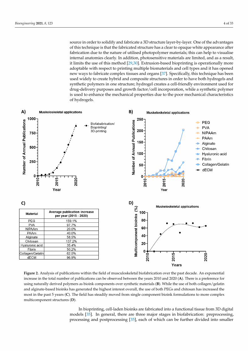
Figure 2. Analysis of publications within the field of musculoskeletal biofabrication over the past decade. An exponential
increase in the total number of publications can be observed between the years 2010 and 2020 (A). There is a preference
for using naturally derived polymers as bioink components over synthetic materials (B). While the use of both colla-
gen/gelatin and alginate-based bioinks has generated the highest interest overall, the use of both PEGs and chitosan has
increased the most in the past 5 years (C). The field has steadily moved from single component bioink formulations to
more complex multicomponent structures (D).
3. Designing Musculoskeletal Bioinks
Significant breakthroughs in material synthesis and crosslinking chemistry over the
past decade have enabled the biofabrication of precursory solutions consisting of both
living components and crosslinkable polymers, so-called bioinks [30,33,39–56]. These pol-
ymers can hold large amounts of water once crosslinked into a hydrogel network [57,58],
which is key to protecting both the cells and biological compounds during and after the
biofabrication process. These bioinks can be fabricated from both biological and synthetic
polymer materials, having more or less inherent biocomplexity, which comes with differ-
ent sets of both benefits and limitations. Simplified, synthetic polymers offer more precise
control over the system but lack the necessary and appropriate biological cues to help
guide cell development, and vice versa. However, the shortcomings of both classes of ma-
terials are nowadays, to a certain degree, possible to overcome by appropriate chemical
and biological modifications as well as hybrid technologies [29,59]. In this section, we will
look more closely at the choice of bioinks used for musculoskeletal soft tissue fabrication
and the clinical relevance of the different classes of materials.
Figure 2. Analysis of publications within the field of musculoskeletal biofabrication over the past decade. An exponentialincrease in the total number of publications can be observed between the years 2010 and 2020 (A). There is a preference forusing naturally derived polymers as bioink components over synthetic materials (B). While the use of both collagen/gelatinand alginate-based bioinks has generated the highest interest overall, the use of both PEGs and chitosan has increased themost in the past 5 years (C). The field has steadily moved from single component bioink formulations to more complexmulticomponent structures (D).
In bioprinting, cell-laden bioinks are fabricated into a functional tissue from 3D digitalmodels [35]. In general, there are three major stages in biofabrication: preprocessing,processing and postprocessing [35], each of which can be further divided into smaller

Bioengineering 2021, 8, 123 5 of 33
steps. In preprocessing, a digital model is generated from imaging (X-ray, computedtomography (CT), magnetic resonance imaging MRI) and the design approach (biomimicry,self-assembly, mini-tissues) is selected. In the processing stage, the bioink material (naturalpolymers, synthetic polymers, ECM), cells (differentiated cells, pluripotent stem cells,multipotent stem cells) and bioprinting technique (lithography-based, extrusion-based,and inkjet/multi-jet bioprinting) are selected. Finally, in the postprocessing steps, accordingto the application (maturation, implantation, in vitro testing), the tissue is utilised [38].
3. Designing Musculoskeletal Bioinks
Significant breakthroughs in material synthesis and crosslinking chemistry over thepast decade have enabled the biofabrication of precursory solutions consisting of bothliving components and crosslinkable polymers, so-called bioinks [30,33,39–56]. Thesepolymers can hold large amounts of water once crosslinked into a hydrogel network [57,58],which is key to protecting both the cells and biological compounds during and after thebiofabrication process. These bioinks can be fabricated from both biological and syntheticpolymer materials, having more or less inherent biocomplexity, which comes with differentsets of both benefits and limitations. Simplified, synthetic polymers offer more precisecontrol over the system but lack the necessary and appropriate biological cues to helpguide cell development, and vice versa. However, the shortcomings of both classes ofmaterials are nowadays, to a certain degree, possible to overcome by appropriate chemicaland biological modifications as well as hybrid technologies [29,59]. In this section, we willlook more closely at the choice of bioinks used for musculoskeletal soft tissue fabricationand the clinical relevance of the different classes of materials.
A total of 4105 publications (out of which 1222 are review articles) containing thekeyword “3D-printing”, or “3D-bioprinting”, or “biofabrication”, and either “bone”, “carti-lage”, “osteochondral”, “tendon”, “muscle”, “osteogenesis”, “chondrogenesis”, or “vas-culogenesis”, and not “cardiac”, were retrieved from the Scopus database from 2010 to2020, disclosing an exponential growth with an average publication increase of 53.3% peryear (Figure 2A). The retrieved data reveals that the use of naturally derived materialsfar outnumbers synthetic based bioink systems throughout the analysed time-period. Asfurther detailed in Figure 2B, the number of original research articles exemplified by someof the most commonly applied material types confirms that both collagen/gelatin andalginate-based bioinks have remained a popular choice over the past decade (876 publica-tions combined). While an increase in publications has been seen across all material types,it should be noted that the use of both polyethylene glycol (PEG, 159.1%) and chitosan(137.2%) have increased the most over the past 5 years (Figure 2C). This may reflect thatmany crosslinking systems can benefit from the added control that synthetic PEG-basedlinkers provide, while the addition of naturally derived chitosan offers a biofunctionalstrategy to increase the viscosity of various bioink formulations. While early biofabricationstudies focused on the development of single-component bioink formulations, the fieldquickly recognised the power of hybrid bioink formulations and shifted towards multi-component bioinks. Between the years 2010 and 2013, 82% of the published papers werebased on single-component bioink formulations, while the majority of papers (67%) pub-lished between 2014 and 2020 utilised multicomponent bioinks (Figure 2D). More detailedinformation around the use of these commonly applied bioink materials are exemplified inthe sections below.
3.1. Synthetic Materials
The application of synthetic polymers as shear-thinning bioinks has proven advan-tageous as it comes with high printability, structural fidelity, batch reproducibility, andindustrial scalability [31–33,60–62]. Synthetic bioinks have thus been used since the be-ginning of the biofabrication era. Key examples includes the use of Polyethylene gly-cols (PEGs) [63–66], Poly(vinyl alcohol) (PVA) [46,67–69], N-isopropylacrylamide (NI-

Bioengineering 2021, 8, 123 6 of 33
PAAm) [70,71], and polyacrylamide (PAAm) [72–74]. In this section, recent developmentsand clinical advantages of these materials are highlighted.
By optimising crosslinking concentrations, Peak et al. was able to tune the function ofdegradable PEG-based bioinks to prolong the delivery of growth factors for up to 28 daysand thus promoted the migration of endothelial umbilical vein cells for long-term cultures,as seen in Figure 3A [63]. This directed chemotaxis strategy holds great translational valueas the retained 4-week bioactivity allows for reduced growth factor concentrations to beused. Furthermore, it can be used as a cell-free, and thus an off-the-shelf, product withtailorable release profiles. In addition to delivering therapeutics, PEG can be designed tohave a broad range of molecular weight distributions and functional arms for chemicalcrosslinking, explaining its widespread use across various platforms [75–77]. While havinga similar chemical structure, additional side-groups provided through Poly(glycidol)s (PG)based bioinks enable even further tailorability, albeit still requiring the addition of naturallyderived biomaterials to induce cellular functionality [62].
Bioengineering 2021, 8, x FOR PEER REVIEW 8 of 33
Figure 3. Key examples of synthetic materials used as bioinks and biomaterial-inks. By tuning the function of degradable
PEG-based bioinks and the biofabrication of high surface area structures using extrusion-based printing technologies and
UV crosslinking, Peak et al. prolonged the delivery of VEGF for up to 28 days to direct the migration of HUVECs encap-
sulated in surrounding GelMA hydrogels after 7 days of culture (A). Change the format as follows: Reprinted with per-
mission from ref. [63]. Copyright 2019 John Wiley and Sons. PNIPAAM-based bioinks printed with extrusion-based tech-
nologies show better chondrocyte viability after washing away residual pNIPAAM, with increasing cell death observed
around the pores, * p < 0.05 and ** p < 0.001 (B). Change the format as follows: Reprinted with permission from ref [70].
Copyright 2015 Acta Biomaterialia. Other examples include acrylamide and glycerol-based biomaterial inks extruded with
directional control to enable alignment of 3T3 fibroblasts using a microperiodic hydrogel scaffold (green) as compared to
the flat glass control (left, C). Reprinted with permission from ref [73]. Copyright 2009 John Wiley and Sons.
3.2. Natural Materials
The application of naturally derived polymers as bioinks is attractive as it includes mo-
tifs that are recognised by cells and can be used to guide the tissue regeneration process.
However, natural hydrogels that can be fabricated on a kg-scale at a low price, narrows
down the library of possible candidates. Commonly used candidates are thus centred on
alginate, chitosan, hyaluronic acid (HA), fibrin, and collagen/gelatin [39,42,49,90–109]. Ad-
ditional work has been performed with decellularised materials as it offers additional bio-
logical complexity and stimulation. With increased complexity, however, comes larger
batch-to-batch variation, a shorter shelf-life and unpredictable outcomes. In this section, the
applicability of these materials as bioinks and their clinical advantages are highlighted.
Alginate is derived from brown seaweed and is an anionic polysaccharide that con-
tains carboxylic acid groups, which allows ionic crosslinking following exposure to cal-
cium chloride [90,107,110]. The molecular weight distributions of alginate and material
concentrations can be easily modulated to tune the viscosity, and subsequently, printabil-
ity, as well as the mechanical properties and degradability to direct cellular responses [90].
Alginate bioinks can be used as both permanent structures [91–94] or as sacrificial tem-
plates [95,96]. Alginate is furthermore often used with the method of freeform reversible
embedding of suspended hydrogels (FRESH) [111–113]. Hinton et al. has demonstrated
Figure 3. Key examples of synthetic materials used as bioinks and biomaterial-inks. By tuning the function of degradablePEG-based bioinks and the biofabrication of high surface area structures using extrusion-based printing technologiesand UV crosslinking, Peak et al. prolonged the delivery of VEGF for up to 28 days to direct the migration of HUVECsencapsulated in surrounding GelMA hydrogels after 7 days of culture (A). Change the format as follows: Reprinted withpermission from ref. [63]. Copyright 2019 John Wiley and Sons. PNIPAAM-based bioinks printed with extrusion-basedtechnologies show better chondrocyte viability after washing away residual pNIPAAM, with increasing cell death observedaround the pores, * p < 0.05 and ** p < 0.001 (B). Change the format as follows: Reprinted with permission from ref [70].Copyright 2015 Acta Biomaterialia. Other examples include acrylamide and glycerol-based biomaterial inks extruded withdirectional control to enable alignment of 3T3 fibroblasts using a microperiodic hydrogel scaffold (green) as compared tothe flat glass control (left, C). Reprinted with permission from ref [73]. Copyright 2009 John Wiley and Sons.

Bioengineering 2021, 8, 123 7 of 33
Other synthetic materials like PVA can be used as both a core component of a bioinkformulation [67] as well as a sacrificial template to support the initial fabrication pro-cess [78]. By depositing sacrificial PVA fibres, multilayered biomaterial-ink constructs withclinically relevant dimensions can be fabricated [78]. These scaffolds may be applied ascell-free products, with a shorter path to the clinic. Using clever chemistry modificationstrategies, PVA hydrogels can furthermore be designed to be hydrolytically degradableto help control the delivery of bioactive factors to surrounding cells [68]. Musculoskeletalfactors such as VEGF, bFGF and BMP can be delivered with tailorable long-term releaseprofiles, reaching over three months of sustained release. Enabling prolonged growthfactor release holds great power as it can sustain bioactive factors throughout the muscu-loskeletal healing processes and not just the initiation. The optimal therapeutic window isfurthermore known to be very different for e.g., VEGF and BMP [79], which may nowadaysbe possible to recreate with these recent developments of tailorable and sustained bioinkdelivery platforms.
NIPAAm is another example of a synthetic polymer used as a bioink, but it has re-verse thermoresponsive properties. It remains liquid below 32 ◦C and solidifies at highertemperatures [80]. This enables easy mixing and loading at room temperature, while itcan be 3D printed onto a heated surface where it solidifies. Although its “on-off” gellingproperties can be useful, its network formation is however highly affected by salt con-centrations [81] it furthermore exhibits complex synthesis routes [71,82], which limit itsuniversal use. It is also critical to remove the NIPAAm components post printing to ensuregood cell viability [70], making it an inappropriate material to work with for bioprintingapplications. As seen in Figure 3B, even structures that have washed away NIPAAm postprinting demonstrate reduced viability around the pores where the sacrificial NIPAAmwas deposited during printing. Additional examples of synthetic bioinks include PAAmblends that can be used to guide cellular growth with high accuracy and directional in-put [72–74]. This is an interesting concept as it allows users to align cellular growth anddirect the interaction between neighboring cells, as seen in Figure 3C. This ability may beof particular interest for musculoskeletal applications as the cellular and matrix orientationis very distinct across different zones within musculoskeletal soft tissues [83–86]. Further-more, the mechanical properties of PAAm can be tailored to facilitate soft–hard interfacesrequired within musculoskeletal regeneration [74]. This capability is highly importantfor musculoskeletal systems, which are comprised of three distinct tissue properties andthe interfaces between them: (1) hard mineralised tissues, (2) soft muscular tissues, and(3) viscoelastic connective tissues [87].
While several impactful advances have been made in the past decade, the evidentcommon denominator that still confines the clinical applicability of synthetic bioinks istheir limited interactive capacity with cells and the requirement of additional biologicalsto successfully direct the musculoskeletal development and prevent cell death. Findingthe right formulations of bioactives to deliver with these synthetic bioinks is a tedioustask, which ends up introducing the very biological variance that synthetic bioinks aredesigned to reduce. Nonetheless, synthetic formulations play a key role in personalisedmedicine as they are easily modified and provide low to no immunogenicity with relativelyhigh controllability [88]. It should, however, be noted that antibodies against syntheticbiomaterials have been reported [89] and should thus be fully considered and evaluatedprior to any clinical translation, especially for chemically modified synthetic polymers thatundergo in situ crosslinking during the bioprinting process.
3.2. Natural Materials
The application of naturally derived polymers as bioinks is attractive as it includesmotifs that are recognised by cells and can be used to guide the tissue regeneration process.However, natural hydrogels that can be fabricated on a kg-scale at a low price, narrowsdown the library of possible candidates. Commonly used candidates are thus centredon alginate, chitosan, hyaluronic acid (HA), fibrin, and collagen/gelatin [39,42,49,90–109].

Bioengineering 2021, 8, 123 8 of 33
Additional work has been performed with decellularised materials as it offers additionalbiological complexity and stimulation. With increased complexity, however, comes largerbatch-to-batch variation, a shorter shelf-life and unpredictable outcomes. In this section,the applicability of these materials as bioinks and their clinical advantages are highlighted.
Alginate is derived from brown seaweed and is an anionic polysaccharide that con-tains carboxylic acid groups, which allows ionic crosslinking following exposure to calciumchloride [90,107,110]. The molecular weight distributions of alginate and material con-centrations can be easily modulated to tune the viscosity, and subsequently, printability,as well as the mechanical properties and degradability to direct cellular responses [90].Alginate bioinks can be used as both permanent structures [91–94] or as sacrificial tem-plates [95,96]. Alginate is furthermore often used with the method of freeform reversibleembedding of suspended hydrogels (FRESH) [111–113]. Hinton et al. has demonstratedthat alginate can be deposited within a thermoreversible support bath, such as gelatin, tofabricate femurs and vascularised structures of clinically relevant sizes [113]. This strategyis interesting as it can generate hollow structures with ease because it is not limited bya layer-by-layer planar fabrication process. Furthermore, it is easy to print liquid bioinkformulations at room temperature [112]. While this strategy further allows the fabricationof large volume structures, the resolution of the printed structures will require furtheroptimisation as it is often restricted to >100 µm [112]. When comparing alginate to othernaturally derived bioinks, Demirtas et al. found chitosan to be superior to alginate whenencapsulating MC3T3-E1 pre-osteoblasts [91]. Chitosan has recently been applied as abioink for 4D printing, allowing a change in structural properties over time [102]. Dueto the dynamic and reversible movement of water within the chitosan-based bioink asa function of temperature-induced pores, Seo et al. were able to tune the shape of theimplants [102]. This reversible shape-morphing is highly interesting from a gradient andstimuli-responsive design aspect. However, the field is still in its infancy, and most currentdevelopments still focus on designing bioinks that exhibit an adequate response to stimuli [114].
Demirtas et al. furthermore reported that the addition of HA was a key component thatsignificantly improved the cellular performance across both alginate and chitosan-basedbioinks [91]. As exemplified in Figure 4A, HA has been widely applied for musculoskeletalapplications [115] as it furthermore displays a low toxicity and inflammatory response,which are key aspects for clinical translation [116]. HA has the proven ability to direct vas-cularisation, osteogenesis, chondrogenesis and cell migration, while providing structuralintegrity post printing [44,107,116–120]. HA is, however, not thermoresponsive, which caneither be an advantage or disadvantage depending on the bioprinting technology used.Although HA has shown great potential over the years, amounting to many commercialproducts, it is still costly to fabricate [121] and is thus often applied as a supplement withother bioink materials. Only 5% of HA-containing bioink publications were pure HAformulations (single component) in the year 2020.
While fibrin is an exciting base material that has proven very useful in clinical trials torepair musculoskeletal tissue—demonstrating both improved bone mineral density andosteocalcin levels [122], when combined with biofabrication technologies it is today mostlyused for cardiac and neural regeneration [38]. This gap might be explained by the lack ofstructural stability provided when applying fibrin-based bioinks, which is a prerequisitefor biofabricating load bearing tissues. Due to its low viscosity, it is mostly confined toinkjet printing which further limits its widespread use, as the shape fidelity and mechanicalproperties of such biofabricated structures are low [105]. To overcome this limitation, fibrinis more commonly used as a bioactive component added to 3D-printed muskuloskeletalimplants for load bearing applications and has shown promising results to decrease theneed of autologous bone transplantation [123]. Fibrin hydrogels have also been designedto have high elasticity and large network gaps to bind and release growth factors for up to21 days and subsequently promote bone tissue repair. So, the clinical potential of fibrin isalready well established, and when it comes to musculoskeletal applications, is currentlypreferred as a supplement post the biofabrication process rather than as a bioink.

Bioengineering 2021, 8, 123 9 of 33
As seen in Figure 2, collagen and gelatin materials have long since been a preferredchoice for many of those designing bioinks, and have been recognised to play a criticalrole in the acceleration of the clinical translation of biofabricated musculoskeletal im-plants [124]. Collagen, and its irreversibly hydrolyzed form gelatin, is a water-solubleprotein often derived from porcine or bovine sources, but has recently gained interest bybeing sourced from other animals including fish [125]. Several examples have highlightedthe potential of both collagen and gelatin-based bioinks [39,45,46,49,109,124,126–128], mostcommonly combined with extrusion-based technologies due to their thermoresponsiveproperties [124,129]. Although both collagen- and gelatin-based medical devices have longsince been used within clinics to repair damaged tissues [124,130–137], it is still far fromthe standard musculoskeletal repair practice, as it is not consistently successful. Problemswith limited tissue regeneration, the formation of mechanically-inferior fibrocartilage anda poor integration with the native tissue persist, resulting in implant failure for mostpatients [138,139]. On a global scale, treatment failure of knee-specific musculoskeletalinjuries is often correlated with larger defect sizes, increasing from a 4.3% failure rate in lesscomplex cases to 87.5% in advanced end-stage osteoarthritis cases [139]. To overcome theselimitations, it is thus important to design large-scale collagen/gelatin implants with morebiomimetic architectures. To this end, Kim et al. combined a collagen- based bioprintingprocess with an in situ bioreactor system to regenerate muscle tissue [140]. By controllingthe mechanical stresses generated from the bioink flow, the coaxial in situ bioprintingtechnology demonstrated alignment of the collagen fibrils as compared to randomisedorientations following a traditional extrusion bioprinting setup, as seen in Figure 4B [140].This biochemical and biophysical stimulation was able to induce osteogenic differentia-tion without any additional growth medium. It was furthermore reported that myofibreswere formed densely, while reduced fibrosis was observed within these novel structuresfollowing a five week in vivo implantation [140].
While this exemplifies an elegant strategy to improve structural biomimicry in col-lagen/gelatin bioinks, researchers still struggle to meet regulatory constraints as thesebioinks are still known to contain highly variable levels of endotoxins [141].

Bioengineering 2021, 8, 123 10 of 33
Bioengineering 2021, 8, x FOR PEER REVIEW 10 of 33
Problems with limited tissue regeneration, the formation of mechanically-inferior fibro-
cartilage and a poor integration with the native tissue persist, resulting in implant failure
for most patients [138,139]. On a global scale, treatment failure of knee-specific musculo-
skeletal injuries is often correlated with larger defect sizes, increasing from a 4.3% failure
rate in less complex cases to 87.5% in advanced end-stage osteoarthritis cases [139]. To
overcome these limitations, it is thus important to design large-scale collagen/gelatin im-
plants with more biomimetic architectures. To this end, Kim et al. combined a collagen-
based bioprinting process with an in situ bioreactor system to regenerate muscle tissue
[140]. By controlling the mechanical stresses generated from the bioink flow, the coaxial
in situ bioprinting technology demonstrated alignment of the collagen fibrils as compared
to randomised orientations following a traditional extrusion bioprinting setup, as seen in
Figure 4B [140]. This biochemical and biophysical stimulation was able to induce osteo-
genic differentiation without any additional growth medium. It was furthermore reported
that myofibres were formed densely, while reduced fibrosis was observed within these
novel structures following a five week in vivo implantation [140].
While this exemplifies an elegant strategy to improve structural biomimicry in colla-
gen/gelatin bioinks, researchers still struggle to meet regulatory constraints as these bio-
inks are still known to contain highly variable levels of endotoxins [141].
Figure 4. Examples of naturally derived bioink applications. Using hyaluronic acid and extrusion-
based printing technologies, designs of both porous cubes and non-porous L3 vertebras can be
achieved with good shape fidelity and tissue regeneration properties (A). Reprinted with permis-
sion from ref. [115]. Printing collagen fibres with a Pri-Actor system enables the hydrostatic forces
to align the fibrils and subsequently the orientation of F-actin of encapsulated hASCs (B). Reprinted
with permission from ref. [140].
Figure 4. Examples of naturally derived bioink applications. Using hyaluronic acid and extrusion-based printing technologies, designs of both porous cubes and non-porous L3 vertebras can beachieved with good shape fidelity and tissue regeneration properties (A). Reprinted with permissionfrom ref. [115]. Printing collagen fibres with a Pri-Actor system enables the hydrostatic forces to alignthe fibrils and subsequently the orientation of F-actin of encapsulated hASCs (B). Reprinted withpermission from ref. [140].
3.3. Composite Materials
To overcome some of the above-listed limitations of individual materials and improvethe biological relevance and translational power, composite bioinks have long since at-tracted attention. While several dual networks have been developed [61,98,104,107,110],new formulations are starting to evolve, consisting of three or even four components toimprove bioink properties and, subsequently, the functionality of musculoskeletal bioinks.Specifically, this section highlights some of the main composite strategies developed to bet-ter balance the controlled bioactivity and mechanical properties of biofabricated structures.
Composite examples with more than two components often include heparin conju-gated bioinks as these can sequester growth factors tethered into the bioink network [142].Other alternatives include laponite-infused bioinks, which can also sequester severalgrowth factors within multicomponent 3D-hydrogel structures [143]. Freeman et al. wereable to use this strategy to control both the temporal and spatial release of VEGF andBMP, demonstrating an increase in vessel infiltration as compared to homogeneous dis-tribution within bulk structures with the same growth factor concentrations, as seen inFigure 5A [143]. These studies that utilise the synergistic effect of dual growth factor deliv-ery are key to advancing the clinical relevance of bioinks. This tight control of growth factordelivery within optimal therapeutic windows is a prerequisite to successfully fabricatingimplants with low therapeutic dosages, reducing the risk of adverse effects often associated

Bioengineering 2021, 8, 123 11 of 33
with burst releases and the subsequent need for supraphysiological doses [144], which isfurther discussed in Section 3.4.2.
Other multicomponent bioink formulations focus on the incorporation of nanopar-ticles [60,145,146]. Chimene and colleagues, for example, were able to tune both themechanical and biological activity of multicomponent bioinks consisting of gelMA toinduce endochondral differentiation even in the absence of osteoinductive agents, as seenin Figure 5B [147]. The ability to induce cellular differentiation without the supply of opti-mised media components in vitro is an important stepping stone to translating developedtherapies to a clinical setting, where the supply of nutrients is sparse, and surrounding cellsare often residing in a diseased and subsequently inflamed environment. By incorporatinghydroxyapatite nanoparticles into a gelatin/PVA bioink mixture and further reinforcingthe structures with PCL, Kim et al. were similarly able to promote calcium depositionand ALP activity as compared to the two-component control structures using gelatin/PVAmixtures alone [69]. This highlights the need to move to more complex formulationscontaining more than two-component mixtures of materials to guide the musculoskeletaltissue regeneration process better.
An interesting multicomponent example is furthermore provided by Leucht et al.,demonstrating how the base material can remain the same, and the bioink flexibilityis instead provided through alternative chemical modification strategies, using blendsof unmodified gelatin, methacryloyl-modified gelatin and acetylated gelatin [43]. Byproviding gelatin bioink with various modifications, the team was able to support theinterplay between vasculogenesis and osteogenesis. The sacrificial nature of includingnonmodified gelatin allowed the authors to reap the rewards of both improved printabilityduring the fabrication process and reduced crosslinking density and elevated swelling,which are key factors for promoting the formation of vascularised tissues. Similarly,Ouyang et al. demonstrated that unmodified gelatin can be used to obtain a wide range ofprintable bioinks [98]. Using a collection of chemical modification protocols and a libraryof base materials, including gelatin, hyaluronic acid, chondroitin sulfate, dextran, alginate,chitosan, heparin, and poly(ethylene glycol), the authors were able to fabricate complexstructures from soft bioinks that were able to support the musculoskeletal mineralisationprocess, as the mechanical properties remained within the optimised window followingthe addition of unmodified gelatin, as seen in Figure 5C [98].
Advances in the past decade further highlight the potential of hybrid approaches tocombine soft and biomimetic hydrogels with synthetic and mechanically robust thermo-plastic materials [29,104,148–154]. Pioneering this interpenetrating network strategy, Visseret al. and Kang et al. showcased that the mechanical properties of hydrogels can increase54-fold, approaching near-native cartilage properties [154], and can be fabricated intoperusable structures with good mechanical integrity following PCL reinforcement [155].By advancing the resolution to fabricate well-defined biphasic structures, de Reuitjer et al.were able to address mechanical properties beyond just the compression strength. By intro-ducing cross-printed fibres, so-called “out-of-plane” printing, additional control over shearand tensile stresses are introduced to withstand the forces from everyday life better, as seenin Figure 5D [156]. These emerging composite strategies can fulfill unmet clinical needsin musculoskeletal tissue engineering. They overcome many of the traditional trade-offsbetween mechanical support and biological function without compromise.

Bioengineering 2021, 8, 123 12 of 33
Bioengineering 2021, 8, x FOR PEER REVIEW 12 of 33
Visser et al. and Kang et al. showcased that the mechanical properties of hydrogels can in-
crease 54-fold, approaching near-native cartilage properties [154], and can be fabricated into
perusable structures with good mechanical integrity following PCL reinforcement [155]. By
advancing the resolution to fabricate well-defined biphasic structures, de Reuitjer et al. were
able to address mechanical properties beyond just the compression strength. By introducing
cross-printed fibres, so-called “out-of-plane” printing, additional control over shear and ten-
sile stresses are introduced to withstand the forces from everyday life better, as seen in Fig-
ure 5D [156]. These emerging composite strategies can fulfill unmet clinical needs in mus-
culoskeletal tissue engineering. They overcome many of the traditional trade-offs between
mechanical support and biological function without compromise.
Figure 5. Examples of hybrid bioink strategies. Using dual extrusion technology to generate core–shell structures of nanopar-
ticle functionalised alginate bioinks promotes vessel volume, vessel thickness as well as connectivity of encapsulated HU-
VECs, * p < 0.05 and ** p < 0.001 (A). Reprinted with permission from ref. [143]. Extrusion of hybrid bioinks made from
combinations of GelMA, kappa-carrageenan, and nanosilicates enables a wide biofabrication window to generate clinically
relevant and resilient constructs, * p < 0.05 (B). Reprinted with permission from ref. [147]. Three dimensional printed tubular
constructs made of a variety of hyaluronic acid, gelatin, PEG, chondroitin sulphate, dextran, alginate, and heparin mixtures
allows the maintenance of Saos-2 cells with osteogenic mineralisation (C). Reprinted with permission from ref. [98]. Other
examples include adding stabilising polyacrylamide fibres in poly(2-hydroxyethyl methacrylate) mixtures, printing various
orientations to tailor the mechanical properties, and increasing the complex shear modulus of implants, * p < 0.05 (D). Re-
printed with permission from ref. [156].
3.4. Functional Properties and Clinical Challenges of Musculoskeletal Bioinks
While in its infancy, the field of biofabrication was mainly focused on structural
shape fidelity and cell viability [60]. Following the mapping of detailed processing and
rheological requirements [60,157] and the subsequent development of a wide library of
Figure 5. Examples of hybrid bioink strategies. Using dual extrusion technology to generate core–shell structures ofnanoparticle functionalised alginate bioinks promotes vessel volume, vessel thickness as well as connectivity of encapsulatedHUVECs, * p < 0.05 and ** p < 0.001 (A). Reprinted with permission from ref. [143]. Extrusion of hybrid bioinks madefrom combinations of GelMA, kappa-carrageenan, and nanosilicates enables a wide biofabrication window to generateclinically relevant and resilient constructs, * p < 0.05 (B). Reprinted with permission from ref. [147]. Three dimensionalprinted tubular constructs made of a variety of hyaluronic acid, gelatin, PEG, chondroitin sulphate, dextran, alginate, andheparin mixtures allows the maintenance of Saos-2 cells with osteogenic mineralisation (C). Reprinted with permission fromref. [98]. Other examples include adding stabilising polyacrylamide fibres in poly(2-hydroxyethyl methacrylate) mixtures,printing various orientations to tailor the mechanical properties, and increasing the complex shear modulus of implants,* p < 0.05 (D). Reprinted with permission from ref. [156].
3.4. Functional Properties and Clinical Challenges of Musculoskeletal Bioinks
While in its infancy, the field of biofabrication was mainly focused on structuralshape fidelity and cell viability [60]. Following the mapping of detailed processing andrheological requirements [60,157] and the subsequent development of a wide library ofpolymers compatible with biofabrication [29,30,32,158], the next era of biofabrication isnow focused on structure–function relationships [31]. This includes formulating bioinkswith controllable physiochemical and biological cues. The microenvironmental proper-ties of the bioinks surrounding the encapsulated cells are major determinants of cellulardevelopment in 3D. In this section, we specifically step through the recent developmentsmade in bioprinting of low-viscosity/soft matrices, the spatial control over growth factordelivery, and the bioprinting of anisotropic 3D cellular niches through high-resolutiongradient structures for improved cell responses and more accurate physiological models ofmusculoskeletal tissues.
Bioengineering 2021, 8, 123 13 of 33
3.4.1. Low-Viscosity Bioinks
It has long since been desirable to biofabricate structures using bioinks with a lowpolymer concentration as this can provide a suitable environment for cellular growth.Soft matrices can permit cell migration, vascularisation and diffusion of nutrients andregulatory molecules inside the scaffold. However, fabrication of a 3D-printed porousstructure made of low-viscosity bioinks has traditionally been challenging as it was longcentred on extrusion-based biofabrication [159]. As using higher polymer concentrationsoften leads to increased stiffness and subsequently reduced tissue outgrowth [160], alterna-tive approaches using sacrificial templates and 3D-printed assisted molding technologiesbecame a popular alternative for extrusion-based 3D bioprinting of nonviscous bioinks.Such a strategy is known as indirect bioprinting, a fabrication method used to create asacrificial framework while printing the final structure of a scaffold [161]. This indirecttechnique opens the opportunity to create hybrid or composite structures with the as-sistance of different biomaterials. Furthermore, indirect bioprinting can realise scaffoldswith advanced architecture as it allows for control over both the external and internalstructure. The most popular strategy is to use Pluronic™ F-127 as a sacrificial ink, a reversethermoresponsive polymer which liquefies at low temperatures and solidifies at highertemperatures with fast viscosity recovery after shearing [162–164]. Using this strategy,Kolesky and colleagues casted GelMA onto 3D-printed Pluronic F127 structures, leaving aperfusable channel for HUVEC endothelialisation after dissolution and removal of PluronicF127 through submersion of the whole construct in a cool liquid [127]. Recent examples ofusing sacrificial inks to fabricate low-viscosity GelMA (≤5 wt.%) include multimaterialformulations, where the addition of alginate to the pluronic F127 reduced the osmotic inter-action between inks and further improved the stability of the sacrificial ink. As such, it waspossible to fabricate interconnected structures of both fast crosslinking gelatin methacrylateand slow crosslinking collagen type I [165]. It should however be noted that concerns havebeen raised around cellular toxicity following exposure to Pluronic F-127 [10]. Looking atalternative materials as sacrificial inks, Norotte et al. were able to fabricate hollow channelsof multicellular pig smooth muscle cells using agarose fibres as a sacrificial ink [166].
Still, the resolution of the final structures is limited to the fibre thickness of theextruded pluronic ink (≥300 µm). Other strategies have thus been developed to overcomethese extrusion-based resolution restrictions, such as in situ crosslinking of the bioinks.By crosslinking the bioink during extrusion, the low-viscosity bioink filaments were ableto be deposited with good shape fidelity and viability [41,103]. This technique however,requires detailed optimisation of the timing of the light irradiation and its intensity. Otherrecent developments include the use of support baths for printing low-viscosity bioinksusing extrusion technologies. The technique, known as (FRESH) enables printing of softmatrices that would otherwise collapse during traditional extrusion printing. Hinton et al.elegantly demonstrated biofabrication of alginate, collagen, and fibrin with an elasticmodulus of <500 kPa and a resolution of approximately 200 µm using this strategy [113].
Biofabrication of low-viscosity bioinks can also be achieved with other technologyplatforms, such as inkjet and laser-based technologies SLA, DLP and volumetric printing.However, most of these technologies require the precursory solution to be completelyliquid, which might also be hard to achieve within biologically compatible temperatures(≤37 ◦C) when using polymers with high molecular weights. By patterning alginate andECM-based bioinks using inkjet technology, Negro et al. demonstrated that it is alsopossible to align several low-viscosity bioinks in a droplet jetting fashion to generatelarger multicomponent hydrogel structures [167]. By mixing liquid formulations of PVA-MA and Gel-MA in a DLP printer, Lim and colleagues were able to fabricate structureswith a 25–50 µm resolution without sedimentation of the encapsulated cells to promotechondrogenic and osteogenic differentiation [46]. Bernal et al. were able to speed up thislight-initiated printing process of low-viscosity bioinks, generating free-floating structuresusing volumetric printing. This rapid fabrication speed holds an important clinical benefit,as it allows for the biofabrication of large constructs with an arbitrary shape within seconds,

Bioengineering 2021, 8, 123 14 of 33
with the proven ability to support the deposition of matrix components present in the nativemeniscus tissue [45]. These advances highlight the importance of both advancing materialformulations as well as technological toolkits to resolve some of the main challenges in thefield of biofabrication.
3.4.2. Controlled Delivery of Growth Factors and Cells
A key aspect to the successful regeneration of musculoskeletal tissues is the spatial–temporal presentation of bioactive components, specifically growth factors (GFs). Whilemany recent advancements have demonstrated the capability to 3D-pattern GFs withinbiofabricated structures [101,168,169], several challenges still remain around controlleddelivery and subsequently clinical translation. Fabricating growth-factor loaded constructsstill comes with a traditional trade-off between maintained bioactivity and sustained releaseprofiles. The most straightforward method to immobilise GFs in hydrogels is by physicalentrapping, mixing the factors with the bioink. Although examples have been able toshow maintained bioactivity using this strategy, for example, highlighting that both dualand triple delivery of GFs is possible, this physical entrapment approach is inefficient andunpredictable [170–175]. A burst release of GF is often seen, losing most of the factors afterjust hours or a few days [175]. So, although the chemical functionalisation of growth factorsis well known to reduce the inherent bioactivity [176–179], often due to steric hindranceor structural modulation, it has been suggested that covalently bound growth factors canachieve more significant osteogenic differentiation as compared to physically entrappedbioactives due to improved retention over longer periods [59,170,173,174,179–182].
However, recent developments within biofabrication are now offering strategies toovercome this traditional balancing act between maintained bioactivity and sustaineddelivery. For instance, using 3D extrusion of core–shell fibres to physically immobiliseGFs within bioinks, Akkineni and colleagues optimised the delivery of BSA, VEGF aswell as BMP-2 from a 3-day burst release to a linear release profile over seven days [101].Freeman et al. further demonstrated detailed delivery control of VEGF and BMP-2 toaccelerate large bone defect healing [143]. The concentration needed for a good therapeuticresponse was significantly lowered (1–100-fold) in this specific study, as compared to otherfabrication strategies [162,183–185], as the biofabrication technology enabled the codeliv-ery of multiple GFs with good temporal control. Seeing good therapeutic effects withreduced GF concentrations is of high importance as GF-loaded hydrogels commonly failclinical trials due to supraphysiological doses triggering several harmful side effects [145].Additional examples include gradient delivery of BMP-2/TGF-β1 or BMP-2/VEGF com-binations [168,169]. In the latter study [169], the synergistic osteogenic and vasculogeniceffects following a slow BMP-2 and quick VEGF release allowed for 5 µg/mL and 2 µg/mLdoses, respectively, which is well below the traditional 150–600µg/mL range often re-quired for human osteogenesis. Such strategies could serve to release the appropriate GFswith both accurate doses and kinetics and a key to advancing the clinical relevance of GFloaded hydrogels.
It should similarly be noted that designing bioinks that require low cell seedingdensities is of equal importance, as clinically sized structures will otherwise require largeamounts of healthy cells, which it may not be possible to obtain from an autologousharvest. To this end, Henrionnet et al. bioprinted a 10 × 10 × 4 mm construct witha seeding density of only 1 × 10⁶ cells/mL. Interestingly, the lower seeding density of1 million cells was shown to induce Collagen II gene expression as compared to a higherconcentration of 2 × 10⁶ cells/mL within the bioprinted alginate, gelatin, and fibrinogenmatrix [186]. While very high cell concentrations may attribute limited oxygen and nutrientdiffusion, and subsequently cell death, this appears not to be the case in this study as2 × 106 cells/mL is still a relatively low concentration. Instead, it may be considered thatthe native cell density in soft musculoskeletal tissues only represents 2% of the hyalinecartilage volume [187–189] and that a low concentration may be more biomimetic. Inaddition, by overcoming traditional limitations of cell harvesting and culture, bioinks that

Bioengineering 2021, 8, 123 15 of 33
enable reduced cell numbers may further hold potential to the reduce risk for tumorigenesisand microembolism [190].
Biofabrication technologies further offer a unique platform to also control the spatiallocation of cells. A recent example of this includes Ker et al. utilising patterns of BMP-2and FGF-2 for tendon–bone interface engineering [191]. Through inkjet technologies, thegrowth factors could be distributed onto oriented polystyrene fibres, which directed thedifferentiation of cells based on their location, enabling the formation of osteoblasts andtenocytes within different regions of the structure [191]. A similar example, using GelMA-based bioinks containing TGF-β1 and BMP-2 deposited as nanolitre droplets using inkjetprinting, allowed for growth factor gradients to guide both osteogenic and chondrogenicdifferentiation within an implant [168]. It is thus evident that controlled delivery of bothgrowth factors and cells are needed to help tune and control the functionality of clinicallyrelevant constructs.
3.4.3. Hierarchical Structures
As the technological tools advance, the field is starting to move from biofabricationof stacked zones to high-resolution gradients, as illustrated in Figure 6A [192], to bettermimic natural tissue hierarchies. The fabrication of gradients has been attempted withother fabrication techniques in past years, to replicate the anisotropic nature of biologicaltissues [193]. The involvement of rapid prototyping, and specifically biofabrication, hereinholds huge potential to drive a more controlled production of gradients with higherresolution—and in high throughput. With detailed control over the implant architecture,powerful biological responses can be observed with imported mimicry of the extracellularanisotropic structure [194,195].
Designing scaffolds with pores is known to help facilitate tissue regeneration byincreasing the diffusion of nutrients, waste and oxygen [27,196–198]. Depending on thesize of the fabricated pores, porous implants can be used to for example improve cellgrowth due to a larger surface area for attachment [196,197], guide vascularisation throughvessel invasion of larger pores [198], interloc with adjacent tissues as larger pores inducetissue ingrowth [199], or induceosteochondral formation before osteogenesis as small poresreduce local oxygen levels [199]. As native tissues are not homogeneously porous, thebiofabrication of gradient pores has long been sought as it may guide multiple tissue typesand interfaces to maximize overall performance. While most studies demonstrate improvedcell seeding efficiency [200], uniformity [201], and mechanical properties [202,203] withgradient pore designs, Diloksumpan et al. reported decreased bone growth in 3D-printedgradient ceramic implants as compared to structures with constant porosity [204]. Whileexact pore size correlations may be difficult to make, it is clear that anisotropic architecturesare able to modulate the regenerative process. However, to the best of our knowledge,there are currently no bioink examples with gradient pore sizes. This gap in the literaturemay be due to limited structural control offered by current bioinks. In addition to limitedshape fidelity, the equilibrium swelling post printing and the changes in pore dimensionsfollowing possible enzymatic/hydrolytic degradation and cellular remodeling of the bioinkmatrix often yields dynamic changes in biofabricated pore sizes over time. Given the impactthat pore size gradients have on biological responses, it is however important that thefield also moves towards controlling and understanding this effect in bioinks in order toimprove the clinical relevance of bioinks.
For bioinks, there is instead more of an interest to biofabricated structures withgradient mechanical properties to direct cellular differentiation, as this is easy to tunewith current platforms. Although no biological components were included, Ober et al.were able to control the active mixing of two fluids to fabricate a continuous materialat a microscale in 2015, setting the stage for bioprinting continuous gradients with ahigh resolution [205]. Freeman and Kelly were later able to leverage gradient stiffnessesin alginate-based bioinks to direct osteogenesis in the periphery of the constructs andadipogenesis in the softer core region of extruded fibres [54]. Similarly using extrusion

Bioengineering 2021, 8, 123 16 of 33
based biofabrication, Nguyen et al. demonstrated that stiffer bioinks guided hypertrophicdifferentiation of stem cells while the soft surface of the tri-layered gradient constructsyielded less collagen type X and more collagen type II expression [206]. Exploring otherbiofabrication technologies to spatially vary mechanical microenvironments, Dobos et al.demonstrated that lithography-based 3D printing (two-photon polymerisation) couldbe used to print GelNOR in the range of 0.2–0.7 kPa [207]. They observed that cellsencapsulated in softer regions started to migrate towards the stiffer areas of the inverseGaussian-like structure [207], a phenomenon known as durotaxis [208]. Similar reportsfrom Lavrentieva and colleagues also highlight increased migration for cells encapsulatedin softer regions while further observing limited cell spreading and differentiation instiffer GelMA bioink formulations, as seen in Figure 6B [209]. Interestingly, by using3D-printed micromixers they were able to further generate a construct with combinedcell and stiffness gradients [209]. While limited functional analysis was performed ofthe dual gradient structure, they were able to fabricate gradients with smooth transitionsand overlapping interfaces which represents a sophisticated stepping stone towards morecomplex architectures with tidemark musculoskeletal regions.
The importance of moving from discrete gradients, with stepwise transitions, tosmooth continuous profiles was demonstrated by Idaszek et al. [210]. Biofabricating afull-thickness structure (>5 mm) using alginate bioinks, uniaxial gradients of both cellsand materials supported heterogeneous musculoskeletal tissue differentiation within oneconstruct [210]. Although Chae et al. biofabricated a three layered tendon implant withdiscrete gradients using extrusion printing; after 9 weeks of implantation in rats, theseimplants were remodeled to constructs with smooth transitions between the bone–tendoninterface [211]. By converging melt-electrowriting with the extrusion of bioinks, de Rui-jter et al., were able to observe a smooth transition between three layers of different bioinks(mixing of components across zones) although it had been printed in a layer-by-layerprocess with 200–400 µm diameter bioink resolution (Figure 6C) [212]. It may be thatthis hybrid printing technology permits extrusion of low-viscosity bioinks that are ableto fuse upon deposition. Interestingly, this exemplifies how continuous gradients may beachievable following post-fabrication/maturation steps, even in low-resolution scaffolds.
So, while there are a relatively limited number of studies that have successfully utilisedbiofabrication to generate high-resolution gradients, there is still a major interest in usingthis route to fabricate biomimetic hierarchical implants. However, there is a great need todevelop robust characterisation techniques with high sensitivity in parallel, before structureto function relationships can be fully deciphered. Extensive work is needed to furthermove to submicron levels, providing reliable methods to mimic tissue heterogeneity atfibrillar levels.

Bioengineering 2021, 8, 123 17 of 33Bioengineering 2021, 8, x FOR PEER REVIEW 17 of 33
Figure 6. The new era of biofabrication is moving from low-resolution layer-by-layer stacking of
objects with a limited volume and into high-resolution printing by decreasing the layer height to
generate a smoother surface (A). Reprinted with permission from ref. [195]. Bioprinting of GelMA
with a controlled stiffness gradient allows encapsulated HUVECs (blue tracker) and adipose de-
rived MSCs (green tracker) to be encapsulated with spatial gradients (B). After 7 days of culture,
immunohistology of f-actin (green), CD31 (red), and DAPI (blue) demonstrated that cells had very
distinct morphologies depending on the stiffness of the local material, making these mechanical
gradients a powerful tool to control cellular phenotypes (B). Reprinted with permission from ref.
[209]. Using melt-electrowriting combined with extrusion-based bioprinting of GelMA bioinks en-
ables the layered distribution of various MSC mixtures (red, green and yellow) demonstrating a
smooth transition between layers, which represents a key step towards more controllable and com-
plex hierarchical structures (C). Reprinted with permission from ref. [212].
4. Challenges in Clinical Translation
Although 3D bioprinting has paved the way to produce customised tissue-engi-
neered structures [213], few technologies have yet been applied in widespread clinical
settings. The ethical, safety and regulatory issues of translating 3D bioprinted tissue-engi-
neered structures should be studied thoroughly. Biocompatibility, maintenance of aseptic
conditions, and ex vivo manipulation present safety concerns, while ethical issues are cor-
related with ownership, cell harvesting, biomaterial, and commercialisation paths. From
a regulatory perspective, the frameworks for bioprinted structures are not clear as they
Figure 6. The new era of biofabrication is moving from low-resolution layer-by-layer stacking ofobjects with a limited volume and into high-resolution printing by decreasing the layer heightto generate a smoother surface (A). Reprinted with permission from ref. [195]. Bioprinting ofGelMA with a controlled stiffness gradient allows encapsulated HUVECs (blue tracker) and adiposederived MSCs (green tracker) to be encapsulated with spatial gradients (B). After 7 days of culture,immunohistology of f-actin (green), CD31 (red), and DAPI (blue) demonstrated that cells had verydistinct morphologies depending on the stiffness of the local material, making these mechanicalgradients a powerful tool to control cellular phenotypes (B). Reprinted with permission from ref. [209].Using melt-electrowriting combined with extrusion-based bioprinting of GelMA bioinks enablesthe layered distribution of various MSC mixtures (red, green and yellow) demonstrating a smoothtransition between layers, which represents a key step towards more controllable and complexhierarchical structures (C). Reprinted with permission from ref. [212].
4. Challenges in Clinical Translation
Although 3D bioprinting has paved the way to produce customised tissue-engineeredstructures [213], few technologies have yet been applied in widespread clinical settings.The ethical, safety and regulatory issues of translating 3D bioprinted tissue-engineeredstructures should be studied thoroughly. Biocompatibility, maintenance of aseptic condi-tions, and ex vivo manipulation present safety concerns, while ethical issues are correlatedwith ownership, cell harvesting, biomaterial, and commercialisation paths. From a reg-

Bioengineering 2021, 8, 123 18 of 33
ulatory perspective, the frameworks for bioprinted structures are not clear as they oftenspan across several health areas (medical devices, biological drugs, regenerative medicine),and new robust guidelines are required to fit this purpose. Despite various studies on3D-bioprinted tissues/organs, concerns associated with the translation characteristicshave often been overlooked and a fit-for-purpose guidance is only starting to form [214].“Technical Considerations for Additive Manufactured Devices” is one current guidelineproduced by the FDA in 2017 to show the design’s pathway, manufacturing considerations,and device testing matter for 3D-printed medical devices. While this provides a step inthe right direction, recognising the unique testing requirements, this guideline is not goodenough for biological products, and a complementary guidance is required for biofabri-cated constructs [215]. It has already been noted by the FDA that this framework will haveto evolve as the understanding of both quality and safety of biofabrication grows. Othershave also flagged that the single-patient-use customisation, which is one cornerstone ofbiofabrication to personalise medical treatments, may actually hold the ability to undergoexemptions from regulatory processes [216] and needs to be addressed further. In thesections below we discuss some of these challenges along with ethical concerns that needto be taken under consideration.
4.1. Regulatory Classifications and Governing Bodies
Biofabricated structures can be classified as biomedical products under a wider rangeof categories, such as drugs, biologics, devices, and even a combination category (includingdrug, biologics, and devices) [214]. In North America, the Food and Drug Administration(FDA; American organisation) guidelines should be followed for any human drugs, tissues,medical devices, and biological products. However, the Health Resources Services Admin-istration is in charge of proving any developed organs, and vascularised organs are notregulated by the FDA [217]. One crucial question is thus to categorize tissue-engineeredstructures to find the proper regulation within a North American market. The FDA isconducting more research studies to find factors influencing the safety and quality, asthere are no worldwide regulations for bioprinting [214,218]. In Europe, bioprinting isinstead considered as part of the tissue engineering provisions and regulated as per itsguideline [218,219]. The European Medical Devices Directive governs these complicatedregulations. Other European organisations are the Active Implantable Medical DevicesDirective, and the In-vitro Diagnostic Medical Devices Directive [219]. Depending on thetype of device, 3D printing technology, software, and biomaterial, various regulations areimplemented. Australia has changed their regulations as per the international definitionsdeveloped by the International Medical Device Regulations Forum [220], allowing low-riskdevices to be mass-produced without any delay (regulating bioprinted tissues/organsas medical devices and out of the biologics category) [220]. All in all, there is no singleguideline in terms of regulatory pathways and the need for a global pathway is essential.
4.2. Translational Pathways
The initial step in clinical translation is identifying the clinical needs to begin thedesign and development of a bioprinted tissue. This is followed by robust sterilisation,characterisation (chemical, mechanical, and physical properties), and in vitro/in vivo bio-compatibility testing. Particularly, any animal study conducted in the US must be approvedby the Institutional Animal Care and Use Committee at each local institution [214]. GoodManufacturing Practice and Good Laboratory Practice are both regulatory guidelines topreserve data integrity by reporting and monitoring, and quality by following predefinedmanufacturing standards [221]. In particular, the cell′s source, processing and manufac-turing, biomaterial properties (source, quality, and biocompatibility), design control, andtesting (mechanical and physical analysis) can be part of the preclinical studies to assess atissue-engineered structure as per the regulations [217]. Additional steps include premarketevaluation and clinical trials. All these aspects are detailed in the sections below.

Bioengineering 2021, 8, 123 19 of 33
4.2.1. Sterilisation
A factor which should be considered early is the sterilisation technique. An inevitablepreclinical evaluation of any biofabrication technology study involved with cell incorpora-tion or therapeutic applications is biomaterial sterilisation. Autoclaving (pressurised steamand high-temperature water) is used widely because it is relatively quick, efficient, and hasa reasonable cost. This technique is not appropriate for biomaterials such as polysaccha-rides due to the effect of high temperature on them [222,223]. Gamma irradiation is anothersterilisation method used chiefly for orthopaedic applications. It works based on transfer-ring the energy from gamma radiation into the electrons of the biomaterial supposed tobe sterilised [224]. UV works based on a similar principle to gamma irradiation, but it haslower energy photons [225]. It should be acknowledged that these standard sterilisationtechniques can affect physiochemical biomaterial properties [225]. As a result, a biomaterialmay not be printable after sterilisation. For instance, Hodder et al. showed that gammairradiation could reduce the viscosity of alginate/methyl cellulose scaffolds due to the sig-nificant methyl cellulose molecular mass deduction affecting the chain mobility in calciumions available at the alginate network. They reported ultraviolet (UV) and autoclaving asappropriate approaches, considering cell viability after sterilisation [225]. Other studieshave shown that polymers degrade when exposed to UV light [226,227], which is likelythe cause for the decrease of the elastic modulus in samples sterilised with UV irradiation.Similarly, bacterial degradation might alter the structural integrity of nonsterile hydrogelaffecting the elastic modulus of alginate scaffolds [228]. It was also reported that othertraditional methods such as lyophilization and ethylene oxide treatment affect the alginatestructure negatively, as reported by Stoppel et al. [229]. Similarly, bacterial degradationmight alter the structural integrity of nonsterile hydrogel affecting the elastic modulus ofalginate scaffolds [228].
4.2.2. In Vitro Evaluation
Biocompatibility is one of the crucial regulatory concerns to ensure that there is no ad-verse interaction between tissue and structure. The chemical composition, fabrication pro-cedure, and sterilisation technique are evaluated. There are International Organisation forStandardisation (ISO) guidelines to evaluate the mentioned parameters appropriately [230].Normally, as part of the biocompatibility examination, cytotoxicity, irritation, sensitisation,toxicity, and genotoxicity are assessed. As part of the assessment, the physical and me-chanical properties are examined. For this, the American Society for Testing and MaterialsInternational (ASTMi), ISO, and the United States Pharmacopeia (USP) standards haveprotocols on how to evaluate physical properties [217]. Second, sterilisation plays a decisiverole from a safety standpoint; the FDA, American National Standards Institute, Associationfor Advancement of Medical Instrumentation, and ISO have published regulations on ster-ilising medical devices using steam, ethylene oxide, dry heat, and ionising radiation [217].However, not all sterilisation techniques are applicable due to the sensitivity of biologicalproducts, which is one of the challenges of using tissue-engineered constructs in humanstudies. Other techniques such as applying heat, acid, detergent, radiation, and chemicalsare implemented instead [231,232]. Third, degradation is a significant concern; ISO-5840offers a guideline to assess the degradation, but it is not comparable to a long-term humancondition. Many studies approved the short-term mechanical stability of tissue-engineeredstructures, but long-term durability is still a challenge [233].
4.2.3. In Vivo Evaluation
Animal studies are utilised extensively as preclinical studies to assess medical devicesfrom a safety point of view. It is recommended to track and record covariables (e.g., cardiacrhythm, respiratory rate) on operative records if the animal study is acute (device-associatedtrends are expected to be transient). For chronic studies, the postimplant or postsurgicalperiod should be carefully monitored as per laboratory research animals’ standards. Atthis step, all physiological information that is similar to the information obtained in human
Bioengineering 2021, 8, 123 20 of 33
intensive care and recovery areas should be captured. Hence, any rationale behind an earlyfailure or success can be explained. Additionally, to monitor pain and body temperature,stress variables can be controlled using established standard assessment paradigms. Duringinterim observation periods, while animals are recovering from the surgical flow, animalweight information should be collected. Also, standard operating procedures should beused to collect clinical chemistry data. During the terminal study period, postmodernimaging methods, such as explant radiography (before histomorphometric analysis) andscanning electron microscopy (to characterise surface behaviours of implants), can be used.The histomorphometric analysis is another imaging technique used to properly interpretacute and chronic biologic responses and these may include mural injury, inflammation,vascularisation, intimal fibrin, and adventitial fibrosis. For further in vivo evaluation, localand downstream tissue assessment can be implemented to check for adverse observations,such as inflammation. As such, pathologic studies should include a systematic descriptiveevaluation of the tissue [234].
Despite the valuable information acquired from such studies, these data cannot bereliable, as animal studies cannot precisely predict tissue-engineered structure functioningin humans. Hence, clinical outcomes in either clinical trials or premarket trials are notclear [235]. This is due to the difference in the anatomy of animals and humans, and second,the difference in the healing kinetics of humans and animals. Furthermore, other factorsassociated with human patients, such as exercise and genetic abnormalities, can influencethe efficacy and postmarket evaluations. Various types of animals (e.g., dogs, pigs, calves,and sheep) have been utilised as standardised animal models [235]; however, none of thesemodels replicate human anatomical and physiological conditions. This issue has yet to beinvestigated, and even ISO-5840 reports that there is no standard protocol to adhere to, dueto various factors affecting animal studies (e.g., design and biomaterial). An animal carecommittee is in charge of considering ethical concerns for any studies involving animals.A similar committee called the Institutional Review Board does the same thing for issuesinvolving human rights, privacy, and welfare. This institution is in charge of regulating,approving, and modifying clinical trials. The FDA is the agency that gives the translationpathway of tissue-engineered products in the United States. Other agencies worldwide alsodo so (e.g., European Medicines Agency in Europe, State Food and Drug Administration inChina, and Therapeutic Goods Administration in Australia) [218].
4.2.4. Premarket Evaluation
The next phase after obtaining preclinical approval is to begin premarket clinicalinvestigations; in this step, safety and efficacy are studied before commercialisation. Thisis the time to examine whether the risk and potential advantages have been studied ornot, by investigating the fabrication procedure, safety, and preclinical studies. Particularly,to translate a tissue-engineered structure and commercialise it, funding from industry,investors, or a capital firm is needed. Occasionally, the government supports research anddevelopment, such as the National Institutes of Health (NIH)
4.2.5. Clinical Trials
Once moved into clinical trials, all products need to be evaluated further because ofethical concerns. These issues involve human rights and conflicts of interest. They mainlyrefer to the type of biomaterial or cell (clinical stage) and clinical trials and the consequencesof the trials [236,237]. From a cell/tissue standpoint, the source of cells and donation isdebatable [237,238]. Additionally, the ownership of human cells/tissues is not clear andis a subject of debate. The FDA has published a standard entitled “Human Cells, Tissues,and Cellular or Tissue-based Products” [239] to indicate how to create tissue-engineeredstructures by minimally influencing cells and their biological characterisation.
Concerning donors, more regulations are needed to protect donors′ privacy. Fur-thermore, once it is time to assess clinical trials, human safety is a most important ethicalconcern for in vitro and animal studies. There is no clear path for the clinical translation

Bioengineering 2021, 8, 123 21 of 33
of tissue-engineered tissues/organs. In the United States, depending on the application,a tissue-engineered product can be examined by the FDA (e.g., implanted tissues), theDepartment of Health and Human Services (e.g., allograft), or even the surgeon (e.g., newsurgical protocols). Good Clinical Practice processes should be in place for all clinical trialsto confirm no violations of human subjects occur.
4.3. Other Considerations
With biofabrication offering an in-house use of technology, it should furthermore beconsidered how the fabrication of patient-specific implants or technologies at hospitals mayraise additional safety concerns [240]. If this is instead outsourced in a strategic manner,both cost-effectiveness and safe transport and/or storability have to be investigated indetail [240]. The manufacturers have to further consider potential problems regarding thelabelling and tracking of the constructs, which may cause difficulties with every implantbeing fully customised and unique. In addition to the implants themself, specialisedsurgical guides are often used in musculoskeletal medicine. The need for biocompatible3D-printed materials as surgical guides is again less clear and requires an updated frame-work [240]. While personalised medicine is an attractive idea, it may be that the next era ofbiofabrication will focus more on stratified medicine patient cohorts over fully personalisedmedical devices to help overcome some of these regulatory hurdles by providing a morestreamlined process.
5. Current Examples of Clinical Applications
While biofabrication is emerging as a new technology that is completely disruptiveto the field of medicine, it is well recognised that several challenges persist before it canbe translated to a clinical setting. There are however some great examples of how thistechnology can today, already make a difference to patients.
While the main interest in 3D models comes from cancer drug development andcosmetic toxicology testing, some models are also used today to screen drugs to treatosteoarthritis pain and slow the disease progression. Although still sparse, both cartilage-on-a-chip and joint-on-a-chip systems have emerged to offer increasingly complex bio-logical systems for in vitro testing [241]. Using a bioreactor setup, Nichols et al. was ableto combine several aspects of the joint microenvironment to study the administration ofdisease progressing cytokines as well as anti-inflammatory drugs [242]. Other studies haveutilised soft lithography techniques to develop miniature cartilage devices that can alsoinduce mechanical stimulation in a controlled manner, to study the effect on tissue regenera-tion [243,244]. Occhetta et al. further studied how anti-inflammatory drugs and commonlyused bioink materials influence tissue repair [244]. While these exciting developmentshold great potential to help advance the development of clinical drugs and new treatmentoptions for osteoarthritis, they still lack full validation and thus implementation in today’sclinical setting. Beyond validating the efficacy and accuracy of these models, additionalwork will have to focus on the high-throughput use of these devices and how it can betterresemble the whole joint space before it can see widespread use in drug developmentpipelines. However, the advantages of these models are that they are not meant to beimplanted in a patient, and can thus follow other clinical translational pathways—whichare still under development.
Biofabricated implants may furthermore be successfully applied to improve surgicaltraining, planning and consultation. This type of application is not as heavily regulatedas it has no direct patient use, it is simply a prototype strategy. Preoperative planning isalready used widely for various surgeries to reduce surgery time and, most importantly,reduce the risk [245]. Recent studies show that 82 percent of preoperative planning hadbetter outcomes using 3D-printed models than standard preoperative planning, with morethan 50% of studies showing a significant decrease in surgery time [246].
In addition to proven intraoperative metrics, Jiang et al. highlight that several subjec-tive benefits are recorded from both surgeons and patients due to increased intraoperative

Bioengineering 2021, 8, 123 22 of 33
guidance and patient engagement, respectively [247]. As surgical simulations have re-vealed that more than 50% of errors are attributable to excessive force used during surgery,and it is not always possible to practice with real human bodies, using 3D-bioprintedtissue models may one day provide an excellent solution for preoperative planning [247].For this purpose, many hydrogel biomaterials have been studied to provide better organmodels for the preoperative surgery of soft tissues. Just like with any in vitro 3D-tissuemodel, there is however much left to learn, as the current technologies do not allow thefull replication of mechanical properties critical to many musculoskeletal tissues [248],especially interfaces such as bone–tendon and muscle–tendon junctions [249]. With recentdevelopments in fibre-oriented 3D structures [250,251], improved control over the modulusof elasticity [252,253], and high-resolution constructs moving towards anisotropic tissuequality [209,210,212,254,255], the field is progressively moving towards models that bothfeel and deform like real human tissue that can aid in both surgical training and planning.
6. Conclusions and Future Perspectives
This review paper firstly discusses the trend of currently available material strategiesfor musculoskeletal biofabrication and their advantages and disadvantages. This includesthe mechanical, rheological, and cross-linking properties along with the cytocompatibility,cell viability, and printability and up-scaled sourcing of the biomaterials, identifying bothclinical challenges and opportunities. With literature trends revealing a strong preferencefor naturally derived materials throughout the last decade, its exponential growth trend inpublications denotes the progression of the biofabrication field beyond a structural integrityfocus and towards more functional outputs. It is furthermore clear that many biomaterialshave by now become well characterised and validated so that their advantageous propertiescan be harnessed across several platforms. This increased confidence that these materialscan be fine-tuned to fit the specific purpose has further sparked the use of multimaterialbioinks to synergistically overcome limitations of the traditional biofabrication window.
Scientists have been able to reap the rewards from these base material developmentsby showcasing several sophisticated strategies to print low-viscosity bioinks, overcomingthe delicate balance of printability and without having to compromise functionality. Recentwork highlights the move to complex multimaterial bioinks that also contain growth factorsand other nutrients crucial to guiding the musculoskeletal tissue regeneration process. Bycombining clever biomaterial platforms with cutting-edge bioprinting technologies, severalstudies are now demonstrating good therapeutic effects without having supraphysiologicaldoses, due to sophisticated spatial- and temporal-delivery control. This updated deliverycontrol further extends to cells, which enables the building of hierarchical structures withsmoother, high-resolution, zones. While these gradient structures have proven the potentialfor users to direct both cellular migration as well as differentiation, understanding whatthis new high-resolution control is fully capable of is still a bit of an enigma, as few studieshave successfully achieved this to date. So, as these technological advances are beingmade, we invite researchers to include more functional outputs to drive this new era ofbiofabrication. Additional work is further needed when it comes to the awareness of boththe ethical and translational pathways, specifically regarding the evolution of technologicaladvances made and the current lack of a clear path for biofabricated tissues and organs.
So, while the field of biofabrication holds the potential to fully disrupt health researchas we know it, the field will have to focus on specific aspects in order to reach efficacyin a clinical setting. This includes, but is not limited to (1) understanding batch-to-batchvariability and robustness of multimaterial bioink formulations that contain more than two-components, (2) balancing the paradoxical need for soft hydrogels to regenerate new tissuewhile maintaining compressive strength similar to the native range of musculoskeletaltissues—several magnitudes higher, (3) managing endotoxin levels and antibody reactionsof bioinks and biomaterial inks, (4) improving the evaluation methods of successful bioink-and tissue-anisotropy, (5) recapitulating soft–hard tissue interfaces that occur within nativemusculoskeletal organs, (6) understanding structure-to-function relationships that promote

Bioengineering 2021, 8, 123 23 of 33
host-tissue integration, and (7) navigating and updating the regulatory pathway—assessinghow novelty can yield undefined benefits to patients while providing a unique risk profile.
As the field of biofabrication works towards this improved functionality, delivery andresolution control, the utilisation of the technology to fabricate 3D models and preoperativeplanning tools offers a unique opportunity to learn more about how 3D-printing processes,and subsequently biofabrication, works in a clinical setting. Although many commercialchallenges still remain, the field of biofabrication can mirror many of the logistical aspects ofsetting up cell-free 3D-printing facilities within hospitals. By looking at the implementationof 3D-printing within hospitals, the field of biofabrication can evaluate the efficiency ofstrategies outsourcing the technologies off-site, and further study the ethical aspects ofstoring patient information and tissue samples as well as the practical aspects of workingtogether with clinicians and patients to ultimately revolutionise the health care system.
Author Contributions: Conceptualization, C.L., S.N. and G.L.; methodology, G.L., M.T., S.N.;writing—original draft preparation, S.N., G.L., M.T.; writing—review and editing, S.N., G.L., M.T.,C.L.; visualization, S.N., G.L., M.T.; funding acquisition, C.L., M.T., G.L. All authors have read andagreed to the published version of the manuscript.
Funding: This research was funded by the New Zealand Health Research Council of EmergingResearcher First (Grants No: 19/679 (G.L.)) and the Explorer Grant No: 21/802 (G.L.), the RoyalSociety Te Aparangi Catalyst Fund (Grant No: 18-UOO-016-CSG (G.L.)), the University of OtagoHealth Sciences Postdoctoral Fellowship (G.L.), The Phil and Penny Knight Campus for AcceleratingScientific Impact start up index (G.L.), Versus Arthritis via a Priorities in Clinical Research Programme(grant no 21977 (C.L., M.T.)), the National Institute for Health Research (NIHR) and Medical ResearchCouncil (MRC) via the UCL Therapeutic Acceleration Support (TAS) Fund (grant no: 564022 and564021 (C.L., M.T.)), and the Engineering and Physical Science Research Council (EPSRC) via theDTP CASE programme (Grant No: EP/ T517793/1 (C.L., M.T.)).
Institutional Review Board Statement: Not applicable.
Informed Consent Statement: Not applicable.
Data Availability Statement: Publicly available datasets were analyzed in this study. This data canbe found here: www.scopus.com.
Acknowledgments: Parts of the graphical abstract (including skeleton, knee, and bone formationimages) were adapted from reference [https://app.biorender.com/biorender-templates] and createdwith BioRender.com (2021).
Conflicts of Interest: The authors declare no conflict of interest.
References1. World Health Organization. Mus Culoskeletal Conditions. Available online: https://www.who.int/news-room/fact-sheets/
detail/musculoskeletal-conditions (accessed on 6 April 2021).2. Moraes, V.Y.; Lenza, M.; Tamaoki, M.J.; Faloppa, F.; Belloti, J.C. Platelet-Rich Therapies for Musculoskeletal Soft Tissue Injuries.
Cochrane Database Syst. Rev. 2013, CD010071. [CrossRef]3. Collins, M.; Raleigh, S.M. Genetic Risk Factors for Musculoskeletal Soft Tissue Injuries. Med. Sport Sci. 2009, 54, 136–149.
[CrossRef]4. Maffulli, N.; Del Buono, A.; Oliva, F.; Giai Via, A.; Frizziero, A.; Barazzuol, M.; Brancaccio, P.; Freschi, M.; Galletti, S.; Lisitano, G.;
et al. Muscle Injuries: A Brief Guide to Classification and Management. Transl. Med. UniSa 2014, 12, 14–18. [PubMed]5. Chan, O.; Del Buono, A.; Best, T.M.; Maffulli, N. Acute Muscle Strain Injuries: A Proposed New Classification System. Knee Surg.
Sports Traumatol. Arthrosc. 2012, 20, 2356–2362. [CrossRef] [PubMed]6. Tamaddon, M.; Gilja, H.; Wang, L.; Oliveira, J.M.; Sun, X.; Tan, R.; Liu, C. Osteochondral Scaffolds for Early Treatment of Cartilage
Defects in Osteoarthritic Joints: From Bench to Clinic. Biomater. Transl. 2020, 1, 3–17.7. Gomoll, A.H.; Filardo, G.; Almqvist, F.K.; Bugbee, W.D.; Jelic, M.; Monllau, J.C.; Puddu, G.; Rodkey, W.G.; Verdonk, P.; Verdonk,
R.; et al. Surgical Treatment for Early Osteoarthritis. Part Ii: Allografts and Concurrent Procedures. Knee Surg. Sports Traumatol.Arthrosc. 2012, 20, 468–486. [CrossRef]
8. Melton, J.T.; Wilson, A.J.; Chapman-Sheath, P.; Cossey, A.J. Trufit Cb Bone Plug: Chondral Repair, Scaffold Design, SurgicalTechnique and Early Experiences. Expert Rev. Med. Devices 2010, 7, 333–341. [CrossRef]
9. Angele, P.; Niemeyer, P.; Steinwachs, M.; Filardo, G.; Gomoll, A.H.; Kon, E.; Zellner, J.; Madry, H. Chondral and OsteochondralOperative Treatment in Early Osteoarthritis. Knee Surg. Sports Traumatol. Arthrosc. 2016, 24, 1743–1752. [CrossRef]

Bioengineering 2021, 8, 123 24 of 33
10. Turnbull, G.; Clarke, J.; Picard, F.; Zhang, W.; Riches, P.; Li, B.; Shu, W. 3d Biofabrication for Soft Tissue and Cartilage Engineering.Med. Eng. Phys. 2020, 82, 13–39. [CrossRef]
11. Dhammi, I.K.; Rehan Ul, H.; Kumar, S. Graft Choices for Anterior Cruciate Ligament Reconstruction. Indian J. Orthop. 2015, 49,127–128. [CrossRef]
12. Salzler, M.J.; Chang, J.; Richmond, J. Management of Anterior Cruciate Ligament Injuries in Adults Aged >40 Years. J. Am. Acad.Orthop. Surg. 2018, 26, 553–561. [CrossRef]
13. Papadimitriou, L.; Manganas, P.; Ranella, A.; Stratakis, E. Biofabrication for Neural Tissue Engineering Applications. Mater. TodayBio 2020, 6, 100043. [CrossRef] [PubMed]
14. Qu, D.; Mosher, C.Z.; Boushell, M.K.; Lu, H.H. Engineering Complex Orthopaedic Tissues Via Strategic Biomimicry. Ann. Biomed.Eng. 2015, 43, 697–717. [CrossRef] [PubMed]
15. Frank, C.B. Ligament Structure, Physiology and Function. J. Musculoskelet. Neuronal Interact. 2004, 4, 199–201.16. Barajaa, M.A.; Nair, L.S.; Laurencin, C.T. Bioinspired Scaffold Designs for Regenerating Musculoskeletal Tissue Interfaces. Regen.
Eng. Transl. Med. 2020, 6, 451–483. [CrossRef]17. Benjamin, M.; Toumi, H.; Ralphs, J.R.; Bydder, G.; Best, T.M.; Milz, S. Where Tendons and Ligaments Meet Bone: Attachment
Sites (‘Entheses’) in Relation to Exercise and/or Mechanical Load. J. Anat. 2006, 208, 471–490. [CrossRef]18. Rossetti, L.; Kuntz, L.A.; Kunold, E.; Schock, J.; Müller, K.W.; Grabmayr, H.; Stolberg-Stolberg, J.; Pfeiffer, F.; Sieber, S.A.; Burgkart,
R.; et al. The Microstructure and Micromechanics of the Tendon–Bone Insertion. Nat. Mater. 2017, 16, 664–670. [CrossRef]19. Moffat, K.L.; Sun, W.-H.S.; Pena, P.E.; Chahine, N.O.; Doty, S.B.; Ateshian, G.A.; Hung, C.T.; Lu, H.H. Characterization of the
Structure–Function Relationship at the Ligament-to-Bone Interface. Proc. Natl. Acad. Sci. USA 2008, 105, 7947–7952. [CrossRef][PubMed]
20. Pavlovich, M.J.; Hunsberger, J.; Atala, A. Biofabrication: A Secret Weapon to Advance Manufacturing, Economies, and Healthcare.Trends Biotechnol. 2016, 34, 679–680. [CrossRef]
21. Ramos, T.; Moroni, L. Tissue Engineering and Regenerative Medicine 2019: The Role of Biofabrication—A Year in Review. TissueEng. Part C Methods 2020, 26, 91–106. [CrossRef]
22. Harley, W.S.; Li, C.C.; Toombs, J.; O’Connell, C.D.; Taylor, H.K.; Heath, D.E.; Collins, D.J. Advances in Biofabrication TechniquesTowards Functional Bioprinted Heterogeneous Engineered Tissues: A Comprehensive Review. Bioprinting 2021, 23, e00147.[CrossRef]
23. Mahendiran, B.; Muthusamy, S.; Sampath, S.; Jaisankar, S.N.; Popat, K.C.; Selvakumar, R.; Krishnakumar, G.S. Recent Trends inNatural Polysaccharide Based Bioinks for Multiscale 3d Printing in Tissue Regeneration: A Review. Int. J. Biol. Macromol. 2021,183, 564–588. [CrossRef] [PubMed]
24. Tiwari, A.P.; Thorat, N.D.; Pricl, S.; Patil, R.M.; Rohiwal, S.; Townley, H. Bioink: A 3d-Bioprinting Tool for Anticancer DrugDiscovery and Cancer Management. Drug Discov. Today 2021, 26, 1574–1590. [CrossRef] [PubMed]
25. Groll, J.; Burdick, J.A.; Cho, D.W.; Derby, B.; Gelinsky, M.; Heilshorn, S.C.; Jüngst, T.; Malda, J.; Mironov, V.A.; Nakayama, K.; et al.A Definition of Bioinks and Their Distinction from Biomaterial Inks. Biofabrication 2018, 11, 013001. [CrossRef]
26. Jammalamadaka, U.; Tappa, K. Recent Advances in Biomaterials for 3d Printing and Tissue Engineering. J. Funct. Biomater. 2018,9, 22. [CrossRef] [PubMed]
27. Cheng, A.; Schwartz, Z.; Kahn, A.; Li, X.; Shao, Z.; Sun, M.; Ao, Y.; Boyan, B.D.; Chen, H. Advances in Porous Scaffold Design forBone and Cartilage Tissue Engineering and Regeneration. Tissue Eng. Part B Rev. 2019, 25, 14–29. [CrossRef]
28. Del Bakhshayesh, A.R.; Asadi, N.; Alihemmati, A.; Tayefi Nasrabadi, H.; Montaseri, A.; Davaran, S.; Saghati, S.; Akbarzadeh, A.;Abedelahi, A. An Overview of Advanced Biocompatible and Biomimetic Materials for Creation of Replacement Structures in theMusculoskeletal Systems: Focusing on Cartilage Tissue Engineering. J. Biol. Eng. 2019, 13, 85. [CrossRef]
29. Lindberg, G.C.J.; Lim, K.S.; Soliman, B.G.; Nguyen, A.; Hooper, G.J.; Narayan, R.J.; Woodfield, T.B.F. Biological FunctionFollowing Radical Photo-Polymerization of Biomedical Polymers and Surrounding Tissues: Design Considerations and CellularRisk Factors. Appl. Phys. Rev. 2021, 8, 011301. [CrossRef]
30. Lim, K.S.; Galarraga, J.H.; Cui, X.; Lindberg, G.C.J.; Burdick, J.A.; Woodfield, T.B.F. Fundamentals and Applications of Pho-tocrosslinking in Bioprinting. Chem. Rev. 2020, 120, 10662–10694. [CrossRef]
31. Levato, R.; Jungst, T.; Scheuring, R.G.; Blunk, T.; Groll, J.; Malda, J. From Shape to Function: The Next Step in Bioprinting. Adv.Mater. 2020, 32, 1906423. [CrossRef]
32. Woodfield, T.; Lim, K.; Morouço, P.; Levato, R.; Malda, J.; Melchels, F. Biofabrication in Tissue Engineering. In ComprehensiveBiomaterials Ii; Elsevier: Oxford, UK, 2017; pp. 587–606.
33. Popov, A.A.; Malferrari, S.; Kalaskar, D. 3d Bioprinting for Musculoskeletal Applications. J. 3D Print. Med. 2017, 1, 191–211.[CrossRef]
34. Malda, J.; Visser, J.; Melchels, F.P.; Jüngst, T.; Hennink, W.E.; Dhert, W.J.; Groll, J.; Hutmacher, D.W. 25th Anniversary Article:Engineering Hydrogels for Biofabrication. Adv. Mater. 2013, 25, 5011–5028. [CrossRef] [PubMed]
35. Vijayavenkataraman, S.; Yan, W.C.; Lu, W.F.; Wang, C.H.; Fuh, J.Y.H. 3d Bioprinting of Tissues and Organs for RegenerativeMedicine. Adv. Drug Deliv. Rev. 2018, 132, 296–332. [CrossRef] [PubMed]
36. Parr, W.C.H.; Burnard, J.L.; Wilson, P.J.; Mobbs, R.J. 3d Printed Anatomical (Bio)Models in Spine Surgery: Clinical Benefits andValue to Health Care Providers. J. Spine Surg. 2019, 5, 549–560. [CrossRef]

Bioengineering 2021, 8, 123 25 of 33
37. Naghieh, S.; Sarker, M.; Izadifar, M.; Chen, X. Dispensing-Based Bioprinting of Mechanically-Functional Hybrid Scaffolds withVessel-Like Channels for Tissue Engineering Applications—A Brief Review. J. Mech. Behav. Biomed. Mater. 2018, 78, 298–314.[CrossRef]
38. Elkhoury, K.; Morsink, M.; Sanchez-Gonzalez, L.; Kahn, C.; Tamayol, A.; Arab-Tehrany, E. Biofabrication of Natural Hydrogelsfor Cardiac, Neural, and Bone Tissue Engineering Applications. Bioact. Mater. 2021, 6, 3904–3923. [CrossRef] [PubMed]
39. Soliman, B.G.; Lindberg, G.C.; Jungst, T.; Hooper, G.J.; Groll, J.; Woodfield, T.B.; Lim, K.S. Stepwise Control of Crosslinking in aOne-Pot System for Bioprinting of Low-Density Bioinks. Adv. Healthc. Mater. 2020, 9, 1901544. [CrossRef]
40. Loebel, C.; Rodell, C.B.; Chen, M.H.; Burdick, J.A. Shear-Thinning and Self-Healing Hydrogels as Injectable Therapeutics and for3d-Printing. Nat. Protoc. 2017, 12, 1521–1541. [CrossRef] [PubMed]
41. Ouyang, L.; Highley, C.B.; Sun, W.; Burdick, J.A. A Generalizable Strategy for the 3d Bioprinting of Hydrogels from NonviscousPhoto-Crosslinkable Inks. Adv. Mater. 2017, 29, 1604983. [CrossRef] [PubMed]
42. Alcala-Orozco, C.R.; Mutreja, I.; Cui, X.; Kumar, D.; Hooper, G.J.; Lim, K.S.; Woodfield, T.B.F. Design and Characterisation ofMulti-Functional Strontium-Gelatin Nanocomposite Bioinks with Improved Print Fidelity and Osteogenic Capacity. Bioprinting2020, 18, e00073. [CrossRef]
43. Leucht, A.; Volz, A.C.; Rogal, J.; Borchers, K.; Kluger, P.J. Advanced Gelatin-Based Vascularization Bioinks for Extrusion-BasedBioprinting of Vascularized Bone Equivalents. Sci. Rep. 2020, 10, 5330. [CrossRef] [PubMed]
44. Highley, C.B.; Song, K.H.; Daly, A.C.; Burdick, J.A. Jammed Microgel Inks for 3d Printing Applications. Adv. Sci. 2019, 6, 1801076.[CrossRef]
45. Bernal, P.N.; Delrot, P.; Loterie, D.; Li, Y.; Malda, J.; Moser, C.; Levato, R. Volumetric Bioprinting of Complex Living-TissueConstructs within Seconds. Adv. Mater. 2019, 31, 1904209. [CrossRef] [PubMed]
46. Lim, K.S.; Levato, R.; Costa, P.F.; Castilho, M.D.; Alcala-Orozco, C.R.; van Dorenmalen, K.M.A.; Melchels, F.P.W.; Gawlitta, D.;Hooper, G.J.; Malda, J.; et al. Bio-Resin for High Resolution Lithography-Based Biofabrication of Complex Cell-Laden Constructs.Biofabrication 2018, 10, 034101. [CrossRef]
47. Petta, D.; Armiento, A.R.; Grijpma, D.; Alini, M.; Eglin, D.; D’Este, M. 3d Bioprinting of a Hyaluronan Bioink through Enzymatic-and Visible Light-Crosslinking. Biofabrication 2018, 10, 044104. [CrossRef]
48. Wang, Z.; Kumar, H.; Tian, Z.; Jin, X.; Holzman, J.F.; Menard, F.; Kim, K. Visible Light Photoinitiation of Cell-Adhesive GelatinMethacryloyl Hydrogels for Stereolithography 3d Bioprinting. ACS Appl. Mater. Interfaces 2018, 10, 26859–26869. [CrossRef]
49. Bertlein, S.; Brown, G.; Lim, K.S.; Jungst, T.; Boeck, T.; Blunk, T.; Tessmar, J.; Hooper, G.J.; Woodfield, T.B.F.; Groll, J. Thiol-EneClickable Gelatin: A Platform Bioink for Multiple 3d Biofabrication Technologies. Adv. Mater. 2017, 29, 1703404. [CrossRef]
50. Daly, A.C.; Freeman, F.E.; Gonzalez-Fernandez, T.; Critchley, S.E.; Nulty, J.; Kelly, D.J. 3d Bioprinting for Cartilage and Osteochon-dral Tissue Engineering. Adv. Healthc. Mater. 2017, 6, 1700298. [CrossRef]
51. Schon, B.S.; Hooper, G.J.; Woodfield, T.B. Modular Tissue Assembly Strategies for Biofabrication of Engineered Cartilage. Ann.Biomed. Eng. 2017, 45, 100–114. [CrossRef] [PubMed]
52. Duchi, S.; Onofrillo, C.; O’Connell, C.D.; Blanchard, R.; Augustine, C.; Quigley, A.F.; Kapsa, R.M.I.; Pivonka, P.; Wallace, G.; DiBella, C.; et al. Handheld Co-Axial Bioprinting: Application to in Situ Surgical Cartilage Repair. Sci. Rep. 2017, 7, 5837. [CrossRef][PubMed]
53. Ahlfeld, T.; Cidonio, G.; Kilian, D.; Duin, S.; Akkineni, A.R.; Dawson, J.I.; Yang, S.; Lode, A.; Oreffo, R.O.C.; Gelinsky, M.Development of a Clay Based Bioink for 3d Cell Printing for Skeletal Application. Biofabrication 2017, 9, 034103. [CrossRef]
54. Freeman, F.E.; Kelly, D.J. Tuning Alginate Bioink Stiffness and Composition for Controlled Growth Factor Delivery and toSpatially Direct Msc Fate within Bioprinted Tissues. Sci. Rep. 2017, 7, 17042. [CrossRef] [PubMed]
55. Blaeser, A.; Duarte Campos, D.F.; Puster, U.; Richtering, W.; Stevens, M.M.; Fischer, H. Controlling Shear Stress in 3d BioprintingIs a Key Factor to Balance Printing Resolution and Stem Cell Integrity. Adv. Healthc. Mater. 2016, 5, 326–333. [CrossRef] [PubMed]
56. He, Y.; Yang, F.; Zhao, H.; Gao, Q.; Xia, B.; Fu, J. Research on the Printability of Hydrogels in 3d Bioprinting. Sci. Rep. 2016, 6,29977. Available online: https://www.nature.com/articles/srep29977#supplementary-information (accessed on 15 August 2021).[CrossRef] [PubMed]
57. Tibbitt, M.W.; Anseth, K.S. Hydrogels as Extracellular Matrix Mimics for 3d Cell Culture. Biotechnol. Bioeng. 2009, 103, 655–663.[CrossRef] [PubMed]
58. Zhang, Y.S.; Khademhosseini, A. Advances in Engineering Hydrogels. Science 2017, 356, eaaf3627. [CrossRef] [PubMed]59. Kyburz, K.A.; Anseth, K.S. Synthetic Mimics of the Extracellular Matrix: How Simple Is Complex Enough? Ann. Biomed. Eng.
2015, 43, 489–500. [CrossRef]60. Schwab, A.; Levato, R.; D’Este, M.; Piluso, S.; Eglin, D.; Malda, J. Printability and Shape Fidelity of Bioinks in 3d Bioprinting.
Chem. Rev. 2020, 120, 11028–11055. [CrossRef]61. Gopinathan, J.; Noh, I. Recent Trends in Bioinks for 3d Printing. Biomater. Res. 2018, 22, 11. [CrossRef] [PubMed]62. Stichler, S.; Jungst, T.; Schamel, M.; Zilkowski, I.; Kuhlmann, M.; Böck, T.; Blunk, T.; Teßmar, J.; Groll, J. Thiol-Ene Clickable
Poly(Glycidol) Hydrogels for Biofabrication. Ann. Biomed. Eng. 2017, 45, 273–285. [CrossRef]63. Peak, C.W.; Singh, K.A.; Adlouni, M.A.; Chen, J.; Gaharwar, A.K. Printing Therapeutic Proteins in 3d Using Nanoengineered
Bioink to Control and Direct Cell Migration. Adv. Healthc. Mater. 2019, 8, 1801553. [CrossRef]64. Cui, X.; Breitenkamp, K.; Finn, M.G.; Lotz, M.; D’Lima, D.D. Direct Human Cartilage Repair Using Three-Dimensional Bioprinting
Technology. Tissue Eng. Part A 2012, 18, 1304–1312. [CrossRef]

Bioengineering 2021, 8, 123 26 of 33
65. Hribar, K.C.; Soman, P.; Warner, J.; Chung, P.; Chen, S. Light-Assisted Direct-Write of 3d Functional Biomaterials. Lab A Chip 2014,14, 268–275. [CrossRef]
66. Wüst, S.; Müller, R.; Hofmann, S. 3d Bioprinting of Complex Channels—Effects of Material, Orientation, Geometry, and CellEmbedding. J. Biomed. Mater. Res. Part A 2015, 103, 2558–2570. [CrossRef]
67. Luo, Y.; Luo, G.; Gelinsky, M.; Huang, P.; Ruan, C. 3d Bioprinting Scaffold Using Alginate/Polyvinyl Alcohol Bioinks. Mater. Lett.2017, 189, 295–298. [CrossRef]
68. Atienza-Roca, P.; Kieser, D.C.; Cui, X.; Bathish, B.; Ramaswamy, Y.; Hooper, G.J.; Clarkson, A.N.; Rnjak-Kovacina, J.; Martens,P.J.; Wise, L.M.; et al. Visible Light Mediated Pva-Tyramine Hydrogels for Covalent Incorporation and Tailorable Release ofFunctional Growth Factors. Biomater. Sci. 2020, 8, 5005–5019. [CrossRef]
69. Kim, H.; Hwangbo, H.; Koo, Y.; Kim, G. Fabrication of Mechanically Reinforced Gelatin/Hydroxyapatite Bio-Composite Scaffoldsby Core/Shell Nozzle Printing for Bone Tissue Engineering. Int. J. Mol. Sci. 2020, 21, 3401. [CrossRef]
70. Kesti, M.; Müller, M.; Becher, J.; Schnabelrauch, M.; D’Este, M.; Eglin, D.; Zenobi-Wong, M. A Versatile Bioink for Three-Dimensional Printing of Cellular Scaffolds Based on Thermally and Photo-Triggered Tandem Gelation. Acta Biomater. 2015, 11,162–172. [CrossRef] [PubMed]
71. Yang, L.; Fan, X.; Zhang, J.; Ju, J. Preparation and Characterization of Thermoresponsive Poly(N-Isopropylacrylamide) for CellCulture Applications. Polymers 2020, 12, 389. [CrossRef] [PubMed]
72. He, Y.; Wang, F.; Wang, X.; Zhang, J.; Wang, D.; Huang, X. A Photocurable Hybrid Chitosan/Acrylamide Bioink for Dlp Based 3dBioprinting. Mater. Des. 2021, 202, 109588. [CrossRef]
73. Barry, R.A.; Shepherd, R.F.; Hanson, J.N.; Nuzzo, R.G.; Wiltzius, P.; Lewis, J.A. Direct-Write Assembly of 3d Hydrogel Scaffoldsfor Guided Cell Growth. Adv. Mater. 2009, 21, 2407–2410. [CrossRef]
74. Wang, J.; Liu, Y.; Zhang, X.; Rahman, S.E.; Su, S.; Wei, J.; Ning, F.; Hu, Z.; Martínez-Zaguilán, R.; Sennoune, S.R.; et al. 3d PrintedAgar/ Calcium Alginate Hydrogels with High Shape Fidelity and Tailorable Mechanical Properties. Polymer 2021, 214, 123238.[CrossRef]
75. Skardal, A.; Devarasetty, M.; Kang, H.W.; Mead, I.; Bishop, C.; Shupe, T.; Lee, S.J.; Jackson, J.; Yoo, J.; Soker, S.; et al. A HydrogelBioink Toolkit for Mimicking Native Tissue Biochemical and Mechanical Properties in Bioprinted Tissue Constructs. Acta Biomater.2015, 25, 24–34. [CrossRef]
76. Gungor-Ozkerim, P.S.; Inci, I.; Zhang, Y.S.; Khademhosseini, A.; Dokmeci, M.R. Bioinks for 3d Bioprinting: An Overview. Biomater.Sci. 2018, 6, 915–946. [CrossRef] [PubMed]
77. Rutz, A.L.; Hyland, K.E.; Jakus, A.E.; Burghardt, W.R.; Shah, R.N. A Multimaterial Bioink Method for 3d Printing Tunable,Cell-Compatible Hydrogels. Adv. Mater. 2015, 27, 1607–1614. [CrossRef]
78. Visser, J.; Peters, B.; Burger, T.J.; Boomstra, J.; Dhert, W.J.; Melchels, F.P.; Malda, J. Biofabrication of Multi-Material AnatomicallyShaped Tissue Constructs. Biofabrication 2013, 5, 035007. [CrossRef] [PubMed]
79. Einhorn, T.A.; Gerstenfeld, L.C. Fracture Healing: Mechanisms and Interventions. Nat. Rev. Rheumatol. 2015, 11, 45–54. [CrossRef][PubMed]
80. Chiantore, O.; Guaita, M.; Trossarelli, L. Solution Properties of Poly(N-Isopropylacrylamide). Die Makromol. Chem. 1979, 180,969–973. [CrossRef]
81. Grimm, O.; Schacher, F.H. Dual Stimuli-Responsive P(Nipaam-Co-Spa) Copolymers: Synthesis and Response in Solution and inFilms. Polymers 2018, 10, 645. [CrossRef] [PubMed]
82. Sanzari, I.; Buratti, E.; Huang, R.; Tusan, C.G.; Dinelli, F.; Evans, N.D.; Prodromakis, T.; Bertoldo, M. Poly(N-Isopropylacrylamide)Based Thin Microgel Films for Use in Cell Culture Applications. Sci. Rep. 2020, 10, 6126. [CrossRef]
83. Schuurman, W.; Gawlitta, D.; Klein, T.J.; ten Hoope, W.; van Rijen, M.H.; Dhert, W.J.; van Weeren, P.R.; Malda, J. ZonalChondrocyte Subpopulations Reacquire Zone-Specific Characteristics During in Vitro Redifferentiation. Am. J. Sports Med. 2009,37 (Suppl. S1), 97–104. [CrossRef]
84. Guilak, F.; Alexopoulos, L.G.; Upton, M.L.; Youn, I.; Choi, J.B.; Cao, L.; Setton, L.A.; Haider, M.A. The Pericellular Matrix as aTransducer of Biomechanical and Biochemical Signals in Articular Cartilage. Ann. N. Y. Acad. Sci. 2006, 1068, 498–512. [CrossRef]
85. Yu, H.; Grynpas, M.; Kandel, R.A. Composition of Cartilagenous Tissue with Mineralized and Non-Mineralized Zones Formed inVitro. Biomaterials 1997, 18, 1425–1431. [CrossRef]
86. Athanasiou, K.A.; Shah, A.R.; Hernandez, R.J.; LeBaron, R.G. Basic Science of Articular Cartilage Repair. Clin. Sports Med. 2001,20, 223–247. [CrossRef]
87. Armitage, O.E.; Oyen, M.L. Hard-Soft Tissue Interface Engineering. Adv. Exp. Med. Biol. 2015, 881, 187–204. [CrossRef]88. Bishop, E.S.; Mostafa, S.; Pakvasa, M.; Luu, H.H.; Lee, M.J.; Wolf, J.M.; Ameer, G.A.; He, T.-C.; Reid, R.R. 3-D Bioprinting
Technologies in Tissue Engineering and Regenerative Medicine: Current and Future Trends. Genes Dis. 2017, 4, 185–195.[CrossRef] [PubMed]
89. Sathish, J.G.; Sethu, S.; Bielsky, M.C.; de Haan, L.; French, N.S.; Govindappa, K.; Green, J.; Griffiths, C.E.; Holgate, S.; Jones,D.; et al. Challenges and Approaches for the Development of Safer Immunomodulatory Biologics. Nat. Rev. Drug Discov. 2013, 12,306–324. [CrossRef]
90. Jia, J.; Richards, D.J.; Pollard, S.; Tan, Y.; Rodriguez, J.; Visconti, R.P.; Trusk, T.C.; Yost, M.J.; Yao, H.; Markwald, R.R.; et al.Engineering Alginate as Bioink for Bioprinting. Acta Biomater. 2014, 10, 4323–4331. [CrossRef] [PubMed]

Bioengineering 2021, 8, 123 27 of 33
91. Demirtas, T.T.; Irmak, G.; Gümüsderelioglu, M. A Bioprintable Form of Chitosan Hydrogel for Bone Tissue Engineering.Biofabrication 2017, 9, 035003. [CrossRef] [PubMed]
92. Choe, G.; Oh, S.; Seok, J.M.; Park, S.A.; Lee, J.Y. Graphene Oxide/Alginate Composites as Novel Bioinks for Three-DimensionalMesenchymal Stem Cell Printing and Bone Regeneration Applications. Nanoscale 2019, 11, 23275–23285. [CrossRef] [PubMed]
93. Mondal, S.; Pal, U. 3d Hydroxyapatite Scaffold for Bone Regeneration and Local Drug Delivery Applications. J. Drug Deliv. Sci.Technol. 2019, 53, 101131. [CrossRef]
94. Rathan, S.; Dejob, L.; Schipani, R.; Haffner, B.; Möbius, M.E.; Kelly, D.J. Fiber Reinforced Cartilage Ecm Functionalized Bioinks forFunctional Cartilage Tissue Engineering. Adv. Healthc. Mater. 2019, 8, 1801501. [CrossRef]
95. Aydin, L.; Kucuk, S.; Kenar, H. A Universal Self-Eroding Sacrificial Bioink That Enables Bioprinting at Room Temperature. Polym.Adv. Technol. 2020, 31, 1634–1647. [CrossRef]
96. Compaan, A.M.; Christensen, K.; Huang, Y. Inkjet Bioprinting of 3d Silk Fibroin Cellular Constructs Using Sacrificial Alginate.ACS Biomater. Sci. Eng. 2017, 3, 1519–1526. [CrossRef] [PubMed]
97. Maturavongsadit, P.; Narayanan, L.K.; Chansoria, P.; Shirwaiker, R.; Benhabbour, S.R. Cell-Laden Nanocellulose/Chitosan-BasedBioinks for 3d Bioprinting and Enhanced Osteogenic Cell Differentiation. ACS Appl. Bio Mater. 2021, 4, 2342–2353. [CrossRef]
98. Ouyang, L.; Armstrong, J.P.K.; Lin, Y.; Wojciechowski, J.P.; Lee-Reeves, C.; Hachim, D.; Zhou, K.; Burdick, J.A.; Stevens, M.M.Expanding and Optimizing 3d Bioprinting Capabilities Using Complementary Network Bioinks. Sci. Adv. 2020, 6, eabc5529.[CrossRef] [PubMed]
99. Min, Q.; Liu, J.; Yu, X.; Zhang, Y.; Wu, J.; Wan, Y. Sequential Delivery of Dual Growth Factors from Injectable Chitosan-BasedComposite Hydrogels. Mar. Drugs 2019, 17, 365. [CrossRef]
100. Comblain, F.; Rocasalbas, G.; Gauthier, S.; Henrotin, Y. Chitosan: A Promising Polymer for Cartilage Repair and Viscosupplemen-tation. Bio-Med. Mater. Eng. 2017, 28, S209–S215. [CrossRef]
101. Akkineni, A.R.; Ahlfeld, T.; Lode, A.; Gelinsky, M. A Versatile Method for Combining Different Biopolymers in a Core/ShellFashion by 3d Plotting to Achieve Mechanically Robust Constructs. Biofabrication 2016, 8, 045001. [CrossRef] [PubMed]
102. Seo, J.W.; Shin, S.R.; Park, Y.J.; Bae, H. Hydrogel Production Platform with Dynamic Movement Using Photo-Crosslinkable/TemperatureReversible Chitosan Polymer and Stereolithography 4d Printing Technology. Tissue Eng. Regen. Med. 2020, 17, 423–431. [CrossRef]
103. Galarraga, J.H.; Kwon, M.Y.; Burdick, J.A. 3d Bioprinting Via an In Situ Crosslinking Technique Towards Engineering CartilageTissue. Sci. Rep. 2019, 9, 19987. [CrossRef]
104. Vega, S.L.; Kwon, M.Y.; Burdick, J.A. Recent Advances in Hydrogels for Cartilage Tissue Engineering. Eur. Cells Mater. 2017, 33,59–75. [CrossRef]
105. Shpichka, A.; Osipova, D.; Efremov, Y.; Bikmulina, P.; Kosheleva, N.; Lipina, M.; Bezrukov, E.A.; Sukhanov, R.B.; Solovieva, A.B.;Vosough, M.; et al. Fibrin-Based Bioinks: New Tricks from an Old Dog. Int. J. Bioprint. 2020, 6, 269. [CrossRef]
106. Barthes, J.; Lagarrigue, P.; Riabov, V.; Lutzweiler, G.; Kirsch, J.; Muller, C.; Courtial, E.J.; Marquette, C.; Projetti, F.; Kzhyskowska,J.; et al. Biofunctionalization of 3d-Printed Silicone Implants with Immunomodulatory Hydrogels for Controlling the InnateImmune Response: An in Vivo Model of Tracheal Defect Repair. Biomaterials 2021, 268, 120549. [CrossRef]
107. Benwood, C.; Chrenek, J.; Kirsch, R.L.; Masri, N.Z.; Richards, H.; Teetzen, K.; Willerth, S.M. Natural Biomaterials and Their Useas Bioinks for Printing Tissues. Bioengineering 2021, 8, 27. [CrossRef]
108. Luetchford, K.A.; Chaudhuri, J.B.; De Bank, P.A. Silk Fibroin/Gelatin Microcarriers as Scaffolds for Bone Tissue Engineering.Mater. Sci. Eng. C 2020, 106, 110116. [CrossRef] [PubMed]
109. Lim, K.S.; Klotz, B.J.; Lindberg, G.C.J.; Melchels, F.P.W.; Hooper, G.J.; Malda, J.; Gawlitta, D.; Woodfield, T.B.F. Visible Light Cross-Linking of Gelatin Hydrogels Offers an Enhanced Cell Microenvironment with Improved Light Penetration Depth. Macromol.Biosci. 2019, 19, 1900098. [CrossRef]
110. Piras, C.C.; Smith, D.K. Multicomponent Polysaccharide Alginate-Based Bioinks. J. Mater. Chem. B 2020, 8, 8171–8188. [CrossRef][PubMed]
111. Lewicki, J.; Bergman, J.; Kerins, C.; Hermanson, O. Optimization of 3d Bioprinting of Human Neuroblastoma Cells Using SodiumAlginate Hydrogel. Bioprinting 2019, 16, e00053. [CrossRef]
112. Shiwarski, D.J.; Hudson, A.R.; Tashman, J.W.; Feinberg, A.W. Emergence of Fresh 3d Printing as a Platform for Advanced TissueBiofabrication. APL Bioeng. 2021, 5, 010904. [CrossRef] [PubMed]
113. Hinton, T.J.; Jallerat, Q.; Palchesko, R.N.; Park, J.H.; Grodzicki, M.S.; Shue, H.-J.; Ramadan, M.H.; Hudson, A.R.; Feinberg, A.W.Three-Dimensional Printing of Complex Biological Structures by Freeform Reversible Embedding of Suspended Hydrogels. Sci.Adv. 2015, 1, e1500758. [CrossRef]
114. Champeau, M.; Heinze, D.A.; Viana, T.N.; de Souza, E.R.; Chinellato, A.C.; Titotto, S. 4d Printing of Hydrogels: A Review. Adv.Funct. Mater. 2020, 30, 1910606. [CrossRef]
115. Poldervaart, M.T.; Goversen, B.; de Ruijter, M.; Abbadessa, A.; Melchels, F.P.W.; Öner, F.C.; Dhert, W.J.A.; Vermonden, T.; Alblas, J.3D Bioprinting of Methacrylated Hyaluronic Acid (Meha) Hydrogel with Intrinsic Osteogenicity. PLoS ONE 2017, 12, e0177628.[CrossRef]
116. Xing, F.; Zhou, C.; Hui, D.; Du, C.; Wu, L.; Wang, L.; Wang, W.; Pu, X.; Gu, L.; Liu, L.; et al. Hyaluronic Acid as a BioactiveComponent for Bone Tissue Regeneration: Fabrication, Modification, Properties, and Biological Functions. Nanotechnol. Rev. 2020,9, 1059–1079. [CrossRef]
Bioengineering 2021, 8, 123 28 of 33
117. Noh, I.; Kim, N.; Tran, H.N.; Lee, J.; Lee, C. 3d Printable Hyaluronic Acid-Based Hydrogel for Its Potential Application as a Bioinkin Tissue Engineering. Biomater. Res. 2019, 23, 3. [CrossRef] [PubMed]
118. Lee, J.; Lee, S.H.; Kim, B.S.; Cho, Y.S.; Park, Y. Development and Evaluation of Hyaluronic Acid-Based Hybrid Bio-Ink for TissueRegeneration. Tissue Eng. Regen. Med. 2018, 15, 761–769. [CrossRef] [PubMed]
119. Wang, L.L.; Highley, C.B.; Yeh, Y.C.; Galarraga, J.H.; Uman, S.; Burdick, J.A. Three-Dimensional Extrusion Bioprinting of Single-and Double-Network Hydrogels Containing Dynamic Covalent Crosslinks. J. Biomed. Mater. Res. Part A 2018, 106, 865–875.[CrossRef] [PubMed]
120. Antich, C.; de Vicente, J.; Jiménez, G.; Chocarro, C.; Carrillo, E.; Montañez, E.; Gálvez-Martín, P.; Marchal, J.A. Bio-InspiredHydrogel Composed of Hyaluronic Acid and Alginate as a Potential Bioink for 3d Bioprinting of Articular Cartilage EngineeringConstructs. Acta Biomater. 2020, 106, 114–123. [CrossRef] [PubMed]
121. Fallacara, A.; Baldini, E.; Manfredini, S.; Vertuani, S. Hyaluronic Acid in the Third Millennium. Polymers 2018, 10, 701. [CrossRef]122. Fang, D.; Long, Z.; Hou, J. Clinical Application of Concentrated Growth Factor Fibrin Combined with Bone Repair Materials in
Jaw Defects. J. Oral Maxillofac. Surg. 2020, 78, 882–892. [CrossRef] [PubMed]123. Lorenz, J.; Al-Maawi, S.; Sader, R.; Ghanaati, S. Individualized Titanium Mesh Combined with Platelet-Rich Fibrin and Depro-
teinized Bovine Bone: A New Approach for Challenging Augmentation. J. Oral Implantol. 2018, 44, 345–351. [CrossRef]124. Klotz, B.J.; Gawlitta, D.; Rosenberg, A.J.W.P.; Malda, J.; Melchels, F.P.W. Gelatin-Methacryloyl Hydrogels: Towards Biofabrication-
Based Tissue Repair. Trends Biotechnol. 2016, 34, 394–407. [CrossRef]125. Liu, D.; Nikoo, M.; Boran, G.; Zhou, P.; Regenstein, J.M. Collagen and Gelatin. Annu. Rev. Food Sci. Technol. 2015, 6, 527–557.
[CrossRef] [PubMed]126. Amler, A.-K.; Thomas, A.; Tüzüner, S.; Lam, T.; Geiger, M.-A.; Kreuder, A.-E.; Palmer, C.; Nahles, S.; Lauster, R.; Kloke, L. 3d
Bioprinting of Tissue-Specific Osteoblasts and Endothelial Cells to Model the Human Jawbone. Sci. Rep. 2021, 11, 4876. [CrossRef][PubMed]
127. Kolesky, D.B.; Truby, R.L.; Gladman, A.S.; Busbee, T.A.; Homan, K.A.; Lewis, J.A. 3d Bioprinting of Vascularized, HeterogeneousCell-Laden Tissue Constructs. Adv. Mater. 2014, 26, 3124–3130. [CrossRef] [PubMed]
128. Bertassoni, L.E.; Cecconi, M.; Manoharan, V.; Nikkhah, M.; Hjortnaes, J.; Cristino, A.L.; Barabaschi, G.; Demarchi, D.; Dokmeci,M.R.; Yang, Y.; et al. Hydrogel Bioprinted Microchannel Networks for Vascularization of Tissue Engineering Constructs. Lab AChip 2014, 14, 2202–2211. [CrossRef] [PubMed]
129. Wang, X.; Ao, Q.; Tian, X.; Fan, J.; Tong, H.; Hou, W.; Bai, S. Gelatin-Based Hydrogels for Organ 3d Bioprinting. Polymers 2017, 9,401. [CrossRef]
130. Kakiuchi, M.; Hosoya, T.; Takaoka, K.; Amitani, K.; Ono, K. Human Bone Matrix Gelatin as a Clinical Alloimplant. Int. Orthop.1985, 9, 181–188. [CrossRef] [PubMed]
131. Gudas, R.; Maciulaitis, J.; Staškunas, M.; Smailys, A. Clinical Outcome after Treatment of Single and Multiple Cartilage Defects byAutologous Matrix-Induced Chondrogenesis. J. Orthop. Surg. 2019, 27, 2309499019851011. [CrossRef] [PubMed]
132. Bartlett, W.; Skinner, J.A.; Gooding, C.R.; Carrington, R.W.J.; Flanagan, A.M.; Briggs, T.W.R.; Bentley, G. Autologous ChondrocyteImplantation Versus Matrix-Induced Autologous Chondrocyte Implantation for Osteochondral Defects of the Knee. J. Bone Jt.Surg. 2005, 87, 640–645. [CrossRef]
133. Nawaz, S.Z.; Bentley, G.; Briggs, T.W.; Carrington, R.W.; Skinner, J.A.; Gallagher, K.R.; Dhinsa, B.S. Autologous ChondrocyteImplantation in the Knee: Mid-Term to Long-Term Results. J Bone Jt. Surg. 2014, 96, 824–830. [CrossRef] [PubMed]
134. Medvedeva, E.V.; Grebenik, E.A.; Gornostaeva, S.N.; Telpuhov, V.I.; Lychagin, A.V.; Timashev, P.S.; Chagin, A.S. Repair ofDamaged Articular Cartilage: Current Approaches and Future Directions. Int. J. Mol. Sci. 2018, 19, 2366. [CrossRef] [PubMed]
135. Carey, J.L.; Remmers, A.E.; Flanigan, D.C. Use of Maci (Autologous Cultured Chondrocytes on Porcine Collagen Membrane) inthe United States: Preliminary Experience. Orthop. J. Sports Med. 2020, 8, 2325967120941816. [CrossRef] [PubMed]
136. Dong, C.; Lv, Y. Application of Collagen Scaffold in Tissue Engineering: Recent Advances and New Perspectives. Polymers 2016,8, 42. [CrossRef] [PubMed]
137. Drury, J.K.; Ashton, T.R.; Cunningham, J.D.; Maini, R.; Pollock, J.G. Experimental and Clinical Experience with a GelatinImpregnated Dacron Prosthesis. Ann. Vasc. Surg. 1987, 1, 542–547. [CrossRef]
138. Khan, I.M.; Gilbert, S.J.; Singhrao, S.K.; Duance, V.C.; Archer, C.W. Cartilage Integration: Evaluation of the Reasons for Failure ofIntegration During Cartilage Repair. A Review. Eur. Cells Mater. 2008, 16, 26–39. [CrossRef]
139. Martín, A.R.; Patel, J.M.; Zlotnick, H.M.; Carey, J.L.; Mauck, R.L. Emerging Therapies for Cartilage Regeneration in CurrentlyExcluded ‘Red Knee’ Populations. NPJ Regen. Med. 2019, 4, 12. [CrossRef]
140. Kim, W.; Jang, C.H.; Kim, G. Bioprinted Hasc-Laden Structures with Cell-Differentiation Niches for Muscle Regeneration. Chem.Eng. J. 2021, 419, 129570. [CrossRef]
141. Groen, W.M.; Utomo, L.; Castilho, M.; Gawlitta, D.; Malda, J.; van Weeren, P.R.; Levato, R.; Korthagen, N.M. Impact of Endotoxinsin Gelatine Hydrogels on Chondrogenic Differentiation and Inflammatory Cytokine Secretion in Vitro. bioRxiv 2020. [CrossRef]
142. Munarin, F.; Kabelac, C.; Coulombe, K.L.K. Heparin-Modified Alginate Microspheres Enhance Neovessel Formation in Hipsc-Derived Endothelial Cells and Heterocellular in Vitro Models by Controlled Release of Vegf. bioRxiv 2021. [CrossRef]
143. Freeman, F.E.; Pitacco, P.; van Dommelen, L.H.A.; Nulty, J.; Browe, D.C.; Shin, J.-Y.; Alsberg, E.; Kelly, D.J. 3d BioprintingSpatiotemporally Defined Patterns of Growth Factors to Tightly Control Tissue Regeneration. Sci. Adv. 2020, 6, eabb5093.[CrossRef]

Bioengineering 2021, 8, 123 29 of 33
144. James, A.W.; LaChaud, G.; Shen, J.; Asatrian, G.; Nguyen, V.; Zhang, X.; Ting, K.; Soo, C. A Review of the Clinical Side Effects ofBone Morphogenetic Protein-2. Tissue Eng. Part B Rev. 2016, 22, 284–297. [CrossRef] [PubMed]
145. Genova, T.; Roato, I.; Carossa, M.; Motta, C.; Cavagnetto, D.; Mussano, F. Advances on Bone Substitutes through 3d Bioprinting.Int. J. Mol. Sci. 2020, 21, 7012. [CrossRef]
146. Fathi-Achachelouei, M.; Knopf-Marques, H.; Ribeiro da Silva, C.E.; Barthès, J.; Bat, E.; Tezcaner, A.; Vrana, N.E. Use ofNanoparticles in Tissue Engineering and Regenerative Medicine. Front. Bioeng. Biotechnol. 2019, 7, 113. [CrossRef]
147. Chimene, D.; Miller, L.; Cross, L.M.; Jaiswal, M.K.; Singh, I.; Gaharwar, A.K. Nanoengineered Osteoinductive Bioink for 3dBioprinting Bone Tissue. ACS Appl. Mater. Interfaces 2020, 12, 15976–15988. [CrossRef] [PubMed]
148. Jing, L.; Li, H.; Tay, R.Y.; Sun, B.; Tsang, S.H.; Cometto, O.; Lin, J.; Teo, E.H.; Tok, A.I. Biocompatible Hydroxylated Boron NitrideNanosheets/Poly(Vinyl Alcohol) Interpenetrating Hydrogels with Enhanced Mechanical and Thermal Responses. ACS Nano2017, 11, 3742–3751. [CrossRef] [PubMed]
149. Han, L.; Xu, J.; Lu, X.; Gan, D.; Wang, Z.; Wang, K.; Zhang, H.; Yuan, H.; Weng, J. Biohybrid Methacrylated Gelatin/PolyacrylamideHydrogels for Cartilage Repair. J. Mater. Chem. B 2017, 5, 731–741. [CrossRef]
150. Boere, K.W.; Visser, J.; Seyednejad, H.; Rahimian, S.; Gawlitta, D.; van Steenbergen, M.J.; Dhert, W.J.; Hennink, W.E.; Vermonden,T.; Malda, J. Covalent Attachment of a Three-Dimensionally Printed Thermoplast to a Gelatin Hydrogel for MechanicallyEnhanced Cartilage Constructs. Acta Biomater. 2014, 10, 2602–2611. [CrossRef] [PubMed]
151. Lin, S.; Cao, C.; Wang, Q.; Gonzalez, M.; Dolbow, J.E.; Zhao, X. Design of Stiff, Tough and Stretchy Hydrogel Composites ViaNanoscale Hybrid Crosslinking and Macroscale Fiber Reinforcement. Soft Matter 2014, 10, 7519–7527. [CrossRef]
152. Bas, O.; De-Juan-Pardo, E.M.; Chhaya, M.P.; Wunner, F.M.; Jeon, J.E.; Klein, T.J.; Hutmacher, D.W. Enhancing Structural Integrityof Hydrogels by Using Highly Organised Melt Electrospun Fibre Constructs. Eur. Polym. J. 2015, 72, 451–463. [CrossRef]
153. Iviglia, G.; Cassinelli, C.; Torre, E.; Baino, F.; Morra, M.; Vitale-Brovarone, C. Novel Bioceramic-Reinforced Hydrogel for AlveolarBone Regeneration. Acta Biomater. 2016, 44, 97. [CrossRef] [PubMed]
154. Visser, J.; Melchels, F.P.W.; Jeon, J.E.; van Bussel, E.M.; Kimpton, L.S.; Byrne, H.M.; Dhert, W.J.A.; Dalton, P.D.; Hutmacher, D.W.;Malda, J. Reinforcement of Hydrogels Using Three-Dimensionally Printed Microfibres. Nat. Commun. 2015, 6, 6933. Availableonline: http://www.nature.com/articles/ncomms7933#supplementary-information (accessed on 15 August 2021). [CrossRef]
155. Kang, H.-W.; Lee, S.J.; Ko, I.K.; Kengla, C.; Yoo, J.J.; Atala, A. A 3d Bioprinting System to Produce Human-Scale Tissue Constructswith Structural Integrity. Nat. Biotechnol. 2016, 34, 312–319. Available online: http://www.nature.com/nbt/journal/v34/n3/abs/nbt.3413.html#supplementary-information (accessed on 15 August 2021). [CrossRef] [PubMed]
156. de Ruijter, M.; Hrynevich, A.; Haigh, J.N.; Hochleitner, G.; Castilho, M.; Groll, J.; Malda, J.; Dalton, P.D. Out-of-Plane 3d-PrintedMicrofibers Improve the Shear Properties of Hydrogel Composites. Small 2018, 14, 1702773. [CrossRef] [PubMed]
157. Mouser, V.H.; Melchels, F.P.; Visser, J.; Dhert, W.J.; Gawlitta, D.; Malda, J. Yield Stress Determines Bioprintability of HydrogelsBased on Gelatin-Methacryloyl and Gellan Gum for Cartilage Bioprinting. Biofabrication 2016, 8, 035003. [CrossRef]
158. Cui, X.; Li, J.; Hartanto, Y.; Durham, M.; Tang, J.; Zhang, H.; Hooper, G.; Lim, K.; Woodfield, T. Advances in Extrusion 3dBioprinting: A Focus on Multicomponent Hydrogel-Based Bioinks. Adv. Healthc. Mater. 2020, 9, 1901648. [CrossRef]
159. Fu, Z.; Naghieh, S.; Xu, C.; Wang, C.; Sun, W.; Chen, X. Printability in Extrusion Bioprinting. Biofabrication 2021, 13, 033001.[CrossRef]
160. Augst, A.D.; Kong, H.J.; Mooney, D.J. Alginate Hydrogels as Biomaterials. Macromol. Biosci. 2006, 6, 623–633. [CrossRef][PubMed]
161. Tamjid, E.; Simchi, A. Fabrication of a Highly Ordered Hierarchically Designed Porous Nanocomposite Via Indirect 3d Printing:Mechanical Properties and in Vitro Cell Responses. Mater. Des. 2015, 88, 924–931. [CrossRef]
162. Daly, A.C.; Pitacco, P.; Nulty, J.; Cunniffe, G.M.; Kelly, D.J. 3d Printed Microchannel Networks to Direct Vascularisation DuringEndochondral Bone Repair. Biomaterials 2018, 162, 34–46. [CrossRef]
163. Kondiah, P.J.; Kondiah, P.P.D.; Choonara, Y.E.; Marimuthu, T.; Pillay, V. A 3d Bioprinted Pseudo-Bone Drug Delivery Scaffold forBone Tissue Engineering. Pharmaceutics 2020, 12, 166. [CrossRef]
164. Gioffredi, E.; Boffito, M.; Calzone, S.; Giannitelli, S.M.; Rainer, A.; Trombetta, M.; Mozetic, P.; Chiono, V. Pluronic F127 HydrogelCharacterization and Biofabrication in Cellularized Constructs for Tissue Engineering Applications. Procedia Cirp 2016, 49,125–132. [CrossRef]
165. Tan, E.Y.S.; Suntornnond, R.; Yeong, W.Y. High-Resolution Novel Indirect Bioprinting of Low-Viscosity Cell-Laden Hydrogels ViaModel-Support Bioink Interaction. 3D Print. Addit. Manuf. 2021, 8, 69–78. [CrossRef]
166. Norotte, C.; Marga, F.S.; Niklason, L.E.; Forgacs, G. Scaffold-Free Vascular Tissue Engineering Using Bioprinting. Biomaterials2009, 30, 5910–5917. [CrossRef] [PubMed]
167. Negro, A.; Cherbuin, T.; Lutolf, M.P. 3d Inkjet Printing of Complex, Cell-Laden Hydrogel Structures. Sci. Rep. 2018, 8, 17099.[CrossRef]
168. Gurkan, U.A.; El Assal, R.; Yildiz, S.E.; Sung, Y.; Trachtenberg, A.J.; Kuo, W.P.; Demirci, U. Engineering Anisotropic BiomimeticFibrocartilage Microenvironment by Bioprinting Mesenchymal Stem Cells in Nanoliter Gel Droplets. Mol. Pharm. 2014, 11,2151–2159. [CrossRef] [PubMed]
169. Park, J.Y.; Shim, J.-H.; Choi, S.-A.; Jang, J.; Kim, M.; Lee, S.H.; Cho, D.-W. 3d Printing Technology to Control Bmp-2 and VegfDelivery Spatially and Temporally to Promote Large-Volume Bone Regeneration. J. Mater. Chem. B 2015, 3, 5415–5425. [CrossRef][PubMed]

Bioengineering 2021, 8, 123 30 of 33
170. Lee, K.; Silva, E.A.; Mooney, D.J. Growth Factor Delivery-Based Tissue Engineering: General Approaches and a Review of RecentDevelopments. J. R. Soc. Interface 2011, 8, 153–170. [CrossRef]
171. Said, S.S.; Pickering, J.G.; Mequanint, K. Advances in Growth Factor Delivery for Therapeutic Angiogenesis. J. Vasc. Res. 2013, 50,35–51. [CrossRef]
172. Rajesh, V.; Dhirendra, K.S. Growth Factor-Delivery Systems for Tissue Engineering: A Materials Perspective. Expert Rev. MedDevices 2006, 3, 29–47. [CrossRef]
173. Silva, A.K.; Richard, C.; Bessodes, M.; Scherman, D.; Merten, O.W. Growth Factor Delivery Approaches in Hydrogels. Biomacro-molecules 2009, 10, 9–18. [CrossRef]
174. Aguilar, L.M.C.; Silva, S.M.; Moulton, S.E. Growth Factor Delivery: Defining the Next Generation Platforms for Tissue Engineering.J. Control. Release 2019, 306, 40–58. [CrossRef] [PubMed]
175. Longoni, A.; Li, J.; Lindberg, G.C.; Rnjak-Kovacina, J.; Wise, L.M.; Hooper, G.J.; Woodfield, T.B.; Kieser, D.C.; Lim, K.S. Strategiesfor Inclusion of Growth Factors into 3d Printed Bone Grafts. Essays Biochem. 2021, 65, 585. [CrossRef]
176. Aimetti, A.A.; Machen, A.J.; Anseth, K.S. Poly(Ethylene Glycol) Hydrogels Formed by Thiol-Ene Photopolymerization forEnzyme-Responsive Protein Delivery. Biomaterials 2009, 30, 6048–6054. [CrossRef] [PubMed]
177. Brown, G.C.J.; Lim, K.S.; Farrugia, B.L.; Hooper, G.J.; Woodfield, T.B.F. Covalent Incorporation of Heparin Improves Chondrogen-esis in Photocurable Gelatin-Methacryloyl Hydrogels. Macromol. Biosci. 2017, 17, 1700158. [CrossRef] [PubMed]
178. Choi, W.I.; Sahu, A.; Vilos, C.; Kamaly, N.; Jo, S.-M.; Lee, J.H.; Tae, G. Bioinspired Heparin Nanosponge Prepared by Photo-Crosslinking for Controlled Release of Growth Factors. Sci. Rep. 2017, 7, 14351. [CrossRef] [PubMed]
179. McCall, J.D.; Anseth, K.S. Thiol-Ene Photopolymerizations Provide a Facile Method to Encapsulate Proteins and Maintain TheirBioactivity. Biomacromolecules 2012, 13, 2410–2417. [CrossRef]
180. Masters, K.S. Covalent Growth Factor Immobilization Strategies for Tissue Repair and Regeneration. Macromol. Biosci. 2011, 11,1149–1163. [CrossRef]
181. Grim, J.C.; Marozas, I.A.; Anseth, K.S. Thiol-Ene and Photo-Cleavage Chemistry for Controlled Presentation of Biomolecules inHydrogels. J. Control. Release 2015, 219, 95–106. [CrossRef]
182. Enriquez-Ochoa, D.; Robles-Ovalle, P.; Mayolo-Deloisa, K.; Brunck, M.E.G. Immobilization of Growth Factors for Cell TherapyManufacturing. Front. Bioeng. Biotechnol. 2020, 8, 620. [CrossRef]
183. Kempen, D.H.R.; Lu, L.; Heijink, A.; Hefferan, T.E.; Creemers, L.B.; Maran, A.; Yaszemski, M.J.; Dhert, W.J.A. Effect of LocalSequential Vegf and Bmp-2 Delivery on Ectopic and Orthotopic Bone Regeneration. Biomaterials 2009, 30, 2816–2825. [CrossRef]
184. Geuze, R.E.; Theyse, L.F.; Kempen, D.H.; Hazewinkel, H.A.; Kraak, H.Y.; Oner, F.C.; Dhert, W.J.; Alblas, J. A Differential Effectof Bone Morphogenetic Protein-2 and Vascular Endothelial Growth Factor Release Timing on Osteogenesis at Ectopic andOrthotopic Sites in a Large-Animal Model. Tissue Eng. Part A 2012, 18, 2052–2062. [CrossRef]
185. Kolambkar, Y.M.; Dupont, K.M.; Boerckel, J.D.; Huebsch, N.; Mooney, D.J.; Hutmacher, D.W.; Guldberg, R.E. An Alginate-BasedHybrid System for Growth Factor Delivery in the Functional Repair of Large Bone Defects. Biomaterials 2011, 32, 65–74. [CrossRef]
186. Henrionnet, C.; Pourchet, L.; Neybecker, P.; Messaoudi, O.; Gillet, P.; Loeuille, D.; Mainard, D.; Marquette, C.; Pinzano, A.Combining Innovative Bioink and Low Cell Density for the Production of 3d-Bioprinted Cartilage Substitutes: A Pilot Study.Stem Cells Int. 2020, 2020, 2487072. [CrossRef]
187. Sophia Fox, A.J.; Bedi, A.; Rodeo, S.A. The Basic Science of Articular Cartilage: Structure, Composition, and Function. SportsHealth 2009, 1, 461–468. [CrossRef]
188. Poole, A.R.; Kojima, T.; Yasuda, T.; Mwale, F.; Kobayashi, M.; Laverty, S. Composition and Structure of Articular Cartilage: ATemplate for Tissue Repair. Clin. Orthop. 2001, 391, S26–S33. [CrossRef] [PubMed]
189. Buckwalter, J.A.; Mankin, H.J. Articular Cartilage: Tissue Design and Chondrocyte-Matrix Interactions. Instr. Course Lect. 1998,47, 477–486.
190. Amer, M.H.; Rose, F.R.A.J.; Shakesheff, K.M.; Modo, M.; White, L.J. Translational Considerations in Injectable Cell-BasedTherapeutics for Neurological Applications: Concepts, Progress and Challenges. NPJ Regen. Med. 2017, 2, 23. [CrossRef][PubMed]
191. Ker, E.D.; Nain, A.S.; Weiss, L.E.; Wang, J.; Suhan, J.; Amon, C.H.; Campbell, P.G. Bioprinting of Growth Factors onto AlignedSub-Micron Fibrous Scaffolds for Simultaneous Control of Cell Differentiation and Alignment. Biomaterials 2011, 32, 8097–8107.[CrossRef] [PubMed]
192. Albritton, J.L.; Miller, J.S. 3d Bioprinting: Improving in Vitro Models of Metastasis with Heterogeneous Tumor Microenvironments.Dis. Models Mech. 2017, 10, 3–14. [CrossRef] [PubMed]
193. Sardelli, L.; Pacheco, D.P.; Zorzetto, L.; Rinoldi, C.; Swieszkowski, W.; Petrini, P. Engineering Biological Gradients. J. Appl.Biomater. Funct. Mater. 2019, 17, 2280800019829023. [CrossRef]
194. Zhang, Y.S.; Xia, Y. Multiple Facets for Extracellular Matrix Mimicking in Regenerative Medicine. Nanomedicine 2015, 10, 689–692.[CrossRef] [PubMed]
195. Xie, J.; Li, X.; Lipner, J.; Manning, C.N.; Schwartz, A.G.; Thomopoulos, S.; Xia, Y. “Aligned-to-Random” Nanofiber Scaffolds forMimicking the Structure of the Tendon-to-Bone Insertion Site. Nanoscale 2010, 2, 923–926. [CrossRef] [PubMed]
196. Hutmacher, D.W. Scaffolds in Tissue Engineering Bone and Cartilage. Biomaterials 2000, 21, 2529–2543. [CrossRef]197. Loh, Q.L.; Choong, C. Three-Dimensional Scaffolds for Tissue Engineering Applications: Role of Porosity and Pore Size. Tissue
Eng. Part B Rev. 2013, 19, 485–502. [CrossRef] [PubMed]

Bioengineering 2021, 8, 123 31 of 33
198. Chiu, Y.-C.; Cheng, M.-H.; Engel, H.; Kao, S.-W.; Larson, J.C.; Gupta, S.; Brey, E.M. The Role of Pore Size on Vascularization andTissue Remodeling in Peg Hydrogels. Biomaterials 2011, 32, 6045–6051. [CrossRef]
199. Karageorgiou, V.; Kaplan, D. Porosity of 3d Biomaterial Scaffolds and Osteogenesis. Biomaterials 2005, 26, 5474–5491. [CrossRef]200. Sobral, J.M.; Caridade, S.G.; Sousa, R.A.; Mano, J.F.; Reis, R.L. Three-Dimensional Plotted Scaffolds with Controlled Pore Size
Gradients: Effect of Scaffold Geometry on Mechanical Performance and Cell Seeding Efficiency. Acta Biomater. 2011, 7, 1009–1018.[CrossRef]
201. Wendt, D.; Marsano, A.; Jakob, M.; Heberer, M.; Martin, I. Oscillating Perfusion of Cell Suspensions through Three-DimensionalScaffolds Enhances Cell Seeding Efficiency and Uniformity. Biotechnol. Bioeng. 2003, 84, 205–214. [CrossRef]
202. Bittner, S.M.; Smith, B.T.; Diaz-Gomez, L.; Hudgins, C.D.; Melchiorri, A.J.; Scott, D.W.; Fisher, J.P.; Mikos, A.G. Fabricationand Mechanical Characterization of 3d Printed Vertical Uniform and Gradient Scaffolds for Bone and Osteochondral TissueEngineering. Acta Biomater. 2019, 90, 37–48. [CrossRef]
203. Ahn, H.; Kim, K.J.; Park, S.Y.; Huh, J.E.; Kim, H.J.; Yu, W.-R. 3d Braid Scaffolds for Regeneration of Articular Cartilage. J. Mech.Behav. Biomed. Mater. 2014, 34, 37–46. [CrossRef] [PubMed]
204. Diloksumpan, P.; Bolaños, R.V.; Cokelaere, S.; Pouran, B.; de Grauw, J.; van Rijen, M.; van Weeren, R.; Levato, R.; Malda, J.Orthotopic Bone Regeneration within 3d Printed Bioceramic Scaffolds with Region-Dependent Porosity Gradients in an EquineModel. Adv. Healthc. Mater. 2020, 9, 1901807. [CrossRef] [PubMed]
205. Ober, T.J.; Foresti, D.; Lewis, J.A. Active Mixing of Complex Fluids at the Microscale. Proc. Natl. Acad. Sci. USA 2015, 112,12293–12298. [CrossRef] [PubMed]
206. Nguyen, L.H.; Kudva, A.K.; Saxena, N.S.; Roy, K. Engineering Articular Cartilage with Spatially-Varying Matrix Compositionand Mechanical Properties from a Single Stem Cell Population Using a Multi-Layered Hydrogel. Biomaterials 2011, 32, 6946–6952.[CrossRef]
207. Dobos, A.; Van Hoorick, J.; Steiger, W.; Gruber, P.; Markovic, M.; Andriotis, O.G.; Rohatschek, A.; Dubruel, P.; Thurner, P.J.; VanVlierberghe, S.; et al. Thiol–Gelatin–Norbornene Bioink for Laser-Based High-Definition Bioprinting. Adv. Healthc. Mater. 2020, 9,1900752. [CrossRef] [PubMed]
208. Hadjipanayi, E.; Mudera, V.; Brown, R.A. Guiding Cell Migration in 3d: A Collagen Matrix with Graded Directional Stiffness.Cell Motil. Cytoskelet. 2009, 66, 121–128. [CrossRef]
209. Lavrentieva, A.; Fleischhammer, T.; Enders, A.; Pirmahboub, H.; Bahnemann, J.; Pepelanova, I. Fabrication of Stiffness Gradientsof Gelma Hydrogels Using a 3d Printed Micromixer. Macromol. Biosci. 2020, 20, 2000107. [CrossRef]
210. Idaszek, J.; Costantini, M.; Karlsen, T.A.; Jaroszewicz, J.; Colosi, C.; Testa, S.; Fornetti, E.; Bernardini, S.; Seta, M.; Kasarełło, K.;et al. 3d Bioprinting of Hydrogel Constructs with Cell and Material Gradients for the Regeneration of Full-Thickness ChondralDefect Using a Microfluidic Printing Head. Biofabrication 2019, 11, 044101. [CrossRef]
211. Chae, S.; Sun, Y.; Choi, Y.-J.; Ha, D.-H.; Jeon, I.; Cho, D.-W. 3d Cell-Printing of Tendon-Bone Interface Using Tissue-DerivedExtracellular Matrix Bioinks for Chronic Rotator Cuff Repair. Biofabrication 2021, 13, 035005. [CrossRef]
212. de Ruijter, M.; Ribeiro, A.; Dokter, I.; Castilho, M.; Malda, J. Simultaneous Micropatterning of Fibrous Meshes and Bioinks for theFabrication of Living Tissue Constructs. Adv. Healthc. Mater. 2019, 8, e1800418. [CrossRef]
213. Guvendiren, M. 3D Bioprinting in Medicine: Technologies, Bioinks, and Applications; Springer: Cham, Switzerland, 2019.214. Lu, L.; Arbit, H.M.; Herrick, J.L.; Segovis, S.G.; Maran, A.; Yaszemski, M.J. Tissue Engineered Constructs: Perspectives on Clinical
Translation. Ann. Biomed. Eng. 2015, 43, 796–804. [CrossRef] [PubMed]215. US Food & Drug Administration. Technical Considerations for Additive Manufactured Medical Devices; US Food & Drug Administra-
tion: Silver Spring, MD, USA, 2016.216. Gilbert, F.; O’Connell, C.D.; Mladenovska, T.; Dodds, S. Print Me an Organ? Ethical and Regulatory Issues Emerging from 3d
Bioprinting in Medicine. Sci. Eng. Ethics 2018, 24, 73–91. [CrossRef]217. Lee, M.H.; Arcidiacono, J.A.; Bilek, A.M.; Wille, J.J.; Hamill, C.A.; Wonnacott, K.M.; Wells, M.A.; Oh, S.S. Considerations for
Tissue-Engineered and Regenerative Medicine Product Development Prior to Clinical Trials in the United States. Tissue Eng. PartB Rev. 2010, 16, 41–54. [CrossRef] [PubMed]
218. Bertram, T.; Hellman, K.B.; Bayon, Y.; Ellison, S.; Wilburn, S. The Regulatory Imperative: International Perspective. Tissue Eng.Part B Rev. 2013, 19, 191–193. [CrossRef]
219. Vinck, I.; Vijverman, A.; Vollebregt, E.; Broeckx, N.; Wouters, K.; Piët, M.; Bacic, N.; Vlayen, J.; Thiry, N.; Neyt, M. Responsible Useof High-Risk Medical Devices: The Example of 3D Printed Medical Devices; Belgian Health Care Knowledge Centre (KCE): Brussels,Belgium, 2018.
220. IMDRF Personalized Medical Devices. Definitions for Personalized Medical Devices. International Medical Device RegulatorsForum: 2018. Available online: http://www.imdrf.org/docs/imdrf/final/technical/imdrf-tech-181018-pmd-definitions-n49.pdf(accessed on 15 August 2021).
221. Bayon, Y.; Vertès, A.A.; Ronfard, V.; Egloff, M.; Snykers, S.; Salinas, G.F.; Thomas, R.; Girling, A.; Lilford, R.; Clermont, G.; et al.Translating Cell-Based Regenerative Medicines from Research to Successful Products: Challenges and Solutions. Tissue Eng. PartB Rev. 2014, 20, 246–256. [CrossRef]
222. Mendes, G.C.; Brandão, T.R.; Silva, C.L. Ethylene Oxide Sterilization of Medical Devices: A Review. Am. J. Infect. Control. 2007,35, 574–581. [CrossRef] [PubMed]

Bioengineering 2021, 8, 123 32 of 33
223. Ahmed, E.M. Hydrogel: Preparation, Characterization, and Applications: A Review. J. Adv. Res. 2015, 6, 105–121. [CrossRef][PubMed]
224. Palmer, I.; Clarke, S.A.; Nelson, J.; Schatton, W.; Dunne, N.J.; Buchanan, F. Identification of a Suitable Sterilisation Method forCollagen Derived from a Marine Demosponge. Int. J. Nano Biomater. 2012, 4, 148–163. [CrossRef]
225. Hodder, E.; Duin, S.; Kilian, D.; Ahlfeld, T.; Seidel, J.; Nachtigall, C.; Bush, P.; Covill, D.; Gelinsky, M.; Lode, A. Investigating theEffect of Sterilisation Methods on the Physical Properties and Cytocompatibility of Methyl Cellulose Used in Combination withAlginate for 3d-Bioplotting of Chondrocytes. J. Mater. Sci. Mater. Med. 2019, 30, 10. [CrossRef]
226. Wasikiewicz, J.M.; Yoshii, F.; Nagasawa, N.; Wach, R.A.; Mitomo, H. Degradation of Chitosan and Sodium Alginate by GammaRadiation, Sonochemical and Ultraviolet Methods. Radiat. Phys. Chem. 2005, 73, 287–295. [CrossRef]
227. Nagai, N.; Matsunobe, T.; Imai, T. Infrared Analysis of Depth Profiles in Uv-Photochemical Degradation of Polymers. Polym.Degrad. Stab. 2005, 88, 224–233. [CrossRef]
228. Arnosti, C.; Repeta, D.J.; Blough, N.V. Rapid Bacterial Degradation of Polysaccharides in Anoxic Marine Systems. Geochim.Cosmochim. Acta 1994, 58, 2639–2652. [CrossRef]
229. Stoppel, W.L.; White, J.C.; Horava, S.D.; Henry, A.C.; Roberts, S.C.; Bhatia, S.R. Terminal Sterilization of Alginate Hydrogels:Efficacy and Impact on Mechanical Properties. J. Biomed. Mater. Res. Part B Appl. Biomater. 2014, 102, 877–884. [CrossRef][PubMed]
230. US Food and Drug Administration. Use of International Standard Iso 10993-1, Biological Evaluation of Medical Devices—Part 1:Evaluation and Testing within a Risk Management Process; U.S. Department of Health and Human Services: Silver Spring, MD,USA, 2020.
231. Food and Drug Administration. Guidance on Specifications: Test Procedures and Acceptance Criteria for Biotechnological/BiologicalProducts; Food and Drug Administration: Rockville, MD, USA, 1999; Volume 64, pp. 44928–44935.
232. US Food and Drug Administration. Guidance for the Preparation of a Premarket Notification Application for a Surgical Mesh; U.S.Department of Health and Human Services: Rockville, MD, USA, 1999.
233. Flanagan, T.C.; Sachweh, J.S.; Frese, J.; Schnöring, H.; Gronloh, N.; Koch, S.; Tolba, R.H.; Schmitz-Rode, T.; Jockenhoevel, S. InVivo Remodeling and Structural Characterization of Fibrin-Based Tissue-Engineered Heart Valves in the Adult Sheep Model.Tissue Eng. Part A 2009, 15, 2965–2976. [CrossRef] [PubMed]
234. US Food and Drug Administration. General Considerations for Animal Studies for Cardiovascular Devices—Guidance for Industry andFda Staff ; U.S. Department of Health and Human Services: Silver Spring, MD, USA, 2010.
235. Zhang, B.L.; Bianco, R.W.; Schoen, F.J. Preclinical Assessment of Cardiac Valve Substitutes: Current Status and Considerations forEngineered Tissue Heart Valves. Front. Cardiovasc. Med. 2019, 6, 72. [CrossRef]
236. Taylor, D.A.; Caplan, A.L.; Macchiarini, P. Ethics of Bioengineering Organs and Tissues. Expert Opin. Biol. Ther. 2014, 14, 879–882.[CrossRef] [PubMed]
237. Oerlemans, A.J.; van den Berg, P.P.; van Leeuwen, E.; Dekkers, W.J. Ethical Issues Regarding the Donation and Source of Cells forTissue Engineering: A European Focus Group Study. Tissue Eng. Part B Rev. 2011, 17, 229–234. [CrossRef] [PubMed]
238. de Vries, R.B.; Oerlemans, A.; Trommelmans, L.; Dierickx, K.; Gordijn, B. Ethical Aspects of Tissue Engineering: A Review. TissueEng. Part B Rev. 2008, 14, 367–375. [CrossRef]
239. Shapiro, J.K.; Wesoloski, B.J. Fda’s Regulatory Scheme for Human Tissue. Hum. Tissue Regul. 2007, 11, 9–12.240. Zadpoor, A.A.; Malda, J. Additive Manufacturing of Biomaterials, Tissues, and Organs. Ann. Biomed. Eng. 2017, 45, 1–11.
[CrossRef]241. Jorgensen, C.; Simon, M. In Vitro Human Joint Models Combining Advanced 3d Cell Culture and Cutting-Edge 3d Bioprinting
Technologies. Cells 2021, 10, 596. [CrossRef]242. Nichols, D.A.; Sondh, I.S.; Little, S.R.; Zunino, P.; Gottardi, R. Design and Validation of an Osteochondral Bioreactor for the
Screening of Treatments for Osteoarthritis. Biomed. Microdevices 2018, 20, 18. [CrossRef]243. Paggi, C.; Venzac, B.; Leijten, J.; Teixeira Leijten, L.M.; Le Gac, S.; Karperien, M. Cartilage-on-Chip: A Multi-Modal Platform to
Study Human Chondrocyte’s Response to Mechanical Stimuli. Osteoarthr. Cartil. 2020, 28, S176–S177. [CrossRef]244. Occhetta, P.; Mainardi, A.; Votta, E.; Vallmajo-Martin, Q.; Ehrbar, M.; Martin, I.; Barbero, A.; Rasponi, M. Hyperphysiological
Compression of Articular Cartilage Induces an Osteoarthritic Phenotype in a Cartilage-on-a-Chip Model. Nat. Biomed. Eng. 2019,3, 545–557. [CrossRef] [PubMed]
245. Mishra, A.; Verma, T.; Vaish, A.; Vaish, R.; Vaishya, R.; Maini, L. Virtual Preoperative Planning and 3d Printing Are Valuable forthe Management of Complex Orthopaedic Trauma. Chin. J. Traumatol. 2019, 22, 350–355. [CrossRef]
246. Segaran, N.; Saini, G.; Mayer, J.L.; Naidu, S.; Patel, I.; Alzubaidi, S.; Oklu, R. Application of 3d Printing in Preoperative Planning.J. Clin. Med. 2021, 10, 917. [CrossRef] [PubMed]
247. Jiang, M.; Chen, G.; Coles-Black, J.; Chuen, J.; Hardidge, A. Three-Dimensional Printing in Orthopaedic Preoperative PlanningImproves Intraoperative Metrics: A Systematic Review. ANZ J. Surg. 2020, 90, 243–250. [CrossRef] [PubMed]
248. Ratinam, R.; Quayle, M.; Crock, J.; Lazarus, M.; Fogg, Q.; McMenamin, P. Challenges in Creating Dissectible Anatomical 3d Printsfor Surgical Teaching. J. Anat. 2019, 234, 419–437. [CrossRef] [PubMed]
249. Baldino, L.; Cardea, S.; Maffulli, N.; Reverchon, E. Regeneration Techniques for Bone-to-Tendon and Muscle-to-Tendon InterfacesReconstruction. Br. Med. Bull. 2016, 117, 25–37. [CrossRef]

Bioengineering 2021, 8, 123 33 of 33
250. Schwab, A.; Helary, C.; Richards, G.; Alini, M.; Eglin, D.; D’Este, M. Tissue Mimetic Hyaluronan Bioink Containing CollagenFibers with Controlled Orientation Modulating Cell Morphology and Alignment. bioRxiv 2020. [CrossRef]
251. Full, S.M.; Delman, C.; Gluck, J.M.; Abdmaulen, R.; Shemin, R.J.; Heydarkhan-Hagvall, S. Effect of Fiber Orientation of Collagen-Based Electrospun Meshes on Human Fibroblasts for Ligament Tissue Engineering Applications. J. Biomed. Mater. Res. Part BAppl. Biomater. 2015, 103, 39–46. [CrossRef] [PubMed]
252. Sensini, A.; Cristofolini, L. Biofabrication of Electrospun Scaffolds for the Regeneration of Tendons and Ligaments. Materials 2018,11, 1963. [CrossRef] [PubMed]
253. Thayer, P.S.; Verbridge, S.S.; Dahlgren, L.A.; Kakar, S.; Guelcher, S.A.; Goldstein, A.S. Fiber/Collagen Composites for LigamentTissue Engineering: Influence of Elastic Moduli of Sparse Aligned Fibers on Mesenchymal Stem Cells. J. Biomed. Mater. Res. PartA 2016, 104, 1894–1901. [CrossRef] [PubMed]
254. Girão, A.F.; Semitela, Â.; Ramalho, G.; Completo, A.; Marques, P.A.A.P. Mimicking Nature: Fabrication of 3d AnisotropicElectrospun Polycaprolactone Scaffolds for Cartilage Tissue Engineering Applications. Compos. Part B Eng. 2018, 154, 99–107.[CrossRef]
255. Di Luca, A.; Longoni, A.; Criscenti, G.; Lorenzo-Moldero, I.; Klein-Gunnewiek, M.; Vancso, J.; van Blitterswijk, C.; Mota, C.;Moroni, L. Surface Energy and Stiffness Discrete Gradients in Additive Manufactured Scaffolds for Osteochondral Regeneration.Biofabrication 2016, 8, 015014. [CrossRef]


