Embed Size (px)
Citation preview
7
Jor ol ol Ctuonatosnphy. )16 (1976) 155-161O Els€vier scientif;P;biishins iompany, Amsrer<tam prinled in The Nerherlands
cHROM.8645
BIOAUTOCRAPHIC DETECTION OF MYCOTOXINS O\ THIN-LAYERCHROMATOGRAMS
ZDENA DURA'KOVA, VLADIMiR BETINA ANd PAVEL NEMECDeptthe.t of Te.hnical Mictobioloe! on.l Biochenisttr. Funttr of Chenhtry, Stoyak polltechnicU,tedsiA,,ldtska t, 8U 37 Btothlaea (czechotlovakia)(Reeived Jdy 18th, 197t
SUMMARY
A method for tlrc bioautographic derection ol mycotoxins on thinlayer chro-matograms by using,{rtemia salina larvae is described. The method was tesred onstandard samples of mycotoxins (affatoxin Br, kojic acid and sterigmatocystin) andon the exftacts from toxicogenic fungi isolated from different sources.
INTRODUCTION
Bioautogaphy is used in the chromatogaphy ofbiologically active substances,especially antibiotics. A recent critical review of the application of bioautography asa special detection method in the paper and thin-iayer chromalography of anti-bioticsr indicated the wide range ofbiological systems used for bioautography: animalviruses, bacteriophages, bacteria, fungi, protozoa, algae and animal celh.
In the study ol the toxiciry of mycotoxins, Afiemia solina lar.vae have beer,shown to b€ a suitable model organism. A method for the bioauto$aphic detectionof mycotoxins on chromatograms has been devetoped that is suitable for tlrc initiatstages of the soeening of mycotoxins when they are available only in cIude extracts,either from purc cultures of fungi or contaminated food, fodder and law material forfoodstuffs as well as in instances when physical and especially chemicsl deteciioncannot be applied to chromatograms. The bioautographic detection is followed in ourstudy of mycotoxins by a systematic analysis by thin-layer chronato$aphy3, which,together with other methods, helps to identify thc known mycotoxins snd to detectunknown mycotoxins,
MATERIALS AND METHODS
Test organismArtemia salina la'var are grorn from commerciatly available eggs in FraDk.s
medium'. To 750 ml of tap warer (rorat hardness of whicb is g-J{;, German scate)two baspoonfuls (c". l2.Og) of sodium chloride (non-iodinaFC),pre added and rhepH of the nedium is adjusled ro ?.4 with I /V potassiurn hydro;jde sotution. One
I'6 Z. 'URA.KOYA, V. BBTINA P. NEMEC
teaspoonful (4r. 0.2 g) of eggs of l ra/,rd is added to the medium, which is incubatedat 28" while subje€ting it to intensive aeration. Ilrvae appear withir 18-24 h. Themedium is made up daily to the original volume with distilled watcr.
We used aflatoxin Br (Dr. L. Shoetwell, U.s. Depadment of Agriculturc,Nortlrcrn Research Laboratories, Peoria, Ill., U.S.A.), kojic acid (this Department)and stedgmatocystln (Dr. P. S. Steyn, National Chemisfy Research Iaboratories,hetoda. South Africa). Standard solutions ol mycotoxins were preparcd at a con-centratlon ol I mg/ml in the following solvents: benzene--ac€tonitrile (98:2) foraflatoxin B1, ethyl ac€tate for kojic acid and chlorofolm lor sterigmatocystin.
Systerns A, B, C, D, E, q G add H from ref.3 (this bsue, p. 143) werc used
Chrcmatogruphy of the standads of mycotoxinsPre-coated thin layers on Silufol sheets (Kavalier, Votice, Czechoslovakia)
(20 x 20 cm) were used; they were cleaned prior to chromatography by developmentin the system in which the sheets werc subsequently developed witl samples. From
our set of systems for the systematic analysis of mycotoxins3 the following were used :
B, toluene-€thyl acetste g0% formic acid (6:3:1); and D, chloroform-methanol (4:1).
On a developed sheet, l0ll of standard solutions of mycotoxins (= l0lg of
substance) were applied and developed in system D to aheightoflo cm. Sioultaneous_ly, Silufot sheets without standads were d€Yeloped in systems B and D.
Bioautogaphic iletection of mycotoxins on chrcmatoSrums' From the developed chromatograms on Silufol sheets, 1.5 x lo_cm strips
were cut out vertically, and identified as follows:strip a: a sheet developed iD system D;slrip b: a sbeet developed iD system D (cleaning) and witb a sanple of
sterii'rnatbcysliD developed again in this system:strip c: a sheet cleaned in system D aDd witb a sample of aflatoxin developcd
asain in svstem D:- slri; d: a sbeet cleaned in svslem D aod with a sample ofkojic acid developed
asain in svstem D:strip e: a sbcet developed lwic€ i! s)€lem D (without sabple)i
' sidp f: a sheet developed onc€ in system B (without sample).Thes€ strips weFe cut into 1.5 x 1.5.cm squares and aalsorb€nt from the
squarcs was scraped into separate test_tub€s. Into each test_tube, 1 ml of the suspen-sion of Artemia saliw laflr'ae was adaled and thoroughly stired, aEd from each tube
0.2-ml portions were pip€tted into four porcelain wclls. On avemgp, 2H0 larvae
were D;sent io each well. Tbe wclls were checked at zero time wift ao SM XX
siere6scoDic mitroscope izeiss, Jena, G.D.R ) at a magnification 1.6 x 6.3 for ftc
Dr;ence of dead laryae. Thc wclls were covercd with micro-scale cover slips io order
io ivevenf evaporation of the solutioDs. lDcubalioo was caffied o t m-22' a d
BIOAUTOGRA?HTC DETrcTION OF MY@TOKNS 157
after 16 h the dead larvae in each well werc counted under the stereoscopic microscop€.The remaining living larvae were then killed by adding a few d{ops ofchloroform andall la ae were counted sgain. On the white ba.kground of the wellr orange larvaewerc clearly visible and could be distitrctly discemd from brown ball-like eggs. Thepercentage mortslity for all samples was calculated2.
Cultiwtion offuagiThe use of the bioautographic detection of unknol,tr mycotoxins in thin-layer
chomatography was tested ori qude extacts of secoddary metabolites from 33 funciisolated from various foodstufs. From well sporulated cultues dn shnt agars (pclmedium: 200 g of potatoes, 100gofca otsJ 20g ofagar and l00Oml olwater). aspore suspension was prepared. Roux flasks of volume 2 I, containing 200 tr of yESmedium5, were inoculated with this suspension and cultivated under;tatic conditionsat 28" for 7-21 days.
Myc€lium of the glown culture and medium were separated by filtration.Myc€lium was exhacted twice witl 50 ml of chloroform, filtered and the combinedchloroform extracls were dried over anhydrous catcium chlo;de and evaDoraled todryness under reduced pressure at 5Oo. The residues were dissolved in 2-ml volumesof chloroform. From the ffltrate of tlrc.medium, 85 ml were taken and the DH wtsadjusted to 3.4 with dilute sulphuric acid (l:lO). The fltrate wal extiacted with 50 mlol chloroform by shaking in a separating funnel ard the aqueous laier was extractealagain with 35 ml of chlorofom in a separating funnel. Th€ co;ibined chloroformextracts were dded over anhydrous calcium chlodde and the solvellt was evaDomtedunder reduced pressure at 5O'. The residues were dissolved in 2-lDl volumes of;hloro-folm.
Testing the toxicit! of exttucts to A. salinaInto separate test-tubes, 100p1 of conc€ntrated chloroform extmcb from
mycelium and from the medium and from the original filtrate of the medium wbrepipetted, allowed to evapomte in a thermostat at 60. and, when cooled, 25 pl of d!melhyl sulphoxide {DMSO) were added to each. thus dissolving the residues. lnlothe control test-tubes, only 25pl ofDMSO were added. Inio each test-tube, I ml oflaNal suspension was added and then the method of testing the standards of myco-toxins was followed. From tlrc results obtained, the percentage mortalitl was calcu_laled for fte samples investigal€d.
Chromatogaphy of exttuctt and bioautographic dete.tian
_On Silufolsheets, already developed. t0-l00lll (depending on the percentagEtnonalrly) or chosen exlracrs from mycelium or medium fhrate w€re applied. Thesheets with samples w€re developed in the sysremr in which rhe separarjon of rhesample of exlrac! was the best. From Silufol sheers, ve(ical srrips wilb samples L5cm wide were cut out along rhe whole chromalogram. These *riis wire cut into scg-neots of I cm regardless of the position of the spols and the adsorbeot was removedfrom them into separate test-tubes. For each strip, a control was also pftpared; thecontrol coDtained adsorbent from the square above'the front of the.ahromatogfDr..
158 z. buRACKovA. v. BETINA, P. NEMEC
i
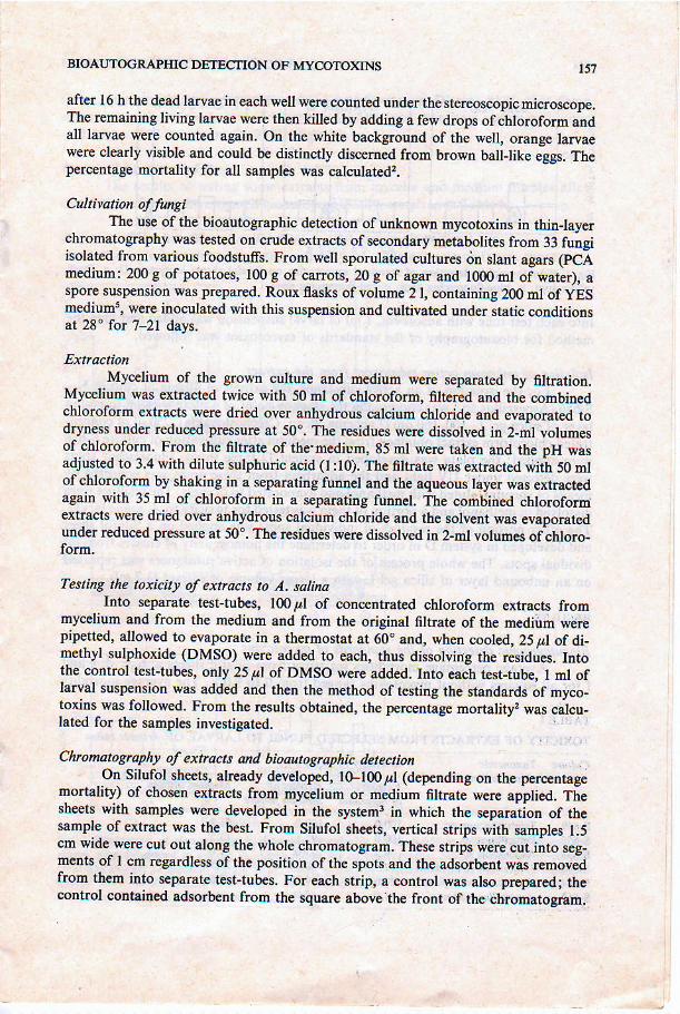
Fic, l Bioauloetsphic de6tion ot standdd mt@toxirs d Silufol shels in stslm D a Darl
velow spol; b, blue-duor*i!8 spot: c. cd'ouore(ina spot
Into each test{ube witl aalsorbent, I ril of larval suspension was added and then the
method for bioautogaphy ol the standards of dycotoxins was followed'
Isolation of ffikrlobtl actil)e substMces fiom the extractFoi the isolation of an active substance, the extract of filtrate of culture No'
5-656 (AsDersillus sp ) was cbosen. On the stanjDg pojnr of a plate with aD unbound
tave|" of siiicig.t t-. tito- | 60 / m ( Lachema' Brno, cz€choslo! akia)' 2 50 t,tl of concln -
i.lrca "no.ofirnl
.*rtact obiained trom rhe medium after cuhivalion ofculture 5-656
were applied; lhe plate qras developed in system D three rimes Fluorescent spols
".i" J!i""d *ai. uv ligbt and ihe adsorbent from $ese areas was collected bv
means of vacuuin, eluteal with methanol and evaporated to dryness' The residues were
alissolved in methanol and; portion olthem *as tested for toxicity to -ll sal a larvae'
The other portion of tie residues of individual spots was applied o-n 6 silufol sheet
and developed in system D in order to determim the homogeneiry ofeluales from in'
dilidual sp;ls. The whole process of the isolation of adjve substances was repeated
on an unbounil layer of sitica gpl L with a larger volume of extract (1 7 mt)'
R.ESULTS
Bioautogruphi. dztedion of the standaftls of mycotoxiasi".ru". o.,ont"g. tr|otr"lilies for strips from silufol sheets a b c' d' e and
f for all sou;s wilhout mvcotoxins ranSed from 0 to 9 0% The averagE mortality
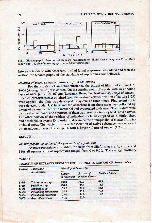
TASLE ITO)qCITY OF EXTRACTS FROM SELECTED FUNCI TO LAkvAB OF Attenia sahtu
Mutuntt of laM (%))
IE tet Extact of MedM fhtuted,,'retirh,a.dtrnftnare
s-649 . Trichoth@ium sP.$656 Pcricilium sP.9658 Alpcrslus 3P:5462 Pehi'dlLtncdtudbetti5-664 ) Pdcilli@ toc,.fo i*5$- , hpea lB EpeN
"ry.217.527.041.0
63.813.55E.6m.52.6
BTOAUTOGRAPHIC DBTECTION OF MYCOTO)qNS
for a square with sterigmatocystin (Rr :0.91 in system D) was 8l%, that for asquarc with aflatoxin (R. : 0.65 in system D) 36% and that for a square with kojicacid (Rr : 0.33 in system D) 2l% (Fie.1).
Testing the extrac^ offungiThe results of testing some extracts from mycelia and m€dium filhates after
cultivation offungi, investigated bioautographically, are given in Table I.
Bioautogruphic detection of metabolites in the extructs from fungiAccording to the percentage mortality in cnrde extracts of fungi, chosen ex-
r59
?
I
I
RF vatu€s
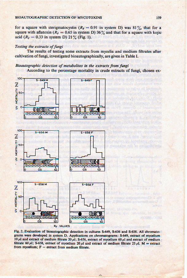
Fig.2. EvalualioD of bioautosraphic d.t€.ridn in cN tffi s{49, $656 dd 9658. AI chroruto-erant lllre &rcloFd io sFtem D. [email protected] on chrcmarog.arE: S-54q dtract of nyceliuml04.rd €tlr&t of Dcdium fltnte 20l; $554 extr&t of myelium 60rl dd qtract of mediuo .filt ale 60pl: sF65E, dtn t of rn c.lium 20d and dlnrt of ftilium 6tr5re 25r,1. M : erlractfrorn tfcelium; F - dtr.c! from mcdim 6lrate,
160 Z. 'URAdKOVA V. BETNA, P. NEMEC
tacts were examined by chromatoeraphy on Silufol sheets and detected bioauto-
$aphically. The chromatographic results and the rcsults of bioauto$aphic detectionare shown itr Fig, 2. On the basis of a chromatographic comparison of the oxtactsof filtrate and mycetium (in system D) with the standards of aflatoxin Bl (Rr : 0 67)and kojic acid (R' : 0.33), it is considercd that with culture No. 5-658 kojic acid andaiatoxins are produc€d.
Isolation of actire subslMces ftom a chtortlatogrumRosidues of substances isolated from individual spots on the chromatogram
on an unbound layer of silica gcl were tested with ,{. ralira and the percentagp mor-tality showed that the active sub'stanc€ on tfte chromatogram was in the region ofblue-green and dark Sreen fluorcscence (see Fig. 2). Adsorbents from these rcgionswerc combin€d, eluted with methanol and evaporat€d to dryness. The rcsidues weredissolved in order to aletermine the toxic efl€ct on ,:1. sal,ta. The remaining solutionof active substances in methanoi was applied on a Silufol sheet along the wholesheet (20 cm) and develop€d in system D-,There were five different fluorescent stripswhich sppeared under UV light. These were removed from t]rc sheet, eluted andevaporat€d xo dryness. Flom individual spots, the actiity of the eluate towards l.ralma was tested. The active ones were as follows: a pink_fluorescing substance (51%
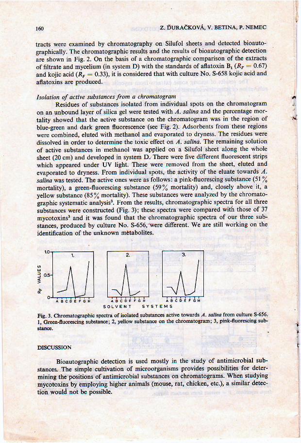
mortalig, a geen-fluorescing substance (59% mortality) and, closely above it, ayelow substanc€ (85% mo.tality). These substances were analyzed by the chromato-gaphic systematic analysis3. From the results, chromatographic specha for all threesubstances werc constructed (Fig.3); these spectra werc compared with those of 37mycotoxlnsi and it was found that the chomatographic spectn of our three sub_stances, produced by culture No. S-656,-were difrerent. We are still working on tlrcidentifcation of the untnown matabolites.
1,
i
It
,ff
I
Fis. 3. Ch@tossphic sp@lri of isolaled substdc€s acdve towdds ,'1. salh. f.om culturc 5-656.I. ciEer-nuot@ing suhtac; 2, }! ow su!6ttu@ on tho ahromtosrmi 3, pink_fu@iDs !ub-
DISCUSSION
Bioautographic aletection is used mostly in the study of antimicrobial sub-
stances;.The simple cultivation of microoigianisms provides possibilities for dcter-
mini4 tle positions ofantimicrobial substanc$ on chromatograms. When studting
mycooxins by enptoying Nghcr animals (mouse, ral chicken, etc.), a similar detec'
tibq would not be poBsible.
I
EIOAUTOGRA?IIIC DEIECTION OF MYCOIO)(INS
In our method of bioautographic dctection, the sensitivity of l. ra1rr4 larva€were tested on $t4Ddard samplcs of mycotoxins2. The advantagp of usidg this mod€lorganbm for the bioautogaphic detection is the simplicity of cultivation of thelarvae.
For testing the method for the detection of standaril mycotoxins, one neutsl(D) and one acidic (B) system l,erc us€d for the tlevelopment of chromatograms onSilufol sheetu. The avemge percentage natural dortality oflarva€ in th€ detectioD ofadsorbcnt from coDtrol squares from Silufol sheets is lower with the Deutral system(H.7ZJ than with th€ acidic systen B (0-q0ZJ.
W[en €mployiog the acidic system (B), the pH ofthe medium is trot substa ial-ly influenc€d after pouridg the edsorbent lrom the squares cut fiom chromatogBmswith larvae cdltivation medium.
The possibility of detecting mycotoxins by inserting the whole square from aSilufol $heet into a test-tube (or by cutting it into smaller squsro6) with lawac suspen-sioD was examitred. This process did not prove to be suitable as the shalp edges ofthe aluminium foildamagedDechanicallytlrcla aewhichhadalrcadystoppedmovingst zdo time of testing,
Ttvo t,?es ol cultues for testing the extracts for the content of mycotodnswerc chosen: (l) fungi of randon contaminated foodstufs snd law materials forfoods werc isolated; (2) cultures ised in the production of some foodstufs were tested.Wei €l al6 isolated a mycotoxin PR-toxin ftom the c|rrhr'f,e Penicilium roqueforti.Their cullure was isolated, howevet from cootamfuated fodaler but, as shown by ourrcsults on the toicity towatds A. saliia of extacts frcm p. rcqueforti (9664) ]cfjedin tfte poduction of cheese, it is nec€ssary to investigate the potential toxigenicity olfungi used in the fooalstufrs industry, as the Fesence of mycotoxins itr products canalso be caus€d by used toxicogenic fungi.
The above method for th€ bioautographic detestion of mycotoxins on thinlay.$ W ],sing Artemia ^ralirra larvae could be applied in sqeening mycotoxins itrcontasinated foodstuffs, rav/ mat€dals for food and foddeN owing to its simplicityand reproducibility.
RBFERENCES
I y. B.tln , t. ctu@to$t.,18 (1973t 41-2 Z. Dt]mkova, \. B€tila, B. HoDikovlt and P. N€m.c, zr,ralt , Baktettot., pabitehk., r"fek-
no6kr. E C,, Abt, 2, in pE.*s.3 z. DuEakov4 v. E€rina and P.N./!@,J, Cl@uatoer,, t16 (i9O t4l.4 S. FEtrk, Frsoml communication.5 N. D. Davis, N. L. Dind atd D. v,I. Adtigq Appl. Mtoobk'|., t4 (196t 37a.6 R. D. Wri, P. E. Still, E. E. Smaley, H. K. Sctc od F,M.Srtoia,,1ppt, Mioobiot.,25 (1973-)
t 1 1 .
161


