Embed Size (px)
Citation preview

Bimodal antagonism of PKA by ARHGAP36
Thesis submitted in accordance with the requirements of the University of Liverpool for the degree of Doctor in Philosophy
By
Rebecca Louise Eccles
April 2016

Bimodal antagonism of PKA by ARHGAP36
Rebecca Louise Eccles
Abstract PKA is a ubiquitous kinase whose activity is controlled by the second messenger
cAMP downstream of GPCR signalling. PKA is a tetrameric holoenzyme composed
of a homodimer of regulatory subunits (PKAR) which each bind and inhibit a
catalytic subunit (PKAC). In this conformation the enzyme is inactive. Upon cAMP
binding to the regulatory subunits, the catalytic subunits are released and can
phosphorylate their substrates. Regulation of PKA is mainly centred around PKAR,
in a large part via binding of the A-kinase anchoring proteins (AKAPs). Here I
characterise a novel Rho GAP, ARHGAP36, and describe how it directly binds the
PKA catalytic subunits. This binding is via a pseudosubstrate motif, which inhibits
PKAC activity by blocking access to substrates. In addition to this, ARHGAP36 also
mediates polyubiquitylation and degradation of PKAC. This is the first description of
ubiquitin-mediated degradation of PKAC. Surprisingly for a cytosolic protein,
degradation is not mediated by the proteasome but by the endolysosomal pathway,
which is usually reserved for transmembrane proteins. This bimodal antagonism of
PKAC by ARHGAP36 leads to suppression of a variety of PKA signalling responses
downstream. I found that ARHGAP36 expression is developmentally regulated and
restricted to embryonic skeletal muscle. It is, however, upregulated in
neuroblastoma cell lines, where I could show that ARHGAP36 modulates PKAC
activity and stability in an endogenous setting.
2

Table of Contents Abstract .......................................................................................................... 2 List of Figures ................................................................................................ 6 List of Tables ................................................................................................. 8 Abbreviations ................................................................................................. 9 Acknowledgements ..................................................................................... 12
Introduction ............................................................................... 13 Chapter 1.1.1. Signal Transduction Pathways .................................................................. 14 1.2. Rho GTPases ............................................................................................... 15 1.3. Ubiquitylation and cellular degradation pathways ................................... 18
1.3.1. Ubiquitin .................................................................................................. 19 1.3.2. The ubiquitylation cascade ..................................................................... 21 1.3.3. Deubiquitylases ...................................................................................... 23 1.3.4. The Proteasome ..................................................................................... 24 1.3.5. Endolysosomal degradation ................................................................... 27 1.3.6. Autophagy .............................................................................................. 30
1.4. Phosphorylation and Kinases .................................................................... 32 1.4.1. PKA ........................................................................................................ 33 1.4.2. PKA regulation ........................................................................................ 36 1.4.3. PKA substrates ....................................................................................... 42 1.4.4. PKA in disease ....................................................................................... 51
1.5. Project Summary ......................................................................................... 54 Materials and Methods ............................................................. 55 Chapter 2.
2.1. Molecular Biology ........................................................................................ 56 2.1.1. Creator Reactions ................................................................................... 56 2.1.2. Gifted Plasmids ...................................................................................... 56 2.1.3. Site-directed mutagenesis ...................................................................... 57 2.1.4. 36i Cloning .............................................................................................. 58 2.1.5. LR Reaction ............................................................................................ 58 2.1.6. Transformation ....................................................................................... 58 2.1.7. Plasmid DNA Purification ....................................................................... 58 2.1.8. Glycerol Stock ........................................................................................ 59 2.1.9. Sequencing ............................................................................................. 59 2.1.10. Agarose gel electrophoresis ................................................................. 59 2.1.11. In situ hybridization ............................................................................... 59
2.2. Cell Biology .................................................................................................. 60 2.2.1. Cell Lines ................................................................................................ 60 2.2.2. DNA Transfection ................................................................................... 61 2.2.3. RNA Transfection ................................................................................... 61 2.2.4. Virus Infection ......................................................................................... 62 2.2.5. Antibodies and Chemicals ...................................................................... 63
2.3. Microscopy .................................................................................................. 65 2.3.1. Immunofluorescence .............................................................................. 65 2.3.2. Live Cell Imaging .................................................................................... 65 2.3.3. Fluorescence microscopy ....................................................................... 65 2.3.4. AKAR FRET (with Markus Müller) .......................................................... 65 2.3.5. Image Analysis (with Markus Müller) ...................................................... 66 2.3.6. Cilia Formation ....................................................................................... 66
2.4. Biochemistry ................................................................................................ 67 2.4.1. Lysis ....................................................................................................... 67
3
2.4.2. Immunoprecipitation ............................................................................... 67 2.4.3. UbiCREST .............................................................................................. 68 2.4.4. Western Blotting ..................................................................................... 68 2.4.5. Peptide Spots ......................................................................................... 68 2.4.6. Peptide Synthesis (Rudolf Volkmer) ....................................................... 69 2.4.7. PepTag Assay (with Carolin Barth) ........................................................ 69
2.5. Mass Spectrometry ..................................................................................... 69 2.5.1. Ubiquitin site identification (with Erik McShane) ..................................... 69 2.5.2. Selected Reaction Monitoring (with Patrick Beaudette) ......................... 71 2.5.3. Identification of Stub1 as an ARHGAP36 interactor (with Erik McShane) .......................................................................................................................... 71 2.5.4. Global protein abundance measurements (iBAQ, with Patrick Beaudette and Erik McShane) ........................................................................................... 72
2.6. Statistics ....................................................................................................... 73 ARHGAP36 is a novel PKAC binding protein and Chapter 3.
pseudosubstrate inhibitor .......................................................................... 74 3.1. Introduction .................................................................................................. 75 3.2. Results .......................................................................................................... 75
3.2.1. ARHGAP36 Characterisation ................................................................. 75 3.2.2. The ARHGAP36 interactome indicates a role in PKA signalling ............ 83
3.3. Discussion ................................................................................................... 93 3.3.1. Non GAP functions of other GAP proteins ............................................. 93 3.3.2. ARHGAP36 is a novel PKAC inhibitory protein ...................................... 94 3.3.3. Other pseudosubstrate Inhibitors of PKAC ............................................. 94 3.3.4. ARHGAP36 in Hedgehog signalling and Medulloblastoma .................... 95
ARHGAP36 mediates PKAC ubiquitylation and degradation Chapter 4.via the endolysosomal pathway ................................................................. 97
4.1. Introduction .................................................................................................. 98 4.2. Results .......................................................................................................... 98
4.2.1. ARHGAP36 causes depletion of PKAC .................................................. 98 4.2.2. ARHGAP36 has no effect on PKAR ..................................................... 100 4.2.3. Pseudosubstrate binding is required but not sufficient to promote PKAC downregulation ............................................................................................... 100 4.2.4. ARHGAP36-N2, encompassing just 77 amino acids, can mediate PKAC degradation ..................................................................................................... 102 4.2.5. ARHGAP36 induced PKAC degradation is rescued by lysosomal inhibitors ......................................................................................................... 102 4.2.6. ARHGAP36 and PKAC colocalise along the endolysosomal pathway . 107 4.2.7. ARHGAP36 mediates PKAC ubiquitylation .......................................... 107 4.2.8. ARHGAP36 promotes PKAC ubiquitylation at a single lysine K285 ..... 110 4.2.9. Lysine 285 is required for ARHGAP36-mediated PKAC poly-ubiquitylation ................................................................................................... 110 4.2.10. PKAC-K285R is resistant to ARHGAP36 mediated degradation ....... 113 4.2.11. PKAC is decorated with K63-linked ubiquitin ..................................... 113 4.2.12. PKAC degradation requires the ESCRT pathway .............................. 118 4.2.13. The ARHGAP36 interactome contains candidate E3 ligases ............. 118 4.2.14. E3 overexpression does not affect PKAC levels ................................ 118 4.2.15. E3 knockdown does not affect PKAC levels ....................................... 121 4.2.16. Stub1 and Praja2 interact with ARHGAP36 ....................................... 121 4.2.17. Stub1 is a possible PKA substrate ..................................................... 125
4.3. Discussion ................................................................................................. 125 4.3.1. Monoubiquitylation vs Polyubiquitylation .............................................. 125 4.3.2. K63 linked ubiquitylation ....................................................................... 126 4.3.3. ARHGAP36 inclusion in MVBs ............................................................. 126
4

4.3.4. E3 ligase identification .......................................................................... 126 4.3.5. Cross Regulation of PKAC and PKAR levels ....................................... 127 4.3.6. PKAR degradation ................................................................................ 127 4.3.7. Hints at PKAC degradation in the literature .......................................... 128 4.3.8. Kinase inhibition by endolysosomal inclusion ....................................... 129 4.3.9. Global or local degradation? ................................................................. 130
ARHGAP36 is a suppressor of PKA signalling .................... 131 Chapter 5.5.1. Introduction ................................................................................................ 132 5.2. Results ........................................................................................................ 132
5.2.1. ARHGAP36 abolishes CREB phosphorylation ..................................... 132 5.2.2. ARHGAP36 inhibits AQP2 trafficking ................................................... 135 5.2.3. ARHGAP36 expression is largely restricted to embryonic skeletal muscle ........................................................................................................................ 135 5.2.4. ARHGAP36 is absent from commonly used cell lines .......................... 138 5.2.5. ARHGAP36 is expressed in neuroblastoma cells ................................ 138 5.2.6. ARHGAP36 and PKAC are expressed at equimolar levels in NGP cells ........................................................................................................................ 141 5.2.7. Endogenous ARHGAP36 interacts with endogenous PKAC ................ 143 5.2.8. ARHGAP36 knockdown leads to an increase in PKAC protein levels and activity in NGP cells ........................................................................................ 143 5.2.9. ARHGAP36 and PKAC expression levels are negatively correlated in NGP cells ........................................................................................................ 145 5.2.10. ARHGAP36 and PKAC colocalise inside Rab5QL vesicles ............... 145
5.3. Discussion ................................................................................................. 148 5.3.1. ARHGAP36 supresses a wide range of PKA signalling responses ...... 148 5.3.2. Arhgap36 expression is developmentally regulated ............................. 148 5.3.3. ARHGAP36 in skeletal muscle ............................................................. 149 5.3.4. ARHGAP36 in neuroblastoma .............................................................. 149 5.3.5. Competitive binding? ............................................................................ 151 5.3.6. Bimodal antagonism ............................................................................. 151
Discussion ............................................................................... 153 Chapter 6.6.1. Summary .................................................................................................... 154 6.2. Open Mechanistic Questions ................................................................... 154
6.2.1. Why is PKAC not degraded by the proteasome? ................................. 154 6.2.2. Lysosomal degradation of cytosolic kinases: a more general mechanism? ................................................................................................... 155 6.2.3. What are the roles of the uncharacterised regions of ARHGAP36? ..... 156 6.2.4. Can ARHGAP36 compete with PKAR for PKAC binding? ................... 156 6.2.5. Which E3 ligase mediates PKAC ubiquitylation, and is recruitment and thus degradation regulated? ........................................................................... 156
6.3. Physiological Relevance and future perspectives ................................. 157 6.3.1. ARHGAP36 and the cAMP-PKA axis during development .................. 157 6.3.2. ARHGAP36 and the cAMP-PKA axis in oncogenesis .......................... 159
References ................................................................................................. 162
5

List of Figures Figure 1-1 The Rho GTPase activation cycle ........................................................... 16 Figure 1-2 Modular domain architecture of the human RhoGEF and RhoGAP
proteins ............................................................................................................. 17 Figure 1-3 Types of ubiquitylation ............................................................................ 20 Figure 1-4 The ubiquitylation cascade ..................................................................... 22 Figure 1-5 The main degradation pathways and their inhibitors .............................. 25 Figure 1-6 The Endolysosomal Pathway .................................................................. 28 Figure 1-7 PKA activation and cAMP generation ..................................................... 34 Figure 1-8 AKAPs provide tailored PKA signalling nodes ........................................ 38 Figure 1-9 PKA mediated CREB activation .............................................................. 43 Figure 1-10 PKA regulation of Gli repressor formation ............................................ 48 Figure 3-1 ARHGAP36 has five predicted isoforms ................................................. 76 Figure 3-2 ARHGAP36 localises to the plasma membrane ..................................... 78 Figure 3-3 ARHGAP36 overexpression does not obviously affect the cytoskeleton 79 Figure 3-4 ARHGAP36 localises to primary cilia ...................................................... 81 Figure 3-5 The arginine rich region is sufficient to target ARHGAP36 to the primary
cilium ................................................................................................................ 82 Figure 3-6 ARHGAP36 interacts with PKAC ............................................................ 85 Figure 3-7 ARHGAP36 recruits PKAC to the plasma membrane and vesicles ........ 85 Figure 3-8 Just 77 amino acids are required to recruits PKAC ................................ 86 Figure 3-9 The N-terminus of ARHGAP36 mediates the interaction with PKAC ...... 88 Figure 3-10 ARHGAP36 contains a PKA pseudosubstrate motif ............................. 88 Figure 3-11 The PKA holoenzyme structure reveals critical residues for the
pseudosubstrate interaction ............................................................................. 89 Figure 3-12 Point mutations on both proteins can abolish the interaction of
ARHGAP36 and PKAC ..................................................................................... 90 Figure 3-13 ARHGAP36 inhibits PKAC in vitro and in vivo ...................................... 92 Figure 4-1 ARHGAP36 downregulates PKAC levels ............................................... 99 Figure 4-2 ARHGAP36 has no effect on PKAR levels ........................................... 101 Figure 4-3 Pseudosubstrate binding alone is not sufficient to mediate PKAC
degradation ..................................................................................................... 103 Figure 4-4 Only 77 amino acids, N2, are required for PKAC degradation ............. 104 Figure 4-5 ARHGAP36 targets PKAC for lysosomal degradation .......................... 106 Figure 4-6 PKAC and ARHGAP36 partially colocalise with markers along the
endocytic pathway .......................................................................................... 108 Figure 4-7 Endogenous PKAC colocalises with HRS, LAMP-1 and is enriched inside
Rab5-Q79L vesicles ....................................................................................... 109 Figure 4-8 ARHGAP36 induces PKAC ubiquitylation ............................................. 111 Figure 4-9 ARHGAP36 mediates PKAC ubiquitylation at K285 ............................. 112 Figure 4-10 PKAC-K285R is resistant to ARHGAP36 induced polyubiquitylation and
degradation ..................................................................................................... 114 Figure 4-11 PKAC-WT is susceptible to CHX and degradation is rescued upon
lysosomal inhibition, whereas PKAC-K285R is unaffected ............................ 115 Figure 4-12 PKAC is decorated with K63 linked ubiquitin ...................................... 117 Figure 4-13 ARHGAP36 induced PKAC degradation requires Vps4 ..................... 119 Figure 4-14 ARHGAP36 has no effect on the localisation of its E3 ligase interactors
........................................................................................................................ 120 Figure 4-15 E3 interactor overexpression has no effect on PKAC levels, even in the
presence of ARHGAP36 ................................................................................. 122 Figure 4-16 Knockdown of Praja2 or Stub1 has no effect on ARHGAP36 mediated
PKAC degradation .......................................................................................... 123 Figure 4-17 ARHGAP36 interacts with Praja2 and Stub1 ...................................... 124
6

Figure 4-18 Stub1 is a PKA substrate .................................................................... 124 Figure 5-1 ARHGAP36 inhibits CREB phosphorylation ......................................... 133 Figure 5-2 ARHGAP36 inhibits CREB phosphorylation via the pseudosubstrate motif
........................................................................................................................ 134 Figure 5-3 ARHGAP36 inhibits AQP2 trafficking .................................................... 136 Figure 5-4 Arhgap36 is expressed in embryonic skeletal muscle .......................... 137 Figure 5-5 Arhgap36 expression is developmentally regulated ............................. 139 Figure 5-6 ARHGAP36 is expressed in neuroblastoma cells ................................. 140 Figure 5-7 ARHGAP36 and PKAC are expressed at equimolar levels in NGP cells
........................................................................................................................ 142 Figure 5-8 Endogenous PKAC interacts with endogenous ARHGAP36 ................ 142 Figure 5-9 ARHGAP36 antagonises PKAC in an endogenous setting .................. 144 Figure 5-10 ARHGAP36 and PKAC levels are negatively regulated in NGP cells . 146 Figure 5-11 ARHGAP36 levels are heterogeneous in SK-N-BE(2) cells ............... 147 Figure 5-12 ARHGAP36 and PKAC colocalise inside Rab5-QL induced vesicles . 147
7

List of Tables Table 2.1 Standard Creator Reaction 56 Table 2.2 Creator Acceptor Plasmids 56 Table 2.3 ARHGAP36 Mutagenesis Primers 57 Table 2.4 PKAC Mutagenesis Primers 58 Table 2.5 36i Primers 58 Table 2.6 Primers used for sequencing 59 Table 2.7 Cell Lines 60 Table 2.8 siRNA sequences 62 Table 2.9 ARHGAP36 targeting shRNA sequences 62 Table 2.10 Inhibitors used within this thesis 63 Table 2.11 ARHGAP36 Antibodies 63 Table 2.12 Primary Antibodies 64
8

Abbreviations
36i ARHGAP36 inhibitor AC Adenyl cyclase ACTH Adrenocorticotropic hormone ADP Adenosine diphosphate AKAP A-kinase anchoring protein AKAR A-kinase activity reporter ALK1 Anaplastic lymphoma kinase
APC Anaphase promoting complex AQP Aquaporin Arp2/3 Actin-related protein-2/3 atg Autophagy related gene ATP Adenosine triphosphate
AVP Arginine vasopressin Baf Bafilomycin BSA Bovine Serum Albumin cAMP Cyclic adenosine monophosphate CASA Chaperone-assisted selected autophagy CBP CREB binding protein CFP Cerulean Flourescent Protein CHX Cycloheximide CK1 Casein kinase 1 CLN5 Ceroid lipofuscinosis neuronal protein 5 CMA Chaperone mediated autophagy CRE cAMP response element CREB Cre Response Element Binding Protein CREM CRE modulator CRH Corticotropin-releasing hormone DMSO Dimethyl sulfoxide DNA Deoxyribonucleic acid DUB Deubiquitylating Enzyme EEA1 Early Endocytic Antigen 1 EGFR Epidermal growth factor receptor eMI Endosomal microautophagy EMT Epithelial-mesenchymal transition EPAC Exchange proteins acrivated by cAMP Epo Epoxomicin ER Endoplasmic reticulum ESCRT Endosomal complexes required for transport EtOH Ethanol FBS Fetal Bovine Serum FL-HCC Fibrolamellar hepatocellular carcinoma FRET Flourescence resonance energy transfer GAP GTPase activating protein GDI GTPase dissociation inhibitor GDP guanosine diphosphate GEF GTPase exchange factor GFP Green Flourescent Protein
9

GliA Gli Activator GliR Gli Repressor GPCR G protein coupled receptor GSK3 Glycogen synthase kinase 3 GST Gluthathione S Transferase GST Glutathione S-transferase GTP Guanosine triphosphate HECT Homology to E6AP carboxy terminus Hh Hedgehog HPA Hypothalamus-Pituitary-Adrenal iBAQ Intensity based absolute quantitation IBMX 3-isobutyl-1-methylxanthine ICER Inducible CRE repressor IF Immunofluorescence IgG Immunoglobulin G IP Immunoprecipitation ITC Isothermal titration calorimetry KO Knock out Leu Leupeptin LIR LC3 interacting region MAP Microtubule associated protein MC2R Melanocortin receptor 2 MET Mesenchymal-epithelial transition mTOR Mammalian target of rapamycin MVB Multivesicular Bodies NCL Neuronal ceroid lipoduscinoses NEM N-Ethylmaleimide NES Nuclear exclusion signal OTU Ovarian tumour protease P/S Penicillin Streptomycin PACT Pericentrin-AKAP-450 centrosomal targeting PAK p21 activated kinase PBS Phosphate Buffered Saline PBS++ Phosphate Buffered Saline supplemented with Ca and Mg pCREB Phosphorylated Cre Response Element Binding Protein PDE Phosphodiesterase PFA Paraformaldehyde PI3K Phosphoinositide 3-kinase PKA Protein Kinase A PKAC Protein Kinase A Catalytic Subunit PKAR Protein Kinase A Regulatory Subunit PKI Protein Kinase Inhibitor RBR Ring between ring RING Really interesting new gene RSK1 p90 Ribosomal S6 kinase-1 sAC Soluble adenyl cyclase SAG Smoothened AGonist SCF Skp Cullin F-box SFRS17A Splicing factor arginine/serine-rich 17A Shh Sonic Hedgehog
10

SILAC Stable isotope labelling by amino acids in cell culture Smo Smoothened SRM Selected Reaction Monitoring StAR Steroidogenic acute regulatory protein SuFu Suppressor of Fused UBD Ubiquitin binding domain UCH Ubiquitin C terminal hydrolase UIM Ubiquitin interacting motif USP Ubiquitin specific peptidase WASP Wiskott-Aldrich syndrome protein WAVE1 (WASP)-family verprolin homologous protein 1 WB Western Blot YFP Yellow Flourescent Protein ZO-1 Zona occuldens-1
11

Acknowledgements
Firstly, thanks to Oliver Rocks, for this project, for the freedom to develop as a
scientist and for always having an open door. Thank you to Sylvie Urbé for taking a
chance on me over six years ago, and to you and Mike Clague for setting me on this
path and teaching me so much. Thanks also for rescuing me from German
bureaucracy and continuing to supervise me.
Thanks go to our many collaborators. Especially to Oli Daumke and the Daumke lab
members past and present, Stephan in particular. Thanks to Annette Hammes and
Nora Mecklenburg for help with in situs and Hedgehog advice. Thanks to the
Klussmann lab for PKA advice and the generous sharing of reagents. Special
thanks to my mass spec guru Erik, for always being up for one more experiment,
and to Patrick for coming through on my last minute requests. Thank you also to all
the past and present Liverpool lab members, especially to Claire, Amos and
Yvonne, for continued support.
Thank you to my ever-expanding AG Rocks family, for support both in and out of the
lab, especially to the original Rock stars Caro, Markus, Matti and Juliane. I wouldn’t
have survived in Berlin without you. Thanks again to Markus and Caro who helped
me massively with this project. Huge thanks also go to Maciej for all the help, for
trying to be a cell biologist, and for continuing the ARHGAP36 story. To all the
Rocks lab members past and present, thank you for making it such a great place to
do science, especially Sreusch, Vicky, Linda, Lennart, Phillip, Laura R and Moritz.
Thanks also to the many weird and wonderful people I met at the MDC who helped
me along the way.
Thanks to John for imparting your post doc wisdom. To Laura and Vicky, thank you
for always entertaining and supporting me, and making Berlin an even better place.
Thank you to Heather for always being there for me and trying to understand the
science!
Thank you to my family for their unwavering belief in me, I hope I made you proud.
Last but not least thank you to James for putting up with my craziness and getting
me through. I couldn’t have done it without you.
12

Introduction Chapter 1.
13

1.1. Signal Transduction Pathways Cellular behaviour is controlled by the concerted action of signal transduction
pathways, which relay signals from the outside in. These pathways were once
believed to be linear and unidirectional, however crosstalk between pathways and
complex regulatory feedback loops are now known to be the norm. Amongst the key
players in signal generation are kinases and GTPases. These signalling cascades
are not just switched on or off; the signals these proteins propagate must also be
precisely regulated in a spatiotemporal manner. Where, when and for how long
these signalling proteins are active is of extreme importance. Thus they can be
recruited to specific subcellular localisations and assembled in macromolecular
complexes together with particular regulators that can tightly control their activity.
Signals must be generated, however they must also be dynamically regulated. The
interplay of proteins controlling activation and attenuation is key. Reversible post-
translational modification (PTM) of proteins is a versatile way to modify protein
behaviour. PTMs can be used to initiate, terminate or tweak signalling responses.
The best-studied PTM is phosphorylation, where the action of kinases is reversed
by phosphatases.
A further layer of regulation is the degradation of signalling components themselves.
This is controlled by another reversible PTM, ubiquitylation, which mediates
subsequent targeting of proteins to the respective degradation machineries. This is
also a tightly regulated, dynamic process. Phosphorylation can prime for
ubiquitylation and ubiquitin itself can be phosphorylated, thus allowing further
complexity in the regulation of protein turnover and thus signalling.
In this thesis I will describe the regulation of a signalling protein, Protein Kinase A
(PKA), by both inhibitory and degradative mechanisms. I will begin by introducing
first the regulation of Rho GTPases, second ubiquitylation and its roles in
proteasomal and lysosomal degradation, and lastly phosphorylation and kinases
with a particular focus on Protein Kinase A.
14

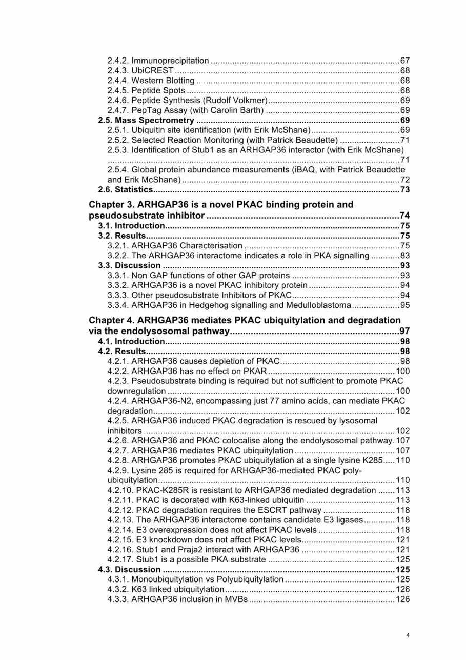
1.2. Rho GTPases
Rho GTPases are master regulators of the actin cytoskeleton. Like other small
GTPases of the Ras-like superfamily, they are molecular switches that cycle
between inactive GDP and active GTP bound states. Only in the latter state can
they bind and relay signals to downstream signalling proteins.
Rho GTPases were first linked to the actin cytoskeleton through the pioneering
studies by Anne Ridley and Alan Hall: Injection of RhoA, Rac1 and Cdc42 into
Swiss 3T3 cells led to formation of stress fibres, lamellipodia and filopodia
respectively (Hall, 1998; Ridley & Hall, 1992). However, in the last 20 years it has
become increasingly obvious that Rho signalling is not so simple. In contrast to the
classical models where RhoA, Rac1 and Cdc42 were thought to cause distinct
cellular behaviours, and to be active in different parts of the migrating cell (Ridley,
2001; Raftopoulou & Hall, 2004), recent studies using FRET biosensors showed all
three Rho proteins to be activated at the leading edge (Machacek et al, 2009). The
Rho proteins have also been implicated in a wide range of other cellular processes,
such as cell cycle progression, cell survival, vesicular trafficking and transcriptional
regulation (Vega & Ridley, 2008). So how is signalling specificity achieved?
The GTPase cycle is tightly controlled by three main factors: GTPase activating
proteins (GAPs), Guanine nucleotide exchange factors (GEFs), and Rho guanine
dissociation inhibitors (GDIs) (Figure 1.1) (Bos et al, 2007). GEF proteins are
positive regulators, catalysing GTP loading (Rossman et al, 2005), whereas GAP
proteins and GDI proteins act as negative regulators. GAPs increase the rate of
GTP hydrolysis, whilst GDIs extract the Rho proteins from the membrane competent
signalling pool and keep them bound in the inactive GDP state (Tcherkezian &
Lamarche-Vane, 2007; Garcia-Mata et al, 2011). Of the twenty Rho proteins, 12 of
them are classically activated and can be differentially regulated by GAPs and
GEFs. In contrast, eight of the Rho proteins are atypical and are mostly GTP bound
(Heasman & Ridley, 2008). Spatio-temporal control of the Rho GTPases is required
to achieve the right signalling outcome. Approximately 80 GAPs, 60 GEFs and three
GDIs provide numerous possibilities for fine-tuning classical Rho protein activity
(Figure 1.2). These multi-domain regulatory proteins have the potential to specify
signalling by targeting Rho proteins to distinct cellular localisations, and act as
molecular scaffolds to recruit further signalling proteins. Determining the GTPase
specificity, localisation and binding partners of these proteins will help to establish
15

GAP
RhoGDP
. RhoGTP
. onoff
PiRh
oG
DP.G
DI effector-
proteins
GEF
GTP
Figure 1.1 The Rho GTPase activation cycleThe Rho proteins are molecular switches which signal from mem-branes. They associate with membranes through their lipid anchors. The Rho GTPase cycle can be controlled negatively by GTPase activating proteins (GAPs) and positively by Guanine nucleotide exchange factors (GEFs). Guanine nucleotide dissociation inhibitors (GDIs) extract the Rho proteins from the membrane and and keep them in an inactive cytosolic complex.
GDP
16

RASGRF2
RASGRF1SOS1SOS2
ECT2L
OCRL
INPP5B
FAM13A
FAM13B
ARHG
AP17
ARHG
AP44
SH3B
P1TAGAP
ARHGAP20
ARHGAP8
ARHGAP1
ARHG
AP19
RAL
BP1
ARH
GAP
5
ARH
GAP
35
ARH
GAP
25
ARHG
AP22
ARHG
AP24
ARHGAP11A
ARHGAP11B
ARHGAP4SRGAP2
SRGAP1SRGAP3
GMIPHMHA1ARHGAP29
OPHN1ARHGAP42ARHGAP10ARHGAP26
ARHGAP2ARHGAP3
ARHGAP32ARHGAP33ARHGAP30ARHGAP31ARHGAP9
ARHGAP27
ARHGAP15
ARHGAP12
RACGAP1ARHGAP21ARHGAP23
ARH
GAP
40AR
HG
AP18
ARH
GAP
28 63P
AG
HR
AAR
HG
AP
6 STARD
8D
LC1
STA
RD
13
MYO
9B
MYO
9A
PIK3R2
PIK3R1
SYDE1
SYDE2
ARAP3
ARAP2
ARAP1DEPDC1B
DEPDC1
DOCK11DOCK9
DOCK10DOCK8DOCK7
DOCK6
DOCK2DOCK5DOCK1
DOCK3DOCK4
ARHGAP39ALS2
ARHGEF33ARHGEF7ARHGEF6
ABRBCR
ARHGEF10ARHGEF10L
ARHGEF17
ECT2
VAV1
VAV3
VAV2
TIAM
1TI
AM2
ARHG
EF4
SPAT
A13
ARHGEF
9PREX
2PR
EX1
ARH
GEF
37AR
HG
EF38
DN
MBP
MCF2L2
MCF2LMCF2
OBSCN
GEFT
KALRNTRIO
ARHGEF40
PLEKHG4B
PLEKHG4
PLEKHG2
PLEKHG3
PLEKHG1
FARP1
FARP2
FGD6
FGD5
FGD3
FGD1
FGD4FGD2
ARHGEF39PLEKHG
7PLEKH
G5
PLEKHG
6ITSN
1ITSN
2AR
HG
EF8AR
HG
EF3AR
HG
EF11A
RH
GE
F12
ARH
GEF1
AR
HG
EF1
8A
RH
GE
F28
AKAP
13AR
HG
EF2
ARHGEF15
NGEFARHG
EF5ARHG
EF19
ARHGEF16
ARHGEF26
GEFGAPDOCK/GEFGEF/GAP
EFhC1RCC1IGc2FN3VPS9RasFYVEC2FFSPECEHMORNIQSEC14
WWPDZArfGapBRCTRasGEFded_cytRhoGEFSH2STARTCHS_TKcFERMMyTH4RasGEFNSTYKc
RGSRBDFBOXDUF3398PHRAIPPcDEPMYScBARHistoneSAMRhoGAPSH3PX
Protein domains:
Color ranges:
Figure 1.2 Modular domain architecture of the human RhoGEF and RhoGAP proteins. Rho guanine nucleotide exchange factors (RhoGEFs) and Rho GTPase activating proteins (RhoGAPs) are multi-domain scaffold proteins that provide subcellular targeting information and connect Rho GTPases to other signaling pathways, thereby specify-ing Rho signalling. Figure kindly provided by Oliver Rocks.
17

mechanisms of Rho regulation, and the crosstalk with other signalling pathways.
Many of these proteins are uncharacterised.
Prior to the start of my PhD, my supervisor Oliver Rocks had carried out a
systematic screen to characterise all the Rho GAP and GEF proteins, especially
their binding proteins. This was done by mass spectrometry using epitope tagged
GAP and GEF proteins as bait. In this interactome screen, an uncharacterised GAP,
ARHGAP36, was found to interact with Protein Kinase A, as well as E3 ubiquitin
ligases (Rocks and Pawson, manuscript in preparation). The regulation of PKAC
activity and stability by ARHGAP36 will be the focus of this thesis.
1.3. Ubiquitylation and cellular degradation pathways
Nowadays it is widely accepted that protein lifetimes are dynamically regulated,
however, this was not always the case. Seminal experiments by Rudolf
Schoenheimer challenged the commonly held belief that proteins are stable entities
and paved the way for modern day protein turnover experiments. He fed mice with
stable isotope labelled amino acids and found that while some were excreted in
urine others were incorporated into tissues. This was the first indication that protein
turnover is dynamically regulated (Schoenheimer et al, 1939).
The lysosome was then discovered in 1953 (De Duve et al, 1953; De Duve &
Wattiaux, 1966) and was thought to be responsible for all cellular protein
degradation. However only a fraction of proteins were stabilised upon inhibition of
the lysosome using weak bases (Poole et al, 1977). Reticulocytes were still able to
degrade haemoglobin, despite being devoid of lysosomes (Rabinovitz & Fisher,
1964). This process also required ATP and took place at neutral pH as opposed to
the acidic pH required for lysosomal degradation (Etlinger & Goldberg, 1977). It was
thus hypothesised that another non-lysosomal mechanism for degradation must
exist. This led to the eventual discovery of the proteasome (Tanaka et al, 1983;
Hough et al, 1986; Hough & Rechsteiner, 1986; Ciechanover, 2005). Later on I will
describe the three main pathways that eukaryotic cells use to degrade proteins:
proteasomal degradation, endolysosomal degradation, and autophagy. But first I will
turn to the unifying feature that is central to all of them: ubiquitin (Clague & Urbé,
2010).
18

1.3.1. Ubiquitin
Ubiquitylation involves the post-translational addition of the 76 amino acid protein
ubiquitin to a lysine residue within the substrate protein. This results in the formation
of an isopeptide bond between the lysine side chain and the exposed carboxyl
terminal glycine tail of ubiquitin (Vijay-Kumar et al, 1987; Pickart & Eddins, 2004).
Ubiquitylation was first recognised as a signal targeting proteins for proteasomal
degradation, however, today it is known to regulate a variety of cellular events, by
affecting protein activity, localisation and interaction (Komander & Rape, 2012).
Addition of one ubiquitin molecule to a substrate is termed monoubiquitylation.
Complexity in the ubiquitin system is achieved by the ability of multiple ubiquitin
molecules to be conjugated to a substrate. Multi-monoubiquitylation can occur via
addition of single ubiquitin molecules to multiple different lysines within a substrate.
Ubiquitin itself contains seven lysines: K6 K11, K27, K29, K33, K48, K63. Each of
these lysines can be utilised for conjugation of further ubiquitin molecules. Linear
ubiquitylation can also be achieved via a peptide bond between the C-terminal
glycine of one ubiquitin and the N-terminal methionine (M1) of another (Walkzak et
al 2012). Polyubiquitin chains can then be formed in a homotypic or heterotypic
manner. Homotypic when the same linkage is utilised within a chain, heterotypic
when differential linkage types are utilised. Heterotypic chains can be branched, if
one ubiquitin is ubiquitylated at two or more sites (Ikeda and Dikic 2008, Behrends
and Harper 2011) (Figure 1.3). The recent discovery that ubiquitin itself can be post-
translationally modified, via phosphorylation or acetylation, further diversifies the
ubiquitin code (Herhaus and Dikic 2015, Swatek and Komander 2016).
All chain linkages have been identified in HEK293 cells, with K48 (52%) and K63-
linked chains (38%) being most abundant (Dammer et al, 2011). From the total
ubiquitin pool in HEK293 cells, 26% was free ubiquitin, 11% incorporated in
polyubiquitin chains, and the majority was identified as monoubiquitylated-
conjugates (Kaiser et al, 2011).
Different ubiquitin chain linkages have different structural properties and thus allow
differential recognition by ubiquitin binding domains (UBDs) of which there are over
20 different types in the human genome (Dikic et al, 2009; Scott et al, 2015). K48
and K11 linked chains are tightly packed together with the different ubiquitin
molecules contacting each other (Cook et al, 1994; Bremm et al, 2010). In contrast,
K63 and M1-linked chains are much more open with no contact between the
19

M1
K6
K11
K27
K33
K48
K63
Mono-Ub Multi-mono-Ub
K29
Homotypic chains Heterotypic chains
Non-branched Branched
Pol
yubi
quity
latio
n
SubstrateSubstrate
SubstrateSubstrate Substrate
Figure 1.3 Types of ubiquitylationProteins can be mono- or multi-monoubiquitylated or polyubiquitylated. There are then eight different types of ubiquitin chain linkage, with different topologies that can be formed. Polyubiquitylation can be homotypic or heterotypic. Heterotypic chains can be nonbranched (shown is K63 and K11), or branched (shown is K48 and K6). Adapted from (Kulathu & Komander, 2012; Heride et al, 2014).
20

different ubiquitin molecules (Komander et al, 2009b). However even with these
similarities, there are UBDs that have specificity for K63 or M1-linked chains
(Husnjak & Dikic, 2012). Many UBDs bind a hydrophobic patch in ubiquitin around
Ile44, which may suggest that binding of several ubiquitin binding proteins to the
same ubiquitin is mutually exclusive.
1.3.2. The ubiquitylation cascade
Ciechanover, Hershko, Rose and colleagues characterised the machinery by which
a chain of ubiquitin molecules are conjugated to a substrate leading to its
degradation (Hershko et al, 1980). They discovered that ubiquitylation requires the
action of three different classes of enzymes in a multistep process (Ciechanover et
al, 1981, 1982; Hershko et al, 1983). The number of enzymes in each class
increases dramatically; there are two E1 ubiquitin activating enzymes, around 40 E2
ubiquitin conjugating enzymes and over 600 E3 ubiquitin ligase enzymes. (Figure
1.4)
The two E1 enzymes, UBA1 and UBA2, catalyse the ATP dependent activation of
the C-terminus of ubiquitin via acyl-adenylation. This is followed by the conjugation
of the activated ubiquitin to the active site cysteine of the E1 via a thioester linkage.
The ubiquitin is then transferred to a similar cysteine within the active site of the E2
via a trans-thioesterification reaction. The E3 ligase, in one way or another, then
allows the transfer of ubiquitin to the substrate. The high number of E3 ligases is
thought to allow substrate specificity.
E3 ligases can be split into three main families, HECT, RING or RING-like, and
RBR. HECT (Homology to E6AP carboxy terminus) family E3 ligases have a two-
step mechanism. They contain a catalytic cysteine, which accepts the ubiquitin from
the E2 before transferring it to the substrate. They catalyse a wide range of chain
linkages (Kim & Huibregtse, 2009).
RING (really interesting new gene) family E3 ligases transfer the ubiquitin directly
from the E2 to the substrate. The same is true for RING-like U-box E3 ligases
(Metzger et al, 2014). They therefore act mostly as scaffolds, and together with the
E2 determine linkage specificity (David et al, 2011). As well as single functioning
units, RING E3 ligases can also exist as multi-subunit E3 ligases (Li et al, 2008),
such as the Cullin-Ring ligases, which associate with substrate receptors and
21

RING1
RING2IBRHECT
E1
E1
Ub
Ub
E2
E1
E2 Ub
E2 Ub E2 Ub E2 Ub
Ub
Substrate Ub
RING
Substrate Ub Substrate Ub
Ub
2 E1s
40 E2s
>600 E3s
Adenylation &Thioesterification
Trans-thioesterification
Figure 1.4 The ubiquitylation cascade(a) E1 ubiquitin activating enzymes mediate adenylation and thioesterification of ubiquitin. Ubquitin is then transferred from the E1 to the E2 ubiquitin conju-gating ezyme by a trans-thioesterification reaction. There are three different types of E3 ligase. HECT ligases accept the ubiquitin from the E2 before trans-ferring it to the substrate. RING ligases transfer ubiquitin directly from the E2 to the substrate. Ring-between-RING (RBR) ligases bind E2s via a RING domain but also accept ubiquitin before transferring it to substrates in a HECT like manner. (b) The number of enzymes increases dramatically in each class. Adapted from (Winklhofer, 2014; Heride et al, 2014)
a.
b.
22

adapter proteins (Petroski & Deshaies, 2005). In the case of the SCF complex
(Skp1, Cullin1, F-box), Skp1 acts as an adaptor between Cul1 and the F-box-
protein, which then provides substrate specificity, whilst the Cullin protein links to
the Ring E3 Rbx1 (Cardozo & Pagano, 2004). ßTRCP is one such F-box protein
that mediates the degradation of a large number of signaling proteins, including the
Gli and β-catenin transcription factors (see below 1.4.3.3 and 4.3.8).
RBR (RING-between-RING) family ligases, of which there are 18 in the human
genome, were only recently identified as a distinct group (Eisenhaber et al, 2007).
They have properties of both RING and HECT ligases (Aguilera et al, 2000). They
bind E2s via a RING domain but also require a conserved catalytic cysteine to
transfer ubiquitin to substrates (Wenzel et al, 2011).
1.3.3. Deubiquitylases
Ubiquitylation is a reversible process. Deubiquitylating enzymes (DUBs) oppose the
action of E3 ligases. DUBs play various roles in ubiquitin regulation. Ubiquitin is
transcribed from four different genes, and is expressed either as a linear fusion of
multiple ubiquitin molecules, or ubiquitin fused to the ribosomal proteins (Wiborg et
al, 1985; Baker & Board, 1991). DUB activity is thus required to generate free
ubiquitin. DUBs are known to associate with the degradation machineries and
recycle ubiquitin from substrates committed for degradation in order to contribute to
ubiquitin homeostasis (Kimura et al, 2009; Clague et al, 2012). They can also
rescue substrates from their fate by removing or editing ubiquitin chains. DUBs are
known to associate with E3 ligases and these interactions may play a role in chain
editing or protect E3s from degradation due to autoubiquitylation. The latter is the
case for USP7 and the E3 ligase Mdm2, which is responsible for regulating p53
stability (Li et al, 2003).
There are around 80 active DUBs in the human genome that fall into five classes.
Four of them are cysteine proteases: Ubiquitin C terminal hydrolases (UCH),
Ubiquitin specific peptidases (USP), ovarian tumour proteases (OTU) and the
Josephins (Komander et al, 2009a). The final class, the JAMM/MPN+ DUBs, are
zinc metalloproteases. DUBs can cleave the isopeptide bond of the terminal
ubiquitin of a chain (exopeptidase activity) or also cut within the chain
(endopeptidase activity). DUBs have different specificities for chain linkages, with
23

some only cleaving one chain-type, whereas others will non-discriminately cleave all
linkages (Clague et al, 2013; Mevissen et al, 2013).
Substrate specificity and roles of the DUBs can also be regulated by their
subcellular localisation. Many have intrinsic localisation properties and can be found
at distinct locations in the cell, such as the plasma membrane, nucleolus,
centrosome, microtubules, ER, and mitochondria (Urbé et al, 2012). For example,
USP33 is a DUB that is associated with the ER, COP coated vesicles and the cis-
Golgi (Thorne et al, 2011). USP19 and USP30 are the only mammalian DUBs that
contain transmembrane domains. USP19 is tethered at the ER and plays a role in
ER associated degradation (ERAD) there (Hassink et al, 2009). USP30 is targeted
to the outer mitochondrial membrane where it opposes ubiquitylation of Parkin
substrates and can also regulate mitochondrial morphology (Nakamura & Hirose,
2008; Bingol et al, 2014; Liang et al, 2015).
1.3.4. The Proteasome
The majority of cytosolic proteins are degraded by the proteasome, a large
multimeric complex that is essentially an assembly of proteases (Figure 1.5). It is
composed of two subunits: the 20S core particle and the 19S regulatory particle.
The core particle is made up of α and β subunits, arranged as a four-stacked ring
structure α7β7β7α7. The β-rings form an inner hydrolytic chamber and the outer α-
rings form a gate to the inner chamber where degradation occurs. The core particle
combines three different proteolytic activities contributed by the β 1, β 2 and β5
subunits, which cleave proteins after acidic, basic or hydrophobic residues
respectively (Finley et al, 2016). Proteasome activity can thus be inhibited by
compounds targeting these subunits, such as epoxomicin, which inhibits the
chymotrypsin-like activity (Meng et al, 1999; Kisselev & Goldberg, 2001).
Bortezomib, another inhibitor of the chymotrypsin-like activity, has successfully been
used in the clinic in the treatment of multiple myeloma (Goldberg, 2012).
The regulatory particle is composed of a hexameric ring of AAA-ATPases, which
mediate the unfolding of substrates and control entry into the core particle. This
unfolding is a key prerequisite for proteasomal substrates, and also prevents the
non-discriminate degradation of cytosolic proteins. The core particle can be capped
at one or both ends by a regulatory particle (Voges et al, 1999). Aside from the six
AAA-ATPases the regulatory particle contains 13 other components. Three of these,
24

UU U
UU
U
U
Mitrochondria
U
Lysosomal Proteases
Ubiquitin
Substrate
UU
Lysosome
Endocytosis
Autophagy
Proteasome
PlasmaMembrane
WortmanninBafilomyin
WortmanninBafilomyin
EpoxomicinLactacystin
Leupeptin
Figure 1.5 The main degradation pathways and their inhibitorsUbiquitylation is a unifying feature of the different degradation pathways. Endocy-tosed and autophagosome engulfed cargo are both degraded in the lysosome and therefore inhibitors have an effect on both pathways. Adapted from (Clague & Urbé, 2010)
25

Rpn10, Rpn13 and Rpn1, have been shown to bind both polyubiquitin chains and a
DUB each (van Nocker et al, 1996; Husnjak et al, 2008; Finley et al, 2016; Shi et al,
2016). Rpn11, a JAMM/MPN+ metalloprotease DUB, is also a component of the
regulatory particle and associates with Rpn10. It is able to cleave the isopeptide
bond between the substrate and the final ubiquitin and thus removes chains ‘en
bloc’ from substrates before their degradation (Yao & Cohen, 2002). Two further
DUBs are associated with the regulatory particle, USP14 and UCH37. These are
thought to be involved in rescuing substrates from degradation via chain editing (Qiu
et al, 2006; Lee et al, 2011; Finley, 2009). The receptor for UCH37 is Rpn13 (Qiu et
al, 2006), whilst the yeast orthologue of USP14, Ubp6 is recruited to Rpn1, thus
suggesting chain recognition is coupled to deubiquitylation (Husnjak et al, 2008;
Chen & Walters, 2015; VanderLinden et al, 2015; Sahtoe et al, 2015; Shi et al,
2016).
The canonical ubiquitin chains associated with proteasomal degradation are linked
via K48, and K48 linked tetra-ubiquitin is sufficient to target substrates for
proteasomal degradation (Thrower et al, 2000). However, all chain linkages except
for K63 were found to accumulate in HEK293T cells upon proteasome inhibition
(Dammer et al, 2011). Interestingly all the DUBs of the proteasome can cleave K63
linked chains, which may provide a proof reading mechanism to prevent
proteasomal degradation of substrates with this chain type (Jacobson et al, 2009).
K11 linked chains formed by the anaphase promoting complex (APC) E3 ligase in
association with the K11-specific E2 UBE2S have been shown to mediate
proteasomal degradation of cell cycle regulators during mitosis (Jin et al, 2008;
Williamson et al, 2009; Matsumoto et al, 2010; Song & Rape, 2010). K27 linked
chains have also been suggested to regulate proteasomal degradation of Parkin
substrates during mitophagy (Geisler et al, 2010). K6 linked chains have been
implicated in Parkin-mediated mitophagy (Durcan et al, 2014; Ordureau et al, 2015).
In addition to playing a clear role in endolysosomal degradation (see below), K63-
linked chains have been shown to have non-degradative roles in some signal
transduction pathways (Conze et al, 2008; Komander & Rape, 2012). This is
particularly well established for the NFkB signaling cascade which also employs M1
linked chains (Iwai & Tokunaga, 2009; Tokunaga et al, 2009). K63 and K6-linked
chains have also been implicated in DNA damage repair mechanisms (Morris &
Solomon, 2004; Thorslund et al, 2015). K29 and K33 linked chains have been
26

shown to have an inhibitory effect on AMPK (AMP-activated protein kinase) (Al-
Hakim et al, 2008). Finally, K33 linked chains have also been implicated in T cell
receptor signalling and post-Golgi trafficking (Huang et al, 2010; Yuan et al, 2014).
1.3.5. Endolysosomal degradation
The endolysosomal pathway is the main route by which plasma membrane proteins,
such as receptors and channels, are turned over by the cell. Endocytosed
membrane proteins enter an early endosomal compartment from where they can
either be recycled back to the plasma membrane or be sorted for degradation in the
lysosome (Figure 1.6). The lysosome contains a collection of proteases, which
require an acidic pH for their action (Haider & Segal, 1972). The acidic environment
of the lysosome is achieved via the vacuolar H+-ATPase (v-ATPase) (Schneider,
1981). Lysosomal degradation can be inhibited in a variety of manners (Figure 1.5).
Bafilomycin can be used to specifically inhibit the v-ATPase, thus preventing
acidification of the lysosome (Yoshimori et al, 1991). Weak bases such as
chloroquine and NH4Cl which accumulate in the lysosome can also be used to
abolish acidification (Poole et al, 1977). Alternatively, leupeptin can be used to
inhibit lysosomal proteases (Libby & Goldberg, 1978).
Trafficking along the endocytic pathway is controlled by the endosomal sorting
complexes required for transport (ESCRT) machinery, which can be split into four
groups: ESCRT-0 –I –II –III. The first three ESCRT complexes all contain ubiquitin-
binding motifs (UBD) with which they engage with ubiquitylated cargo (Williams &
Urbé, 2007; Hurley & Stenmark, 2011).
ESCRT-0 is composed of two proteins, HRS and STAM, which initially select
ubiquitylated cargo at the endosomal membrane. They bind to each other via coiled
coil domains (Asao et al, 1997; Prag et al, 2007) and to ubiquitin via their ubiquitin
interacting motifs (UIM) and VHS domains (Urbé et al, 2003; Mizuno et al, 2003;
Hong et al, 2009; Ren & Hurley, 2010). Together they contain four (mammals) or
five (yeast) ubiquitin binding modules. HRS is targeted to early endocytic
compartments via its FYVE domain that binds with high specificity to
Phosphatidylinositol 3-phosphate (PI3P), the main phosphoinositide on early
endosomes (Gillooly et al, 2000; Raiborg et al, 2001). HRS then binds clathrin and
mediates the sorting of proteins into clathrin-coated microdomains of early
27

UU
U
Plasma Membrane Early/Sorting Endosome MVB > Late Endosome Lysosome
Lysosomal Proteases
Ubiquitin
Transmembrane Protein
Figure 1.6 The Endolysosomal PathwayMembrane proteins, such as activated transmembrane receptors, can be removed from the plasma membrane via endocytosis. From the early/sorting endosomes they can either be recycled back to the plasma membrane or trafficked along the endocytic pathway for eventual degradation in the lyso-some. ESCRT-0 mediates the selection of ubiquitylated cargo, and the concerted action of ESCRT-I -II and -III leads to the internalisation of cargo into intraluminal vesicles of MVBs. The fusion of the lysosome and MVBs then allows the delivery of the lysosomal proteases which mediate subsequent deg-radation. Adapted from (Williams & Urbé, 2007).
ESCRT-0 ESCRT-I -II -III
28

endosomes (Raiborg et al, 2002; Lloyd et al, 2002; Clague, 2002). STAM is also
thought to bind clathrin (McCullough et al, 2006). HRS then recruits the ESCRT-I
complex via its interaction with Tsg101 (Lu et al, 2003; Bache et al, 2003).
ESCRT-I and ESCRT-II have been proposed to be involved in initial membrane
deformation to form buds that confine cargo (Wollert & Hurley, 2010). ESCRT-II
initiates the ordered assembly of the ESCRT-III complex (Teis et al, 2008; Henne et
al, 2012). ESCRT-III proteins are found in an autoinhibited state in the cytoplasm,
then upon recruitment to the endosome they have the ability to oligomerise
(Zamborlini et al, 2006). ESCRT-III is thought to gather and confine ubiquitylated
cargo and endosomal ubiquitin receptors at sites of internal vesicle formation and to
initiate the scission of intraluminal vesicles (Wollert et al, 2009; Wollert & Hurley,
2010; Hurley & Hanson, 2010; Chiaruttini et al, 2015). The ESCRT-III component
Vps2 recruits the AAA-ATPase Vps4, which is required for the recycling of ESCRT
components and for the formation of intraluminal vesicles, two processes that may
thus be coupled (Bishop & Woodman, 2000; Sachse et al, 2004). ESCRT-III does
not contain an intrinsic UBD but rather engages with DUBs to deubiquitylate cargo
prior to the inward budding of the intraluminal vesicles and thus allows ubiquitin
recycling (Williams & Urbé, 2007). Multivesicular bodies (MVBs) can then fuse with
lysosomes which deliver the acidic hydrolases responsible for degradation of the
cargo (Luzio et al, 2010; Wartosch et al, 2015).
Lysosomal targeting is preferentially mediated via K63 linked polyubiquitylation.
ESCRT-0 binds with slightly higher affinity to K63 than K48 linked chains (Ren &
Hurley, 2010). K63 linked chains also accumulate rapidly upon lysosomal inhibition
(Dammer et al, 2011). This linkage has been shown to be critical for degradation of
the epidermal growth factor receptor (EGFR), TrkA and dopamine receptors
(Geetha et al, 2005; Huang et al, 2006, 2013; Vina-Vilaseca & Sorkin, 2010).
Monoubiquitylation is thought to be sufficient to promote internalization of
membrane proteins like EGFR but K63 linked polyubiquitylation is required for
lysosomal sorting by the ESCRT machinery (Huang et al, 2013). It has also been
shown in yeast that monoubiquitylation of the Gap1 permease is sufficient for its
internalization, but K63 linked polyubiquitylation is required for its sorting into MVBs
(Lauwers et al, 2009). A combination of K11 and K63 chains have been implicated
in the degradation of MHC-I (Boname et al, 2010), whilst K29 linked ubiquitin chains
have been shown to regulate endolysomal trafficking of Notch pathway components
(Chastagner et al, 2006).
29

DUBs are thought to play distinct roles at different stages of the endocytic pathway.
As well as interacting with ESCRT-III, the K63-linkage specific DUB AMSH and the
non-discriminating DUB USP8 can bind to the SH3 domain of STAM via their
microtubule interacting and transport (MIT) domains (McCullough et al, 2006;
Clague & Urbé, 2006; Komander et al, 2009a; Faesen et al, 2011). At this early
stage they are thought to determine the fate of cargo between recycling and
degradation. Depletion of AMSH has been shown to promote degradation of EGFR,
suggesting that it usually favours receptor recycling (McCullough et al, 2004). USP8
has pleotropic roles including the stabilisation of ESCRT-0 (Row et al, 2006, 2007),
and has been shown to promote recycling of some plasma membrane proteins
including the Hedgehog receptor Smoothened (Li et al, 2012; Xia et al, 2012). USP8
has also recently been shown to be required for the correct trafficking of mannose-
6-phosphate receptor and thus for the delivery of newly synthesised lysosomal
proteases to the endocytic pathway (MacDonald et al, 2014).
1.3.6. Autophagy
The word autophagy literally means 'self-eating': it is derived from the Greek words
‘auto’ meaning self and ‘phagein’ meaning eating. This was originally thought to be
a non-specific process that mediates the bulk degradation of cytosolic material.
However it is now known that there are many different types of autophagy which
serve to sequester proteins in both non-selective and selective manners, including
macroautophagy, microautophagy and chaperone-mediated autophagy (CMA)
(Klionsky, 2005).
In classical macroautophagy cytosolic material is surrounded by a double-limiting
membrane that then closes to form an autophagosome. The autophagosome then
fuses with the lysosome, to degrade engulfed material. This involves over 30
autophagy-related genes (atg) that were originally identified in yeast and are mostly
conserved in mammals (Klionsky et al, 2003; Reggiori & Klionsky, 2002). In contrast
to the classical macroautophagy, microautophagy and CMA do not require the
formation of an autophagosome nor do they involve the atg genes. However, all of
these mechanisms rely on the lysosome to terminally degrade proteins.
CMA is mainly thought to be mediated by the cytosolic chaperone Hsc70, which
recognises substrates with the sequence KFERQ (Chiang & Dice, 1988; Chiang et
al, 1989). Through the action of LAMP-2A (lysosome-associated membrane protein
30

2A), the so-called CMA receptor, substrates are then unfolded and translocated
through the lysosomal membrane with the aid of a luminal form of Hsc70 (Cuervo &
Dice, 1996; Agarraberes et al, 1997; Salvador et al, 2000). This does not involve
ubiquitylation of the cargo (Kaushik & Cuervo, 2012). Recently another process was
described called chaperone-assisted selected autophagy (CASA). This also involves
Hsc70, as well as other chaperones. However, it further requires ubiquitylation
mediated by Stub1, the autophagic ubiquitin adaptor p62 and formation of an
autophagosome (Arndt et al, 2010; Ulbricht & Höhfeld, 2013)
Microautophagy involves the sequestration of cargo directly into the lysosome via
membrane invagination (Marzella et al, 1981; Kunz et al, 2004). This has only been
described in yeast, however recently microautophagy by late endosomes has been
postulated in mammals (endosomal microautophagy, e-MI) (Sahu et al, 2011). This
is thought to require components of the ESCRT pathway, specifically Tsg101 and
Vps4. This can occur in a selective manner via Hsc70 mediated recruitment of
KFERQ motif containing proteins. In contrast to CMA, this does not involve LAMP-
2A or cargo unfolding. Hsc70 is thought to associate with the endosomal limiting
membrane via an electrostatic interaction mediated by a basic region in its C-
terminus. This process is not thought to require ubiquitin. Apparently eMI can also
occur in a non-selective fashion in which cytosolic proteins are incorporated
passively and trapped in MVBs upon intraluminal vesicle budding (Sahu et al, 2011).
Macroautophagy can be either non-selective or selective. Non-selective autophagy
occurs in response to different types of cellular stress and amino acid starvation,
through inhibition of mammalian target of rapapmycin (mTOR). (Yang & Klionsky,
2010). Multiple selective macroautophagy pathways have now been identified and
are named after their specific cargo: Mitophagy (mitochondria), aggrephagy
(aggregates), proteophagy (proteasomes) among others (Khaminets et al, 2016).
General macroautophagy starts with the initiation of a phagopore, which then
expands and closes to give the double-membrane autophagosome. Initiation and
development of an autophagosome requires the sequential action of four different
groups of proteins (Mizushima et al, 2011; Klionsky & Schulman, 2014). Nucleation
of the phagopore is mediated by the ULK1-kinase complex, followed by the PtdIns-
3-kinase (PI3K) complex composed of Vps34, p150, Beclin and ATG14L. A second
Vps34, Beclin and UVRAG containing PI3K complex is thought to be involved in the
autophagosome maturation process (Vanhaesebroeck et al, 2010). PI3K inhibitors
31

such as wortmannin can therefore be used to block the pathway, however this also
inhibits progression along the endolysosomal pathway (Figure 1.5). The
transmembrane protein Atg9 is thought to be involved in trafficking of membrane
sources to the phagopore assembly site. Ubiquitin-like proteins (UBLs) are then
involved in the autophagosome expansion process. Specifically, these are Atg8
(LC3 and GAPARAP in mammals) and Atg12. Atg12 is conjugated to Atg5 in a
process that requires the action Atg7 and Atg10, which are E1 and E2 like enzymes.
An Atg5-Atg12-Atg16 complex then acts in an E3-like manner to mediate the
conjugation of Atg8 to the lipid phosphatidylethanolamine (PE). This process also
requires the action of the E1 and E2 like enzymes Atg7 and Atg3 (Klionsky &
Schulman, 2014).
For selective autophagy, recognition of cargo is achieved via a variety of autophagy
receptors, which contain LC3 interacting regions (LIR) and can deliver cargo to the
autophagic membrane (Stolz et al, 2014). Ubiquitin has now been widely implicated
in the regulation of many selective autophagy mechanisms (Kirkin et al, 2009;
Khaminets et al, 2016). This involves autophagic receptors which contain UBDs and
thus can specifically recognize ubiquitylated cargo and also simultaneously bind the
LC3/GABARAP proteins (Khaminets et al, 2016). For example, the UBDs of
autophagy receptors OPTN and NDP52 are required for mitophagy (Lazarou et al,
2015). Most recently, phosphorylated ubiquitin has been shown to act as a signal to
trigger mitophagy (Durcan & Fon, 2015). In aggrephagy, K63 linked ubiquitylation
has been implicated in targeting of Tau and Sod1 aggregates for degradation by the
autophagosome (Tan et al, 2008).
1.4. Phosphorylation and Kinases
Phosphorylation is a reversible post-translational modification. Kinases catalyse the
transfer of the gamma-phosphate group from ATP onto substrates, usually onto Ser,
Thr and Tyr residues. Phosphatases reverse this reaction. The first kinase was
described in 1954 and was later identified as casein kinase 2 (Burnett & Kennedy,
1954). The seminal studies by Krebs and Fischer that later won them the Nobel
Prize then described how phosphorylation was a reversible process and could
modulate the activity of enzymes (Krebs & Fischer, 1956). It is now known that there
are over 500 protein kinases encoded in the human genome (Manning et al, 2002),
and a staggering 700,000 potential phosphorylation sites (Ubersax & Ferrell, 2007).
The interplay of different kinases in regulating the same substrate by multi-step
32

phosphorylation processes is commonplace. Phosphorylation has now been shown
to modulate all aspects of protein biology, affecting catalytic activity, protein-protein
interaction, stability, and subcellular localisation. It is thus no surprise that
phosphorylation plays a central role in a variety of signalling processes (Cohen,
2000).
1.4.1. PKA
Protein Kinase A (PKA) was the second ever kinase to be identified (Walsh et al,
1968). It is a broad spectrum Ser/Thr kinase of the AGC subfamily (PKA, PKG,
PKC) (Pearce et al, 2010). PKA is a tetrameric holoenzyme comprised of a
homodimer of regulatory (PKAR) subunits that each bind a catalytic (PKAC) subunit.
In this conformation the enzyme is inactive, as PKAR binds PKAC via its catalytic
domain, blocking access to substrates. PKA is then activated upon cAMP binding to
PKAR, leading to the release of PKAC (Figure 1.7a) (Reimann et al, 1971; Corbin et
al, 1975). PKA was thus initially known as the cAMP-dependent kinase.
1.4.1.1. PKAR
There are four PKAR isoforms PKARIα, PKARIβ, PKARIIα and PKARIIβ. Depending
on the regulatory subunit makeup, the holoenzyme is termed either type I or type II
PKA. PKARIα and PKARIIα are ubiquitously expressed. PKARIβ is expressed
predominantly in the central nervous system. PKARIIβ is expressed mainly in the
brain, as well as in neuroendocrine, adipose and reproductive tissues (Skalhegg &
Tasken, 2000).
All PKAR subunits are composed of an N-terminal D/D domain for dimerization and
two C-terminal cAMP binding domains (Taylor et al, 2012). A flexible linker region in
between these N and C terminal domains contains the PKAC binding site. This
resembles a peptide substrate of PKAC. Whereas the RI subunits are
pseudosubstrate inhibitors, containing an Ala or a Gly, the RII subunits are actual
substrates, containing an acceptor Ser (Johnson & Lewis, 2001). This site is auto-
phosphorylated within the holoenzyme.
33

α γβGDP
AC
αGTP
ATPcAMP
R
R
C
C
R
R
C C
Inactive
Active
GPCR
Ligand
γβ
G proteins
R
R
C
C
R
R
C C
Inactive
Active
Figure 1.7 PKA activation and cAMP generation (a) PKA is a tetrameric holoenzyme composed of a heterodimer of regulatory subunits (R) which each bind and inhibit a catalytic subunit (C). cAMP (represented by the pink dots) binding to PKAR leads to the release of PKAC, which is already phos-phorylated (represented by the yellow dots) in the catalytic site and thus active. (b) Ligand binding to GPCRs mediates dissociation of the heterotri-meric G proteins. Activated GTP bound Gαs stimulates the adenyl cyclase (AC) to produce cAMP. cAMP can then bind PKAR and thus release PKAC. Adapted from (Pearce et al, 2010)
a.
b.
34

1.4.1.2. PKAC
PKAC itself is constitutively active. Most protein kinases are regulated by dynamic
phosphorylation of the activation loop, however for PKAC this activation loop
phosphorylation occurs soon after synthesis, and is very stable (Steichen et al,
2010, 2012). Thr197 is phosphorylated by PDK1 or, in trans, by another PKAC
molecule. PKAC activity is thus not regulated by turnover of this phosphorylation but
solely by binding to PKAR.
PKAC also undergoes cis-autophosphorylation at Ser338, which occurs at the
ribosome and is required for its maturation and subsequent activating Thr197
phosphorylation (Keshwani et al, 2012). PKAC is also myristoylated at its N-
terminus. This enhances its structural stability (Yonemoto et al, 1993; Bastidas et al,
2012) and is also thought to contribute to membrane association of both the
holoenzyme and the catalytic subunit alone (Gangal et al, 1999; Gaffarogullari et al,
2011).
PRKACα and PRKACβ are the two main isoforms of the PKAC subunits in human
and are highly conserved. In humans another isoform was detected, PRKACγ, that
is found specifically in the testis (Beebe et al, 1990). Two other related kinases have
also been identified in humans, PRKX and PRKY, which are encoded on the X and
Y chromosomes respectively. PRKX was found to bind the regulatory subunits and
become activated by cAMP (Zimmermann et al, 1999), however it is unclear if the
same is the case for PRKY (Schiebel et al, 1997). Interestingly PRKX is thought
only to form holoenzyme complexes with RI subunits (Zimmermann et al, 1999).
PRKACα and PRKACβ can then be differentially spliced to give Cα1, Cα2, Cβ1 and
Cβ2. For PRKACα the canonical Cα1 is ubiquitously expressed and the best
studied. In contrast Cα2 is found only in male germ cells (also termed CaS). A third
variant has also been described but has not been characterised (Strausberg et al,
2002). For PRKACβ, Cβ1 is ubiquitously expressed in mouse, albeit at much lower
levels than Cα1. Cβ2 is predominantly found in the brain (Uhler et al, 1986).
Additional PRKACβ splice variants are thought to exist in humans (Ørstavik et al,
2001).
35

1.4.2. PKA regulation
1.4.2.1. cAMP production
cAMP (cyclic adenosine monophosphate) was identified over 60 years ago
(Sutherland & Rall, 1958). This discovery and the fact that cAMP production is
coupled to hormone signalling led to Earl Sutherland receiving the Nobel Prize in
1971. cAMP is a ubiquitous second messenger. We now know its production is
stimulated by ligand binding to G protein coupled receptors (GPCRs), of which there
are over 800 in the genome (Figure 1.7a) (O’Hayre et al, 2013). They comprise
seven transmembrane domains, as well as a N-terminal extracellular domain and a
C-terminal intracellular domain. As their name suggests, they signal via
heterotrimeric G proteins, which link the receptor to its downstream effectors. G
proteins are made up of α, β , and γ subunits (Pierce et al, 2002). Ligand binding
activates the receptor, which can then act as a Guanine nucleotide exchange
factors (GEF) to mediate GDP dissociation from and GTP-binding to the Gα subunit,
resulting in the dissociation of the beta-gamma subunits and recruitment of
downstream effectors to the alpha subunits (Gilman, 1987; Johnston & Siderovski,
2007). There are four classes of α subunit: Gαs, Gαi, Gαq and Gα12. Gαs stimulates
activity of the adenyl cyclase whereas Gαi inhibits it (Gilman, 1987). Gαq and Gα12
couple to phospholipase C and Rho GEFs respectively. Upon G protein activation
the β and γ subunits remain associated as a heterodimer, and can also modulate
adenyl cyclase activity as well as other downstream effectors (Tang & Gilman,
1991).
There are nine membrane-associated isoforms of the adenyl cyclase (AC1-9)
(Sunahara & Taussig, 2002). These consist of 12 transmembrane passes, which are
split into two tandem repeating domains of six transmembrane passes, each
followed by a cytosolic catalytic loop (Krupinski et al, 1989). G proteins are thought
to bind at the interface of these loops to stimulate activation (Hurley, 1999). The
activity of some isoforms can also be differentially modulated by Ca2+ (Halls &
Cooper, 2011). PKA itself can phosphorylate and inhibit AC5/6, which is thought to
contribute to a refractory period (Iwami et al, 1995; Chen et al, 1997). PKA
phosphorylation of the GPCR can also lead to inhibition of signalling via G protein
switching. This has been shown for the β-adrenergic receptor and also the
prostacyclin receptor (Daaka et al, 1997; Lawler et al, 2001).
36

It should also be noted that there is one soluble isoform of the adenyl cyclase
(sAC/AC10), which is predominantly expressed in testis and is not responsive to G
proteins (Braun & Dods, 1975; Buck et al, 1999). This is rather regulated by
intracellular calcium and has been postulated to be a bicarbonate sensor (Chen et
al, 2000; Steegborn et al, 2005).
cAMP levels are negatively regulated by phosphodiesterases. There are 11 different
members of the phosphodiesterase family (Conti & Beavo, 2007; Omori & Kotera,
2007). These are encoded for by different genes and undergo differential splicing,
leading to a large number of isoforms. Some are specific for cAMP (PDE4, PDE7,
PDE8), some for cGMP (PDE5, PDE6, PDE9) while the rest catalyse the hydrolysis
of both (Houslay, 2010).
cAMP synthesis can be stimulated by Forskolin, a natural compound isolated from
the Indian medical plant Coleus forskohlii (Metzger & Lindner, 1981). It directly
activates all membrane bound adenyl cyclases, except AC9. IBMX can also be used
to increase cAMP levels, which inhibits phosphodiesterase activity in a non-selective
manner (Costa et al, 1975). Maximal stimulation can be obtained by combined use
of Forskolin and IBMX.
PKA is the main cellular effector of cAMP, however it was later found that the EPAC
(Exchange proteins activated by cAMP) proteins are also activated by cAMP (de
Rooij et al, 1998). These are GEFs for the small GTPase Rap (de Rooij et al, 1998).
Specific cAMP analogues have been developed that can selectively activate EPAC
proteins in order to dissect the distinct roles of PKA and EPAC proteins downstream
of cAMP, such as 8-pCPT-2-O-Me-cAMP, also known as 007 (Gloerich & Bos,
2010).
1.4.2.2. AKAPs
Compartmentalised signalling by PKAC was first hypothesised in the 1970s.
Prostaglandin and epinephrine stimulation both caused a similar increase in cAMP
levels and PKA activity in the rat heart, however only epinephrine led to an increase
in glycogen phosphorylase activity (Keely, 1977). This suggested differential
activities of PKAC within the same cell. Cellular PKA activities were later shown to
be compartmentalised by the action of A-kinase anchoring proteins (AKAPs).
AKAPs bind R subunits and thus target the PKA holoenzyme to specific subcellular
37

R
RC
C
AKAP
AC
PDEKinase
Phosphatase
GPCRForskolin
IBMX
Figure 1.8 AKAPs provide tailored PKA signalling nodesAKAPs bind PKAR and can thus target the holoenzyme to different subcellu-lar locations, such as the plasma membrane, placing PKA close to particular substrates. They can also bind further proteins that can regulate PKA signal-ling. Such as GPCRs, the adenyl cyclase (AC) for cAMP production, phos-phodiesterases (PDE) for cAMP hydrolysis, phosphateses to reverse substrate phosphorylation and other kinases allwoing for crosstalk with other pathways. cAMP levels can be modulated by Forskolin which stimulates the adenyl cyclase, and IBMX which inhibits phospodiesterases. Adapted from (Skroblin et al, 2010)
38

locations, providing spatial information and placing PKA in the vicinity of particular
substrates (Skroblin et al, 2010). The first AKAP was identified in 1982, when
Protein kinase A was found to co-purify with microtubule associated protein 2
(MAP2) from microtubule preparations isolated from rat brain (Vallee et al, 1981). It
was subsequently shown that MAP2 binds RII subunits and thereby recruits PKA to
microtubules (Theurkauf & Vallee, 1982).
Over 50 AKAPs have now been identified. They are similar only in the way they bind
PKA regulatory subunits, whereas they target PKA to different subcellular
compartments via distinct mechanisms. AKAPs bind the N-terminal D/D domain of
PKA regulatory subunits via an amphipathic helix of 14-18 residues (Skroblin et al,
2010). AKAPs in general preferentially bind RII subunits, however both dual
specificity and RI specific AKAPs have been described. These proteins also have
numerous other binding partners and thus act as scaffolds to recruit other proteins
that modulate PKA signalling (Figure 1.8). These include adenyl cyclases and
phosphodiesterases for cAMP generation and turnover respectively, protein
phosphatases to reverse PKA substrate phosphorylation and other protein kinases,
allowing for the integration of other signalling pathways. AKAPs therefore provide
different PKA signalling modules for particular signalling contexts, allowing for
modular specialisation of this ubiquitously used pathway. The same AKAP can also
bind different sets of proteins at different times, further adding to the complexity of
their role in PKA signalling. Since the identification of MAP2 as a microtubule
targeting AKAP, AKAPs have been found that target PKA to diverse cellular
locations, such as the nucleus, centrosome, plasma membrane, Golgi and
mitochondria (Skroblin et al, 2010). AKAP mediated PKA signalling is thought to be
the key mechanism by which specific cAMP signalling outcomes are achieved.
1.4.2.3. PKAC binding proteins
In contrast there are few proteins that bind directly to the C subunits, as most
regulation is thought to be via the regulatory proteins. The protein kinase inhibitor
protein (PKI) is the most famous of these proteins, mainly as it is widely used
experimentally to inhibit PKAC. PKI binds directly to PKAC and inhibits it via a
pseudosubstrate motif, similar to that found in PKAR (Scott et al, 1985). This
mechanism of pseudosubstrate inhibition rather than blocking the ATP pocket
increases specificity for PKAC over other kinases. The ATP binding pocket is
39

extremely conserved across kinases, whereas the catalytic site is more diverse
(Murray, 2008).
PKI cannot compete with PKAR for PKAC within the holoenzyme, but rather binds
free PKAC (Herberg & Taylor, 1993). PKI proteins contain a nuclear export signal
(Wen et al, 1995), which is hidden unless PKI is bound to PKAC (Dalton & Dewey,
2006). PKI proteins are thus thought to be involved in shuttling nuclear PKAC back
to the cytoplasm. PKAR subunits that are not bound to cAMP can then compete for
PKAC in order to reform the holoenzyme (Herberg & Taylor, 1993).
PDE7A1, a cAMP specific phosphodiesterase, also contains two PKAC
pseudosubstrate sites in its N-terminus (Han et al, 2006). It thus inhibits PKAC
directly via pseudosubstrate binding, and indirectly via hydrolysing cAMP.
Two PKAC binding proteins are involved in NFkB signalling. IkB, a negative
regulator of the NFkB pathway, binds PKAC directly and blocks ATP binding, thus
inhibiting kinase activity (Zhong et al, 1997). AKIP1 (A-kinase interacting protein 1),
which can bind the transcription factor p65 subunit of NFkB (Gao et al, 2008), can
also bind PKAC in the A-helix at its N-terminus. This does not affect PKA activity,
but AKIP1 contains a nuclear localisation signal and is thought to play a role in
nuclear shuttling of PKAC (Sastri et al, 2005). p65 is also a PKA substrate and thus
AKIP1 helps to mediate its phosphorylation, which enhances NFkB pathway
activation.
The G protein Gαo binds PKAC and prevents it from translocating to the nucleus.
The binding mechanism is unknown, however Gαo does not affect PKAC activity
(Ghil et al, 2006). The role of Gαo signalling is unclear. It belongs to the Gαi family,
but does not seem to inhibit the adenyl cyclase.
A dual role of RSK1 (p90 ribosomal S6 kinase-1) in PKA regulation has been
proposed based on work from the Patel laboratory. Active RSK1 binds PKAC to
promote PKAR binding and inactivation. In contrast, inactive RSK1 binds PKAR and
displaces PKAC. RSK1 is thought to bind PKAR in the cytoplasm, and PKAC in the
nucleus (Chaturvedi et al, 2006, 2009; Gao & Patel, 2009; Gao et al, 2010, 2012).
Caveolin-1 is proposed to bind and inhibit PKAC, although the mechanism is not
known (Razani et al, 1999). The binding site on PKAC has not been identified, but
40

the scaffolding domain and C-terminal portion of Caveolin-1 can both mediate the
interaction. How Caveolin-1 inhibits PKAC is unclear; the binding domains do not
resemble substrates. Interestingly upon Caveolin-1 overexpression PKAC was seen
to translocate from the cytosol to Caveolin-rich microdomains at the plasma
membrane (Razani et al, 1999; Razani & Lisanti, 2001).
1.4.2.4. Regulation of PKA protein levels
PKAC and PKAR are encoded on different chromosomes, however their expression
is thought to be co-regulated to result in approximately equal protein levels. This
ensures optimal responsiveness of the kinase to cAMP. This has been shown
across a wide variety of cells types (Hofmann et al, 1977). Compensatory
mechanisms have been demonstrated in mice where knock out of either PKARIβ
and PKARIIβ led to an increase in PKARIα levels (Amieux et al, 1997). Previous
experiments in PKAC null cells showed that PKAR was much less stable and also
synthesised to a lesser degree (Steinberg & Agard, 1981a). In contrast, when PKAC
is exogenously overexpressed, a corresponding increase in PKAR levels could be
seen (Uhler & McKnight, 1987).
Conversely, degradation of PKAR has been shown to play a role in synaptic
plasticity by causing persistent PKAC signalling (Sweatt & Kandel, 1989). PKAR
degradation via the ubiquitin-proteasome system is thought to alter the R/C ratio
and thus achieve continual and eventually autonomous PKAC activation, even in the
presence of basal cAMP levels (Greenberg et al; Hegde et al, 1993; Chain et al,
1995, 1999). Until recently the E3 ubiquitin ligase involved in this turnover was
unknown.
One of the more recent AKAPs to be discovered was Praja2, which is also a largely
neuronal E3 ubiquitin ligase (Lignitto et al, 2011). Praja2 has dual specificity AKAP
properties. It binds the D/D domain of both RI and RII subunits via an amphipathic
helix located in its N-terminus. Interestingly it was identified as the E3 ligase that
mediates the ubiquitylation and proteasomal degradation of the PKAR subunits
(Lignitto et al, 2011). PKAC activation leads to Praja2 phosphorylation and
enhanced E3 activity, causing ubiquitylation and degradation of PKAR, which in turn
leads to more free and active PKAC. Praja2 thus mediates a positive feed forward
loop. This was the first identification of an E3 ubiquitin ligase that regulates PKAR.
There is currently no mechanism described for PKAC degradation, even though
41

degradation of activated kinases is a common mechanism of their regulation (Lu &
Hunter, 2009; Liu et al, 2012). However some studies in the literature provide hints
that PKAC is degraded upon chronic stimulation (Hemmings, 1986; Armstrong et al,
1995; Richardson et al, 1990).
1.4.3. PKA substrates
PKA is a serine/ threonine kinase, which phosphorylates substrates with the
sequence R-R-X-S/T-Y, where X is variable and Y is a hydrophobic residue
(Songyang et al, 1994). PKA phosphorylates a myriad of substrates with new
targets constantly being identified (Shabb, 2001). Here I will outline a selection of
these to highlight the diverse roles of PKA in a variety of cellular pathways.
1.4.3.1. Nuclear substrates
PKA mediates many of its long-term effects by modulating gene expression through
regulation of transcription factors. CREB, cAMP response element binding protein,
was the first transcription factor for which activity was shown to be regulated by
phosphorylation (Gonzalez & Montminy, 1989). CREB becomes activated upon
phosphorylation at Ser133 (Figure 1.9). Phospho-CREB (p-CREB) binds to
palindromic CRE (cAMP response element) sequences within DNA (Montminy et al,
1986; Comb et al, 1986; Short et al, 1986). In this activated state p-CREB can
recruit transcriptional co-activators such as CBP (CREB binding protein) and p300.
These proteins are acetyl tansferases and are also thought to interact with RNA
polymerase II complexes (Kee et al, 1996; Vo & Goodman, 2001). Upon activation,
PKAC translocates to the nucleus to phosphorylate CREB. CREB activity peaks
after approximately 30 minutes of stimulation, at which point maximal amounts of
active PKAC have accumulated in the nucleus. Activity is then attenuated via the
action of the phosphatases PP-1 and PP-2 on Ser-133 (Hagiwara et al, 1992;
Wadzinski et al, 1993).
The CREB family contains two further transcription factors, CREM (CRE modulator)
and ATF-1. CREM is highly expressed in neuroendocrine tissues, whereas CREB
and ATF-1 are expressed ubiquitously. CREM proteins can also be alternatively
spliced to produce ICER (inducible CRE repressor), which lacks an activation
domain, and is thus a negative transcriptional regulator (Molina et al, 1993).
Transient expression of ICER is induced by the binding of phosphorylated CREB
family proteins to CRE elements. ICER then acts in a negative feedback loop to
42

CRE CRE
CBP
R
R
C
C
R
R
C
C
CREB CREBX
cAMP
Cytoplasm
Nucleus
Figure 1.9 PKA mediated CREB activationcAMP activation of PKA allows phosphorylation of CREB at Ser133. This mediates binding to cofactors, such as CREB binding protein (CBP) and allows the activation of transcription from CRE sequences. Adapted from (Altarejos & Montminy, 2011).
43

switch off other CRE genes, which is of particular importance for hormone secretion
in the pituitary gland (Mazzucchelli & Sassone-Corsi, 1999).
Although CREB is clearly regulated by PKA, this is not the only mechanism for its
activation. It has been shown in recent years that phosphorylation of CREB family
proteins can also be stimulated by a vast array of stimuli and multiple different
kinases (Johannessen et al, 2004).
PKA has many other nuclear substrates that can affect transcription. I have
described above how p65, the NFkB subunit and transcription factor is also
activated by PKA phosphorylation (Gao et al, 2008). PKA has also been shown to
phosphorylate many different nuclear hormone receptors in order to mediate their
transcriptional activity (Shao & Lazar, 1999). However, PKA does not just affect
transcription factors. Histone H1 was the first PKA substrate for which the
phosphorylation site was identified (Langan, 1969). This was initially found upon
glucagon stimulation in liver cells, and was also later observed in mouse
neuroblastoma cells (Ajiro et al, 1990). PKA was later shown to also phosphorylate
Histone H3 (Wei et al, 1999; DeManno et al, 1999).
Recently a role for PKA phosphorylation has been identified in the regulation of
splicing (Kvissel et al, 2007). SFRS17A (splicing factor arginine/serine-rich 17A)
was also recently found to be an AKAP that can target PKA to splicing factor
compartments within the nucleus (Jarnaess et al, 2009). Whereas previous models
hypothesised that only free activated PKAC translocates to the nucleus, it has
recently been shown that functional holoenzyme complexes also exist in the nucleus
(Sample et al, 2012). They hypothesised that cAMP produced at the plasma
membrane would normally not be able to activate nuclear PKA holoenzymes due to
cytosolic phosphodiesterase activity. Consequently, PKAC translocation from the
activated cytoplasmic pool would normally be responsible for nuclear activity.
However once a certain cAMP threshold is overcome, cAMP may directly activate
this nuclear pool, leading to faster signalling kinetics. The soluble adenyl cyclase
(sAC) has also been localised to the nucleus in some cells, and may play a role in
nuclear PKA activation (Zippin et al, 2004).
Through these nuclear substrates it becomes apparent how PKA can mediate long-
term effects and widely affect gene transcription.
44

1.4.3.2. PKA in cell migration and Rho protein regulation
PKA can both positively and negatively regulate cell migration, through the
phosphorylation of a variety of proteins, including Integrins and Rho proteins, which
I will discuss below (Howe, 2011). PKA activity during cell migration is spatially
regulated and this is thought to be mediated by AKAP-Lbc (Howe et al, 2005;
Paulucci-Holthauzen et al, 2009). AKAP-Lbc is, as the name suggests, an AKAP,
however it is also a Rho GEF protein (Diviani et al, 2001, 2006). AKAP-Lbc GEF
activity can be stimulated by activated Gα12 and is thought to play a role in stress
fibre formation (Diviani et al, 2001). Phosphorylation of AKAP-Lbc by PKA mediates
14-3-3 binding, which inhibits its GEF activity (Diviani et al, 2004; Jin et al, 2004).
Integrins play an important role in cell migration as they mediate cell-extracellular
matrix contacts (Harburger & Calderwood, 2009). PKA can phosphorylate α4 -
integrin at Ser988, which inhibits its interaction with Paxillin (Han et al, 2001).
Dynamic paxillin binding to the cytoplasmic tail of α4-integrin is required for proper
cell migration. Phosphorylated α4-integrin is spatially restricted to the leading edge
of the migrating cell, in order to enhance lamellipodia formation there, and restrict
formation elsewhere (Goldfinger et al, 2003). It was later shown that α4-integrins are
actually non-canonical AKAPs which specifically bind the type I holoenzyme (Lim et
al, 2007). Integrins are also hypothesised to activate PKA in response to mechanical
stress, which is thought to be via integrin-mediated activation of Gαs (Lim et al,
2008; Alenghat et al, 2009).
Rac1 has been shown to be sequestered in AKAP complexes mediated by
AKAP220 and WAVE1. AKAP220 anchors PKA close to cortical actin. In response
to high calcium levels AKAP220 further recruits IQGAP2, a Ras-GAP-like
scaffolding protein (Logue et al, 2011b, 2011a). PKA phosphorylation of IQGAP2
then leads to recruitment of active Rac1 to influence actin remodelling. WAVE1, is
an AKAP that mediates subcellular targeting of two different PKA complexes, one at
the mitochondria and one on actin (Wong & Scott, 2004). WAVE1 is a member of
the Wiskott-Aldrich syndrome protein (WASP) family of scaffolding proteins that
coordinate actin reorganization (Machesky & Insall, 1998). Actin associated WAVE1
binds Rac as well as SRGAP3/WRP, a WAVE-associated Rac-GAP, and the Arp2/3
complex (Miki et al, 1998; Westphal et al, 2000; Soderling et al, 2002).
Rac1 itself was also shown to act in an AKAP manner, by interacting with PKAR via
an amphipathic helix in its C-terminus (Bachmann et al, 2013). Rac1 interacts
45

preferentially with PKAR in the holoenzyme context, and dissociates from PKAR
upon PKAC release. Rac1 is not thought to be a PKA substrate itself, however its
downstream effector PAK (p21 activated kinase) is regulated by PKA
phosphorylation (Howe & Juliano, 2000).
PKA phosphorylation has been implicated in regulating the association of Rho and
GDI proteins. GDI proteins extract the Rho proteins from the membrane competent
signalling pool and thus binding leads to termination of signalling (Garcia-Mata et al,
2011). Phosphorylation of GDI by PKA at Ser174 was shown to inhibit its interaction
with RhoA (Qiao et al, 2008). Conversely PKA was also found to phosphorylate
RhoA at Ser188 and Cdc42 at Ser185, in proximity of their lipid anchors causing
enhanced GDI binding (Lang et al, 1996; Forget et al, 2002; Ellerbroek et al, 2003).
Interestingly, Tkachenko and colleagues found that PKA governs a RhoA-RhoGDI
protrusion-retraction pacemaker in migrating cells (Tkachenko et al, 2011). By using
activity sensors for both proteins in live migrating cells, they found PKA and RhoA
activation correlated both temporally and spatially. RhoA activation led to protrusion
of the leading edge, where PKA activation closely followed. This activation was
proposed to be via mechanical stimulation due to the formation of adhesions within
the protrusion. PKA is then thought to phosphorylate RhoA at Ser188, and thus
cause displacement of RhoA from the membrane via enhanced GDI binding. This
leads to termination of protrusions and in turn adhesions, forming a self-inhibiting
feedback loop. This is an intriguing new insight into how PKA activity is dynamically
regulated at the leading edge. PKA inhibition via PKI significantly increases both
duration and amplitude of the protrusions. This additional intersection of the Rho
and PKA signalling pathways further supports the notion that this crosstalk is critical
for controlled migration.
Other cytoskeleton related AKAPs include Pericentrin and AKAP350, which are both
targeted to the centrosome via PACT domains (Diviani et al, 2000; Gillingham &
Munro, 2000), and MAP2, the first identified AKAP, which is localised to
microtubules (Theurkauf & Vallee, 1982). Multiple AKAPs localise to the plasma
membrane, including AKAPP79/150, Gravin and AKAP18. This is mediated via a
basic region in AKAPP79/150 that binds phospholipids. Gravin requires
myristolylation and phospholipid binding, whereas AKAP18 requires both
myristoylation and palmitoylation for membrane association (Dell’Acqua et al, 1998;
Trotter et al, 1999; Malbon et al, 2004).
46

1.4.3.3. PKA phosphorylation in the Hedgehog signalling pathway
PKA is emerging as a negative master regulator of the Hedgehog (Hh) signalling
pathway in mammals (Figure 1.10) (Chen & Jiang, 2013). Hh signalling responses
are mediated by the Gli transcription factors. There are three Gli proteins in
mammals Gli1, Gli2 and Gli3. Gli2 and Gli3 respond first to signalling and induce the
expression of Gli1, which amplifies the response (Dai et al, 1999). In the absence of
a stimulus, Gli3, and to a lesser extent Gli2, undergo partial cleavage from
transcriptional activators (GliA) into transcriptional repressors (GliR) (Tian et al,
2005; Hui & Angers, 2011). Binding of Hh ligands to the twelve-transmembrane-
pass receptor Ptch1 relieves its suppression of the GPCR Smoothened (Smo)
leading to the accumulation of transcriptionally active GliA.
PKA initiates the multistep phosphorylation of a cluster of sites within Gli2/3 (Pan et
al, 2009; Wang et al, 1999, 2000). PKA phosphorylation primes for further
phosphorylation by GSK3 and CK1, generating a so-called phosphodegron (Tempé
et al, 2006). This leads to recognition of Gli2 and Gli3 by the SCFβTRCP ubiquitin E3
ligase complex (Tempé et al, 2006; Wang & Li, 2006). They are then subsequently
ubiquitylated and partially cleaved by the proteasome, resulting in the loss of the
transcriptional activation domain. PKA is often used as a priming kinase for the
generation of phospho-degrons. Within the phosphorylated cluster, PKA directly
phosphorylates six sites (termed p1-6) (Niewiadomski et al, 2014). Loss of
phosphorylation at p1-4 is enough to inhibit GliR production. However loss of
phosphorylation at all six sites is required for full GliA activity and nuclear
accumulation. PKA thus not only mediates GilR formation but also inhibits the
generation of GliA.
Sufu is a negative regulator of the pathway that is thought to bind and sequester Gli
proteins in the cytosol and promote their conversion into transcriptional repressors
(Humke et al, 2010). Upon pathway activation by Hedgehog, the Gli proteins
translocate to the tip of the primary cilium and dissociate from Sufu, before
translocating to the nucleus to activate transcription (Tukachinsky et al, 2010).
Activation of signalling also promotes the proteasomal degradation of Sufu (Yue et
al, 2009). In an additional negative regulatory feedback loop PKA, and GSK3β, are
thought to phosphorylate Sufu and thus stabilise it (Chen et al, 2011).
PKA thus tonically suppresses Hh pathway activation, but how is PKA activated in
this context? Recently an orphan GPCR was characterised, GPR161, which
47

GliSufu
Ptch Smo
GliR
Gli GliSufu
Ptch Smo
Gli
Shh
GliA
R
R
CC
C
R
R
Cytoplasm
Nucleus
Hh signal OFF Hh signal ON
CC CK1GSK3
SCFβ
TRCP
Proteasome
Figure 1.10 PKA regulation of Gli repressor formationIn the absence of Hh ligand, Ptch represses Smo. PKA phosphorylates Gli2/3, initiating further phosphorylation by GSK3 and CK1. This leads to recognition of Gli2/3 by the SCF βTRCP E3 ligase complex, ubiquitylation and partial processing by the proteasome into a Gli repressor form (GliR). Shh binding to Ptch relieves its suppression of Smo and allows Gli activa-tor formation (GliA). Adapted from (Niewiadomski et al, 2014)
48

localises specifically to cilia. GPR161 is thought to be constitutively basally active,
and coupled to Gαs which thus leads to constant cAMP production (Mukhopadhyay
et al, 2013). Interestingly Shh signalling leads to GPR161 internalisation and
removal from the cilium, which would reduce cAMP levels and contribute to
suppressing PKA signalling in the cilium (Mukhopadhyay et al, 2013; Pal et al,
2016). Furthermore, upon stimulation, Smo has been proposed to couple to Gαi in
order to inhibit adenyl cyclase activation (Ogden et al, 2008; Barzi et al, 2011; Riobo
et al, 2006).
Complete PKAC knockout mice arrest at E9 with complete ventralisation of the
neural tube (Tuson et al, 2011). This phenotype is strikingly similar to null mutants of
Ptch1 and Sufu, both negative regulators of the Hh pathway. This phenotype
suggests that a primary function of PKA in the early embryo is to restrain Hh
pathway activity.
A role of PKA phosphorylation of the fly homologue of Gli, Ci, has long been
proposed (Chen et al, 1998). Drosophila mutants that lack PKA catalytic activity also
show a gain of Hh pathway activity (Chen et al, 1999). In the fly, PKA
phosphorylation also leads to formation of a Ci repressor form (Dai et al, 1999).
However, upon pathway activation in the fly, PKA has been proposed to switch
substrates and instead phosphorylate and activate Smo (Ranieri et al, 2014). PKA is
thus hypothesised to have a dual function in the fly Hh pathway, suppressing the
pathway in the absence of Hh and supporting activation upon Hh stimulation.
Hh is a classic morphogen and its diffusion creates a graded signal to control cell
patterning and differentiation in various embryonic tissues (Jiang & Hui, 2008). Hh
signalling also regulates stem cell maintenance in adult tissues (Beachy et al, 2004).
This perhaps explains why aberrant Hh signalling is found in a variety of cancers
(Teglund & Toftgård, 2010). Thus tight regulation of this pathway is critical.
1.4.3.4. The role of PKA in water homeostasis
The water channel aquaporin-2 (AQP2) is important in water homeostasis in the
distal collecting duct cells of the kidney. The subcellular localisation of AQP2 is
controlled by the hormone vasopressin and subsequent activation of PKA. In the
absence of a stimulus, AQP2 is located on vesicles in the cytoplasm. Upon
49

AC
α GTPγ
βαγβ GDP
AC
R
R
C
C
R
R
C
V2RVasopressin
AQP2
AQP3
AQP4
p-AQP2
cAMP
Figure 1.11 PKA regulation of AQP2 trafficking In the proximal cells of the kidney, in the absence of stimulus AQP2 is located on vesicles in the cytoplasm. Vasopressin binding to its receptor V2R, leads to disso-ciation of the heterotrimeric G proteins (α, β, γ), activation of the adenyl cyclase (AC), and subsequent cAMP production. Activated PKA then mediates the phos-phorylation of AQP2 at Ser256 (p-AQP2), which directs its insertion into the apical membrane, in order to increase water reabsorption. Water can then leave the proximal cells through AQP3 and AQP4 located in the basolateral membrane.
50

vasopressin stimulation of the V2 receptor, which is located in the basolateral
membrane, PKA is activated and phosphorylates AQP2 at Ser256 (Katsura et al,
1997; Fushimi et al, 1997). This leads to translocation and insertion of AQP2 into
the apical plasma membrane (Figure 1.11). As each AQP2 monomer forms an
independent pore this leads to a significant increase in permeability and
reabsorption of water from the urine. Reabsorbed water can then exit the principal
cells via AQP3 and AQP4 that are located in the basolateral membrane (Nedvetsky
et al, 2009). This PKA activity is thought to be AKAP dependent, as peptide
disrupters of AKAP-PKAR D/D domain binding abolished this effect (Klussmann et
al, 1999). A potential AKAP, described as a 90 kDa RII binding protein was
identified on AQP2 vesicles (Jo et al, 2001). Another AKAP, AKAP11δ also
colocalised together with AQP2 on vesicles, as well as with PDE4D (Stefan et al,
2007). This suggests PKA is specifically targeted to AQP2 containing vesicles,
ready to respond to vasopressin. PDE4D may keep local cAMP levels low under
basal conditions in order to prevent aberrant trafficking and excessive water
reabsorption. Precise regulation of this process is required, as aberrations are
associated with disease. Excess water retention stimulated by abnormally high
levels of vasopression can lead to chronic heart failure (Schrier, 2011). Loss of
vasopressin secretion leads to central diabetes insipidus, whereas mutations in both
AQP2 and the vasopressin V2 receptor can lead to nephrogenic diabetes insipidus
(Deen & Knoers, 1998).
1.4.4. PKA in disease
Clearly perturbations of the PKA pathway can have vast implications, and not
surprisingly lead to various diseases, some of which are already mentioned above.
Here I will discuss a few examples of particular interest.
1.4.4.1. PKA in diseases of the Endocrine System relating to Cushing’s
Syndrome
A recurring theme is the deregulation of PKA in cancers of endocrine origin,
especially those of the HPA axis (hypothalamus-pituitary-adrenal gland). Stress
leads to the release of corticotropin-releasing hormone (CRH) by the hypothalamus,
which stimulates the pituitary gland to release adrenocorticotropic hormone (ACTH).
ACTH binds its receptor at the adrenal gland, the melanocortin receptor 2 (MC2R).
This leads to stimulation of the adenyl cyclase, cAMP production and thus activation
of PKA (Ramachandran et al, 1987; Simpson & Waterman, 1988). PKA is thought to
51

be involved in both short and long term responses in the regulation of
steroidogenesis. In the short term PKA phosphorylates and stabilises the
mitochondrial protein StAR (steroidogenic acute regulatory protein) (Arakane et al,
1997; Clark et al, 2001). StAR then mediates the transport of cholesterol from the
outer to the inner mitochondrial membrane, and thus regulates cortisol production
(Epstein & Orme-Johnson, 1991; Clark et al, 1994; Artemenko et al, 2001; Manna et
al, 2015). In the long term, PKA upregulates the transcription of genes involved in
cortisol production (Simpson & Waterman, 1988).
Overproduction of cortisol can lead to Cushing’s syndrome, characterised by
metabolic aberrations including central obesity, myopathy, hypertension and
osteoporosis (Hatipoglu, 2012). Cushing’s syndrome has a variety of causes, some
of which I will detail below, all leading to excessive cortisol secretion. This can be
due to adrenal tumours autonomously producing cortisol, or tumours producing
ACTH or CRH (Lodish & Stratakis, 2016). Overproduction of cortisol due to pituitary
tumours that secrete ACTH is classified as Cushing’s disease.
Deregulated PKA signalling was first implicated in cortisol-producing tumours of the
adrenal cortex in patients with McCune Albright Syndrome (Weinstein et al, 1991).
This is a rare disease that occurs sporadically, characterised by multiple tumours of
the endocrine system. Weinstein and colleagues identified mutations in the gene
coding for the G protein GαS, GNAS, as the cause for this syndrome. These
mutations caused constitutive activation of GαS, thus leading to increased adenyl
cyclase activity and cAMP production.
Carney Complex is another rare disorder that leads to multiple neoplasms including
those of the adrenal and pituitary gland that can lead to Cushing’s syndrome
(Correa et al, 2015). It can be both inherited and sporadic. Carney Complex is
caused by a variety of mutations in PKARIα; over 120 have been identified so far
(Salpea & Stratakis, 2014). Most of these lead to haplo-insufficiency of PKARIα and
thus defects in PKA signalling (Kirschner et al, 2000; Correa et al, 2015). Some
mutations have also been identified that inhibit PKAC binding (Greene et al, 2008).
Both Carney Complex and McCune Albright Syndrome are ultimately caused by
uncontrolled PKA signalling.
Inactivating mutations in phosphodiesterases, PDE11A and PDE8B, were later
identified in adrenal hyperplasia that also leads to Cushing’s syndrome (Horvath et
52

al, 2006, 2008). Most recently mutation of PKAC itself have been identified in
adrenal tumours that lead to Cushing’s syndrome (Beuschlein et al, 2014; Cao et al,
2014; Goh et al, 2014; Sato et al, 2014). This mutation, L206R, is thought to inhibit
PKAR binding (Cheung et al, 2015), again leading to uncontrolled PKA signalling.
1.4.4.2. PKA in other cancers
As in normal physiology PKA plays a variety of roles in a wide range of cancers
(Sapio et al, 2014).
Around the same time as the PKAC mutants were described in adrenal tumours, a
fusion protein of PKAC was identified in fibrolamellar hepatocellular carcinoma (FL-
HCC) (Honeyman et al, 2014). PKAC is fused to the C-terminus of DNAJB1, a
molecular chaperone. It was initially hypothesised that this fusion protein has lost
the ability to bind PKAR, however, further studies showed that this was not the case
(Cheung et al, 2015). This chimeric PKAC is thought to be overexpressed under the
control of the DNAJB1 promoter and is also intrinsically more active, leading to
enhanced signalling (Cheung et al, 2015).
Increased PKAR1α expression has been observed in a wide variety of cancers,
including colorectal, ovarian, breast, melanoma and cholangiocarcinoma (a cancer
of the bile ducts) (Bradbury et al, 1994; McDaid et al, 1999; Miller, 2002; Mantovani
et al, 2008; Loilome et al, 2011). This has led to the hypothesis that type II PKAR is
preferentially expressed in non-proliferating tissues, and type I PKAR is transiently
upregulated in proliferating cells, and constitutively upregulated in cancer (Cho-
Chung et al, 1995; Sapio et al, 2014). Type I PKAR binds cAMP with higher affinity
than type II, and thus is thought to be able to activate PKA signalling in the presence
of lower cAMP levels (Rannels et al, 1985; Robinson-Steiner et al, 1984). It has thus
been speculated that ‘isozyme switching’ may be a potential strategy for cancer
therapy (Cho-Chung & Nesterova, 2005). Overexpression of RIIβ led to growth
arrest in colon cancer cells (Nesterova et al, 1996). Furthermore, treatment of cells
with RIα antisense oligonucleotides caused a decrease in RIα levels, and led to
stabilisation and increased RII levels. This led to differentiation in leukaemia cells,
and induced growth arrest in a variety of epithelial cancer cell lines (Cho-Chung et
al, 1999; Nesterova et al, 2000). A single subcutaneous injection of RIα antisense
given to mice with colon carcinomas led to decreased RIα expression and a striking
suppression of tumour growth for up to 14 days (Nesterova & Cho-Chung, 1995).
53

As mentioned above perturbation of Hh signalling has been found in a range of
cancers. Given the role of PKA as a negative master regulator in this pathway, PKA
has unsurprisingly been implicated as a tumour suppressor in both medulloblastoma
and basal-cell carcinoma, two tumour-types that are driven by Hh signalling (He et
al, 2014; Iglesias-Bartolome et al, 2015).
PKA has most recently been implicated as a mediator of mesenchymal-epithelial
transition (MET) in a variety of cancer cell lines. This is thought to be through
phosphorylation and activation of the histone demethylase PHF2, which enhances
the expression of epithelial genes (Pattabiraman et al, 2016). Importantly PKA-
induced MET made cells much more susceptible to chemotherapeutics, as well as
causing a dramatic reduction in their potential to metastasise. This is thus a further
mechanism by which PKA can convey tumour suppressive qualities, as the
mesenchymal state is associated with increased tumourigenic potential.
1.5. Project Summary
Previous mass spectrometry data from a systematic screen, aimed at characterising
Rho regulatory proteins, identified ARHGAP36 as a novel interacting partner of
PKAC (Rocks and Pawson manuscript in preparation). The initial aim of this thesis
was to confirm and characterise the interaction between ARHGAP36 and PKAC,
and investigate a potential interplay between the Rho and PKA signalling pathways.
In this thesis, I found that ARHGAP36 interacts with PKAC via a pseudosubstrate
inhibition motif, akin to those found in the PKA regulatory subunits and the PKI
proteins. Furthermore and unexpectedly, ARHGAP36 promotes polyubiquitylation of
PKAC at a single lysine, K285. Surprisingly for a cytosolic protein, this mediates its
degradation via the endolysosomal pathway. This bimodal antagonism of PKAC by
ARHGAP36 results in a suppression of a wide variety of PKA signalling responses,
including CREB activation and AQP2 trafficking. The ability of ARHGAP36 to inhibit
PKAC and promote its degradation was independent of its Rho GAP domain.
54

Materials and Methods Chapter 2.
55

2.1. Molecular Biology
2.1.1. Creator Reactions
cDNAs for overexpression of ARHGAP36 were available as donor plasmids of the
Creator system described previously from the Pawson Lab Toronto (Colwill et al,
2006). The full-length sequence refers to human ARHGAP36 UniProt ID: Q6ZRI8-2.
Subcloning into acceptor expression plasmids was then done by Creator Reaction
as shown in Table 2.1. The reaction was pipetted at room temperature and
incubated for 15 min before heat inactivation of the Cre enzyme for 10 min at 70 °C.
After cooling the total reaction was transformed into DH5α and plated onto sucrose
chloramphenicol plates. If possible three colonies were picked to be screened.
Colonies were then grown overnight in approximately 5 ml LB media containing
kanamycin. Purified DNA was digested at 37 °C for 1 h with the restriction enzymes
AscI and PacI, to cut out the insert from the backbone. Resulting fragments were
then assessed by agarose gel electrophoresis. Where necessary clones were
sequenced by Source Bioscience with primers O19, O20 or RE.
Component Amount
Donor Plasmid 500 ng
Acceptor Plasmid 500 ng
BSA 1 µl
Cre 10x Buffer 1 µl
Cre Enzyme 0.5 µl
Water Up to 10 µl
Total 10 µl
Table 2.1 Standard Creator Reaction
Clone Number Acceptor Plasmid v180 Triple Flag v3531 Cerulean (CFP) v3534 Citrine (YFP) v3535 Cherry
Table 2.2 Creator Acceptor Plasmids
2.1.2. Gifted Plasmids
Mouse PKAC-Venus YFP was provided by Manuela Zaccolo (Zaccolo & Pozzan,
2002). Human PRKACA (GenBank: BC039846.1) was cloned into the Gateway
system (Life Technologies). K285R mutants were generated by site directed
56

mutagenesis. Human PRKAR1α (GenBank: BC036285) was cloned into the
gateway system. Praja2, Stub1 and Huwe1 were also available as Gateway donors
and cloned into the Gateway expression system. Cerulean CFP-PKI was kindly
provided by Susan Taylor (University of California, San Diego). AKAR-4-NES and
PKI-Cherry were provided by Jin Zhang (Johns Hopkins) and were described
previously (Herbst et al, 2011). LAMP1-YFP was a gift from Lee Haynes (University
of Liverpool). GFP-HRS was described previously (Urbé et al, 2003). GFP-Vps4 and
the EQ mutant were a gift from Phil Woodman (Bishop & Woodman, 2000), GFP-
Rab5-Q79L was a gift from Volker Haucke (FMP, Berlin).
2.1.3. Site-directed mutagenesis
Subsequent mutation and deletion constructs of ARHGAP36 and PKAC were
created by site-directed mutagenesis (QuikChange, Agilent Technologies) according
to manufacturer’s guidelines using the Pfu polymerase. Following the reaction DNA
was incubated with Dpn1, to digest methylated bacterial template DNA, leaving only
unmethylated mutant DNA. Part of the reaction was then transformed into XL10
Gold bacteria (Invitrogen). All primers were purchased from Sigma.
ARHGAP36 Primer Sequence Template N F cttctgggatccaaaaggaagttaattaaccaggtaagttcc dC R ggaacttacctggttaattaacttccttttggatcccagaag N1 F cggtcaggctgcgggcttaattaaccaggtaag dC R cttacctggttaattaagcccgcagcctgaccg N2 F cttctgggatccaaaaggaagttaattaaccaggtaagttcc Iso3 R ggaacttacctggttaattaacttccttttggatcccagaag ARG F tcgccgtgggcgtcgattaattaaccaggtaag N2 R cttacctggttaattaatcgacgcccacggcga dARG F tcaggctgcgggcggtgcagtgtctg WT R cagacactgcaccgcccgcagcctga dN F ccaggggcgcgccatgagtctcaatcc WT R ggattgagactcatggcgcgcccctgg dC F gataactgggatgtcctcttcttaattaaccaggtaagttcc WT R ggaacttacctggttaattaagaagaggacatcccagttatc GAP F gataactgggatgtcctcttcttaattaaccaggtaagttcc dN R ggaacttacctggttaattaagaagaggacatcccagttatc dGAP F gggatccaaaaggaagcaggtgcctccccata WT R tatggggaggcacctgcttccttttggatccc Linker F agacactgcaccggcgcgcccctg N2 R caggggcgcgccggtgcagtgtct RRV F tcagccagactatccacagaatctgcaccatcatccccacggcgagtg WT R gggagttcactcgccgtggggatgatggtgcagattctgtggatagtc Iso3 (delN1) F ggggcgcgcccgtcgtcggg WT R cccgacgacgggcgcgcccc Table 2.3 ARHGAP36 Mutagenesis Primers
57

PKAC F R K286R atcgttgaccccattcctgaggttcccaaagcg cgctttgggaacctcaggaatggggtcaacgat E127A ggtgggagaacatcgcgccaccagctaca tgtagctggtggcgcgatgttctcccacc E170A tcgatgagaagattcgcgggcttcaggtccc gggacctgaagcccgcgaatcttctcatcga E230A ggagtcctcatctacgcgatggctgctggttac gtaaccagcagccatcgcgtagatgaggactcc Table 2.4 PKAC Mutagenesis Primers
2.1.4. 36i Cloning
36i was amplified with the primers below and inserted into pmCherry-C1 (Clontech)
with BsrGI and EcoRI, or into pmCherry-N1 (Clontech) with EcoRI and AgeI by
ligation.
36i F R N1 gcgcggaattcgccaccatggagcccaccttgccccgg cgcgcaccggtccgctcagctcagccagactatccac C1 gcgcgtgtacagcgagcccaccttgccccgg cgcgcgaattcctacagctcagccagactatc Table 2.5 36i Primers
2.1.5. LR Reaction
Gateway donor constructs were cloned into Gateway expression vectors using the
LR clonase II (Invitrogen) according to manufacturers guidelines.
2.1.6. Transformation
Unless otherwise indicated, 100 µl DH5α were briefly thawed on ice before addition
of plasmid or reaction to be transformed. Bacteria were incubated for 30 min on ice,
before heat shock at 42 °C for 45 s. Bacteria were cooled on ice for minimum 2 min
before addition of 500 µl LB media and 20 mM Glucose. Bacteria were shaken for 1
h at 37 °C before plating on LB agar plates with the required antibiotic, then left to
form colonies overnight at 37 °C.
2.1.7. Plasmid DNA Purification
For minipreps single colonies were picked and grown in approximately 5 ml LB
media in presence of the required selection antibiotic overnight at 37 °C shaking.
DNA was purified from pelleted bacteria using the Zymogen Research ZR Plasmid
Miniprep Classic kit (D4054), according to manufacturer’s instructions. When more
DNA was required, midipreps were done from 100 ml bacterial culture, using the
58

Invitrogen Midi kit (K210005). DNA concentrations were measured using a
NanoDrop (PeqLab).
2.1.8. Glycerol Stock
Glycerol stocks were prepared from the same cultures as those for minipreps. 700
µl bacteria in LB media was combined with 300 µl glycerol, thoroughly mixed and
frozen at -80 °C.
2.1.9. Sequencing
When necessary clones were sequenced by Source Bioscience (Berlin).
Sequencing Primers Sequence Binds
O19 TGCTCACATGTTCTTTCCTG All Creator- before insert- F O20 TGGATTTGTTCAGAACGCTC All Creator- after insert- R
RE CGA AGT TAT GGC GCG AGG G Creator Expression- between tag and insert
M13F TGT AAA ACG ACG GCC AGT Gateway M13R CAG GAA ACA GCT ATG ACC Gateway Table 2.6 Primers used for sequencing
2.1.10. Agarose gel electrophoresis
Agarose gels of 0.8-1 % were prepared by dissolving agarose in TAE (40 mM Tris,
20 mM acetic acid, 1 mM Na2EDTA). DNA samples were loaded in Orange G
loading buffer (50 % Glycerol, 25 mM EDTA, 0.5 % Orange G). After running in TAE
buffer, gels were incubated in a 0.025 mg/ml Ethidium Bromide bath for 10 min
before imaging in a Transilluminator.
2.1.11. In situ hybridization
Specific fragments of the Arhgap36 gene were amplified from whole mouse E12.5
embryo cDNA using the following primers: GCATCTGTCAATGTTGTCCG and
GGTGGCAAATTTGCCCTTCTTCC. The PCR product was then cloned into the
pGEM-T Easy plasmid using T4 DNA ligase (Promega). In vitro transcription of the
antisense probe was performed using the DIG-RNA labelling kit (Roche). Paraffin
sections were rehydrated by successive incubations in decreasing concentrations of
Ethanol before additional incubation in Xylol. Subsequently, the sections were post-
fixed in 4 % PFA for 20 min and incubated in a 10 µg/ml Proteinase K solution for 8
min. The tissue was incubated with the RNA-probe overnight at 65 °C. Following
59

successive washing with SSC/Formamide and MABT, the sections were incubated
in blocking reagent (Roche) and goat serum. The sections were then incubated with
an alkaline-phosphatase-coupled against digoxigenin (DIG, Roche). After further
washing with MABT and NTMT, NBT/ BCIP Purple (Roche) was used as a
chromogenic substrate for the alkaline phosphatase (Boehringer) in order to detect
the RNA probe. The sections were then mounted with Roti-Histokitt II (Roth) and
imaged on a Leica DMI 6000B inverted microscope.
2.2. Cell Biology
2.2.1. Cell Lines
All cell lines were cultured at 37 °C with 5 % CO2, and split every 2-3 days at the
indicated ratios. Cells were washed once with PBS (PAA) before incubation with
Trypsin/EDTA (PAA) for the indicated times, followed by resuspension in the
indicated media and replating. Penicillin/streptomycin (P/S) was purchased from
PAA. All media were from Gibco.
a.
Cell Line Origin Species Media Additives
MDCK Madin-Darby canine kidney II Dog MEM 10 % FBS, 1 % P/S
HEK293T Human Embryonic Kidney Human DMEM 10 % FBS, 1 % P/S
HeLa Cervical Cancer Human DMEM 10 % FBS, 1 % P/S NIH-3T3 Fibroblast Mouse DMEM 10 % FBS, 1 % P/S U2OS Osteocarcoma Human DMEM 10 % FBS, 1 % P/S
MCD4 Collecting duct cells Mouse DMEM/F12
10 % FBS, 10 µM dexamethasone
NGP Neuroblastoma Human RPMI 10 % FBS, 1 % P/S
SK-N-BE(2) Neuroblastoma Human DMEM/F12 10 % FBS, 1 % P/S
b.
Cell Line Splitting Density Trypsinisation Time DNA Transfection
RNA Transfection
MDCK 1:8 10-15 min Effectene - HEK293T 1:8 3-5 min PEI RNAiMAX HeLa 1:10 3-5 min PEI RNAiMAX NIH-3T3 1:10 3-5 min Lipofectamine 3000 U2OS 1:5 3-5 min Lipofectamine 3000 - MCD4 1:4 3-5 min PEI NGP 1:3 3 min Lipofectamine 3000 RNAiMAX SK-N-BE(2) 1:5 3-5 min Lipofectamine 3000 RNAiMAX
Table 2.7 Cell Lines The table shows the cell lines used within this thesis, how they were cultured and which reagents were used for transfection.
60

2.2.2. DNA Transfection
For live cell microscopy, cells were transfected the same day as they were seeded,
a minimum of 3 h later, after cells had adhered. For immunofluorescence (IF) and
other applications, cells were transfected the day after seeding, or for NGP cells two
days after seeding.
Effectene (Qiagen): DNA and Enhancer were incubated together in Buffer EC for 5
min, before addition of Effectene for a further 10 min. The mixture was then
vortexed for 10 s before adding to cells. Enhancer and Effectene were both vortexed
before use, and used at a ratio of 3:1:1 (µl:µl:µg) with DNA. Medium on cells was
changed to OptiMEM (Invitrogen) before transfection.
PEI: DNA and PEI were incubated together in OptiMEM at a ratio of 1:3 (µg:µl) for
15-60 min, before adding dropwise to cells. PEI (Polysciences Inc) was prepared by
dissolving in water at 1 mg/ml pH 7 and sterile filtered before use.
Lipofectamine 3000 (Thermo): DNA and p3000 reagent were added together in
OptiMEM and combined with Lipofectamine in OptiMEM at a ratio of 1:2:3 (DNA
(µg): p3000 (µl): Lipofectamine(µl)). The mixture was incubated for 5 min before
adding dropwise to cells. Medium on cells was changed to OptiMEM before
transfection.
2.2.3. RNA Transfection
HEK293T cells were transfected one day after seeding in the absence of
penicillin/streptomycin using Lipofectamine RNAiMAX (Invitrogen) and 40 nM final
concentration siRNA according to manufacturer’s guidelines. 48 h later cells were
harvested. NGP cells were cultured for two days before transfection with 50 nM
ARHGAP36 SMARTpool or individual oligos, also using Lipofectamine RNAiMAX.
24 h later cells were harvested. All oligos were purchased from Dharmacon, except
for HRS which was custom synthesised.
61

Target Species No. Sequence Praja2 Human J-006916-08 GAAGCACCCUAAACCUUGA OTP
J-006916-07 AGACUGCUCUGGCCCAUUU
J-006916-06 GCAGGAGGGUAUCAGACAA J-006916-05 GUUAGAUUCUGUACCAUUA Stub1 Human J-007201-10 UGGAAGAGUGCCAGCGAAA OTP
J-007201-09 GAAGGAGGUUAUUGACGCA
J-007201-08 GUGGAGGACUACUGAGGUU J-007201-07 CGCUGGUGGCCGUGUAUUA Huwe1 Human J-007185-10 UAACAUCAAUUGUCCACUU OTP
J-007185-09 GAGCCCAGAUGACUAAGUA
J-007185-08 GCAGUUGGCGGCUUUCUUA J-007185-07 GCUUUGGGCUGGCCUAAUA ARHGAP36 Human J-032590-12 GCGGGUCAGCUCCGAGAAA OTP
J-032590-11 CCUCGGAGACGGACAUCGA
J-032590-10 UAUGAGAUUUACCGGGAUU J-032590-09 GCAUGCAGAGAGAGCGCUA HRS Human Sense GUCAACGACAAGAACCCACdTdT Duplex Anti-sense dTdTCAGUUGCUGUUCUUGGGTG Table 2.8 siRNA sequences
2.2.4. Virus Infection
Cells were infected with lentiviruses produced previously in the Pawson lab
containing shRNA in a pLKO.1 vector and a puromycin selection cassette. The
stuffer control virus contains the pLKO.1 plasmid containing a 1.9 kb stuffer
sequence in place of the shRNA cassette. 24 h after infection puromycin was added
at 2 µg/ml for SK-N-BE(2) and 0.5 µg/ml for NGP. Cells were then expanded in the
presence of puromycin.
Virus Number shRNA Sequence A8 GATGACAATCAGAATGTGCAT A9 GCTGTATCACACAAGACATTT A10 CACACAAGACATTTGGCATTA G11 GCAGAGATGTTTCTGTGTCAT G12 CAACCGTATGACTTCCACTAA
Table 2.9 ARHGAP36 targeting shRNA sequences
62

2.2.5. Antibodies and Chemicals
Chemicals Target Source Vehicle Final Concentration Bafilomycin Lysosome Cayman DMSO 100 nM Leupeptin Lysosome US Biological Water 500 µM Epoxomicin Proteasome Cayman DMSO Hek: 50 nM; MDCK: 100 nM
Cycloheximide Protein Synthesis Calbiochem DMSO Hek: 50 µg/ml;
MDCK: 1 µg/ml
Forskolin Adenyl Cyclase Cayman EtOH 10 µM
IBMX Phosphodiesterases Cayman EtOH 100 µM
SAG Hh Pathway Enzo Water 0.2 µM PKI PKAC Santa Cruz DMSO 10 µM Table 2.10 Inhibitors used within this thesis
Source Species Verified IF WB IP Order Number
Atlas Rabbit Y 1:200 1:500 ~0.5 µg/ 1 mg protein HPA002064
Thermo Rabbit Y 1:200 1:500 ~1 µg/1 mg protein PA5-31619
Sigma Rabbit - 1:100 1:500 - SAB2102046 Millipore Rabbit - 1:100 1:500 - 07-2142 Abgent Rabbit - 1:100 1:500 - AP16204b
Table 2.11 ARHGAP36 Antibodies
63

Antibody Species Source Clone/Order Number IF WB IP
EEA1 Rabbit CST 3288 1:100 - - Rab7 Rabbit CST 9367 1:100 - - GAPDH Rabbit CST 2118 - 1:10,000 -
GFP Rabbit AbCam ab290 1:1000 1:10,000 0.5 µl
GFP Chicken AbCam ab13970 1:1000 - - Flag Mouse Sigma M2 1:500 1:5,000 - His Mouse Sigma H1029 - 1:500 1 µl Ubiquitin Mouse Covance P4G7 - 1:500 - PKAC Mouse BD 610981 1:200 1:1000 - PKAC Rabbit CST 4782 1:200 1:1000 - pCREB Rabbit CST 9198 1:200 1:500 - pSubstrate Rabbit CST 9624 - 1:1000 1 µl RIa Mouse BD 610610 1:200 1:1000 - RIb Sheep R&D AF4177 - 1:500 RIIa Rabbit Santa Cruz sc-909 1:200 - - RIIa Mouse BD 612243 1:200 1:1000 RIIb Rabbit Santa Cruz sc-25424 1:200 - - RIIb Mouse BD 610626 1:200 1:1000
AQP2 Rabbit Enno Klussmann H27 1:1000 - -
ZO-1 Rat Santa Cruz sc33725 1:1000 - - HRS Rabbit Sylvie Urbe 958/3 1:500 1:1000 - AMSH Rabbit Sylvie Urbe 850/3 1:500 1:500 - Stub1 Rabbit CST 2080 - 1:1000 - Praja2 Rabbit Bethyl A302-991A - 1:1000 - GST Mouse CST 26H1 - 1:5000 - Acetylated Tubulin Mouse Sigma T6793 1:1000 - -
Polaris Goat Acris AP16448PU-N 1:100 - -
Tubulin Mouse Sigma DM1a, T6199 1:1000 1:10,000 -
β-catenin Mouse BD 610153 - 1:5,000 - Vinculin Mouse Sigma V9264 1:2000 - - Phalloidin-594 - Biotium CF594 1:200 - - Cell Tracker-647 - Molecular
Probes C34565 250 nM - -
Table 2.12 Primary Antibodies
Alexa Fluor secondary antibodies for immunofluorescence were purchased from
Molecular probes and used at 1:1000. 488, 555 and 647 conjugates were used. All
HRP coupled antibodies for western blot were purchased from Biorad.
64

2.3. Microscopy
2.3.1. Immunofluorescence
Cells seeded on glass coverslips, were washed once with warm PBS containing
calcium and magnesium (PBS++), before fixing with 4 % PFA in PBS for 10 min.
Cells were washed 2 x 5 min with PBS between every further step. Cells were then
permeabilised with 0.2 % Triton X-100/ 100 mM glycine in PBS for 10 min, and
blocked for a minimum of 20 min in 3 % BSA in PBS. Primary antibodies were
incubated in blocking solution for minimum 1 h at room temperature or at 4 °C over
night. Alexa Fluor secondary antibodies were incubated for 30 min, also in blocking
solution. Coverslips were mounted using ProLong Gold (Invitrogen). For
experiments with Cell Tracker 647, cells were washed with PBS and incubated for
30 min with 250 nM Cell Tracker in serum free media, before fixing as normal.
2.3.2. Live Cell Imaging
Cells were seeded on MatTek dishes and transfected later the same day after
having time to adhere. Transfected cells were imaged the following day.
2.3.3. Fluorescence microscopy
Confocal laser scanning microscopy was used for both live and fixed cells, and
performed on a Fluoview 1000 confocal laser scanning microscope (Olympus)
equipped with a UPLSAPO 60X/1.3 NA silicon immersion oil immersion lens.
Images were taken with the following excitation (Ex) and emission (Em) settings:
Hoechst Ex: 405 nm diode laser (50 mW) Em: 425-475 nm; mCerulean Ex: 440 nm
diode laser (25 mW) Em: 460-500 nm; GFP, AlexaFluor488 Ex: Multi-Line Argon
laser 488 nm (40 mW) Em: 500-545 nm; mCitrine, Venus Ex: Multi-Line Argon laser
515 nm (40 mW) Em: 530-545 nm; AlexaFluor555 Ex: 559 nm diode laser (20 mW)
Em: 570-625 nm; mCherry Ex: 559 nm diode laser (20 mW) Em: 575-675 nm;
AlexaFluor647 Ex: 635 nm diode laser (20 mW) Em: 655-755 nm.
2.3.4. AKAR FRET (with Markus Müller)
1.3 x 10⁵ HEK293T cells were seeded per well on poly-L-lysine (0.01 %, Sigma)
coated 12 well plates. The following day cells were transfected with 650 ng AKAR4-
NES FRET sensor together with 150 ng Cherry tagged ARHGAP36 constructs or
control. After 24 h transfection, cells were serum starved for 5 h, treated with 10 µM
Forskolin and 100 µM IBMX for 30 min and subsequently imaged. Intensity-based
65

FRET ratio-imaging experiments were then performed on an inverted
epifluorescence IX81 microscope (Olympus) equipped with a MT20 xenon-arc
burner (Olympus) and an UPLSAPO air 10x/0.4 NA objective controlled by
xcellence software (Olympus). Images were acquired with a Hamamatsu ImagEM
Enhanced EM-CCD camera at a 16-bit depth. Donor and FRET-acceptor images
were acquired with the following settings: 430/25 (excitation), zt442RDC (dichroic
mirror) and emission changing between 483/32 (donor-channel) and 542/27 (FRET-
acceptor-channel) using a filter wheel. Images were analysed with ImageJ. Images
were background corrected and regions of interest were defined by AKAR4-Venus
fluorescence channel. Average intensities of acceptor channel were divided by
donor channel to calculate FRET efficiency.
2.3.5. Image Analysis (with Markus Müller)
Line scans to determine fluorescence intensity profiles were analysed using ImageJ
software. Plot Profile analysis was used to create a graph of pixel intensity plotted
against the distance along the line. To reduce noise, pixel intensity was averaged on
a line width of 10 pixels. Intensity was then normalized to the lowest and the highest
intensity within the selection. For analysis of protein level ratios of ARHGAP36 and
PKAC in immunofluorescence images of NGP cells, automated cell segmentation
was performed with CellProfiler software (Kamentsky et al, 2011) based on DAPI,
CellTracker Deep Red and ARHGAP36 images. Average PKAC and ARHGAP36
intensities were measured in each cell outline. Each single cell intensity was
normalised to the median intensity of all cells in each image. Pearson’s sample
correlation analysis was performed in OriginPro (OriginLab).
2.3.6. Cilia Formation
NIH-3T3 cells were seeded on coverslips, the following day they were starved for 48
h in serum free medium. Cells were transfected for the final 24 h before fixing.
MDCK cells seeded on coverslips were grown for up to nine days in complete
medium, and the media changed every day. Cells were then transfected for the final
24 h before fixing.
66

2.4. Biochemistry
2.4.1. Lysis
As HEK293T cells easily detach they were harvested by merely pipetting up and
down in PBS++ on ice. Cells were then pelleted by spinning at 400 RCF for 5 min at
4˚C. Unless otherwise stated HEK293T cell pellets were lysed in NP40 (1 % NP40,
50 mM Tris-HCL pH 8, 150 mM NaCl,), with complete protease inhibitor cocktail
(Roche) and 10 µM N-Ethylmaleimide (NEM, Sigma) for 20 min on ice. NGP cells
were lysed directly in RIPA (50 mM Tris pH 7.4, 150 mM NaCl, 0.1 % SDS, 1 %
NP40, 0.5 % Sodium deoxycholate) supplemented with complete protease inhibitor
cocktail (Roche), 10 µM NEM. For Forskolin/IBMX stimulation experiments lysis
buffer was also supplemented with PhosSTOP (Roche). All other cell lines were
also lysed directly in RIPA. Lysates were then cleared by spinning at 18,000 RCF
for 5 min at 4 ˚C. Cleared lysates were then transferred to new eppendorf tubes.
Protein concentration was measured using the Precision Red Protein Assay
(Cytoskeleton) according to manufacturers instructions. Absorbance was measured
at 600nm.
2.4.2. Immunoprecipitation
HEK293T cells were transfected when 80 % confluent and harvested as above the
following day. For Ubiquitin immunoprecipitations (IPs), His-Ubiquitin was always
co-transfected for improved detection at the same ratio as the substrate protein, with
less ARHGAP36. For a 10cm plate specifically 5:5:3 µg ratio of
PKA:Ubiquitin:ARHGAP36 was used. Cleared lysates were added to either Flag-M2
affinity gel (Sigma), or Protein G Sepharose beads (Sigma) coupled with anti-GFP
(ab290, Abcam) or anti-His (H1029, Sigma). After minimum 1 h rotation at 4 ˚C,
beads were washed three times in lysis buffer and eluted with 2x Sample Buffer. To
try to avoid antibodies becoming uncoupled from the beads, samples were
incubated first at 37 °C for 20 min, then the supernatant was separated from the
beads and boiled. Lysate samples were always saved and run together with the
eluates to check for expression levels.
For endogenous ARHGAP36 IP NGP cells directly lysed in RIPA supplemented with
EDTA-free complete protease inhibitor cocktail (Roche), 10 µM NEM, 5 mM MgCl2
and 1 mM ADP were subjected to protein assay before IP. 4.5 mg protein was
incubated for 1 h at 4 ˚C with 4 µg ARHGAP36 Thermo antibody or IgG control.
67

Beads were then added for 30 min, washed three times in lysis buffer and eluted
with 2x Sample Buffer.
2.4.3. UbiCREST
The assay kit was purchased from Boston Biochem and the experiment carried out
according to manufacturer’s guidelines and as described previously (Hospenthal et
al, 2015; Mevissen et al, 2013). Specifically, HEK293T cells were transiently
transfected with PKAC-YFP, Flag-ARHGAP36 and His-Ubiquitin as for Ubiquitin IPs.
Cells were lysed in the presence of 10 µM NEM. Lysates were subjected to one
single GFP IP, which after washing, was then split and incubated with the DUBs for
45 min at 37 °C. Eluates were run on 12 % SDS-PAGE gels, and membranes
probed with GFP and Ubiquitin antibodies.
2.4.4. Western Blotting
Boiled samples were separated on SDS-PAGE gels of varying percentage with a 4
% stacking gel. Gels were run first at 80 V then at up to 200 V until proteins were
sufficiently separated, in SDS Running Buffer (100 mM Tris, 100 mM Hepes, 0.1 %
SDS). Gels were then transferred onto 0.45 µm Nitrocellulose (Amersham) using the
Wet Blot System (BioRad) for 90 min at 100 V in Wet Blot Transfer Buffer (25 mM
Tris, 190 mM Glycine, 20 % Methanol). Membranes were then incubated with
Ponceau Red to determine transfer efficiency. After washing with TBST (50mM Tris,
150mM NaCl, 0.01% Tween20), membranes were blocked with 5 % Milk Powder in
TBST for approximately 1 h, before incubating with the primary antibody overnight at
4 °C. For phospho antibodies 1 % BSA/TBST was used for blocking. Membranes
were then washed 3 x 5 min in TBST before incubation with HRP-coupled
secondary antibodies for 1 h at RT. After washing 3 x 5 min in TBST membranes
were then incubated with the LumiGlo Peroxide Substrate (CST) for 1 min. Films
were exposed for varying times before developing.
2.4.5. Peptide Spots
Peptide spots were produced by automatic SPOT synthesis on Whatman 50
cellulose membranes using Fmoc (9-fluorenylmethyloxycarbonyl) chemistry with the
AutoSpot-Robot ResPep-SL (Intavis Bioanalytical Instruments) as previously
described (Hundsrucker et al, 2010). Fmoc-protected amino acids and derivatized
cellulose-membranes (amino-modified acid-stable cellulose membrane with PEG-
68

spacer) were purchased from Intavis. Membranes were activated for 1 min in
MeOH, washed 3 x 10 min in TBST followed by blocking for minimum 2 h in 3 %
filtered BSA. Membranes were then incubated with 0.1 µg/µl GST-PKAC (Biovision)
overnight at 4 °C. The next day membranes were washed 3 x 10 min in TBST,
before primary antibody incubation with α-GST, for 2 h at RT. Membranes were then
again washed before secondary antibody incubation for 1 h at RT, followed by final
washing and developing as for western blotting.
2.4.6. Peptide Synthesis (Rudolf Volkmer)
36i and the corresponding scrambled control peptides were synthesised on a 433A
peptide synthesiser (Applied Biosystems) on Rapp resin columns (Rapp Polymere,
Tuebingen, Germany) using Fmoc (N-(9-fluorenyl)methoxycarbonyl) chemistry. The
peptides were purified and analysed by high performance liquid chromatography on
Polyencap A300 columns (Bischoff, Leonberg, Germany) and electrospray mass
spectrometry (TSQ 700, Finnigan MAT, Bremen, Germany). The sequences of the
peptides are:
36i: E-P-T-L-P-R-E-F-T-R-R-G-R-R-G-A-V-S-V-D-S-L-A-E-L
Scrambled: V-R-R-F-P-T-R-A-E-E-T-S-L-E-G-A-G-R-S-R-V-L-L-P-D
2.4.7. PepTag Assay (with Carolin Barth)
In order to assess PKA activity the Peptag Assay (Promega) was used as described
previously (Christian et al, 2011). A1 Peptide (L-R-R-A-S-L-G) was incubated with or
without 25 ng recombinant PKACα (Biovision) together with 10 µM scrambled, 36i or
PKI peptides for 45 min shaking at 30 °C. The separation of phosphorylated and
non-phosphorylated peptide was achieved by agarose gel (0.8 %) electrophoresis.
PKI (5-24) peptide was purchased from Santa Cruz, the sequence is:
TTYADFIASGRTGRRNAIHD. 36i and scrambled peptides were synthesised as
described above.
2.5. Mass Spectrometry
2.5.1. Ubiquitin site identification (with Erik McShane)
For ubiquitin site identification HEK293T cells were cultured in SILAC DMEM (Life
Technologies) containing 1% penicillin/streptomycin, and 10% dialysed FBS
(Sigma-‐Aldrich) containing either heavy (Lys8 and Arg10, Cambridge isotope
laboratories) or light (Lys0 and Arg0, Sigma-Aldrich) versions of amino acids, for a
minimum five passages before being transiently transfected with PEI (Ong et al,
69

2002). Cells were harvested after 24 h transfection, lysed by sonicating in RIPA
buffer (50 mM Tris HCl, pH 7.4, 150 mM NaCl, 0.25 % Na-deoxycholate, 1 mM
EDTA) containing 0.1 % SDS, supplemented with NEM, DNAse and complete
protease inhibitor cocktail (Roche). Lysates were subjected to IP using Flag-M2
affinity gel (Sigma). Heavy and light corresponding samples were mixed before the
last washing step.
Proteins eluted in Guanidine-HCl were precipitated in ethanol over night at 4 °C as
previously described (Sury et al, 2015). Proteins were spun down and ethanol
decanted. Protein pellet was resuspended in 6 M urea and 2 M thiourea in 10 mM
HEPES (pH 8). Proteins were denatured using DTT followed by alkylation of
cysteines by 2-chloroacetamide (Nielsen et al, 2008). Proteins were digested by
endoproteinase LysC and then diluted in 50 mM Ammonium bicarbonate (pH 8) in
water before being further digested by trypsin over night at room temperature.
Resulting peptide solution was desalted on stage tips by washing in 5 % acetonitrile
and 0.1 % formic acid (Rappsilber et al, 2003). Samples were eluted in 80 %
acetonitrile and 0.1 % formic acid and vacuum dried before being diluted in 5 %
acetonitrile and 0.1 % formic acid. The peptides were separated on a 15 cm column
packed in house with ReproSil-Pur 120 C18-AQ 3 µm resin (Dr. Maisch GmbH),
using a 1 h linear gradient of increasing acetonitrile concentration with a flow rate of
250 nl/ min on a high pressure liquid chromatography (HPLC) system
(ThermoScientific). Separated peptides were ionized using an electrospray
ionization source (ThermoScientific) and analysed on Q Exactive mass
spectrometer (ThermoScientific). The Orbitrap resolution was set to 70,000 (target
value 3,000,000; maximum injection time of 20 ms) for full scans and 17,500
(maximum injection time 60 ms; target value 1,000,000) for MS/MS spectra. The
system was run in a data dependent mode selecting the top 10 most intense ions for
higher energy collision induced dissociation.
Raw files were analysed using the MaxQuant software 1.5.1.1 (Cox & Mann, 2008)
using the default setting but with ‘Requantify’ and ‘match between runs’ activated.
Lys8 and Arg10 were set as the heavy label. Ubiquitin leaves a signature GlyGly
modification of Lysines (+114.0429 Da) after tryptic digestion. The GlyGly(K)
modification, acetylation of protein N-termini and oxidation of methionine were set
as variable modifications. C-terminal carbamidomethylation was set as a fixed
modification. Trypsin/P was set as protease for the in silico digest of the Human
Uniprot database (2014-01) in addition to a data base containing common
70

contaminants. The false discovery rate was set to 1 % both at the peptide and
protein level and was estimated by in parallel matching the MS/MS spectra against
a database contain the reversed sequences of the Uniprot database (Cox & Mann,
2008).
Plotting of the SILAC ratios was done using R version 2.15.1 (R Foundation for
Statistical Computing, Vienna, Austria). Spectra were visualized using the
MaxQuant viewer and figures were modified in Illustrator (Adobe).
2.5.2. Selected Reaction Monitoring (with Patrick Beaudette)
For Selected Reaction Monitoring (SRM) chain linkage identification, 1x 15 cm dish
of HEK293T cells were used for each condition. Cells were lysed with NP40, and
subjected to IP using GFP-Trap Beads (Chromotek) due to their ability to withstand
harsh washing conditions, in order to elute only the GFP tagged protein itself. After
1 h rotating at 4 °C, beads were washed with 2 M Guanidine-HCl followed by water,
then eluted by boiling in 2x SDS sample buffer. Eluates were run briefly on an SDS-
PAGE gel followed by an in-gel digestion with trypsin. After a solid-phase extraction
and desalting, peptides were eluted, lyophilized and reconstituted with a 0.1 %
formic acid/ 3 % ACN buffer containing 100 fmol/µl of heavy tryptic peptide
standards corresponding to all the different polyubiquitin linkage types (Mirzaei et al,
2010). Peptides were separated on a reversed-phase column (20 cm length, 75 µm
ID, 3 µm Dr. Maisch C18) with a gradient from 3 to 36 % ACN in 38 min and SRM
measurements performed using a Q-Trap 6500 (AB Sciex). The top two most
intense transitions were selected and their peaks integrated with MultiQuant 3.0
software (AB Sciex). In parallel, the samples were measured in data-dependent
acquisition mode using an Orbitrap Q-Exactive instrument (Thermo) with an on-line
chromatography equivalent to what was described for the SRM analysis. The raw
files were analysed with MaxQuant 1.5.2.8 and the signal intensity for PKAC used to
normalize the SRM-MS data to account for variation between the IPs.
2.5.3. Identification of Stub1 as an ARHGAP36 interactor (with Erik McShane)
HEK293T cells transfected with YFP-ARHGAP36, were lysed with NP40 and
subjected to regular GFP IP. Eluates were then separated by SDS-PAGE. Proteins
in the SDS-PAGE gel were fixed by incubation in 50 % methanol and 10 % acetic
acid solution. The gel was then stained with Coomassie blue using the Novex colloid
blue stain Kit (Thermo Scientific) following the manufacturer’s instructions. After the
71

gel was de-stained overnight, the unidentified band at approximately 35 kDa was cut
out, then further cut into smaller pieces, and transferred to an Eppendorf tube.
Proteins were then reduced, alkylated and digested “in-gel” as described previously
(Shevchenko et al, 2007). In brief, gel pieces were washed by switching between 50
% ethanol in 50 mM ammoniumbicarbonate (ABC buffer) solution or just ABC buffer.
Gel pieces were fully dehydrated in 100 % ethanol and then speed vacced. 10 mM
DTT in ABC was then added and incubated at 56 °C for 1 h to reduce the proteins.
After decanting the DTT solution, 55 mM iodoacetamide in ABC buffer was added to
block free sulfhydryl groups. After 45 min the gel pieces were washed in ABC buffer
before being dehydrated as described above. The proteins were digested by
incubating the gel pieces with trypsin at 37 °C overnight. Peptides were eluted from
the gel by sequentially adding 3 % trifluoracetic acid and 30 % acetonitrile followed
by 100 % acetonitrile. The resulting peptide solution was vacuum dried to get rid of
organic solvents before peptides were stored on StageTips as described above in
section 2.5.1.
2.5.4. Global protein abundance measurements (iBAQ, with Patrick Beaudette
and Erik McShane)
For iBAQ, NGP cells were harvested, pelleted and flash-frozen prior to precipitation
of proteins with equal volumes of chloroform, methanol and water (Wessel &
Flügge, 1984). Proteins were solubilized in 6 M urea/2 M thiourea buffer containing
10 mM HEPES and treated with benzonase to digest DNA and reduce viscosity. 50
µg lysate was subjected to reduction with TCEP (tris(2-carboxyethyl)phosphine) and
alkylation with CAA (2-chloroacetamide) before digestion with trypsin. Peptides were
extracted and desalted with a C18 StageTip prior to elution and reconstitution in 3 %
ACN/0.1 % formic acid, followed by separation on a reversed-phase column (20 cm
length, 75 µm ID, 3 µm Dr. Maisch C18) with a gradient from 5 to 45 % ACN in 120
min, while MS and MS/MS spectra were acquired in data-dependent mode on a Q-
Exactive Plus interment (Thermo). Raw data files were then analysed with
MaxQuant 1.5.2.8.
In order to calculate protein abundances we applied the ‘intensity based absolute
quantification’ algorithm [iBAQ] (Schwanhäusser et al, 2011). In short, we first
calculated the number of theoretically observable peptides per protein by in silico
trypsin-digesting the Uniprot database. We then divided the protein intensities by the
theoretical observable peptide count.
72

2.6. Statistics
An unpaired Student’s T-test was used to evaluate statistical significance were
required. Values are expressed as the mean ± s.e.m or s.d as indicated.
Significance was set at the 95 % confidence level and ranked as * p<0.05, **
p<0.01, *** p<0.001.
73

ARHGAP36 is a novel PKAC binding Chapter 3.protein and pseudosubstrate inhibitor
74

3.1. Introduction
The starting point for my PhD thesis was a finding from a systematic screen carried
out by my supervisor Oliver Rocks in the laboratory of Tony Pawson (Rocks and
Pawson, manuscript in preparation). This screen set out to characterise the
regulatory proteins of the Rho family GTPases. The interactome and localisation of
all GAP and GEF proteins was analysed by mass spectrometry and imaging
respectively. Proteins of significant interest were then selected for further extensive
characterisation. One such protein was ARHGAP36, a novel GAP protein. Flag-
tagged ARHGAP36 was found to interact with several proteins involved in PKA
signalling, including the catalytic subunits of the kinase itself (PKACα and PKACβ).
This alone is interesting: control of this pathway is mostly mediated through the
regulatory subunits (which were not identified in the interactome) with few proteins
binding directly to the catalytic subunits themselves. Rho GTPases and their
regulatory proteins are known to be regulated by PKA phosphorylation (Tkachenko
et al, 2011). In addition, many AKAP proteins have functions related to the
cytoskeleton, and some of them are Rho related signalling proteins themselves
(Klussmann et al, 2001; Wong & Scott, 2004). I was thus initially interested in the
crosstalk of the Rho and PKA signalling pathways. In this chapter I aim to discuss
the following questions:
• What is known about ARHGAP36?
• Do ARHGAP36 and PKAC interact?
• How do ARHGAP36 and PKAC interact?
• What is the nature of the interaction?
3.2. Results
3.2.1. ARHGAP36 Characterisation
ARHGAP36 is a relatively small GAP protein. Uniprot describes five isoforms,
ranging in size between 46 and 61 kDa. These isoforms vary only in the very N-
terminus and all share a Rho GAP domain and an arginine rich region (Figure.
3.1a). Isoform 1 contains an additional signal peptide, and is described in NCBI as a
precursor. SignalP confirms the presence of a signal peptide and cleavage site
(Petersen et al, 2011). ARHGAP36 is located between 131,058,242-131,089,883 on
the forward strand of the X chromosome. ENSEMBL corroborates the existence of
isoforms 1-4 at the transcript level (Figure 3.1b).
75

147-185 226-426
516aaisoform2 1-32: MGGCIPFLKAARALCPRIMPPLLLLSAFIFLV → MRho-GAPArg RhoGAP 58.3 kDa
411aaisoform3 1-136: MissingRho-GAPArg RhoGAP 46.2 kDa
535aaisoform4 1-31: MGGCIPFLKAARALCPRIMPPLLLLSAFIFL → MAWILDCLFASAFEPRPRRRho-GAPArg RhoGAP 60.6 kDa
499aaisoform5 1-48: MissingRho-GAPArg RhoGAP 55.6 kDa
547aaisoform1 Rho-GAPArg RhoGAP canonical sequence61.7 kDa
131059K 131061K 131063K 131065K 131067K 131069K 131071K 131089K131073K 131075K 131077K 131079K 131081K 131083K 131085K 131087K
IntronExon
a.
b.
isoform2
isoform3
isoform4
isoform1
Figure 3.1 ARHGAP36 has five predicted isoforms Uniprot describes five ARHGAP36 isoforms, Q6ZR18 1-5, depicted in (a), four out of the five sequences are corroborated at the transcript level in Ensmbl and shown in the graphic in (b) created using Fancy Gene.
76

Upon overexpression, isoform 1 localised on cytoplasmic punctae, whilst Isoform 2
and the shortest isoform, 3, localised mostly to the plasma membrane in MDCK
cells, as well as to some discrete cytoplasmic structures (Figure 3.2a). In hindsight,
N-terminal tagging may have blocked cleavage of the signal peptide of Isoform 1,
and C-terminally tagged constructs should have been tested for this isoform.
However, as Rho GTPases signal from membranes, I focused on the interesting
plasma membrane localisation of Isoform 2 and 3, which may be of functional
relevance. I therefore set out to map the determinants for membrane localisation. I
refer to isoform 2 of ARHGAP36 from here on, unless otherwise indicated.
3.2.1.1. ARHGAP36 membrane localisation is encoded by the arginine-rich
region
I next generated a panel of ARHGAP36 expression constructs in order to delete
discrete sections or to express them in isolation. All truncations were N-terminally
tagged with either a CFP or YFP fluorophore. Interestingly deletion of the GAP
domain had no effect on ARHGAP36 localisation, whereas the GAP domain alone
appeared cytosolic (Figure 3.2b). Deletion of the N-terminus of ARHGAP36
rendered the protein cytosolic, whereas deletion of the C-terminus had no effect.
Expression of the N-terminus alone was sufficient to localise ARHGAP36. The
localisation requirements could be further pinpointed to the 36 amino acid arginine
rich region. Expression of this region alone, ARHGAP36-ARG, or any construct
containing it, resulted in localisation at the plasma membrane. Deletion of the
arginine rich region in the context of the full-length protein was sufficient to render
the protein cytosolic. Note the shorter expression constructs are not excluded from
the nucleus.
3.2.1.2. ARHGAP36 localisation does not depend on the GAP domain
It was intriguing that the GAP domain did not contribute to ARHGAP36 membrane
localisation. Overexpression of an active GAP protein may be expected to have an
effect on the cytoskeleton due to perturbation of Rho GTPase activity. I therefore
overexpressed ARHGAP36 and looked at effects on actin, microtubules and focal
adhesions, visualised by phalloidin, tubulin and vinculin respectively. I saw no
obvious effect on any of these markers in cells overexpressing YFP-ARHGAP36
(Figure 3.3). GAP proteins usually have a conserved ‘arginine finger’ which confers
catalytic activity (Rittinger et al, 1997; Cherfils & Zeghouf, 2013), however
ARHGAP36 has a threonine in place of this arginine (isoform 2: position 227).
77

Iso3
dGAP GAP dN dC dARG N N1 N2ARG
Iso2Iso1
Figure 3.2 ARHGAP36 localises to the plasma membrane (a) Confocal live cell imaging micrographs of MDCK cells transiently transfected with CFP- or YFP- tagged ARGAP36 isoforms. Scale Bars: 10 µm. (b) As in (a) except cells transiently transfected with CFP- or YFP- tagged ARHGAP36 truncations based on Isoform 2. A comparison to Isoform 2 is shown, with construct boundaries indicated. Dashed line indicates deleted regions. All images are representative of both the CFP- and YFP- tagged forms, from three similar experiments.
a.
b.
Construct Boundaries Localises
dGAP 1-194/396-516 Y
GAP 195-395 N
dN 195-516 N
dC 1-395 Y
dARG 1-117/155-516 N
ARG 118-154 Y
N 1-194 Y
N1 1-117 N
N2 118-194 Y
78

YFP-ARHGAP36 Phalloidin Tubulin Merge
Vinculin
Figure 3.3 ARHGAP36 overexpression does not obviously affect the cytoskeleton. MDCK cells were transfected with YFP-ARHGAP36, then fixed the following day and subjected to immunofluorescence using antibodies against GFP, tubulin or vinculin, as well as Phalloidin-555.
YFP-ARHGAP36 Phalloidin Merge
79

Together with the lack of effect on the cytoskeleton, this led me to hypothesise that
ARHGAP36 may not be an active GAP protein. Indeed, ARHGAP36 showed no
activity toward RhoA, Cdc42 and Rac1, in an ongoing specificity screen using FRET
activity sensors for these three GTPases (unpublished observation Markus Müller).
However, specificity for one of the lesser-studied GTPases could not be ruled out.
3.2.1.3. ARHGAP36 localises to primary cilia
In 2014, a study by Rack and colleagues reported that ARHGAP36 overexpression
caused massive Hh pathway activation (Rack et al, 2014). This activation was
independent of Smoothened, and proposed to occur at the level of the Gli
transcription factors, however, the mechanism by which ARHGAP36 achieved this
was unclear. They also reported that ARHGAP36 localised to the primary cilium.
Shh signalling requires a primary cilium: this organelle harbours the receptor Ptch
and Gli proteins can be seen to translocate to the tip of the primary cilium upon
pathway activation. Rack and colleagues showed that the N-terminus of mouse
ARHGAP36, as well as human ARHGAP36-Iso3 localised at primary cilia in NIH-
3T3 cells. I wanted to see whether I could confirm this result and if so, explore
whether cilia localisation properties are the same as those required for membrane
localisation. I thus established cilia formation protocols in both NIH-3T3 and MDCK
cells. Most cells are capable of forming primary cilia upon growth arrest. For cilium
formation in NIH-3T3 cells, I simply serum-starved the cells for 48 hours. In order to
induce cilium formation in MDCK cells, I cultured the cells for up to 9 days in full
media and changed the media daily. These cells are epithelial and must first
polarize before they can form a cilium. Once the cells form a complete tightly
packed epithelium, they start to become taller, differentiate basolateral and apical
membrane domains, and form primary cilia. I identified cilia by immunostaining for
acetylated Tubulin and Polaris (Ift88- Intraflagellar transport protein 88 homolog).
Cilia formation was much more reproducible and showed higher penetrance in
MDCK cells, with almost 100% of cells displaying cilia, and these were much longer
and more identifiable than in NIH-3T3 cells. This is in agreement with published
findings by others (Ott & Lippincott-Schwartz, 2012). In MDCK cells locating the
cilium was obvious merely based on the acetylated tubulin stain, with no need for
the Polaris co-stain. In these cells I could see clear localisation of both Isoform 2
and 3 at the primary cilium (Figure 3.4). As a negative control I used YFP to ensure
the fluorophore or GFP staining did not by itself confer ciliary localisation.
80

Iso3 dN N N1 N2 ARG
Mer
ge
Iso2YFP
α-G
FPα-
acet
ylat
edtu
bulin
Figure 3.4 ARHGAP36 localises to primary ciliaMDCK cells were cultured on coverslips for seven days before transfection with the indicated constructs and fixation the following day. Cells were subjected to immunofluorescence with antibodies against GFP and acetylated tubulin. Scale bars: 5 μm. All constructs that localise to the plasma membrane also localise to cilia.
81

Mergeα-GFPα-acetylated
tubulin Polaris
N2
AR
G
Figure 3.5 The arginine rich region is sufficient to target ARHGAP36 to the primary cilium NIH-3T3 cells were starved for 48 hours before fixing. 24 hours prior to fixation cells were transfected with the indicated constructs. Cells were subjected to immunofluorescence using antibodies against GFP, acetylated tubulin and polaris. Scale bars: 10 μm.
82

ARHGAP36-dN was unable to localise to the primary cilium, whereas the N-
terminus alone was sufficient for localisation. I further mapped the localisation
requirements to the ARG rich region, the same 36 amino acids that are required for
membrane localisation (see Figure 3.2b).
NIH-3T3 cells have much more cytoplasmic acetylated tubulin, making it more
difficult to identify the cilium. The Polaris staining was very weak and gave high
background in these cells. In addition, the combination of transfected, ciliated cells
was low, despite optimisation attempts. Transfecting ciliated cells is generally
difficult, as in order to form cilia the cells need to differentiate and stop dividing,
whereas many transfection reagents require cells to divide to ensure good
expression. These cells are also notoriously difficult to transfect in the first place. I
was able to show that ARHGAP36-ARG as well as the larger fragment
encompassing the ARG region, ARHGAP36-N2, localised to the primary cilium
(Figure 3.5). It is likely that full-length protein does localise to cilia, as in MDCK cells
(Figure 3.4), however in NIH-3T3 cells only the shorter, more easily transfected
constructs could be visualised there. Note that Rack and colleagues also only
showed Isoform 3, the shortest variant, to localise on primary cilia in NIH3T3 cells
(Rack et al, 2014).
Three different ARHGAP36 antibodies I was testing at the time also localised
strikingly to the primary cilia. However, these antibodies had not yet been verified by
knock down and it is also not clear whether ARHGAP36 is expressed in MDCK
cells. It would be interesting to check whether ARHGAP36 is upregulated upon cilia
formation and whether knocking down endogenous ARHGAP36 has an effect on
cilium formation itself.
3.2.2. The ARHGAP36 interactome indicates a role in PKA signalling
Our interest in characterising ARHGAP36 stems from prior interactome data (Rocks
& Pawson, manuscript in preparation). In that previous study, Flag-ARHGAP36 was
overexpressed in HEK293T cells and interacting partners were identified by
immunoprecipitation and subsequent mass spectrometry. Interestingly the PKA
catalytic subunits, PKACα and PKACβ, were among the strongest hits. Other
interactors identified were Praja2, the E3 ubiquitin ligase recently found to be an
AKAP, which mediates ubiquitylation and proteasomal degradation of PKAR
(Lignitto et al, 2011). Two other E3 ubiquitin ligases, Stub1 and Huwe1, were also
83

identified. I wanted to first confirm the interaction with PKAC, and then establish the
nature of this interaction.
3.2.2.1. ARHGAP36 interacts with PKAC
In order to first confirm the interaction of ARHGAP36 with PKAC I performed co-
immunoprecipitation (IP) experiments. I transiently overexpressed Flag-tagged
ARHGAP36, or Flag-Cherry as a control, together with PKAC-YFP, in HEK293T
cells for 24 hours. I then pulled down the Flag-tagged proteins using Flag-coupled
beads and looked for interacting PKAC-YFP by western blotting. PKAC-YFP
interacted strongly and specifically with Flag-ARHGAP36 (Figure 3.6a). I then
wanted to know if ARHGAP36 could interact with endogenous PKAC. I pulled down
overexpressed YFP-tagged ARHGAP36 or YFP-Cherry as a control from singly
transfected HEK293T cells. Endogenous PKAC also interacted strongly and
specifically with YFP-ARHGAP36 (Figure 3.6b).
3.2.2.2. ARHGAP36 relocalises PKAC
In MDCK cells PKAC-YFP is mostly cytosolic, with some Golgi localisation,
presumably due to AKAP recruitment of PKA holoenzymes (Skroblin et al, 2010). In
addition some aggregates can be seen that most likely result from overexpression
(Figure 3.7a). Upon coexpression with ARHGAP36, PKAC is recruited and
completely relocalises to the plasma membrane and to vesicles (Figure 3.7b).
3.2.2.3. Just 77 amino acids are required to recruit PKAC
I then wanted to map the determinants required for ARHGA36 recruitment of PKAC.
To this end I utilised my mutation and deletion constructs previously designed to
map ARHGAP36 localisation. Isoform 3, the shortest ARHGAP36 isoform, could
also recruit PKAC (Figure 3.8). Deletion of the GAP domain had no effect on the
ability of ARHGAP36 to recruit PKAC. Deletion of the N-terminus abolished the
interaction, whereas the N-terminus alone was sufficient to recruit PKAC. From
these imaging experiments it could not be ruled out that ARHGAP36 and PKAC
interact in the cytosol, however IP experiments confirmed that the interaction site
correlates with the ARHGAP36 membrane localisation determinants and lies within
the N-terminus. PKAC-YFP did not interact with Flag-dN, whereas Flag-N did
(Figure 3.9). ARGAP36-N2, encompassing just 77 amino acids, was sufficient to
84

++-
-+
+-
-
PKAC-YFPFlag-ARHGAP36
Flag-CherryYFP-ARHGAP36
YFP-Cherry
LysateIP-Flag
IP-Flag Flag
GFP (PKAC)
Lysate
IP-GFPPKAC
IP-GFP GFP
40
40
4070
7070
70 100
100
55
55
ARHGAP36 PKAC-YFP Merge
a. b.
Figure 3.6 ARHGAP36 interacts with PKAC (a) HEK293T cells were trans-fected with PKAC-YFP and Flag-ARHGAP36 or a Flag-Cherry control. Lysates were subjected to IP using a Flag antibody, and immunoblotted with GFP or Flag antibodies. Blots representative of three similar experiments. (b) HEK293T cells were transfected with YFP-ARHGAP36 or a YFP-Cherry control. Lysates were subjected to IP using a GFP antibody, and immunoblot-ted with GFP or PKAC antibodies. Blots representative of three similar experi-ments.
Figure 3.7 ARHGAP36 recruits PKAC to the plasma membrane and vesi-cles (a) MDCK cells transfected with PKAC-YFP or Flag-ARHGAP36, were fixed and stained with GFP or Flag antibodies. Images were collected by confocal microscopy. Scale bars: 10µM (b) As in (a) except cells were trasn-fected with PKAC-YFP and Flag-ARHGAP36 together. Images representa-tive of three independent experiments.
PKAC-YFPARHGAP36a.
b.
85

Figure 3.8 Just 77 amino acids are required to recruit PKAC. Confocal live micrographs of MDCK cells transiently transfected with CFP-tagged ARGAP36 truncations together with PKAC-YFP. Images representative of three independent experiments. Scale Bars: 10 µm. All trunca-tions are based on isoform 2 and are depicted below.
Iso3 dGAP GAP dN dC dARG N N1 N2ARG
AR
HG
AP
36P
KA
CM
erge
Iso2
Construct Boundaries Localises Recruits PKAC
Isoform3 118-516 Y Y
dGAP 1-194/396-516 Y Y
GAP 195-395 N N
dN 195-516 N N
dC 1-395 Y Y
dARG 1-117/155-516 N N
ARG 118-154 Y N
N 1-194 Y Y
N1 1-117 N N
N2 118-194 Y Y
86

recruit PKAC (Figure 3.8). ARHGAP36-ARG was the only truncation that localised
to membranes as the full-length ARHGAP36 but was unable to recruit PKAC.
3.2.2.4. ARHGAP36 contains an N-terminal pseudosubstrate domain
To further pinpoint the interaction site I used peptide-spotting experiments. 25 amino
acid peptide spots were synthesised, with each spot shifted by five amino acids to
provide overlap for the entire ARHGAP36 sequence. The spots were then incubated
with purified GST-PKAC and interaction detected using a GST antibody as for
western blotting (Figure 3.10a). One spot, B12, interacted most strongly. This spot
spans part of the arginine rich region and the following ‘linker’ region, so called as it
joins the arginine rich region to the GAP domain. This fits with my previous findings,
as this sequence is located in the middle of ARHGAP36-N2, the minimal construct
that could recruit PKAC. Further amino acid scanning experiments were performed,
where each amino acid from the positive spot was in turn mutated to alanine or
aspartic acid (Figure 3.10b). This revealed the requirement of two arginines, R153
and R154 for PKAC binding. The surrounding sequence (RRGAV) resembles that of
the consensus motif (R-R-X-S/(A/G)-Y) for PKAC substrates/inhibitors found in the
PKAR and PKI proteins. X is variable, Y a hydrophobic residue and S/(A/G) the
phosphorylation/pseudophosphorylation site, respectively (Kemp et al, 1976; Taylor
et al, 2012). This region of ARHGAP36 aligns perfectly with the pseudosubstrate
inhibitor sites within PKAR and PKI (Figure 3.10c). I thus mutated three amino acids
corresponding to R94, R95, V98 in PKAR, which had been shown to dock into the
active site of PKAC (Buechler et al, 1993; Zhang et al, 2012)(Figure 3.11), to
aspartic acid R153D/R154D/V157D (ARHGAP36-RRV). This mutation had no effect
on ARHGAP36 localisation, however ARHGAP36-RRV could no longer recruit
PKAC to membranes (Figure 3.12a). IP experiments confirmed that ARHGAP36-
RRV could no longer interact with endogenous PKAC either (Figure 3.12b). Further
analysis of the structure of PKAC in complex with PKAR, revealed three amino
acids in the active site cleft of PKAC, critical for the interaction (Kim et al, 2005)
(Figure 3.11). Mutation of these three amino acids E127A/E170A/E230A (PKAC-
EEE) was enough to abolish PKAC recruitment by ARHGAP36 (Figure 3.12c). I thus
identified point mutations on both proteins that abolish their interaction.
3.2.2.5. ARHGAP36 inhibits PKAC activity
As ARHGAP36 binds PKAC via a pseudosubstrate site, in the same manner as
PKAR and the PKI proteins, I next asked if ARHGAP36 also inhibits PKAC. With
87

ABCDEF
1 2 3 4 5 6 7 8 9 10 11 12 13 14 15 16 17
141-165
EPTLPREFTRRGRRGAVSVDSLAEL
Rho-GAPArg
hsARHGAP36:hsPRKAR1A :hsPRKAR1B :hsPRKAR2A :hsPRKAR2B :hsPKIa :hsPKIb :hsPKIg :
FTRRGRRGAVSVDSLAEVKGRRRRGAISAEVYTEVKARRRRGGVSAEVYTEPSRFNRRVSVCAETYNPINRFTRRASVCAEAYNPSGRTGRRNAIHDILVSSSARAGRRNALPDIQSSACDRTGRRNAVPDIQGDS
: 164: 107: 107: 107: 122: 30: 37: 30
>A
>D
* E P T L P R E F T R R G R R G A V S V D L AS E L
a.
c.
b.
Figure 3.10 ARHGAP36 contains a PKA pseudosubstrate motif (a) Immobilised peptide ‘spots’. Overlapping 25-mer peptides each shifted along by five amino acids in the entire ARHGAP36 sequence, were probed for interaction with GST-PKAC and immunoblotted using a GST antibody. The sequence of the spot with strongest inter-action is shown. (b) Alanine and Aspartate scans of the spot indicated in (a) were treated the same as in (a). Asterisk indicates the control spot with the original sequence. (c) Alignment of human ARHGAP36 with the human isoforms of PRKAR and PKI revealing its pseudosubstrate motif RRxAY.
70
7055
100
40
35
IP-GFPLysate
dN N WT
dN NWT
Con
trol
Con
trolFigure 3.9 The N-terminus of
ARHGAP36 mediates the interac-tion with PKAC. HEK293T cells were transfected with PKAC-YFP together with Flag-tagged wild-type ARHGAP36 (WT), dN, N or Flag-Cherry control. Lysates were immu-noprecipitated using a GFP antibody, and immunoblotted with GFP or Flag antibodies.
GFP (PKAC)
Flag
88

Regulatorysubunit
Catalyticsubunit
Pseudo-substrate
E127
D328R95
E230
E170R94
V98
L205
P202
Figure 3.11 The PKA holoenzyme structure reveals critical residues for the pseudosubstrate interaction. Structure of the PKAC-PKARIIβ complex (in blue, sand, pdb 3TNP.pdb)(Zhang et al, 2012). The pseudo-substrate motif of the regulatory subunit is coloured in pink. The inset shows the three residues in the pseudosubstrate sequence (R94, R95, V98) that were mutated in the corresponding ARHGAP36 sequence, together with selected contacts in the catalytic subunit. Figure created by Oliver Daumke (MDC).
89

ARHGAP36-RRV PKAC-YFP Merge
ARHGAP36
GFP (ARHGAP36)
PKAC
Lysate IP-GFP
WT
RR
V
CtrlARHGAP36: WT
RR
V
Ctrl
70
55
100
42
PKAC-EEE-YFP Merge
a.
c.
b.
Figure 3.12 Point mutations on both proteins can abolish the interaction of ARHGAP36 and PKAC (a) Confocal live micrographs of MDCK cells coex-pressing CFP-ARHGAP36-RRV and PKAC-YFP. Images representative of three similar experiments. (b) HEK293T cells were transfected with CFP-ARHGAP36, the RRV mutant or a CFP-Cherry control. Lysates were immuno-precipitated using a GFP antibody, and immunoblotted with GFP or PKAC antibodies. (c) Confocal live micrographs of MDCK cells expressing PKAC-YFP-EEE alone or together with CFP-ARHGAP36. Images representative of three similar experiments. Scale bars: 10 μm throughout.
90

help from Carolin Barth (AG Rocks, MDC), we first used an in vitro assay, which
assesses the ability of purified PKAC to phosphorylate a substrate peptide (L-R-R-
A-S-L-G). The peptide is then separated on an agarose gel, simply by its charge, as
upon phosphorylation, modified residues become negatively charged. In the
absence of purified PKAC, all the peptide is non-phosphorylated, whereas in the
presence it is all phosphorylated. A scrambled peptide had no effect on
phosphorylation, whereas the PKI (5-24) peptide could significantly inhibit
phosphorylation. We then tested ‘36i’, a 25 amino acid peptide comprising the
pseudosubstrate motif, based on spot B12 from the peptide spot experiments. 36i
could also significantly inhibit substrate phosphorylation (Figure 3.13a). I would of
course have liked to do this experiment with full-length purified ARHGAP36,
however my attempts to purify ARHGAP36 were unsuccessful as the protein was
always insoluble.
I next wanted to know if full-length ARHGAP36 could inhibit PKAC in vivo. For this I
used the AKAR4-NES FRET sensor. This is made up of cerulean and venus based
(cpVE172) fluorophores, with a substrate site (LRRATLVD) and a FHA1 phospho-
binding domain in between (Herbst et al, 2011; Depry et al, 2011) (Figure 3.13b). So
when the substrate site is phosphorylated the FHA1 domain can bind and FRET can
occur between the two fluorophores, which are brought closer together. There is
also an additional nuclear export signal (NES), to ensure the sensor remains in the
cytosol. I first attempted to measure the FRET efficiency of single cells before and
after stimulation with forskolin, in order to see whether the sensor worked. However
our microscopy set up at the time did not allow for constant perfusion of liquid, and
therefore I had problems with losing focus upon addition of stimulants. Together with
Markus Müller (AG Rocks, MDC) we then tried these experiments on a larger scale,
assessing many stimulated cells at one time-point then comparing between
conditions. We co-expressed mCherry tagged ARHGAP36 variants, or mCherry
itself as a control, so that we could always normalise FRET measurements to the
expression level of the constructs. Full-length ARHGAP36 and ARHGAP36-N2
could significantly reduce FRET activity, whereas ARHGAP36-RRV and
ARHGAP36-dN had no effect (Figure 3.13c & d). Overexpressing constructs
encoding 36i or full-length PKI also significantly reduced FRET activity. Together
these experiments show that ARHGAP36 interacts directly with PKAC and inhibits
its kinase activity via a pseudosubstrate domain.
91

d.
b.
DM
SO
Scr
36i
PK
I
phos
pho
non-
phos
pho
+GST-PKAC
0.0
0.5
1.0
1.5
PK
A ac
tivity
rela
tive
to H
2O
*** ***H
2O
H2O
DM
SO
Scr 36
i
PK
I
H2O
Figure 3.13 ARHGAP36 inhibits PKAC in vitro and in vivo (a) Recombinant PKACα (25 ng) was incubated with 10 μM scrambled, 36i or PKI peptide, and its activity determined by its ability to phosphorylate the substrate peptide PepTag A1. Phospho-rylated and non-phosphorylated peptide were separated by agarose electrophoresis, and densitometrically analysed. Representative gel shown. Recombinant PKACα activity is shown as the ratio of phosphorylated to non-phosphorylated peptide (mean of eight repeats +/- SEM, *** p<0.001 versus all three controls). Figure contributed by Carolin Barth. (b) Representation of AKAR4-NES FRET sensor (Herbst et al, 2011). (c) AKAR4-NES FRET sensor was expressed together with the indicated mCherry tagged constructs. Cells were serum starved for five hours, treated with 10 µM Forskolin and 100 µM IBMX for 30 min and subsequently imaged. Mean FRET emission ratio (acceptor/donor intensity) of three independent experiments normal-ised to control +/- s.d. *** p<0.001, n.s. not significant versus control and RRV (d) Representative images of HEK293T cells expressing AKAR4-NES FRET sensor together with the indicated mCherry tagged constructs. Cells were treated as in (c). Shown are YFP intensity images and pseudocoloured FRET ratio images (acceptor/donor intensity) reflecting the relative PKA activity levels. The histograms show the pixel distribution within the FRET emission ratio images. Scale bars: 100 µm. Panels c-d contributed by Markus Müller.
a.
Con
trol
WT
N2
RR
V dN 36i
PK
Ic. YFP FRET-ratio
Con
trol
RR
VW
T
norm
. fre
quen
cyno
rm.
frequ
ency
0.5 1.8FRET-ratio
norm
. fre
quen
cy
0.6 1.4
0.0
0.6
0.7
0.8
0.9
1.0
norm
alis
ed e
mis
sion
ratio
***
n.s.
******
***
n.s.
92

3.3. Discussion
Main Chapter Findings
• ARHGAP36 localises to the plasma membrane and the primary cilium via
its arginine rich region.
• ARHGAP36 binds PKAC directly via a pseudosubstrate site, recruiting it
to the plasma membrane
• ARHGAP36 inhibits PKAC activity in vitro and in vivo.
3.3.1. Non GAP functions of other GAP proteins
GEF and GAPs are diverse multidomain proteins that have numerous roles outside
of regulating GTPase activity. ARHGAP36 may not be an active GAP protein. Other
GAP proteins have already been shown to have functions independent of their GAP
activity, even if they are still active. Overexpression of the GAP protein N-Chimaerin
leads to lamellipodia and filopodia formation. Although this was shown to depend on
Cdc42 and Rac1 activity, N-chimearin mutants lacking GAP activity could still cause
the cellular phenotype (Kozma et al, 1996). Recently it was shown that ARHGAP30
regulates p53 acetylation in a Rho independent manner (Wang et al, 2014).
Catalytic mutants still had the same effects. The shorter ARHGAP30 isoform could
not affect p53 acetylation, pointing to a role of the Glutamine-rich domain contained
only in the longer-isoform. The other domains of Rho proteins are thus important in
mediating protein-protein interactions and thus their diverse roles. ARHGAP11A
binds p53 and surprisingly this is actually via the GAP domain (Xu et al, 2013).
ARHGAP11A promoted p53 function upon DNA damage, and there was no effect of
mutating the catalytic arginine, showing the effect to be independent of Rho GTPase
activation. Another Rho GAP family protein, OCRL, contains many other domains
critical for its function, and mutation of this protein leads to Lowe syndrome and
Dent disease (Mehta et al, 2014). OCRL is an inositol 5-phosphatase, and binding
of a C-terminal ASH domain to Rab GTPases mediates its subcellular localisation
and stimulates its phosphatase activity (Hyvola et al, 2006). Its GAP domain is
actually catalytically inactive, however it is still thought to mediate binding to Rac1
and Cdc42 and rather act as an effector (Faucherre et al, 2003). Taken together this
suggests that the GAP domain itself can also have functions independent of
catalytic activity. The scaffolding roles of GAP proteins, even in context of GAP
activity, contribute massively to their functions (Okada et al, 2011).
93

3.3.2. ARHGAP36 is a novel PKAC inhibitory protein
ARHGAP36 further extends the PKA regulatory repertoire. Regulation of this
signalling pathway is mostly centred around PKAR, with much emphasis in the field
on the role of AKAPs. ARHGAP36 interacts directly with PKAC via a
pseudosubstrate motif that binds in the same manner as PKAR and the PKI proteins
to the PKAC catalytic site. I thus conclude that binding would be at least in parts
competitive. ARHGAP36 should thus be able to uncouple PKAC from upstream
control via PKAR.
ARHGAP36 could act as a tonic suppressor of PKA to greatly reduce the sensitivity
of certain cells to cAMP-releasing stimuli. It might also oppose basal PKA activity,
particularly in situations with stoichiometric excess of free PKAC. Compensatory
mechanisms have long been suggested to balance the relative expression levels of
PKAR and PKAC, so that proper responsiveness of the PKA holoenzyme to cAMP
is maintained (Hofmann et al, 1977; Amieux et al, 1997). ARHGAP36 may
contribute to such a mechanism by providing additional buffering capacity.
3.3.3. Other pseudosubstrate Inhibitors of PKAC
Isothermal titration calorimetry (ITC) experiments performed by Stephan Grunwald
(AG Daumke, MDC) also support that ARHGAP36 inhibits PKAC in a similar
manner to the PKI proteins. He found similar high nanomolar binding constants for
both 36i and PKI (5-24) peptides for purified His-PKAC. Most protein kinase
inhibitors bind the ATP-binding site, and thus specificity is a problem (Murray, 2008).
PKI and 36i block substrate binding and therefore are much more specific to PKA. I
have thus identified a new pseudosubstrate peptide inhibitor which can be used to
inhibit PKA in vitro, and possibly also in vivo.
It would be interesting to repeat these ITC experiments with full-length ARHGAP36
protein, and with a series of mutants, to establish if other parts of the protein can
contribute to binding of PKAC. For PKI a phenylalanine located N-terminally to the
pseudosubstrate motif contributes to binding affinity (Glass et al, 1989).
Interestingly, the peptide spotting experiments did identify a possible role for F148 in
PKAC binding (Figure 3.10b).
The PKI proteins have been implicated in nuclear export of PKAC (Fantozzi et al,
1992, 1994). They contain a nuclear export signal (NES) (Wen et al, 1995),
94

however, this is thought to be masked unless they are bound to PKAC (Dalton &
Dewey, 2006). They can freely diffuse between the nucleus and cytoplasm, and
upon binding active PKAC in the nucleus, the NES is uncovered and PKAC is
translocated back to the cytoplasm. This is thought to then promote holoenzyme
reformation. PKI thus participates in regulating the transcriptional response of PKA
in the nucleus. ARHGAP36 is specifically targeted to the plasma membrane via its
arginine rich region and can also recruit PKAC there. I would therefore hypothesise
that ARHGAP36 rather regulates PKA activity at the plasma membrane. Similar to
the role of PKI in the nucleus, ARHGAP36 could bind recently activated PKAC to
ensure its timely inactivation in dynamic signalling events. ARHGAP36 would thus
contribute to resetting the PKA pathway.
PKI is a rather small protein of only 75 amino acids. By comparison ARHGAP36 is
much larger and thus the possibilities for protein-protein interaction and further
functions are increased. The ARHGAP36 interactome contained many more
interesting proteins. Although ARHGAP36 is not a canonical AKAP, as it interacts
with PKAC rather than PKAR, it may still act as a scaffold to recruit other proteins
that could further modulate PKA signalling.
The only other pseudosubstrate inhibitor of PKAC is PDE7A1, which actually
contains two pseudosubstrate sites within its N-terminus (Han et al, 2006). It is also
a phosphodiesterase, so can further regulate PKA activity by degrading cAMP.
3.3.4. ARHGAP36 in Hedgehog signalling and Medulloblastoma
Overexpressed ARHGAP36 has been shown to activate the Hh signalling pathway
(Rack et al, 2014). Rack et al claimed that different parts of the protein had
differential effects. Whereas the full-length protein activates Hh signalling, the N-
terminus had an inhibitory effect on Gli activation. However they could not explain
how either effect occurs. They did report that overexpressed full-length ARGAP36
could interact with overexpressed Sufu. Sufu is a negative regulator of Hh signalling,
thought to bind the Gli proteins, and promote repressor formation. They did not
explore how ARHGAP36 binds to Sufu or discuss how this could contribute to the
observed effects on signalling. One theory would be that ARHGAP36 sequesters
Sufu away from the Gli proteins, in order to promote their conversion into
transcriptionally active forms.
95

In the absence of a Hh stimulus, the Gli proteins are ubiquitylated and, particularly in
the case of Gli3, undergo partial proteolysis into repressor forms. This cleavage
process is carefully regulated by multi-step phosphorylation by various kinases. PKA
has been shown to directly phosphorylate the Gli proteins, in a priming event
leading to further phosphorylation by GSK3β and CK1. This leads to βTRCP binding
and subsequent ubiquitylation by the SCF E3 ligase complex, this mediates their
partial cleavage into transcriptional repressors. PKA is thought to be a negative
master regulator of the pathway. PKA knock out mice even phenocopy Ptch1 and
Sufu knock outs in the developing embryo (Tuson et al, 2011). We thus
hypothesised that ARHGAP36 mediates its effects via its inhibitory role on PKA.
Maciej Czajkowski (AG Rocks) could show an increase in Gli transcript levels upon
ARHGAP36 overexpression, as well as with ARHGAP36-N2, the minimal construct
that binds and targets PKAC. More importantly ARHGAP36-RRV could no longer
mediate an increase in Gli1 transcript levels. Therefore if the ARHGAP36
pseudosubstrate domain is mutated, and it can no longer bind and inhibit PKAC, it
can no longer lead to Hh activation.
Rack et al also reported that ARHGAP36 was upregulated in medulloblastoma, the
most common brain tumour in childhood. This disease is frequently associated with
abnormal Hh signalling (Teglund & Toftgård, 2010). Medulloblastomas are normally
split into four subtypes, with Type 2 being classified by upregulated Hh signalling
(Kool et al, 2012). However ARHGAP36 was upregulated within subtypes 3 & 4, and
its expression did not correlate with Gli1 or Ptch1 levels (Rack et al, 2014). They
could show that isoforms 2, 3 and 5 were those found to be upregulated. This
argues that these are the physiological (or pathophysiological) relevant isoforms. I
have shown here that isoforms 2 and 3 bind PKAC. Isoform 5 is an intermediate
version, its N-terminus starting between Isoforms 2 and 3. It contains no extra or
different domains, and contains the pseudosubstrate motif, I would thus also expect
isoform 5 to bind and inhibit PKAC. ARHGAP36 inhibition of PKAC thus provides a
simple rationale for how Gli activation is achieved, and provides a mechanism by
which ARHGAP36 could promote medulloblastoma formation. ARHGAP36 could
also oppose PKAC in further disease contexts.
Unfortunately medulloblastoma cell lines are not thought to well represent the
disease, otherwise this would have been an obvious choice to explore the role of
endogenous ARHGAP36 on PKAC.
96

ARHGAP36 mediates PKAC ubiquitylation Chapter 4.and degradation via the endolysosomal pathway
97

4.1. Introduction
I consistently observed a dramatic decrease in PKAC protein levels upon
overexpression of ARHGAP36. Loss of PKAC could be caused by either an effect at
the transcriptional or post-translational level. However, I also observed a reduction
of the over-expressed form, which is not under the control of the endogenous PKAC
promoter. This points towards a post-translational regulatory mechanism such as
degradation. The regulatory subunits are known to be degraded by the proteasome
upon ubiquitylation by the E3-ligase Praja2 (Lignitto et al, 2011). Many activated
kinases are ubiquitylated and degraded (Lu & Hunter, 2009; Liu et al, 2012),
however thus far there is no known mechanism for ubiquitin-mediated PKAC
degradation. In this chapter I aim to discuss the following questions:
• Does ARHGAP36 affect PKAC stability?
• How is PKAC degraded?
• Is PKAC ubiquitylated?
• What other proteins are involved?
4.2. Results
4.2.1. ARHGAP36 causes depletion of PKAC
Overexpression of ARHGAP36 in MDCK cells results in a striking loss of
endogenous PKAC (Figure 4.1a, compare ARHGAP36 overexpressing cells with
surrounding untransfected cells). In order to visualise an intermediate stage that
may inform on the mechanism by which PKAC is degraded, I fixed cells after eight
and 24 hours of transfection and monitored endogenous PKAC. At the eight hour
timepoint, PKAC was concentrated on vesicles that colocalised with ARHGAP36,
whilst it was completely lost after 24 hours of ARHGAP36 expression in the majority
of transfected cells (Figure 4.1b). Overexpression of YFP had no effect on PKAC at
either time-point. At first it was difficult to see this decrease in PKAC levels by
western blotting, as only a small percentage of cells were transfected with
ARHGAP36, but the entire cell population is assessed. Transfection efficiency thus
had to be optimised first. HEK293T cells were used as they have the SV40 large T
antigen for enhanced replication of DNA plasmids. Transfection of cells with PEI at
high confluency (around 80%), resulted in transfection efficiency close to 100%. I
always used YFP-tagged ARHGAP36 constructs, so efficiency could be easily
judged by fluorescence microscopy before harvesting, and also later by western
98

α-G
FPM
erge
ARHGAP36 ARHGAP368hours 24hours
a.
40 PKAC
GAPDH
GFP70100
55
Con
trol
ARH
GAP
36
c.
ARHGAP36 Mergeα-PKAC
b.
Figure 4.1 ARHGAP36 downregulates PKAC levels (a) MDCK cells were trans-fected with YFP-ARHGAP36. The next day cells were fixed and subjected to immuno-fluorescence using antibodies against PKAC and GFP. Images were collected by confocal microscopy. Scale bars: 10 μm. Images representative of three similar experiments (b) As in (a) except cells where also trans-fected with a YFP control were indicated and fixed after 8 or 24 hours. (c) HEK293T cells were transfected with YFP-ARHGAP36 or YFP-Cherry control for 24 hours. Lysates were immunoblotted with the indicated antibodies. Blots representative of three similar experiments.
α-P
KA
C
YFP YFP
99

blotting using an anti-GFP antibody. Once thus optimised, endogenous PKAC
levels could also be seen by western blot (Figure 4.1c).
4.2.2. ARHGAP36 has no effect on PKAR
As most regulation of PKA signalling is mediated through PKAR, I first wanted to
investigate if this effect on PKAC was secondary to an effect on PKAR. I performed
the same experiments as for PKAC, overexpressing ARHGAP36 and assessing
PKAR levels by IF in MDCK and by WB in HEK293T. ARHGAP36 had no effect on
any PKAR subunit tested, either by IF or by WB (Figure 4.2a & b).
4.2.3. Pseudosubstrate binding is required but not sufficient to promote PKAC
downregulation
I next hypothesised that degradation of PKAC may require binding of any
pseudosubstrate, promoting its dissociation from the regulatory subunits and
release from the protective holoenzyme setting. It is known that some components
of protein complexes are much less stable in isolation. For example, ribosomal
proteins are synthesised at a high rate and accumulate in the nucleus to allow for
ready assembly of ribosomes when required. However most of these proteins do not
become incorporated into the ribosome and any free ribosomal proteins are quickly
degraded via the proteasome, in order to prevent over accumulation. Ribosomal
proteins that do assemble into ribosomes become stable as components of the
complex and have a much longer half-life (Lam et al, 2007). This is also true for
subunits of the T Cell Antigen Receptor (TCR) that contain hydrophobic sequences
recognised for degradation in the endoplasmic reticulum unless they are bound by
other members of the complex (Bonifacino et al, 1990). It has also been shown for
PKAR that RIα half live increases from three to 14 hours upon incorporation into the
holoenzyme (Amieux et al, 1997). I thus overexpressed PKI, which binds PKAC in
the same manner as ARHGAP36 in the substrate binding pocket, and assessed
PKAC levels. Both N and C terminally tagged constructs were tested, as it is
possible that the fluorophore could mask the pseudosubstrate site in this small
protein. Neither N nor C terminally tagged PKI had an effect on PKAC levels (Figure
4.3a), arguing that down regulation of PKAC is a specific property of ARHGAP36.
36i expression, just 25aa comprising the ARHGAP36 pseudosubstrate motif, was
equally unable to promote PKAC degradation (Figure 4.3b). I then questioned
whether ARHGAP36 pseudosubstrate binding was even necessary to mediate
PKAC-degradation. Perhaps binding of a secondary protein via a different site is
100

R S
ubun
itsA
RH
GA
P36
Mer
geα-RIα α-RIIα α-RIIβ
BD SC BD SC BD
a.
b.
GFP
RIIα
RIIβ
PKAC 40
55
55
10070
55
GAPDH35
Con
trol
AR
HG
AP
36
RIβ55
RIα55
Figure 4.2 ARHGAP36 has no effect on PKAR levels (a) MDCK cells transfected with YFP-ARHGAP36 were fixed after 24 hours and subjected to immunofluores-cence using antibodies against GFP and the indicated PKA regulatory (R) subunits from the indicated companies, SC- Santa Cruz, BD- Becton Dickinson. Scale bars: 10 μm. (b) HEK293T cells were transfected with YFP-ARHGAP36 or YFP control. Lysates were immunoblotted with the indicated antibodies. Blots representative of three similar experiments.
101

enough to catalyse this. However the RRV pseudosubstrate mutant, which varies
from the WT ARHGAP36 by only three amino acids, could no longer cause
degradation of PKAC (Figure 4.3c). This suggests a direct interaction via the
pseudosubstrate domain is required but not sufficient to promote downregulation of
PKAC.
4.2.4. ARHGAP36-N2, encompassing just 77 amino acids, can mediate PKAC
degradation
I next set out to map the determinants in ARHGAP36 that are necessary and
sufficient to promote downregulation of PKAC. ARHGAP36-N was able to promote
down regulation of PKAC, and surprisingly so could ARHGAP36-N2, just 77 amino
acids containing the pseudosubstrate domain. Otherwise all constructs that interact
with PKAC could also cause its degradation (Figure 4.4, ARHGAP36-Iso3, -dGAP, -
dC). This supports the fact that pseudosubstrate binding is absolutely required for
degradation. ARHGAP36-ARG (aa118-154), which contains the two critical
arginines but not the rest of the pseudosubstrate motif (153-RRGAV-157), can no
longer cause PKAC degradation, despite the fact that it still localises similarly to full-
length ARHGAP36.
4.2.5. ARHGAP36 induced PKAC degradation is rescued by lysosomal
inhibitors
Most cytosolic proteins are degraded via the proteasome, therefore I tried to rescue
PKAC degradation using proteasomal inhibitors such as MG132 and epoxomicin,
but to no avail. The transient endosome-like vesicular PKAC staining observed prior
to its degradation also pointed to a different mechanism. I next wanted to test the
hypothesis that PKAC degradation occurs via the endolysosomal pathway, using
inhibitors of the vacuolar ATPase that is responsible for lysosomal acidification and
activation of lysosomal enzymes (Yoshimori et al, 1991). However, this was far from
straight forward. In order to trigger PKAC degradation, I first needed to overexpress
ARHGAP36, but as degradation is quite rapid, it was important to optimise the time
window for treatment with inhibitors. I first tried transfecting and treating at the same
time, however the proteasomal inhibitor MG132 and bafilomycin interfered with
expression. The PEI transfection reagent works by allowing complexed DNA to bind
membranes, be endocytosed and subsequently released into the cytoplasm
(Boussif et al 1995). Bafilomycin has previously been shown to interfere with the
progress of endocytosed material from early to late endosomes (Clague et al, 1994)
102

36i-C
herr
yP
KI-C
herr
y
α-PKAC Merge
CFP
-PK
IC
herr
y-36
i
α-PKAC Mergeb.
a.
α-PKAC Merge
AR
HG
AP
36-R
RV
Figure 4.3 Pseudosubstrate binding alone is not sufficient to mediate PKAC degradation (a) MDCK cells transfected with CFP-PKI or PKI-cherry were fixed after 24 hours and subjected to immu-nofluorescence against PKAC. CFP-PKI signal was amplified using α-GFP. Images representative of three similar experiments. (b) The same as in (a) except cells were transfected with Cherry-36i or 36i-Cherry. 36i signal was not amplified using antibodies. Images repre-sentative of three similar experiments. (c) The same as in (a) except YFP-ARHGAP36-RRV was transfected. YFP-ARHGAP36-RRV signal was amplified with α-GFP. Images representative of three simi-lar experiments. Scale bars: 10 µm throughout.
c.
103

Iso3 dGAP GAP dN dC dARG N N1 N2ARG
GFP
α-P
KA
CM
erge
*
Iso2
Construct Boundaries Binds Degrades
Isoform3 118-516 Y Y
dGAP 1-194/396-516 Y Y
GAP 195-395 N N
dN 195-516 N N
dC 1-395 Y Y
dARG 1-117/155-516 N N
ARG 118-154 N N
N 1-194 Y Y
N1 1-117 N N
N2 118-194 Y Y
36i 141-165 Y N
RRV 1-156 (R153D/R154D/V157D) N N
Figure 4.4 Only 77 amino acids, N2, are required for PKAC degradation MDCK cells were transfected with the indicated YFP-tagged ARHGAP36 trun-cation constructs for 24 hours before fixing. Cells were subjected to immuno-fluorescence with the indicated antibod-ies. Images were collected by confocal microscopy. Images representative of at least two similar experiments. Scale Bars: 10 µm. A summary of these trun-cations and those in Figure 4.3 is shown. Depicted is the structure com-pared to full-length ARHGAP36-Isoform2, whether they bind to PKAC and degrade it.
104

and may affect the release of DNA into the cytosol. Therefore I first had to wait after
transfection to allow for proper ARHGAP36 expression, before an additional
incubation with and without inhibitors. The second problem I encountered was that
cells start to express ARHGAP36 at any point after transfection, thus after longer
expression the ARHGAP36 transfected cells show quite heterogenous PKAC levels
and localisation. By immunofluorescence microscopy, cells just starting to express
ARHGAP36 present with the intermediate PKAC phenotype, which is also what a
rescue may look like. In order to alleviate this confusing effect, I combined inhibitor
treatment with cycloheximide (CHX), to also inhibit new protein synthesis. Cells can
then no longer start to express ARHGAP36 during the inhibitor treatment, and are
somewhat synchronised. PKAC levels should then be completely depleted in all
ARHGAP36 transfected cells treated with CHX, therefore making the effect of
inhibitors more obvious in comparison.
I co-overexpressed PKAC-Flag with YFP-cherry or YFP-ARHGAP36 in HEK293T
cells for 12 hours. The following day I pre-treated the cells with epoxomicin or
bafilomycin for 30 minutes prior to addition of CHX for a further six hours. In the
absence of ARHGAP36 there was no effect of epoxomicin, bafilomycin or CHX on
PKAC protein levels. This fits with previous observations that PKAC has a long half-
life (T1/2 = 100 hours, (Schwanhäusser et al, 2011)). However in the presence of
ARHGAP36, PKAC levels were much decreased, and a second higher molecular
weight band became apparent. The addition of epoxomicin had no effect on PKAC,
although the proteasomal substrate β -Catenin rapidly accumulated over the same
timecourse (Figure 4.5a&b). In contrast, bafilomycin treatment partially restored
PKAC levels.
The same effect on PKAC could be observed at the endogenous level by
immunofluorescence in MDCK cells. Cells were first transfected with ARHGAP36 for
eight hours, then pretreated with epoxomicin or bafilomycin for 30 minutes, before
addition of CHX for a further eight hours. With the addition of CHX PKAC levels
were clearly reduced in ARHGAP36 overexpressing cells. Bafilomycin, but not
epoxomicin, caused a build up of PKAC on enlarged vesicles that colocalised with
ARHGAP36 (Figure 4.5c).
105

55
40
Flag (PKAC)
GAPDH
GFP (Ctrl/ARHGAP36)
55
70100
Epo
Baf
CHX
-Epo
Baf
-
YFP-Cherry ARHGAP36
0
1
PK
AC
loca
lisat
ion
(% c
ells
)
c.ARHGAP36 α-PKAC Merge
CH
XE
po +
CH
XB
af +
CH
X
0
20
40
CHX Epo+CHX Baf+CHX
DepletedPartially depletedEnlarged vesicles
a.
60
100
130170
GAPDH
ß-Catenin (long)
ß-Catenin (short)100
130
Epo
Baf
-
b.
Figure 4.5 ARHGAP36 targets PKAC for lysosomal degradation (a) HEK293T cells were transfected with PKAC-Flag and YFP-ARHGAP36 or YFP-Cherry control as indicated. Before harvesting cells were pre-treated for 30 minutes with Epoxomi-cin (Epo, 50 nM) or Bafilomycin (Baf, 100 nM), before cycloheximide (CHX, 50 µg/ml) addition for a further six hours. Lysates were immunoblotted with the indicated antibodies. Flag-PKAC levels were densitometrically evaluated and normalised to the first lane shown. Blots representative of three independent experiments. (b) HEK293T cells were treated with Epoxomicin, Bafilomycin or DMSO vehicle control as in (a). Lysates were immunoblotted for the proteasomal substrate ß-Catenin. (c) MDCK cells were transfected with YFP-ARHGAP36 or YFP control for eight hours and, where indicated, were pre-treated for 30 minutes with Epoxomycin (100 nM) or Bafilomycin (100 nM), before cycloheximide (1 µg/ml) addition for a further eight hours. Cells were then fixed after a total of 16 hours transfection and subjected to immunofluorescence using antibodies against GFP and PKAC. Images were collected by confocal microscopy. Images representative of three independent experiments. Scale bars: 10 µm. The bar plot shows the percentage of YFP-ARHGAP36 transfected cells with the respective phenotype from a representative experiment, 100 cells counted per condition.
PK
AC
106

4.2.6. ARHGAP36 and PKAC colocalise along the endolysosomal pathway
In order to confirm PKAC is degraded in the lysosome I wanted to assess
colocalisation with markers of the endolysosomal pathway. In U2OS cells
overexpressed PKAC partially colocalised together with ARHGAP36 and the early
endosomal marker EEA1, as well as with the MVB marker HRS (Figure 4.6a). In
HeLa cells partial colocalisation could be seen with HRS, EEA1 and also with the
late endosomal marker Rab7 (Figure 4.6b & c). Furthermore when HeLa cells were
pretreated with bafilomycin for 30 mins and CHX for another six hours before fixing,
PKAC-positive vesicles were affected in the same way as EEA1, with colocalisation
becoming more pronounced. Before treatment both vesicle populations were
diffusely distributed throughout the cell, whereas after treatment vesicles became
more tubular and concentrated in the perinuclear region, in agreement with previous
observations (Clague et al, 1994) (Figure 4.6c).
In MDCK cells endogenous PKAC partially colocalised together with ARHGAP36
and overexpressed HRS, as well as overexpressed LAMP-1, a lysosomal marker
(Figure 4.7). Endogenous PKAC could also clearly be seen inside enlarged vesicles
caused by the overexpression of Rab5-Q79L, a constitutively active GTPase variant
(Stenmark et al, 1994). These experiments suggest that ARHGAP36 targets PKAC,
a cytosolic protein, for endolysosomal degradation. The endolysosomal degradation
pathway is usually reserved for transmembrane receptors. One other cytosolic
kinase, GSK3 has been suggested to enter MVBs, but its fate is unknown as it does
not seem to be subsequently degraded (Taelman et al, 2010). The absence of
ubiquitin from this story caused much debate, as ubiquitylation of a cargo is
generally thought to be required for its recognition by the ESCRT machinery and
subsequent internalisation into MVBs (Metcalfe & Bienz, 2011). I thus wanted to
explore whether PKAC is ubiquitylated and how this may be regulated by
ARHGAP36.
4.2.7. ARHGAP36 mediates PKAC ubiquitylation
I first wanted to see if PKAC was ubiquitylated and whether the amount of
ubiquitylated PKAC is increased in the presence of ARHGAP36. I pulled down
PKAC-YFP in the presence or absence of Flag-ARHGAP36 and assessed its
ubiquitylation status. I also cotransfected His tagged ubiquitin. In the presence of
ARHGAP36, the amount of PKAC-YFP that was pulled down with anti-GFP
appeared reduced and several higher molecular weight forms became apparent.
107

ARHGAP36 PKAC-YFP Merge
α-E
EA
1α-
HR
Sα-
Rab
7α-
HR
S
ARHGAP36 PKAC-YFP Mergeb.
a.
α-EEA1
Con
trol
Baf
+ C
HX
Mergec. ARHGAP36 PKAC-YFP
Figure 4.6 PKAC and ARHGAP36 partially colocalise with markers along the endocytic pathway (a) Confocal micrographs of U2OS cells transfected with PKAC-YFP and Flag-ARHGAP36. Cells were fixed and subjected to immunofluo-rescence using antibodies against GFP, Flag, EEA-1 and HRS. (b) As in (a) except HeLa cells were used, and cells were stained with antibodies against GFP, Flag, HRS and Rab7. (c) As in (b) except where indicated cells were pretreated for 30 min with 100 nM Bafilomycin (Baf), before addition of 1 µg/ml CHX for a further six hours before fixing. Cells were stained with antibodies against GFP, Flag and EEA-1. Scale bars: 10 µm throughout.
108

GFP-HRS
LAMP1-YFP
ARHGAP36 α-PKAC Merge
GFP-Rab5-QL
0
1 2 μm
norm
. int
ensi
ty
Rab5-QLPKAC
Figure 4.7 Endogenous PKAC colocalises with HRS, LAMP-1 and is enriched inside Rab5-Q79L vesicles. Confocal micrographs of MDCK cells transfected with Flag-ARHGAP36 together with GFP-HRS, LAMP1-YFP, or GFP-Rab5-QL. Cells were fixed and subjected to immunofluorescence using antibodies against GFP, Flag and PKAC. Scale bars: 10 µm. For Rab5-QL line scan fluorescence intensity profiles of the indicated area are shown below. In red Rab5-QL, in blue PKAC.
109

Probing the same immunoprecipitates with an anti-ubiquitin antibody, showed that a
small amount of ubiquitin was present in the absence of ARHGAP36, however this
was massively increased in the presence of ARHGAP36 (Figure 4.8a).
4.2.8. ARHGAP36 promotes PKAC ubiquitylation at a single lysine K285
In order to identify any ubiquitin sites on PKAC regulated by ARHGAP36, I designed
SILAC-based mass spectrometry experiments, together with Erik McShane (AG
Selbach, MDC). SILAC (Stable isotope labelling by amino acids in cell culture)
involves culturing cells in media supplemented with heavy isotope labelled amino
acids, specifically arginine and lysine, for a minimum of five passages, until these
isotopes become incorporated into all newly synthesised proteins (Ong et al, 2002;
Ong & Mann, 2007). Cell lysates derived from cells grown in ‘light’ or normal media,
can then be mixed together 1:1 with ‘heavy’ labelled cells and subsequently
subjected to tryptic digest for quantitation by mass spectrometry. As trypsin cleaves
after arginines and lysines, each peptide should be efficiently labelled (Olsen et al,
2004). The relative intensity of heavy and light counterparts of each peptide then
informs on the relative abundance of the parent protein in each condition. I labelled
HEK293T cells with heavy or light media for a minimum of five passages, before
transfecting cells with PKAC-Flag and YFP-ARHGAP36 or YFP-Cherry, so that
PKAC would be differently labelled in the presence and absence of ARHGAP36. I
lysed cells in RIPA, subjected proteins to Flag IP, then prior to the last washing step
mixed heavy and light samples. After elution in 6M guanidine hydrochloride,
samples were processed by Erik McShane. We found many PKAC peptides but only
a single ubiquitin site, K285, identifiable by the additional isopeptide linked Gly-Gly
remnant on the lysine side chain (Peng et al, 2003). This was also the only peptide
to be regulated by ARHGAP36 (Figure 4.8b-d & Figure 4.9).
4.2.9. Lysine 285 is required for ARHGAP36-mediated PKAC poly-
ubiquitylation
I next mutated this site to arginine, K285R, so it could no longer be ubiquitylated to
see if PKAC could be stabilised. I pulled down YFP tagged PKAC-WT or PKAC-
K285R in the presence and absence of Flag-ARHGAP36 and His-ubiquitin and
assessed their ubiquitylation status. The lack of higher molecular weight bands with
PKAC-K285R is immediately apparent, even upon probing with the GFP-antibody.
The corresponding ubiquitin smear seemed to be completely lost with PKAC-K285R
(Figure 4.10a). PKAC-K285R still bound to ARHGAP36, thus the lack of
110

70
55
100
170130
GFP Ub
IP-GFP
+ +- - + +- -+ ++ + + + + +
+ - + - + - + -
a.
PKAC-YFPARHGAP36
His-Ub
−4 −2 0 2 4
6.5
7.0
7.5
8.0
8.5
9.0
9.5
10.0
Peptide evidences PKAC
Control (H) / ARHGAP36 (L) [log2]
Inte
nsity
[log
10]
GGRFGNLKNGVNDIK
Catalyticsubunit
Regulatorysubunit
Pseudo-substrate
K285
Unique Peptides Identified:RFGNLKNGVNDIKILQAVNFPFLVKKWESPAQNTAHLDQFERRILQAVNFPFLVKWESPAQNTAHLDQFERFPSHFSSDLKVRFPSHFSSDLKWFATTDWIAIYQRDNSNLYMVMEYVPGGEMFSHLRDNSNLYMVMEYVPGGEMFSHLRRFYAAQIVLTFEYLHSLDLIYRQIEHTLNEKTLGTGSFGRKVEAPFIPKVEAPFIPK
b.
c. d.
Figure 4.8 ARHGAP36 induces PKAC ubiquitylation (a) HEK293T cells were transfected with PKAC-YFP or YFP-Cherry control, Flag-ARHGAP36 and His-Ubiquitin as indicated. Lysates were subjected to GFP IP. Blots representative of three similar experiments (b) SILAC-labelled HEK293T cells were transfected with PKAC-Flag in the presence or absence of YFP-ARHGAP36. Lysates were subjected to Flag IP. Heavy/Light (H/L) ratio plot of the normalised peptide evidences matching PKAC. Only one peptide, highlighted in red, was upregu-lated in the presence of ARHGAP36 and this was the only ubiquitylated PKAC peptide identified. Panel b courtesy of Erik McShane (AG Selbach, MDC) (c) Sequences of unique peptides identified in (b). The underlined lysine in the top sequence is the ubiquitin modified K285. (d) Structure of the PKAC-PKARIIβ complex showing the position of K285 on PKAC that undergoes ubiquitylation. Panel d courtesy of Oliver Daumke (MDC).
111

Figure 4.9 ARHGAP36 mediates PKAC ubiquitylation at K285One out of two MS/MS spectra identifying the GlyGly modified peptide of PKAC. Amino acid sequence, modified amino acid and the corresponding ions are shown. The mass shift introduced by the ubiquitylation is annotated in both the b (blue) and y (red) series. Figure kindly provided by Erik McShane.
y₁-NH₃130.0863
y₁147.1128
y₂-NH₃243.1703
y₂260.1969
b₂304.1768
b₃361.1983
y₃375.2238
b₄-NH₃458.2146
y₄-H₂O471.2562
y₄-NH₃472.2402 b₄
475.2412
y₄489.2667
y₅-H₂O570.3246
y₅-NH₃571.3086 y₆-NH₃
628.3301
y₆645.3566
y₇-H₂O741.389
y₇-NH₃742.373
y₇759.3995
b₆-NH₃813.4366
b₆830.4631
b₇-NH₃927.4795
b₇944.5061
y₈-NH₃984.5109
y₈1001.537
y₅588.3352
050
100
100 200 300 400 500 600 700 800 900 1000 1100 1200
R Fb₂
Gb₃
Nb₄
L KGGy₈
b₆N
y₇
b₇G
y₆
V
y₅
N
y₄
D
y₃
I
y₂
K
y₁ K (128.094) + GG (114.043)
L (113.084) + K (128.094) + GG (114.043)
Inte
nsity
[%]
m/z
112
polyubiquitylation could not be attributed to a lack of binding to ARHGAP36. This
was supported by inspection of the published holoenzyme structure, which shows
that K285R is at a considerable distance from the pseudo/substrate binding domain
(see Figure 4.8d). It also appears to be readily accessible, however a part of PKAR
is missing from this structure that could possibly cover this site (Zhang et al, 2012).
PKAC-WT was also pulled down via His-Ubiquitin IP and presented with multiple
prominent higher molecular weight forms in the presence but not the absence
ARHGAP36. In contrast, these high molecular weight species were not apparent for
the K285R mutant. PKAC-K285R presence was much reduced in the His-Ubiquitin
IP, to the level of PKAC-WT in absence of ARHGAP36 (Figure 4.10b). Note that
PKAC-K285R does still appear to be mono-ubiquitylated (see Figure 4.10a, GFP IP,
GFP blot, lane 4, arrow).
Direct binding to ARHGAP36 was required for PKAC ubiquitylation, as the
pseudosubstrate mutant ARHGAP36-RRV, is unable to promote ubiquitylation of
PKAC (Figure 4.10c).
4.2.10. PKAC-K285R is resistant to ARHGAP36 mediated degradation
CHX chase experiments showed that both PKAC-WT and PKAC-K285R are stable
for at least nine hours, in agreement with previous data (Schwanhäusser et al,
2011). However when ARHGAP36 was co-transfected, PKAC-WT was turned over
within three hours, whereas PKAC-K285R was stable throughout (Figure 4.10d). By
IF, PKAC-WT was clearly lost in the presence of ARHGAP36 and CHX after six
hours. Epoxomicin could not rescue this effect, but a build up of PKAC-positive
vesicles could be seen with addition of either bafilomycin or the lysosomal protease
inhibitor leupeptin. The polyubiquitylation deficient mutant PKAC-K285R was
unaffected by either treatment and remained stable throughout (Figure 4.11). I thus
conclude that polyubiquitylation of PKAC at K285 is absolutely required for
ARHGAP36 mediated lysosomal degradation.
4.2.11. PKAC is decorated with K63-linked ubiquitin
Different ubiquitin chain linkages are known to be involved in different degradation
or signalling pathways. While K48-linked ubiquitin chains normally target substrates
to the proteasome, K63-linked chains have been implicated in lysosomal targeting
(Komader and Rape 2012). I wanted to determine the ubiquitin chain linkages
113

70
100
170130
IP-His
GFP
70
+ - + -PKAC-YFP- + - +K285R-YFP
+ +- -Flag-ARHGAP36+ ++ +His-Ub
Lysate
1007055
40
10070
55
Flag (PKAC)
GAPDH
GFP
ARHGAP36: Con
trol
WT
RR
V
GFP(PKAC)
GAPDH
PKAC-YFPK285R-YFP
0 3 6 9CHX+Flag-Cherry +Flag-ARHGAP36
0 3 6 9
Flag (ARHGAP36)Flag (Flag-Cherry)
-+ + + +
+ + + +- - -- - - -
0 3 6 9 0 3 6 9
-+ + + +
+ + + +- - -- - - -
c.
a.
b.
d.
70
4070
GFP Ub
70
130
100
170
IP-GFP+ - + -PKAC-YFP- + - +K285R-YFP
+ +- -Flag-ARHGAP36+ ++ +His-Ub
+ - + -PKAC-YFP- + - +K285R-YFP
+ +- -Flag-ARHGAP36+ ++ +His-Ub
Lysate
+ - + -- + - +
+ +- -+ ++ +
40
70
55 Flag
Figure 4.10 PKAC-K285R is resistant to ARHGAP36 induced polyubiquity-lation and degradation (b) HEK293T cells were transfected with PKAC-YFP or K285R-YFP, Flag-ARHGAP36 or Flag-Cherry control, and His-Ubiquitin as indicated. Lysates were subjected to GFP IP. Blots representative of three independent experi-ments. (b) The same as in (a) except lysates were subjected to His IP and immunob-lotted with anti-GFP. Blots representative of three independent experiments. (c) HEK293T cells were transfected with PKAC-Flag and CFP-ARHGAP36, the RRV mutant or CFP-Cherry control. Lysates were immunoblotted with the indicated antibodies. (d) HEK293T cells were transfected with PKAC-YFP or PKAC-K285R-YFP, and Flag-ARHGAP36 or Flag-Cherry control as indicated. 12 hours after trans-fection cells were treated with 50 µg/ml cycloheximide (CHX) and harvested at the indicated time points. Lysates were immunoblotted with indicated antibodies. Blots representative of three similar experiments.
Mono-Ub
114

Con
trol
CH
XBa
f +C
HX
Leu
+CH
X
ARHGAP36 PKAC-YFP Merge ARHGAP36 K285R-YFP Merge
Epo
+C
HX
Figure 4.11 PKAC-WT is susceptible to CHX and degradation is rescued upon lysosomal inhibition, whereas PKAC-K285R is unaffected. Confocal micro-graphs of MDCK cells transfected with PKAC-YFP or PKAC-K285R-YFP and Flag-ARHGAP36. 12 hours after transfection, cells were pre-treated for 30 minutes with Bafilomycin (Baf,100 nM), Leupeptin (Leu, 500 µM) or Epoxomicin (Epo, 100 nM), before cycloheximide (CHX, 1 µg/ml) addition for a further five hours. Cells were fixed and subjected to immunofluorescence using antibodies against GFP and Flag. Images representative of three independent experiments. Scale bars: 10 µm.
115

attached to PKAC, especially as the ubiquitylation pattern consistently showed
multiple discrete higher molecular weight bands. I first employed the UbiCREST
assay developed in the lab of David Komander, which utilises the cleavage
specificity of the DUBs to establish which linkages are within the chain (Mevissen et
al, 2013; Hospenthal et al, 2015). After pulling down PKAC-YFP in the presence of
Flag-ARHAGP36, the IP was split into aliquots and incubated with a panel of DUBs.
USP2, a non linkage-specific DUB, reliably removed the whole smear, confirming
this modification as ubiquitin (Figure 4.12a). None of the other DUBs were able to
remove the entire smear, even after increasing the incubation time from the
recommended 30 min to 45 min. However I consistently saw the appearance of a
lower molecular weight species upon incubation with AMSH, a K63 chain editing
DUB (McCullough et al, 2004, 2006; Komander et al, 2009b). This is most likely a
mono-ubiquitylated species that would be resistant to AMSH. This partial cleavage
may indicate the presence of complex mixed chains, which have also been
implicated in lysosomal sorting (Boname et al, 2010). However, discussion with
experts in the field revealed others had also experienced problems with DUB
efficiency to completely remove chains from immunoprecipitated substrates
(personal communication Anja Bremm, Buchmann Institute for Molecular Life
Sciences, Frankfurt).
We then turned to Selected Reaction Monitoring mass spectrometry (SRM-MS) to
try to quantitatively assess the type of ubiquitin chain linkages associated with
PKAC. This utilises AQUA-peptides, as they allow Absolute QUAntification (Gerber
et al, 2003; Kettenbach et al, 2011). These are heavy isotope labelled peptides
corresponding to the digested forms of each of the possible ubiquitin chain linkages.
These peptides are spiked as specific standards into the reaction at known amounts
such that any ubiquitin linkages attached to the substrate can be accurately
quantified (Mirzaei et al, 2010). I pulled down PKAC in the absence or presence of
ARHGAP36 and washed under stringent conditions in order to get rid of any
proteins that are non-covalently bound to PKAC and only detect PKAC-specific
post-translational modifications. SRM-MS was then performed by Patrick Beaudette
(AG Dittmar, MDC). In the absence of ARHGAP36, a small amount of K48-linked
ubiquitin chains were detected. In the presence of ARHGAP36 there was no change
in K48 levels, but a strong increase in K63 (Figure 4.12b). No other chain linkages
were identified at significant levels. This confirms that PKAC is decorated with K63-
linked ubiquitin chains and further supports that PKAC is degraded by the lysosome.
116

70
100
130170
No
DU
BU
SP
2O
TUB
1AM
SH
Cez
anne
Otu
lin
OTU
D3
Trab
id
Yod1
PKAC Ubiquitin
No
DU
BU
SP
2O
TUB
1AM
SH
Cez
anne
Otu
lin
OTU
D3
Trab
id
Yod1
a.
*
Di-Ub?Mono-Ub?
Poly-Ub
0.0
0.2
0.4
norm
alis
ed in
tens
ity 0.6
Ub chain type K48 K63
ARHGAP36PKAC + +
+-
Figure 4.12 PKAC is decorated with K63 linked ubiquitin (a) HEK293T cells were transfected with His-Ubiquitin, PKAC-YFP and Flag-ARHGAP36. Lysates was subjected to a single GFP IP, then divided and incubated for 45 minutes at 37°C with the indicated DUB. Eluates were immunoblotted with antibodies against PKAC or Ubiquitin. Asterisk indicates background antibody band. Blots representative of three independent experiments. (b) HEK293T cells were trans-fected with PKAC-YFP in the presence or absence of Flag-ARHGAP36 and subjected to GFP IP followed by SRM-MS based relative quantitation of polyu-biquitin linkage specific peptides. Data was normalised using the signal intensity of PKAC derived from a shotgun MS analysis of the same samples. Samples were measured in duplicate and the top two transitions used for quantitation. The error bars represent the standard deviation of the four calculated area ratios. Panel b contributed by Patrick Beaudette (AG Dittmar, MDC).
b.
117

4.2.12. PKAC degradation requires the ESCRT pathway
Lysosomal targeting of ubiquitylated cargo relies on the ESCRT complexes. To
provide further proof that PKAC is degraded at the lysosome, I wanted to assess the
involvement of the ESCRT machinery. I first attempted to knock down the ESCRT-0
component HRS, however this proved difficult. In HEK293T cells I could only
partially reduce HRS protein levels, despite attempting both single and double knock
down protocols (Figure 4.13a). Knock down efficiency was better in HeLa cells,
however transfection efficiency of ARHGAP36 plasmids was not sufficient to see a
global decrease in PKAC levels. Instead I used Vps4, the AAA-ATPase that is
involved in the recycling of ESCRT components in the final step when intraluminal
vesicles pinch off into the MVB lumen (Williams & Urbé, 2007). I overexpressed
Vps4-WT or the GTPase-deficient mutant, Vps4-E223Q (Bishop & Woodman, 2000;
Sachse et al, 2004), and compared the turnover of PKAC-Flag over time. In the
absence of ARHGAP36 neither Vps4-WT nor Vps4-EQ had an effect on PKAC-
Flag. Upon cotransfection of ARHGAP36 and Vps4-WT, PKAC-Flag was readily
degraded. In contrast, in Vps4-EQ cotransfected cells, PKAC-Flag still appeared to
be ubiquitylated, however it was no longer degraded (Figure 4.13b). This suggests a
role for the ESCRT machinery in ARHGAP36 induced PKAC degradation.
4.2.13. The ARHGAP36 interactome contains candidate E3 ligases
The original mass spectrometry screen that identified PKAC as an interactor of
ARHGAP36 also turned up other hits. Three of them were ubiquitin E3 ligases:
Praja2, Stub1 and Huwe1. We thought that it was conceivable that these could be
the E3 ligases responsible for ubiquitylation of PKAC, with ARHGAP36 acting as a
scaffold to bring substrate and ligase together. Praja2 is a RING E3 ligase (Yu et al,
2002), which is already known to ubiquitylate the PKA regulatory subunits,
mediating their proteasomal degradation (Lignitto et al, 2011). Stub1, aka CHIP, is a
small (35kDa) U-box domain containing E3, while Huwe1 is a very large (481kDa)
HECT domain containing E3.
4.2.14. E3 overexpression does not affect PKAC levels
YFP-expression constructs for each ligase were overexpressed in MDCK cells to
assess localisation of the encoded proteins. All E3s appeared mostly cytosolic with
no apparent distinctive intracellular structures (Figure 4.14a). I next wanted to
assess if ARHGAP36 could recruit any of the E3 ligases, however there was no
change in E3 localisation upon co-expression of ARHGAP36 (Figure 4.14b). I then
118

Flag (PKAC)
0 3 6 0 3 6Vps4 Vps4-EQ
+Cherry-ARHGAP36
CHX 0 3 6 0 3 670-55-
40-GAPDH
GFP (Vps4)
ARHGAP36
100-35-
100-
Vps4 Vps4-EQ
0
1
Lipo
NT1
HR
SLi
po
NT1
HR
S
Control ARHGAP36
HRSPKAC
GAPDH
GFP
42-
35-
100-
70-
55-
100-
a.
b.
Figure 4.13 ARHGAP36 induced PKAC degradation requires Vps4 (a) HEK293T cells were transfected with 40 nM HRS siRNA the day after seeding. At 48 hours cells were transfected with YFP-ARHGAP36 or YFP-Cherry control. Cells were harvested 72 hours after knock down. Lysates were immunoblotted with the indicated antibodies. Lipo: reagent only control. NT1: non-targeting oligo control. ( b) HEK293T cells were transfected with GFP-Vps4-WT or GFP-Vps4-EQ, PKAC-Flag and Cherry-ARHGAP36 or Cherry control as indicated. Cells were treated with cycloheximide (CHX, 50 µg/ml) and harvested at the indicated time points. Lysates were immunoblotted with the indicated antibodies. PKAC-Flag levels were densitometrically evaluated and normalised to the amount present in the first lane of each gel. Blots representative of three independent experiments.
PK
AC
119

Praja2 Stub1 Huwe1
AR
HG
AP
36E
3M
erge
Praja2 Stub1 Huwe1a.
b.
Figure 4.14 ARHGAP36 has no effect on the localisation of its E3 ligase interactors (a) YFP-tagged E3 ligases were overex-pressed in MDCK cells and images collected by live cell confocal microscopy. (b) YFP-tagged E3 ligases were co-expressed with CFP-ARHGAP36 in MDCK cells and images collected by live cell confocal microscopy. Scale bars: 10 µm.
120

wanted to see if any of the E3 ligases could cause PKAC degradation, however
overexpression did not have any effect on endogenous PKAC levels, or localisation,
by IF (Figure 4.15a). After eight hours of ARHGAP36 expression, endogenous
PKAC is still present and co-localises with ARHGAP36 on vesicles (Figure 4.1b) I
co-overexpressed the E3 ligases for this length of time, to see if they could increase
the degradation rate of PKAC. PKAC was still found on vesicles, with no change in
comparison to cells cotransfected with ARHGAP36 and a control plasmid (Figure
4.15b). Huwe1 expression was problematic throughout, due to its large size it was
hard to express and transfection efficiency was low. Therefore solid conclusions
could not be drawn for this E3. Huwe1 is also listed in the CRAPome, a mass
spectrometry database collating commonly identified background proteins
(Mellacheruvu et al, 2013). It was found in 17% of experiments, suggesting this hit
could be unspecific. For this reason, as well as difficulties working with a protein of
this size, the other E3s were prioritised.
4.2.15. E3 knockdown does not affect PKAC levels
Having established that overexpression of the E3 ligases had no effect on PKAC
levels, I next sought to knock them down to see if PKAC levels could be stabilised.
Praja2 and Stub1 were knocked down using a smart pool comprised of four oligos
for 48 hours with a single hit in HEK293T cells. Praja2 and Stub1 knock down were
confirmed at the protein level via antibody staining. There was no effect on PKAC
levels upon either E3 knock down. Co-expression of ARHGAP36 for the last 24
hours of knock down consistently caused a decrease in PKAC levels independent of
the presence of either ligase (Figure 4.16a). I then knocked down Praja2 and Stub1
together, to see if they could compensate for each other. However simultaneous
knock down of both Praja2 and Stub1 had no effect on PKAC, even when
ARHGAP36 was overexpressed (Figure 4.16b). Huwe1 was also knocked down
alone and in combination with Praja2 and Stub1. Although its knock down was not
verified by antibody staining, initial experiments revealed no alteration in PKAC
levels, even in the presence of ARHGAP36. Interestingly ARHGAP36
overexpression promoted a decrease in Stub1 levels.
4.2.16. Stub1 and Praja2 interact with ARHGAP36
In order to at least confirm the interaction of Praja2 and Stub1 with ARHGAP36, I
performed immunoprecipitation experiments. ARHGAP36 was able to pull down
both Praja2 and Stub1 (Figure 4.17a). Conversely, Stub1 could pull down both
121

Praja2 Stub1 Huwe1
GFP
α-P
KA
CM
erge
ARHGAP36α-PKAC MergeE3
Pra
ja2
Stu
b1H
uwe1
Con
trol
a.
b.
*
*
**
Figure 4.15 E3 interactor overexpression has no effect on PKAC levels, even in the presence of ARHGAP36 (a) YFP-tagged E3 ligases were overex-pressed in MDCK cells. After 24 hours cells were fixed and subjected to immuno-flouresence using antibodies against GFP and PKAC. Asterisk in PKAC channel indicates E3 transfected cells. (b) YFP-tagged E3 ligases were co-expressed with Flag-ARHGAP36 in MDCK cells. After eight hours the cells were fixed and subjected to immunofluorescence using antibodies against GFP, ARHGAP36 and PKAC. Scale bars: 10 µm.
122

42
100
70
55
PKAC
GFP
55 Tubulin
Stub135
170130 Praja2
Tubulin55
YFP-ARHGAP36YFP-Cherry
- + - + - + - ++ - + - + - + -
siRNA
42
Praja2
PKAC
170
Stub13555 Tubulin
GFP10070
55
YFP-ARHGAP36YFP-Cherry
- + - + - + - + - ++ - + - + - + - + -
Lipo
NT1Stub
1Praj
a2 Stub1
+Praj
a2
siRNA
a.
b.
Lipo
NT1Stub
1Praj
a2
Figure 4.16 Knockdown of Praja2 or Stub1 has no effect on ARHGAP36 mediated PKAC degradation (a) HEK293T cells were transfected using Lipofectamine RNAiMAX with 40 nM siRNA SMARTpools against Praja2, Stub1, the control oligo NT1, or the reagent alone (Lipo). After 24hours cells were transfected with YFP-ARHGAP36 or YFP-Cherry using PEI. After a further 24 hours cells were harvested, yielding a 48 hour total knockdown. Lysates were probed with the indicated antibodies. (b) As in (a) except where indicated, cells were treated with 40 nM each Stub1 and Praja2 together.
123

LysateIP-Flag
(ARHGAP36)
Praja2
Stub1Con
trol
Praja2
Stub1Con
trol
Flag (ARHGAP36)
GFP 70
55
100
130170
70 PK
ACA
RH
GA
P36
Con
trol
GFP
LysateIP-Flag(Stub1)
PK
ACA
RH
GA
P36
Con
trol
70
55
100
130
40 Flag (Stub1)
70
100
55
170
70
55
40
35
100
130
IP-GFPLysate
pSub
GFP
pSub
GFP
YFP-Stub1YFP-cherry
F/IMBX
+
++++
+- -- -
- -
+
++++
+- -- -
- -
Lysate
7055
170
70
55
40
35
100130
IP-pSub
a. b.
YFP-ARHGAP36YFP-Cherry
++- -++ - -
a. b.
*
*
Figure 4.17 ARHGAP36 interacts with Praja2 and Stub1 (a) HEK293T cells were transfected with Flag-ARHGAP36 together with YFP-Praja2, YFP-Stub1 or YFP-Cherry control. Lysates were subjected to Flag IP and immunoblotted with antibodies against Flag and GFP. (b) As in (a) except cells were transfected with Flag-Stub1 together with PKAC-YFP, YFP-ARHGAP36 or YFP-Cherry control.
Figure 4.18 Stub1 is a PKA substrate (a) HEK293T cells were transfected with YFP-ARHGAP36 or a YFP-Cherry control. Lysates were subjected to GFP IP and immunoblotted with PKA p-Substrate and GFP antibodies. Arrow indicates the band which was subsequently identified as Stub1. Asterisk indicates the heavy chain of the antibody used for IP. (b) HEK293T cells were transiently transfected with YFP-Stub1 or a YFP-Cherry control. 24hours after transfection, cells were stimulated with 10 μM Forskolin and 100 μM IBMX for 20 mins, before harvesting. Lysates were subjected to IP using a PKA p-Substrate antibody and immunoblot-ted with antibodies against p-Substrate and GFP. Asterisk indicates the heavy chain of the antibody used for IP.
124

cotransfected ARHGAP36 and PKAC (Figure 4.17b). PKAC is already known to
interact at least indirectly with Praja2 via PKAR (Lignitto et al, 2011). The interaction
of ARHGAP36 with Praja2 and Stub1 was thus confirmed, however the nature of
these interactions is still unclear.
4.2.17. Stub1 is a possible PKA substrate
Immunoblotting of an YFP-ARHGAP36 IP with a PKA phospho-substrate antibody
suggested that one particular substrate was consistently pulled down with
ARHGAP36 (Figure 4.18a). To identify this protein of around 35 kDa, the YFP-
ARHGAP36 IP was repeated, the eluate run on a gel, stained and the band cut out
and analysed by mass spectrometry by Erik McShane (AG Selbach, MDC). The top
hit was none other than Stub1. PKA phosphorylation site prediction software turned
up three candidate sites, two of which had also been identified in phosphosite. GFP-
Stub1 could also be pulled down with the PKA p-Substrate antibody (Figure 4.18b).
4.3. Discussion
Main Chapter Findings
• ARHGAP36 mediates PKAC degradation
• Unusually for a cytosolic protein, PKAC degradation occurs at the lysosome
• ARHGAP36 mediates K63 linked ubiquitylation of PKAC at a single site,
K285
4.3.1. Monoubiquitylation vs Polyubiquitylation
ARHGAP36 promotes ubiquitylation of a single site on PKAC, K285. Mutation of this
site to arginine, K285R, stabilises PKAC completely and abolishes its
polyubiquitylation. PKAC-K285R still appears to be mono-ubiquitylated, however
this does not lead to its degradation. Further mass spectrometric analysis of PKAC-
K285R in the presence of ARHGAP36 did identify another ubiquitin site, K279.
However, ubiquitylation at this site was only present on the mutant protein, and was
much less abundant than at K285 in PKAC-WT. This could be an attempt of the E3
ligase to compensate, by ubiquitylating the next closest lysine, upon mutation of the
primary site. This mono-ubiquitylation is however not sufficient to trigger
downregulation of PKAC. Interestingly, it has previously been shown for EGFR that
monoubiquitylation is sufficient for initial internalisation, but polyubiquitylation is
125

required for further endosomal sorting and lysosomal degradation (Huang et al,
2013).
4.3.2. K63 linked ubiquitylation
I have shown that ARHGAP36 mediates K63 linked ubiquitylation of PKAC. This
chain linkage is already widely implicated in endolysosomal trafficking (Duncan et al,
2006; Lauwers et al, 2009; Erpapazoglou et al, 2012). The UbiCREST assay may
implicate the presence of more complex mixed chains (e.g. K63 and K11-linked
chains), which have previously been implicated in endolysosomal targeting
(Boname et al, 2010). However, as others also experienced problems with
immounoprecipitated substrates, and the SRM data only identified an increase in
K63 linked chains upon ARHGAP36 co-expression, this would rather argue for an
exclusive role of this chain linkage. Further experiments would be needed to clarify
this point.
4.3.3. ARHGAP36 inclusion in MVBs
Some overexpressed ARHGAP36 appears to translocate inside Rab5-Q79L
enlarged endosomes together with endogenous PKAC. However, the majority of the
protein does not appear to be degraded, as it was stable throughout CHX chase
experiments as assessed by WB (Figure 4.10d & 4.13b) and IF (Figure 4.11). The
ubiquitin E3 ligase and multi subunit adaptor protein c-Cbl has also been seen
inside MVBs. It ubiquitylates EGFR and has been proposed to remain associated
with its substrate along its endocytic journey (de Melker et al, 2001). The ESCRT-I
protein Tsg101 is a well-known marker for internal vesicles, and is even thought to
be co-degraded with cargo as a possible feedback mechanism to limit receptor
degradation (Malerød et al, 2011).
4.3.4. E3 ligase identification
The E3 ligase involved in ARHGAP36-mediated ubiquitylation of PKAC has yet to
be identified. Since I have not found any evidence for an involvement of Praja2 in
this process, I would argue for completely different mechanisms of degradation for
PKAR and PKAC. Their degradation is spatially separated, PKAC in the lysosome,
and PKAR in the proteasome, and this seems to involve different sets of proteins.
As there are over 600 E3 ligases, identifying the E3 responsible for PKAC
ubiquitylation will be a daunting task. Stub1 does not seem to control PKAC stability,
126

however it appears to be phosphorylated by PKAC and is itself downregulated by
ARHGAP36. Phosphorylation of Stub1 by PKAC will need to be further confirmed,
as the phospho-substrate antibody used for IP can potentially also recognise Akt
and PKC substrates, albeit to a lesser extent. Regardless of the kinase, it would be
interesting to see if there is an effect of this modification on Stub1 function. Further
experiments would also be required to establish how ARHGAP36 affects Stub1
stability. For both Praja2 and Stub1 it would be interesting to know how they bind to
ARHGAP36, and whether simultaneous binding to PKAC is possible.
4.3.5. Cross Regulation of PKAC and PKAR levels
It has previously been shown indirectly that in a wide variety of tissues PKAC and
PKAR subunit proteins are present at approximately the same levels (Hofmann et
al, 1977). Safeguarding mechanisms to ensure proper responsiveness of the
holoenzyme to cAMP have thus long been hypothesised. In S49 mutant cell lines
that lack PKAC, the PKAR subunits were shown to be much less stable (Steinberg
& Agard, 1981a). It could also be shown that much less PKAR subunits are
synthesised in these cells. Conversely, in wild type S49 cells that do express PKAC,
kinase activation was shown to stimulate PKAR synthesis (Steinberg & Agard,
1981b). Thus active free PKAC can lead to an increase in PKAR. It was later
confirmed that PKARIα transcription is indeed stimulated by cAMP (Solberg et al,
1997). In cells where PKAC is exogenously overexpressed, a corresponding
increase in PKAR is also observed (Uhler & McKnight, 1987). Furthermore, PKARIβ
and PKARIIβ knock out mice both show a compensatory increase in PKARIα levels
(Amieux et al, 1997). ARHGAP36 may provide additional buffering capacity in
situations of PKAC excess.
4.3.6. PKAR degradation
PKA signalling is known to play a critical role in synaptic plasticity. This requires
sustained signalling through PKAC (Sweatt & Kandel, 1989). The ubiquitin-
proteasome system has long been implicated in this mechanism through the
degradation of PKAR. This shifts the balance of the R/C ratio and leads to persistent
and eventually autonomous PKAC activation, despite basal cAMP levels
(Greenberg et al; Hegde et al, 1993; Chain et al, 1995, 1999). Recently Praja2, a
neuronal E3 ligase, was shown to mediate PKAR ubiquitylation and proteasomal
degradation (Lignitto et al, 2011). Praja2 is an AKAP, which binds PKAR via an
amphipathic a-helix located N-terminally from its RING domain. Praja2 is also a PKA
127

substrate, and phosphorylation is thought to increase its catalytic activity. Praja2
thus mediates a positive feed forward loop. Elevation of cAMP-levels causes
dissociation of the holoenzyme, thus activation of PKAC, which phosphorylates
Praja2, which in turn ubiquitylates PKAR. PKAR degradation thus leads to an
increase in free and active PKAC. Lignitto and co-authors could show that siRNA
knockdown of Praja2 in rat brain leads to decreased p-CREB levels upon
stimulation. They further demonstrated that Praja2 knockdown affects long-term
potentiation in the rat brain. ARHGAP36 could be involved in resetting the pathway
in the brain.
4.3.7. Hints at PKAC degradation in the literature
Whereas many protein kinases are ubiquitylated and degraded upon activation (Lu
& Hunter, 2009; Liu et al, 2012), this is the first proposed mechanism for ubiquitin-
dependent turnover of PKAC. PKAC is intrinsically active, with its activity regulated
by binding to PKAR. Therefore it was mostly thought that PKA signalling is reset
through holoenzyme reassociation.
Loss of PKAC upon prolonged stimulation was first observed for a porcine kidney
cell line (Hemmings, 1986). In this case the loss of PKAC was strongly accentuated
upon IBMX addition, which inhibits phosphodiesterase activity. This argues that
normally cAMP would be degraded over short periods, so the holoenzyme can
reassociate. Then mechanisms may need to come into play to degrade any
remaining free PKAC left over at longer time points. ARHGAP36 could be involved
in such a mechanism.
It has been shown in thyroid follicular cells that under constant hormone stimulation
the PKA pathway becomes unresponsive, and CREB-dependent transcriptional
activation ceases. Over shorter periods this was shown to be due to
dephosphorylation of CREB at Ser-133, however after longer periods of sustained
activation phosphatase inhibitors were unable to rescue this effect. It was claimed
that this was due to a loss of PKAC, however the quality of western blotting was
questionable (Armstrong et al, 1995). Loss of PKAC with constant stimulation was
also seen over time in a growth hormone producing pituitary cell line, however this
was assessed indirectly via PKI-binding assays (Richardson et al, 1990a).
Nevertheless ARHGAP36 may play a role in such a hormone-induced refractory
period.
128

One recent paper implicated a cell-type specific role of the Regγ (PSME3)
proteasome activator in regulating PKAC stability (Liu et al, 2014). It had previously
been described that Regγ could mediate ubiquitin and ATP independent
degradation of SRC-3 and p21 (Li et al, 2006, 2007). PKAC was shown to be more
abundant and stable in Regγ knock out cells. I saw no effect of proteasome
inhibitors on ARHGAP36 induced PKAC degradation.
4.3.8. Kinase inhibition by endolysosomal inclusion
Surprisingly for a cytosolic protein, ARHGAP36 targets PKAC not to the
proteasome, but to the endolysosomal pathway. One other cytosolic kinase, GSK3,
has also been seen to enter MVBs, however it is not subsequently degraded by the
endolysosomal system (Taelman et al, 2010). GSK3ß is a central component of the
destruction complex in the canonical Wnt signalling pathway (Wu & Pan, 2010). In
the absence of a Wnt stimulus, GSK3ß together with CK1 takes part in the
sequential phosphorylation of β-catenin, which as a result is recognised by the
SCFßTRCP ubiquitin-ligase, ubiquitylated and degraded at the proteasome
(MacDonald et al, 2009). Upon pathway activation, β-catenin ubiquitylation is
prevented, so that newly synthesised beta-catenin can accumulate and drive target
gene expression in the nucleus. GSK3ß inactivation is critical for this. Taelman and
colleagues claimed that when the activated Frizzled receptor and its co-receptor
LRP5/6 become endocytosed, GSK3 that is bound to the cytoplasmic tail of LRP5/6
becomes internalised into MVBs. They used cryoimmunoelectron microscopy to
show convincingly that some GSK3 is located inside MVBs upon Wnt stimulation.
They could also show that accumulation of β-catenin and Wnt pathway activation
requires HRS and Vps4, implying a role for the ESCRT machinery. However they
did not show or discuss a role for ubiquitin in this process, although the ESCRT
pathway, and particularly HRS are known to specifically recognise ubiquitylated
cargo for translocation into the internal vesicles of MVBs. The authors thus
proposed that GSK3 may be actively sequestered in the endolysosomal pathway
without providing evidence for downregulation of this critical kinase. This paper led
to much discussion in the field (Metcalfe & Bienz, 2011).
In contrast to this story, I have shown that PKAC is ubiquitylated, providing a
mechanism by which it can engage with the ESCRT machinery. I have also shown
that this process requires active Vps4, suggesting a role for the ESCRT pathway.
129

Furthermore, unlike GSK3, ARHGAP36-mediated translocation of PKAC into the
endolysosomal pathway results in its degradation.
Interestingly GSK3 is also thought to be inhibited by pseudosubstrate binding of
LRP6 (Cselenyi et al, 2008; Mi et al, 2006; Piao et al, 2008; Wu et al, 2009). Thus
parallels can be drawn with PKAC, which is subjected to bimodal inhibition by
ARHGAP36. Pseudosubstrate binding ensures immediate inhibition of the kinase,
and internalisation into MVBs ensures this inhibition is sustained, as the kinase can
no longer access its substrates. For PKAC, degradation is the final step. One could
argue that this may turn out to be a general mechanism for long-term signal
termination of cytosolic signalling proteins.
The Taelman paper implies that entry into the endolysosomal pathway could be
facilitated by any membrane associated interaction partner that itself is sorted into
the endocytic pathway. ARHGAP36 could thus mediate PKAC endolysosomal
sorting as it independently localises to the plasma membrane and also to vesicular
structures. I have shown that the ARHGAP36 arginine rich region is sufficient to
target ARHGAP36 to membranes (Figure 3.2). The positively charged arginines
could hydrostatically associate with the negatively charged plasma membrane. A
polylysine charged region plays a role in KRas4b association with the membrane
(Hancock et al, 1990; Jang et al, 2015), and basic regions in ESCRT-III proteins are
thought to mediate binding to endosomal membranes (Williams & Urbé, 2007).
Isoform 1 of ARHGAP36 contains a signal peptide, however this could also be a
transmembrane protein. More investigation will be required to determine this, and
whether it is of physiological relevance. It is also tempting to speculate whether
ARHAGP36 could play a more general role in endocytic sorting.
4.3.9. Global or local degradation?
From my experiments with overexpressed ARHGAP36, all cellular PKAC was
degraded, however this may not be the case in the endogenous context. A pool of
PKAC may be turned over, for example at the membrane or within the primary cilia,
in response to acute upregulation or stabilisation of ARHGAP36. This could be
coupled to specific GPCR receptor stimulation within this space. Alternatively PKAC
degradation could also take place in a constitutive manner in order to decrease
sensitivity of cells to cAMP, leading to a tonic suppression of the pathway wherever
ARHGAP36 is expressed.
130

ARHGAP36 is a suppressor of PKA Chapter 5.signalling
131

5.1. Introduction
ARHGAP36 has a dramatic inhibitory effect on PKA, firstly by direct inhibition and
secondly by promoting its degradation. One would expect that as a result of this,
signalling downstream of PKA is abolished. In this chapter I will aim to answer the
following questions:
• Does ARHGAP36 expression affect PKA signalling outputs?
• Where is ARHGAP36 expressed?
• Does ARHGAP36 regulate PKAC in an endogenous setting?
5.2. Results
5.2.1. ARHGAP36 abolishes CREB phosphorylation
PKA phosphorylation controls many cellular processes. The transcriptional effects of
PKA are partially mediated through the cAMP response element binding protein
(CREB). This transcription factor is activated upon phosphorylation by PKA
(Gonzalez & Montminy, 1989). Upon activation, CREB binds CRE sequences and
activates transcription of target genes. CREB activation in the nucleus can be easily
assessed by monitoring its phosphorylation status using phospho-specific
antibodies. I transfected MDCK cells in OptiMEM with YFP-ARHGAP36 or YFP as a
control. In unstimulated cells, there was hardly any phospho-CREB (p-CREB) signal
in untransfected cells or those transfected with either construct (Figure 5.1a). Upon
stimulation with 10 µM Forskolin and 100 µM IBMX for 20 min, all untransfected cell
nuclei became positive for p-CREB staining. The same was true for YFP-transfected
cells, however YFP-ARHGAP36 transfected cells remained p-CREB negative
(Figure 5.1b). I counted the p-CREB positive nuclei of transfected cells and could
show that ARHGAP36 causes more than a 90% reduction in CREB activation
(Figure 5.1c). Overexpression of 36i, the 25 amino acids comprising the
pseudosubstrate motif, also effectively inhibited CREB phosphorylation (Figure 5.2).
Both N- and C-terminally tagged constructs were tested, as it is possible that the
fluorophore could mask the pseudosubstrate site in this small protein. Both were
found to be equally effective. CREB inhibition could thus be attributed to the
pseudosubstrate binding and direct inhibition of PKAC, since 36i does not trigger
PKAC downregulation. Both N- and C -terminally tagged PKI constructs could also
inhibit CREB phosphorylation, serving as a positive control for inhibition of this PKA-
dependant process. Importantly, the pseudosubstrate mutant ARHGAP36-RRV,
132

AR
HG
AP
36Y
FP
+For
skol
in/IB
MX
p-C
RE
B +
Nuc
lei
0
20
40
60
80
100
ARHGAP36YFP
***
AR
HG
AP
36Y
FP
unst
imul
ated
Hoechst Mergeα-pCREBα-GFP
Hoechst Mergeα-pCREBα-GFP
Figure 5.1 ARHGAP36 inhibits CREB phosphorylation (a) MDCK cells express-ing YFP-ARHGAP36 or YFP control in OptiMEM were fixed without any stimulation and subjected to immunofluorescence using antibodies against GFP and phospho-CREB. Images were collected by confocal microscopy. Images representative of three independent experiments. Scale bars: 10 μm. (b) As in (a) except cells were treated with 10 μM Forskolin and 100 μM IBMX for 20 minutes before fixation. (c) Quantitative analysis of nuclear phospho-CREB staining in cells expressing the indicated constructs as in (b) (n>100 for each of three independent experiments, shown as mean ±s.d. *** p<0.001).
c.
133

CFP
-PK
I
Hoechst
Che
rry-
36i
+For
skol
in/IB
MX
36i-C
herr
yP
KI-C
herr
y
Mergeα-pCREB
Figure 5.2 ARHGAP36 inhibits CREB phosphorylation via the pseudo-substrate motif MDCK cells expressing Cherry-36i, 36i-Cherry, CFP-PKI, PKI- Cherry or CFP-ARHGAP36-RRV in OptiMEM were treated with 10 μM Forskolin and 100 μM IBMX for 20 minutes, fixed and subjected to immunofluorescence using an antibody against phospho-CREB, and for CFP constructs signal was amplified using a GFP antibody. Images were collected by confocal microscopy. Scale bars: 10 μm.
AR
HG
AP
36-R
RV
134

which cannot interact with nor inhibit PKAC, was unable to suppress CREB
phosphorylation.
5.2.2. ARHGAP36 inhibits AQP2 trafficking
Trafficking of the water channel aquaporin-2 (AQP2) is also a PKA dependant
process. This occurs in the collecting duct cells of the kidney and is an important
process for water homeostasis. Under basal conditions AQP2 is located on vesicles
in the cytoplasm. However, upon vasopressin stimulus and cAMP production,
activated PKA phosphorylates AQP2 (Katsura et al, 1997; Fushimi et al, 1997). This
mediates the translocation and insertion of AQP2 into the plasma membrane, where
it can affect water reabsorption. Using a mouse kidney collecting duct cell line,
MCD4, which stably expresses AQP2 (Iolascon et al, 2007), I could assess the
effect of ARHGAP36 on AQP2 translocation. I transfected MCD4 cells overnight in
OptiMEM with YFP-ARHGAP36 or a YFP control plasmid. Cells were then fixed and
stained for GFP and AQP2, as well as for the tight junction marker ZO-1 in order to
identify the cell membrane. In unstimulated cells, AQP2 was indeed localised mainly
to vesicles in the cytoplasm and did not colocalise with ZO-1 (Figure 5.3a). When
cells were stimulated prior to fixation with 10 µM Forskolin and 100 µM IBMX for 20
min, a portion of AQP2 relocalised to the plasma membrane. YFP transfection had
no effect on AQP2 translocation, whereas in YFP-ARHGAP36 expressing cells,
AQP2 remained on vesicles in the cytoplasm (Figure 5.3b). This could be clearly
shown by the lack of colocalisation with ZO-1, which is also depicted as a line-scan
intensity plot in Figure 5.3b. In contrast, clear colocalisation of AQP2 and ZO-1
could be seen in YFP-expressing cells.
5.2.3. ARHGAP36 expression is largely restricted to embryonic skeletal
muscle
With such an extreme effect on PKAC itself, as well as signalling downstream, we
hypothesised that ARHGAP36 expression must be tightly regulated both spatially
and perhaps temporally throughout development. In order to address this question I
designed an in situ hybridisation probe, to recognise Arhgap36 mouse mRNA. At
that time we did not have a validated antibody.
I initially tested my probe on sections from E14 and E16 mice. I saw a striking
expression in the tongue, the diaphragm, and the intercostal muscles (Figure 5.4).
In discussion with experts, we realised that Arhgap36 mRNA was present in all
135

Con
trol
AR
HG
AP
36 unst
imul
ated
0
15 μm
norm
. int
ensi
ty
0
15 μm
norm
. int
ensi
ty
Con
trol
AR
HG
AP
36 +For
skol
in/IB
MX AQP2
ZO1
AQP2ZO1
α-ZO1 Mergeα-AQP2α-GFPa.
b. α-ZO1 Mergeα-AQP2α-GFP
0
15 μm
0
15 μm
AQP2
ZO1
AQP2
ZO1norm
. int
ensi
tyno
rm. i
nten
sity
Figure 5.3 ARHGAP36 inhibits AQP2 trafficking (a) MCD4 cells stably expressing Aquaporin2 (AQP2) were transfected with YFP-ARHGAP36 or YFP control in OptiMEM. 24 hours post-transfection, without any stimulation, cells were fixed and subjected to immunofluorescence using antibodies against GFP, AQP2 and the tight junction protein ZO-1, to visualise the plasma membrane at cell-cell contact sites. Images were collected by confocal microscopy. Scale bars: 10 µm. Line scan fluorescence intensity profiles are shown on the right. In red: AQP2, in blue: ZO-1. Images representative of three similar experiments. (b) As in (a) except cells were treated with 10 µM Forskolin for 20 minutes before fixing.
136

Arhgap36 anti-sense probe Arhgap36 sense probe
4
2
1
5
*
3
Figure 5.4 Arhgap36 is expressed in embryonic skeletal muscle. In situ hybrid-ization using an Arhgap36-specific probe on wild-type E14 mice. Left panel anti-sense probe, right panel negative control using sense probe. Positive blue staining can be seen in: 1- extraocular muscles, 2- back muscles, 3- intercostal muscles, 4-diaphragm, 5- muscles of the hindlimb. Note that the heart (*), a cardiac muscle, is not positively stained.
137

skeletal muscles. Upon seeing such strong expression in skeletal muscle, I wanted
to use the C2C12 muscle cell line as a model, however no Arhgap36 mRNA could
be detected. As these cells are derived from adult mouse muscle fibres, I wondered
whether Arhgap36 is actually still expressed in adult muscle. In collaboration with
Nora Mecklenburg (AG Hammes-Lewin, MDC) and Maciej Czajkowski (AG Rocks,
MDC) Arhgap36 expression was surveyed across the developmental stages. These
expression data suggest that Arhgap36 is present in skeletal muscle from formation,
up until birth. Expression starts as early as E10 in the somites, however is quickly
lost after birth, and completely gone at P2 (Figure 5.5).
5.2.4. ARHGAP36 is absent from commonly used cell lines
The restricted expression pattern explains why we had failed to detect Arhgap36
mRNA by qRT-PCR in a number of commonly used cell lines, such as those used in
this thesis so far. I then queried disease databases and found that ARHGAP36 was
upregulated in neuroblastoma cell lines (nextbio.com), and even identified as part of
a neuroblastoma signature (Lee et al, 2014). qRT-PCR revealed a substantial
increase in mRNA levels in SK-N-BE(2) cells, and an even greater increase in NGP
cells (25 and approximately 3000 fold respectively compared to HeLa cells, Maciej
Czajkowski, AG Rocks, unpublished data). Both of these cell lines are derived from
human neuroblastoma. I then wanted to confirm ARHGAP36 expression at the
protein level. I ran 10 µg of protein lysate from NGP and SK-N-BE(2) cells, as well
as HeLa, HEK293T and U2OS for comparison, and assessed expression by WB
with a commercial ARHGAP36 antibody. I detected a strong band at the relevant
size only in NGP cells (Figure 5.6a). This band between 40 and 50 kDa could
correspond to human Isoform 3, which is predicted to have a MW of 46 kDa. I have
already confirmed that Isoform 3 can both interact with PKAC and cause its
degradation (Figure 3.8 and 4.4).
5.2.5. ARHGAP36 is expressed in neuroblastoma cells
I next attempted to knock down ARHGAP36 in NGP cells using siRNA in order to
first confirm the specificity of the antibody. Cells were cultured for 48 hours before
transfection with Lipofectamine RNAiMAX, NT1 or a smart pool comprising four
oligos targeting ARHGAP36. 24 hours after siRNA transfection, cells were lysed
directly in the well with RIPA buffer. Western blotting revealed that the ARHGAP36
band at 46 kDa is specific, as it is reduced upon treatment with siRNA (Figure 5.6b).
When I ran much more SK-N-BE(2) protein I could also see an ARHGAP36 band at
138

Arh
gap3
6
E14.5 E16.5 E18.5 P0 P2
Figure 5.5 Arhgap36 expression is developmentally regulated. In situ hybridization using an Arhgap36-specific probe on wild-type mice at the indicated developmental stages. Blue indicates posi-tive Arhgap36 staining. Images are representative of the expression in at least three different embryos for each time point. Scale bars: 1 mm. Figure contributed by Maciej Czajkowski (AG Rocks, MDC) and Nora Mecklenberg (AG Hammes, MDC).
139

a.
b.
Tubulin
ARHGAP36
Lipo
NT1
siAR
HG
AP36
55
40
HeL
a
HE
K29
3T
NG
P
U2O
S
GAPDH
SK
-N-B
E(2
)
Figure 5.6 ARHGAP36 is expressed in neuroblastoma cells (a) 10 μg lysate of the indicated cell lines were immunoblotted with the indicated antibodies. A band at the r ight s ize, 46 kDa, corresponding to isoform 3 of ARHGAP36, was only obsereved in NGP cells. (b) ARHGAP36 was knocked down using an siRNA SMARTpool in NGP cells for 24 hours. Lipo: reagent only control, NT1: non-targeting oligo control. 10 μg lysate was immunoblotted with the indicated antibodies. (c) ARHGAP36 was stably knocked down in SK-N-BE(2) cells using lentiviral infection of shRNA. Stuffer: control. A9, A10, G11, G12: ARHGAP36 shRNA. 20 μg lysate was immunoblotted with the indicated antibodies. All blots representative of three similar experiments.
Stu
ffer
A9
G11
A10
G12
ARHGAP36
Tubulin
shARHGAP36
c.
70100
55
40
70100
55
40
70
10040 PKAC
ARHGAP36
140

46 kDa. I therefore also attempted to knock down ARHGAP36 using siRNA in SK-N-
BE(2) cells, however there was no obvious effect. I then set out to create stable
ARHGAP36 knockdown cell lines using lentiviral infection of ARHGAP36 targeting
shRNAs. Cells were infected with viruses one day after seeding, and the following
day 2 µg/µl puromycin was added to select for cells which had integrated the viral
plasmid. Cells were then expanded in the presence of puromycin. As a control I
infected cells with a virus containing a random stuffer sequence as opposed to
ARHGAP36 shRNA. These cells have therefore undergone the same procedure as
ARHGAP36 knock down cells, namely viral infection, followed by puromycin
selection. I could demonstrate knockdown of the 46 kDa band with 3 out of 4
ARHGAP36 shRNAs (Figure 5.6c). ARHGAP36 is thus also expressed in SK-N-
BE(2) cells, albeit at much lower levels. I therefore focused on NGP cells to
characterise the role of endogenous ARHGAP36.
5.2.6. ARHGAP36 and PKAC are expressed at equimolar levels in NGP cells
I next conducted a snapshot analysis of the NGP proteome with the help of Erik
McShane (AG Selbach, MDC) and Patrick Beaudette (AG Dittmar, MDC). Intensity
based absolute quantitation (iBAQ, Schwanhäusser et al, 2011) was used to
compare protein levels of ARHGAP36 to PKAC and other components of the
pathway. This technique divides the protein intensity, derived from the sum of all
peptide intensities, by the number of theoretically observable peptides, derived from
an in silico trypsin digest of the proteome. For absolute quantitation, protein
standards must be spiked in at known amounts, to create a standard curve,
however without this it is still possible to compare protein levels to one another.
PKACα was the majorly expressed catalytic isoform, and was expressed in
approximately equimolar levels to ARHGAP36 (Figure 5.7a). PKACβ was expressed
at much lower levels such that the pooled total of catalytic subunits was
approximately equal to ARHGAP36 (Figure 5.7b & c). ARHGAP36 is thus present at
sufficiently high levels to theoretically form a 1:1 complex with PKAC.
The predominantly expressed R subunit was R1α, which was 3 fold more highly
expressed than ARHGAP36. R2α was expressed at only slightly higher levels than
ARHGAP36, whereas R2β was expressed at a much lower level. Finally, no
peptides were detected for R1β. The sum total of PKAR subunits was 4.4 fold that
of ARHGAP36 (Figure 5.7c). We did not detect PKI.
141

a.
protein abundance [iBAQ, log2]
Freq
uenc
y
15 20 25 30 35
-3-2
-10
12
3
His
tone
3
GA
PD
H
PK
AC
IKK
A
NFK
B1
0
50
100
150
200
250
300ARHGAP36PKAC
-10
-50
510
Histo
ne 3
GAP
DH
PKAR
tota
l
PKAR
Iα
PKAR
IIα
PKAR
IIβ
PKAC
tota
l
PKAC
α
PKAC
β
ARHG
AP36
IKKA
NFKB
1
c.
b.
x / P
KA
C [l
og2,
abu
ndan
ce]
x / A
RH
GA
P36
[log
10, a
bund
ance
]
IgG
36Lysa
te IP
ARHGAP36
PKAC40
Figure 5.8 Endogenous PKAC interacts with endogenous ARHGAP36 (a) NGP cell lysates were immunoprecipitated with an ARHGAP36 antibody or an IgG rabbit control. Seperate gels were immunoblotted for ARHGAP36 or PKAC. 25% less lysate and eluate were run for ARHGAP36 than for PKAC. Blots representative of three simi-lar experiments. (b) As in (a) except where indicated cells were stimulated for 30 min with 10 µM Forskolin and 100 µM IBMX (F/I).
Figure 5.7 ARHGAP36 and PKAC are expressed at equimolar levels in NGP cells (a) Histogram displaying the distribution of the relative abundance of all mea-sured proteins. ARHGAP36 and PKAC are found within the same bin (pink). (b) Bar plot of log 10 abundance of the indicated proteins relative to ARHGAP36. GAPDH and PKAC are expressed in a 437 fold and 0.95 fold abundance relative to ARHGAP36, respectively. PKAC is the summed total of PKACα and PKACβ. (c) Bar plot of log 2 abundance of the indicated proteins relative to PKAC total, the summed total of PKACα and PKACβ. PKAR is the summed total of RIα, RIIα and RIIβ. Figures kindly provided by Erik McShane (AG Selbach, MDC).
ARHGAP36
- + - +Lysate
-
PKAC 4055
F/I
IgG 36 36
IPa. b.
142

5.2.7. Endogenous ARHGAP36 interacts with endogenous PKAC
After confirming the presence of ARHGAP36 and PKAC in NGP cells, the next step
was to see if endogenous ARHGAP36 regulates endogenous PKAC in these cells.
First I attempted to co-immunoprecipitate ARHGAP36 and PKAC. I supplemented
my RIPA lysis buffer with ADP and MgCl2 as nucleotide and metal ion presence was
shown to be critical for pseudosubstrate binding to PKAC (Herberg & Taylor, 1993).
15 cm dishes of NGP cells were scraped directly in 500 µl RIPA buffer in order to
concentrate lysates as much as possible. 4.5 mg NGP lysates were incubated first
with 4 µg ARHGAP36 antibody or IgG rabbit control for one hour at 4 °C, before
addition of beads for a further 30 min. I could pull down endogenous PKAC with
endogenous ARHGAP36 (Figure 5.8a), proving that this interaction does happen at
the endogenous level in vivo. Interestingly stimulation of cells with 10 µM Forskolin
and 100 µM IBMX for 30 minutes prior to harvesting did not increase the amount of
PKAC pulled down with ARHGAP36 (Figure 5.8b).
5.2.8. ARHGAP36 knockdown leads to an increase in PKAC protein levels and
activity in NGP cells
I then wanted to see whether ARHGAP36 knock down had an effect on PKAC
stability or activity. I knocked down ARHGAP36 for 24 hours and stimulated cells
with 10 µM Forskolin and 100 µM IBMX for 30 minutes prior to harvesting directly in
RIPA. Lysis buffer was supplemented with phosSTOP to specifically inhibit cellular
phosphatases and ensure any effects on phosphorylation were captured. Knock
down of ARHGAP36 led to a reproducible and significant increase in PKAC levels
(37%) and also activity, as seen by an increase in pCREB levels (78%) (Figure
5.9a). ARHGAP36 knock down and similar, albeit overall weaker effects on PKAC
and p-CREB levels could also be seen when deconvoluting the SMARTpool into
individual oligos (Figure 5.9b). Out of the 4 oligos (9-12), oligo 11 was clearly not as
effective at knocking down ARHGAP36, and thus had a lesser effect on PKAC and
p-CREB. The extent of ARHGAP36 knockdown negatively correlates with PKAC
and p-CREB levels, as determined by the Pearson’s sample correlation coefficient. I
also created NGP cell lines with stable knock down of ARHGAP36 using lentiviral
infection of shRNA, as for SK-N-BE(2). Using these cells I could further show a
reproducible and significant increase in CREB phosphorylation upon knockdown,
even in the absence of stimulus, with two different viruses (Figure 5.9c 68% and
107%). However, I did not see a clear effect on PKAC levels, which may be
accounted for by compensatory mechanisms in the stable cell line setting. As
143

a.
ARHGAP36
PKAC
pCREB
Tubulin
55
40
40
55
Lipo
NT1si3
6
0
0.5
1.0
1.5
2.0
Lipo NT1 si36
PKACpCREB **
**
b.
pCREB
PKAC
Lipo
NT1#9 #1
0#1
1#1
2#9
-12siARHGAP36
ARHGAP36
Tubulin
55
40
55
55
0
0.4
0.8
1.2
0.8 1.0 1.2 1.4 1.6 1.8
AR
HG
AP
36
prot
ein
leve
ls
PKAC protein levels pCREB protein levels
0
0.4
0.8
1.2
0.8 1.0 1.2 1.4 1.6
controls
oligos 9,10,12pooled oligos
r = -0.87 r = -0.98
Figure 5.9 ARHGAP36 antagonises PKAC in an endogenous setting (a) NGP cells were treated with an siRNA SMARTpool against ARHGAP36 for 24 hours. Cells were stimulated with 10 μM Forskolin and 100 μM IBMX before harvesting. Lysates were immunoblotted with the indicated antibodies. Lipo: reagent only control. NT1: non-targeting oligo control. Bands were densitometrically evaluated, normalised first to tubulin then to Lipo. Mean of five independent experiments +/- s.d. ** p<0.01 compared to NT1. (b) As in (a) except cells were treated with both individual and pooled oligos against ARHGAP36. Bands were densitometrically evaluated, normalised first to tubulin then to NT1. Scatter plot shows pCREB or PKAC values plotted against ARHGAP36 values. Linear regression analysis indicates a negatively correlated distribution (Pearson’s sample correlation coefficient, PKAC: r = -0.87, pCREB: r = -0.98). (c) ARHGAP36 was stably knocked down in NGP cells using lenti-viral infection of shRNA. Stuffer: control. A9, G11: ARHGAP36 shRNA. Lysates were immunoblotted with the indicated antibodies. Bands were densitometrically evalu-ated, normalised first to tubulin then to Stuffer. Mean of three independent experi-ments +/- s.d. ** p<0.01 or *** p<0.001 compared to Stuffer.
pCREBTubulin
Stu
ffer
G11
A9
sh36
ARHGAP36
PKAC
*****
2.0
2.5
1.5
1.0
0.5
0.0
Stuffer G11 A9
c.
pCR
EB
prot
ein
leve
ls
PK
AC
or p
CR
EB
prot
ein
leve
ls
oligo 11
144

discussed in Chapter 4, manipulation of PKAC or PKAR levels commonly leads to a
corresponding increase or decrease in the other protein (Steinberg & Agard, 1981a,
1981b; Uhler & McKnight, 1987).
5.2.9. ARHGAP36 and PKAC expression levels are negatively correlated in
NGP cells
Immunofluorescence of wild-type NGP cells showed that endogenous ARHGAP36
localised like the overexpressed protein, to the plasma membrane, and also some
vesicular structures (Figure 5.10a). Upon siRNA knockdown of ARHGAP36, this
antibody staining was partially reduced, confirming specificity of the ARHGAP36
antibody also for immunofluorescence. At steady state however, ARHGAP36
expression appeared to be heterogeneous in this non-clonal cell line, with protein
levels varying dramatically from cell to cell. Interestingly where ARHGAP36 levels
where high, PKAC levels were low (Figure 5.10b). Together with Markus Müller (AG
Rocks, MDC) I was able to quantify this effect, by utilising cell tracker (Molecular
Probes), a dye that can freely pass into cells, but once taken up is converted into a
product that can no longer escape. This allowed individual cells to be segmented
and their levels of ARHGAP36 and PKAC quantitated. The ratio of PKAC to
ARHGAP36 could then be represented (Figure 5.10b pseudo-coloured image). The
levels of ARHGAP36 and PKAC were plotted against each other and showed a
negative correlation (Figure 5.10b scatter plot, Pearson’s coefficient: -0.296). By
immunofluorescence microscopy, SK-N-BE(2) cells also appeared to have
heterogeneous ARHGAP36 expression levels. However the majority of cells showed
just background nuclear staining. A very small number of cells exhibited a significant
ARHGAP36 signal (less than 1%), which was associated predominantly with
vesicles and accumulated on structures in the perinuclear region (Figure 5.11).
5.2.10. ARHGAP36 and PKAC colocalise inside Rab5QL vesicles
We could not see any convincing colocalisation between endogenous ARHGAP36
and PKAC. This is not surprising as the two proteins show negative correlation and I
have shown that ARHGAP36 causes the efficient degradation of interacting PKAC. I
thus overexpressed GFP-Rab5-Q79L, in order to induce enlarged endosomes and
hopefully trap some PKAC before it is degraded. I could indeed see endogenous
PKAC inside these enlarged endosomes, colocalising together with ARHGAP36
(Figure 5.12). I had expected to perhaps see ARHGAP36 only on the outer
membrane of Rab5-QL in endosomes, however some ARHGAP36 was also present
145

a. MergeHoechst
α-ARHGAP36
Intensity(ARHGAP36)
Inte
nsity
(PK
AC
)
Figure 5.10 ARHGAP36 and PKAC levels are negatively correlated in NGP cells (a) Confocal micrographs of NGP cells subjected to immunofluorescence using an antibody against ARHGAP36. Scale bar: 10 μm. (b) Confocal micro-graphs of Cell Tracker Deep Red treated NGP cells that were subjected to immunofluorescence using antibodies against ARHGAP36 and PKAC. Images representative of three similar experiments. Cells were segmented and the ratio of average PKAC over ARHGAP36 fluorescence intensity of single cells is shown in the pseudocolour image. Scale bar: 10 μm. The scatterplot shows the normalised single cell average intensity of PKAC plotted against ARHGAP36 (red dots, n=225 cells). Linear regression analysis (continuous line) indicates a negatively correlated distribution (Pearson’s sample correlation coefficient r = -0.296). Dashed line indicates theoretical maximal positive correlation (r = 1). Image analysis was performed with the help of Markus Müller.
α-PKAC Mergeα-ARHGAP36
DAPI Ratio I(PKAC/36)CellTracker
0
2
r = -0.2960 1 2 3
0
1
2
3
b.
146

α-ARHGAP36 α-PKAC MergeGFP-Rab5-QL
Figure 5.12 ARHGAP36 and PKAC colocalise inside Rab5-QL induced vesicles. Confocal micrographs of NGP cells transfected with GFP-Rab5-QL. Cells were fixed and subjected to immunofluorescence using antibod-ies against GFP, ARHGAP36 and PKAC. Images representative of three similar experiments. Scale bar: 5 μm
Merge
Hoechst
α-ARHGAP36
Figure 5.11 ARHGAP36 levels are heterogeneous in SK-N-BE(2) cells Confocal micrographs of SK-N-BE(2) cells subjected to immunofluores-cence using an antibody against ARHGAP36. Scale bar: 10 μm.
147

inside. In Chapter 4 I had already observed overexpressed ARHGAP36 inside these
enlarged endosomes together with endogenous PKAC. However whereas PKAC
was only localised inside, ARHGAP36 was still present on the outer membrane of
the vesicles, as well as elsewhere in the cell (Figure 4.7). CHX chase experiments
had also shown that overexpressed ARHGAP36 protein levels do not change with
CHX treatment, in the same time that overexpressed PKAC is efficiently degraded
(Figure 4.10d). It would be interesting to repeat the CHX chase experiments in NGP
cells looking at both endogenous ARHGAP36 and PKAC. This would help to
establish whether a subpopulation of ARHGAP36 is degraded together with PKAC.
A precedence for this has already been discussed above with respect to the E3
ligase Cbl and the ESCRT-I component Tsg101 which become internalised into
MVBs together with cargo (de Melker et al, 2001; Malerød et al, 2011).
5.3. Discussion
Main Chapter Findings
• ARHGAP36 suppresses PKA signalling
• ARHGAP36 expression is tissue specific and developmentally regulated
• ARHGAP36 is expressed in neuroblastoma cell lines
• Endogenous ARHGAP36 controls endogenous PKAC activity and stability
5.3.1. ARHGAP36 supresses a wide range of PKA signalling responses
I have shown that PKA signalling responses are suppressed in the presence of
ARHGAP36. CREB is the main mediator of the PKA transcriptional response, and is
ubiquitously expressed. AQP2 trafficking is a more specific process that takes place
in the collecting duct cells of the kidney. I have also described in Chapter 3 how
Hedgehog signalling is activated in the presence of ARHGAP36, as PKA is a
negative master regulator of this pathway. ARHGAP36 can thus oppose PKA
signalling in a wide variety of contexts. These are just three examples, however I
hypothesise that ARHGAP36 could antagonise PKAC wherever it is expressed.
5.3.2. Arhgap36 expression is developmentally regulated
We found that Arhgap36 is regulated during development and specifically found in
embryonic skeletal muscle. Previous work also indicated developmental regulation
of Arhgap36. Rack and colleagues assayed whole embryos at E7, E10, E15 and
E17, and found increasing Arhgap36 mRNA levels throughout the developmental
148

stages. When looking at specific adult tissues they also observed no expression
except in the spinal cord and to a lesser extent the brain.
5.3.3. ARHGAP36 in skeletal muscle
Myogenesis involves specification of embryonic precursors into myoblasts, which
then differentiate and finally fuse into multinucleate myotubes. This process is
controlled by four transcription factors. Three of the four, MyoD, Myf5 and Myr4 are
determination factors, whereas Myogenin is a differentiation factor (Buckingham &
Rigby, 2014). Myogenin thus controls the conversion of myoblasts into myofibres.
A study comparing differences in wild-type versus myogenin knock-out mice, found
that Arhgap36 expression is massively reduced in the absence of this transcription
factor (Davie et al, 2007). In fact Arhgap36 was one of the most downregulated
genes in the entire screen. They could further confirm that Arhgap36, at the time
only known as RIKEN1100001E04, was expressed in the same fibres as myogenin
in the tongue of embryonic wild-type mice. They could also show myogenin and
MyoD bound to the Arhgap36 sequence upstream of the transcription initiation site.
They further hypothesised the presence of another possible binding site within the
first intron.
We carried out preliminary experiments to see if Arhgap36 expression is reinitiated
upon injury. Together with the lab of Simone Spuler (ECRC, Berlin) we tried
regeneration experiments, where adult mouse tibialis anterior muscle was injected
with cholera toxin, or DMSO as a control, in order to wound the muscle and initiate
the wound-healing program. Arhgap36 presence was assessed by in situ
hybridisation 3, 6 and 12 days after injection. We saw no upregulation of Arhgap36
upon wound healing and muscle regeneration. This is surprising as the same gene
program controls the activation of satellite cells and their differentiation into new
muscle fibres (Montarras et al, 2013). ARHGAP36 can be found repeatedly
upregulated in biopsies obtained from myopathy patients or probands after muscle
exercise (NCBI GEO profiles). It will be interesting to reassess the role of
ARHGAP36 in muscle regeneration in future gene-targeting studies.
5.3.4. ARHGAP36 in neuroblastoma
Neuroblastoma is a rare childhood cancer of neuroendocrine origin. It is an
extremely heterogenous disease, with diverse outcomes: tumours can metastasise
149

or even spontaneously regress (Cheung & Dyer, 2013). It is thus thought that many
cases are never even detected (Brodeur & Bagatell, 2014). It is derived from neural
crest cells, and is mostly diagnosed associated with the adrenal gland.
Approximately 22% of tumours have amplified MYCN and 8-10% ALK1, but apart
from this there is a lack of candidates for targeted treatment.
Neuroblastoma cell lines are known to contain heterogenous cell populations
(Thiele, 1998). This is thought to be due to the fact that the tumours originally derive
from multi-potent neural crest progenitors. Cells were characterised as having
mainly neuronal or melanocytic properties, two neural crest derivative cell types.
Some studies have aimed to isolate these different cell types from mixed
populations, however these cells have been shown to have the capacity to
spontaneously differentiate and change phenotype (Biedler et al, 1978; Ciccarone et
al, 1989). It could be of interest to characterize the properties of cells with high
ARHGAP36 levels, to determine if they are more neuronal or have properties of
other neural crest cell derivatives.
NGP cells were isolated from the lung after treatment, but the original site of the
tumour was unknown (Brodeur & Goldstein, 1976). The SK-N-BE(2) line was
isolated from bone marrow after treatment, its original site is also unknown.
Interestingly another cell line, SK-N-BE(1), was isolated from the same patient just 5
months earlier before treatment (Biedler et al, 1978). It would be interesting to
compare the number of ARHGAP36 expressing cells in these two lines. A number of
these paired lines exist.
ARHGAP36 was identified as an interactor of CLN5 in a mass spectrometry screen
in SH-SY5Y cells (Scifo et al, 2013). These cells are a neuronal type subclone of the
SKNSH neuroblastoma cell line. CLN5 (ceroid lipofuscinosis neuronal protein 5) is a
lysosomal protein that has been implicated in endocytic sorting (Mamo et al, 2012).
CLN5 mutations lead to a variant of neuronal ceroid lipoduscinoses (NCL)
(Savukoski et al, 1998). Most of these mutations were found to abolish the
lysosomal localisation of CLN5 (Isosomppi et al, 2002; Schmiedt et al, 2010). As
ARHGAP36 mediates the endolysosomal degradation of PKAC in neuroblastoma
cells, this interaction is of particular interest.
150

5.3.5. Competitive binding?
We previously hypothesised that ARHGAP36 binds PKAC in the same manner as
PKI and thus would not be able to compete for PKAC binding in the holoenzyme
setting. In this case one would expect to see increased binding between
ARHGAP36 and PKAC upon pathway activation, when PKAC is released by PKAR.
However, this was not the case. Stimulation of NGP cells with forskolin did not
increase the interaction (Figure 5.8). It could be that PKA activity in these cells is
uncoupled from cAMP. Indeed, in initial western blotting experiments, stimulation did
not lead to a major increase in phosphorylation of substrates compared to basal
levels. However, another possibility is that ARHGAP36 could compete with PKAR
for binding. ARHGAP36 could potentially bind to the holoenzyme via an alternate
site in order to displace PKAR and facilitate pseudosubstrate binding. In this case
binding may be independent of cellular cAMP levels. Further experiments would be
required to determine if ARHGAP36 has the potential to bind PKAR. The
ARHGAP36-RRV pseudosubstrate mutant that no longer binds PKAC will be useful
in establishing this.
5.3.6. Bimodal antagonism
It is hard to dissect the inhibitory and degradative properties of ARHGAP36, as one
leads to the other, and if PKA is absent it obviously cannot be active. However, I
could see that expression of ARHGAP36-36i, which contains the pseudosubstrate
domain but cannot trigger PKAC degradation, was sufficient to inhibit CREB
activation. In NGP cells I saw an increase in PKAC protein levels upon ARHGAP36
knock down, arguing that ARHGAP36 also mediates PKAC degradation here. It
would be interesting to knock down the E3 ligases identified in the interactome in
NGP cells and assess if PKAC protein levels are affected. Our mass spectrometry
data demonstrated that Stub1 and Huwe1 are indeed expressed in NGP cells,
however Praja2, a supposedly neuronal specific gene, was not detected and thus is
not within the top 4518 abundantly expressed proteins.
It is of course possible that the degradative properties of ARHGAP36 could be
uncoupled from PKAC inhibition dependent on the cell-type or the signalling state of
the cell. In response to low levels of EGF stimulus, EGFR is thought to be mostly
recycled back to the plasma membrane, whereas in response to high levels of EGF
it is degraded in the lysosome (Sigismund et al, 2008). The same principle could
apply for ARHGAP36 regulation of PKAC. Pseudosubstrate binding alone could
151

mediate short-term inhibition in response to low-level stimulus, whereas degradation
may only take place upon high level or repetitive exposure to stimulus. In general,
degradation could be responsible for a tonic suppression of the pathway, whereas
pseudosubstrate binding alone may mediate acute reversible inhibition.
152

Discussion Chapter 6.
153

6.1. Summary
In this thesis I have shown that ARHGAP36 binds the PKA catalytic subunit directly
and inhibits it via a pseudosubstrate motif. Furthermore, ARHGAP36 binding leads
to depletion of PKAC levels. ARHGAP36 mediates K63 linked polyubiquitylation of
PKAC at a single site, K285. Unexpectedly for a cytosolic protein PKAC is then
degraded via the endolysosomal system in a Vps4 dependent manner. ARHGAP36
can thus drastically suppress PKA signalling in a wide variety of settings. With such
a striking effect on a ubiquitous pathway it is not surprising that ARHGAP36
expression is extremely restricted. I observed it only in embryonic skeletal muscles.
It is, however, upregulated in neuroblastoma cells, where I could show that it
modulates PKAC activity and stability in an endogenous setting.
6.2. Open Mechanistic Questions
I will discuss here some of the outstanding questions from my work. One of the key
points raised throughout these sections is how the interaction between ARHGAP36
and PKAC is regulated.
6.2.1. Why is PKAC not degraded by the proteasome?
PKAC degradation is mediated via the endolysosomal pathway, and not by the
proteasome which is unusual for a cytosolic protein. It is possible that PKAC is not
amenable to unfolding by the AAA-ATPases of the proteasomal regulatory particle,
which is a prerequisite for entry into the core particle and thus degradation (Finley et
al, 2016). Interestingly, PKAC has been implicated in phosphorylation of regulatory
particle proteasomal subunits, which leads to enhanced proteasome activity (Zhang
et al, 2007; Lokireddy et al, 2015). PKAC may thus be unsusceptible to proteasomal
degradation in order to allow it to phosphorylate proteasomal subunits and stimulate
their activity without itself being degraded. I found that PKAC is decorated with K63
linked chains. As well as mediating endolysosomal trafficking, this may ensure it is
not degraded by the proteasome. All the DUBs associated with the proteasome
have the ability to cleave K63 linked chains, and thus rescue substrates from
degradation (Jacobson et al, 2009). Endosomal ubiquitin receptor binding of K63-
linked chains has also been argued to block proteins modified with this linkage from
proteasomal ubiquitin receptor binding and thus shield them from proteasomal
degradation (Nathan et al, 2013).
154

In contrast to PKAC, PKAR is degraded by the proteasome. In the regulation of
synaptic plasticity, PKAR degradation facilitates an alteration in the R/C ratio and
thus an increase in PKAC activity (Greenberg et al, 1987; Sweatt & Kandel, 1989).
The spatial separation of PKAR and PKAC degradation may be important in such
mechanisms, by ensuring that PKAC is not co-degraded. Endolysosomal
degradation of PKAC could also be a mechanism to degrade only the fraction of
cellular PKAC that is localised together with ARHGAP36 at the plasma membrane
or on endosomes. This could help to further compartmentalise PKA signalling
responses, and ensure timely inactivation of PKAC in this context. This would also
ensure that inactivation and degradation of PKA in one part of the cell, does not lead
to a loss of activity at other subcellular localisations.
6.2.2. Lysosomal degradation of cytosolic kinases: a more general
mechanism?
Other cytosolic kinases are also known to be degraded by the lysosome, however
they take different routes to reach this destination. PKAC is trafficked along the
endocytic pathway before arriving at the lysosome. In contrast, Src and Ret, both
non-receptor tyrosine kinases, have been shown to be sequestered into
autophagosomes which then fuse with lysosomes (Sandilands et al, 2011, 2012).
This has so far been shown to be ubiquitin independent. This process occurs in
response to the loss of their binding partner focal adhesion kinase (FAK), a
signalling hub at focal adhesions, which would normally anchor them there.
Autophagy of Src and Ret is thought to be a mechanism that promotes cancer cell
survival by allowing cells to cope with FAK loss and the subsequent increase in
these untethered kinases. This has been termed ‘signalophagy’: autophagy of
signalling proteins (Khaminets et al, 2016). The identification here that PKAC is also
degraded in the lysosome, albeit in a process that depends on ubiquitin and the
ESCRT-machinery, may suggest that lysosomal degradation of cytosolic signalling
proteins is a more general way to achieve long-term signal termination. This is a
well-established mechanism for transmembrane receptor tyrosine kinases, such as
the Epidermal Growth Factor Receptor (EGFR) (Lu & Hunter, 2009; Liu et al, 2012).
Like PKAC, GSK3β is also sequestered by the endosomal pathway into MVBs,
although it is not thought to be subsequently degraded (Taelman et al, 2010).
However, this still results in protection of cytosolic substrates from GSK3β
phosphorylation.
155

6.2.3. What are the roles of the uncharacterised regions of ARHGAP36?
Just 25 amino acids were required for pseudosubstrate inhibition of PKAC (36i), and
77 amino acids for its degradation (ARHGAP36-N2). I have therefore only
characterised the function of a small part of ARHGAP36. It will be interesting to find
out what the rest of the protein contributes. In this respect the GAP domain is
particularly interesting as it may still mediate binding to Rho proteins, even if it is
catalytically inactive, and thus serve to integrate Rho and PKA signalling pathways.
The Rho GAP family protein OCRL contains a catalytically inactive GAP domain, but
still binds to Rac1 and Cdc42, and is rather thought to act as a Rho effector protein
(Faucherre et al, 2003; Mehta et al, 2014). It is possible that binding of ARHGAP36
to Rho-family members either blocks or stimulates PKAC binding, thus adding
another layer of regulation to the interaction.
6.2.4. Can ARHGAP36 compete with PKAR for PKAC binding?
It still needs to be determined if ARHGAP36 can compete with PKAR for binding to
PKAC, or whether it merely binds to free PKAC in the same manner as PKI. Another
part of ARHGAP36 may be involved in binding to PKAR and mediate its
displacement from the PKAC catalytic site. If ARHGAP36 cannot compete with
PKAR for PKAC binding, then inhibition and degradation is thus coupled to cAMP
levels, as PKAC is only released from PKAR upon cAMP binding. This would
constitute another incidence where activation of a kinase is coupled to its
degradation (Lu & Hunter, 2009; Liu et al, 2012). It would be interesting to see if
ARHGAP36 itself is reactive to other cellular stimuli.
6.2.5. Which E3 ligase mediates PKAC ubiquitylation, and is recruitment and
thus degradation regulated?
This is the first description of ubiquitin-mediated degradation of PKAC. The E3
ligase mediating PKAC ubiquitylation has still to be identified. As there are over 600
E3 ligases, this will be a daunting task. ARHGAP36 interacts with multiple E3
ligases, that were identified in an unbiased fashion, however I have so far not
obtained any evidence that Praja2, Stub1 or Huwe1 are involved in the degradation
of PKAC. Perhaps ARHGAP36 recruitment of an E3 ligase is also regulated, such
that ubiquitylation and degradation of PKAC is only initiated upon high or repeated
exposure to stimulus. There is already precedence for such degradation of PKAC in
the literature. Constant stimulation of thyroid follicular cells, growth-hormone
producing pituitary cells and porcine kidney cells all led to an eventual decrease in
156

PKAC protein levels (Hemmings, 1986; Armstrong et al, 1995; Richardson et al,
1990a). ARHGAP36 could thus be involved in a hormone-induced refractory period.
Alternatively this could occur in a constitutive manner, to completely suppress PKA
signalling wherever ARHGAP36 is expressed. The inhibitory and degradative
properties of ARHAGP36 may be differentially regulated, and possibly segregated,
dependent on the cell type.
6.3. Physiological Relevance and future perspectives
6.3.1. ARHGAP36 and the cAMP-PKA axis during development
6.3.1.1. PKA in Myogenesis
A role for PKA has been implicated at multiple stages of myogenesis (Knight &
Kothary, 2011). It was found that PKA phosphorylation of CREB, stimulated by non-
canonical Wnt signalling, led to the initial expression of the myogenic transcription
factors (Chen et al, 2005). It is not clear whether this is direct, however Myf5
contains a CRE element, as does Pax3, a transcription factor further upstream of
the myogenic programme. ARHGAP36 expression is under the control of myogenic
transcription factors (Davie et al, 2007). PKA activity itself may thus indirectly lead to
ARHGAP36 expression. The interplay of Shh and Wnt signals regulates the
specification of the dermomyotome, which gives rise to the myotome and myogenic
progenitors, from the somites. Wnt regulation of PKA signalling thus provides a
mechanism of how Wnt could also antagonize Shh signalling (Pourquié, 2005).
Later in myogenesis the role of PKA in differentiation of myoblasts is unclear, with
some studies claiming it inhibits myoblast fusion and others claiming it enhances it
(Winter et al, 1993; Mukai & Hashimoto, 2008; Knight & Kothary, 2011). cAMP
levels are thought to drop dramatically after myoblast fusion (Siow et al, 2002).
Older studies focused mainly on CREB activation and transcription downstream of
PKAC, however it is now becoming apparent that PKAC has many different roles
throughout the cell. It is thus conceivable that PKA may have differential activities
within the same cell. PKAC is clearly dynamically regulated throughout myogenesis.
It will be interesting to explore exactly how ARHGAP36 contributes to its activity
profile during muscle development.
157

It has also been hypothesised that PKAC and PKAR levels and lifetimes vary
between myoblasts and myotubes. PKAC is supposedly longer lived in myoblasts
than myotubes, whereas for PKAR it is the opposite (Lorimer et al, 1987; Lorimer &
Sanwal, 1989). The lifetimes of both subunits decrease in myoblasts upon cAMP
stimulation. It will be extremely interesting to explore the role of ARHGAP36 in
regulating PKAC stability throughout myogenesis.
6.3.1.2. Dissecting the role and cellular localisation of ARHGAP36 in
development
The restricted embryonic expression of ARHGAP36 made it extremely difficult to
investigate the role of endogenous ARHGAP36. The generation of knock-out (KO)
mice will facilitate this. Cell lines derived from Arhgap36 KO mice will allow the
comparison of PKAC activity, levels and turnover between KO and wild-type cells.
Analysing the phenotypes of Arhgap36 mutant mice will also facilitate pinpointing
when and where Arhgap36 expression is important in myogenesis. The plan is to
generate and assess both skeletal muscle specific and complete knockouts.
Non-myogenic associated phenotypes of the complete KO will also help to ascertain
whether Arhgap36 is expressed in further discrete tissues that I may have missed
thus far. We are also developing antibodies to recognise mouse Arhgap36 for this
purpose. This will allow us to assess the specific subcellular localisation of
Arhgap36 wherever it is expressed. Ultimately, these approaches will help to
establish whether Arhgap36 acts in a local or global manner to inhibit and degrade
PKAC.
I described in Chapter 3 that ARHGAP36 can localise to the primary cilium (Figure
3.4 & 5). It will be interesting to see if this is the case during muscle development. It
was recently shown that primary cilia are dynamically regulated during myogenesis
(Fu et al, 2014). Interfering with cilia formation inhibits myogenic differentiation and
instead promotes proliferation. Fu and colleagues also showed that these cilia
contribute to Hh signalling in myoblasts. Cell lines derived from Arhgap36 KO mice
will allow us to assess whether Arhgap36 plays a role in cilia formation and
maintenance. We will also be able to explore whether Arhgap36 enables Hh
signalling via PKAC inhibition in this context. PKAC is generally thought to be
localised at the basal body (Tuson et al, 2011), however it was recently detected
within the cilium itself (Mick et al, 2015). Furthermore, overexpression of a cilia-
158

targeted PKI caused a reduction in Gli3 repressor processing, indicating a role for
PKAC within the cilium (Mick et al, 2015). As ARHGAP36 can localise to the cilium
independently, it could be a physiological and dynamic regulator of PKAC within
cilia.
Maciej Czajkowski (AG Rocks, MDC) has already started to generate complete KO
mice. So far only heterozygous females have been born, suggesting that deletion in
males of the one and only copy of Arhgap36, encoded on the X-chromosome, is
embryonically lethal.
6.3.2. ARHGAP36 and the cAMP-PKA axis in oncogenesis
6.3.2.1. ARHGAP36 upregulation in cancer
ARHGAP36 is upregulated in neuroblastoma (nextbio.com) and was identified as
part of a neuroblastoma signature (Lee et al, 2014). I could show in Chapter 5 that
ARHGAP36 is expressed in neuroblastoma cells, and it can modulate both PKAC
activity and stability in an endogenous setting. ARHGAP36 has also very recently
been shown to be upregulated in pheochromocytoma (Croisé et al, 2016). This
tumour arises from the chromaffin cells of the adrenal medulla. It is interestingly also
of neural crest origin, and has similarities to neuroblastoma in its genetic landscape
(Castro-Vega et al, 2016). It will be interesting to see if ARHGAP36 is also
upregulated in other cancers of neural crest origin, such as melanoma, glioma and
glioblastoma. As described in Chapter 3, ARHGAP36 was also found upregulated in
a subset of medullablastoma.
6.3.2.2. The diverse roles of PKAC in cancer
As in normal physiology, PKAC plays complicated and sometimes seemingly
opposing roles in cancer biology. PKAC has been identified as both an oncogene
and tumour suppressor in a variety of malignancies (Sapio et al, 2014).
Much attention has been given recently to the discovery that activating mutations of
PKAC result in tumours of the adrenal cortex leading to Cushing’s syndrome
(Beuschlein et al, 2014; Cao et al, 2014; Goh et al, 2014; Sato et al, 2014). These
mutations are thought to inhibit PKAR binding. A fusion protein of PKAC has also
been described in fibrolamellar hepatocellular carcinoma (FL-HCC) (Honeyman et
al, 2014). PKAC is fused to the C-terminus of the molecular chaperone DNAJB1.
159

PKAC retains kinase activity and is overproduced under the control of the DNAJB1
promoter. In both cases signalling is thus uncontrolled. There were conflicting
reports on whether the DNAJB1-PKAC fusion also retained PKAR binding
(Honeyman et al, 2014; Cheung et al, 2015). It would be interesting to see if these
mutant proteins can still bind ARHGAP36, as Cao and colleagues claimed PKI could
still inhibit activity of the PKAC Leu206 mutant. Peptides based on ARHGAP36 may
be an option for targeted therapy to inhibit this mutant PKAC.
GPCR coupled GαS signalling through PKA is thought to be tumour suppressive in
medulloblastoma (He et al, 2014). This supports our hypothesis that the reason
ARHGAP36 is upregulated in medulloblastoma is that it oncogenically activates
Hedgehog signalling via inhibition of PKAC.
The GαS-PKA axis is also thought to act as a tumour suppressor pathway in basal-
cell carcinoma (Iglesias-Bartolome et al, 2015). It was even shown that PKI is
oncogenic in this context: transgenic mice expressing PKI in the epidermis rapidly
develop skin lesions. We would thus expect ARHGAP36 to have a similar, if not a
more extreme phenotype. It would be interesting to see if ARHGAP36 is
upregulated in this cancer. Iglesias-Bartolome and colleagues showed that GαS
mediated activation of PKA normally leads to Shh pathway inhibition, as well as
Hippo pathway inhibition. PKA has recently been implicated in regulating Hippo
signalling (Kim et al, 2013; Yu et al, 2013). This is yet another pathway where
ARHGAP36 could modulate the tumour suppressive function of PKAC.
Most recently PKA has been in the spotlight for regulating mesenchymal to epithelial
transition (MET) in a number of cancer cell lines (Pattabiraman et al, 2016). This is
partially via phosphorylation and activation of PHF2, a histone demethylase, which
derepresses genes required for the epithelial state. Importantly PKA-induced MET
renders cells much more susceptible to chemotherapeutics and dramatically
reduces their potential to metastasise and initiate secondary tumours. The PKA
pathway has thus had a recent revival as a potential drug target in cancer. It would
be interesting to know if ARHGAP36 can contribute to epithelial to mesenchymal
transition (EMT) via suppression of PKA signalling. Tumour-initiating cells with
mesenchymal properties may be more likely to express ARHGAP36 in order to
suppress PKAC and the epithelial state. The study from Pattabiraman and
colleagues also highlights the importance of heterogeneity within tumour cell
populations. It further identifies PKAC transcriptional signatures that can be utilised
160

as a readout for future studies, such as an increase in E-Cadherin and a decrease
in EMT-inducing transcription factors Snail, Twist1 and Zeb1 (Pattabiraman et al,
2016). It will be interesting to see whether ARHGAP36 upregulation in cancer
negatively correlates with this signature, and vice versa.
Dependent on the tissue setting and signalling context in which PKA functions,
aberrant ARHGAP36 expression could have either tumour suppressive or
oncogenic roles. In terms of targeting oncogenic PKA signalling, ARHGAP36 as a
pseudosubstrate inhibitor should be very specific for PKA as opposed to other
kinases. Its bimodal inhibitory mechanism also indicates it could be a much more
potent suppressor of PKA than PKI, which is only a transient inhibitor of activity.
Dissecting the role of ARHGAP36 and its interplay with PKAC will allow further
understanding of the context-specific roles of PKA signalling, and its impact in both
development and disease.
161

References Agarraberes FA, Terlecky SR & Dice JF (1997) An intralysosomal hsp70 is required for a
selective pathway of lysosomal protein degradation. J. Cell Biol. 137: 825–34
Aguilera M, Oliveros M, Martínez-Padrón M, Barbas JA & Ferrús A (2000) Ariadne-1: a vital
Drosophila gene is required in development and defines a new conserved family of
ring-finger proteins. Genetics 155: 1231–44
Ajiro K, Shibata K & Nishikawa Y (1990) Subtype-specific cyclic AMP-dependent histone H1
phosphorylation at the differentiation of mouse neuroblastoma cells. J. Biol. Chem.
265: 6494–500
Al-Hakim AK, Zagorska A, Chapman L, Deak M, Peggie M & Alessi DR (2008) Control of
AMPK-related kinases by USP9X and atypical Lys(29)/Lys(33)-linked polyubiquitin
chains. Biochem. J. 411: 249–60
Alenghat FJ, Tytell JD, Thodeti CK, Derrien A & Ingber DE (2009) Mechanical control of
cAMP signaling through integrins is mediated by the heterotrimeric Galphas protein. J.
Cell. Biochem. 106: 529–38
Altarejos JY & Montminy M (2011) CREB and the CRTC co-activators: sensors for hormonal
and metabolic signals. Nat. Rev. Mol. Cell Biol. 12: 141–51
Amieux PS, Cummings DE, Motamed K, Brandon EP, Wailes LA, Le K, Idzerda RL &
McKnight GS (1997) Compensatory regulation of RIalpha protein levels in protein
kinase A mutant mice. J. Biol. Chem. 272: 3993–8
Arakane F, King SR, Du Y, Kallen CB, Walsh LP, Watari H, Stocco DM & Strauss JF (1997)
Phosphorylation of steroidogenic acute regulatory protein (StAR) modulates its
steroidogenic activity. J. Biol. Chem. 272: 32656–62
Armstrong R, Wen W, Meinkoth J, Taylor S & Montminy M (1995) A refractory phase in
cyclic AMP-responsive transcription requires down regulation of protein kinase A. Mol.
Cell. Biol. 15: 1826–32
Arndt V, Dick N, Tawo R, Dreiseidler M, Wenzel D, Hesse M, Fürst DO, Saftig P, Saint R,
Fleischmann BK, Hoch M & Höhfeld J (2010) Chaperone-Assisted Selective Autophagy
Is Essential for Muscle Maintenance. Curr. Biol. 20: 143–148
Artemenko IP, Zhao D, Hales DB, Hales KH & Jefcoate CR (2001) Mitochondrial processing
of newly synthesized steroidogenic acute regulatory protein (StAR), but not total StAR,
mediates cholesterol transfer to cytochrome P450 side chain cleavage enzyme in
adrenal cells. J. Biol. Chem. 276: 46583–96
Asao H, Sasaki Y, Arita T, Tanaka N, Endo K, Kasai H, Takeshita T, Endo Y, Fujita T &
Sugamura K (1997) Hrs is associated with STAM, a signal-transducing adaptor
molecule. Its suppressive effect on cytokine-induced cell growth. J. Biol. Chem. 272:
32785–91
Bache KG, Brech A, Mehlum A & Stenmark H (2003) Hrs regulates multivesicular body
formation via ESCRT recruitment to endosomes. J. Cell Biol. 162: 435–42
Bachmann VA, Riml A, Huber RG, Baillie GS, Liedl KR, Valovka T & Stefan E (2013)
162

Reciprocal regulation of PKA and Rac signaling. Proc. Natl. Acad. Sci. U. S. A. 110:
8531–6
Baker RT & Board PG (1991) The human ubiquitin-52 amino acid fusion protein gene shares
several structural features with mammalian ribosomal protein genes. Nucleic Acids
Res. 19: 1035–40
Barzi M, Kostrz D, Menendez A & Pons S (2011) Sonic Hedgehog-induced proliferation
requires specific Gα inhibitory proteins. J. Biol. Chem. 286: 8067–74
Bastidas AC, Deal MS, Steichen JM, Keshwani MM, Guo Y & Taylor SS (2012) Role of N-
terminal myristylation in the structure and regulation of cAMP-dependent protein
kinase. J. Mol. Biol. 422: 215–29
Beachy PA, Karhadkar SS & Berman DM (2004) Tissue repair and stem cell renewal in
carcinogenesis. Nature 432: 324–31
Beebe SJ, Oyen O, Sandberg M, Frøysa A, Hansson V & Jahnsen T (1990) Molecular
cloning of a tissue-specific protein kinase (C gamma) from human testis--representing
a third isoform for the catalytic subunit of cAMP-dependent protein kinase. Mol.
Endocrinol. 4: 465–75
Beuschlein F, Fassnacht M, Assié G, Calebiro D, Stratakis C a, Osswald A, Ronchi CL,
Wieland T, Sbiera S, Faucz FR, Schaak K, Schmittfull A, Schwarzmayr T, Barreau O,
Vezzosi D, Rizk-Rabin M, Zabel U, Szarek E, Salpea P, Forlino A, et al (2014)
Constitutive activation of PKA catalytic subunit in adrenal Cushing’s syndrome. N. Engl.
J. Med. 370: 1019–28
Biedler JL, Roffler-Tarlov S, Schachner M & Freedman LS (1978) Multiple neurotransmitter
synthesis by human neuroblastoma cell lines and clones. Cancer Res. 38: 3751–7
Bingol B, Tea JS, Phu L, Reichelt M, Bakalarski CE, Song Q, Foreman O, Kirkpatrick DS &
Sheng M (2014) The mitochondrial deubiquitinase USP30 opposes parkin-mediated
mitophagy. Nature 510: 370–5
Bishop N & Woodman P (2000) ATPase-defective mammalian VPS4 localizes to aberrant
endosomes and impairs cholesterol trafficking. Mol. Biol. Cell 11: 227–39
Boname JM, Thomas M, Stagg HR, Xu P, Peng J & Lehner PJ (2010) Efficient
internalization of MHC I requires lysine-11 and lysine-63 mixed linkage polyubiquitin
chains. Traffic 11: 210–20
Bonifacino JS, Cosson P & Klausner RD (1990) Colocalized transmembrane determinants
for ER degradation and subunit assembly explain the intracellular fate of TCR chains.
Cell 63: 503–13
Bos JL, Rehmann H & Wittinghofer A (2007) GEFs and GAPs: critical elements in the control
of small G proteins. Cell 129: 865–77
Bradbury AW, Carter DC, Miller WR, Cho-Chung YS & Clair T (1994) Protein kinase A (PK-
A) regulatory subunit expression in colorectal cancer and related mucosa. Br. J.
Cancer 69: 738–42
Braun T & Dods RF (1975) Development of a Mn-2+-sensitive, ‘soluble’ adenylate cyclase in
163

rat testis. Proc. Natl. Acad. Sci. U. S. A. 72: 1097–101
Bremm A, Freund SM V & Komander D (2010) Lys11-linked ubiquitin chains adopt compact
conformations and are preferentially hydrolyzed by the deubiquitinase Cezanne. Nat.
Struct. Mol. Biol. 17: 939–47
Brodeur GM & Bagatell R (2014) Mechanisms of neuroblastoma regression. Nat. Rev. Clin.
Oncol. 11: 704–713
Brodeur GM & Goldstein MN (1976) Histochemical demonstration of an increase in
acetylcholinesterase in established lines of human and mouse neuroblastomas by
nerve growth factor. Cytobios 16: 133–8
Buck J, Sinclair ML, Schapal L, Cann MJ & Levin LR (1999) Cytosolic adenylyl cyclase
defines a unique signaling molecule in mammals. Proc. Natl. Acad. Sci. U. S. A. 96:
79–84
Buckingham M & Rigby PWJ (2014) Gene regulatory networks and transcriptional
mechanisms that control myogenesis. Dev. Cell 28: 225–38
Buechler YJ, Herberg FW & Taylor SS (1993) Regulation-defective mutants of type I cAMP-
dependent protein kinase. Consequences of replacing arginine 94 and arginine 95. J.
Biol. Chem. 268: 16495–503
Burnett G & Kennedy EP (1954) The enzymatic phosphorylation of proteins. J. Biol. Chem.
211: 969–80
Cao Y, He M, Gao Z, Peng Y, Li Y, Li L, Zhou W, Li X, Zhong X, Lei Y, Su T, Wang H, Jiang
Y, Yang L, Wei W, Yang X, Jiang X, Liu L, He J, Ye J, et al (2014) Activating Hotspot
L205R Mutation in PRKACA and Adrenal Cushing’s Syndrome. Science (80-. ).:
1249480
Cardozo T & Pagano M (2004) The SCF ubiquitin ligase: insights into a molecular machine.
Nat. Rev. Mol. Cell Biol. 5: 739–51
Castro-Vega LJ, Lepoutre-Lussey C, Gimenez-Roqueplo A-P & Favier J (2016) Rethinking
pheochromocytomas and paragangliomas from a genomic perspective. Oncogene 35:
1080–1089
Chain DG, Casadio A, Schacher S, Hegde AN, Valbrun M, Yamamoto N, Goldberg AL,
Bartsch D, Kandel ER & Schwartz JH (1999) Mechanisms for generating the
autonomous cAMP-dependent protein kinase required for long-term facilitation in
Aplysia. Neuron 22: 147–56
Chain DG, Hegde AN, Yamamoto N, Liu-Marsh B & Schwartz JH (1995) Persistent
activation of cAMP-dependent protein kinase by regulated proteolysis suggests a
neuron-specific function of the ubiquitin system in Aplysia. J. Neurosci. 15: 7592–603
Chastagner P, Israël A & Brou C (2006) Itch/AIP4 mediates Deltex degradation through the
formation of K29-linked polyubiquitin chains. EMBO Rep. 7: 1147–53
Chaturvedi D, Cohen MS, Taunton J & Patel TB (2009) The PKARIalpha subunit of protein
kinase A modulates the activation of p90RSK1 and its function. J. Biol. Chem. 284:
23670–81
164

Chaturvedi D, Poppleton HM, Stringfield T, Barbier A & Patel TB (2006) Subcellular
localization and biological actions of activated RSK1 are determined by its interactions
with subunits of cyclic AMP-dependent protein kinase. Mol. Cell. Biol. 26: 4586–600
Chen AE, Ginty DD & Fan C-M (2005) Protein kinase A signalling via CREB controls
myogenesis induced by Wnt proteins. Nature 433: 317–22
Chen X & Walters KJ (2015) Structural plasticity allows UCH37 to be primed by RPN13 or
locked down by INO80G. Mol. Cell 57: 767–8
Chen Y, Cann MJ, Litvin TN, Iourgenko V, Sinclair ML, Levin LR & Buck J (2000) Soluble
adenylyl cyclase as an evolutionarily conserved bicarbonate sensor. Science 289:
625–8
Chen Y, Cardinaux JR, Goodman RH & Smolik SM (1999) Mutants of cubitus interruptus
that are independent of PKA regulation are independent of hedgehog signaling.
Development 126: 3607–16
Chen Y, Gallaher N, Goodman RH & Smolik SM (1998) Protein kinase A directly regulates
the activity and proteolysis of cubitus interruptus. Proc. Natl. Acad. Sci. U. S. A. 95: 2349–54
Chen Y, Harry A, Li J, Smit MJ, Bai X, Magnusson R, Pieroni JP, Weng G & Iyengar R
(1997) Adenylyl cyclase 6 is selectively regulated by protein kinase A phosphorylation
in a region involved in Galphas stimulation. Proc. Natl. Acad. Sci. U. S. A. 94: 14100–4
Chen Y & Jiang J (2013) Decoding the phosphorylation code in Hedgehog signal
transduction. Cell Res. 23: 186–200
Chen Y, Yue S, Xie L, Pu X, Jin T & Cheng SY (2011) Dual Phosphorylation of suppressor
of fused (Sufu) by PKA and GSK3beta regulates its stability and localization in the
primary cilium. J. Biol. Chem. 286: 13502–11
Cherfils J & Zeghouf M (2013) Regulation of small GTPases by GEFs, GAPs, and GDIs.
Physiol. Rev. 93: 269–309
Cheung J, Ginter C, Cassidy M, Franklin MC, Rudolph MJ, Robine N, Darnell RB &
Hendrickson WA (2015) Structural insights into mis-regulation of protein kinase A in
human tumors. Proc. Natl. Acad. Sci. U. S. A. 112: 1374–9
Cheung N-K V & Dyer M a (2013) Neuroblastoma: developmental biology, cancer genomics
and immunotherapy. Nat. Rev. Cancer 13: 397–411
Chiang HL & Dice JF (1988) Peptide sequences that target proteins for enhanced
degradation during serum withdrawal. J. Biol. Chem. 263: 6797–805
Chiang HL, Terlecky SR, Plant CP & Dice JF (1989) A role for a 70-kilodalton heat shock
protein in lysosomal degradation of intracellular proteins. Science 246: 382–5
Chiaruttini N, Redondo-Morata L, Colom A, Humbert F, Lenz M, Scheuring S & Roux A
(2015) Relaxation of Loaded ESCRT-III Spiral Springs Drives Membrane Deformation.
Cell 163: 866–79
Cho-Chung YS, Nesterova M, Pepe S, Lee GR, Noguchi K, Srivastava RK, Srivastava AR,
Alper O, Park YG & Lee YN (1999) Antisense DNA-targeting protein kinase A-RIA
165

subunit: a novel approach to cancer treatment. Front. Biosci. 4: D898–907
Cho-Chung YS & Nesterova M V (2005) Tumor reversion: protein kinase A isozyme
switching. Ann. N. Y. Acad. Sci. 1058: 76–86
Cho-Chung YS, Pepe S, Clair T, Budillon A & Nesterova M (1995) cAMP-dependent protein
kinase: role in normal and malignant growth. Crit. Rev. Oncol. Hematol. 21: 33–61
Christian F, Szaszák M, Friedl S, Drewianka S, Lorenz D, Goncalves A, Furkert J, Vargas C,
Schmieder P, Götz F, Zühlke K, Moutty M, Göttert H, Joshi M, Reif B, Haase H,
Morano I, Grossmann S, Klukovits A, Verli J, et al (2011) Small molecule AKAP-protein
kinase A (PKA) interaction disruptors that activate PKA interfere with
compartmentalized cAMP signaling in cardiac myocytes. J. Biol. Chem. 286: 9079–96
Ciccarone V, Spengler BA, Meyers MB, Biedler JL & Ross RA (1989) Phenotypic
diversification in human neuroblastoma cells: expression of distinct neural crest
lineages. Cancer Res. 49: 219–25
Ciechanover A (2005) Proteolysis: from the lysosome to ubiquitin and the proteasome. Nat.
Rev. Mol. Cell Biol. 6: 79–87
Ciechanover A, Elias S, Heller H & Hershko A (1982) ‘Covalent affinity’ purification of
ubiquitin-activating enzyme. J. Biol. Chem. 257: 2537–42
Ciechanover A, Heller H, Katz-Etzion R & Hershko A (1981) Activation of the heat-stable
polypeptide of the ATP-dependent proteolytic system. Proc. Natl. Acad. Sci. U. S. A.
78: 761–5
Clague MJ (2002) Membrane transport: a coat for ubiquitin. Curr. Biol. 12: R529–31
Clague MJ, Barsukov I, Coulson JM, Liu H, Rigden DJ & Urbé S (2013) Deubiquitylases
from genes to organism. Physiol. Rev. 93: 1289–315
Clague MJ, Liu H & Urbé S (2012) Governance of Endocytic Trafficking and Signaling by
Reversible Ubiquitylation. Dev. Cell 23: 457–467
Clague MJ & Urbé S (2006) Endocytosis: the DUB version. Trends Cell Biol. 16: 551–9
Clague MJ & Urbé S (2010) Ubiquitin: same molecule, different degradation pathways. Cell
143: 682–5
Clague MJ, Urbé S, Aniento F & Gruenberg J (1994) Vacuolar ATPase activity is required for
endosomal carrier vesicle formation. J. Biol. Chem. 269: 21–4
Clark BJ, Ranganathan V & Combs R (2001) Steroidogenic acute regulatory protein
expression is dependent upon post-translational effects of cAMP-dependent protein
kinase A. Mol. Cell. Endocrinol. 173: 183–92
Clark BJ, Wells J, King SR & Stocco DM (1994) The purification, cloning, and expression of
a novel luteinizing hormone-induced mitochondrial protein in MA-10 mouse Leydig
tumor cells. Characterization of the steroidogenic acute regulatory protein (StAR). J.
Biol. Chem. 269: 28314–22
Cohen P (2000) The regulation of protein function by multisite phosphorylation--a 25 year
update. Trends Biochem. Sci. 25: 596–601
Colwill K, Wells CD, Elder K, Goudreault M, Hersi K, Kulkarni S, Hardy WR, Pawson T &
166

Morin GB (2006) Modification of the Creator recombination system for proteomics
applications--improved expression by addition of splice sites. BMC Biotechnol. 6: 13
Comb M, Birnberg NC, Seasholtz A, Herbert E & Goodman HM (1986) A cyclic AMP- and
phorbol ester-inducible DNA element. Nature 323: 353–6
Conti M & Beavo J (2007) Biochemistry and physiology of cyclic nucleotide
phosphodiesterases: essential components in cyclic nucleotide signaling. Annu. Rev.
Biochem. 76: 481–511
Conze DB, Wu C-J, Thomas JA, Landstrom A & Ashwell JD (2008) Lys63-linked
polyubiquitination of IRAK-1 is required for interleukin-1 receptor- and toll-like receptor-
mediated NF-kappaB activation. Mol. Cell. Biol. 28: 3538–47
Cook WJ, Jeffrey LC, Kasperek E & Pickart CM (1994) Structure of tetraubiquitin shows how
multiubiquitin chains can be formed. J. Mol. Biol. 236: 601–9
Corbin JD, Keely SL & Park CR (1975) The distribution and dissociation of cyclic adenosine
3’:5'-monophosphate-dependent protein kinases in adipose, cardiac, and other tissues.
J. Biol. Chem. 250: 218–25
Correa R, Salpea P & Stratakis CA (2015) Carney complex: an update. Eur. J. Endocrinol.
173: M85–97
Costa M, Manen CA & Russell DH (1975) In vivo activation of cAMP-dependent protein
kinase by aminophylline and 1-methyl, 3-isobutylxanthine. Biochem. Biophys. Res.
Commun. 65: 75–81
Cox J & Mann M (2008) MaxQuant enables high peptide identification rates, individualized
p.p.b.-range mass accuracies and proteome-wide protein quantification. Nat.
Biotechnol. 26: 1367–72
Croisé P, Houy S, Gand M, Lanoix J, Calco V, Tryoen-Tóth P, Brunaud L, Lomazzi S,
Paramithiotis E, Chelsky D, Ory S & Gasman S (2016) Cdc42 and Rac1 activity is
reduced in human pheochromocytoma and correlates with FARP1 and ARHGEF1
expression. Endocr. Relat. Cancer. 23: 281-93
Cselenyi CS, Jernigan KK, Tahinci E, Thorne CA, Lee LA & Lee E (2008) LRP6 transduces
a canonical Wnt signal independently of Axin degradation by inhibiting GSK3’s
phosphorylation of beta-catenin. Proc. Natl. Acad. Sci. U. S. A. 105: 8032–7
Cuervo AM & Dice JF (1996) A receptor for the selective uptake and degradation of proteins
by lysosomes. Science 273: 501–3
Daaka Y, Luttrell LM & Lefkowitz RJ (1997) Switching of the coupling of the beta2-
adrenergic receptor to different G proteins by protein kinase A. Nature 390: 88–91
Dai P, Akimaru H, Tanaka Y, Maekawa T, Nakafuku M & Ishii S (1999) Sonic Hedgehog-
induced activation of the Gli1 promoter is mediated by GLI3. J. Biol. Chem. 274: 8143–
52
Dalton GD & Dewey WL (2006) Protein kinase inhibitor peptide (PKI): a family of
endogenous neuropeptides that modulate neuronal cAMP-dependent protein kinase
function. Neuropeptides 40: 23–34
167

Dammer EB, Na CH, Xu P, Seyfried NT, Duong DM, Cheng D, Gearing M, Rees H, Lah JJ,
Levey AI, Rush J & Peng J (2011) Polyubiquitin linkage profiles in three models of
proteolytic stress suggest the etiology of Alzheimer disease. J. Biol. Chem. 286:
10457–65
David Y, Ternette N, Edelmann MJ, Ziv T, Gayer B, Sertchook R, Dadon Y, Kessler BM &
Navon A (2011) E3 ligases determine ubiquitination site and conjugate type by
enforcing specificity on E2 enzymes. J. Biol. Chem. 286: 44104–15
Davie JK, Cho J-H, Meadows E, Flynn JM, Knapp JR & Klein WH (2007) Target gene
selectivity of the myogenic basic helix-loop-helix transcription factor myogenin in
embryonic muscle. Dev. Biol. 311: 650–64
Deen PM & Knoers N V (1998) Vasopressin type-2 receptor and aquaporin-2 water channel
mutants in nephrogenic diabetes insipidus. Am. J. Med. Sci. 316: 300–9
Dell’Acqua ML, Faux MC, Thorburn J, Thorburn A & Scott JD (1998) Membrane-targeting
sequences on AKAP79 bind phosphatidylinositol-4, 5-bisphosphate. EMBO J. 17:
2246–60
DeManno DA, Cottom JE, Kline MP, Peters CA, Maizels ET & Hunzicker-Dunn M (1999)
Follicle-stimulating hormone promotes histone H3 phosphorylation on serine-10. Mol.
Endocrinol. 13: 91–105
Depry C, Allen MD & Zhang J (2011) Visualization of PKA activity in plasma membrane
microdomains. Mol. Biosyst. 7: 52–8
Dikic I, Wakatsuki S & Walters KJ (2009) Ubiquitin-binding domains - from structures to
functions. Nat. Rev. Mol. Cell Biol. 10: 659–671
Diviani D, Abuin L, Cotecchia S & Pansier L (2004) Anchoring of both PKA and 14-3-3
inhibits the Rho-GEF activity of the AKAP-Lbc signaling complex. EMBO J. 23: 2811–
20
Diviani D, Baisamy L & Appert-Collin A (2006) AKAP-Lbc: a molecular scaffold for the
integration of cyclic AMP and Rho transduction pathways. Eur. J. Cell Biol. 85: 603–10
Diviani D, Langeberg LK, Doxsey SJ & Scott JD (2000) Pericentrin anchors protein kinase A
at the centrosome through a newly identified RII-binding domain. Curr. Biol. 10: 417–20
Diviani D, Soderling J & Scott JD (2001) AKAP-Lbc anchors protein kinase A and nucleates
Galpha 12-selective Rho-mediated stress fiber formation. J. Biol. Chem. 276: 44247–
57
Duncan LM, Piper S, Dodd RB, Saville MK, Sanderson CM, Luzio JP & Lehner PJ (2006)
Lysine-63-linked ubiquitination is required for endolysosomal degradation of class I
molecules. EMBO J. 25: 1635–45
Durcan TM & Fon EA (2015) The three ‘P’s of mitophagy: PARKIN, PINK1, and post-
translational modifications. Genes Dev. 29: 989–99
Durcan TM, Tang MY, Pérusse JR, Dashti EA, Aguileta MA, McLelland G-L, Gros P, Shaler
TA, Faubert D, Coulombe B & Fon EA (2014) USP8 regulates mitophagy by removing
K6-linked ubiquitin conjugates from parkin. EMBO J. 33: 2473–91
168

De Duve C, Gianetto R, Appelmanns F & Wattieux R (1953) Enzymic content of the
mitochondria fraction. Nature 172: 1143–4
De Duve C & Wattiaux R (1966) Functions of lysosomes. Annu. Rev. Physiol. 28: 435–92
Eisenhaber B, Chumak N, Eisenhaber F & Hauser M-T (2007) The ring between ring fingers
(RBR) protein family. Genome Biol. 8: 209
Ellerbroek SM, Wennerberg K & Burridge K (2003) Serine phosphorylation negatively
regulates RhoA in vivo. J. Biol. Chem. 278: 19023–31
Epstein LF & Orme-Johnson NR (1991) Regulation of steroid hormone biosynthesis.
Identification of precursors of a phosphoprotein targeted to the mitochondrion in
stimulated rat adrenal cortex cells. J. Biol. Chem. 266: 19739–45
Erpapazoglou Z, Dhaoui M, Pantazopoulou M, Giordano F, Mari M, Léon S, Raposo G,
Reggiori F & Haguenauer-Tsapis R (2012) A dual role for K63-linked ubiquitin chains in
multivesicular body biogenesis and cargo sorting. Mol. Biol. Cell 23: 2170–83
Etlinger JD & Goldberg AL (1977) A soluble ATP-dependent proteolytic system responsible
for the degradation of abnormal proteins in reticulocytes. Proc. Natl. Acad. Sci. U. S. A.
74: 54–8
Faesen AC, Luna-Vargas MPA, Geurink PP, Clerici M, Merkx R, van Dijk WJ, Hameed DS,
El Oualid F, Ovaa H & Sixma TK (2011) The differential modulation of USP activity by
internal regulatory domains, interactors and eight ubiquitin chain types. Chem. Biol. 18:
1550–61
Fantozzi DA, Harootunian AT, Wen W, Taylor SS, Feramisco JR, Tsien RY & Meinkoth JL
(1994) Thermostable inhibitor of cAMP-dependent protein kinase enhances the rate of
export of the kinase catalytic subunit from the nucleus. J. Biol. Chem. 269: 2676–86
Fantozzi DA, Taylor SS, Howard PW, Maurer RA, Feramisco JR & Meinkoth JL (1992) Effect
of the thermostable protein kinase inhibitor on intracellular localization of the catalytic
subunit of cAMP-dependent protein kinase. J. Biol. Chem. 267: 16824–8
Faucherre A, Desbois P, Satre V, Lunardi J, Dorseuil O & Gacon G (2003) Lowe syndrome
protein OCRL1 interacts with Rac GTPase in the trans-Golgi network. Hum. Mol.
Genet. 12: 2449–56
Finley D (2009) Recognition and processing of ubiquitin-protein conjugates by the
proteasome. Annu. Rev. Biochem. 78: 477–513
Finley D, Chen X & Walters KJ (2016) Gates, Channels, and Switches: Elements of the
Proteasome Machine. Trends Biochem. Sci. 41: 77–93
Forget M-A, Desrosiers RR, Gingras D & Béliveau R (2002) Phosphorylation states of Cdc42
and RhoA regulate their interactions with Rho GDP dissociation inhibitor and their
extraction from biological membranes. Biochem. J. 361: 243–54
Fu W, Asp P, Canter B & Dynlacht BD (2014) Primary cilia control hedgehog signaling
during muscle differentiation and are deregulated in rhabdomyosarcoma. Proc. Natl.
Acad. Sci. U. S. A.: 1–6
Fushimi K, Sasaki S & Marumo F (1997) Phosphorylation of serine 256 is required for
169

cAMP-dependent regulatory exocytosis of the aquaporin-2 water channel. J. Biol.
Chem. 272: 14800–4
Gaffarogullari EC, Masterson LR, Metcalfe EE, Traaseth NJ, Balatri E, Musa MM, Mullen D,
Distefano MD & Veglia G (2011) A myristoyl/phosphoserine switch controls cAMP-
dependent protein kinase association to membranes. J. Mol. Biol. 411: 823–36
Gangal M, Clifford T, Deich J, Cheng X, Taylor SS & Johnson DA (1999) Mobilization of the
A-kinase N-myristate through an isoform-specific intermolecular switch. Proc. Natl.
Acad. Sci. U. S. A. 96: 12394–9
Gao N, Asamitsu K, Hibi Y, Ueno T & Okamoto T (2008) AKIP1 enhances NF-kappaB-
dependent gene expression by promoting the nuclear retention and phosphorylation of
p65. J. Biol. Chem. 283: 7834–43
Gao X, Chaturvedi D & Patel TB (2010) p90 ribosomal S6 kinase 1 (RSK1) and the catalytic
subunit of protein kinase A (PKA) compete for binding the pseudosubstrate region of
PKAR1alpha: role in the regulation of PKA and RSK1 activities. J. Biol. Chem. 285:
6970–9
Gao X, Chaturvedi D & Patel TB (2012) Localization and retention of p90 ribosomal S6
kinase 1 in the nucleus: implications for its function. Mol. Biol. Cell 23: 503–515
Gao X & Patel TB (2009) Regulation of protein kinase A activity by p90 ribosomal S6 kinase
1. J. Biol. Chem. 284: 33070–8
Garcia-Mata R, Boulter E & Burridge K (2011) The ‘invisible hand’: regulation of RHO
GTPases by RHOGDIs. Nat. Rev. Mol. Cell Biol. 12: 493–504
Geetha T, Jiang J & Wooten MW (2005) Lysine 63 polyubiquitination of the nerve growth
factor receptor TrkA directs internalization and signaling. Mol. Cell 20: 301–12
Geisler S, Holmström KM, Skujat D, Fiesel FC, Rothfuss OC, Kahle PJ & Springer W (2010)
PINK1/Parkin-mediated mitophagy is dependent on VDAC1 and p62/SQSTM1. Nat.
Cell Biol. 12: 119–31
Gerber SA, Rush J, Stemman O, Kirschner MW & Gygi SP (2003) Absolute quantification of
proteins and phosphoproteins from cell lysates by tandem MS. Proc. Natl. Acad. Sci. U.
S. A. 100: 6940–5
Ghil S, Choi J-M, Kim S-S, Lee Y-D, Liao Y, Birnbaumer L & Suh-Kim H (2006)
Compartmentalization of protein kinase A signaling by the heterotrimeric G protein Go.
Proc. Natl. Acad. Sci. U. S. A. 103: 19158–63
Gillingham AK & Munro S (2000) The PACT domain, a conserved centrosomal targeting
motif in the coiled-coil proteins AKAP450 and pericentrin. EMBO Rep. 1: 524–9
Gillooly DJ, Morrow IC, Lindsay M, Gould R, Bryant NJ, Gaullier JM, Parton RG & Stenmark
H (2000) Localization of phosphatidylinositol 3-phosphate in yeast and mammalian
cells. EMBO J. 19: 4577–88
Gilman AG (1987) G proteins: transducers of receptor-generated signals. Annu. Rev.
Biochem. 56: 615–49
Glass DB, Lundquist LJ, Katz BM & Walsh DA (1989) Protein kinase inhibitor-(6-22)-amide
170

peptide analogs with standard and nonstandard amino acid substitutions for
phenylalanine 10. Inhibition of cAMP-dependent protein kinase. J. Biol. Chem. 264:
14579–84
Gloerich M & Bos JL (2010) Epac: defining a new mechanism for cAMP action. Annu. Rev.
Pharmacol. Toxicol. 50: 355–75
Goh G, Scholl UI, Healy JM, Choi M, Prasad ML, Nelson-Williams C, Kuntsman JW, Korah
R, Suttorp A-C, Dietrich D, Haase M, Willenberg HS, Stålberg P, Hellman P, Akerström
G, Björklund P, Carling T & Lifton RP (2014) Recurrent activating mutation in PRKACA
in cortisol-producing adrenal tumors. Nat. Genet.: 1–6
Goldberg AL (2012) Development of proteasome inhibitors as research tools and cancer
drugs. J. Cell Biol. 199: 583–8
Goldfinger LE, Han J, Kiosses WB, Howe AK & Ginsberg MH (2003) Spatial restriction of
alpha4 integrin phosphorylation regulates lamellipodial stability and alpha4beta1-
dependent cell migration. J. Cell Biol. 162: 731–41
Gonzalez GA & Montminy MR (1989) Cyclic AMP stimulates somatostatin gene transcription
by phosphorylation of CREB at serine 133. Cell 59: 675–80
Greenberg SM, Castellucci VF, Bayley H & Schwartz JH A molecular mechanism for long-
term sensitization in Aplysia. Nature 329: 62–5
Greene EL, Horvath AD, Nesterova M, Giatzakis C, Bossis I & Stratakis CA (2008) In vitro
functional studies of naturally occurring pathogenic PRKAR1A mutations that are not
subject to nonsense mRNA decay. Hum. Mutat. 29: 633–9
Hagiwara M, Alberts A, Brindle P, Meinkoth J, Feramisco J, Deng T, Karin M, Shenolikar S &
Montminy M (1992) Transcriptional attenuation following cAMP induction requires PP-
1-mediated dephosphorylation of CREB. Cell 70: 105–13
Haider M & Segal HL (1972) Some characteristics of the alanine aminotransferase- and
arginase-inactivating system of lysosomes. Arch. Biochem. Biophys. 148: 228–37
Hall A (1998) Rho GTPases and the Actin Cytoskeleton. Science (80-. ). 279: 509–514
Halls ML & Cooper DMF (2011) Regulation by Ca2+-signaling pathways of adenylyl
cyclases. Cold Spring Harb. Perspect. Biol. 3: a004143
Han J, Liu S, Rose DM, Schlaepfer DD, McDonald H & Ginsberg MH (2001) Phosphorylation
of the integrin alpha 4 cytoplasmic domain regulates paxillin binding. J. Biol. Chem.
276: 40903–9
Han P, Sonati P, Rubin C & Michaeli T (2006) PDE7A1, a cAMP-specific
phosphodiesterase, inhibits cAMP-dependent protein kinase by a direct interaction with
C. J. Biol. Chem. 281: 15050–7
Hancock JF, Paterson H & Marshall CJ (1990) A polybasic domain or palmitoylation is
required in addition to the CAAX motif to localize p21ras to the plasma membrane. Cell
63: 133–9
Harburger DS & Calderwood DA (2009) Integrin signalling at a glance. J. Cell Sci. 122: 159–
63
171

Hassink GC, Zhao B, Sompallae R, Altun M, Gastaldello S, Zinin N V, Masucci MG &
Lindsten K (2009) The ER-resident ubiquitin-specific protease 19 participates in the
UPR and rescues ERAD substrates. EMBO Rep. 10: 755–61
Hatipoglu BA (2012) Cushing’s syndrome. J. Surg. Oncol. 106: 565–71
He X, Zhang L, Chen Y, Remke M, Shih D, Lu F, Wang H, Deng Y, Yu Y, Xia Y, Wu X,
Ramaswamy V, Hu T, Wang F, Zhou W, Burns DK, Kim SH, Kool M, Pfister SM,
Weinstein LS, et al (2014) The G protein α subunit Gαs is a tumor suppressor in Sonic
hedgehog-driven medulloblastoma. Nat. Med. 20: 1035–42
Heasman SJ & Ridley AJ (2008) Mammalian Rho GTPases: new insights into their functions
from in vivo studies. Nat. Rev. Mol. Cell Biol. 9: 690–701
Hegde AN, Goldberg AL & Schwartz JH (1993) Regulatory subunits of cAMP-dependent
protein kinases are degraded after conjugation to ubiquitin: a molecular mechanism
underlying long-term synaptic plasticity. Proc. Natl. Acad. Sci. U. S. A. 90: 7436–40
Hemmings BA (1986) cAMP mediated proteolysis of the catalytic subunit of cAMP-
dependent protein kinase. FEBS Lett. 196: 126–30
Henne WM, Buchkovich NJ, Zhao Y & Emr SD (2012) The endosomal sorting complex
ESCRT-II mediates the assembly and architecture of ESCRT-III helices. Cell 151: 356–
71
Herberg FW & Taylor SS (1993) Physiological inhibitors of the catalytic subunit of cAMP-
dependent protein kinase: effect of MgATP on protein-protein interactions.
Biochemistry 32: 14015–22
Herbst KJ, Allen MD & Zhang J (2011) Spatiotemporally regulated protein kinase A activity is
a critical regulator of growth factor-stimulated extracellular signal-regulated kinase
signaling in PC12 cells. Mol. Cell. Biol. 31: 4063–75
Heride C, Urbé S & Clague MJ (2014) Ubiquitin code assembly and disassembly. Curr. Biol.
24: R215–20
Hershko A, Ciechanover A, Heller H, Haas AL & Rose IA (1980) Proposed role of ATP in
protein breakdown: conjugation of protein with multiple chains of the polypeptide of
ATP-dependent proteolysis. Proc. Natl. Acad. Sci. U. S. A. 77: 1783–6
Hershko A, Heller H, Elias S & Ciechanover A (1983) Components of ubiquitin-protein ligase
system. Resolution, affinity purification, and role in protein breakdown. J. Biol. Chem.
258: 8206–14
Hofmann F, Bechtel PJ & Krebs EG (1977) Concentrations of cyclic AMP-dependent protein
kinase subunits in various tissues. J. Biol. Chem. 252: 1441–7
Honeyman JN, Simon EP, Robine N, Chiaroni-Clarke R, Darcy DG, Lim IIP, Gleason CE,
Murphy JM, Rosenberg BR, Teegan L, Takacs CN, Botero S, Belote R, Germer S,
Emde A-K, Vacic V, Bhanot U, LaQuaglia MP & Simon SM (2014) Detection of a
recurrent DNAJB1-PRKACA chimeric transcript in fibrolamellar hepatocellular
carcinoma. Science 343: 1010–4
Hong Y-H, Ahn H-C, Lim J, Kim H-M, Ji H-Y, Lee S, Kim J-H, Park EY, Song HK & Lee B-J
172

(2009) Identification of a novel ubiquitin binding site of STAM1 VHS domain by NMR
spectroscopy. FEBS Lett. 583: 287–92
Horvath A, Boikos S, Giatzakis C, Robinson-White A, Groussin L, Griffin KJ, Stein E, Levine
E, Delimpasi G, Hsiao HP, Keil M, Heyerdahl S, Matyakhina L, Libè R, Fratticci A,
Kirschner LS, Cramer K, Gaillard RC, Bertagna X, Carney JA, et al (2006) A genome-
wide scan identifies mutations in the gene encoding phosphodiesterase 11A4
(PDE11A) in individuals with adrenocortical hyperplasia. Nat. Genet. 38: 794–800
Horvath A, Mericq V & Stratakis CA (2008) Mutation in PDE8B, a cyclic AMP-specific
phosphodiesterase in adrenal hyperplasia. N. Engl. J. Med. 358: 750–2
Hospenthal MK, Mevissen TET & Komander D (2015) Deubiquitinase-based analysis of
ubiquitin chain architecture using Ubiquitin Chain Restriction (UbiCRest). Nat. Protoc.
10: 349–61
Hough R, Pratt G & Rechsteiner M (1986) Ubiquitin-lysozyme conjugates. Identification and
characterization of an ATP-dependent protease from rabbit reticulocyte lysates. J. Biol.
Chem. 261: 2400–8
Hough R & Rechsteiner M (1986) Ubiquitin-lysozyme conjugates. Purification and
susceptibility to proteolysis. J. Biol. Chem. 261: 2391–9
Houslay MD (2010) Underpinning compartmentalised cAMP signalling through targeted
cAMP breakdown. Trends Biochem. Sci. 35: 91–100
Howe AK (2011) Cross-talk between calcium and protein kinase A in the regulation of cell
migration. Curr. Opin. Cell Biol. 23: 554–61
Howe AK, Baldor LC & Hogan BP (2005) Spatial regulation of the cAMP-dependent protein
kinase during chemotactic cell migration. Proc. Natl. Acad. Sci. U. S. A. 102: 14320–5
Howe AK & Juliano RL (2000) Regulation of anchorage-dependent signal transduction by
protein kinase A and p21-activated kinase. Nat. Cell Biol. 2: 593–600
Huang F, Kirkpatrick D, Jiang X, Gygi S & Sorkin A (2006) Differential regulation of EGF
receptor internalization and degradation by multiubiquitination within the kinase
domain. Mol. Cell 21: 737–48
Huang F, Zeng X, Kim W, Balasubramani M, Fortian A, Gygi SP, Yates NA & Sorkin A
(2013) Lysine 63-linked polyubiquitination is required for EGF receptor degradation.
Proc. Natl. Acad. Sci. U. S. A. 110: 15722–7
Huang H, Jeon M-S, Liao L, Yang C, Elly C, Yates JR & Liu Y-C (2010) K33-linked
polyubiquitination of T cell receptor-zeta regulates proteolysis-independent T cell
signaling. Immunity 33: 60–70
Hui C-C & Angers S (2011) Gli proteins in development and disease. Annu. Rev. Cell Dev.
Biol. 27: 513–37
Humke EW, Dorn K V, Milenkovic L, Scott MP & Rohatgi R (2010) The output of Hedgehog
signaling is controlled by the dynamic association between Suppressor of Fused and
the Gli proteins. Genes Dev. 24: 670–82
Hundsrucker C, Skroblin P, Christian F, Zenn H-M, Popara V, Joshi M, Eichhorst J, Wiesner
173

B, Herberg FW, Reif B, Rosenthal W & Klussmann E (2010) Glycogen synthase kinase
3beta interaction protein functions as an A-kinase anchoring protein. J. Biol. Chem.
285: 5507–21
Hurley JH (1999) Structure, mechanism, and regulation of mammalian adenylyl cyclase. J.
Biol. Chem. 274: 7599–602
Hurley JH & Hanson PI (2010) Membrane budding and scission by the ESCRT machinery:
it’s all in the neck. Nat. Rev. Mol. Cell Biol. 11: 556–66
Hurley JH & Stenmark H (2011) Molecular mechanisms of ubiquitin-dependent membrane
traffic. Annu. Rev. Biophys. 40: 119–42
Husnjak K & Dikic I (2012) Ubiquitin-Binding Proteins: Decoders of Ubiquitin-Mediated
Cellular Functions. Annu. Rev. Biochem. 81: 291–322
Husnjak K, Elsasser S, Zhang N, Chen X, Randles L, Shi Y, Hofmann K, Walters KJ, Finley
D & Dikic I (2008) Proteasome subunit Rpn13 is a novel ubiquitin receptor. Nature 453:
481–8
Hyvola N, Diao A, McKenzie E, Skippen A, Cockcroft S & Lowe M (2006) Membrane
targeting and activation of the Lowe syndrome protein OCRL1 by rab GTPases. EMBO
J. 25: 3750–61
Iglesias-Bartolome R, Torres D, Marone R, Feng X, Martin D, Simaan M, Chen M, Weinstein
LS, Taylor SS, Molinolo AA & Gutkind JS (2015) Inactivation of a Gα(s)-PKA tumour
suppressor pathway in skin stem cells initiates basal-cell carcinogenesis. Nat. Cell Biol.
17: 793–803
Iolascon A, Aglio V, Tamma G, D’Apolito M, Addabbo F, Procino G, Simonetti MC, Montini
G, Gesualdo L, Debler EW, Svelto M & Valenti G (2007) Characterization of two novel
missense mutations in the AQP2 gene causing nephrogenic diabetes insipidus.
Nephron. Physiol. 105: p33–41
Isosomppi J, Vesa J, Jalanko A & Peltonen L (2002) Lysosomal localization of the neuronal
ceroid lipofuscinosis CLN5 protein. Hum. Mol. Genet. 11: 885–91
Iwai K & Tokunaga F (2009) Linear polyubiquitination: a new regulator of NF-kappaB
activation. EMBO Rep. 10: 706–13
Iwami G, Kawabe J, Ebina T, Cannon PJ, Homcy CJ & Ishikawa Y (1995) Regulation of
adenylyl cyclase by protein kinase A. J. Biol. Chem. 270: 12481–4
Jacobson AD, Zhang N-Y, Xu P, Han K-J, Noone S, Peng J & Liu C-W (2009) The lysine 48
and lysine 63 ubiquitin conjugates are processed differently by the 26 s proteasome. J.
Biol. Chem. 284: 35485–94
Jang H, Abraham SJ, Chavan TS, Hitchinson B, Khavrutskii L, Tarasova NI, Nussinov R &
Gaponenko V (2015) Mechanisms of membrane binding of small GTPase K-Ras4B
farnesylated hypervariable region. J. Biol. Chem. 290: 9465–77
Jarnaess E, Stokka AJ, Kvissel A-K, Skålhegg BS, Torgersen KM, Scott JD, Carlson CR &
Taskén K (2009) Splicing factor arginine/serine-rich 17A (SFRS17A) is an A-kinase
anchoring protein that targets protein kinase A to splicing factor compartments. J. Biol.
174

Chem. 284: 35154–64
Jiang J & Hui C-C (2008) Hedgehog signaling in development and cancer. Dev. Cell 15:
801–12
Jin J, Smith FD, Stark C, Wells CD, Fawcett JP, Kulkarni S, Metalnikov P, O’Donnell P,
Taylor P, Taylor L, Zougman A, Woodgett JR, Langeberg LK, Scott JD & Pawson T
(2004) Proteomic, functional, and domain-based analysis of in vivo 14-3-3 binding
proteins involved in cytoskeletal regulation and cellular organization. Curr. Biol. 14: 1436–50
Jin L, Williamson A, Banerjee S, Philipp I & Rape M (2008) Mechanism of ubiquitin-chain
formation by the human anaphase-promoting complex. Cell 133: 653–65
Jo I, Ward DT, Baum MA, Scott JD, Coghlan VM, Hammond TG & Harris HW (2001) AQP2
is a substrate for endogenous PP2B activity within an inner medullary AKAP-signaling
complex. Am. J. Physiol. Renal Physiol. 281: F958–65
Johannessen M, Delghandi MP & Moens U (2004) What turns CREB on? Cell. Signal. 16:
1211–27
Johnson LN & Lewis RJ (2001) Structural basis for control by phosphorylation. Chem. Rev.
101: 2209–42
Johnston CA & Siderovski DP (2007) Receptor-mediated activation of heterotrimeric G-
proteins: current structural insights. Mol. Pharmacol. 72: 219–30
Kaiser SE, Riley BE, Shaler TA, Trevino RS, Becker CH, Schulman H & Kopito RR (2011)
Protein standard absolute quantification (PSAQ) method for the measurement of
cellular ubiquitin pools. Nat. Methods 8: 691–6
Kamentsky L, Jones TR, Fraser A, Bray M-A, Logan DJ, Madden KL, Ljosa V, Rueden C,
Eliceiri KW & Carpenter AE (2011) Improved structure, function and compatibility for
CellProfiler: modular high-throughput image analysis software. Bioinformatics 27:
1179–80
Katsura T, Gustafson CE, Ausiello DA & Brown D (1997) Protein kinase A phosphorylation is
involved in regulated exocytosis of aquaporin-2 in transfected LLC-PK1 cells. Am. J.
Physiol. 272: F817–22
Kaushik S & Cuervo AM (2012) Chaperone-mediated autophagy: a unique way to enter the
lysosome world. Trends Cell Biol. 22: 407–17
Kee BL, Arias J & Montminy MR (1996) Adaptor-mediated recruitment of RNA polymerase II
to a signal-dependent activator. J. Biol. Chem. 271: 2373–5
Keely SL (1977) Activation of cAMP-dependent protein kinase without a corresponding
increase in phosphorylase activity. Res. Commun. Chem. Pathol. Pharmacol. 18: 283–
90
Kemp BE, Benjamini E & Krebs EG (1976) Synthetic hexapeptide substrates and inhibitors
of 3’:5'-cyclic AMP-dependent protein kinase. Proc. Natl. Acad. Sci. U. S. A. 73: 1038–
42
Keshwani MM, Klammt C, von Daake S, Ma Y, Kornev AP, Choe S, Insel PA & Taylor SS
175

(2012) Cotranslational cis-phosphorylation of the COOH-terminal tail is a key priming
step in the maturation of cAMP-dependent protein kinase. Proc. Natl. Acad. Sci. U. S.
A. 109: E1221–9
Kettenbach AN, Rush J & Gerber SA (2011) Absolute quantification of protein and post-
translational modification abundance with stable isotope-labeled synthetic peptides.
Nat. Protoc. 6: 175–86
Khaminets A, Behl C & Dikic I (2016) Ubiquitin-Dependent And Independent Signals In
Selective Autophagy. Trends Cell Biol. 26: 6–16
Kim C, Xuong N-H & Taylor SS (2005) Crystal structure of a complex between the catalytic
and regulatory (RIalpha) subunits of PKA. Science 307: 690–6
Kim HC & Huibregtse JM (2009) Polyubiquitination by HECT E3s and the determinants of
chain type specificity. Mol. Cell. Biol. 29: 3307–18
Kim M, Kim M, Lee S, Kuninaka S, Saya H, Lee H, Lee S & Lim D-S (2013) cAMP/PKA
signalling reinforces the LATS-YAP pathway to fully suppress YAP in response to actin
cytoskeletal changes. EMBO J. 32: 1543–55
Kimura Y, Yashiroda H, Kudo T, Koitabashi S, Murata S, Kakizuka A & Tanaka K (2009) An
inhibitor of a deubiquitinating enzyme regulates ubiquitin homeostasis. Cell 137: 549–
59
Kirkin V, McEwan DG, Novak I & Dikic I (2009) A role for ubiquitin in selective autophagy.
Mol. Cell 34: 259–69
Kirschner LS, Carney JA, Pack SD, Taymans SE, Giatzakis C, Cho YS, Cho-Chung YS &
Stratakis CA (2000) Mutations of the gene encoding the protein kinase A type I-alpha
regulatory subunit in patients with the Carney complex. Nat. Genet. 26: 89–92
Klionsky DJ (2005) Autophagy. Curr. Biol. 15: R282–3
Klionsky DJ, Cregg JM, Dunn WA, Emr SD, Sakai Y, Sandoval I V, Sibirny A, Subramani S,
Thumm M, Veenhuis M & Ohsumi Y (2003) A unified nomenclature for yeast
autophagy-related genes. Dev. Cell 5: 539–45
Klionsky DJ & Schulman BA (2014) Dynamic regulation of macroautophagy by distinctive
ubiquitin-like proteins. Nat. Struct. Mol. Biol. 21: 336–45
Klussmann E, Edemir B, Pepperle B, Tamma G, Henn V, Klauschenz E, Hundsrucker C,
Maric K & Rosenthal W (2001) Ht31: the first protein kinase A anchoring protein to
integrate protein kinase A and Rho signaling. FEBS Lett. 507: 264–8
Klussmann E, Maric K, Wiesner B, Beyermann M & Rosenthal W (1999) Protein kinase A
anchoring proteins are required for vasopressin-mediated translocation of aquaporin-2
into cell membranes of renal principal cells. J. Biol. Chem. 274: 4934–8
Knight JD & Kothary R (2011) The myogenic kinome: protein kinases critical to mammalian
skeletal myogenesis. Skelet. Muscle 1: 29
Komander D, Clague MJ & Urbé S (2009a) Breaking the chains: structure and function of the
deubiquitinases. Nat. Rev. Mol. Cell Biol. 10: 550–563
Komander D & Rape M (2012) The ubiquitin code. Annu. Rev. Biochem. 81: 203–29
176

Komander D, Reyes-Turcu F, Licchesi JDF, Odenwaelder P, Wilkinson KD & Barford D
(2009b) Molecular discrimination of structurally equivalent Lys 63-linked and linear
polyubiquitin chains. EMBO Rep. 10: 466–73
Kool M, Korshunov A, Remke M, Jones DTW, Schlanstein M, Northcott PA, Cho Y-J, Koster
J, Schouten-van Meeteren A, van Vuurden D, Clifford SC, Pietsch T, von Bueren AO,
Rutkowski S, McCabe M, Collins VP, Bäcklund ML, Haberler C, Bourdeaut F, Delattre
O, et al (2012) Molecular subgroups of medulloblastoma: an international meta-
analysis of transcriptome, genetic aberrations, and clinical data of WNT, SHH, Group 3,
and Group 4 medulloblastomas. Acta Neuropathol. 123: 473–84
Kozma R, Ahmed S, Best A & Lim L (1996) The GTPase-activating protein n-chimaerin
cooperates with Rac1 and Cdc42Hs to induce the formation of lamellipodia and
filopodia. Mol. Cell. Biol. 16: 5069–80
Krebs EG & Fischer EH (1956) The phosphorylase b to a converting enzyme of rabbit
skeletal muscle. Biochim. Biophys. Acta 20: 150–7
Krupinski J, Coussen F, Bakalyar HA, Tang WJ, Feinstein PG, Orth K, Slaughter C, Reed
RR & Gilman AG (1989) Adenylyl cyclase amino acid sequence: possible channel- or
transporter-like structure. Science 244: 1558–64
Kulathu Y & Komander D (2012) Atypical ubiquitylation - the unexplored world of
polyubiquitin beyond Lys48 and Lys63 linkages. Nat. Rev. Mol. Cell Biol. 13: 508–23
Kunz JB, Schwarz H & Mayer A (2004) Determination of four sequential stages during
microautophagy in vitro. J. Biol. Chem. 279: 9987–96
Kvissel A-K, Ørstavik S, Eikvar S, Brede G, Jahnsen T, Collas P, Akusjärvi G & Skålhegg BS
(2007) Involvement of the catalytic subunit of protein kinase A and of HA95 in pre-
mRNA splicing. Exp. Cell Res. 313: 2795–809
Lam YW, Lamond AI, Mann M & Andersen JS (2007) Analysis of nucleolar protein dynamics
reveals the nuclear degradation of ribosomal proteins. Curr. Biol. 17: 749–60
Lang P, Gesbert F, Delespine-Carmagnat M, Stancou R, Pouchelet M & Bertoglio J (1996)
Protein kinase A phosphorylation of RhoA mediates the morphological and functional
effects of cyclic AMP in cytotoxic lymphocytes. EMBO J. 15: 510–9
Langan TA (1969) Phosphorylation of liver histone following the administration of glucagon
and insulin. Proc. Natl. Acad. Sci. U. S. A. 64: 1276–83
Lauwers E, Jacob C & André B (2009) K63-linked ubiquitin chains as a specific signal for
protein sorting into the multivesicular body pathway. J. Cell Biol. 185: 493–502
Lawler OA, Miggin SM & Kinsella BT (2001) Protein kinase A-mediated phosphorylation of
serine 357 of the mouse prostacyclin receptor regulates its coupling to G(s)-, to G(i)-,
and to G(q)-coupled effector signaling. J. Biol. Chem. 276: 33596–607
Lazarou M, Sliter DA, Kane LA, Sarraf SA, Wang C, Burman JL, Sideris DP, Fogel AI &
Youle RJ (2015) The ubiquitin kinase PINK1 recruits autophagy receptors to induce
mitophagy. Nature 524: 309–14
Lee MJ, Lee B-H, Hanna J, King RW & Finley D (2011) Trimming of ubiquitin chains by
177

proteasome-associated deubiquitinating enzymes. Mol. Cell. Proteomics 10:
R110.003871
Lee S, Rahnenführer J, Lang M, De Preter K, Mestdagh P, Koster J, Versteeg R, Stallings
RL, Varesio L, Asgharzadeh S, Schulte JH, Fielitz K, Schwermer M, Morik K &
Schramm A (2014) Robust selection of cancer survival signatures from high-throughput
genomic data using two-fold subsampling. PLoS One 9: e108818
Li M, Brooks CL, Wu-Baer F, Chen D, Baer R & Gu W (2003) Mono- versus
polyubiquitination: differential control of p53 fate by Mdm2. Science 302: 1972–5
Li S, Chen Y, Shi Q, Yue T, Wang B & Jiang J (2012) Hedgehog-regulated ubiquitination
controls smoothened trafficking and cell surface expression in Drosophila. PLoS Biol.
10: e1001239
Li W, Bengtson MH, Ulbrich A, Matsuda A, Reddy VA, Orth A, Chanda SK, Batalov S &
Joazeiro CAP (2008) Genome-wide and functional annotation of human E3 ubiquitin
ligases identifies MULAN, a mitochondrial E3 that regulates the organelle’s dynamics
and signaling. PLoS One 3: e1487
Li X, Amazit L, Long W, Lonard DM, Monaco JJ & O’Malley BW (2007) Ubiquitin- and ATP-
independent proteolytic turnover of p21 by the REGgamma-proteasome pathway. Mol.
Cell 26: 831–42
Li X, Lonard DM, Jung SY, Malovannaya A, Feng Q, Qin J, Tsai SY, Tsai M-J & O’Malley
BW (2006) The SRC-3/AIB1 coactivator is degraded in a ubiquitin- and ATP-
independent manner by the REGgamma proteasome. Cell 124: 381–92
Liang J-R, Martinez A, Lane JD, Mayor U, Clague MJ & Urbé S (2015) USP30
deubiquitylates mitochondrial Parkin substrates and restricts apoptotic cell death.
EMBO Rep. 16: 618–27
Libby P & Goldberg AL (1978) Leupeptin, a protease inhibitor, decreases protein
degradation in normal and diseased muscles. Science 199: 534–6
Lignitto L, Carlucci A, Sepe M, Stefan E, Cuomo O, Nisticò R, Scorziello A, Savoia C, Garbi
C, Annunziato L & Feliciello A (2011) Control of PKA stability and signalling by the
RING ligase praja2. Nat. Cell Biol. 13: 412–22
Lim CJ, Han J, Yousefi N, Ma Y, Amieux PS, McKnight GS, Taylor SS & Ginsberg MH
(2007) Alpha4 integrins are type I cAMP-dependent protein kinase-anchoring proteins.
Nat. Cell Biol. 9: 415–421
Lim CJ, Kain KH, Tkachenko E, Goldfinger LE, Gutierrez E, Allen MD, Groisman A, Zhang J
& Ginsberg MH (2008) Integrin-mediated protein kinase A activation at the leading
edge of migrating cells. Mol. Biol. Cell 19: 4930–41
Liu H, Urbé S & Clague MJ (2012) Selective protein degradation in cell signalling. Semin.
Cell Dev. Biol. 23: 509–14
Liu S, Lai L, Zuo Q, Dai F, Wu L, Wang Y, Zhou Q, Liu J, Liu J, Li L, Lin Q, Creighton CJ,
Costello MG, Huang S, Jia C, Liao L, Luo H, Fu J, Liu M, Yi Z, et al (2014) PKA
turnover by the REGγ-proteasome modulates FoxO1 cellular activity and VEGF-
178

induced angiogenesis. J. Mol. Cell. Cardiol. 72: 28–38
Lloyd TE, Atkinson R, Wu MN, Zhou Y, Pennetta G & Bellen HJ (2002) Hrs regulates
endosome membrane invagination and tyrosine kinase receptor signaling in
Drosophila. Cell 108: 261–9
Lodish M & Stratakis CA (2016) A genetic and molecular update on adrenocortical causes of
Cushing syndrome. Nat. Rev. Endocrinol. 12: 255–262
Logue JS, Whiting JL & Scott JD (2011a) Sequestering Rac with PKA confers cAMP control
of cytoskeletal remodeling. Small GTPases 2: 173–176
Logue JS, Whiting JL, Tunquist B, Langeberg LK & Scott JD (2011b) Anchored protein
kinase A recruitment of active Rac GTPase. J. Biol. Chem. 286: 22113–21
Loilome W, Juntana S, Namwat N, Bhudhisawasdi V, Puapairoj A, Sripa B, Miwa M, Saya H,
Riggins GJ & Yongvanit P (2011) PRKAR1A is overexpressed and represents a
possible therapeutic target in human cholangiocarcinoma. Int. J. cancer 129: 34–44
Lokireddy S, Kukushkin NV & Goldberg AL (2015) cAMP-induced phosphorylation of 26S
proteasomes on Rpn6/PSMD11 enhances their activity and the degradation of
misfolded proteins. Proc. Natl. Acad. Sci. U. S. A. 112: E7176–85
Lorimer IA, Mason ME & Sanwal BD (1987) Levels of type I cAMP-dependent protein kinase
regulatory subunit are regulated by changes in turnover rate during skeletal
myogenesis. J. Biol. Chem. 262: 17200–5
Lorimer IA & Sanwal BD (1989) Regulation of cyclic AMP-dependent protein kinase levels
during skeletal myogenesis. Biochem. J. 264: 305–8
Lu Q, Hope LW, Brasch M, Reinhard C & Cohen SN (2003) TSG101 interaction with HRS
mediates endosomal trafficking and receptor down-regulation. Proc. Natl. Acad. Sci. U.
S. A. 100: 7626–31
Lu Z & Hunter T (2009) Degradation of activated protein kinases by ubiquitination. Annu.
Rev. Biochem. 78: 435–75
Luzio JP, Gray SR & Bright NA (2010) Endosome-lysosome fusion. Biochem. Soc. Trans.
38: 1413–6
MacDonald BT, Tamai K & He X (2009) Wnt/beta-catenin signaling: components,
mechanisms, and diseases. Dev. Cell 17: 9–26
MacDonald E, Urbé S & Clague MJ (2014) USP8 controls the trafficking and sorting of
lysosomal enzymes. Traffic 15: 879–88
Machacek M, Hodgson L, Welch C, Elliott H, Pertz O, Nalbant P, Abell A, Johnson GL, Hahn
KM & Danuser G (2009) Coordination of Rho GTPase activities during cell protrusion.
Nature 461: 99–103
Machesky LM & Insall RH (1998) Scar1 and the related Wiskott-Aldrich syndrome protein,
WASP, regulate the actin cytoskeleton through the Arp2/3 complex. Curr. Biol. 8:
1347–56
Malbon CC, Tao J & Wang H (2004) AKAPs (A-kinase anchoring proteins) and molecules
that compose their G-protein-coupled receptor signalling complexes. Biochem. J. 379:
179

1–9
Malerød L, Pedersen NM, Sem Wegner CE, Lobert VH, Leithe E, Brech A, Rivedal E, Liestøl
K & Stenmark H (2011) Cargo-dependent degradation of ESCRT-I as a feedback
mechanism to modulate endosomal sorting. Traffic 12: 1211–26
Mamo A, Jules F, Dumaresq-Doiron K, Costantino S & Lefrancois S (2012) The role of
ceroid lipofuscinosis neuronal protein 5 (CLN5) in endosomal sorting. Mol. Cell. Biol.
32: 1855–66
Manna PR, Stetson CL, Slominski AT & Pruitt K (2015) Role of the steroidogenic acute
regulatory protein in health and disease. Endocrine 51: 7–21
Manning G, Whyte DB, Martinez R, Hunter T & Sudarsanam S (2002) The protein kinase
complement of the human genome. Science 298: 1912–34
Mantovani G, Bondioni S, Lania AG, Rodolfo M, Peverelli E, Polentarutti N, Veliz Rodriguez
T, Ferrero S, Bosari S, Beck-Peccoz P & Spada A (2008) High expression of PKA
regulatory subunit 1A protein is related to proliferation of human melanoma cells.
Oncogene 27: 1834–43
Marzella L, Ahlberg J & Glaumann H (1981) Autophagy, heterophagy, microautophagy and
crinophagy as the means for intracellular degradation. Virchows Arch. B. Cell Pathol.
Incl. Mol. Pathol. 36: 219–34
Matsumoto ML, Wickliffe KE, Dong KC, Yu C, Bosanac I, Bustos D, Phu L, Kirkpatrick DS,
Hymowitz SG, Rape M, Kelley RF & Dixit VM (2010) K11-linked polyubiquitination in
cell cycle control revealed by a K11 linkage-specific antibody. Mol. Cell 39: 477–84
Mazzucchelli C & Sassone-Corsi P (1999) The inducible cyclic adenosine monophosphate
early repressor (ICER) in the pituitary intermediate lobe: role in the stress response.
Mol. Cell. Endocrinol. 155: 101–13
McCullough J, Clague MJ & Urbé S (2004) AMSH is an endosome-associated ubiquitin
isopeptidase. J. Cell Biol. 166: 487–92
McCullough J, Row PE, Lorenzo O, Doherty M, Beynon R, Clague MJ & Urbé S (2006)
Activation of the endosome-associated ubiquitin isopeptidase AMSH by STAM, a
component of the multivesicular body-sorting machinery. Curr. Biol. 16: 160–5
McDaid HM, Cairns MT, Atkinson RJ, McAleer S, Harkin DP, Gilmore P & Johnston PG
(1999) Increased expression of the RIalpha subunit of the cAMP-dependent protein
kinase A is associated with advanced stage ovarian cancer. Br. J. Cancer 79: 933–9
Mehta ZB, Pietka G & Lowe M (2014) The cellular and physiological functions of the Lowe
syndrome protein OCRL1. Traffic 15: 471–87
de Melker AA, van der Horst G, Calafat J, Jansen H & Borst J (2001) c-Cbl ubiquitinates the
EGF receptor at the plasma membrane and remains receptor associated throughout
the endocytic route. J. Cell Sci. 114: 2167–78
Mellacheruvu D, Wright Z, Couzens AL, Lambert J-P, St-Denis NA, Li T, Miteva Y V, Hauri
S, Sardiu ME, Low TY, Halim VA, Bagshaw RD, Hubner NC, Al-Hakim A, Bouchard A,
Faubert D, Fermin D, Dunham WH, Goudreault M, Lin Z-Y, et al (2013) The
180

CRAPome: a contaminant repository for affinity purification-mass spectrometry data.
Nat. Methods 10: 730–6
Metcalfe C & Bienz M (2011) Inhibition of GSK3 by Wnt signalling--two contrasting models.
J. Cell Sci. 124: 3537–44
Metzger H & Lindner E (1981) The positive inotropic-acting forskolin, a potent adenylate
cyclase activator. Arzneimittelforschung. 31: 1248–50
Metzger MB, Pruneda JN, Klevit RE & Weissman AM (2014) RING-type E3 ligases: master
manipulators of E2 ubiquitin-conjugating enzymes and ubiquitination. Biochim.
Biophys. Acta 1843: 47–60
Mevissen TET, Hospenthal MK, Geurink PP, Elliott PR, Akutsu M, Arnaudo N, Ekkebus R,
Kulathu Y, Wauer T, El Oualid F, Freund SM V, Ovaa H & Komander D (2013) OTU
deubiquitinases reveal mechanisms of linkage specificity and enable ubiquitin chain
restriction analysis. Cell 154: 169–84
Mi K, Dolan PJ & Johnson GVW (2006) The low density lipoprotein receptor-related protein
6 interacts with glycogen synthase kinase 3 and attenuates activity. J. Biol. Chem. 281: 4787–94
Mick DU, Rodrigues RB, Leib RD, Adams CM, Chien AS, Gygi SP & Nachury M V. (2015)
Proteomics of Primary Cilia by Proximity Labeling. Dev. Cell 35: 497–512
Miki H, Suetsugu S & Takenawa T (1998) WAVE, a novel WASP-family protein involved in
actin reorganization induced by Rac. EMBO J. 17: 6932–41
Miller WR (2002) Regulatory subunits of PKA and breast cancer. Ann. N. Y. Acad. Sci. 968:
37–48
Mirzaei H, Rogers RS, Grimes B, Eng J, Aderem A & Aebersold R (2010) Characterizing the
connectivity of poly-ubiquitin chains by selected reaction monitoring mass
spectrometry. Mol. Biosyst. 6: 2004–14
Mizuno E, Kawahata K, Kato M, Kitamura N & Komada M (2003) STAM proteins bind
ubiquitinated proteins on the early endosome via the VHS domain and ubiquitin-
interacting motif. Mol. Biol. Cell 14: 3675–89
Mizushima N, Yoshimori T & Ohsumi Y (2011) The role of Atg proteins in autophagosome
formation. Annu. Rev. Cell Dev. Biol. 27: 107–32
Molina CA, Foulkes NS, Lalli E & Sassone-Corsi P (1993) Inducibility and negative
autoregulation of CREM: an alternative promoter directs the expression of ICER, an
early response repressor. Cell 75: 875–86
Montarras D, L’honoré A & Buckingham M (2013) Lying low but ready for action: the
quiescent muscle satellite cell. FEBS J. 280: 4036–50
Montminy MR, Sevarino KA, Wagner JA, Mandel G & Goodman RH (1986) Identification of a
cyclic-AMP-responsive element within the rat somatostatin gene. Proc. Natl. Acad. Sci.
U. S. A. 83: 6682–6
Morris JR & Solomon E (2004) BRCA1 : BARD1 induces the formation of conjugated
ubiquitin structures, dependent on K6 of ubiquitin, in cells during DNA replication and
181

repair. Hum. Mol. Genet. 13: 807–17
Mukai A & Hashimoto N (2008) Localized cyclic AMP-dependent protein kinase activity is
required for myogenic cell fusion. Exp. Cell Res. 314: 387–97
Mukhopadhyay S, Wen X, Ratti N, Loktev A, Rangell L, Scales SJ & Jackson PK (2013) The
ciliary G-protein-coupled receptor Gpr161 negatively regulates the sonic hedgehog
pathway via cAMP signaling. Cell 152: 210–223
Murray AJ (2008) Pharmacological PKA inhibition: all may not be what it seems. Sci. Signal.
1: re4
Nakamura N & Hirose S (2008) Regulation of mitochondrial morphology by USP30, a
deubiquitinating enzyme present in the mitochondrial outer membrane. Mol. Biol. Cell
19: 1903–11
Nathan JA, Kim HT, Ting L, Gygi SP & Goldberg AL (2013) Why do cellular proteins linked
to K63-polyubiquitin chains not associate with proteasomes? EMBO J. 32: 552–65
Nedvetsky PI, Tamma G, Beulshausen S, Valenti G, Rosenthal W & Klussmann E (2009)
Regulation of aquaporin-2 trafficking. Handb. Exp. Pharmacol.: 133–57
Nesterova M & Cho-Chung YS (1995) A single-injection protein kinase A-directed antisense
treatment to inhibit tumour growth. Nat. Med. 1: 528–33
Nesterova M, Noguchi K, Park YG, Lee YN & Cho-Chung YS (2000) Compensatory
stabilization of RIIbeta protein, cell cycle deregulation, and growth arrest in colon and
prostate carcinoma cells by antisense-directed down-regulation of protein kinase A
RIalpha protein. Clin. Cancer Res. 6: 3434–41
Nesterova M, Yokozaki H, McDuffie E & Cho-Chung YS (1996) Overexpression of RII beta
regulatory subunit of protein kinase A in human colon carcinoma cell induces growth
arrest and phenotypic changes that are abolished by site-directed mutation of RII beta.
Eur. J. Biochem. 235: 486–94
Nielsen ML, Vermeulen M, Bonaldi T, Cox J, Moroder L & Mann M (2008) Iodoacetamide-
induced artifact mimics ubiquitination in mass spectrometry. Nat. Methods 5: 459–60
Niewiadomski P, Kong J, Ahrends R, Ma Y, Humke E, Khan S, Teruel M, Novitch B &
Rohatgi R (2014) Gli protein activity is controlled by multisite phosphorylation in
vertebrate hedgehog signaling. Cell Rep. 6: 168–181
van Nocker S, Sadis S, Rubin DM, Glickman M, Fu H, Coux O, Wefes I, Finley D & Vierstra
RD (1996) The multiubiquitin-chain-binding protein Mcb1 is a component of the 26S
proteasome in Saccharomyces cerevisiae and plays a nonessential, substrate-specific
role in protein turnover. Mol. Cell. Biol. 16: 6020–8
O’Hayre M, Vázquez-Prado J, Kufareva I, Stawiski EW, Handel TM, Seshagiri S & Gutkind
JS (2013) The emerging mutational landscape of G proteins and G-protein-coupled
receptors in cancer. Nat. Rev. Cancer 13: 412–24
Ogden SK, Fei DL, Schilling NS, Ahmed YF, Hwa J & Robbins DJ (2008) G protein Galphai
functions immediately downstream of Smoothened in Hedgehog signalling. Nature 456:
967–70
182

Okada H, Uezu A, Mason FM, Soderblom EJ, Moseley MA & Soderling SH (2011) SH3
domain-based phototrapping in living cells reveals Rho family GAP signaling
complexes. Sci. Signal. 4: rs13
Olsen J V, Ong S-E & Mann M (2004) Trypsin cleaves exclusively C-terminal to arginine and
lysine residues. Mol. Cell. Proteomics 3: 608–14
Omori K & Kotera J (2007) Overview of PDEs and their regulation. Circ. Res. 100: 309–27
Ong S-E, Blagoev B, Kratchmarova I, Kristensen DB, Steen H, Pandey A & Mann M (2002)
Stable isotope labeling by amino acids in cell culture, SILAC, as a simple and accurate
approach to expression proteomics. Mol. Cell. Proteomics 1: 376–86
Ong S-E & Mann M (2007) Stable isotope labeling by amino acids in cell culture for
quantitative proteomics. Methods Mol. Biol. 359: 37–52
Ordureau A, Heo J-M, Duda DM, Paulo JA, Olszewski JL, Yanishevski D, Rinehart J,
Schulman BA & Harper JW (2015) Defining roles of PARKIN and ubiquitin
phosphorylation by PINK1 in mitochondrial quality control using a ubiquitin replacement
strategy. Proc. Natl. Acad. Sci. U. S. A. 112: 6637–42
Ørstavik S, Reinton N, Frengen E, Langeland BT, Jahnsen T & Skålhegg BS (2001)
Identification of novel splice variants of the human catalytic subunit Cbeta of cAMP-
dependent protein kinase. Eur. J. Biochem. 268: 5066–73
Ott C & Lippincott-Schwartz J (2012) Visualization of live primary cilia dynamics using
fluorescence microscopy. Curr. Protoc. Cell Biol. Chapter 4: Unit 4.26
Pal K, Hwang S-H, Somatilaka B, Badgandi H, Jackson PK, DeFea K & Mukhopadhyay S
(2016) Smoothened determines β -arrestin-mediated removal of the G protein-coupled
receptor Gpr161 from the primary cilium. J. Cell Biol. 212: 861–75
Pan Y, Wang C & Wang B (2009) Phosphorylation of Gli2 by protein kinase A is required for
Gli2 processing and degradation and the Sonic Hedgehog-regulated mouse
development. Dev. Biol. 326: 177–89
Pattabiraman DR, Bierie B, Kober KI, Thiru P, Krall J a., Zill C, Reinhardt F, Tam WL &
Weinberg R a. (2016) Activation of PKA leads to mesenchymal-to-epithelial transition
and loss of tumor-initiating ability. Science (80-. ). 351: aad3680–aad3680
Paulucci-Holthauzen A a, Vergara L a, Bellot LJ, Canton D, Scott JD & O’Connor KL (2009)
Spatial distribution of protein kinase A activity during cell migration is mediated by A-
kinase anchoring protein AKAP Lbc. J. Biol. Chem. 284: 5956–67
Pearce LR, Komander D & Alessi DR (2010) The nuts and bolts of AGC protein kinases.
Nat. Rev. Mol. Cell Biol. 11: 9–22
Peng J, Schwartz D, Elias JE, Thoreen CC, Cheng D, Marsischky G, Roelofs J, Finley D &
Gygi SP (2003) A proteomics approach to understanding protein ubiquitination. Nat.
Biotechnol. 21: 921–6
Petersen TN, Brunak S, von Heijne G & Nielsen H (2011) SignalP 4.0: discriminating signal
peptides from transmembrane regions. Nat. Methods 8: 785–6
Petroski MD & Deshaies RJ (2005) Function and regulation of cullin-RING ubiquitin ligases.
183

Nat. Rev. Mol. Cell Biol. 6: 9–20
Piao S, Lee S-H, Kim H, Yum S, Stamos JL, Xu Y, Lee S-J, Lee J, Oh S, Han J-K, Park B-J,
Weis WI & Ha N-C (2008) Direct inhibition of GSK3beta by the phosphorylated
cytoplasmic domain of LRP6 in Wnt/beta-catenin signaling. PLoS One 3: e4046
Pickart CM & Eddins MJ (2004) Ubiquitin: structures, functions, mechanisms. Biochim.
Biophys. Acta 1695: 55–72
Pierce KL, Premont RT & Lefkowitz RJ (2002) Seven-transmembrane receptors. Nat. Rev.
Mol. Cell Biol. 3: 639–50
Poole B, Ohkuma S & Warburton MJ (1977) The accumulation of weakly basic substances
in lysosomes and the inhibition of intracellular protein degradation. Acta Biol. Med. Ger.
36: 1777–88
Pourquié O (2005) Signal transduction: a new canon. Nature 433: 208–9
Prag G, Watson H, Kim YC, Beach BM, Ghirlando R, Hummer G, Bonifacino JS & Hurley JH
(2007) The Vps27/Hse1 complex is a GAT domain-based scaffold for ubiquitin-
dependent sorting. Dev. Cell 12: 973–86
Qiao J, Holian O, Lee B-S, Huang F, Zhang J & Lum H (2008) Phosphorylation of GTP
dissociation inhibitor by PKA negatively regulates RhoA. Am. J. Physiol. Cell Physiol.
295: C1161–8
Qiu X-B, Ouyang S-Y, Li C-J, Miao S, Wang L & Goldberg AL (2006)
hRpn13/ADRM1/GP110 is a novel proteasome subunit that binds the deubiquitinating
enzyme, UCH37. EMBO J. 25: 5742–53
Rabinovitz M & Fisher JM (1964) Characteristics Of The Inhibition Of Hemoglobin Synthesis
In Rabbit Reticulocytes By Threo-Alpha-Amino-Beta-Chlorobutyric Acid. Biochim.
Biophys. Acta 91: 313–22
Rack PG, Ni J, Payumo AY, Nguyen V, Crapster JA, Hovestadt V, Kool M, Jones DTW, Mich
JK, Firestone AJ, Pfister SM, Cho Y-J & Chen JK (2014) Arhgap36-dependent
activation of Gli transcription factors. Proc. Natl. Acad. Sci. U. S. A. 111: 11061–6
Raftopoulou M & Hall A (2004) Cell migration: Rho GTPases lead the way. Dev. Biol. 265:
23–32
Raiborg C, Bache KG, Gillooly DJ, Madshus IH, Stang E & Stenmark H (2002) Hrs sorts
ubiquitinated proteins into clathrin-coated microdomains of early endosomes. Nat. Cell
Biol. 4: 394–8
Raiborg C, Bremnes B, Mehlum A, Gillooly DJ, D’Arrigo A, Stang E & Stenmark H (2001)
FYVE and coiled-coil domains determine the specific localisation of Hrs to early
endosomes. J. Cell Sci. 114: 2255–63
Ramachandran J, Tsubokawa M & Gohil K (1987) Corticotropin receptors. Ann. N. Y. Acad.
Sci. 512: 415–25
Ranieri N, Thérond PP & Ruel L (2014) Switch of PKA substrates from Cubitus interruptus to
Smoothened in the Hedgehog signalosome complex. Nat. Commun. 5: 5034
Rannels SR, Cobb CE, Landiss LR & Corbin JD (1985) The regulatory subunit monomer of
184

cAMP-dependent protein kinase retains the salient kinetic properties of the native
dimeric subunit. J. Biol. Chem. 260: 3423–30
Rappsilber J, Ishihama Y & Mann M (2003) Stop and go extraction tips for matrix-assisted
laser desorption/ionization, nanoelectrospray, and LC/MS sample pretreatment in
proteomics. Anal. Chem. 75: 663–70
Razani B & Lisanti MP (2001) Two distinct caveolin-1 domains mediate the functional
interaction of caveolin-1 with protein kinase A. Am. J. Physiol. Cell Physiol. 281: C1241–50
Razani B, Rubin CS & Lisanti MP (1999) Regulation of cAMP-mediated signal transduction
via interaction of caveolins with the catalytic subunit of protein kinase A. J. Biol. Chem.
274: 26353–60
Reggiori F & Klionsky DJ (2002) Autophagy in the eukaryotic cell. Eukaryot. Cell 1: 11–21
Reimann EM, Brostrom CO, Corbin JD, King CA & Krebs EG (1971) Separation of
regulatory and catalytic subunits of the cyclic 3’,5'-adenosine monophosphate-
dependent protein kinase(s) of rabbit skeletal muscle. Biochem. Biophys. Res.
Commun. 42: 187–94
Ren X & Hurley JH (2010) VHS domains of ESCRT-0 cooperate in high-avidity binding to
polyubiquitinated cargo. EMBO J. 29: 1045–54
Richardson JM, Howard P, Massa JS & Maurer RA (1990a) Post-transcriptional regulation of
cAMP-dependent protein kinase activity by cAMP in GH3 pituitary tumor cells.
Evidence for increased degradation of catalytic subunit in the presence of cAMP. J.
Biol. Chem. 265: 13635–40
Richardson M, Massa S & Maurer A (1990b) Post-transcriptional Regulation of CAMP-
dependent Activity by CAMP in GH3 Pituitary Tumor Cells Protein Kinase. 265: 13635–
13640
Ridley AJ (2001) Rho GTPases and cell migration. J. Cell Sci. 114: 2713–22
Ridley AJ & Hall A (1992) The small GTP-binding protein rho regulates the assembly of focal
adhesions and actin stress fibers in response to growth factors. Cell 70: 389–99
Riobo NA, Saucy B, Dilizio C & Manning DR (2006) Activation of heterotrimeric G proteins
by Smoothened. Proc. Natl. Acad. Sci. U. S. A. 103: 12607–12
Rittinger K, Walker PA, Eccleston JF, Nurmahomed K, Owen D, Laue E, Gamblin SJ &
Smerdon SJ (1997) Crystal structure of a small G protein in complex with the GTPase-
activating protein rhoGAP. Nature 388: 693–7
Robinson-Steiner AM, Beebe SJ, Rannels SR & Corbin JD (1984) Microheterogeneity of
type II cAMP-dependent protein kinase in various mammalian species and tissues. J.
Biol. Chem. 259: 10596–605
de Rooij J, Zwartkruis FJ, Verheijen MH, Cool RH, Nijman SM, Wittinghofer A & Bos JL
(1998) Epac is a Rap1 guanine-nucleotide-exchange factor directly activated by cyclic
AMP. Nature 396: 474–7
Rossman KL, Der CJ & Sondek J (2005) GEF means go: turning on RHO GTPases with
185

guanine nucleotide-exchange factors. Nat. Rev. Mol. Cell Biol. 6: 167–180
Row PE, Liu H, Hayes S, Welchman R, Charalabous P, Hofmann K, Clague MJ, Sanderson
CM & Urbé S (2007) The MIT domain of UBPY constitutes a CHMP binding and
endosomal localization signal required for efficient epidermal growth factor receptor
degradation. J. Biol. Chem. 282: 30929–37
Row PE, Prior IA, McCullough J, Clague MJ & Urbé S (2006) The ubiquitin isopeptidase
UBPY regulates endosomal ubiquitin dynamics and is essential for receptor down-
regulation. J. Biol. Chem. 281: 12618–24
Sachse M, Strous GJ & Klumperman J (2004) ATPase-deficient hVPS4 impairs formation of
internal endosomal vesicles and stabilizes bilayered clathrin coats on endosomal
vacuoles. J. Cell Sci. 117: 1699–708
Sahtoe DD, van Dijk WJ, El Oualid F, Ekkebus R, Ovaa H & Sixma TK (2015) Mechanism of
UCH-L5 activation and inhibition by DEUBAD domains in RPN13 and INO80G. Mol.
Cell 57: 887–900
Sahu R, Kaushik S, Clement CC, Cannizzo ES, Scharf B, Follenzi A, Potolicchio I, Nieves E,
Cuervo AM & Santambrogio L (2011) Microautophagy of cytosolic proteins by late
endosomes. Dev. Cell 20: 131–9
Salpea P & Stratakis CA (2014) Carney complex and McCune Albright syndrome: an
overview of clinical manifestations and human molecular genetics. Mol. Cell.
Endocrinol. 386: 85–91
Salvador N, Aguado C, Horst M & Knecht E (2000) Import of a cytosolic protein into
lysosomes by chaperone-mediated autophagy depends on its folding state. J. Biol.
Chem. 275: 27447–56
Sample V, DiPilato LM, Yang JH, Ni Q, Saucerman JJ & Zhang J (2012) Regulation of
nuclear PKA revealed by spatiotemporal manipulation of cyclic AMP. Nat. Chem. Biol.
8: 375–82
Sandilands E, Serrels B, McEwan DG, Morton JP, Macagno JP, McLeod K, Stevens C,
Brunton VG, Langdon WY, Vidal M, Sansom OJ, Dikic I, Wilkinson S & Frame MC
(2011) Autophagic targeting of Src promotes cancer cell survival following reduced
FAK signalling. Nat. Cell Biol. 14: 51–60
Sandilands E, Serrels B, Wilkinson S & Frame MC (2012) Src-dependent autophagic
degradation of Ret in FAK-signalling-defective cancer cells. EMBO Rep. 13: 733–40
Sapio L, Maiolo F Di, Illiano M, Esposito A, Chiosi E, Spina A & Naviglio S (2014) Targeting
Protein Kinase A In Cancer Therapy : An Update. : 843–855
Sastri M, Barraclough DM, Carmichael PT & Taylor SS (2005) A-kinase-interacting protein
localizes protein kinase A in the nucleus. Proc. Natl. Acad. Sci. U. S. A. 102: 349–54
Sato Y, Maekawa S, Ishii R, Sanada M, Morikawa T, Shiraishi Y, Yoshida K, Nagata Y,
Sato-Otsubo A, Yoshizato T, Suzuki H, Shiozawa Y, Kataoka K, Kon A, Aoki K, Chiba
K, Tanaka H, Kume H, Miyano S, Fukayama M, et al (2014) Recurrent somatic
mutations underlie corticotropin-independent Cushing’s syndrome. Science 344: 917–
186

20
Savukoski M, Klockars T, Holmberg V, Santavuori P, Lander ES & Peltonen L (1998) CLN5,
a novel gene encoding a putative transmembrane protein mutated in Finnish variant
late infantile neuronal ceroid lipofuscinosis. Nat. Genet. 19: 286–8
Schiebel K, Winkelmann M, Mertz A, Xu X, Page DC, Weil D, Petit C & Rappold GA (1997)
Abnormal XY interchange between a novel isolated protein kinase gene, PRKY, and its
homologue, PRKX, accounts for one third of all (Y+)XX males and (Y-)XY females.
Hum. Mol. Genet. 6: 1985–9
Schmiedt M-L, Bessa C, Heine C, Ribeiro MG, Jalanko A & Kyttälä A (2010) The neuronal
ceroid lipofuscinosis protein CLN5: new insights into cellular maturation, transport, and
consequences of mutations. Hum. Mutat. 31: 356–65
Schneider DL (1981) ATP-dependent acidification of intact and disrupted lysosomes.
Evidence for an ATP-driven proton pump. J. Biol. Chem. 256: 3858–64
Schoenheimer R, Ratner S & Rittenberg D (1939) The Process Of Continuous Deamination
And Reamination Of Amino Acids In The Proteins Of Normal Animals. Science 89: 272–3
Schrier RW (2011) Use of diuretics in heart failure and cirrhosis. Semin. Nephrol. 31: 503–
12
Schwanhäusser B, Busse D, Li N, Dittmar G, Schuchhardt J, Wolf J, Chen W & Selbach M
(2011) Global quantification of mammalian gene expression control. Nature 473: 337–
42
Scifo E, Szwajda A, Debski J, Uusi-Rauva K, Kesti T, Dadlez M, Gingras AC, Tyynel?? J,
Baumann MH, Jalanko A & Lalowski M (2013) Drafting the CLN3 protein interactome in
SH-SY5Y human neuroblastoma cells: A label-free quantitative proteomics approach.
J. Proteome Res. 12: 2101–2115
Scott D, Oldham NJ, Strachan J, Searle MS & Layfield R (2015) Ubiquitin-binding domains:
mechanisms of ubiquitin recognition and use as tools to investigate ubiquitin-modified
proteomes. Proteomics 15: 844–61
Scott JD, Fischer EH, Demaille JG & Krebs EG (1985) Identification of an inhibitory region of
the heat-stable protein inhibitor of the cAMP-dependent protein kinase. Proc. Natl.
Acad. Sci. U. S. A. 82: 4379–83
Shabb JB (2001) Physiological substrates of cAMP-dependent protein kinase. Chem. Rev.
101: 2381–411
Shao D & Lazar MA (1999) Modulating nuclear receptor function: may the phos be with you.
J. Clin. Invest. 103: 1617–8
Shevchenko A, Tomas H, Havli\[sbreve] J, Olsen J V & Mann M (2007) In-gel digestion for
mass spectrometric characterization of proteins and proteomes. Nat. Protoc. 1: 2856–
2860
Shi Y, Chen X, Elsasser S, Stocks BB, Tian G, Lee B-H, Shi Y, Zhang N, de Poot SAH,
Tuebing F, Sun S, Vannoy J, Tarasov SG, Engen JR, Finley D & Walters KJ (2016)
187

Rpn1 provides adjacent receptor sites for substrate binding and deubiquitination by the
proteasome. Science 351:
Short JM, Wynshaw-Boris A, Short HP & Hanson RW (1986) Characterization of the
phosphoenolpyruvate carboxykinase (GTP) promoter-regulatory region. II. Identification
of cAMP and glucocorticoid regulatory domains. J. Biol. Chem. 261: 9721–6
Sigismund S, Argenzio E, Tosoni D, Cavallaro E, Polo S & Di Fiore PP (2008) Clathrin-
mediated internalization is essential for sustained EGFR signaling but dispensable for
degradation. Dev. Cell 15: 209–19
Simpson ER & Waterman MR (1988) Regulation of the synthesis of steroidogenic enzymes
in adrenal cortical cells by ACTH. Annu. Rev. Physiol. 50: 427–40
Siow NL, Choi RCY, Cheng AWM, Jiang JXS, Wan DCC, Zhu SQ & Tsim KWK (2002) A
cyclic AMP-dependent pathway regulates the expression of acetylcholinesterase during
myogenic differentiation of C2C12 cells. J. Biol. Chem. 277: 36129–36
Skalhegg BS & Tasken K (2000) Specificity in the cAMP/PKA signaling pathway. Differential
expression,regulation, and subcellular localization of subunits of PKA. Front. Biosci. 5: D678–93
Skroblin P, Grossmann S, Schäfer G, Rosenthal W & Klussmann E (2010) Mechanisms of
protein kinase A anchoring. Int. Rev. Cell Mol. Biol. 283: 235–330
Soderling SH, Binns KL, Wayman GA, Davee SM, Ong SH, Pawson T & Scott JD (2002)
The WRP component of the WAVE-1 complex attenuates Rac-mediated signalling.
Nat. Cell Biol. 4: 970–5
Solberg R, Sandberg M, Natarajan V, Torjesen PA, Hansson V, Jahnsen T & Taskén K
(1997) The human gene for the regulatory subunit RI alpha of cyclic adenosine 3’, 5'-
monophosphate-dependent protein kinase: two distinct promoters provide differential
regulation of alternately spliced messenger ribonucleic acids. Endocrinology 138: 169–
81
Song L & Rape M (2010) Regulated degradation of spindle assembly factors by the
anaphase-promoting complex. Mol. Cell 38: 369–82
Songyang Z, Blechner S, Hoagland N, Hoekstra MF, Piwnica-Worms H & Cantley LC (1994)
Use of an oriented peptide library to determine the optimal substrates of protein
kinases. Curr. Biol. 4: 973–82
Steegborn C, Litvin TN, Levin LR, Buck J & Wu H (2005) Bicarbonate activation of adenylyl
cyclase via promotion of catalytic active site closure and metal recruitment. Nat. Struct.
Mol. Biol. 12: 32–7
Stefan E, Wiesner B, Baillie GS, Mollajew R, Henn V, Lorenz D, Furkert J, Santamaria K,
Nedvetsky P, Hundsrucker C, Beyermann M, Krause E, Pohl P, Gall I, MacIntyre AN,
Bachmann S, Houslay MD, Rosenthal W & Klussmann E (2007) Compartmentalization
of cAMP-dependent signaling by phosphodiesterase-4D is involved in the regulation of
vasopressin-mediated water reabsorption in renal principal cells. J. Am. Soc. Nephrol.
18: 199–212
188

Steichen JM, Iyer GH, Li S, Saldanha SA, Deal MS, Woods VL & Taylor SS (2010) Global
consequences of activation loop phosphorylation on protein kinase A. J. Biol. Chem.
285: 3825–32
Steichen JM, Kuchinskas M, Keshwani MM, Yang J, Adams JA & Taylor SS (2012)
Structural basis for the regulation of protein kinase A by activation loop
phosphorylation. J. Biol. Chem. 287: 14672–80
Steinberg RA & Agard DA (1981a) Turnover of regulatory subunit of cyclic AMP-dependent
protein kinase in S49 mouse lymphoma cells. Regulation by catalytic subunit and
analogs of cyclic AMP. J. Biol. Chem. 256: 10731–4
Steinberg RA & Agard DA (1981b) Studies on the phosphorylation and synthesis of type I
regulatory subunit of cyclic AMP-dependent protein kinase in intact S49 mouse
lymphoma cells. J. Biol. Chem. 256: 11356–64
Stenmark H, Parton RG, Steele-Mortimer O, Lütcke A, Gruenberg J & Zerial M (1994)
Inhibition of rab5 GTPase activity stimulates membrane fusion in endocytosis. EMBO J.
13: 1287–96
Stolz A, Ernst A & Dikic I (2014) Cargo recognition and trafficking in selective autophagy.
Nat. Cell Biol. 16: 495–501
Strausberg RL, Feingold EA, Grouse LH, Derge JG, Klausner RD, Collins FS, Wagner L,
Shenmen CM, Schuler GD, Altschul SF, Zeeberg B, Buetow KH, Schaefer CF, Bhat
NK, Hopkins RF, Jordan H, Moore T, Max SI, Wang J, Hsieh F, et al (2002) Generation
and initial analysis of more than 15,000 full-length human and mouse cDNA
sequences. Proc. Natl. Acad. Sci. U. S. A. 99: 16899–903
Sunahara RK & Taussig R (2002) Isoforms of mammalian adenylyl cyclase: multiplicities of
signaling. Mol. Interv. 2: 168–84
Sury MD, McShane E, Hernandez-Miranda LR, Birchmeier C & Selbach M (2015)
Quantitative proteomics reveals dynamic interaction of c-Jun N-terminal kinase (JNK)
with RNA transport granule proteins splicing factor proline- and glutamine-rich (Sfpq)
and non-POU domain-containing octamer-binding protein (Nono) during neuronal
differ. Mol. Cell. Proteomics 14: 50–65
Sutherland EW & Rall TW (1958) Fractionation and characterization of a cyclic adenine
ribonucleotide formed by tissue particles. J. Biol. Chem. 232: 1077–91
Sweatt JD & Kandel ER (1989) Persistent and transcriptionally-dependent increase in
protein phosphorylation in long-term facilitation of Aplysia sensory neurons. Nature
339: 51–4
Taelman VF, Dobrowolski R, Plouhinec J-L, Fuentealba LC, Vorwald PP, Gumper I, Sabatini
DD & De Robertis EM (2010) Wnt signaling requires sequestration of glycogen
synthase kinase 3 inside multivesicular endosomes. Cell 143: 1136–48
Tan JMM, Wong ESP, Kirkpatrick DS, Pletnikova O, Ko HS, Tay S-P, Ho MWL, Troncoso J,
Gygi SP, Lee MK, Dawson VL, Dawson TM & Lim K-L (2008) Lysine 63-linked
ubiquitination promotes the formation and autophagic clearance of protein inclusions
189

associated with neurodegenerative diseases. Hum. Mol. Genet. 17: 431–9
Tanaka K, Waxman L & Goldberg AL (1983) ATP serves two distinct roles in protein
degradation in reticulocytes, one requiring and one independent of ubiquitin. J. Cell
Biol. 96: 1580–5
Tang WJ & Gilman AG (1991) Type-specific regulation of adenylyl cyclase by G protein beta
gamma subunits. Science 254: 1500–3
Taylor SS, Ilouz R, Zhang P & Kornev AP (2012) Assembly of allosteric macromolecular
switches: lessons from PKA. Nat. Rev. Mol. Cell Biol. 13: 646–58
Tcherkezian J & Lamarche-Vane N (2007) Current knowledge of the large RhoGAP family of
proteins. Biol. Cell 99: 67–86
Teglund S & Toftgård R (2010) Hedgehog beyond medulloblastoma and basal cell
carcinoma. Biochim. Biophys. Acta 1805: 181–208
Teis D, Saksena S & Emr SD (2008) Ordered assembly of the ESCRT-III complex on
endosomes is required to sequester cargo during MVB formation. Dev. Cell 15: 578–89
Tempé D, Casas M, Karaz S, Blanchet-Tournier M-F & Concordet J-P (2006) Multisite
protein kinase A and glycogen synthase kinase 3beta phosphorylation leads to Gli3
ubiquitination by SCFbetaTrCP. Mol. Cell. Biol. 26: 4316–26
Theurkauf WE & Vallee RB (1982) Molecular characterization of the cAMP-dependent
protein kinase bound to microtubule-associated protein 2. J. Biol. Chem. 257: 3284–90
Thiele CJ (1998) Neuroblastoma Cell Lines
Thorne C, Eccles RL, Coulson JM, Urbé S & Clague MJ (2011) Isoform-specific localization
of the deubiquitinase USP33 to the Golgi apparatus. Traffic 12: 1563–74
Thorslund T, Ripplinger A, Hoffmann S, Wild T, Uckelmann M, Villumsen B, Narita T, Sixma
TK, Choudhary C, Bekker-Jensen S & Mailand N (2015) Histone H1 couples initiation
and amplification of ubiquitin signalling after DNA damage. Nature 527: 389–93
Thrower JS, Hoffman L, Rechsteiner M & Pickart CM (2000) Recognition of the polyubiquitin
proteolytic signal. EMBO J. 19: 94–102
Tian L, Holmgren RA & Matouschek A (2005) A conserved processing mechanism regulates
the activity of transcription factors Cubitus interruptus and NF-κB. Nat. Struct. Mol. Biol.
12: 1045–1053
Tkachenko E, Sabouri-Ghomi M, Pertz O, Kim C, Gutierrez E, Machacek M, Groisman A,
Danuser G & Ginsberg MH (2011) Protein kinase A governs a RhoA-RhoGDI
protrusion-retraction pacemaker in migrating cells. Nat. Cell Biol. 13: 660–7
Tokunaga F, Sakata S, Saeki Y, Satomi Y, Kirisako T, Kamei K, Nakagawa T, Kato M,
Murata S, Yamaoka S, Yamamoto M, Akira S, Takao T, Tanaka K & Iwai K (2009)
Involvement of linear polyubiquitylation of NEMO in NF-kappaB activation. Nat. Cell
Biol. 11: 123–32
Trotter KW, Fraser ID, Scott GK, Stutts MJ, Scott JD & Milgram SL (1999) Alternative
splicing regulates the subcellular localization of A-kinase anchoring protein 18
isoforms. J. Cell Biol. 147: 1481–92
190

Tukachinsky H, Lopez L V & Salic A (2010) A mechanism for vertebrate Hedgehog
signaling: recruitment to cilia and dissociation of SuFu-Gli protein complexes. J. Cell
Biol. 191: 415–28
Tuson M, He M & Anderson K V. (2011) Protein kinase A acts at the basal body of the
primary cilium to prevent Gli2 activation and ventralization of the mouse neural tube.
Development 138: 4921–4930
Ubersax JA & Ferrell JE (2007) Mechanisms of specificity in protein phosphorylation. Nat.
Rev. Mol. Cell Biol. 8: 530–41
Uhler MD, Chrivia JC & McKnight GS (1986) Evidence for a second isoform of the catalytic
subunit of cAMP-dependent protein kinase. J. Biol. Chem. 261: 15360–3
Uhler MD & McKnight GS (1987) Expression of cDNAs for two isoforms of the catalytic
subunit of cAMP-dependent protein kinase. J. Biol. Chem. 262: 15202–7
Ulbricht A & Höhfeld J (2013) Tension-induced autophagy: May the chaperone be with you.
Autophagy 9: 920–922
Urbé S, Liu H, Hayes SD, Heride C, Rigden DJ & Clague MJ (2012) Systematic survey of
deubiquitinase localization identifies USP21 as a regulator of centrosome- and
microtubule-associated functions. Mol. Biol. Cell 23: 1095–103
Urbé S, Sachse M, Row PE, Preisinger C, Barr FA, Strous G, Klumperman J & Clague MJ
(2003) The UIM domain of Hrs couples receptor sorting to vesicle formation. J. Cell Sci.
116: 4169–79
Vallee RB, DiBartolomeis MJ & Theurkauf WE (1981) A protein kinase bound to the
projection portion of MAP 2 (microtubule-associated protein 2). J. Cell Biol. 90: 568–76
VanderLinden RT, Hemmis CW, Schmitt B, Ndoja A, Whitby FG, Robinson H, Cohen RE,
Yao T & Hill CP (2015) Structural basis for the activation and inhibition of the UCH37
deubiquitylase. Mol. Cell 57: 901–11
Vanhaesebroeck B, Guillermet-Guibert J, Graupera M & Bilanges B (2010) The emerging
mechanisms of isoform-specific PI3K signalling. Nat. Rev. Mol. Cell Biol. 11: 329–41
Vega FM & Ridley AJ (2008) Rho GTPases in cancer cell biology. FEBS Lett. 582: 2093–
101
Vijay-Kumar S, Bugg CE & Cook WJ (1987) Structure of ubiquitin refined at 1.8 A resolution.
J. Mol. Biol. 194: 531–44
Vina-Vilaseca A & Sorkin A (2010) Lysine 63-linked polyubiquitination of the dopamine
transporter requires WW3 and WW4 domains of Nedd4-2 and UBE2D ubiquitin-
conjugating enzymes. J. Biol. Chem. 285: 7645–56
Vo N & Goodman RH (2001) CREB-binding protein and p300 in transcriptional regulation. J.
Biol. Chem. 276: 13505–8
Voges D, Zwickl P & Baumeister W (1999) The 26S proteasome: a molecular machine
designed for controlled proteolysis. Annu. Rev. Biochem. 68: 1015–68
Wadzinski BE, Wheat WH, Jaspers S, Peruski LF, Lickteig RL, Johnson GL & Klemm DJ
(1993) Nuclear protein phosphatase 2A dephosphorylates protein kinase A-
191

phosphorylated CREB and regulates CREB transcriptional stimulation. Mol. Cell. Biol.
13: 2822–34
Walsh DA, Perkins JP & Krebs EG (1968) An adenosine 3’,5'-monophosphate-dependant
protein kinase from rabbit skeletal muscle. J. Biol. Chem. 243: 3763–5
Wang B, Fallon JF & Beachy PA (2000) Hedgehog-regulated processing of Gli3 produces an
anterior/posterior repressor gradient in the developing vertebrate limb. Cell 100: 423–
34
Wang B & Li Y (2006) Evidence for the direct involvement of {beta}TrCP in Gli3 protein
processing. Proc. Natl. Acad. Sci. U. S. A. 103: 33–8
Wang G, Wang B & Jiang J (1999) Protein kinase A antagonizes Hedgehog signaling by
regulating both the activator and repressor forms of Cubitus interruptus. Genes Dev.
13: 2828–37
Wang J, Qian J, Hu Y, Kong X, Chen H, Shi Q, Jiang L, Wu C, Zou W, Chen Y, Xu J & Fang
J-Y (2014) ArhGAP30 promotes p53 acetylation and function in colorectal cancer. Nat.
Commun. 5: 4735
Wartosch L, Bright NA & Luzio JP (2015) Lysosomes. Curr. Biol. 25: R315–6
Wei Y, Yu L, Bowen J, Gorovsky MA & Allis CD (1999) Phosphorylation of histone H3 is
required for proper chromosome condensation and segregation. Cell 97: 99–109
Weinstein LS, Shenker A, Gejman P V, Merino MJ, Friedman E & Spiegel AM (1991)
Activating mutations of the stimulatory G protein in the McCune-Albright syndrome. N.
Engl. J. Med. 325: 1688–95
Wen W, Taylor SS & Meinkoth JL (1995) The expression and intracellular distribution of the
heat-stable protein kinase inhibitor is cell cycle regulated. J. Biol. Chem. 270: 2041–6
Wenzel DM, Lissounov A, Brzovic PS & Klevit RE (2011) UBCH7 reactivity profile reveals
parkin and HHARI to be RING/HECT hybrids. Nature 474: 105–8
Wessel D & Flügge UI (1984) A method for the quantitative recovery of protein in dilute
solution in the presence of detergents and lipids. Anal. Biochem. 138: 141–3
Westphal RS, Soderling SH, Alto NM, Langeberg LK & Scott JD (2000) Scar/WAVE-1, a
Wiskott-Aldrich syndrome protein, assembles an actin-associated multi-kinase scaffold.
EMBO J. 19: 4589–600
Wiborg O, Pedersen MS, Wind A, Berglund LE, Marcker KA & Vuust J (1985) The human
ubiquitin multigene family: some genes contain multiple directly repeated ubiquitin
coding sequences. EMBO J. 4: 755–9
Williams RL & Urbé S (2007) The emerging shape of the ESCRT machinery. Nat. Rev. Mol.
Cell Biol. 8: 355–68
Williamson A, Wickliffe KE, Mellone BG, Song L, Karpen GH & Rape M (2009) Identification
of a physiological E2 module for the human anaphase-promoting complex. Proc. Natl.
Acad. Sci. U. S. A. 106: 18213–8
Winklhofer KF (2014) Parkin and mitochondrial quality control: toward assembling the
puzzle. Trends Cell Biol. 24: 332–41
192

Winter B, Braun T & Arnold HH (1993) cAMP-dependent protein kinase represses myogenic
differentiation and the activity of the muscle-specific helix-loop-helix transcription
factors Myf-5 and MyoD. J. Biol. Chem. 268: 9869–78
Wollert T & Hurley JH (2010) Molecular mechanism of multivesicular body biogenesis by
ESCRT complexes. Nature 464: 864–9
Wollert T, Wunder C, Lippincott-Schwartz J & Hurley JH (2009) Membrane scission by the
ESCRT-III complex. Nature 458: 172–7
Wong W & Scott JD (2004) AKAP signalling complexes: focal points in space and time. Nat.
Rev. Mol. Cell Biol. 5: 959–70
Wu D & Pan W (2010) GSK3: a multifaceted kinase in Wnt signaling. Trends Biochem. Sci.
35: 161–168
Wu G, Huang H, Garcia Abreu J & He X (2009) Inhibition of GSK3 phosphorylation of beta-
catenin via phosphorylated PPPSPXS motifs of Wnt coreceptor LRP6. PLoS One 4:
e4926
Xia R, Jia H, Fan J, Liu Y & Jia J (2012) USP8 promotes smoothened signaling by
preventing its ubiquitination and changing its subcellular localization. PLoS Biol. 10:
e1001238
Xu J, Zhou X, Wang J, Li Z, Kong X, Qian J, Hu Y & Fang J-Y (2013) RhoGAPs attenuate
cell proliferation by direct interaction with p53 tetramerization domain. Cell Rep. 3:
1526–38
Yang Z & Klionsky DJ (2010) Eaten alive: a history of macroautophagy. Nat. Cell Biol. 12:
814–22
Yao T & Cohen RE (2002) A cryptic protease couples deubiquitination and degradation by
the proteasome. Nature 419: 403–7
Yonemoto W, McGlone ML & Taylor SS (1993) N-myristylation of the catalytic subunit of
cAMP-dependent protein kinase conveys structural stability. J. Biol. Chem. 268: 2348–
52
Yoshimori T, Yamamoto A, Moriyama Y, Futai M & Tashiro Y (1991) Bafilomycin A1, a
specific inhibitor of vacuolar-type H(+)-ATPase, inhibits acidification and protein
degradation in lysosomes of cultured cells. J. Biol. Chem. 266: 17707–12
Yu F-X, Zhang Y, Park HW, Jewell JL, Chen Q, Deng Y, Pan D, Taylor SS, Lai Z-C & Guan
K-L (2013) Protein kinase A activates the Hippo pathway to modulate cell proliferation
and differentiation. Genes Dev. 27: 1223–32
Yu P, Chen Y, Tagle DA & Cai T (2002) PJA1, encoding a RING-H2 finger ubiquitin ligase, is
a novel human X chromosome gene abundantly expressed in brain. Genomics 79:
869–74
Yuan W-C, Lee Y-R, Lin S-Y, Chang L-Y, Tan YP, Hung C-C, Kuo J-C, Liu C-H, Lin M-Y, Xu
M, Chen ZJ & Chen R-H (2014) K33-Linked Polyubiquitination of Coronin 7 by Cul3-
KLHL20 Ubiquitin E3 Ligase Regulates Protein Trafficking. Mol. Cell 54: 586–600
Yue S, Chen Y & Cheng SY (2009) Hedgehog signaling promotes the degradation of tumor
193

suppressor Sufu through the ubiquitin-proteasome pathway. Oncogene 28: 492–9
Zaccolo M & Pozzan T (2002) Discrete microdomains with high concentration of cAMP in
stimulated rat neonatal cardiac myocytes. Science 295: 1711–5
Zamborlini A, Usami Y, Radoshitzky SR, Popova E, Palu G & Göttlinger H (2006) Release of
autoinhibition converts ESCRT-III components into potent inhibitors of HIV-1 budding.
Proc. Natl. Acad. Sci. U. S. A. 103: 19140–5
Zhang F, Hu Y, Huang P, Toleman CA, Paterson AJ & Kudlow JE (2007) Proteasome
function is regulated by cyclic AMP-dependent protein kinase through phosphorylation
of Rpt6. J. Biol. Chem. 282: 22460–71
Zhang P, Smith-Nguyen E V, Keshwani MM, Deal MS, Kornev AP & Taylor SS (2012)
Structure and allostery of the PKA RIIβ tetrameric holoenzyme. Science 335: 712–6
Zhong H, SuYang H, Erdjument-Bromage H, Tempst P & Ghosh S (1997) The
transcriptional activity of NF-kappaB is regulated by the IkappaB-associated PKAc
subunit through a cyclic AMP-independent mechanism. Cell 89: 413–24
Zimmermann B, Chiorini JA, Ma Y, Kotin RM & Herberg FW (1999) PrKX is a novel catalytic
subunit of the cAMP-dependent protein kinase regulated by the regulatory subunit type
I. J. Biol. Chem. 274: 5370–8
Zippin JH, Farrell J, Huron D, Kamenetsky M, Hess KC, Fischman DA, Levin LR & Buck J
(2004) Bicarbonate-responsive ‘soluble’ adenylyl cyclase defines a nuclear cAMP
microdomain. J. Cell Biol. 164: 527–34
194


