Embed Size (px)
Citation preview
Volume 71 | Number 6 | June 2012 Pediatric ReseaRch 653copyright © 2012 International Pediatric Research Foundation, Inc.
ArticlesBasic Science Investigationnature publishing group
IntroductIon: Few data exist on regional brain bilirubin content in the neonatal period when acute bilirubin-induced neurologic damage (BIND) may occur, and no information is available on regional brain expression of cytochrome P450 monooxygenases (cyps) that oxidize bilirubin.Methods: Bilirubin content was analyzed by high-performance liquid chromatography and Cyp1a1, 1a2, and 2a3 mRNa expres-sion was analyzed by quantitative PcR (qPcR) in cortex (cx), cer-ebellum (cll), superior colliculi (sc), and inferior colliculi (Ic) of 17-d-old hyperbilirubinemic (jj) Gunn rat pups before and after administration of sulphadimethoxine to acutely displace bilirubin from plasma albumin.results: There was no difference in bilirubin content among brain regions in untreated rats. after intraperitoneal sulphadimethoxine, bilirubin content peaked at fourfold in cx and sc at 1 h; but at 11- to 13-fold in cll and Ic at 24 h; returning to control levels at 72 h. The Cyp mRNa peaked at 30–70 times control at 1 h in cx and sc, but at 3–9 times control at 24 h in cll and Ic.dIscussIon: The close relationship in distinct brain regions between the extent of bilirubin accumulation and induction of mRNa of Cyps suggests cyps may have a role in protecting selected brain areas from bilirubin neurotoxicity.
Although physiological neonatal jaundice is believed to protect newborns against oxidative stress (1), severe unconjugated
hyperbilirubinemia may cause irreversible bilirubin-induced neurologic damage (BIND) (2). Recent findings emphasize that diffusion of unconjugated bilirubin (UCB) into cells, causing neurotoxicity, is related to the unbound, free fraction of plasma UCB (Bf), rather than the total plasma UCB level (3–5).
Visible staining with UCB in the basal ganglia of both new-born babies who died from extreme hyperbilirubinemia and of jaundiced Gunn rats (the classic animal model of neonatal hyperbilirubinemia and Crigler-Najjar type I syndrome) after a bilirubin load (6–8) is called “kernicterus.” However, there are very few reports in humans that strictly describe such selective bilirubin accumulation in specific brain regions (9). Similarly, kernicterus in experimental animals seems to occur
only under extreme conditions, e.g., when agents acutely dis-placing UCB from albumin engender a sudden entry of UCB into the central nervous system. Moreover, previous studies utilized poor analytical methods to document the increases in brain UCB content, so that they were limited to these experi-mental conditions (6,7).
The first aim of this work was to apply a sensitive, spe-cific high-performance liquid chromatography method (10) to assess tissue UCB levels spontaneously present in Gunn (jj) rats with jaundice due to a homozygous mutation, lead-ing to loss of function of bilirubin-UDP glucuronosyl trans-ferase (UGT1A1), and their heterozygous (Jj) and wild-type (JJ) littermates. The second aim was to study serial changes in regional brain UCB levels when jj Gunn rats were treated with sulphadimethoxine to acutely displace UCB from albu-min, mimicking the effects of acute increases in unconjugated hyperbilirubinemia that can occur in jaundiced newborns and patients with Crigler-Najjar type I.
Hyperbilirubinemic Gunn rats partially compensate for their inability to conjugate UCB by its oxidation catalyzed by hepatic cytochrome P450 monooxygenases (Cyps) 1a1, 1a2 (11–13), and 2a3 (Cyp2a5 in humans) (14). Export of UCB by two ATP-binding cassette transporters (ABCc1 and ABCb1) has a role in protecting cultured central nervous system cells from UCB toxicity (15–19). Therefore, the mRNA expression of the three Cyps and two ABC transporters was assessed in selected brain regions by quantitative PCR (qPCR).
ResultsTotal Bilirubin and Albumin in PlasmaAs expected (Table 1), the jj rats had much higher plasma bili-rubin concentrations at all postnatal ages as compared with both JJ and Jj animals (P < 0.001). In jj animals, the biliru-bin concentration peaked at day 9 (d9; around 250 µmol/l), remained comparable until d17, and then decreased to about 80 µmol/l at d60 (d60 jj vs. d2, d9, and d17 jj: P < 0.01). In heterozygous Jj Gunn rats, plasma bilirubin concentrations at d2 and d9 were 9- and 13-fold higher, respectively, than in
Received 06 July 2011; accepted 25 January 2012; advance online publication 21 March 2012. doi:10.1038/pr.2012.23
Bilirubin accumulation and Cyp mrnA expression in selected brain regions of jaundiced Gunn rat pupssilvia Gazzin1, Jaroslav Zelenka2, lucie Zdrahalova2, Renata Konickova2, Carlos Coda Zabetta1, Pablo J. Giraudi1, Andrea l. Berengeno1, Alan Raseni3, Maria C. Robert1, libor Vitek4, and Claudio tiribelli5
International Pediatric Research Foundation, Inc.
2012
10.1038/pr.2012.23
06 July 2011
25 January 2012
Basic Science Investigation
Articles
21 March 2012
the first two authors contributed equally to this work.1Fondazione Italiana Fegato (Italian liver Foundation), trieste, Italy; 2Institute of Clinical Biochemistry and laboratory Diagnostics, 1st Faculty of Medicine, Charles university, Prague, Czech Republic; 3s.C. laboratorio Analisi Cliniche, IRCCs Burlo Garofolo, trieste, Italy; 4Department of Internal Medicine, 1st Faculty of Medicine, Charles university, Prague, Czech Republic; 5Department of Medical sciences, 1st Faculty of Medicine, Charles university, Prague, Czech Republic. Correspondence: silvia Gazzin ([email protected])
654 Pediatric ReseaRch Volume 71 | Number 6 | June 2012 copyright © 2012 International Pediatric Research Foundation, Inc.
Articles Gazzin et al.
age-matched JJ animals (P < 0.001), but decreased to levels comparable with JJ animals at d17. In JJ animals, plasma bili-rubin levels remained low and constant from d2 to adulthood.
Plasma albumin levels increased significantly from d2 to adulthood (P < 0.01, Table 1), without differences among genotypes.
In both JJ and Jj animals, during the whole postnatal period, the plasma bilirubin/albumin molar ratio (B/A) was below 0.6, the cutoff value for increased risk of bilirubin neurotoxicity (20). In contrast, in hyperbilirubinemic jj rats, the ratio was clearly >1 at d2 and at d9, declining to 0.55 at d17. At d60, the B/A ratio in jj rats was within a safe range (Table 1).
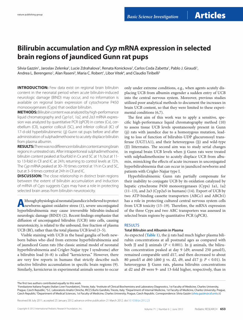
Organ UCB Levels in Untreated Gunn Rat Pups at d9 and d17In jj Gunn rats, at both ages, UCB content in the liver was almost 10 times that of the brain, with intermediate levels in spleen and kidneys, and was significantly greater than in the same organs of Jj and JJ rats (Figure 1).
During development, the organ bilirubin content signifi-cantly decreased (by 37–66%) in liver, kidney, and brain of JJ and Jj rats, whereas no statistically significant changes were detected in the same organs of jj animals. Splenic bilirubin content increased significantly with age in jj Gunn rats but did not change in normobilirubinemic animals (Figure 1).
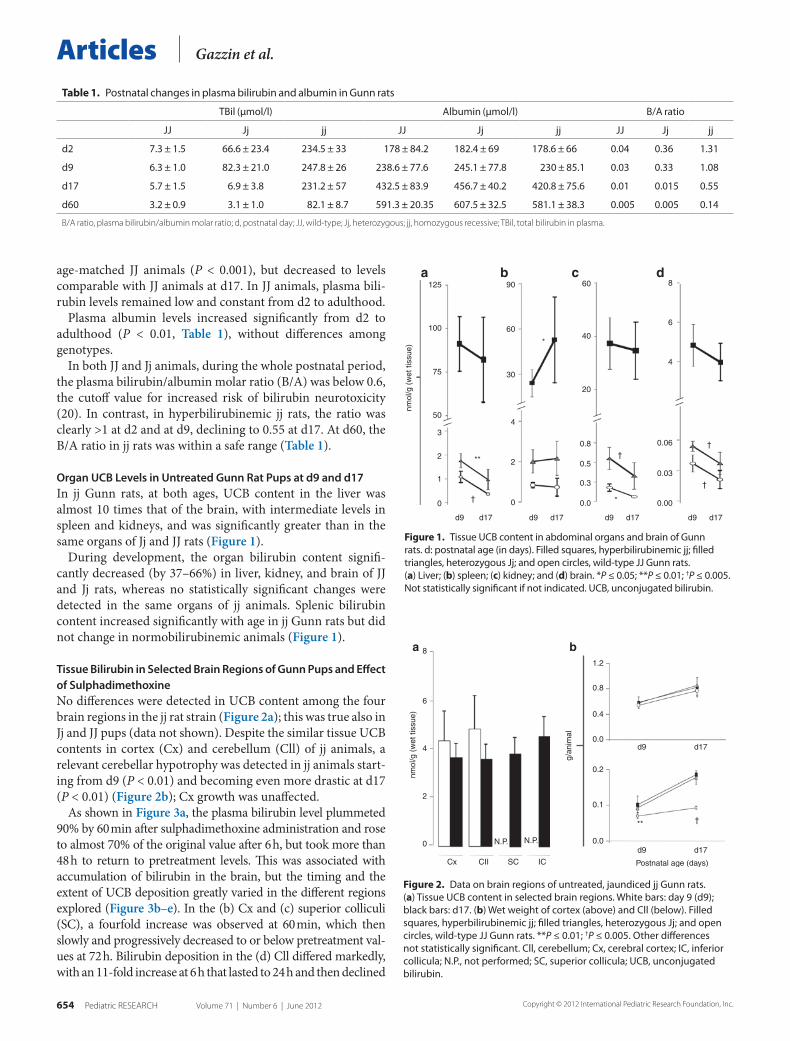
Tissue Bilirubin in Selected Brain Regions of Gunn Pups and Effect of SulphadimethoxineNo differences were detected in UCB content among the four brain regions in the jj rat strain (Figure 2a); this was true also in Jj and JJ pups (data not shown). Despite the similar tissue UCB contents in cortex (Cx) and cerebellum (Cll) of jj animals, a relevant cerebellar hypotrophy was detected in jj animals start-ing from d9 (P < 0.01) and becoming even more drastic at d17 (P < 0.01) (Figure 2b); Cx growth was unaffected.
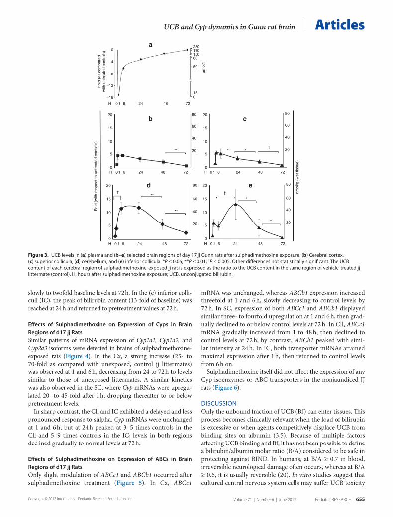
As shown in Figure 3a, the plasma bilirubin level plummeted 90% by 60 min after sulphadimethoxine administration and rose to almost 70% of the original value after 6 h, but took more than 48 h to return to pretreatment levels. This was associated with accumulation of bilirubin in the brain, but the timing and the extent of UCB deposition greatly varied in the different regions explored (Figure 3b–e). In the (b) Cx and (c) superior colliculi (SC), a fourfold increase was observed at 60 min, which then slowly and progressively decreased to or below pretreatment val-ues at 72 h. Bilirubin deposition in the (d) Cll differed markedly, with an 11-fold increase at 6 h that lasted to 24 h and then declined
125 90
60
60
40
20
0.8
0.5
0.3
0.0 0.00
0.03
0.06
4
6
8
30
4
2
0
100
75
50
3
2
1
0
d9 d17 d9 d17 d9 d17 d9 d17
nmol
/g (
wet
tiss
ue)
**
*
*†
†
†
†
a b c d
Figure 1. tissue uCB content in abdominal organs and brain of Gunn rats. d: postnatal age (in days). Filled squares, hyperbilirubinemic jj; filled triangles, heterozygous Jj; and open circles, wild-type JJ Gunn rats. (a) liver; (b) spleen; (c) kidney; and (d) brain. *P ≤ 0.05; **P ≤ 0.01; †P ≤ 0.005. Not statistically significant if not indicated. uCB, unconjugated bilirubin.
8
6
4
nmol
/g (
wet
tiss
ue)
g/an
imal
2
0
Cx CII SC IC
N.P.N.P.
1.2
0.8
0.4
0.0
0.2
0.1
0.0d9 d17
d9 d17
Postnatal age (days)
** †
a b
Figure 2. Data on brain regions of untreated, jaundiced jj Gunn rats. (a) tissue uCB content in selected brain regions. White bars: day 9 (d9); black bars: d17. (b) Wet weight of cortex (above) and Cll (below). Filled squares, hyperbilirubinemic jj; filled triangles, heterozygous Jj; and open circles, wild-type JJ Gunn rats. **P ≤ 0.01; †P ≤ 0.005. Other differences not statistically significant. Cll, cerebellum; Cx, cerebral cortex; IC, inferior collicula; N.P., not performed; sC, superior collicula; uCB, unconjugated bilirubin.
table 1. Postnatal changes in plasma bilirubin and albumin in Gunn rats
tBil (µmol/l) Albumin (µmol/l) B/A ratio
JJ Jj jj JJ Jj jj JJ Jj jj
d2 7.3 ± 1.5 66.6 ± 23.4 234.5 ± 33 178 ± 84.2 182.4 ± 69 178.6 ± 66 0.04 0.36 1.31
d9 6.3 ± 1.0 82.3 ± 21.0 247.8 ± 26 238.6 ± 77.6 245.1 ± 77.8 230 ± 85.1 0.03 0.33 1.08
d17 5.7 ± 1.5 6.9 ± 3.8 231.2 ± 57 432.5 ± 83.9 456.7 ± 40.2 420.8 ± 75.6 0.01 0.015 0.55
d60 3.2 ± 0.9 3.1 ± 1.0 82.1 ± 8.7 591.3 ± 20.35 607.5 ± 32.5 581.1 ± 38.3 0.005 0.005 0.14
B/a ratio, plasma bilirubin/albumin molar ratio; d, postnatal day; JJ, wild-type; Jj, heterozygous; jj, homozygous recessive; TBil, total bilirubin in plasma.
Volume 71 | Number 6 | June 2012 Pediatric ReseaRch 655copyright © 2012 International Pediatric Research Foundation, Inc.
ArticlesUCB and Cyp dynamics in Gunn rat brain
slowly to twofold baseline levels at 72 h. In the (e) inferior colli-culi (IC), the peak of bilirubin content (13-fold of baseline) was reached at 24 h and returned to pretreatment values at 72 h.
Effects of Sulphadimethoxine on Expression of Cyps in Brain Regions of d17 jj RatsSimilar patterns of mRNA expression of Cyp1a1, Cyp1a2, and Cyp2a3 isoforms were detected in brains of sulphadimethoxine- exposed rats (Figure 4). In the Cx, a strong increase (25- to 70-fold as compared with unexposed, control jj littermates) was observed at 1 and 6 h, decreasing from 24 to 72 h to levels similar to those of unexposed littermates. A similar kinetics was also observed in the SC, where Cyp mRNAs were upregu-lated 20- to 45-fold after 1 h, dropping thereafter to or below pretreatment levels.
In sharp contrast, the Cll and IC exhibited a delayed and less pronounced response to sulpha. Cyp mRNAs were unchanged at 1 and 6 h, but at 24 h peaked at 3–5 times controls in the Cll and 5–9 times controls in the IC; levels in both regions declined gradually to normal levels at 72 h.
Effects of Sulphadimethoxine on Expression of ABCs in Brain Regions of d17 jj RatsOnly slight modulation of ABCc1 and ABCb1 occurred after sulphadimethoxine treatment (Figure 5). In Cx, ABCc1
mRNA was unchanged, whereas ABCb1 expression increased threefold at 1 and 6 h, slowly decreasing to control levels by 72 h. In SC, expression of both ABCc1 and ABCb1 displayed similar three- to fourfold upregulation at 1 and 6 h, then grad-ually declined to or below control levels at 72 h. In Cll, ABCc1 mRNA gradually increased from 1 to 48 h, then declined to control levels at 72 h; by contrast, ABCb1 peaked with simi-lar intensity at 24 h. In IC, both transporter mRNAs attained maximal expression after 1 h, then returned to control levels from 6 h on.
Sulphadimethoxine itself did not affect the expression of any Cyp isoenzymes or ABC transporters in the nonjaundiced JJ rats (Figure 6).
DIsCussIONOnly the unbound fraction of UCB (Bf) can enter tissues. This process becomes clinically relevant when the load of bilirubin is excessive or when agents competitively displace UCB from binding sites on albumin (3,5). Because of multiple factors affecting UCB binding and Bf, it has not been possible to define a bilirubin/albumin molar ratio (B/A) considered to be safe in protecting against BIND. In humans, at B/A ≥ 0.7 in blood, irreversible neurological damage often occurs, whereas at B/A ≥ 0.6, it is usually reversible (20). In vitro studies suggest that cultured central nervous system cells may suffer UCB toxicity
0
−4
−8
−12Fol
d (a
s co
mpa
red
with
unt
reat
ed c
ontr
ols)
Fol
d (w
ith r
espe
ct to
unt
reat
ed c
ontr
ols)
nmol
/g (
wet
tiss
ue)
−16
20
20
15
10
5
0
20
15
10
5
0
15
10
5
0
20
15
10
5
0
H 01 6 24 48 72
H 01 6 24 48 72
H 01 6 24 48 72
H 01 6 24 48 72
H 01 6 24 48 72
015
50
60150170230
µmol
/l
80
60
40
20
80
60
40
20
80
60
40
20
80
60
40
20
** *
*
**
**
*†
††
†
a
b c
ed
Figure 3. uCB levels in (a) plasma and (b–e) selected brain regions of day 17 jj Gunn rats after sulphadimethoxine exposure. (b) Cerebral cortex, (c) superior collicula, (d) cerebellum, and (e) inferior collicula. *P ≤ 0.05; **P ≤ 0.01; †P ≤ 0.005. Other differences not statistically significant. the uCB content of each cerebral region of sulphadimethoxine-exposed jj rat is expressed as the ratio to the uCB content in the same region of vehicle-treated jj littermate (control). H, hours after sulphadimethoxine exposure; uCB, unconjugated bilirubin.

656 Pediatric ReseaRch Volume 71 | Number 6 | June 2012 copyright © 2012 International Pediatric Research Foundation, Inc.
Articles Gazzin et al.
Fol
d (w
ith r
espe
ct to
unt
reat
ed c
ontr
ols)
H 1 6 24 48 72H 1 6
*
*†
††
†
†
†
24 48 72
H 1 6 24 48 72H 1 6 24 48 72
120
80
4020
8
4
0
6
4
2
0
80
60
40
20
2
1
0
20
14
8
2
0
a
c
b
d
Figure 4. Cyp mRNA expression in selected brain regions of day 17 jj Gunn rats after sulphadimethoxine exposure. (a) Cerebral cortex, (b) superior colli-cula, (c) cerebellum, and (d) inferior collicula. *P ≤ 0.05; †P ≤ 0.005. Other differences not statistically significant. White bars, Cyp1a1; gray bars, Cyp1a2; and black bars, Cyp2a3 mRNA expression. Cyp, cytochrome P450 monooxygenase; H, hours after sulphadimethoxine exposure.
Fol
d (w
ith r
espe
ct to
unt
reat
ed c
ontr
ols)
H 1 6 24 48 72 H 1 6 24 48 72 H 1 6 24 48 72 H 1 6 24 48 72
H 1 6 24 48 72 H 1 6 24 48 72 H 1 6 24 48 72 H 1 6 24 48 72
2
1
0
2
3 5
4
3
2
1
0
1
0
2
3
1
0
2
3
1
0
4
2
0
4
2
0
4
6
2
0
*
*
*
*
**
*
†
a
c
b
d
Figure 5. ABC mRNA expression in selected brain regions of day 17 jj Gunn rats after sulphadimethoxine exposure. (a) Cerebral cortex, (b) superior collicula, (c) cerebellum, and (d) inferior collicula. *P ≤ 0.05; †P ≤ 0.005. Other differences not statistically significant. White bars, ABCc1; black bars, ABCb1 mRNA expression. H, hours after sulphadimethoxine exposure.

Volume 71 | Number 6 | June 2012 Pediatric ReseaRch 657copyright © 2012 International Pediatric Research Foundation, Inc.
ArticlesUCB and Cyp dynamics in Gunn rat brain
at even lower ratios (<0.5) (3,21,22), but caution is advised in extrapolation of the in vitro data to the much more compli-cated situation in vivo.
Hyperbilirubinemia in jj Gunn rats results from a deficiency of hepatic UGT1A1, similar to that seen in human patients with Crigler-Najjar type I and analogous to the decreased conjugat-ing activity seen in human neonates during the first days of life (23). As in severely jaundiced newborns, the high levels of UCB in plasma of jj Gunn rat pups, with B/A ratios exceeding 1.0 at d2 and d9, resulted in accumulation of UCB in the tissues in the early neonatal period; in jj Gunn pups, this causes marked cerebellar hypotrophy from d9 onward (24,25). In heterozy-gous (Jj) Gunn rats, the milder, early, temporary hyperbiliru-binemia did not impair cerebellar growth, suggesting that the 0.36 B/A ratio found at d2 may be safe. It is interesting that d2 jj rats showed no signs of bilirubin toxicity (4,25), suggesting that it takes time for UCB to accumulate in the central nervous system and/or for neurotoxicity to develop.
In jj Gunn rats, we found UCB content to be higher in liver than in spleen and kidneys, and much lower in whole brain (Figure 1). As reported in humans (20), visceral bilirubin con-centrations decreased less rapidly than plasma bilirubin levels from d9 to d17, whereas brain UCB content declined more rap-idly (24%) than plasma bilirubin levels (6.5%) during the same period. The decline of UCB content in all organs is likely in part related to the large increase in plasma albumin from d9 to d17, enhancing the decrease in plasma B/A ratio and Bf concentra-tion as total plasma UCB levels decline. This is consistent with previous reports of lower bilirubin concentrations in liver, spleen, kidney, and whole brain of adult Gunn rats than in pups (26).
The differences in UCB content and rates of decline among the organs (Figure 1) indicate, however, that tissue UCB lev-els must also be controlled by other mechanisms. The produc-tion rates of UCB in the organ and the permeability to UCB of the blood–organ barriers (10) determine the supply of UCB. Because Gunn (jj) rats cannot conjugate bilirubin, the cellular export of UCB (15,16,18,19) and UCB oxidation (12,14,26–28) might play a role in UCB removal from the brain of these animals.
Our study in jj rats after sulphadimethoxine administra-tion provides important new data on the time dependency of the consequent sudden, massive accumulation and gradual clearance of UCB in different brain regions (Figure 3b–e). Notable are the much greater and longer-lasting accumu-lations of UCB in the Cll and IC, whose functions (motor coordination and auditory, respectively) are impaired in bilirubin encephalopathy of sudden onset after sulphadi-methoxine administration (29), and the more gradual onset in jaundiced neonates and jj Gunn rats. By contrast, UCB accumulation after sulphadimethoxine was limited and brief in the Cx and IC, whose functions (cognition and vision, respectively) are unimpaired in BIND. The spatial nearness of the SC and IC suggest further that their markedly differ-ent kinetics and severity of UCB accumulation after sulpha-dimethoxine are unlikely to be due to differences blood sup-ply or blood–brain barriers, but most probably are linked to cellular mechanisms for removal of UCB.
This conclusion is further supported by the relationships between expression of Cyps known to oxidize UCB and the dynamics of tissue UCB in the selected brain regions. Figures 4 and 5 show an immediate and massive upregulation of the Cyp mRNAs and much smaller immediate upregulation of the ABC transporters in the unaffected brain regions (Cx and SC), in striking contrast to the delayed and relatively puny upregulation of Cyps and ABC transporters in the affected brain regions (Cll and IC). The timing seems to fit with the decline in tissue UCB levels after 1 h in the unaffected regions but only after 6 h in the affected regions (Figure 4). This suggests that these Cyps (and, presumably, UCB oxida-tion) provide major protection against acute accumulation of UCB in the unaffected regions, with the ABC transporters playing a minor role. Because only mRNA expression was assessed, confirmation of this hypothesis will require further studies of upregulation of enzyme and transporter protein levels and activities. It needs to be determined also if similar, differential, short-term upregulation of Cyps is stimulated by the relatively rapid, albeit less massive and precipitous, spontaneous increases in brain UCB content during the week after birth in untreated jj Gunn pups
The similarity of increments in tissue UCB levels 1 h after sulpha administration suggests that there are similar rates of passive diffusion of UCB in all four brain regions dur-ing this early accumulation of UCB. Selective upregulation of Cyps, enhancing UCB oxidation, more likely accounts for the subsequent decline in tissue UCB levels. Based on this reasoning, we speculate that limited passive diffusion
Fol
d (w
ith r
espe
ct to
unt
reat
ed c
ontr
ols)
2
1
0
2
1
0
2
1
0
2
1
0
CT
RL
Cyp
1a1
Cyp
1a2
Cyp
2a3
AB
Cb1
AB
Cc1
CT
RL
Cyp
1a1
Cyp
1a2
Cyp
2a3
AB
Cb1
AB
Cc1
CT
RL
Cyp
1a1
Cyp
1a2
Cyp
2a3
AB
Cb1
AB
Cc1
CT
RL
Cyp
1a1
Cyp
1a2
Cyp
2a3
AB
Cb1
AB
Cc1
* *
a
c d
b
Figure 6. Cyp and ABC mRNA modulation by sulphadimethoxine in selected brain regions of day 17 JJ Gunn rats. (a) Cerebral cortex, (b) superior collicula, (c) cerebellum, and (d) inferior collicula. *P ≤ 0.05. Other differences not statistically significant. White bars, controls (vehicle-in-jected animals) and black bars, sulpha-treated rats. Cyp, cytochrome P450 monooxygenase.
658 Pediatric ReseaRch Volume 71 | Number 6 | June 2012 copyright © 2012 International Pediatric Research Foundation, Inc.
Articles Gazzin et al.
across the blood–brain and blood–cerebrospinal fluid bar-riers, rather than oxidation or export of UCB, is also the dominant factor determining the similarity of UCB content observed among the four brain regions of untreated jj Gunn rats at d17 (Figure 2). The reasons why the upregulation dif-fers among different portions of the brain need to be further investigated.
Our data suggest that the historical concept of kernicterus, based on selective damage due to regional bilirubin accumula-tion in the brain, should be reassessed. Our study shows that regional modulation of Cyps may account for the differences among brain regions in severity and duration of bilirubin accumulation during sudden increases in Bf. It remains to be determined whether BIND in jaundiced Gunn rat pups and neonates is similarly influenced by modulation of intracellular defensive mechanisms, such as oxidation of UCB.
MetHODsAnimalsHyperbilirubinemic Gunn rats (jj) (30) with congenital deficiency of UGT1A1 (23) and their heterozygous (Jj) and normobilirubinemic wild-type (JJ) littermates (31) were bred in the animal facility of the CSPA, University of Trieste. Parturitions were synchronized to obtain a sufficient number of littermate pups of each genotype and postnatal age (d ± 1).
Animal care and procedures were conducted according to the guidelines approved by Italian Law (decree 116-92) and by European
Community directive 86-609-EEC. The study was approved by the animal care and use committee of the University of Trieste.
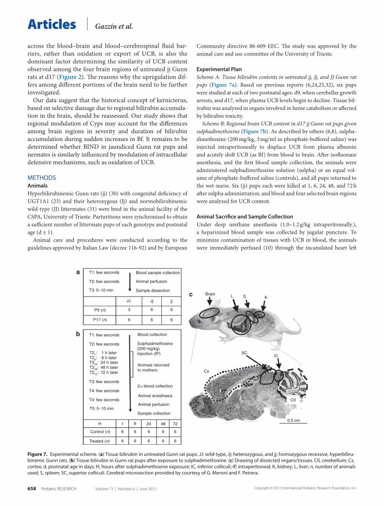
Experimental PlanScheme A: Tissue bilirubin contents in untreated jj, Jj, and JJ Gunn rat pups (Figure 7a). Based on previous reports (6,24,25,32), six pups were studied at each of two postnatal ages: d9, when cerebellar growth arrests, and d17, when plasma UCB levels begin to decline. Tissue bil-irubin was analyzed in organs involved in heme catabolism or affected by bilirubin toxicity.
Scheme B: Regional brain UCB content in d17 jj Gunn rat pups given sulphadimethoxine (Figure 7b). As described by others (6,8), sulpha-dimethoxine (200 mg/kg, 3 mg/ml in phosphate-buffered saline) was injected intraperitoneally to displace UCB from plasma albumin and acutely shift UCB (as Bf) from blood to brain. After isofluorane anesthesia, and the first blood sample collection, the animals were administered sulphadimethoxine solution (sulpha) or an equal vol-ume of phosphate-buffered saline (controls), and all pups returned to the wet-nurse. Six (jj) pups each were killed at 1, 6, 24, 48, and 72 h after sulpha administration, and blood and four selected brain regions were analyzed for UCB content.
Animal Sacrifice and Sample CollectionUnder deep urethane anesthesia (1.0–1.2 g/kg intraperitoneally.), a heparinized blood sample was collected by jugular puncture. To minimize contamination of tissues with UCB in blood, the animals were immediately perfused (10) through the incanulated heart left
T1: few seconds Blood sample collection
Blood collection
Sulphadimethoxine(200 mg/kg)injection (IP)
Animals returnedto mothers
Animal anesthesia
Animal perfusion
Sample collection
1
6
6
6
6
6
6
24
6
6
48
6
6
72
6
2nd blood collection
Animal perfusion
Sample dissection
T2: few seconds
T3: 5−10 min
T1: few seconds
T21: 1 h laterT26: 6 h laterT224: 24 h laterT248: 48 h laterT272: 72 h later
T2: few seconds
T3: few seconds
T4: few seconds
T4: few seconds
T5: 5−10 min
H
Control (n)
Treated (n)
JJ Jj jj
6
6
6
6
3P9 (n)
P17 (n) 6
Brain L S
SCIC
CII
0.5 cm
Cx
K
a
c
b
Figure 7. experimental scheme. (a) tissue bilirubin in untreated Gunn rat pups. JJ: wild-type, Jj: heterozygous, and jj: homozygous recessive, hyperbiliru-binemic Gunn rats. (b) tissue bilirubin in Gunn rat pups after exposure to sulphadimethoxine. (c) Drawing of dissected organs/tissues. Cll, cerebellum; Cx, cortex; d, postnatal age in days; H, hours after sulphadimethoxine exposure; IC, inferior colliculi; IP, intraperitoneal; K, kidney; l, liver; n, number of animals used; s, spleen; sC, superior colliculi. Cerebral microsection provided by courtesy of G. Meroni and F. Petrera.
Volume 71 | Number 6 | June 2012 Pediatric ReseaRch 659copyright © 2012 International Pediatric Research Foundation, Inc.
ArticlesUCB and Cyp dynamics in Gunn rat brain
ventricle using a peristaltic pump, until the effluent was visibly free of red cells.
After decapitation, the Cx, Cll, SC, and IC were dissected and cleaned of meninges and choroid plexuses as described previously for Cx (33). In scheme A, the abdominal organs mainly involved in metabolism of bilirubin (liver, spleen, kidney) were also collected (Figure 7c). Each tissue/organ sample was divided into two parts, one for tissue bilirubin quantification and the second one for mRNA extraction followed by qPCR analysis. Samples were immediately stored at −80 °C until analysis. All sampling and subsequent analyses were performed under dim light, using foil-wrapped tubes, to mini-mize bilirubin photooxidation.
Tissue Bilirubin QuantificationUCB content was determined on rapidly thawed samples using high-performance liquid chromatography with diode array detec-tor (Agilent, Santa Clara, CA) as described (10). Briefly, 300 pmol of mesobilirubin in dimethylsulfoxide (internal standard) was added, and samples were homogenized on ice. Bile pigments were then extracted into chloroform/hexane (5:1 vol/vol) at pH 6.0, and con-taminants removed by extraction of the pigments in a minimum volume of methanol/sodium carbonate (pH 10). The resulting polar droplet was loaded onto a C-8 reverse-phase column (Phenomenex, Torrance, CA), and separated pigments were detected at 440 nm. The concentration of UCB, determined from the area under the curve of the UCB peak in reference to the internal standard of mesobilirubin, was calculated as nmol/g of wet tissue weight.
Total Bilirubin and Albumin Determination in PlasmaAfter centrifugation (2,500 rpm, 20 min at room temperature), plasma was collected and immediately frozen at –20 °C until assayed. Plasma total bilirubin and albumin were quantified, respectively, by the diazo-reaction (Boehringer-Mannheim Kit 1552414, Monza, Italy) and by the ALB-Plus BCG (Roche Diagnostic, Milan, Italy) kits on an automated Roche–Hitachi analyzer (Roche Diagnostic, Milan, Italy). Samples with hemolysis were discarded.
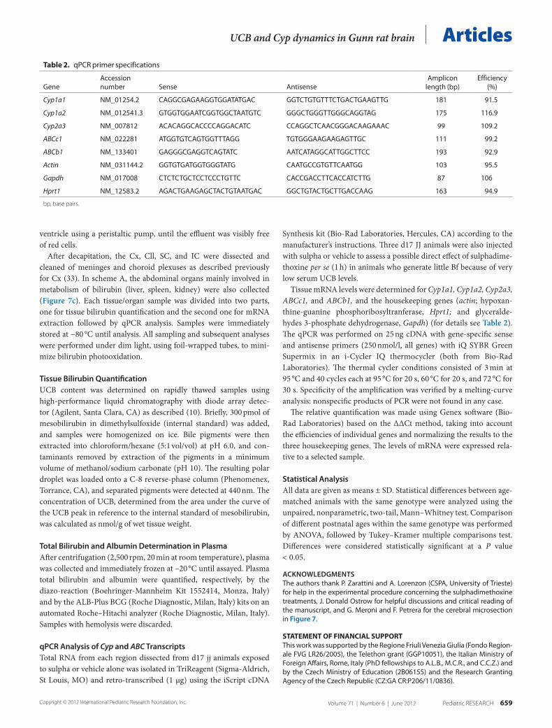
qPCR Analysis of Cyp and ABC TranscriptsTotal RNA from each region dissected from d17 jj animals exposed to sulpha or vehicle alone was isolated in TriReagent (Sigma-Aldrich, St Louis, MO) and retro-transcribed (1 µg) using the iScript cDNA
Synthesis kit (Bio-Rad Laboratories, Hercules, CA) according to the manufacturer’s instructions. Three d17 JJ animals were also injected with sulpha or vehicle to assess a possible direct effect of sulphadime-thoxine per se (1 h) in animals who generate little Bf because of very low serum UCB levels.
Tissue mRNA levels were determined for Cyp1a1, Cyp1a2, Cyp2a3, ABCc1, and ABCb1, and the housekeeping genes (actin; hypoxan-thine-guanine phosphoribosyltranferase, Hprt1; and glyceralde-hydes 3-phosphate dehydrogenase, Gapdh) (for details see Table 2). The qPCR was performed on 25 ng cDNA with gene-specific sense and antisense primers (250 nmol/l, all genes) with iQ SYBR Green Supermix in an i-Cycler IQ thermocycler (both from Bio-Rad Laboratories). The thermal cycler conditions consisted of 3 min at 95 °C and 40 cycles each at 95 °C for 20 s, 60 °C for 20 s, and 72 °C for 30 s. Specificity of the amplification was verified by a melting-curve analysis: nonspecific products of PCR were not found in any case.
The relative quantification was made using Genex software (Bio-Rad Laboratories) based on the ΔΔCt method, taking into account the efficiencies of individual genes and normalizing the results to the three housekeeping genes. The levels of mRNA were expressed rela-tive to a selected sample.
Statistical AnalysisAll data are given as means ± SD. Statistical differences between age-matched animals with the same genotype were analyzed using the unpaired, nonparametric, two-tail, Mann–Whitney test. Comparison of different postnatal ages within the same genotype was performed by ANOVA, followed by Tukey–Kramer multiple comparisons test. Differences were considered statistically significant at a P value < 0.05.
ACKNOWLEDGMENTSthe authors thank P. Zarattini and A. lorenzon (CsPA, university of trieste) for help in the experimental procedure concerning the sulphadimethoxine treatments, J. Donald Ostrow for helpful discussions and critical reading of the manuscript, and G. Meroni and F. Petrera for the cerebral microsection in Figure 7.
stAteMent oF FInAncIAl suPPortthis work was supported by the Regione Friuli Venezia Giulia (Fondo Region-ale FVG lR26/2005), the telethon grant (GGP10051), the Italian Ministry of Foreign Affairs, Rome, Italy (PhD fellowships to A.l.B., M.C.R., and C.C.Z.) and by the Czech Ministry of education (2B06155) and the Research Granting Agency of the Czech Republic (CZ:GA CR:P206/11/0836).
table 2. qPCR primer specifications
GeneAccession number sense Antisense
Amplicon length (bp)
efficiency (%)
Cyp1a1 NM_01254.2 CAGGCGAGAAGGtGGAtAtGAC GGtCtGtGtttCtGACtGAAGttG 181 91.5
Cyp1a2 NM_012541.3 GtGGtGGAAtCGGtGGCtAAtGtC GGGCtGGGttGGGCAGGtAG 175 116.9
Cyp2a3 NM_007812 ACACAGGCACCCCAGGACAtC CCAGGCtCAACGGGACAAGAAAC 99 109.2
ABCc1 NM_022281 AtGGtGtCAGtGGtttAGG tGtGGGAAGAAGAGttGC 111 99.2
ABCb1 NM_133401 GAGGGCGAGGtCAGtAtC AAtCAtAGGCAttGGCttCC 193 92.9
Actin NM_031144.2 GGtGtGAtGGtGGGtAtG CAAtGCCGtGttCAAtGG 103 95.5
Gapdh NM_017008 CtCtCtGCtCCtCCCtGttC CACCGACCttCACCAtCttG 87 106
Hprt1 NM_12583.2 AGACtGAAGAGCtACtGtAAtGAC GGCtGtACtGCttGACCAAG 163 94.9
bp, base pairs.

660 Pediatric ReseaRch Volume 71 | Number 6 | June 2012 copyright © 2012 International Pediatric Research Foundation, Inc.
Articles Gazzin et al.
REFERENCES 1. Benaron DA, Bowen FW. Variation of initial serum bilirubin rise in new-
born infants with type of illness. Lancet 1991;338:78–81. 2. Bhutani VK, Johnson L. 2005 Kernicterus: a preventable neonatal brain
injury. J Arab Neonatal Forum 2005;2:12–24. 3. Calligaris SD, Bellarosa C, Giraudi P, Wennberg RP, Ostrow JD, Tiribelli
C. Cytotoxicity is predicted by unbound and not total bilirubin concentra-tion. Pediatr Res 2007;62:576–80.
4. Takahashi M, Sugiyama K, Shumiya S, Nagase S. Penetration of bilirubin into the brain in albumin-deficient and jaundiced rats (AJR) and Nagase analbuminemic rats (NAR). J Biochem 1984;96:1705–12.
5. Ahlfors CE, Wennberg RP. Bilirubin-albumin binding and neonatal jaun-dice. Semin Perinatol 2004;28:334–9.
6. Cannon C, Daood MJ, O’Day TL, Watchko JF. Sex-specific regional brain bilirubin content in hyperbilirubinemic Gunn rat pups. Biol Neonate 2006;90:40–5.
7. Aono S, Semba R, Sato H, Kashiwamata S. Mode of bilirubin deposi-tion in the cerebellum of developing jaundiced Gunn rats. Biol Neonate 1989;55:119–23.
8. Daood MJ, Watchko JF. Calculated in vivo free bilirubin levels in the cen-tral nervous system of Gunn rat pups. Pediatr Res 2006;60:44–9.
9. Hansen TW. Pioneers in the scientific study of neonatal jaundice and ker-nicterus. Pediatrics 2000;106:E15.
10. Zelenka J, Lenícek M, Muchová L,et al. Highly sensitive method for quan-titative determination of bilirubin in biological fluids and tissues. J Chro-matogr B Analyt Technol Biomed Life Sci 2008;867:37–42.
11. Kapitulnik J, Gonzalez FJ. Marked endogenous activation of the CYP1A1 and CYP1A2 genes in the congenitally jaundiced Gunn rat. Mol Pharma-col 1993;43:722–5.
12. De Matteis F, Lord GA, Kee Lim C, Pons N. Bilirubin degradation by uncoupled cytochrome P450. Comparison with a chemical oxidation sys-tem and characterization of the products by high-performance liquid chro-matography/electrospray ionization mass spectrometry. Rapid Commun Mass Spectrom 2006;20:1209–17.
13. Pons N, Pipino S, De Matteis F. Interaction of polyhalogenated compounds of appropriate configuration with mammalian or bacterial CYP enzymes. Increased bilirubin and uroporphyrinogen oxidation in vitro. Biochem Pharmacol 2003;66:405–14.
14. Abu-Bakar A, Moore MR, Lang MA. Evidence for induced microsomal bilirubin degradation by cytochrome P450 2A5. Biochem Pharmacol 2005;70:1527–35.
15. Hankø E, Tommarello S, Watchko JF, Hansen TW. Administration of drugs known to inhibit P-glycoprotein increases brain bilirubin and alters the regional distribution of bilirubin in rat brain. Pediatr Res 2003;54:441–5.
16. Watchko JF, Daood MJ, Mahmood B, Vats K, Hart C, Ahdab-Barmada M. P-glycoprotein and bilirubin disposition. J Perinatol 2001;21:Suppl 1:S43–7; discussion S59–62.
17. Corich L, Aranda A, Carrassa L, Bellarosa C, Ostrow JD, Tiribelli C. The cytotoxic effect of unconjugated bilirubin in human neuroblastoma
SH-SY5Y cells is modulated by the expression level of MRP1 but not MDR1. Biochem J 2009;417:305–12.
18. Calligaris S, Cekic D, Roca-Burgos L,et al. Multidrug resistance associ-ated protein 1 protects against bilirubin-induced cytotoxicity. FEBS Lett 2006;580:1355–9.
19. Falcão AS, Bellarosa C, Fernandes A,et al. Role of multidrug resistance-associated protein 1 expression in the in vitro susceptibility of rat nerve cell to unconjugated bilirubin. Neuroscience 2007;144:878–88.
20. Strauss KA, Robinson DL, Vreman HJ, Puffenberger EG, Hart G, Mor-ton DH. Management of hyperbilirubinemia and prevention of kernict-erus in 20 patients with Crigler-Najjar disease. Eur J Pediatr 2006;165: 306–19.
21. Ostrow JD, Pascolo L, Shapiro SM, Tiribelli C. New concepts in bilirubin encephalopathy. Eur J Clin Invest 2003;33:988–97.
22. Watchko JF. Kernicterus and the molecular mechanisms of bilirubin-induced CNS injury in newborns. Neuromolecular Med 2006;8:513–29.
23. Chowdhury JR, Kondapalli R, Chowdhury NR. Gunn rat: a model for inherited deficiency of bilirubin glucuronidation. Adv Vet Sci Comp Med 1993;37:149–73.
24. Conlee JW, Shapiro SM. Development of cerebellar hypoplasia in jaun-diced Gunn rats: a quantitative light microscopic analysis. Acta Neuro-pathol 1997;93:450–60.
25. Keino H, Sato H, Semba R, Aono S, Aoki E, Kashiwamata S. Mode of prevention by phototherapy of cerebellar hypoplasia in a new Sprague-Dawley strain of jaundiced Gunn rats. Pediatr Neurosci 1985;12: 145–50.
26. Schmid R, Hammaker L. Metabolism and disposition of c14-bilirubin in congenital nonhemolytic jaundice. J Clin Invest 1963;42:1720–34.
27. Kapitulnik J, Ostrow JD. Stimulation of bilirubin catabolism in jaundiced Gunn rats by an induced of microsomal mixed-function monooxygenases. Proc Natl Acad Sci USA 1978;75:682–5.
28. Zaccaro C, Sweitzer S, Pipino S,et al. Role of cytochrome P450 1A2 in bili-rubin degradation Studies in Cyp1a2 (-/-) mutant mice. Biochem Pharma-col 2001;61:843–9.
29. Ahlfors CE, Shapiro SM. Auditory brainstem response and unbound bil-irubin in jaundiced (jj) Gunn rat pups. Biol Neonate 2001;80:158–62.
30. Gunn CH. Hereditary acholuric jaundice in a new mutant strain of rats. J Hered 1938;29:137–9.
31. Zarattini P, Gazzin S, Stebel M. Improvement of a historical animal model for Crigler Najjar Type I syndrome: development of the normobiliru-binemic JJ genotype as a true control for the Gunn jaundiced rat, 2011. Experimental Models: 110-114. (http://www.felasa.eu/media/uploads/ Proceedings_FELASA-ScandLAS-2010_small_SEC.pdf).
32. Gazzin S, Berengeno AL, Strazielle N,et al. Modulation of Mrp1 (ABCc1) and Pgp (ABCb1) by bilirubin at the blood-CSF and blood-brain barriers in the Gunn rat. PLoS ONE 2011;6:e16165.
33. Gazzin S, Strazielle N, Schmitt C,et al. Differential expression of the multi-drug resistance-related proteins ABCb1 and ABCc1 between blood-brain interfaces. J Comp Neurol 2008;510:497–507.


