Embed Size (px)
Citation preview

1 3
Arch Toxicol (2014) 88:1537–1548DOI 10.1007/s00204-014-1217-7
MOleculAr TOxIcOlOgy
BDE‑47 and 6‑OH‑BDE‑47 modulate calcium homeostasis in primary fetal human neural progenitor cells via ryanodine receptor‑independent mechanisms
Kathrin Gassmann · Timm Schreiber · Milou M. L. Dingemans · Guido Krause · Claudia Roderigo · Susanne Giersiefer · Janette Schuwald · Michaela Moors · Klaus Unfried · Åke Bergman · Remco H. S. Westerink · Christine R. Rose · Ellen Fritsche
received: 29 October 2013 / Accepted: 19 February 2014 / Published online: 6 March 2014 © Springer-Verlag Berlin Heidelberg 2014
this hypothesis, we investigated acute effects of BDe-47 and 6-OH-BDe-47 on [ca2+]i in human neural progenitor cells (hNPcs) and unraveled involved signaling pathways. Short-time differentiated hNPcs were exposed to BDe-47, 6-OH-BDe-47, and multiple inhibitors/stimulators of pre-sumably involved signaling pathways to determine possible effects on [ca2+]i by single-cell microscopy with the fluo-rescent dye Fura-2. Initial characterization of calcium sign-aling pathways confirmed the early developmental stage of hNPcs. In these cells, BDe-47 (2 μM) and 6-OH-BDe-47 (0.2 μM) induce [ca2+]i transients. This increase in [ca2+]i is due to extracellular ca2+ influx and intracellular release of ca2+, mainly from the endoplasmic reticulum (er). While extracellular ca2+ seems to enter the cytoplasm upon 6-OH-BDe-47 by interfering with the cell membrane and independent of ca2+ ion channels, er-derived ca2+ is released following activation of protein lipase c and ino-sitol 1,4,5-trisphosphate receptor, but independently of ryanodine receptors. These findings illustrate that immature developing hNPcs respond to low concentrations of 6-OH-BDe-47 by an increase in [ca2+]i and provide new mecha-nistic explanations for such BDe-induced calcium disrup-tion. Thus, these data support the possibility of a critical window of PBDe exposure, i.e., early human brain devel-opment, which has to be acknowledged in risk assessment.
Keywords Brominated flame retardant · calcium · Human neural progenitor cell · Neurotoxicity · Polybrominated diphenyl ether · ryanodine receptor
Introduction
Polybrominated diphenyl ethers (PBDes) are ubiqui-tously present in the environment, animals, and humans.
Abstract Polybrominated diphenyl ethers (PBDes) are bioaccumulating flame retardants found in rising concen-trations in human tissue. epidemiological and animal stud-ies have raised concern for their potential to induce devel-opmental neurotoxicity (DNT). considering the essential role of calcium homeostasis in neurodevelopment, PBDe-induced disturbance of intracellular calcium concentra-tion ([ca2+]i) may underlie PBDe-induced DNT. To test
Kathrin gassmann, Timm Schreiber, and Milou M. l. Dingemans have contributed equally to this work.
Electronic supplementary material The online version of this article (doi:10.1007/s00204-014-1217-7) contains supplementary material, which is available to authorized users.
K. gassmann · T. Schreiber · S. giersiefer · J. Schuwald · M. Moors · K. unfried · e. Fritsche (*) leibniz research Institute for environmental Medicine, Auf’m Hennekamp 50, 40225 Duesseldorf, germanye-mail: [email protected]
S. giersiefer e-mail: [email protected]
M. M. l. Dingemans · r. H. S. Westerink Neurotoxicology research group, Toxicology Division, Faculty of Veterinary Medicine, Institute for risk Assessment Sciences, utrecht university, utrecht, The Netherlands
g. Krause · c. roderigo · c. r. rose Institute of Neurobiology, Heinrich-Heine-university, universitaetsstrasse 1, 40225 Duesseldorf, germany
Å. Bergman Department of environmental chemistry, Stockholm university, Stockholm, Sweden
e. Fritsche Heinrich-Heine-university, universitaetsstrasse 1, 40225 Duesseldorf, germany

1538 Arch Toxicol (2014) 88:1537–1548
1 3
Among those, 2,2′,4,4′-tetrabromodiphenyl ether (BDe-47) is the predominant congener in human blood samples (Frederiksen et al. 2009). In vivo, BDe-47 is converted to hydroxylated metabolites (Marsh et al. 2006; Staskal et al. 2006), and these are found in high amounts in human blood (Athanasiadou et al. 2008), including fetal blood (Qiu et al. 2009). currently, the greatest concern for potential adverse health effects of PBDes relates to their developmental neurotoxicity (DNT). Previous data demonstrated that high and low doses of PBDes admin-istered perinatally during the brain growth spurt modify neurobehavior in rodents ranging from altered motor activity over changes in sweet preference to impairment of cognitive functions (costa and giordano 2007; Fon-num and Mariussen 2009). lately, several epidemiologi-cal studies revealed that prenatal exposure to PBDes, including BDe-47, also affects neurological outcome in children (eskenazi et al. 2013; gascon et al. 2011; Herbstman et al. 2010; Kicinski et al. 2012; roze et al. 2009). The precise molecular mechanisms of PBDe DNT are not clear (Dingemans et al. 2011); however, PBDes disrupt calcium (ca2+) homeostasis in rat pheochromo-cytoma (Pc12) cells, human neuroblastoma (SH-Sy5y) cells, and in isolated rat, mouse and rabbit organelles in vitro (Dingemans et al. 2008; Kim et al. 2011; Koda-vanti and Ward 2005; yu et al. 2008). ca2+ is an early-response second messenger and as such plays a key role in a number of physiological processes including cell proliferation, differentiation and apoptosis as well as a wide variety of specific neuronal processes including dendritic spine growth, synaptic plasticity and neuro-transmission (Berridge et al. 2000; ciccolini et al. 2003). Influx as well as ca2+ release from intracellular stores, such as endoplasmic reticulum (er) and mitochondria, defines the magnitude, time course and spatial spread of the ca2+ signal (Berridge et al. 2000; Delmas and Brown 2002). Therefore, it is suggested that PBDes exert their DNT potential through interference with [ca2+]i tran-sients in developing brain cells. Because this hypothesis is based on data generated in tumor cell lines or isolated organelles from rodents, here we test this concept in pri-mary human neural cells and investigate the involved mechanisms, e.g., if ryanodine receptors (ryrs) are primary biological targets of these PBDes congeners (Kim et al. 2011). The present study demonstrates that (1) acute low concentration BDe-47 and 6-OH-BDe-47 exposure disturbs [ca2+]i homeostasis in primary human neural progenitor cells (hNPcs) and (2) that these BDes affect [ca2+]i transients even in immature hNPcs, which are devoid of functional ryrs, by interfering with pro-tein lipase c (Plc) and the inositol 1,4,5-trisphosphate receptor (IP3r).
Materials and methods
chemicals
BDe-47 and 6-OH-BDe-47 were synthesized and purified (~99 % purity) at Stockholm university as described by Marsh et al. (1999) or were a kind gift of Jacob de Boer, Free university of Amsterdam. All other chemicals used (unless otherwise noted) were purchased from Sigma–Aldrich (Taufstein, germany) and were of the highest purity available.
cell culture
Normal hNPcs from three different donors (16–18 gesta-tional weeks, gW) were purchased from lonza (Verviers SPrl, Belgium) and were cultured as described earlier (Moors et al. 2007, 2009). For details, see Supplemental Material.
electrophysiology
Somatic whole-cell recordings were performed as described in Kafitz et al. (2008). For details, see Supple-mental Material.
calcium imaging
ratiometric calcium imaging was performed as described previously (Dingemans et al. 2008; Meier et al. 2008). Briefly, differentiated hNPcs were loaded with 5–15 μM Fura-2 AM (Molecular Probes, Invitrogen) in saline for 20 min at room temperature. Subsequently, cells were washed with external saline and placed on the stage of a microscope equipped with a TIll Photonics Polychrome IV (TIll Photonics gmbH, gräfelfing, germany). Flu-orescence was recorded every 0.5–6 s at 510 nm (exci-tation wavelengths: 340 or 357 nm and 380 nm) with a ccD camera (TIll Photonics gmbH), and the fluores-cence ratio (F340/F380 or F357/380) was calculated. Data collection, digital camera and polychromator con-trol were performed by imaging software (TIllvisION, version 4.01). changes in the fluorescence ratio were analyzed using custom-made excel macros (Microsoft corp., redmond, WA, uSA) or Igor Pro Software (Wav-emetrics, lake Oswego, Or, uSA).
For details regarding (6-OH-)BDe-47, lanthanum (la3+), thapsigargin (Tg), carbonyl cyanide 4-(trifluo-romethoxy) phenylhydrazone (FccP), u73122, 2-ami-noethoxydiphenyl borate (2-APB), caffeine, ryanodine or 4-chloro-m-cresol (4-cMc) application, see Supplemental Material.

1539Arch Toxicol (2014) 88:1537–1548
1 3
rNA preparation and reverse transcription polymerase chain reaction
rNA preparation was performed as described in gassmann et al. (2010). For details, see Supplemental Material.
lDH assay
cell viability was measured using a lactate-dehydroge-nase (lDH) assay (cytoTox-One, Promega) as previously described (Moors et al. 2009).
Data analysis and statistics
To determine effects on [ca2+]i, fluorescence ratio of each cell under baseline conditions was normalized. A respond-ing cell was considered a cell with an increase in the nor-malized fluorescence ratio above 1.2 (≥1.02 in case of glutamate application). The mean of the average and the maximum amplitude were calculated from all cells whether or not they reached the threshold of 1.2. For the average amplitude, this method enabled detection of small shifts in baseline (<1.2). Data are presented as mean ± SeM from at least three independent experiments with the number of cells indicated. Statistical analyses were performed using graphPad Prism and SPSS Statistics. We compared contin-uous data using paired Student’s t test, and categorical data were compared using Kruskal–Wallis and Mann–Whitney test. For multifactor analysis, ANOVA with the Bonferroni post hoc test was used. A p value <0.05 was considered sta-tistically significant.
Results
electrophysiology
Membrane potential and characteristics of voltage-gated ion channels were determined in 14 days differentiated hNPcs using whole-cell patch-clamp measurements. cells had a mean membrane potential of −42.4 ± 10.7 mV, a membrane resistance of 1.0 ± 0.6 gΩ and a membrane capacitance of 6.4 ± 3.8 pF (n = 11). The voltage-step pro-tocol revealed the presence of outward currents (n = 10/11 cells; Suppl. Fig. 1A). In two of these cells, we detected small fast inward currents. In none of the cells tested, injec-tion of depolarizing currents resulted in action potential generation in the current-clamp mode.
general characterization of calcium signaling in hNPcs
First, calcium imaging experiments were performed to reveal the maturation state of hNPcs by presence/
functionality of receptors. To probe for the presence of voltage-gated ion channels, we applied saline containing 80 mM potassium by puff application for 2.5 s to the cells. Only a negligible fraction (1.6 %) of cells responded to the application of potassium with a calcium increase (Fig. 1a). In contrast, virtually all cells responded to puff application of ATP (0.5 or 1 mM for 1 s; Fig. 1a), which is mediated through purinergic P2 receptors (rev. in Butt 2011). Also, store-operated calcium channels (SOcc) are operational, because la3+ suppressed the plateau phase of ATP-depend-ent [ca2+]i transients (Fig. 1a). Additionally, puff applica-tion of acetylcholine (Ach; 0.5 mM for 1 or 2 s) caused a [ca2+]i increase in about 40 % of all cells (Fig. 1a). The calcium increase induced by Ach was completely blocked by atropine in 48/53 cells (data not shown), indicating that it was due to the activation of muscarinic Ach receptors. The percentage of cells responding to ATP and acetylcho-line as well as the amplitude of the induced calcium tran-sients were independent of the differentiation time (either 12, 24 or 48 h in culture) and the application form (bath application or puff application), although the bath applica-tion yielded slightly higher amplitudes (Suppl. Fig. 1B). Application of glutamate (1 mM; 1–2.5 s) induced very mild [ca2+]i transients with small amplitudes compared to those induced by ATP or Ach in the majority of cells (Fig. 1a; Suppl. Fig. 1B). At early differentiation time points (12 h), the number of responding cells was signifi-cantly smaller than at later time points (Suppl. Fig. 1B). Therefore, for all additional experiments, a differentiation time of 24 or 48 h was chosen. In none of the cells stud-ied (n = 60), application of gABA (1 mM, 2.5 s) induced an increase in [ca2+]i (Fig. 1a). Investigations on expres-sion and function of ryrs revealed presence of ryr3 and absence of ryr1 and ryr2 in hNPc (gW 16–18) and presence of all three subtypes in fetal control mrNA from gW 20–33 (Fig. 1b). Despite presence of ryr3, neither the ryr sensitizer caffeine (20 mM), nor ryanodine (20 nM), nor the ryr agonist 4-cMc (500 μM) altered spontaneous baseline [ca2+]i transients of the ethanol solvent control hNPc (Fig. 1c, d)
BDe-47 and 6-OH-BDe-47 cause an increase in [ca2+]i in hNPc
Differentiated hNPc was short-term exposed to non-cyto-toxic concentrations of BDe-47 and OH-BDe-47 (Suppl. Fig. 2). BDe-47 (20 μM) or 6-OH-BDe-47 (0.2, 2 and 20 μM) initiated a significant [ca2+]i increase in 83 % or 45, 54 and 100 % of responding cells, respectively (vehicle: 27 %; Fig. 2a). The maximum [ca2+]i amplitude of the sol-vent control amounted to 1.16 ± 0.01, whereas amplitude was concentration-dependently increased by both BDe-47 (2, 20 μM) and 6-OH-BDe-47 (0.2, 2 and 20 μM),

1540 Arch Toxicol (2014) 88:1537–1548
1 3
Fig. 1 general characterization of calcium signaling in differenti-ated hNPcs. a representative traces of ratiometric calcium imag-ing of 24–48 h differentiated NPcs that were constantly perfused with saline. Arrows indicate application of neurotransmitters for 1 s. In case of lanthanum, individual cells were exposed to 0.5 mM ATP for 25 min, and la3+ was added at t = 10 min and washed out at t = 17 min. For glutamate and gABA, the graph was smoothed by calculating the mean fluorescence of three timepoints. b mrNA expression patterns (rT-Pcr) of the different isoforms of the ryr
in 24 or 48 h differentiated hNPc. Human fetal brain rNA (gW 20–33) was used as positive control for the designed primers. Primers for β-actin served as loading control. Negative control (DePc water instead of cDNA) indicates the purity of the Pcr mixture. To test the functionality of the ryr3 in our model system, we exposed 48 h differentiated NPcs to the ryr agonist 4-chloro-m-cresol (4-cMc, 500 μM), ryanodine (20 nM) and the ryr sensitizer caffeine (20 mM). The diagrams show responding cells (c) and amplitudes (d). Data are from 4 to 9 independent experiments (mean ± SeM)

1541Arch Toxicol (2014) 88:1537–1548
1 3
Fig. 2 BDe-47 and 6-OH-BDe-47 cause an increase in [ca2+]i in hNPcs during acute exposure. results from individ-ual cells exposed to 0.2–20 μM BDe-47 or 0.02–20 μM 6-OH-BDe-47 for 20 min, applied at t = 5 min, are quantified as bars indicating the percentage of cells showing an increase in [ca2+]i within 20 min after application (a) and the average and maximal amplitudes (b). Data are from three independent experiments (mean ± SeM). Numbers above bars indicate the number of cells used for the data analysis. *p < 0.05 com-pared to control. representative traces of normalized F340/F380 ratio during exposure to BDe-47 (c) or 6-OH-BDe-47 (d). Note the difference in scaling for 20 μM

1542 Arch Toxicol (2014) 88:1537–1548
1 3
amounting to a significantly higher maximum amplitude of 1.22 ± 0.03, 2.11 ± 0.15 and 1.30 ± 0.05, 1.44 ± 0.08 and 5.74 ± 0.45, respectively (Fig. 2b).
Thus, 6-OH-BDe-47 was effective already at lower concentrations than BDe-47, and the increase in ampli-tude was larger. There was also a difference in response time: while the increase in [ca2+]i occurred with multi-ple peaks 3–5 min after BDe-47 application (2, 20 μM), [ca2+]i, elevation took place immediately and persistently after 6-OH-BDe-47 at concentrations ≥0.2 μM. Only 20 μM 6-OH-BDe-47 induced a transient initial [ca2+]i increase within the first 5 min after application fol-lowed by a late (>10 min) [ca2+]i increase accompanied by a general elevation of the baseline. Some single cells showed a very strong [ca2+]i increase without recovery (Fig. 2c, d).
Increases in [ca2+]i originate from extracellular and intracellular stores
After removal of extracellular ca2+, [ca2+]i, transients were still observed in response to application of 6-OH-BDe-47 (20 μM), while BDe-47 (20 μM) initiated only a few fast transient [ca2+]i increases (Fig. 3a, b). Addi-tionally, the number of responsive cells decreased from 83 to 26 %, and the maximal amplitude of the [ca2+]i tran-sients decreased significantly 85 ± 6 % from 2.11 ± 0.15 to 1.16 ± 0.07 (Fig. 3d). In contrast, 6-OH-BDe-47 expo-sure under ca2+-free conditions did not change the num-ber of responding cells, but eliminated the late [ca2+]i increase thereby lowering the average and the maximal amplitude of [ca2+]i (1.48 ± 0.02 and 2.58 ± 0.11, respec-tively, Fig. 3a, d). This late increase was not due to inter-action with SOcc, because 100 μM la3+, which blocked ATP-mediated SOcc completely (Fig. 1a), did not affect 6-OH-BDe-47-dependent ca2+ influx (Fig. 3f). In contrast to la3+, all 6-OH-BDe-47-, but not ATP-dependent [ca2+]i transients were blocked when hNPcs were pretreated with the osmolyte ectoine for 4 h (Fig. 3c, Suppl. Fig. 3), sug-gesting direct interaction of 6-OH-BDe-47 with the cell membrane.
To identify the intracellular stores responsible for 6-OH-BDe-47-induced increase in [ca2+]i, we depleted er and mitochondrial ca2+ stores with Tg and FccP, respectively, in ca2+-free medium. Tg and Tg/FccP pretreatment transiently increased [ca2+]i, but after several minutes the baseline stabilized. exposure to 20 μM 6-OH-BDe-47 in ca2+-free medium in the presence of Tg reduced the num-ber of responding cells to 84 %. Additionally, the aver-age and maximum amplitude of the response decreased further to 1.13 ± 0.01 (27 ± 2 %) and 1.41 ± 0.02 (26 ± 1 %) in comparison with 6-OH-BDe-47 expo-sure in ca2+-free medium only (Fig. 3c, d). However, the
6-OH-BDe-47-induced shift of the baseline still remained in presence of Tg or Tg/FccP (Fig. 3c, d), but appeared with a delay of several minutes. The quantification of these data indicate that OH-BDe-dependent [ca2+]i transients from intracellular stores mainly involve the er and not the mitochondria (Fig. 3d).
6-OH-BDe-47-induced, er-derived [ca2+]i transients are transmitted through Plc and the IP3r
general characterization of calcium signaling in hNPcs revealed absence of functional ryr. Therefore, we deter-mined alternative molecular targets mediating er-derived ca2+ transients after 6-OH-BDe-47 exposure. Inhibition of the IP3r in ca2+-free medium with 100 μM 2-APB resulted in a transient increase in [ca2+]i that returned to baseline after some minutes. Stimulation of such IP3r-inhibited hNPcs with 6-OH-BDe-47 reduced the number of responding cells significantly to 61 % of 6-OH-BDe-47 alone. Moreover, the average and maximum ampli-tudes were decreased by 40 ± 10 % (from 1.30 ± 0.02 to 1.18 ± 0.03) and 44 ± 7 % (from 1.73 ± 0.04 to 1.41 ± 0.05), respectively (Fig. 3e, f). Inhibition of Plc, which signals upstream of the IP3r, with the inhibitor u73122 (10 μM), also caused an initial transient increase in [ca2+]i and finally resulted in a significant decrease in number of responding cells to 19 % compared to 6-OH-BDe-47 alone. Furthermore, average and maximum ampli-tudes declined by 77 ± 3 % and 82 ± 1 % to 1.07 ± 0.01 and 1.13 ± 0.01, respectively (Fig. 3e, f). These data show that Plc and IP3r are involved in OH-BDe-47-induced [ca2+]i transients from the er. A graphical summary of investigated targets involved in ca2+-signaling is given in Fig. 4.
Fig. 3 Acute exposure to BDe-47 or 6-OH-BDe-47 leads to influx of extracellular ca2+ and release of ca2+ from intracellular stores via Plc–IP3r pathway in differentiated hNPcs. results are shown as representative traces of [ca2+]i measurements of individual cells exposed to 20 μM 6-OH-BDe-47 (a) or BDe-47 (b) for 15 min, applied at t = 5 min or t = 15 min with or without external calcium and after preincubation with ectoine and thapsigargin (c). The dia-grams show the respective quantification of responding cells and the amplitudes (d). Data are from at least three independent experi-ments (mean ± SeM). Numbers above bars indicate the number of cells used for the data analysis. Asterisks significances compared to 6-OH-BDe-47 1.8 mM ca2+, section signs significances com-pared to 6-OH-BDe-47 0 mM ca2+ (p < 0.05) (e). representative traces of [ca2+]i measurements of individual cells exposed to 20 μM 6-OH-BDe-47 for 15 min, applied at t = 5 or 15 min in combina-tion with 2-APB and u73122. The diagrams show responding cells and amplitudes (f). Data are from three independent experiments (mean ± SeM). Numbers above bars indicate the number of cells used for data analysis. Asterisks significances compared to 6-OH-BDe-47 1.8 mM ca2+, section signs significances compared to 6-OH-BDe-47 0 mM ca2+ (p < 0.05)
▸

1543Arch Toxicol (2014) 88:1537–1548
1 3
1544 Arch Toxicol (2014) 88:1537–1548
1 3
Discussion
Prenatal exposure to PBDes affects neurodevelopment of children (gascon et al. 2011; Herbstman et al. 2010; roze et al. 2009) and rodents (rev. by Fonnum and Mariussen 2009). Similar to polychlorinated biphenyls (PcBs), where interference with calcium homeostasis is discussed as one of the major mechanisms for PcB-induced DNT, also PBDes might exert their adverse effects on the develop-ing brain by disturbing calcium signaling (rev. by Fonnum and Mariussen 2009). We have already shown that 1 week of BDe-47 and BDe-99 exposure did not alter hNPc cal-cium responses toward ATP, acetylcholine and glutamate (Schreiber et al. 2010). However, the acute influences of PBDes on [ca2+]i had so far not been investigated in pri-mary human neural cells, e.g., hNPc. Moreover, signaling pathways involved in alterations of hNPc ca2+ homeosta-sis had not been studied yet. Therefore, the aim of the pre-sent study was to determine the acute effects of BDe-47
and 6-OH-BDe-47 on [ca2+]i homeostasis of hNPcs and to identify signaling pathways involved in 6-OH-BDe-47-induced [ca2+]i transients.
For these studies, we chose the cell model of human neurospheres, which grow as three-dimensional (3D) cel-lular aggregates of neural stem/progenitor cells (NS/Pcs) in free floating culture (reynolds et al. 1992; reynolds and Weiss 1992). Such aggregates mimic basic processes of early brain development in vitro because beyond NS/Pc proliferation, cells migrate radially out of the sphere and differentiate into neuronal (β(III)-tubulin) and glial (gFAP, O4) marker-expressing cells (ebert et al. 2008; Fritsche et al. 2005, 2011; Moors et al. 2007, 2009, 2010; Tegenge et al. 2011; Svendsen et al. 1998). Thereby, the ratio of differentiating NPcs is approximately 10 % neuronal and 90 % glial cells (Moors et al. 2007), resembling the physio-logical distribution of brain cells in humans (Baumann and Pham-Dinh 2001).
general characterization of the maturation state of hNPcs used in this study (gW 16–18) with regard to basic electrophysiological properties revealed that the cells reflect an early maturation state. This is supported by (1) the voltage-step protocol revealing the presence of outward currents (Suppl. Fig. 1A), most likely representing a mix-ture of potassium currents similar to data reported earlier (Piper et al. 2000). Moreover, (2) application of gABA did not induce an increase in [ca2+]i in any of our sam-ples (Fig. 1a). This reflects the young developmental stage of the cells because the gABAergic system in the human cerebral cortex and white matter does not start to develop before the second half of gestation (xu et al. 2011). Addi-tionally, (3) hNPcs possess immature ryrs, also indica-tive of an early developmental state. For one, only ryr3 is expressed in hNPcs, while whole fetal brain (gW 20–33) expresses all three ryr isoforms 1–3 (Fig. 1b). exclusive ryr3 expression with absence of ryr1 and 2 seems to be characteristic for neural progenitor cells, not neurons, and thus supports the nature of the hNPc cell system (Haak et al. 2001; Matyash et al. 2002). Secondly, this ryr3 is not functional in hNPcs because stimulation with the direct ryr agonists 4-cMc or ryanodine or the receptor sensi-tizer caffeine did not elicit any increase in [ca2+]i (Fig. 1c, d). However, comparable concentrations of 4-cMc and caffeine led to calcium release due to ryr3 receptor acti-vation in myotubes earlier (Fessenden et al. 2000; Ward et al. 2001). caffeine- and/or ryanodine-insensitive calcium stores seem to be characteristic for the neural progenitor cell pool in rodents (Maric et al. 2000; Owens et al. 2000), supporting the mrNA data on the progenitor stage of the cells. Such non-functioning ryrs were also found in mouse brains of early developmental stages (e11), which started to turn active at e13 (Faure et al. 2001) additionally point-ing to a developmental maturation of the ryrs overtime. In
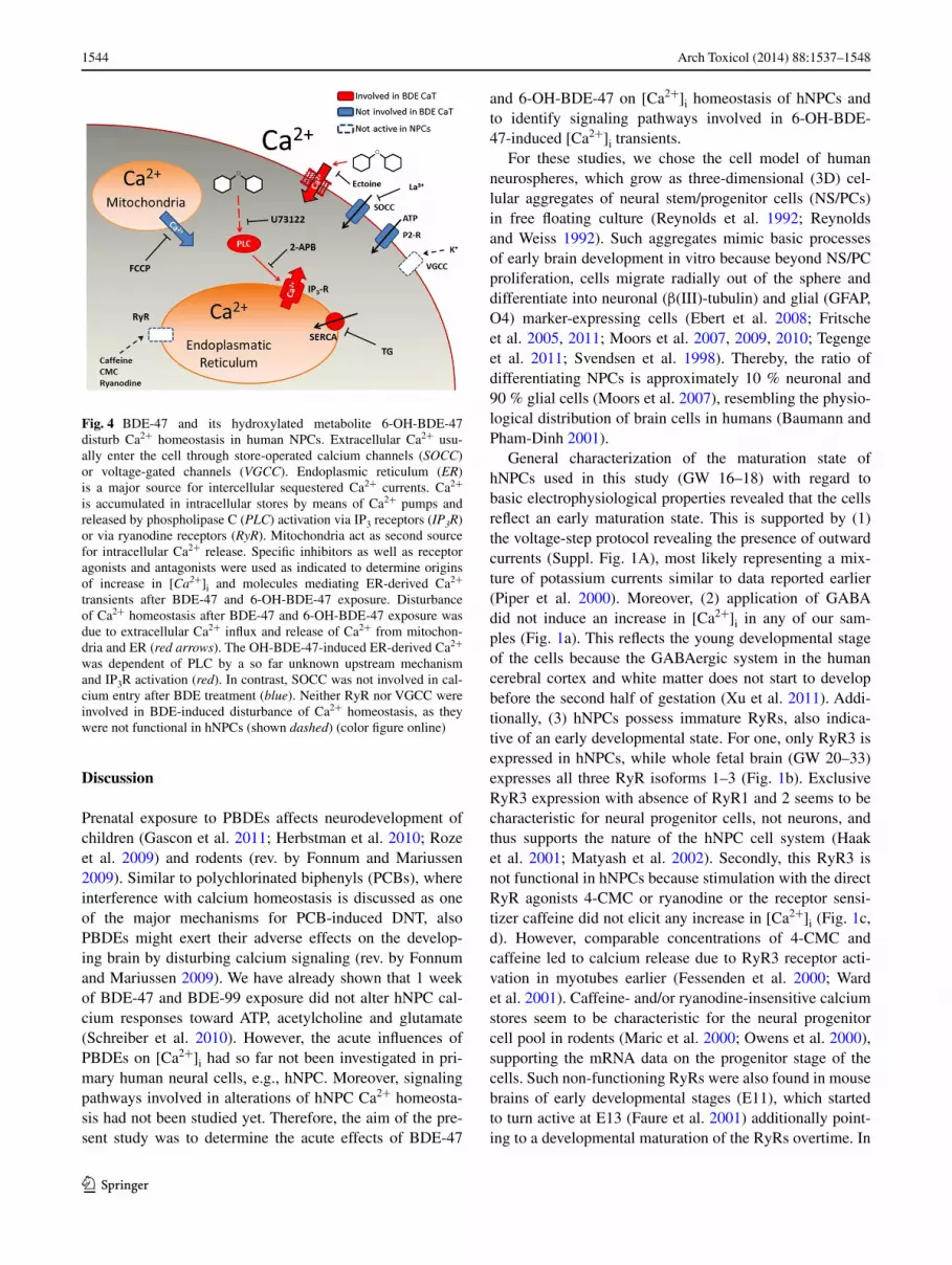
Fig. 4 BDe-47 and its hydroxylated metabolite 6-OH-BDe-47 disturb ca2+ homeostasis in human NPcs. extracellular ca2+ usu-ally enter the cell through store-operated calcium channels (SOCC) or voltage-gated channels (VGCC). endoplasmic reticulum (ER) is a major source for intercellular sequestered ca2+ currents. ca2+ is accumulated in intracellular stores by means of ca2+ pumps and released by phospholipase c (PLC) activation via IP3 receptors (IP3R) or via ryanodine receptors (RyR). Mitochondria act as second source for intracellular ca2+ release. Specific inhibitors as well as receptor agonists and antagonists were used as indicated to determine origins of increase in [Ca2+]i and molecules mediating er-derived ca2+ transients after BDe-47 and 6-OH-BDe-47 exposure. Disturbance of ca2+ homeostasis after BDe-47 and 6-OH-BDe-47 exposure was due to extracellular ca2+ influx and release of ca2+ from mitochon-dria and er (red arrows). The OH-BDe-47-induced er-derived ca2+ was dependent of Plc by a so far unknown upstream mechanism and IP3r activation (red). In contrast, SOcc was not involved in cal-cium entry after BDe treatment (blue). Neither ryr nor Vgcc were involved in BDe-induced disturbance of ca2+ homeostasis, as they were not functional in hNPcs (shown dashed) (color figure online)

1545Arch Toxicol (2014) 88:1537–1548
1 3
contrast, ATP and Ach evoked [ca2+]i transients in hNPcs, while glutamate caused only small [ca2+]i increases in 24–48 h versus 12 h differentiated hNPcs (Suppl. Fig. 1B). These changes in glutamate reaction are likely reflecting a developmental regulation of ligand-induced ca2+ responses in vitro, which was also seen for other neural cell models (gafni et al. 2004; He and Mccarthy 1994; Takeda et al. 1995). Overall, the hNPcs utilized in this study seem to reflect an early stage in the normal trajectory of developing neural tissue and therefore provide a model to study DNT of chemicals on early stages of development. This informa-tion is important for data interpretation and hazard assess-ment, as disturbances of the development of the nervous system are dependent on multiple critical windows of sus-ceptibility (rice and Barone Jr. 2000).
exposure to BDe-47 (≥2 μM) and its hydroxylated metabolite 6-OH-BDe-47 (≥0.2 μM) caused [ca2+]i transients in hNPcs. These effects of PBDes on primary human brain cells appear to be principally similar, but are observed at 5–10 times lower concentrations than on the rat tumor Pc12 cell line (Dingemans et al. 2007, 2008). effects of PBDes on [ca2+]i homeostasis were also seen in SH-Sy5y cells after 1.5–24 h (25.6 μM of the penta-brominated mixture De-71; yu et al. 2008) and in micro-somes and mitochondria isolated from different rat brain regions (approx. 7–20 μM De-71; Kodavanti and Ward 2005). The two latter studies investigated only parent BDe compounds, and effects were measured by endpoint deter-mination and not in a time kinetic like in the present study. Although this makes direct comparison of data difficult, these results still suggest that these primary human neural cells utilized in this study seem to be more sensitive toward disturbance of ca2+ homeostasis by some (OH)-PBDes than immortalized cell lines or cell organelles from rodent brains.
Not only PBDes but also the known developmentally neurotoxic PcBs disrupt ca2+ homeostasis, which is dis-cussed as one of the major mechanisms for PcB-induced DNT (rev. in Fonnum and Mariussen 2009). Due to struc-tural similarities between PcBs and PBDes, the involved extracellular as well as intracellular PcB target structures mediating [ca2+]i transients may be similar for PBDes. Therefore, we first tested if PBDe-induced [ca2+]i tran-sients were generated from the extracellular milieu and/or from intracellular mitochondria or er. While BDe-47-in-duced [ca2+]i transients were fed by calcium mainly from the extracellular milieu and to a much smaller extent from intracellular sources, 6-OH-BDe-47-induced [ca2+]i tran-sients are mainly nourished by the er with the extracellular space contributing to the magnitude of response (Fig. 3a, b, d). Due to the higher potency of 6-OH-BDe-47 compared to BDe-47 in inducing [ca2+]i transients and the low ca2+-induced amplitudes in 26 % responding cells after removal
of extracellular calcium, further experiments only focused on the underlying molecular mechanisms of the hydroxy-lated metabolite 6-OH-BDe-47.
We employed two strategies for investigating the role of extracellular calcium in 6-OH-BDe-47-induced [ca2+]i transients. In addition to measurements in calcium-free saline, we preexposed cells to the osmolyte ectoine, which inhibited any 6-OH-BDe-47-dependent calcium influx into the cells (Fig. 3c, d) without affecting responses to ATP (Suppl. Fig. 3). ectoine belongs to the group of osmolytes, which are organic, low molecular weight com-pounds with high water binding capacities that have the ability to increase the mobility of lipids and thus fluidity of membranes (Harishchandra et al. 2010), while they do not interfere with cellular functions (Brown 1978; lentzen and Schwarz 2006). ectoine forms hydrogen bonds with water molecules. These complexes increase hydrophilic interac-tions at the membrane and thereby increase lipid fluidity. It is probable that due to these increased hydrophilic inter-actions at the membrane, 6-OH-BDe-47-interaction with the membrane is directly prohibited causing the observed ablation of [ca2+]i responses. Such direct interaction with the cell membrane by integration into lipid bilayers was also reported for the structurally similar, ortho-substituted PcB52 (Tan et al. 2004). That 6-OH-BDe-47-induced [ca2+]i transients in hNPcs are produced by direct inter-face with the cell membrane, and not mediated by mem-brane-bound proteins like l-type Vgcc (Inglefield and Shafer 2000a), gABA-r (Inglefield and Shafer 2000b) or SOcc (Inglefield et al. 2001) is supported by the observa-tions that (1) ectoine does not interfere with stimulation of membrane-bound protein function, i.e., P2-r (Suppl. Fig. 3), (2) l-type Vgcc and gABA-r are not functional in hNPc (Fig. 1a) and (3) inhibition of SOcc by la3+ did only antagonize the increase in [ca2+]i provoked by ATP (Fig. 1a), not by 6-OH-BDe-47 (Fig. 3f).
Polychlorinated biphenyls can evoke ca2+ release from neural er stores by stimulation of ryrs or IP3rs. Because ryrs are the most sensitive to PcBs, they seem to be criti-cal molecular targets in PcB DNT (rev. in Pessah et al. 2010). Moreover, Kim et al. (2011) recently found that 6-OH-BDe-47 (10 μM) rapidly released accumulated ca2+ from microsomes isolated from skeletal muscle and heart in a ryr-dependent fashion. Additionally, it inhibited 4-cMc-induced ca2+ discharge after longer exposure due to prohi-bition of ryanodine-ryr binding. These data explained at least partially PBDe effects on cytoplasmic ca2+ previously observed in Pc12 cells by Dingemans et al. (2008, 2010). However, this study indicates that concentrations as low as 0.2 μM 6-OH-BDe-47 are sufficient to induce [ca2+]i tran-sients in hNPcs independent of ryrs because due to the early developmental stage, these cells do not possess func-tional ryr (Fig. 1b, c, d). Instead, 6-OH-BDe-47-induced

1546 Arch Toxicol (2014) 88:1537–1548
1 3
[ca2+]i transients are mediated by Plc and IP3r (Fig. 3e, f). As disturbances in ca2+ homeostasis observed in this study are clearly independent of ryrs, we identified a novel signaling pathway involved in 6-OH-BDe-47-induced disruption of ca2+-signaling (Fig. 4), which may be spe-cific for NPcs and thus affect early human brain develop-ment as a vulnerable window of exposure. With regard to the total period of human BDe exposure during the long time course of brain development, depending on the devel-opmental stage—previously reported ryr-dependent and here identified independent mechanisms may act synergisti-cally regarding the total DNT potential of BDes. BDe-47 reduced neuronal and oligodendrocyte differentiation of hNPcs (Schreiber et al. 2010). If this disruption in NPc dif-ferentiation is causally related to the disturbed calcium sign-aling observed in this study or if it is an additional mode of action that 6-OH-BDe-47 exerts on neurosphere differentia-tion needs to undergo further elucidation.
Conclusion
In summary, exposure to BDe-47 (≥2 μM) or 6-OH-BDe-47 (≥0.2 μM) triggers an increase in [ca2+]i in hNPcs due to influx of extracellular ca2+ and ryr-inde-pendent release of ca2+ from the er. Also, hNPcs seem to be more sensitive to BDe-induced disturbances of ca2+ homeostasis than tumor cells or isolated rodent orga-nelles, possibly pointing to a high BDe sensitivity during early stages of human brain development. We calculated that infant exposure can result in a brain concentration of 0.5–1.1 μM PBDe (Schreiber et al. 2010). considering that approximately 45 % of detectable PBDes are present as metabolites in human fetal blood (Qiu et al. 2009), OH-PBDe effects on hNPcs in our study are observed at rel-evant concentrations close to human exposure and might provide an additional mechanistic explanation for the epi-demiologic findings that PBDes are developmentally neu-rotoxic for humans.
Acknowledgments We acknowledge the expert technical help of ulrike Huebenthal and thank Marta Barenys for critical read-ing of the manuscript. Parts of this project were funded by the ger-man Federal Ministry for the environment, Nature conservation and Nuclear Safety (BMu), by the german research Foundation (DFg grK1427), by the research commission of the Department of Medi-cine, Heinrich-Heine university Duesseldorf and the Faculty of Vet-erinary Medicine, utrecht university. The authors declare no actual or potential competing financial interests.
References
Athanasiadou M, cuadra SN, Marsh g, Bergman A, Jakobs-son K (2008) Polybrominated diphenyl ethers (PBDes) and
bioaccumulative hydroxylated PBDe metabolites in young humans from Managua, Nicaragua. environ Health Perspect 116:400–408
Baumann N, Pham-Dinh D (2001) Biology of oligodendrocyte and myelin in the mammalian central nervous system. Physiol rev 81:871–927
Berridge MJ, lipp P, Bootman MD (2000) The versatility and univer-sality of calcium signalling. Nat rev Mol cell Biol 1:11–21
Brown AD (1978) compatible solutes and extreme water stress in eukaryotic micro-organisms. Adv Microb Physiol 17:181–242
Butt AM (2011) ATP: a ubiquitous gliotransmitter integrating neuron–glial networks. Semin cell Dev Biol 22(2):205–213
ciccolini F, collins TJ, Sudhoelter J, lipp P, Berridge MJ, Bootman MD (2003) local and global spontaneous calcium events regu-late neurite outgrowth and onset of gABAergic phenotype during neural precursor differentiation. J Neurosci 23:103–111
costa lg, giordano g (2007) Developmental neurotoxicity of poly-brominated diphenyl ether (PBDe) flame retardants. Neurotoxi-cology 28:1047–1067
Delmas P, Brown DA (2002) Junctional signaling microdomains: bridging the gap between the neuronal cell surface and ca2+ stores. Neuron 36:787–790
Dingemans MM, ramakers gM, gardoni F, van Kleef rg, Berg-man A, Di luca M, van den Berg M, Westerink rH, Vijverberg HP (2007) Neonatal exposure to brominated flame retardant BDe-47 reduces long-term potentiation and postsynaptic pro-tein levels in mouse hippocampus. environ Health Perspect 115: 865–870
Dingemans MM, de groot A, van Kleef rg, Bergman A, van den Berg M, Vijverberg HP, Westerink rH (2008) Hydroxylation increases the neurotoxic potential of BDe-47 to affect exocytosis and calcium homeostasis in Pc12 cells. environ Health Perspect 116:637–643
Dingemans MM, Heusinkveld HJ, Bergman A, van den Berg M, West-erink rH (2010) Bromination pattern of hydroxylated metabolites of BDe-47 affects their potency to release calcium from intracel-lular stores in Pc12 cells. environ Health Perspect 118:519–525
Dingemans MM, van den Berg M, Westerink rH (2011) Neurotoxic-ity of brominated flame retardants: (in)direct effects of parent and hydroxylated polybrominated diphenyl ethers on the (developing) nervous system. environ Health Perspect 119:900–907
ebert AD, McMillan el, Svendsen cN (2008) Isolating, expanding, and infecting human and rodent fetal neural progenitor cells. curr Protoc Stem cell Biol chap 2, unit 2D.2
eskenazi B, chevrier J, rauch SA, Kogut K, Harley Kg, Johnson c, Trujillo c, Sjödin A, Bradman A (2013) In utero and childhood polybrominated diphenyl ether (PBDe) exposures and neurode-velopment in the cHAMAcOS study. environ Health Perspect 121:257–262
Faure AV, grunwald D, Moutin MJ, Hilly M, Mauger JP, Marty I, De Waard M, Villaz M, Albrieux M ( 2001) Developmental expres-sion of the calcium release channels during early neurogenesis of the mouse cerebral cortex. eur J Neurosci 14(10):1613–1622
Fessenden JD, Wang y, Moore rA, chen Sr, Allen PD, Pessah IN (2000) Divergent functional properties of ryanodine recep-tor types 1 and 3 expressed in a myogenic cell line. Biophys J 79:2509–2525
Fonnum F, Mariussen e (2009) Mechanisms involved in the neuro-toxic effects of environmental toxicants such as polychlorin-ated biphenyls and brominated flame retardants. J Neurochem 111:1327–1347
Frederiksen M, Vorkamp K, Thomsen M, Knudsen le (2009) Human internal and external exposure to PBDes—a review of levels and sources. Int J Hyg environ Health 212:109–134
Fritsche e, cline Je, Nguyen NH, Scanlan TS, Abel J (2005) Poly-chlorinated biphenyls disturb differentiation of normal human

1547Arch Toxicol (2014) 88:1537–1548
1 3
neural progenitor cells: clue for involvement of thyroid hormone receptors. environ Health Perspect 113:871–876
Fritsche e, gassmann K, Schreiber T (2011) Neurospheres as a model for developmental neurotoxicity testing. Methods Mol Biol 758:99–114
gafni J, Wong PW, Pessah IN (2004) Non-coplanar 2,2’,3,5’,6-penta-chlorobiphenyl (PcB 95) amplifies ionotropic glutamate receptor signaling in embryonic cerebellar granule neurons by a mecha-nism involving ryanodine receptors. Toxicol Sci 77:72–82
gascon M, Vrijheid M, Martínez D, Forns J, grimalt JO, Torrent M, Sunyer J (2011) effects of pre and postnatal exposure to low lev-els of polybromodiphenyl ethers on neurodevelopment and thy-roid hormone levels at 4 years of age. environ Int 37:605–611
gassmann K, Abel J, Bothe H, Haarmann-Stemmann T, Merk HF, Quasthoff KN, rockel TD, Schreiber T, Fritsche e (2010) Spe-cies-specific differential Ahr expression protects human neural progenitor cells against developmental neurotoxicity of PAHs. environ Health Perspect 118(11):1571–1577
Haak ll, Song lS, Molinski TF, Pessah IN, cheng H, russell JT (2001) Sparks and puffs in oligodendrocyte progenitors: cross talk between ryanodine receptors and inositol trisphosphate receptors. J Neurosci 21:3860–3870
Harishchandra rK, Wulff S, lentzen g, Neuhaus T, galla HJ (2010) The effect of compatible solute ectoines on the structural organi-zation of lipid monolayer and bilayer membranes. Biophys chem 150:37–46
He M, Mccarthy KD (1994) Oligodendroglial signal transduction systems are developmentally regulated. J Neurochem 63:501–508
Herbstman JB, Sjodin A, Kurzon M, lederman SA, Jones rS, rauh V, Needham ll, Tang D, Niedzwiecki M, Wang ry, Perera F (2010) Prenatal exposure to PBDes and neurodevelopment. envi-ron Health Perspect 118(5):712–719
Inglefield Jr, Shafer TJ (2000a) Polychlorinated biphenyl-stimulation of ca(2+) oscillations in developing neocortical cells: a role for excitatory transmitters and l-type voltage-sensitive ca(2+) chan-nels. J Pharmacol exp Ther 295:105–113
Inglefield Jr, Shafer TJ (2000b) Perturbation by the PcB mixture Aroclor 1254 of gABA(A) receptor-mediated calcium and chlo-ride responses during maturation in vitro of rat neocortical cells. Toxicol Appl Pharmacol 164:184–195
Inglefield Jr, Mundy Wr, Shafer TJ (2001) Inositol 1,4,5-triphos-phate receptor-sensitive ca(2+) release, store-operated ca(2+) entry, and cAMP responsive element binding protein phosphoryl-ation in developing cortical cells following exposure to polychlo-rinated biphenyls. J Pharmacol exp Ther 297:762–773
Kafitz KW, Meier SD, Stephan J, rose cr (2008) Developmental profile and properties of sulforhodamine 101—labeled glial cells in acute brain slices of rat hippocampus. J Neurosci Methods 169:84–92
Kicinski M, Viaene MK, Den Hond e, Schoeters g, covaci A, Dirtu Ac, Nelen V, Bruckers l, croes K, Sioen I, Baeyens W, Van larebeke N, Nawrot TS (2012) Neurobehavioral func-tion and low-level exposure to brominated flame retardants in adolescents: a cross-sectional study. environ Health 11:86. doi:10.1186/1476-069x-11-86
Kim KH, Bose DD, ghogha A, riehl J, Zhang r, Barnhart cD, lein PJ, Pessah IN (2011) Para- and ortho-substitutions are key determinants of polybrominated diphenyl ether activity toward ryanodine receptors and neurotoxicity. envrion Health Perspect 119:519–526
Kodavanti Pr, Ward Tr (2005) Differential effects of commercial polybrominated diphenyl ether and polychlorinated biphenyl mixtures on intracellular signaling in rat brain in vitro. Toxicol Sci 85:952–962
lentzen g, Schwarz T (2006) extremolytes: natural compounds from extremophiles for versatile applications. Appl Microbiol Biotech-nol 72:623–634
Maric D, Maric I, Barker Jl (2000) Developmental changes in cell calcium homeostasis during neurogenesis of the embryonic rat cerebral cortex. cereb cortex 10:561–573
Marsh g, Hu J, Jakobsson e, rahm S, Bergman A (1999) Synthesis and characterization of 32 polybrominated diphenyl ethers. envi-ron Sci Technol 33:3033–3037
Marsh g, Athanasiadou M, Athanassiadis I, Sandholm A (2006) Iden-tification of hydroxylated metabolites in 2,2’,4,4’-tetrabromodi-phenyl ether exposed rats. chemosphere 63:690–697
Matyash M, Matyash V, Nolte c, Sorrentino V, Kettenmann H (2002) requirement of functional ryanodine receptor type 3 for astrocyte migration. FASeB J 16:84–86
Meier SD, Kafitz KW, rose cr (2008) Developmental profile and mechanisms of gABA-induced calcium signaling in hippocam-pal astrocytes. glia 56:1127–1137
Moors M, cline Je, Abel J, Fritsche e (2007) erK-dependent and -independent pathways trigger human neural progenitor cell migration. Toxicol Appl Pharmacol 221:57–67
Moors M, rockel TD, Abel J, cline Je, gassmann K, Schreiber T, Schuwald J, Weinmann N, Fritsche e (2009) Human neuro-spheres as three-dimensional cellular systems for developmental neurotoxicity testing. environ Health Perspect 117:1131–1138
Moors M, Vudattu NK, Abel J, Kramer u, rane l, ulfig N, cecca-telli S, Seyfert-Margolies V, Fritsche e, Maeurer MJ (2010) Inter-leukin-7 (Il-7) and Il-7 splice variants affect differentiation of human neural progenitor cells. genes Immun 11:11–20
Owens DF, Flint Ac, Dammerman rS, Kriegstein Ar (2000) cal-cium dynamics of neocortical ventricular zone cells. Dev Neuro-sci 22:25–33
Pessah IN, cherednichenko g, lein PJ (2010) Minding the calcium store: ryanodine receptor activation as a convergent mechanism of PcB toxicity. Pharmacol Ther 125:260–285
Piper Dr, Mujtaba T, rao MS, lucero MT (2000) Immunocytochem-ical and physiological characterization of a population of cultured human neural precursors. J Neurophysiol 84(1):534–548
Qiu x, Bigsby rM, Hites rA (2009) Hydroxylated metabolites of polybrominated diphenyl ethers in human blood samples from the united States. environ Health Perspect 117:93–98
reynolds BA, Weiss S (1992) generation of neurons and astrocytes from isolated cells of the adult mammalian central nervous sys-tem. Science 255:1707–1710
reynolds BA, Tetzlaff W, Weiss S (1992) A multipotent egF-respon-sive striatal embryonic progenitor cell produces neurons and astrocytes. J Neurosci 12:4565–4574
rice D, Barone S Jr (2000) critical periods of vulnerability for the developing nervous system: evidence from humans and animal models. environ Health Perspect 108(Suppl 3):511–533
roze e, Meijer l, Bakker A, Van Braeckel KN, Sauer PJ, Bos AF (2009) Prenatal exposure to organohalogens, including bro-minated flame retardants, influences motor, cognitive, and behavioral performance at school age. environ Health Perspect 117:1953–1958
Schreiber T, gassmann K, götz c, Hübenthal u, Moors M, Krause g, Merk HF, Nguyen NH, Scanlan TS, Abel J, rose cr, Fritsche e (2010) Polybrominated diphenyl ethers induce developmental neurotoxicity in a human in vitro model: evidence for endocrine disruption. environ Health Perpect 118:572–578
Staskal DF, Hakk H, Bauer D, Diliberto JJ, Birnbaum lS (2006) Tox-icokinetics of polybrominated diphenyl ether congeners 47, 99, 100, and 153 in mice. Toxicol Sci 94:28–37
Svendsen cN, ter Borg Mg, Armstrong rJ, rosser Ae, chandran S, Ostenfeld T, caldwell MA (1998) A new method for the rapid and long term growth of human neural precursor cells. J Neurosci Methods 85:141–152
Takeda M, Nelson DJ, Soliven B (1995) calcium signaling in cul-tured rat oligodendrocytes. glia 14:225–236

1548 Arch Toxicol (2014) 88:1537–1548
1 3
Tan y, chen cH, lawrence D, carpenter DO (2004) Ortho-substi-tuted PcBs kill cells by altering membrane structure. Toxicol Sci 80:54–59
Tegenge MA, rockel TD, Fritsche e, Bicker g (2011) Nitric oxide stimulates human neural progenitor cell migration via cgMP-mediated signal transduction. cell Mol life Sci 68:2089–2099
Ward cW, Protasi F, castillo D, Wang y, chen Sr, Pessah IN, Allen PD, Schneider MF (2001) Type 1 and type 3 ryanodine receptors generate different ca(2+) release event activity in both intact and permeabilized myotubes. Biophys J 81:3216–3230
xu g, Broadbelt Kg, Haynes rl, Folkerth rD, Borenstein NS, Bel-liveau rA, Trachtenberg Fl, Volpe JJ, Kinney Hc (2011) late development of the gABAergic system in the human cerebral cortex and white matter. J Neuropathol exp Neurol 70:841–858
yu K, He y, yeung lW, lam PK, Wu rS, Zhou B (2008) De-71-in-duced apoptosis involving intracellular calcium and the Bax–mitochondria–caspase protease pathway in human neuroblastoma cells in vitro. Toxicol Sci 104:341–351


![[47] Phospholipid hydroperoxide glutathione peroxidase](https://img.dokumen.tips/doc/110x75/63488ecff4145ce0ba02c82b/47-phospholipid-hydroperoxide-glutathione-peroxidase.jpg)


