Embed Size (px)
Citation preview

695
Bacterial Endosymbionts from the Genus Camponotus (Hymenoptera: Formicidae)
by
John J. Peloquin1, Stephen G. Miller3 , Stephen A. Klotz2, Richard
Stouthammer1 , Lloyd R. Davis, Jr.3 , &John H. Klotz1
ABSTRACT
Gram-negative prokaiyotic endosymbionts from follicle cells of eight species of North American Camponotus were isolated and their 16S rRNA-encoding DNA was amplified, cloned and sequenced. A BLASTn similarity search showed that the endosymbiont of Camponotus jloridanus is most similar to Candidatus camponotii. Other than this ant endosymbiont, the most similar sequences were from various enterobacteria commonly found in the gut of insects and other animals. The most parsimonious phylogenetic tree generated by PAUP analysis on manually aligned sequences supports a monophyletic relationship of the ant endosymbionts when the l 6S RNA sequences of two other insect-associated enterobacteria (Yersinia pestis, and Buchnera aphidicola) are included. The most parsimonious tree does not support, however, a monophyletic grouping of endosymbionts of the North American species when Old World isolates of Camponotus-associated eubacterial sequences are added to the analysis. These results suggest that the association and divergence of the endosymbiont with their ant host is ancient.
INTRODUCTION
The endosymbiotic bacteria of Formicidae were among the first discovered in insects (Blochmann 1884, 1886), but their widespread distribution in other insects and biological significance has only recently become appreciated (Baumann 1998; Heddi et al., 1993; Nardon 1999). In Camponotus, prokaiyotic endosymbionts are located in the midgut epithelium and follicle cells of the ovarioles within specialized mycetocytes. They are gram-negative, nonmotile, variable in shape, and lie freely in the cytoplasm without a surrounding membrane (Buchner 1965; Dasch et al. 1984). The bacteria are transmitted transovarily from the symbiont-rich follicle cells of a queen
1Department of Entomology, University of California, Riverside , Riverside, CA 92521 2Department of Medicine, Section of Infectious Diseases, PO Box 245039, Tucson, AZ, 85724-5039 3USDA-ARS-CMAVE, Gainesville, FL 32608

696 Sociobiology Vol. 38, No. 38, 2001
into her developing oocytes, and from there enter her offspring's follicle cells and midgut epithelium during embryogenesis (Buchner 1965; Steinhaus 1946). The bacteria's role in the ant is unknown but suspected to be nutritional, possibly supplying essential amino acids to their host (Dasch et al. 1984). In the past, classification of bacteria has been based on morphological and biochemical characteristics. With the advent of molecular techniques, microbial systematics has been revolutionized (Woese 1987). We describe electron microscopy of the bacterial endosymbionts and isolation of their DNA from the ovarioles of eight species of North American carpenter ants. We then amplified, cloned, and sequenced portions of this l 6S rRNA-encoding DNA and used it to construct phylogenetic trees
MATERIALS AND METHODS
Ants Newly mated queens of eight species of Camponotuswere collected in
the field and maintained in the laboratory on a 10°/o sucrose water and cricket diet. Queens of C. castaneus, C. jloridanus, C. impressus, C. snellingi, C. socius, and C. tortuganus, were collected in Alachua Co., Florida and C. pennsylvanicus and C. noveboracensis in Maine ..
Ant Tissue extraction Prior to dissection, live queens were surface-sterilized by immersion
for 3-5 minutes in 5.25°/o sodium hypochlorite. Using aseptic technique, the ants were transferred with forceps to 10°/o sodium thiosulfate for 3-5 minutes, then to lOOo/o ethanol for one minute, and finally to sterile distilled water where they were rinsed 3 times. Ants were dissected under aseptic conditions in sterile Grace's medium (Gibco-BRL).
I
PCR, cloning, and DNA sequence analysis Excised ovarian tissue was placed in 200µ1 of20mM Tris HCl, 1 OmM
MgCl (pH= 6.8), then homogenized with a tissue grinder, and sonicated for 10 s. DNA was isolated from the homogenate as follows: a) to disrupt gram positive bacteria, two units ofmutanolysin (2 µl of 1 U/µl stock) were added to the material, mixed, and then incubated at 37°C for 60 m; b) homogenates were centrifuged 30 sec at 1200g, and the supernatant removed; c) 700µ1 of 30mM Tris HCl with 25 mg/ml lysozyme was added to the pellet, mixed, and incubated at 37°C for 30 m; d) 20 µl of 20°/o SDS ( 1I10th volume) was added to the sample and mixed, followed by20µ1 of0.2M EDTA (l/lOth volume); f) the sample was extracted with buffer-saturated, neutralized phenol. and then with 24: 1 chloroform/isoamyl alcohol; g) the upper phase was transferred
Peloquin, J.J. et al.: Endosymbionts of Camponotus 697
to a clean tube and a final extraction was performed with watersaturated ether; h) the upper organic phase was removed, then 1/10 volume of 3. 0 M sodium acetate was added, along with 2 volumes 100% ETOH, and the sample was mixed and stored at -20° C.
DNA encoding eubacterial 16S rRNA was amplified by PCR. Reactions were performed in 500 mL tubes using the 'universal' eubacterial 16S rRNA PCR primers rl8F (5'-catggctcagattgaacgctggcg-3'), and rl494R (5'-cccctacggttaccttgttacgac-3')(Britschgi and Fallon, 1994; Cary et al., 1993; Chen et al., 1994; Goldenberger and Altwegg, 1995; Lee etal., 1993; Marzach etal., 1998; McCabe et al., 1999; Smith etal., 1996; Woo et al., 1999). The remainder of the reaction was in 71.5ml water, 10 ml lOx PCR buffer (500 mM KCL, lOOmMTris-HCl pH 9.0@ 25, l.0°/oTritonOX-100, 15mMMgC12), 16mldNTPs(l.25mMofeach dNTP stock), 0.5 ml of each primer (200 mM stock), 0.5 ml 5U /ml Taq polymerase (Promega, Madison WI), and 1 ml of template DNA. Each reaction was overlain with 100 ml mineral oil (Sigma, St. Louis MO) for 1 mat 94, 1 mat 50, 2 mat 72, repeated for 30 cycles, and finished with an extension of 8 m at 72.
PCR-amplified DNA fragments were separated on 1 o/o TAE agarose gels containing ethidium bromide (Sambrook et al., 1989). A 1000-bp DNA ladder (Gibco-BRL catalog number 15615-016) was also run as a size standard to estimate DNA length by comparison of electrophoretic mobility of PCR-amplified fragments.
PCR products were extracted from the gels, purified, and ligated into the pCR-II (Invitrogen, Carlsbad CA) cloning vector. 'One Shot' (lnvitrogen, Carlsbad CA) competent cells were transformed with an aliquot of the ligation reaction. Transf ormants were analyzed as mini preps (Sambrook et al., 1989). Plasmid clones containing appropriately sized inserts were chosen for sequence analysis (performed by University of Florida DNA Sequencing Core Facility). DNA sequences were acquired from the forward and reverse universal priming sites in pCR-II using forward and reverse Ml3-universal primers supplied by the Florida sequencing facility. Sequences internal to the EcorRv site of pCR-II were obtained using primers designed to anneal to conserved eubacterial sequences (ie., 773F, 5'-gcgtggggagcaaacagg-3' and 771R, 5'-atcctgtttgctccccacg-3'). Data obtained from the Core Facility DNA sequencing were encoded and sequence comparisons within this data set. and between archived data from other insect-associated bacteria (Yersinia pestis, Buchnera aphidicola) were performed with applications in the Wisconsin GCG package. The various rRNA encoding DNA sequences were aligned then and phylogenetic analysis of the aligned DNA sequence data from the ant endosymbionts and other bacteria was performed using Phyloge-
698 Sociobiology Vol. 38, No. 38, 2001
netic Analysis Using Parsimony (PAUP) ver. 4. 08 for power PC (Swofford, 2001). Because our sequences for the endosymbionts from C. castaneus and C. noveboracensiswere limited and outside the region of the other symbiont sequences, we excluded them from PAUP analysis. Sequence information from C. castaneus and C. noveboracensis was analyzed by a BLASTN similarity search using a server at the National Center for Biotechnology Information(http://www. ncbL nlm. nih.gov I BLAST I) to find the Genbank-submitted sequences that most closely resembled our submitted sequences.
Electron Microscopy Crushed ovaries were cultured in aerobic and anaerobic Bactec
bottles (Towson, MD), thioglycolate broth, and upon chocolate and blood agar at 28°C. Specimens for transmission electron microscopy were fixed in glutaraldehyde/paraformaldehyde, rinsed in phosphate buffer, dehydrated through gradient alcohol/acetonitrile and embedded in Epon. Thick sections were stained with toluidine blue and thin sections (700 angstroms) were stained with lead citrate/uranyl acetate , and examined with a Joel 1200 Transmission Scanning Electron Microscope (Joel Corp., Boston, MA).
RESULTS
Electron microscopy Fig. 1 shows the intimate intracellular relationship of these bacteria
with the ovarian tissue of the insect. Gram stain of crushed ovaries demonstrated long, filamentous gram-negative bacilli. Electron microscopy revealed numerous bacilli without cell walls consistent with gram-negative bacteria in the follicles surrounded by ground substance.
Bacteria The endosymbionts of Camponotus are gram-negative pleomorphic
bacilli. Their appearance in TEM suggests that they lack a cell wall. We were unable to culture isolated bacteria under either aerobic or anaerobic conditions on any of blood, chocolate, or gram-negative agar, Bactec aerobic and anaerobic media, or in thioglycolate broth.
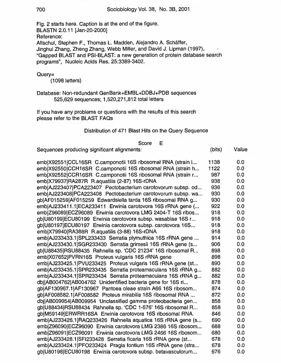
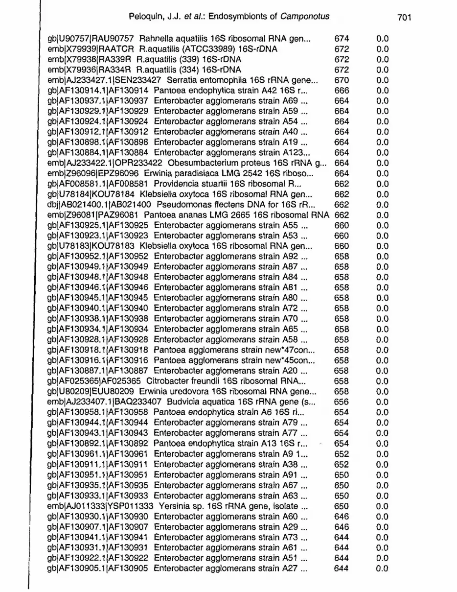
Phylogenetic analysis of endosymbiont sequences: Fig. 2 shows a BLASTn (Altschul et al., 1997) computer sequence
search comparing the entire data set we had for l 6S rRNA sequence from C. jloridanus. The highest scoring sequences were those of Camponotus-derived symbiotic bacteria (genbank accession number X9255 l). The next most similar sequence in the database was obtained

Peloquin, J.J. et al.: Endosymbionts of Camponotus 699
Fig.1. Top: Lightmicroscopeviewofovocyte. Black arrow shows follicle epithelium and white arrow, bacteria in the ovocyte. Middle: Transmission electron photomicrograph of the bacteria in an ovocyte. Bottom: Higher power view of the bacteria. Black bar in the middle and lower panels equals 1µ.
from Rahnella aquatalis (genbank accession number X79937). Almost the same high similarity scores were seen with Enterobacterial sequences isolated from the genera Erwinia, Serratia and Proteus. Sequences from rRNA of Yersinia pestis, Buchnera aphidicola, and our Camponotus symbionts were used as starting data in a PAUP (Swofford, 2001) analysis. The most parsimonious trees constructed by PAUP after bootstrap analysis (500 separate samplings) grouped all of the endosymbionts of Camponotus within a single clade (Fig. 3), and the 100 most parsimonious trees also grouped them into a monophyletic taxon. When l 6S rRNA sequences from Old World endosymbionts of Camponotus ( C. balzanl C. silivicola, C. nawai, C. vitiosus, C. japonicus, C. vagus, C. kiusiuensis, C. quadrinotatus, and C. rufipes) were compared with the bacterial sequences from our North American species, the most parsimonious trees did not show the latter as a monophyletic group. The endosymbionts of all Camponotus, however, were placed in a monophyletic group with reference to Buchnera aphidicola, and Yersinia pestis (Fig. 3).
BLAST analysis of sequences of the 16srRNAfrom C. castaneusand C. noveboracensis returned sequences derived from other Camponotus symbionts and Enterobacteriacea. The 10 sequences in the database most similar to C. castaneus and C. noveboracensis are listed in Table 1.
700 Sociobiology Vol. 38, No. 3B, 2001
Fig. 2 starts here. Caption is at the end of the figure. BLASTN 2.0.11 [Jan-20-2000] Reference: Altschul, Stephen F., Thomas L. Madden, Alejandro A. Schaffer, Jinghui Zhang, Zheng Zhang, Webb Miller, and David J. Lipman (1997), "Gapped BLAST and PSI-BLAST: a new generation of protein database s.earch programs", Nucleic Acids Res. 25:3389-3402.
Query= (1098 letters)
Database: Non-redundant GenBank+EMBL+DDBJ+PDB sequences 525,629 sequences; 1,520,271,812 total letters
If you have any problems or questions with the results of this search please refer to the BLAST FAQs
Distribution of 471 Blast Hits on the Query Sequence
Score E Sequences producing significant alignments:
emblX92551ICCL16SR C.camponotii 16S ribosomal RNA (strain I... emblX92550/CCH16SR C.camponotii 16S ribosomal RNA (strain h .. . emblX92552ICCR16SR C.camponotii 16S ribosomal RNA (strain r .. . emblX79937IRA287R R.aquatilis (2-87) 16S-rDNA embjAJ223407jPCA223407 Pectobacterium carotovorum subsp. od .. . emblAJ223408IPCA223408 Pectobacterium carotovorum subsp. wa .. . gblAF015259IAF015259 Edwardsiella tarda 16S ribosomal RNA g .. . embjAJ233411.1IECA233411 Erwinia carotovora 16S rRNA gene( .. . emblZ96089IECZ96089 Erwinia carotovora LMG 2404-T 16S ribos .. . gblU80199IECU80199 Erwinia carotovora subsp. wasabiae 16S r .. . gbjU80197IECU80197 Erwinia carotovora subsp. carotovora 16S .. . emb1X79940IRA388R R.aquatilis (3-88) 16S-rDNA embjAJ233433.1ISPL233433 Serratia plymuthica 16S rRNA gene .. . emblAJ233430.1 ISGR233430 Serratia grimesii 16S rRNA gene (s .. . gblU88435IRSU88435 Rahnella sp. 'CDC 21234' 16S ribosomal R .. . emblX07652/PVRN16S Proteus vulgaris 16S rRNA gene emb1AJ233425.1 IPVU233425 Proteus vulgaris 16S rRNA gene (st... emblAJ233435.1 ISPR233435 Serratia proteamaculans 16S rRNA g .. . emb/AJ233434.1 ISPR233434 Serratia proteamaculans 16S rRNA g .. . dbj!AB004762IAB004762 Unidentified bacteria gene for 16S ri ... gblAF130967 .1IAF130967 Pantoea oleae strain A66 16S ribosom .. . gblAF008582.1 IAF008582 Proteus mirabilis 16S ribosomal RNA .. . dbjlAB009954IAB009954 Unclassified gamma proteobacteria gen .. . gblU88434IRSU88434 Rahnella sp. 'CDC 1-576' 16S ribosomal R .. . gblM59149/ERWRR16SA Erwinia carotovora 16S ribosomal RNA. emblAJ233426.1IRAQ233426 Rahnella aquatica 16S rRNA gene (s .. . emblZ96090IECZ96090 Erwinia carotovora LMG 2386 16S ribosom .. . emblZ96091jECZ96091 Erwinia carotovora LMG 2466 16S ribosom .. . emblAJ233428.1 ISF1233428 Serratia ficaria 16S rRNA gene (st... embjAJ233424.1IPF0233424 Pragia fontium 16S rRNA gene (stra .. . gbjU80198jECU80198 Erwinia carotovora subsp. betavasculorum .. .
(bits)
1138 1122 987 938 936 930 930 922 918 918 918 918 914 906 898 898 890 882 882 878 874 872 858 858 846 690 688 680 678 678 676
Value
0.0 0.0 0.0 0.0 0.0 0.0 0.0 0.0 0.0 0.0 0.0 0.0 0.0 0.0 0.0 0.0 0.0 0.0 0.0 0.0 0.0 0.0 0.0 0.0 0.0 0.0 0.0 0.0 0.0 0.0 0.0
Peloquin, J.J. et a/.: Endosymbionts of Camponotus
gbjU90757jRAU90757 Rahnella aquatilis 16S ribosomal RNA gen... 674 embjX79939jRAATCR R.aquatilis (ATCC33989) 16S-rDNA 672 embjX79938jRA339R R.aquatilis (339) 16S-rDNA 672 embjX79936jRA334R R.aquatilis (334) 16S-rDNA 672 embjAJ233427 .1 jSEN233427 Serratia entomophila 16S rRNA gene... 670 gbjAF130914.1jAF130914 Pantoea endophytica strain A42 16S r... 666 gbjAF130937.1jAF130937 Enterobacter agglomerans strain A69 ... 664 gbjAF130929.1jAFl30929 Enterobacter agglomerans strain A59 ... 664 gbjAF130924.1jAF130924 Enterobacter agglomerans strain A54 ... 664 gbjAF130912.1jAF130912 Enterobacter agglomerans strain A40 ... 664 gbjAF130898.1jAFl30898 Enterobacter agglomerans strain A 19 ... 664 gbjAF130884.1jAFl30884 Enterobacter agglomerans strain A 123... 664 embjAJ233422.1 \0PR233422 Obesumbacterium proteus 16S rRNA g... 664 emb\Z96096IEPZ96096 Erwinia paradisiaca LMG 2542 16S riboso... 664 gbjAF008581.1 jAF008581 Providencia stuartii 16S ribosomal R... 662 gbjU78184\KOU78184 Klebsiella oxytoca 16S ribosomal RNA gen... 662 dbjjAB021400.1 jAB021400 Pseudomonas flectens DNA for 16S rR... 662 embjZ96081jPAZ96081 Pantoea ananas LMG 2665 16S ribosomal RNA 662 gbjAF130925.1jAFl30925 Enterobacter agglomerans strain A55 ... 660 gbjAF130923.1jAFl30923 Enterobacter agglomerans strain A53 ... 660 gbjU78183jKOU78183 Klebsiella oxytoca 16S ribosomal RNA gen... 660 gbjAF130952.1jAFl30952 Enterobacter agglomerans strain A92 ... 658 gbjAF130949.1jAF130949 Enterobacter agglomerans strain A87 ... 658 gbjAF130948.1jAF130948 Enterobacter agglomerans strain A84 ... 658 gbjAF130946.1jAFl30946 Enterobacter agglomerans strain A81 ... 658 gbjAF130945.1jAFl30945 Enterobacter agglomerans strain A80 ... 658 gbjAF130940.1jAF130940 Enterobacter agglomerans strain A72 ... 658 gbjAF130938.1jAFl30938 Enterobacter agglomerans strain A70 ... 658 gbjAF130934.1jAF130934 Enterobacter agglomerans strain A65 ... 658 gb\AF130928.1\AF130928 Enterobacter agglomerans strain A58 ... 658 gbjAF130918.1jAF130918 Pantoea agglomerans strain new*47con... 658 gbjAF130916.1jAFl30916 Pantoea agglomerans strain new• 45con... 658 gbjAF130887 .1jAF130887 Enterobacter agglomerans strain A20 ... 658 gb\AF025365jAF025365 Citrobacter freundii 16S ribosomal RNA... 658 gbjU80209jEUU80209 Erwinia uredovora 16S ribosomal RNA gene... 658 embjAJ233407.1jBA0233407 Budvicia aquatica 16S rRNA gene (s... 656 gbjAF130958.1jAF130958 Pantoea endophytica strain A6 16S ri... 654 gbjAF130944.1 jAF130944 Enterobacter agglomerans strain A79 ... 654 gbjAF130943.1jAF130943 Enterobacter agglomerans strain A77 ... 654 gbjAF130892.1jAF130892 Pantoea endophytica strain A13 16S r... 654 gbjAF130961.1jAFl30961 Enterobacter agglomerans strain A9 1... 652 gbjAF130911.1jAFl30911 Enterobacter agglomerans strain A38 ... 652 gbjAF130951.1 jAF130951 Enterobacter agglomerans strain A91 ... 650 gbjAF130935.1jAFl30935 Enterobacter agglomerans strain A67 ... 650 gbjAF130933.1jAF130933 Enterobacter agglomerans strain A63 ... 650 embjAJ011333jYSP011333 Yersinia sp. 16S rRNA gene, isolate... 650 gbjAF130930.1jAFl30930 Enterobacter agglomerans strain A60 ... 646 gbjAF130907.1IAF130907 Enterobacter agglomerans strain A29 ... 646 gb\AF130941.1IAF130941 Enterobacter agglomerans strain A73 ... 644 gbjAF130931.1 jAF130931 Enterobacter agglomerans strain A61 ... 644 gbjAF130922.1jAF130922 Enterobacteragglomerans strain A51 ... 644 gbjAF130905.1jAF130905 Enterobacter agglomerans strain A27 ... 644
0.0 0.0 0.0 0.0 0.0 0.0 0.0 0.0 0.0 0.0 0.0 0.0 0.0 0.0 0.0 0.0 0.0 0.0 0.0 0.0 0.0 0.0 0.0 0.0 0.0 0.0 0.0 0.0 0.0 0.0 0.0 0.0 0.0 0.0 0.0 0.0 0.0 0.0 0.0 0.0 0.0 0.0 0.0 0.0 0.0 0.0 0.0 0.0 0.0 0.0 0.0 0.0
701

702 Sociobiology Vol. 38, No. 38, 2001
dbilD78010ID78010 Xenorhabdus poinarii DNA for 16S Ribosoma ... gblAF130963.1IAF130963 Pantoea toletana strain A64 16S ribo .. . emblAJ233429.11SF0233429 Serratia fonticola 16S rRNA gene( .. . gblAF130936.1IAF130936 Enterobacter agglomerans strain A68 .. . emblAJ009930.1jEPAJ9930 Erwinia pyrifoliae 16S rRNA, tRNA-G .. . embjAJ010485.1jEAM010485 Erwinia amylovora 16S rRNA gene, t... embjX83265IEA16SRR E.amylovora 16S rRNA gene gbjAF130966.1IAF130966 Pantoea toletana strain A78 16S ribo .. . gbjAF130899.1IAF130899 Enterobacter agglomerans strain A21 .. . dbjiAB004745jAB004745 Serratia ficaria gene for 16S ribosom ... dbjjD78007jD78007 Xenorhabdus bovienii DNA for 16S Ribosoma ... emblZ49828IYE320692 Y.enterocolitica gene for 16S ribosomal... gblAF130968.1IAF130968 Pantoea toletana strain A75 16S ribo ... dbjlD78008ID78008 Xenorhabdus japonicus DNA for 16S Ribosom .. . dbjiD78009ID78009 Xenorhabdus nematophilus DNA for 16S Ribo .. . gbjM59155jHAFRR16SA Hafnia alvei 16S ribosomal RNA. gblAF130942.1IAF130942 Enterobacter agglomerans strain A74 ...
642 0.0 638 0.0 638 0.0 636 e-180 632 e-179 632 e-179 632 e-179 630 e-178 628 e-178 624 e-176 622 e-176 622 e-176 618 e-175 617 e-174 609 e-172 609 e-172 589 e-166
Fig. 2. BlastN results of search using C. floridanus endosymbiont 16S rRNA sequences showing sequence similarities to Gram negative Enterobacteria.
Electron Microscopy Bacteria
DISCUSSION
The observed lack of cell walls in the endosymbionts is unlike gramnegative bacteria, but similar to mycoplasma and the wall-less L forms of various bacteria. The fact that we were unable to culture them suggests that these bacteria require a specific environment to grow perhaps uniquely provided by the follicle's intracellular environment, as would be expected from an endosymbiont that has co-evolved with its host. Buchnera and Wolbachiaendosymbionts show simil~r requirements for their growth (Heddi etal., 1999; Johanowicz and Hoy, 1998).
Phylogenetic analysis of ovarian endosymbionts: The BLASTn search in Fig. 2 displaying sequences in decreasing
similarity strongly suggests that the closest phylogenetic relative of the C. florid.anus endosymbiont is Candidatus camponotii (Schroder et al., 1996), a previously identified bacterial endosymbiont of Camponotus. Listed in decreasing order of similarity with these endosymbiontderived sequences are ones obtained from various enterobacteria. Interestingly, the endosymbionts of Camponotus sp., though morphologically dissimilar to enterobacteria in lacking cell walls, appear by l 6S RNA sequences to be most closely related to the Enterobacteriaceae commonly found in the gut of insects and other animals. Corroborative evidence for this statement can be found in Table 1. As expected, sequences of C. castaneus and C. noveboracensis were more similar to

Peloquin, J.J. et al.: Endosymbionts of Camponotus 703
Yersinia
Buchnera
balzani
silvicola
tortuganus *
snellingi *
herculeanus *
vagus
japonicus
pennsylvanicus *
floridanus *
socius *
kiusiuensis
quadrinotatus
nawa1
vitiosus
rufipes
impressus *
Fig. 3. Most parsimonious phylogenetic tree constructed by PAUP (default settings) showing the polyphyletic relationship of North American Camponotus endosyrnbionts (with *) when compared with Old World symbiont sequences.

Table 1. BLASTn search results, 10 closest sequences in Genbank:
Camponotus castaneus
giJ8250193JembJAJ245598.llEOF245598 C. pennsylvanicus symbiont 16S rRNA gil121281SlembJX92551.ljCCL16SR C.camponotii 16S rRNA lignaperda str. giJ825019llembJAJ245595.llEOF245595 Endosymbiont 16S RRNA C. socius gil1212814JembJX92550.1JCCH16SR C.camponotii 16S rRNA heculeanus strain giJ7708202JembiAJ25071S.liCHE250715 C. herculeanus symbiont16S rRNA giJ8250192lembJAJ245597.llEOF245597 C. rufipes symbiont 16S rRNA gil436814iembJX75274.liYPD16SRN Yersinia pestis (D-28) 16S rRNA giJ48619JembiX67464.1JYP16SRRN Y.pestis 16S rRNA gene giJ1212816JembJX92552.1JCCR16SR C.camponotii 16S rRNA rufipes strain giJ8250189JembJAJ245593.1JEOF245593 Endosymbiont C. sericeiventris
Camponotus noveboracensis
giJ7708202JembJAJ250715.llCHE250715 Camponotus herculeanus 16S rRNA giJ1212814lemblX92550.llCCH16SR C.camponotii 16S rRNA herculeanus str. giJ121281SlemblX92551.1JCCL16SR C.camponotii 16S rRNA ligniperda strain giJ8439279JembJAJ245596.llEOF245596 Endosymbiont of Camponotus balzani gil8250193JembJAJ245598.1JEOF245598 Endosymbiont of C. pennsylvanicus giJ8250190!embJAJ245594.llEOF245594 C. castaneus endosymbiont 16S rRNA giJ2570284Jgb!US0207.1JERU80207 Erwinia rubrifaciens 16S rRNA gil4582065jembjAJ23341E3.llERU233418 Erwinia rubrifaciens 16S rRNA gil2584756JembJZ96098.1JERZ96098 Erwinia rubrifaciens LMG 2709 16S rRNA giJ4754850Jgb!AF130918.lJAF130918 Pantoea agglomerans 16S rRNA
-.,J 0 .f>.
en 0 0 5· O" Q" 0
<O '< < 0
(,) _o:>
z !=> (,)
_Ill
~ 0 ~

Peloquin, J.J. et al.: Endosymbionts of Camponotus 705
other Camponotus symbionts, than to Yersinia pestis, Erwinia rubrifaciens, and Panteoa agglomerans. There is mounting evidence that many of the Enterobacteriaceae have properties expected of bacteria having a mutualistic relationship with their insect host, excepting nitrogen metabolism (Epsky etal., 1997; Epsky et al., 1998; Hendrichs et al., 1993; Lauzon et al., 1998; MacCollom et al., 1994; Peloquin et al., 2000; Prokopy et al., 1993).
The most parsimonious phylogenetic tree (Fig. 3) generated by the PAUP program for the endosymbionts supported a monophyletic relationship when the l 6S RNA sequences of other insect-associated enterobacteria (Yersinia pestis, Buchnera aphidicola) were included (Fig. 3). When additional old world isolates of Camponotus-associated eubacterial sequences were examined, using Yersinia, andBuchneraas outgroups. a monophyletic grouping of the North American ant endosymbionts could not be supported.
These results suggest that the phylogenetic divergence of the endosymbionts and association with their ant hosts is ancient. Additional sequence information from the ants may provide more details on these relationships (Boursaux-Eude and Gross 2000; Sauer et al. 2000).
Evidence indicates that competition exists between symbiotic prokruyotes within the host organism (Charles et al., 1997; Grenier et al., 1994; Heddi etal. 1998; Heddi etal. 1999; Nardon et al. 1998). Progenitors of extant prokaryotes may have produced antibiotics or other substances that displaced or manipulated the original endosymbionts (Fredenhagen et al. 1986; Fredenhagen et al. 1987; Jigami et al. 1986; Nardon et al. 1992). Alternatively, exposure to some environmental factor may have cleared the way for inoculation by other bacteria, which then evolved an intimate association with their ant hosts.
Multiple independent acquisition of endosymbionts would be more strongly supported if similar results were obtained from PAUP with additional data obtained from other ant endosymbionts. At least one other study supports a paraphyletic hypothesis of the origins of ant symbiotic bacteria (Sameshima et al. 1999). The association between endosymbiont and host maybe dynamic and variable, with competition occurring amongst them possibly mediated by antibiotic substances (Fredenhagen et al. 1986; Fredenhagen et al. 1987). This constant flux of endosymbionts may give new capabilities to the host/ endosymbiont association, depending on the properties of the new bacteria. The endosymbionts may influence the biology of the association considerably more than is accepted in the traditional concept applied to metazoans in which the nuclear genome solely determines the biology of the host (Heddi et al., 1999).
706 Sociobiology Vol. 38, No. 38, 2001
REFERENCES
Altschul, S.F., T.L. Madden, A.A. Schaffer, J. Zhang, Z. Zhang, W. Miller, and D.J. Lipman 1997. Gapped BLAST and PSI-BLAST: a new generation of protein database search programs. Nucleic Acids Res 25: 3389-3402.
Baumann, P. 1998. Symbiotic associations involving microorganisms. Bioscience 48: 254-255.
Blochmann, F. 1884. Ueber eine Metamorphose der Ovarialeiern und uber den Beginn der Blastoderm-bildung bei den Ameisen. Verhandlungen des Naturhistorisch-Medizinischen Vereins zu Heidelberg 3: 243-247.
Blochmann, F. 1886. Ueber die Reifung der Eier bei Ameisen und Wespen. Verhandlungen des Naturhistorisch-Medizinischen Vereins zu Heidelberg: 141-172.
Boursaux-Eude, C., and R. Gross 2000. New insights into symbiotic associations between ants and bacteria. Research in Microbiology 151: 513-519.
Britschgi, T.B .. and R.D. Fallon 1994. PCR-amplification of mixed 16S rRNA genes from an anaerobic, cyanide-degrading consortium. FEMS (Federation of European Microbiological Societies) Microbiology Ecology 13: 225-231.
Buchner, P. 1965. "Endosymbiosis of animals with plant microorganisms," Interscience Publishers, New York, NY.
Cary, S.C., W. Warren, E. Anderson, and S.J. Giovannoni 1993. Identification and localization of bacterial endosymbionts in hydrothermal vent taxa with symbiont-specific polymerase chain reaction amplification and in situ hybridization techniques. Molecular Marine Biology and Biotechnology 2: 51-62.
Charles, H.,A. Heddi,J. Guillaud, C. Nardon, andP. Nardon 1997.Amolecular aspect of symbiotic interactions between the weevil Sitophilus oryzae and its endosymbiotic bacteria: Over-expression of a chaperonin. Biochem. Biophys. Res. Commun. 239: 769-774.
Chen, S.-M., J.S. Dumler, J.S. Bakken, and D.H. Walker 1994. Identification of a granulocytotropic Ehrlichia species as the etiologic agent of human
I
disease. J. Clin. Microbiol. 32: 589-595. . Dasch, G., E. and C.K. Weiss 1984. Endosymbionts of insects. In "Bergey's
Manual of Systematic Bacteriology" (N. A. Krieg, ed.), Vol. I, pp. 881-883. Williams and Wilkins, Baltimore.
Epsky, N.D., B.D. Dueben, R.R. Heath, C.R. Lauzon, and R.J. Prokopy 1997. Attraction of Anastrepha suspensa (Diptera: Tephritidae) to volatiles from avian fecal material. Florida Entomologist 80: 270-277.
Epsky, N.D .. R.R. Heath, B.D. Dueben, C.R. Lauzon, A.T. Proveaux. and G.B. MacCollom 1998. Attraction of 3-methyl-1-butanol and ammonia identified from Enterobacter agglomerans to Anastrepha suspensa. Journal of Chemical Ecology 24: 1867-1880.
Fredenhagen, A., P. Kenny. H. Kita, H. Komura, Y. Naya, K. Nakanishi, K. Nishimaya, M. Sugiura, andS. Tamura 1986. Roleofintracellularsymbiotes in planthoppers. IUPAC proceeding. Pesticide chemistry: 101-108.
Fredenhagen, A .. S. Tamura, P. Kenny, H. Komura, Y. Naya, and K. Nakanishi 1987. Andrimid, a new peptide antibiotic produced by an intracellular
Peloquin, J.J. et al.: Endosymbionts of Camponotus 707
bacterial symbiont isolated from a brown planthopper. Journal of the American Chemical Society 109: 4409-4411.
Goldenberger, D., and M. Altwegg 1995. Eubacterial PCR: Contaminating DNA in primer preparations and its elimination by UV light. Journal of Microbiological Methods 21: 27-32.
Grenier,A.M., C. Nardon, and P. Nardon 1994. The Role ofSymbiotes in Flight Activity of Sitophilus Weevils. Entomologia Experimentalis etApplicata 70: 201-208.
Heddi, A., H. Charles, C. Khatchadourian, G. Bonnot, and P. Nardon 1998. Molecular characterization of the principal symbiotic bacteria of the weevil Sitophilus oryzae: A peculiar G - C content of an endocytobiotic DNA. J. Mol. Evol. 47: 52-61.
Heddi, A., A.M. Grenier, C. Khatchadourian, H. Charles, and P. Nardon 1999. Four intracellular genomes direct weevil biology: nuclear, mitochondrial, principal endosymbiont, and Wolbachia. Proc. Nat. Acad. Sci. 96: 6814-6819.
Heddi, A., F. Lefebvre, and P. Nardon 1993. Effect of endocytobiotic bacteria on mitochondrial enzymatic activities in the weevil Sitophilus oryzae (Coleoptera, Curculionidae). Insect Biochem Mol Biol 23: 403-411.
Hendrichs, J., C.R. Lauzon, S.S. Cooley, and R.J. Prokopy .1993. Contribution of natural food sources to adult longevity and fecundity of Rhagoletis ponwnella (Diptera: Tephritidae). Ann. Ent. Soc. Am. 86: 250-264.
Jigami, Y., N. Harada, H. Uemura, H. Tanaka, K. Ishikawa, S. Nakasoto, H. Kita, and M. Sugiura 1986. Identification of a Polymyxin produced by a symbiotic Microorganism isolated from the Brown Planthopper, Nilaparavata lugens. Agricultural and Biological Chemistry 50: 1637-1639. .
Johanowicz, D.L., and M.A. Hoy 1998. The manipulation of arthropod reproduction by Wolbachia endosymbionts. Florida Entomologist 81: 310-317.
Lauzon, C.R .. R.E. Sjogren, S.E. Wright, and R.J. Prokopy 1998. Attraction of Rhagoletis pomonella (Diptera: Tephritidae) flies to odor of bacteria: Apparent confinement to specialized members of enterobacteriaceae. Environ. Entomol. 27: 853-857.
Lee, I.M., RW. Hammond, RE. Davis, and D.E. Gundersen 1993. Universal amplification and analysis of pathogen 16S rDNA for classification and identification of mycoplasma-like organisms. Phytopath. 83: 834-842.
MacCollom, G.B., C.R. Lauzon, E.B. Payne, and W.W. Currier 1994. Apple maggot (Diptera: Tephritidae) trap enhancement with washed bacterial cells. Environ. Entomol. 23: 354-359.
Marzach, C., F. Veratti, and D. Bosco 1998. Direct PCR detection of phytoplasmas in experimentally infected insects. Annals of Applied Biology 133: 45-54.
McCabe, K.M., Y.-H. Zhang, B.-L. Huang, E.A. Wagar, and E.R.B. McCabe 1999. Bacterial species identification after DNA amplification with a universal primer pair. Molecular Genetics and Metabolism 66: 205-211.
Nardon, P. 1999. Symbiosis as an example of an acquired character: NeoLamarckism or Darwinism? Bulletin De La Societe Zoologique De France 124: 39-52.
708 Sociobiology Vol. 38, No. 38, 2001
Nard on, P., A.M. Grenier, and A. Heddi 1998. Endocytobiote control by the host in the weevil Sitophilus oryzae, Coleoptera, Curculionidae. Symbiosis 25: 237-250.
Nardon, P., C. Nardon, B. Delobel, Y. Rahbe, and J. Guillaud 1992. Characteristics and Development of the Tyrosine-Rich Protein Granules in the Adipose Tissue of the Curculionid Beetle Sitophilus oryzae. Tissue & Cell 24: 157-170.
Peloquin, J.J., L. Kuzina, C.R. Lauzon, and T.A. Miller 2000. Transformation of internal extracellular bacteria isolated from Rhagoletis completa cresson gut with enhanced green fluorescent protein. Current Microbiology 40: 367-371.
Prokopy, R.J., S.S. Cooley, L. Galarza, C. Bergweiler, and C.R. Lauzon 1993. Bird droppings compete with bait sprays for Rhagoletis pomonella (Walsh) flies (Diptera: Tephritidae). Canad. Entomol. 125: 413-422.
Sambrook, J., T. Maniatis, and E.F. Fritsch 1989. '"Molecular cloning: a laboratory manual," 2nd/Ed. Cold Spring Harbor Laboratory, Cold Spring Harbor, N.Y.
Sameshima, S., E. Hasegawa, 0. Kitade, N. Minaka, and T. Matsumoto 1999. Phylogenetic comparison of endosymbionts with their host ants based on molecular evidence. Zoological Science (Tokyo). 16: 993-1000.
Sauer, C., E. Stackebrandt, J. Gadau, B. Hoelldobler, and R. Gross 2000. Systematic relationships and cospeciation of bacterial endosymbionts and their carpenter ant host species: Proposal of the new taxon Candidatus Blochmannia gen. nov. International Journal of Systematic and Evolutionary Microbiology 50: 1877-1886.
Schroder, D., H. Deppisch, M. Obermayer, G. Krohne, E. Stackebrandt, B. H61ldobler, W. Goebel, and R. Gross 1996. Intracellular endosymbiotic bacteria of Camponotus species (carpenter ants): systematics, evolution and ultrastructural characterization. Molecular Microbiology 21: 4 79-489.
Smith, J.G., L. Kong, G.K. Abruzzo, C.J. Gill, A.M. Flattery, P.M. Scott, D. Bramhill, C. Cioffe, C.M. Thompson, and K. Bartizal 1996. PCR detection of colonization by Helicobacter pylori in conventional, euthymic mice based on the 16S ribosomal gene sequence. Clin Diagn Lab Immunol 3: 66-72.
Steinhaus, E.A. 1946. "Insect microbiology; an account of the microbes associated with insects and ticks, with special reference to the biologic relationships involved," Comstock Publishing Company, Ithaca, New York.
Swofford, D.L. 2001. PAUP*. Phylogenetic Analysis Using Parsimony (*and Other Methods). Sinauer Associates, Sunderland, Massachusetts.
Woese, C.R. 1987. Bacterial evolution. Microbiological Reviews 51: 221-271. Woo, T.H.S., B.K.C. Patel, M. Cinco, L.D. Smythe, M.A. Norris, M.L. Symonds,
M.F. Dohnt, and J. Piispanen 1999. Identification of Leptospira bijlexa by real-time homogeneous detection of rapid cycle PCR product. Journal of Microbiological Methods 35: 23-30.
@


