Embed Size (px)
Citation preview
Copyright Undertaking
This thesis is protected by copyright, with all rights reserved.
By reading and using the thesis, the reader understands and agrees to the following terms:
1. The reader will abide by the rules and legal ordinances governing copyright regarding the use of the thesis.
2. The reader will use the thesis for the purpose of research or private study only and not for distribution or further reproduction or any other purpose.
3. The reader agrees to indemnify and hold the University harmless from and against any loss, damage, cost, liability or expenses arising from copyright infringement or unauthorized usage.
IMPORTANT
If you have reasons to believe that any materials in this thesis are deemed not suitable to be distributed in this form, or a copyright owner having difficulty with the material being included in our database, please contact [email protected] providing details. The Library will look into your claim and consider taking remedial action upon receipt of the written requests.
Pao Yue-kong Library, The Hong Kong Polytechnic University, Hung Hom, Kowloon, Hong Kong
http://www.lib.polyu.edu.hk
DESIGN AND FABRICATION OF MULTIFUNCTIONAL
POLYMER BASED NANOCOMPOSITES FOR
BONE TISSUE ENGINEERING
CHEN LING
Ph.D
The Hong Kong Polytechnic University
2014
THE HONG KONG POLYTECHNIC UNIVERSITY
DEPARTMENT OF INDUSTRIAL AND
SYSTEMS ENGINEERING
Design and Fabrication of Multifunctional
Polymer Based Nanocomposites for Bone Tissue
Engineering
CHEN Ling
A thesis submitted in partial fulfillment of the
requirements for the degree of Doctor of Philosophy
August 2013

CERTIFICATE OF ORIGINALITY
I hereby declare that this thesis is my own work and that, to the best of my knowledge
and belief, it reproduces no material previously published or written, nor material that
has been accepted for the award of any other degree or diploma, except where due
acknowledgement has been made in the text.
(Signed)
CHEN LING (Name of student)
i
Abstract
Abstract of thesis entitled “Design and Fabrication of Multifunctional
Polymer Based Nanocomposites for Bone Tissue Engineering”
Submitted by CHEN Ling
For the Degree of Doctor of Philosophy
at The Hong Kong Polytechnic University in August, 2013
Biodegradable polymer scaffolds with drug delivery function have received great
research attention in bone tissue engineering due to their biocompatibility and
biodegradability as temporary support systems for cell adhesion, growth, differentiation
and tissue regeneration. The drug release systems of the scaffolds can deliver
biologically active molecules with the desired behavior, so as to reduce the amount of
drug administration and shorten the therapeutic period. Scaffolds with shape memory
effects have been considered as smart materials for potential applications in minimally
invasive surgery. Efforts have also been devoted to the development of composite
scaffolds combining biodegradable polymers and bioactive ceramics with enhanced
osteoconductive properties and mechanical strength. However, there is still no best
method for three-dimensional scaffold fabrication and the influences of bioactive
inclusion and porosity of the scaffold on drug delivery behavior, shape memory effect,
biodegradability, bioactivity and cellular response have not yet been well addressed.
The purpose of this study is to develop a technique to produce a multifunctional porous
composite and investigate the influence of bioactive inclusion on its mechanical
ii
properties, biodegradability, drug release behavior, shape memory effect and cellular
response for bone tissue engineering.
Among the developed fabrication technologies, the solvent casting/particulate leaching
method is the most commonly used scaffold fabrication method due to its simplicity,
efficiency and ability to independently control the porosity and pore size of the scaffold.
Nevertheless, it is limited to fabricating scaffolds with uniform pore distribution. To
solve this distribution problem, a method using polymer coagulation, thermal
compression molding and salt leaching has been reported however, the problem of
decomposition of the active drug and polymer may be induced. In this study, a new
technique has been developed to fabricate multifunctional scaffolds. This technique,
called PC-DC technique, applying polymer coagulation (PC) and drug coating (DC) to
solvent casting/particulate leaching method and using cold compression molding
instead of conventional hot compression molding. This low temperature composite
technology can contribute to a wider range of choices of drug loadings, since thermal
decomposition of drug inclusion and polymer matrix can be avoided. This technique not
only independently controls the pore size and porosity of the scaffolds, but also reduces
solvent residual. A uniform distribution of pores throughout the polymer matrix can be
achieved by this technique.
Poly(ethylene glycol)/dexamethasone (PEG/Dex)-coated porous poly-D-L-lactide/nano-
hydroxyapatite (PDLLA/nano-HAp) composites with homogenous pore networks and
controllable porosity and pore size have been fabricated by the PC-DC technique. The
compressive moduli and strengths of the composites were improved by the nano-HAp
iii
addition, which were close to those of cancellous bone. The improved wettability of the
scaffold by PEG/Dex coating and nano-HAp filling was confirmed. The drug loading
ability and total drug release amount of the scaffolds increased with increasing porosity
level and/or the nano-HAp content. The improved bioactivity of the scaffolds was
validated by the apatite formation on the scaffolds with nano-HAp addition after
incubation in simulated body fluid (SBF). The compressive moduli and strengths of the
scaffolds after incubation in SBF were affected by the combination of degradation,
weight loss, apatite deposition and incubation time. Nano-HAp incorporation can
decelerate the polymer degradation and mass loss. Moreover, the PDLLA/nano-HAp
scaffolds showed a pH compensating effect to reduce the risk of chronic inflammation
complications. Cyclic thermomechnical and physical shape recovery tests were firstly
conducted to investigate the shape memory effect of the porous PDLLA/nano-HAp
scaffolds. The results showed that desirable shape memory behavior could be achieved
when the nano-HAp fracture was 10 wt%. Good biocompatibility, bioactivity and
osteoconductivity of the PDLLA based samples were confirmed by investigating the
ability of the scaffolds for MG63 cell adhesion, proliferation and differentiation.
This study provides a new technique for the fabrication of multifunctional
biodegradable drug loaded bone scaffolds with shape memory function, which is highly
desirable in the bone repair applications. The findings of the study not only lead to a
better understanding of the effects of nano-HAp, structure factor, surface coating on the
mechanical properties, degradability, drug release behavior, shape memory effects and
the cellular response of the composites in bone tissue engineering, but also provide
guidelines for design and fabrication of the multifunctional composites with

iv
controllable properties for certain application requirements. A finite element model
simulating the behavior of the scaffolds implanted into a human body and animal model
experiments for further evaluation of the in vivo performance of the composite scaffolds
are recommended for future study.
v
Acknowledgements
I would like to express my sincerest thanks to my supervisor, Prof. Chak Yin TANG for
giving me this great opportunity to join his group and broaden my research horizon. The
lessons I learned from him were not only in regard to scientific knowledge, but also
responsibility, perseverance, and integrity, which will bring me lifelong benefits.
Without his constructive advice, clear guidance and continuous support over the past
four years, I would not have completed this research. I am forever grateful for
everything that he has done for my continuous improvement.
My sincere gratitude is also accorded to my co-supervisors, Dr. Da Zhu CHEN of
Shenzhen University and Dr. Chi Pong TSUI of The Hong Kong Polytechnic University
(PolyU), for their selfless help, valuable suggestions about the experimentation and
documentation in this study. I would like to express my particular gratitude to Mr. S.Y.
Lau (PolyU) in the Materials Engineering Laboratory, Mr. Hardy LUI (PolyU) in the
Material Research Centre, for their patience and thoroughness in instructing me to use
those equipments for my research.
My appreciation is expressed to Dr. Mo YANG (PolyU) in the Interdisciplinary
Division of Biomedical Engineering for his professional advice and providing the
resources in his laboratory. I would also like to thank Ms. Xuan LIU (PolyU) from the
Institute of Textiles and Clothing, who taught me the basic cell culture techniques and
provided indispensible assistance for the in vitro biological tests. Special thanks go to
Dr. Harry KU and Dr Francisco CARDONA of the Centre of Excellence in Engineering
Fibre Composites (University of Southern Queensland) for their guidance and

vi
assistance during the three-month Research Student Out-going Attachment Programme.
My final acknowledgement should be reserved to my family: my parents, who raised
me and supported me; my husband, who trusted and encouraged me all the time. Their
unyielding support gave me confidence and strength to overcome every difficult
moment and finish this thesis.
vii
Table of Contents
Abstract .............................................................................................................................. i
Acknowledgements ........................................................................................................... v
Table of Contents ............................................................................................................ vii
List of Figures ................................................................................................................. xii
Chapter 1. Introduction ..................................................................................................... 1
1.1 Background ............................................................................................................. 1
1.2 Objectives ................................................................................................................ 5
1.3 Need and Significance ............................................................................................. 7
Chapter 2. Literature Review .......................................................................................... 10
2.1 Bone Biology ......................................................................................................... 10
2.1.1 Bone Structure and Function .......................................................................... 10
2.1.2 Bone Cells ....................................................................................................... 11
2.1.3 Bone Repair .................................................................................................... 15
2.2 Requirements of Scaffolds for Bone Tissue Engineering ..................................... 16
2.2.1 Biocompatibility ............................................................................................. 16
2.2.2 Bioactive Surface Properties ........................................................................... 19
2.2.3 Macro-architecture and Mechanical Properties .............................................. 21
2.2.4 Biodegradability ............................................................................................. 24
2.3 Recent Development of Materials for Bone Tissue Engineering .......................... 26
viii
2.3.1 Biodegradable Oganic Polymers .................................................................... 27
2.3.2 Research Trends of Advanced Scaffold Materials ......................................... 36
2.3.3 Polymer/Bioactive Particles Composites ........................................................ 37
2.3.4 Multifunctional Bone Scaffolds ...................................................................... 42
2.4 Fabrication Techniques of Polymer Based Bone Scaffold .................................... 46
2.4.1 Solvent Casting/Particulate Leaching ............................................................. 47
2.4.2 Gas Foaming/Particulate Leaching ................................................................. 48
2.4.3 Phase Separation and Freeze Drying .............................................................. 50
2.4.4 Electrospinning ............................................................................................... 51
2.4.5 Solid Freeform Fabrication ............................................................................. 52
Chapter 3. Development of PC-DC Scaffold Fabrication Technique and
Characterization of Multifunctional Bone Scaffold ........................................................ 58
3.1 Overview of the Methodology .............................................................................. 58
3.2 Introduction ........................................................................................................... 59
3.3 Fabrication of PDLLA/nano-HAp Scaffold with PEG/Dex Coating .................... 61
3.3.1 Materials ......................................................................................................... 61
3.3.2 PDLLA/nano-HAp Scaffold Preparation ....................................................... 62
3.3.3 PEG/Dex Coating ........................................................................................... 63
3.4 Characterization of PDLLA/nano-HAp Scaffold with PEG/Dex Coating ............ 65
3.4.1 Morphology of the Scaffold............................................................................ 65
3.4.2 Microstructure of the Scaffold by Micro-computed Tomography ................. 69
ix
3.4.3 Wettability of the Scaffold .............................................................................. 70
3.4.4 Detection of Dex Loading .............................................................................. 72
3.4.5 Mechanical Properties .................................................................................... 73
3.5 Summary ............................................................................................................... 75
Chapter 4. In Vitro Human Osteoblast-like Cell Response to the PDLLA/nano-HAp
Composites ...................................................................................................................... 77
4.1 Introduction ........................................................................................................... 77
4.2 Cell Culture ........................................................................................................... 79
4.3 Characterization of the Scaffolds with Cells ......................................................... 81
4.3.1 Cell Adhesion ................................................................................................. 81
4.3.2 MTT Test for Cell Proliferation ..................................................................... 84
4.3.3 Alkaline Phosphatase Assay ........................................................................... 86
4.3.4 Mineralization of MG63 ................................................................................. 88
4.3.5 Cell Morphology ............................................................................................. 90
4.4 Summary ............................................................................................................... 98
Chapter 5. In Vitro Bioactivity, Degradation and Drug Release Capacity of
PDLLA/nano-HAp Composites ...................................................................................... 99
5.1 Introduction ........................................................................................................... 99
5.2 Preparation of SBF .............................................................................................. 101
5.3 Sample Preparation and Incubation Process ....................................................... 103
5.4 In Vitro Bioactivity: Apatite Formation .............................................................. 105
x
5.4.1 Morphology Observation .............................................................................. 105
5.4.2 Characterizations of the Bioactive Layer Formed on the Composites ......... 107
5.5 Degradation of the Composites ........................................................................... 112
5.5.1 Molecular Weight Change ............................................................................ 112
5.5.2 Mass Loss ..................................................................................................... 113
5.6 Mechanical Properties Evaluation after Incubation in SBF ................................ 115
5.7 In vitro Drug Release Study ................................................................................ 120
5.7.1 Dex Loading Capacity .................................................................................. 120
5.7.2 Drug Release Behavior ................................................................................. 121
5.7.3 pH Value Change .......................................................................................... 124
5.8 Summary ............................................................................................................. 125
Chapter 6. Shape Memory Effect of the PDLLA/nano-HAp Scaffolds ....................... 127
6.1 Introduction ......................................................................................................... 127
6.2 Preparation of Samples ........................................................................................ 129
6.3 Thermal Analyses ................................................................................................ 129
6.3.1 Thermal Degradation Properties ................................................................... 129
6.3.2 Melting Properties ........................................................................................ 131
6.3.3 Dynamic Mechanical Properties ................................................................... 132
6.4 Shape Memory Effect .......................................................................................... 134
6.4.1 Cyclic Thermomechanical Compression Test .............................................. 134
xi
6.4.2 Physical Shape Memory Test ....................................................................... 138
6.5 Summary ............................................................................................................. 141
Chapter 7. Conclusions and Statement of Originality ................................................... 142
7.1 Overall Conclusions ............................................................................................ 142
7.2 Originality and Contributions of the Research Work .......................................... 144
7.3 Research Outputs ................................................................................................. 147
7.4 Suggestions for Future Work .............................................................................. 148
References ..................................................................................................................... 150
Appendix: Feasibility Study on Fabrication of Polymer Biocomposite by Microwave
Sintering ........................................................................................................................ 179
A1. Introduction ........................................................................................................ 179
A2. Sample Preparation ............................................................................................. 181
A3. Processing and Properties of PP/MWCNT/HAp Composites ............................ 184
A3.1 Sintering Time .............................................................................................. 184
A3.2 Morphology of PP/MWCNT/HAp Composites ........................................... 184
A3.3 Composition Characterization ...................................................................... 188
A3.4 HAp Distribution in the Composites ............................................................ 188
A4. Summary............................................................................................................. 190
xii
List of Figures
Figure 2.1 Synthesis of poly(grycolic acid) from glycolic acid ............................... 28
Figure 2.2 Types of lactide (meso-lactide, L-lactide and D-lactide) and their
corresponding polymers (poly(meso-lactide), poly(L-lactide), and
poly(D-lactide) ) ...................................................................................... 30
Figure 2.3 Synthesis of poly(lactide-co-glycolide) from lactide and glycolide ....... 34
Figure 2.4 Synthesis of poly(ε-caprolactone) by ring opening polymerization of ε-
caprolactone ............................................................................................. 35
Figure 3.1 Overview of the methodology of this study ............................................ 58
Figure 3.2 A schematic representation of (a) scaffold fabrication and (b) drug
coating process ........................................................................................ 64
Figure 3.3 Photograph of the PDLLA/nano-HAp/NaCl mixture in a state of gel
paste ......................................................................................................... 64
Figure 3.4 Photographs of the PDLLA/nano-HAp composites (cylinder: ∅10×5 mm)
(a) material before salt leaching process, (b) material after salting process.
................................................................................................................. 65
Figure 3.5 The SEM micrographs of PDLLA/nano-HAp composites without
PEG/Dex coated: (a) Low magnification of 1:9; High magnification of (b)
1:7, (c) 1:8, (d) 1:9; PEG/Dex coated PDLLA/nano-HAp composites: (e)
Low magnification of 1:7; High magnification of (f) 1:7, (g) 1:8; (h) 1:9.
................................................................................................................. 66
Figure 3.6 EDX analysis results: (a) EDX spectra of the PDLLA/nano-HAp
composite, and Ca and P element distribution maps: (b), (c) on composite
surface; (d), (e) in composite matrix. ...................................................... 67
xiii
Figure 3.7 3D micro-CT reconstructed images of (a) PDLLA/nano-HAp composites
and (b) PEG/Dex coated PDLLA/nano-HAp composites with different
porosity (PDLLA/NaCl weight ratios of (i) 1:7, (ii) 1:8, and (iii) 1:9) ... 68
Figure 3.8 Water contact angles of the surface of unfilled PDLLA, PDLLA/nano-
HAp composites and PEG/Dex coated PDLLA/nano-HAp composites
with different porosities (PDLLA/NaCl weight ratios of 1:7, 1:8 and 1:9).
(*indicates statistically significant difference for p<0.05; #indicates
statistically significant difference for p<0.01, n=3). ............................... 71
Figure 3.9 FTIR spectra of PEG/Dex coated PDLLA/nano-HAp composite, unfilled
PDLLA, nano-HAp, Dex, and PEG. ....................................................... 73
Figure 4.1 Optical images of the MG63 cells grown on the culture flask at (a)
magnification of ×10 and (b) magnification of ×4 .................................. 80
Figure 4.2 Fluorescence images of MG63 cells attached on the (a) 1:7 PDLLA
scaffold, (b) 1:9 PDLLA scaffold, (c) 1:9 PDLLA/nano-HAp scaffold, (d)
PEG/Dex coated PDLLA/nano-HAp scaffold and PDLLA control film
after 4 hours culture. Cell nuclei were stained with DAPI. The cell
numbers counted from the images by Image J are shown in the pictures
accordingly. ............................................................................................. 83
Figure 4.3 MTT assay on the PDLLA based scaffolds with same MG63 cell seeding
density after 1 and 7 days culture. Results are mean ± SD. *, #, & and
∞ indicate the existence of statistically significant differences as
compared with the results of unfilled PDLLA film, 1:7 PDLLA scaffold,
1:9 PDLLA scaffold, and 1:9 PDLLA scaffold with 50 wt.% of nano-
HAp filled, respectively (n=3, p<0.05). .................................................. 85
xiv
Figure 4.4 ALP activities of the MG63 cells on the PDLLA based scaffolds and
control after 7 and 14 days culture. Results are mean ± SD. *, #, & and
∞ indicate the existence of statistically significant differences as
compared with the results of unfilled PDLLA film, 1:7 PDLLA scaffold,
1:9 PDLLA scaffold, and 1:9 PDLLA scaffold with 50 wt.% of nano-
HAp filled, respectively (n=3, p<0.05). .................................................. 87
Figure 4.5 Alizarin Red staining of PDLLA based scaffolds and control film after 7
days and 14 days culture. With increasing culture time, all the samples
exhibited darker red color. ....................................................................... 88
Figure 4.6 Alizarin Red staining for mineralization in the MG63 cells on the
PDLLA based scaffolds and control after 7 and 14 days culture. Results
are mean ± SD. *, #, & and ∞ indicate the existence of statistically
significant differences as compared with the results of unfilled PDLLA
film, 1:7 PDLLA scaffold, 1:9 PDLLA scaffold, and 1:9 PDLLA
scaffold with 50 wt.% of nano-HAp filled, respectively (n=3, p<0.05). 89
Figure 4.7 SEM (a,c,e,g) and fluorescence (b,d,f,h) micrographs of MG63 cells after
(a,b) 4 hours, (c,d) 3 days, (e,f) 7 days culture on the unfilled PDLLA
film at low and high magnification (insert), (g) MG63 cells division and
(h) cytoplastic webbing of MG63 cells. ............................................ 92-93
Figure 4.8 SEM (a,c,e) and fluorescence (b,d,f) images of MG63 cells after 4 hours
(a,b), 3 days (c,d), and 7 days (e,f) culture on the unfilled PDLLA
scaffold with polymer/porogen ratio of 1:7. ............................................ 94
xv
Figure 4.9 SEM (a,c,e) and fluorescence (b,d,f) images of MG63 cells after 4 hours
(a,b), 3 days (c,d), and 7 days (e,f) culture on the unfilled PDLLA
scaffold with polymer/porogen ratio of 1:9. ............................................ 95
Figure 4.10 SEM (a,c,e) and fluorescence (b,d,f) images of MG63 cells after 4 hours
(a,b), 3 days (c,d), and 7 days (e,f) culture on the PDLLA/50 wt% nano-
HAp scaffold with polymer/porogen ratio of 1:9.. .................................. 96
Figure 4.11 SEM (a,c,e) and fluorescence (b,d,f) images of MG63 cells after 4 hours
(a,b), 3 days (c,d), and 7 days (e,f) culture on the PDLLA/50 wt% nano-
HAp scaffold with polymer/porogen ratio of 1:9 and PEG/Dex coating.
................................................................................................................. 97
Figure 5.1 Schematic diagram of the scaffold fully immersed in the SBF ............. 104
Figure 5.2 SEM micrographs of the porous scaffolds after (a, b, c, d) 7 days and (e, f,
g, h) 28 days incubation in SBF at low and high magnification (inset): (a,
e) unfilled PDLLA, PDLLA filled with (b, f) 20 wt%, (c, g) 40 wt%, (d,
h) 60 wt% of nano-HAp. ............................................................... 105-106
Figure 5.3 EDX spectrum to confirm the presence of apatite formation on the
surface of the PDLLA/60 wt% nano-HAp composite after incubation in
SBF at 37 °C for 28 days. ...................................................................... 108
Figure 5.4 TF-XRD graphs of (a) unfilled PDLLA before immersion in SBF; (b)
PDLLA/60 wt% nano-HAp composite before immersion in SBF; and (c)
PDLLA/60 wt% nano-HAp composite after immersion in SBF for 28
days ........................................................................................................ 109
xvi
Figure 5.5 Schematic diagram of the mechanism of bonelike apatite formation on
the PDLLA/nano-HAp composite scaffold in SBF (formation sequence is
from (a) to (f)) ....................................................................................... 110
Figure 5.6 Weight average molecular weight ( ) changes of PDLLA scaffolds
with 0, 20, 40, 60 wt% of nano-HAp after incubation in SBF for different
time periods. Results are mean ± SD. *, #, and & indicate the existence
of statistically significant differences as compared with the results of 0
day, 7 days, and 14 days, respectively (n=3, p<0.05). .......................... 113
Figure 5.7 Weight losses of the PDLLA composites filled with different amounts of
nano-HAp (0, 20, 40, and 60 wt%) after incubation in SBF for different
time. Results are mean ± SD. *, # and & indicate the existence of
statistically significant differences as compared with the results of
unfilled PDLLA, 20 wt% and 40 wt% nano-HAp filled PDLLA scaffolds,
respectively (n=3, p<0.05)..................................................................... 114
Figure 5.8 Changes in compressive moduli (E) of the PDLLA composites with
different amounts of nano-HAp after incubation in SBF for different time.
Results are mean ± SD. *, #, and & indicate the existence of statistically
significant differences as compared with the results of unfilled PDLLA,
PDLLA with 20 wt% and 40 wt% of nano-HAp, respectively (n=5,
p<0.05). ................................................................................................. 116
Figure 5.9 Changes in compressive strength (ζ10) of the PDLLA samples with
different amounts of nano-HAp after incubation in SBF for different time.
Results are mean ± SD. *, #, and & indicate the existence of statistically
significant differences as compared with the results of unfilled PDLLA,
xvii
PDLLA with 20 wt% and 40 wt% of nano-HAp, respectively (n=5,
p<0.05). ................................................................................................. 119
Figure 5.10 The cumulative release of Dex from the PDLLA/nano-HAp compsites as
a function of time (a) 1:7 PDLLA composites with and without nano-
HAp fillers; (b) 1:8 PDLLA composites with and without nano-HAp
fillers; (c) 1:9 PDLLA composites with and without nano-HAp fillers; (d)
1:7, 1:8 and 1:9 PDLLA/nano-HAp composites; (e) 1:8 PDLLA
composites with different amounts of nano-HAp (0, 20, 40, and 60 wt.%).
Results are mean ± SD, n=3. ................................................................. 122
Figure 5.11 The pH changes of PBS after immersion of porous PDLLA with 0, 20,
40, 60 wt.% of nano-HAp, (a) pH versus incubation time and (b) The
Weibull probability plot of pH value data. ............................................ 125
Figure 6.1 TGA and DTG thermograms for PDLLA scaffolds with different
amounts of nano-HAp filled .................................................................. 131
Figure 6.2 DSC thermograms for PDLLA scaffolds with different amounts of nano-
HAp filled .............................................................................................. 132
Figure 6.3 DMA thermograms for PDLLA scaffolds with different amounts of
nano-HAp filled ..................................................................................... 133
Figure 6.4 Photographs of electromechanical universal testing machine (MTS,
CMT4202) with a enviromental chamber ............................................. 134
Figure 6.5 Schematic describing of cyclic thermomechanical compression test.... 135
Figure 6.6 3D temperature-strain-stress curves and 2D strain-stress curves of cyclic
thermo-mechanical compressive test of (a) unfilled PDLLA scaffolds and
xviii
scaffolds with (b) 10, (c) 30 and (d) 50 wt% of nano-HAp at mechanical
deformation of 50% ............................................................................... 136
Figure 6.7 3D temperature-strain-stress curves and 2D strain-stress curves of cyclic
thermo-mechanical compressive test of (a) unfilled PDLLA scaffolds and
scaffolds with (b) 10, (c) 30 and (d) 50 wt% of nano-HAp at mechanical
deformation of 70% ............................................................................... 137
Figure 6.8 The shape recoverability of the porous scaffolds as a function of nano-
HAp fraction and time at 65°C .............................................................. 139
Figure A.1 Schematic diagram for the procedures of powder dispersion ............... 182
Figure A.2 FE-SEM micrographs of the fractured surfaces: (a) unfilled PP, sintered
PP/MWCNT/HAp composites of (b) 100:1:0, (c) 100:1:5, (d) 100:1:15
and (e) 100:1:30. .................................................................................... 185
Figure A.3 SEI-SEM micrographs of the fracture surfaces: (a) unfilled PP, and
sintered PP/MWCNT/HAp composites of (b) 100:1:0, (c) 100:1:5, (d)
100:1:15 and (e) 100:1:30 ..................................................................... 187
Figure A.4 XRD patterns of the microwave sintered PP/MWCNT/HAp composites
with different compositions ................................................................... 188
Figure A.5 BS-SEM micrographs of polished surfaces of sintered PP/MWCNT/HAp
composites of (a) 100:1:0, (b) 100:1:5, (c) 100:1:15 and (d) 100:1:30 . 189
Figure A.6 (a) Magnified BS-SEM micrograph of polished surface of 100:1:30, (b)
EDX spectra of the area in micrograph (a) as pointed by the arrow ..... 190
xix
List of Tables
Table 2.1 Mechanical property of HAp, (A/W) glass-ceramics, 45S5 Bioglass®,
and human bones (Giesen et al. 2001, Rezwan et al. 2006, Sabir et al.
2009) ........................................................................................................ 38
Table 2.2 Summary of the methods used to process bone scaffolds (Liu et al. 2007,
Hutmacher 2000) ..................................................................................... 56
Table 3.1 Total and open porosity of PDLLA/nano-HAp composites before and
after PEG/Dex coating. ............................................................................ 70
Table 3.2 Compressive modulus (E) and the compressive strength (ζ10) of the
PEG/Dex coated porous PDLLA/nano-HAp composites and unfilled
PDLLA scaffolds. .................................................................................... 74
Table 5.1 Order, amounts and purities of the reagents used in preparation of 1000
ml of SBF .............................................................................................. 102
Table 5.2 Ion concentration of SBF in comparison with human blood plasma .... 103
Table 5.3 Weight gains after drug coating and initial drug loading amounts of the
PEG/Dex coated porous PDLLA/nano-HAp composites and unfilled
PDLLA .................................................................................................. 121
Table 6.1 Value for Rf and Rr calculated for the porous scaffolds at mechanical
deformation of 50% and 70% ................................................................ 138
Table 6.2 Shape recovery time of the porous scaffolds recovered from “U” shape to
their original “I” shape at 65 °C ............................................................ 140
Table A.1 Time required for microwave sintering of PP/MWCNT/HAp composites
with different compositions ................................................................... 184
1
Chapter 1. Introduction
1.1 Background
Bone tissue which provides the human body with physical support, has the function of
protecting the internal organs and housing biological elements for hematopoiesis, as
well as keeping the acid-base and electrolyte balance (Sommerfeldt 2001). These
functions, together with the unique ability to adapt its mass and morphology to
functional demands and to effect repairs without leaving scars, make it the ultimate
smart material (Salgado et al. 2004). That explains why bone is considered to be one of
the most important tissues in the human body. However, bone deficiency or failure
resulting from bone metastases, congenital defects, and trauma is considered as a major
human health problem, especially due to the aging of the population worldwide. This
can significantly alter people‟s body balance and quality of life. According to a new
market report published by Transparency Market Research, the population aged 60
years and above is expected to double by 2020 that represents the key driver of global
orthopedic biomaterial market, which is estimated to reach a market worth USD
5,519.9million in 2019 (Medtronic Inc., 2013). Therefore, it is critical for both
physicians and scientists to get a deeper understanding of the biological aspects of bone
and develop appropriate strategies to solve this health problem.
Even though some progress has been made in the bone regenerative medicine field,
standard therapies, such as autograft and allograft applied for orthopaedic defect
treatments (Rose & Oreffo 2002). The autograft is a part of bone taken from a patient‟s
own body to complement host repair, while an allograft is a bone harvested from one
2
donor and implanted into the patient. However, both autograft and allograft suffer from
certain limitations (Giannoudis et al. 2005). Autograft is restricted owing to the
inadequate amount of autograft material and the donor site morbidity. And allograft is
limited by the shortage of donors and higher rates of side effects and/or infection
occurrences (Chen et al. 2002). Other therapies such as synthetic prostheses and
medical devices cannot substitute all the functions of bone or they may show a
significant rate of complications, like resorption of bone transplants and inflammations
(Chen et al. 2001). Repairs or reconstructions of large bone tissue defects have always
been a thorny problem for surgeons. Hence, improved clinical strategies and adequate
bone replacements for full recovery of the patients are imperative.
Tissue engineering is defined as “an interdisciplinary field that applies the principles of
engineering and life sciences in the development of biological substitutes that restore,
maintain, or improve tissue function” (Langer & Vacanti 1993). Bone tissue
engineering potentially provides an alternative approach to treat the loss or malfunction
of bone tissue without the restrictions of current therapies. Differing from the traditional
biomaterial (metal, ceramic, and polymer) implantation approach, tissue engineering
aims to make use of the self-healing potential of human tissue to induce the new tissue
regeneration. There are three general strategies for tissue engineering: (i) Implantation
of isolated cells or cell substitutes; (ii) Implantation of tissue-inducing substances; and
(iii) Implantation of scaffold with cells within it (Langer & Vacanti 1993). As bone is a
3D structure, and cells do not grow in a 3D fashion, a 3D scaffold mimicking bone
structure should be used so that new tissue can be grown in a 3D manner, especially for
a large bone defect. Therefore, in bone tissue engineering, more attention has been paid
3
to the third strategy that involves isolating cells from the patient, followed by culturing
of the cell in temporary 3D scaffolds, regeneration of the new tissue and replacing the
biodegrading scaffolds.
Three-dimensional porous scaffolds play an important role in tissue engineering that
they serve as a temporary template for cell interaction and formation of a bone-
extracellular matrix to provide structural support until a new tissue formed (Chen et al.
2002, Hutmacher et al. 2007, Liu et al. 2007). Design and fabrication of these scaffolds
becomes one of the most significant challenges in tissue engineering. Considering the
sensitiveness and complexity of a human body, scaffolds need to retain several features
as follows: (i) the chemical composites of the scaffold should be biocompatible in case
that they cause unresolved inflammatory responses, minimal immunity or cytotoxicity;
(ii) three-dimensional architecture with predefined shape, controllable interconnected
porosity and desired pore size are needed to direct cell growth and immigration; (iii)
good mechanical properties are imperative in order to help the scaffold support the
patients during their normal activities; (iv) the degradation rate of the scaffold needs to
match the new tissue growth rate so that scaffold can provide sufficient support during
the full regeneration process of the impaired tissues (Rezwan et al. 2006, Chung & Park
2007, Bose et al. 2012). Biomaterials used for orthopedic applications are mainly metals,
ceramics, and polymers. Within the category of polymers, biodegradable polymers
attract much more attention because they can be gradually degraded and replaced by the
newly regenerated tissues which can avoid secondary surgery to remove the implants
after tissue recovery or when the drug supply is depleted (Liu et al. 2007, Biondi et al.
2008). However, scaffolds derived from unfilled polymers are lack of osteoconduction
4
and mechanical strength. Recently, composites derived from the combination of
bioactive ceramics and biodegradable polymers have been developed to improve these
properties (Boccaccini et al. 2005, Zhang et al. 2009, Chuenjitkuntaworn et al. 2010).
Efforts have also been devoted to the development of scaffolds with drug release
function. This kind of scaffold in or near the damaged bone can not only act as a
physical support but also release growth factor, anti-inflammatory drug or antibiotic to
provide the benefits of high therapeutic efficacy and fast recover rate (Kim et al. 2004,
Biondi et al. 2008, Mourino & Boccaccini 2010). Scaffolds with shape memory effect
(SME) have also been investigated and considered as intelligent scaffolds with potential
applications in intelligent medical devices, and implants for minimally invasive surgery,
since they can be precisely positioned in the body in a temporary small shape and gain
their application-relevant shape after implantation (Lendlein & Langer 2002, Gunes &
Jana 2008, Leng et al. 2011). However, the influences of bioactive inclusions on the
drug delivery behavior, shape memory effect, biodegradability and cell response of
multifunctional scaffolds have not yet been well addressed.
Over the last two decades, several fabrication technologies have been developed for
processing biodegradable polymers into three-dimensional porous scaffolds, for
example, fiber bonding, emulsion freeze drying, solvent casting/particulate leaching,
gas foaming/particulate leaching, phase separation, high-pressure processing,
electrospinning, solid freeform fabrication (SFF) (Hutmacher 2001, Hollister 2005, Liu
et al. 2007). The pore size, porosity, surface topography, and even mechanical
properties of scaffolds can be affected by the fabrication methods applied for scaffold
preparation. Currently, no best fabrication method has been developed, while all the
5
methods have their own advantages and limitations. For example, both electrospinning
and phase separation have the advantages of easy processing and can fabricate scaffolds
with high porosity; nevertheless, they have the same problems with residual solvent
which can be harmful to human tissue. SFF can fabricate scaffolds with predefined
porous architecture using a data file created by computer aided design software;
however, fabricated scaffolds generally have limited pore size and insufficient
mechanical strength (Ravichandran et al. 2012). Furthermore, directly mixing the drug
with the polymer is a commonly used method to obtain drug delivery scaffolds (Kim et
al. 2003), and it is limited for water soluble drug loaded scaffold preparation using the
particular leaching method. A new method need to be developed for fabrication of
composite scaffolds with biocompatibility, controllable pore size and porosity,
improved mechanical properties, biodegradability, bioactivity, drug release function,
and shape memory effect to improve the performance of the bone scaffolds to the
greatest extent.
1.2 Objectives
This study aims to develop a technique to fabricate a multifunctional porous composite
with similar mechanical strength to that of natural bone, controllable shape memory
behavior, osteoconduction and drug release function, for bone tissue engineering.
The main objectives of this study are listed as follows:
To develop a new processing method for multifunctional polymeric composite
fabrication;
To study the effect of processing conditions on the microstructure and drug
6
release behavior, and the mechanical properties of the composites;
To study the influence of fraction of the inorganic filler phase on the mechanical
properties, drug release behavior, biodegradability and shape memory behavior of
the composites;
To investigate the effect of bioactive inclusions on the bioactivity of the
fabricated composites in simulated body fluid (SBF) by apatite deposition on their
surface; and
To investigate the influence of the drug coating, bioactive inclusions and
porosity of the composites on human osteoblast-like cell attachment, proliferation,
and differentiation.
To achieve these objectives, multifunctional porous dexamethasone (Dex)-releasing
poly(D-L-lactide)/nano-hydroxyapatite (PDLLA/nano-HAp) composite was fabricated
as a model composite system for bone tissue engineering. It could not only act as a
temporary physical support, but also release Dex to prevent inflammation. Its
degradability and shape memory effect could avoid secondary surgery and had high
potential for minimum invasive surgery which might reduce the pain of a patient and
shorten the therapy period. A method which involves polymer coagulation, cold
compression molding, particulate leaching and drug coating (PC-DC technique) was
developed in this research for scaffold fabrication. Poly(D-L-lactide) (PDLLA), a US
Food and Drug Administration (FDA) approved biodegradable polymer, has been
fabricated into porous scaffolds for various tissue regenerations (Wu et al. 2008b, Xu et
al. 2009), and was chosen as the base material. However, scaffolds developed from
unfilled polymers are limited by low mechanical strength and osteoconductivity.
7
Calcium phosphate (CaP) is the major mineral composition of natural bone and
possesses osteconductive properties (Douglas et al. 2009). Hydroxyapatite (HAp) is one
of the most frequently used CaP in bone tissue engineering due to its great bioactivity.
Compared with micro-HAp, nano-HAp with a larger surface area exhibits improved
mechanical properties of the composites due to the strong hydrogen bonding
interactions between the nano-HAp and the polymer (Zhou et al. 2007, Ren et al. 2008).
Therefore, nano-HAp was chosen as the reinforcing and bioactive phase in the
polymeric matrix. Dex was selected as the drug releasing model because of its great
effect in inducing and maintaining the osteoblastic phenotype of exposing stem cells
and its high anti-inflammatory capacity (Yoon et al. 2003). The microstructure,
mechanical properties, bioactivity, degradability, shape memory behavior, drug release
function, and cellular response of the multifunctional Dex-releasing PDLLA/nano-HAp
composites were investigated.
1.3 Need and Significance
Development of polymeric scaffolds with appropriate chemical composition, structure,
and functions is critical for their success in tissue engineering applications (Armentano
et al. 2010). Considerable work has been done to fabricate scaffolds with the goal of
achieving high degrees of porosity and good control over pore size, morphology,
degradation and drug release rate so as to facilitate cell attachment, proliferation and
tissue in-growth. Recently, biodegradable polymeric composites incorporated with a
nano-sized bioactive ceramic phase have achieved much recognition as bone scaffolds
because both the mechanical and osteoconductive properties of the scaffolds have be
improved. Shape memory polymers (SMPs) have been considered as an intelligent
8
material with potential applications in implants for minimally invasive surgery since
they can be precisely positioned in the body in a temporary small shape and attain their
application-relevant shape after implantation (Wischke & Lendlein 2010). In view of
this effect, development of multifunctional polymer based composites with
degradability, shape memory effect, and drug release function is indispensable in bone
tissue engineering. These multifunctional scaffolds may have a high potential in the
field of orthopedics to treat large bone defects by providing a substrate for the cell
adhesion, growth and releasing bioactive molecules to promote bone regeneration. A
comprehensive understanding of the relationships among the pore structure, drug
delivery, degradation, mechanical properties, shape memory effect, and cellular
response of the multifunctional scaffolds was achieved. Patients will benefit greatly by
single surgery, less pain and fast healing time by the shape memory effect (minimum
invasive surgery) and controllable degradability (avoiding second surgery) of the
scaffolds. The risk of future surgery will be reduced due to minimum invasion with
increased scaffold functionality and reliability.
This study provides a novel way for the fabrication of multifunctional scaffold material
with drug releasing function, shape memory effect, and controllable biodegradation rate.
This technique not only independently controls the pore size and porosity of the
scaffolds, but also shortens the scaffold fabrication time by reducing the solvent
evaporation time in the polymer coagulation, salt leaching and solvent casting steps.
Solvent residual can be reduced by polymer coagulation and vacuum drying. The low
temperature fabrication technique, avoiding thermal decomposition of the drug
inclusion and polymer matrix, contributes to a wider range of drugs and polymers for
9
fabricating drug releasing biodegradable porous shape memory polymer composites.
Moreover, the subsequent PEG/Dex coating process is an alternative approach to
fabricate drug loaded scaffold, especially for water soluble drugs. The multifunctional
scaffold material fabricated in this study has the advantages of controllable
biodegradability of the scaffold thereby avoiding secondary surgery for taking out the
implants after tissue recovery; similar mechanical properties and microstructure
structure to human cancellous bone; the drug releasing function provides relatively high
therapeutic efficacy and a faster recover rate; excellent bioactivity and biocompatibility
facilitating cell adhesion, growth, and differentiation; reduction of surgery risk by
minimal invasive techniques.
10
Chapter 2. Literature Review
After describing the background and research objectives of this study, an overall review
of the substantial literature is presented in this chapter. Basic knowledge of bone
biology, which is fundamental for bone scaffold fabrication, is placed at the beginning
of this chapter. The biological, chemical, and biological requirements of a bone scaffold
are then explained. To fulfill the requirements of a bone scaffold, selection of the
material and fabricating method is essential. Therefore, the development of bone
scaffold materials, the current available fabrication techniques, as well as their
limitations and research trends, are discussed at the end of this chapter.
2.1 Bone Biology
2.1.1 Bone Structure and Function
In order to fabricate a scaffold mimicking natural bone structure for bone tissue
engineering, understanding the structure and function of such tissue is the first step.
Bone is a stiff skeletal material providing the principal support and protection of the
body. Bone has the major role in load bearing and prevents fractures caused by fatigue.
The protection function of bone is widely recognized, especially in vital areas (e.g.
head). Dense-porous-dense sandwich structures can be found in the cranial vault, which
can absorb maximum energy to avoid serious injury. In addition to these two major
functions, bone is a hematopoietic organ to produce red blood cells throughout one‟s
life. Moreover, hormones, fibroblast growth factor 23 and osteocalcin, secreted from the
bone have the function of mediating phosphate metabolism and energy metabolism
(Burr & Akkus 2014).
11
Bone is composed of 60% mineral (primarily apatite), and its organic component
contributes about 40% to its composition. Almost 95% of the organic material is
collagen (type I, III, V collagen) and the rest is proteoglycans and numerous non-
collagenous proteins. Bone is organized as a multiscale composite material, from the
nanometer to millimeter scale. The micro- and macrostructure of a human long bone
were illustrated by Park & Lakes (1992). At the macroscopic level, bone is a composite
material comprising dense cortical (or compact) bone and more porous cancellous (or
spongy, trabecular) bone. Cancellous bone is typically found in the ends on long bones
with a cortical bone formed outside the shell. At the microscopic level, cortical bone is
composed of Haversian systems (or osteon) which is made up of from 4-20 concentric
lamellae around the Haversian canal carrying a blood vessel, nerves, and lymphastics.
At the nanostructural level, bone is made up of collagen fibers with apatite crystals. The
collagen/apatite fibrils are the basic components arranged into lamella sheets (Burr &
Akkus 2014). Cancellous bone also consists of lamellae arranged approximately parallel
to the trabecular surface.
2.1.2 Bone Cells
After describing the bone structure and functions, a brief introduction to the cells of
bone tissue and the cells used in bone tissue engineering are described. Cells involved
in the formation and remodeling of bone tissue include four different types: osteoblasts,
osteocytes, bone lining cells, and osteoclasts.
12
Osteoblasts are fully-differentiated cells that originated from either bone marrow
stromal cells or mesenchymal stem cells (Marie 2008). Osteoblasts cells are derived
from preosteoblasts when the preosteoblasts are stimulated by some factors, such as
bone morphogenetic proteins, for differentiation (Zaidi 2007). Osteoblasts are usually
found lining areas of newly developed unmineralized tissue and are composed primarily
of collagen type I. They are responsible for the formation of bone.
Osteocytes are the main cellular components of bone and can be found in the whole
body of the bone, surrounded with a mineralized matrix. They are the fully mature
osteoblasts. An osteocytye is a cell responsible for maintaining the mineralized tissue
through limited abilities of both synthesis and resorption of the matrix.
Bone lining cells are found in the lining surface of bone. They form a thin layer
covering both the inner and outer surface of bone. Bone lining cells are flat, slender,
elongated and inactive. They are responsible for preparing the surface of bone by
removing nonmineralized collagen fibrils, and then deposit a smooth layer of collagen
over the bone surface (Matsuo & Irie 2008).
Osteoclasts are bone-destroying cells, whose role is the resorption of bone. They are
large and multinucleated cells differentiated from the hematopoietic stem cells found in
circulating blood. They exhibit two distinct plasma membrane regions: a ruffled portion
and a sealing region (Väänänen et al. 2008). Osteoclasts disappear and presumably die
after they have done their job.
13
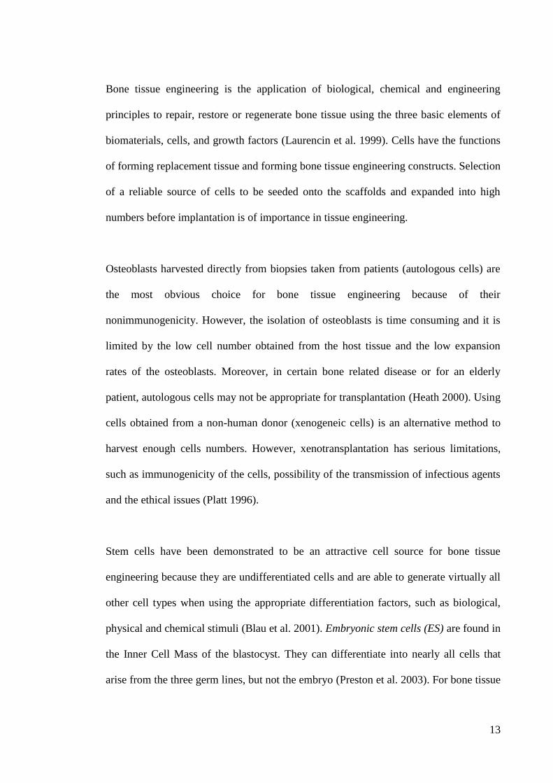
Bone tissue engineering is the application of biological, chemical and engineering
principles to repair, restore or regenerate bone tissue using the three basic elements of
biomaterials, cells, and growth factors (Laurencin et al. 1999). Cells have the functions
of forming replacement tissue and forming bone tissue engineering constructs. Selection
of a reliable source of cells to be seeded onto the scaffolds and expanded into high
numbers before implantation is of importance in tissue engineering.
Osteoblasts harvested directly from biopsies taken from patients (autologous cells) are
the most obvious choice for bone tissue engineering because of their
nonimmunogenicity. However, the isolation of osteoblasts is time consuming and it is
limited by the low cell number obtained from the host tissue and the low expansion
rates of the osteoblasts. Moreover, in certain bone related disease or for an elderly
patient, autologous cells may not be appropriate for transplantation (Heath 2000). Using
cells obtained from a non-human donor (xenogeneic cells) is an alternative method to
harvest enough cells numbers. However, xenotransplantation has serious limitations,
such as immunogenicity of the cells, possibility of the transmission of infectious agents
and the ethical issues (Platt 1996).
Stem cells have been demonstrated to be an attractive cell source for bone tissue
engineering because they are undifferentiated cells and are able to generate virtually all
other cell types when using the appropriate differentiation factors, such as biological,
physical and chemical stimuli (Blau et al. 2001). Embryonic stem cells (ES) are found in
the Inner Cell Mass of the blastocyst. They can differentiate into nearly all cells that
arise from the three germ lines, but not the embryo (Preston et al. 2003). For bone tissue
14
engineering, osteoblasts can be differentiated from ES cells with the help of
dexamethasone, as reported by Buttery (2001). Multipotent stem cells also known as
adult stem cells (ASC) can be found in the fully differentiated tissues, such as muscle,
cartilage, bone, the nervous systems and, probably, the liver and pancreas (Heath 2000).
They can differentiate into cell lineages from the tissue in which the ASC resides. In
addition, ASC have been found to have a higher degree of differentiation plasticity
(differentiate into muscle, brain, and fat cells) (Toma et al. 2001).
Human mesenchymal stem cells (MSC), stem cells that reside in bone marrow, have
drawn much interest in the bone tissue engineering field. These cells have the name
mesenchymal stem cells, given by Caplan (1994), who reported that these cells could be
easily isolated, expended and directed to differentiate into cells with mesenchymal
origin and form bone, cartilage, fat, and other tissues. MSCs have been used not only in
bone tissue engineering research but also in clinical trials (Li et al. 2006). Ex vivo-
expanded MSCs combined with three-dimensional porous biomaterials carriers have
been implanted into a sheep skull to directly repair a cranial defect (Shang et al. 2001).
Moreover, systemic infusion of MSCs into subjects with osteoporosis has led to the
attenuation of the disease (Ichioka et al. 2002). The use of MSCs in bone tissue
engineering applications offers powerful novel tools in the clinical strategy
development for the site-specific bone defects repair and the attenuation of bone
disorders (e.g. osteoporosis).
Additionally, in bone tissue engineering research, human clonal osteoblast cell lines are
widely used as osteoblastic models. The most commonly used osteoblast-like cell lines
15
include SaOs2, MG63, Te85 (HOS), and U2OS (HTB96). All these cell lines are
derived from osteosarcomas but differ in their responsiveness to certain hormones.
Osteosarcomas are malignant tumors of bone derived from cells of the osteoblast
lineage. They are poorly differentiated and pleomorphic with high mitotic activity. The
reason for their widespread use as osteoblast models is that osteosarcoma cells can
express osteoblastic genes, synthesize bone matrix proteins and respond to calcium-
regulating hormones (Gartland et al. 2005).
2.1.3 Bone Repair
Bone is a tissue with the ability of defect-healing to regenerate new tissue and blood
vessels and maintain physical and biological functions. In order to fabricate a suitable
scaffold to treat bone damage, it is essential to understand the bone healing process.
Bone fracture commonly occurs due to falls, accidents or sports injuries. It is taken as
an example to study the bone repair. There are four stages of secondary fracture healing:
inflammatory response, soft callus formation, hard callus formation and bone
remodeling (Li & Stocum 2014). Stage 1: When trauma occurs, the blood supply of the
bone is disturbed and a hematoma (blood clot) forms. Platelets trapped in the hematoma
release a platelet-derived growth factor (PDGF) and a transforming growth factor
(TGF-β) to initiate an inflammatory response. At Stage 2, the periosteal MSCs begin to
produce new vessels, proliferate and differentiate into osteoblasts. The hematoma is
replaced by fibrocartilage. After that, the cartilage is replaced by hard callus; and the
osteoblasts form woven bone on the calcified matrix at Stage 3. At the final stage of
bone repair, a remodeling process proceeds with hard callus resorption by osteoclasts
and lamellar bone formation by osteoblasts.
16
The repair of bone critically depends on the defect size. A defect heals spontaneously
when it is a non-critical size. However, a critical size defect cannot completely be
repaired and filled with new bone tissue by the bone self-healing ability (Meyer &
Wiesmann 2006). Research is currently in progress to develop bone-like scaffolds
containing cells and/or bone growth factors that helps bone regeneration after
implantation, for critical defect healing and is known as bone tissue engineering (Chung
& Park 2007). The requirements of a bone scaffold are studied and presented in Section
2.2.
2.2 Requirements of Scaffolds for Bone Tissue Engineering
To succeed as a bone regenerating scaffold, it should: (i) be biocompatibility inducing
minimal immune response or cytotoxicity; (ii) posses bioactive surface properties which
promote cell adhesion, bone bonding and stimulate osteogenesis; (iii) have precise
controllable interconnected porous structure with predefined shape, porosity and pore
size that can allow cell ingrowth, immigration and differentiation; (iv) exhibit
mechanical properties similar to those of the replacing host bone to support the patients‟
normal activities; (v) have a controllable degradation rate which matches the new tissue
growth rate; and (vi) be sterilisable for clinical use (Rezwan et al. 2006, Chung & Park
2007, Bose et al. 2012). The details of these requirements are described as follows:
2.2.1 Biocompatibility
Biocompatibility is the single most important factor that distinguishes a biomaterial
from any other material. “Biocompatibility is the ability of a material to perform an
17
appropriate host response in a specific situation” (Donaruma 1988). This means that the
material should perform an appropriate response to the tissue without eliciting an
immune response, not simply to exist in the human body, and the appropriateness may
be different from one situation to another.
As a scaffold for bone tissue regeneration, biocompatibility is also an important
requirement. When a scaffold is exposed to a living organism, there is a natural
tendency for the living organism to respond to this foreign object. Many interactions
may occur at the scaffold-tissue interface. The interactions, such as coagulation,
immune surveillance, healing, inflammation, mutagenicity, and carcinogenicity,
between a host and an implanted scaffold are extremely complex. These biologic
responses to materials are important considerations in the design of medical devices,
such as scaffolds. The host response is influenced by the material characteristics which
are also considered in the context of the biocompatibility (Williams 2008). The major
material characteristics include chemical composition, structure, morphology,
crystallinity, elastic constant, wettability, porosity of the bulk materials, surface
composition, surface engineering, surface electrical properties, corrosion parameters
and ion release profile (for metal), degradation profile and degraded product toxicity
(for polymer and ceramic) and the ware debris release profile (Williams 2008).
To develop a scaffold for tissue engineering, understanding of the molecular
mechanisms of the interactions, controlling the interactions, and optimizing the scaffold
need to be considered. Therefore, the biocompatibility of the scaffold must be evaluated.
The biocompatibility tests can be divided into two levels: biosafety and biofunctionality
18
(Pizzoferrato et al. 1995). The first level tests are concerned with the biosafety of the
materials in terms of cytotoxicity, hemotoxicity, genotoxicity and histotoxicity. The
tests at the second level are designed for biofunctionality issues, to assess the host
response evoked by the materials in a specific application. The second level tests
include cytocompatibility, immunocompatibility, hemocompatibility, histocompatibility,
and infectability (Zhang 2004). The basic testing methods can be found in the
International Standards Organization 10993 standards: international standards for
biological evaluation of medical devices. Understanding the historic context and the
biocompatibility of materials used in biomedical field will facilitate the design and
fabrication of new medical devices.
An in vitro test (test with cells cultured outside body) is usually done before an in vivo
test (test in body), because it is cheaper and less time consuming; in vivo results are
difficult to obtain and have a lot of restrictions. The interaction between cells and
biomaterials in vitro can mimic that in vivo. In vitro cytotoxicity tests are used in
evaluating a wide range of devices and materials for medical applications (ISO 10993-
5-2009). There are three types of test: extract test, direct contact test and indirect contact
test. The methods used in cytotoxicity determination can be grouped into four
categories: morphological means for cell damage assessments; chemical measurements
of cell damage; cell growth assessments; specific aspects of cellular metabolism
measurements.
19
2.2.2 Bioactive Surface Properties
Chemical compositions of the scaffold surface are related to the cell attachment and
dictate the protein adsorption behavior with cellular interactions (Dos Santos et al.
2009). The initial cell adhesion plays a decisive role in determining cell survival. The
cells continue to grow and differentiate only after the adhesion is achieved. Once the
scaffold is implanted, its surface will be covered with the body fluid and protein layer.
The cells will attach to the surface through various biomolecules in the adsorbed layer.
The important molecules called integrins, a kind of cell surface receptor, can be
absorbed on the scaffold surface when it has appropriate chemical properties. These
integrin receptors can transmit biochemical signal between the intracellular and
extracellular compartments. After the integrin receptors interact with the cells, focal
adhesions will be formed. Hydrophilicity or wettability, which is controlled by the
chemical compositions of the scaffold surface, influences the cell adhesion. Techniques
such as self-assembled monolayers (SAMs) have been used for the tailoring of surface
chemistry. It was shown that the in vitro osteoblasts adhesion and differentiation were
better on hydrophilic surfaces than on hydrophobic ones (Keselowsky et al. 2005).
Chemical gradient polymers mixed with hydrophobic and hydrophilic plasma polymers
were recently developed and investigated (Zelzer et al. 2008). The results from this
study also confirmed that the hydrophilic part of the polymer is more cytotropic (having
an affinity for cells).
After focal adhesion, the cells will spread and migrate to find a more suitable place to
secure their shape stability and future developments. Not only the chemical composition,
but also the topography of the scaffold surface, can control and affect cell response to
20
the bone implant materials, such as cell adhesion, migration, and proliferation (Anselme
et al. 2010). A phenomenon named „contact guidance‟ has been observed when there is
a systematic orientation of the cells in the direction of grooves on the substrate (Chen et
al. 2007, Ismail et al. 2007). It has been shown that the enhancement of microstructural
roughness could facilitate the osteogenic cells migration on the materials surface (Du et
al. 1999, Albrektsson & Johansson 2001). Osteoblast-like cells, such as MG63 cells,
show positive interaction with substrates with rougher surfaces (Lincks et al. 1998),
nevertheless, contradicted findings are also reported (Setzer et al. 2009). This is due to
the influence of the cell model used in different studies.
All these indicate that the cytotropic surface properties are important for a bone scaffold.
Surface modification of the bone scaffold is one of the promising strategies to achieve
the desired biological response. Numerous technologies have been developed for
surface modification. The surface of a scaffold can be functionalized with some
bioactive molecules, such as protein or peptides, either by physical adsorption or
chemical modification (Shin et al. 2003). The collagen, cell adhesive protein, peptide
and growth factors adsorbed onto the surface of the scaffolds show a positive effect on
cell attachment, growth and differentiation (Patel et al. 2008). Arg-Gly-Asp (RGD) is
the most commonly used peptide for surface modification to enhance cell adhesion
(Hersel et al. 2003). Kokubo (1991) reported that the formation of a bonelike apatite
layer on the surface of an artificial biomaterial after being implanted in the body is the
essential requirement for bonding to living bone. Oyane (2003) reported that the apatite
formation in a simulated body fluid (SBF) can be reproduced as the artificial
biomaterial implanted in a living body. Therefore, the biomimetic apatite formation on
21
the artificial biomaterials after soaking in SBF not only has been used to assess the
bioactivity of materials, but also becomes a strategy to improve the surface bioactivity
of materials (Song et al. 2004). Apart from these chemical methods, some
physicochemical methods such as glow discharge gas plasma treatment and ion
irradiation have been used to modify the surface composition, and surface energy to
improve cell adhesion (Wan et al. 2004).
2.2.3 Macro-architecture and Mechanical Properties
Highly porous interconnected three-dimensional scaffolds with a minimum pore size of
100 μm facilitate cell migration, and proper transportation of nutrients and metabolic
wastes (Hutmacher 2000). The architectural characteristics of the scaffolds are usually
determined by the fabrication method.
Porosity and interconnectivity are the important parameters for bone tissue engineering
scaffolds because they directly affect the diffusion of physiological nutrients and gases
into the scaffold resulting from the activity of the cells. Kuboki (1998) reported that the
porosity of scaffolds is crucial for bone formation since no osteogenesis is induced in
solid hydroxyapatite, while in the porous hydroxyapatite new bone formed after 2
weeks implantation. Increased porosity is benefit for nutrients and oxygen diffusion,
resulting in increased cell proliferation (Khademhosseini & Langer 2007). In vivo
studies of porosity gradient (80%-88%) poly(L-lactide-co-D,L-lactide)/β-tricalcium
phosphate scaffolds showed similar results that more tissue growth occurred in the areas
with higher porosity (Roy et al. 2003).
22
In addition to porosity, pore size of the scaffold is also an important issue that affects
cellular interaction. If the pore sizes are too small, this will disturb cell migration,
extracellular matrix production, and neovascularization. The minimum requirement for
a pore size of 100 μm is suggested by Karageorgiou and Kaplan (2005). They also
recommend that pore size larger than 300 µm is better for achieving a higher rate of
bone regeneration and vascularization. A comparing study of hydroxyapatite scaffolds
with 70% porosity and 800 μm pore size versus scaffolds with 60% porosity and 400
μm pore size, in vitro and in vivo, has been conducted (Kruyt et al. 2003). The scaffolds
with 60% porosity and 400 μm pore size performed better in vitro while scaffolds with
70% porosity and 800 μm pore size showed more bone formation in vivo (Kruyt et al.
2003). Although the results are different from in vitro and in vivo studies, both the
porosity and pore size affect cell proliferation and tissue regeneration.
Several methods have been used to measure the porosity and pore size of the scaffolds
(Karageorgiou & Kaplan 2005). The simplest way to estimate the total porosity (Pt) is
by using the Equation 2.1:
2.1
where is the density of the material and is the apparent density of the
scaffold. Mercury intrusion porosimetry and the liquid displacement method are also
available methods to measure the porosity of the scaffold (Maspero et al. 2002, Nazarov
et al. 2004). Scanning electron microscopy (SEM) images can be analyzed by computer
software to measure the porosity and pore size (Kim et al. 2002). In addition,
microcomputed tomography (micro-CT) has been applied to determine the porosity and
pore size of a 3D porous scaffold (Cartmell et al. 2004).
23
Mechanical properties of the scaffold play an important role in supporting the body,
maintaining the spaces for cell in-growth and providing the correct physical stimuli to
cells (Salgado et al. 2004, Agrawal & Ray 2001). They can be influenced by the
porosity and pore size of the scaffolds. It is important to balance the porosity value and
the mechanical needs for the target tissue. Moreover, the mechanical properties of the
scaffold should better match those of surrounding bone tissues in case of early injury
(Hutmacher 2000). Too low a mechanical strength is not suitable because it can not bear
the in-situ load from the surrounding tissues while too a high mechanical strength is
inappropriate since a stiff implant can stress shield the host tissues from the normal
physiological loading, increase the pain, and consequently give rise to bone resorption
and a depression of the osteoblastic activity (Iolascon et al. 2010).
Natural bone has been used as the template for bone scaffolds design and fabrication.
Bone, as a composite material which consists of collagen and calcium phosphate, has
been studied extensively (Currey 2002). The mechanical properties of bone (e.g.
Young‟s modulus, shear modulus, strength, viscoelastic properties, and fracture
mechanical properties) depend not only on the composition, but also on the structure of
the bone (e.g. geometric shape of the components, bond between fibers and matrix, and
bonds at points of contact of the fibers) (Fung 1993). Bone is slightly viscoelastic and
its strain-displacement relationship is anisotropic. Moreover, bone can be organized into
two different types: Compact bone (solid, only porous for canaliculi, osteocyte lacunae,
blood channels and erosion cavities) and cancellous bone (porosity visible to the naked
eye, rods and plates of bone are multiply-connected). So when we are discussing the
24
mechanical properties of bone, the specific type and part of the bone should be clarified.
For example, the tensile strength of compact bone has a range from 50 to 300 MPa, and
the modulus varies from 3 to more than 30 GPa (Cowins 1989, Rezwan et al. 2006). For
cancellous bone, it has much lower strength and modulus than that of compact bone
because of its high porosity. Normally, the modulus and tensile strength of human
cancellous bone are in the range of 0.01 to 2 GPa, and 0.1 to 30 MPa, respectively
(Black & Hastings 1998). The mechanical properties are not only different between
various types but also within the same type. The mechanical properties of the femur,
tibia, humerus and radius (human compact bones in different parts of the human body)
are different.
Most scaffolds fabricated from polymer exhibits insufficient mechanical properties.
Reinforcing with inorganic particles, fibers, and sheets have been used and dispersed in
the polymer matrix to enhance the mechanical properties of the scaffolds (Xu et al.
2009, Swetha et al. 2010, Blaker et al. 2011). The architecture of the scaffold should be
balanced with the mechanical properties. How to fabricate the polymer based scaffolds
with high porosity and appropriate mechanical properties is still a challenge in bone
tissue engineering.
2.2.4 Biodegradability
Great efforts have been made in developing biodegradable biomaterials for medical and
related applications over last five decades (Shalaby & Karen 2003). Biodegradable
materials are designed to overcome the side effects of permanent or non-degradable
devices (generally metal-based), such as physical irritations, chronic inflammatory local
25
reactions, stress shielding, corrosion and repeat surgery. Since the 1960s, when the first
biodegradable sutures were approved, a range of new generation biodegradable
polymers has been developed and accepted for used in medical applications (Middleton
& Tipton 2000).
Scaffolds with biodegradability can be gradually degraded or resorbed, and replaced by
regenerated tissue after implantation. This avoids secondary surgery to remove the
implants after tissue recovery. Degradation behavior of bone scaffolds is of crucial
importance in tissue engineering, because the degradation rate is essentially linked to
cell growth, host response and tissue regeneration (Babensee et al. 1998). The scaffold
degradation rate needs to be controllable and match the rate of the bone regeneration
(Nair & Laurencin 2007). In this way, the scaffold will completely degrade when the
injury site is totally repaired. Several factors, such as scaffold composition,
microstructure, chemical and physical properties, molecular weight as well as
environmental conditions can affect the degradation kinetics of scaffolds (Wu & Ding
2004, Söntjens et al. 2012). Therefore, controlling and evaluation of the degradation
behavior of composite scaffolds are of importance for the success of a biodegradable
scaffold for bone tissue engineering. Current efforts have been focused on custom
designing and synthesizing biodegradable polymers with tailored mechanical properties
and controllable degradation rates (Nair & Laurencin 2007).
Synthesis of a copolymer is one of the common strategies to control the degradation rate
of a polymer. Co-polyester, poly(lactide-co-glycolide) (PLGA) is a copolymer of
glycolide with L- or DL-lactide PLGA. The degradability of PLGA can be controlled
26
with varied ratios of glycolide and lactide. The copolymer of 50% glycolide and 50%
DL-lactide is very hydrolytically unstable and degrades faster than the copolymers at
either end of the co-polymer composition range (Miller et al. 1977). Kim and his co-
authors (2003) developed a composite scaffold consisting of poly(d,l-lactide) (PDLLA),
PLGA (LA/GA=55/55), poly(lactide-b-ethylene glycol-b-lactide) (PLA-b-PEG-b-PLA)
triblock copolymer and lactide. Its degradation rate can be tuned with different material
compositions. More recently, a biomimetic approach using PLGA microspheres loaded
with lysozyme (an antibacterial protein) as a filler material to control the degradation
rate of a chitosan scaffold has been reported (Liu et al. 2012). How to precisely control
the degradation rate of the scaffold matching the bone growth rate, and at the same time
maintain adequate mechanical properties to support the body is another challenge in
bone tissue engineering.
Scaffold degradation evaluation could be either tested in vitro or in vivo. For the in vitro
studies, the scaffolds are immersed in a phosphate buffered saline (PBS, pH 7.4) at
37°C. For in vivo study, scaffolds are implanted into a suitable animal model and then
retrieved at predetermined time points. The molecular weight loss and scaffold weight
loss at different time intervals are measured to determine the degradation rate of the
scaffold. Gel permeation chromatography (GPC) and viscometry are widely used to
measure the molecular weight changes of the polymeric scaffolds.
2.3 Recent Development of Materials for Bone Tissue Engineering
Tissue engineering is defined as “an interdisciplinary field that applies the principles of
engineering and life sciences toward the development of biological substitutes that
27
restore, maintain, or improve tissue function” (Langer & Vacanti 1993). More
specifically, tissue engineering implies the combination of cells, scaffolds, and
bioactive peptides used to guide the repair or formation of tissue. Bone tissue
engineering potentially provides an alternative approach to treat the loss or malfunction
of particular tissue. Bone tissue engineering requires a three-dimensional scaffold which
serves as a 3D template for cell attachment, ingrowth, proliferation and bone-
extracellular matrix formation to provide a structural support to a newly formed tissue
(Chen et al. 2002, Liu et al. 2007). In recent years, there have been tremendous
advances in the development of biomaterials for bone tissue engineering.
2.3.1 Biodegradable Oganic Polymers
Both synthetic polymers (e.g. poly(glycolic acid), poly(lactic acid), and polyurethane)
and natural polymers (e.g. collagen, polysaccharides, and starches) have been
extensively investigated in bone tissue engineering. Biodegradable polymers have
become promising candidates for scaffold implants which can be gradually reabsorbed
by regenerated tissue, avoid secondary surgery to remove the implants after tissue
recovery or depletion of the drug supply and shorten the recovery time (Liu et al. 2008).
Natural polymers can be considered as the first biodegradable biomaterials used
clinically. They have some advantages such as bioactivity, natural remodeling, and
ability to present receptor-binding ligands to cells (Nair & Laurencin 2007). Compared
with the natural polymers, the synthetic polymers have more predictable properties and
can be tailored for specific applications. In recent years, there have been tremendous
advances in the development of synthetic biodegradable polymers used for bone
scaffolds. These materials have been fabricated into three-dimensional porous scaffolds
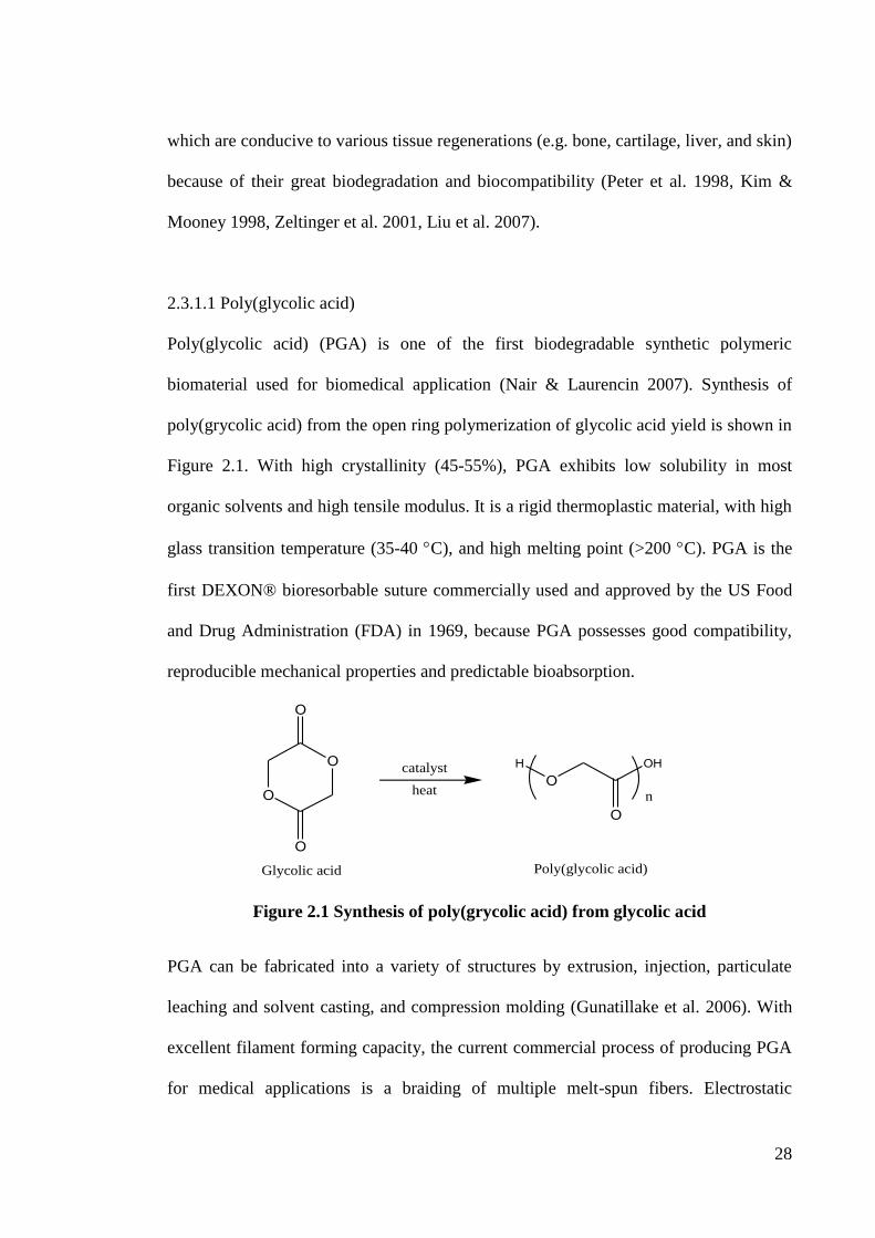
28
which are conducive to various tissue regenerations (e.g. bone, cartilage, liver, and skin)
because of their great biodegradation and biocompatibility (Peter et al. 1998, Kim &
Mooney 1998, Zeltinger et al. 2001, Liu et al. 2007).
2.3.1.1 Poly(glycolic acid)
Poly(glycolic acid) (PGA) is one of the first biodegradable synthetic polymeric
biomaterial used for biomedical application (Nair & Laurencin 2007). Synthesis of
poly(grycolic acid) from the open ring polymerization of glycolic acid yield is shown in
Figure 2.1. With high crystallinity (45-55%), PGA exhibits low solubility in most
organic solvents and high tensile modulus. It is a rigid thermoplastic material, with high
glass transition temperature (35-40 C), and high melting point (>200 C). PGA is the
first DEXON® bioresorbable suture commercially used and approved by the US Food
and Drug Administration (FDA) in 1969, because PGA possesses good compatibility,
reproducible mechanical properties and predictable bioabsorption.
O
O
O
O
HO
OH
On
Glycolic acid Poly(glycolic acid)
catalyst
heat
Figure 2.1 Synthesis of poly(grycolic acid) from glycolic acid
PGA can be fabricated into a variety of structures by extrusion, injection, particulate
leaching and solvent casting, and compression molding (Gunatillake et al. 2006). With
excellent filament forming capacity, the current commercial process of producing PGA
for medical applications is a braiding of multiple melt-spun fibers. Electrostatic
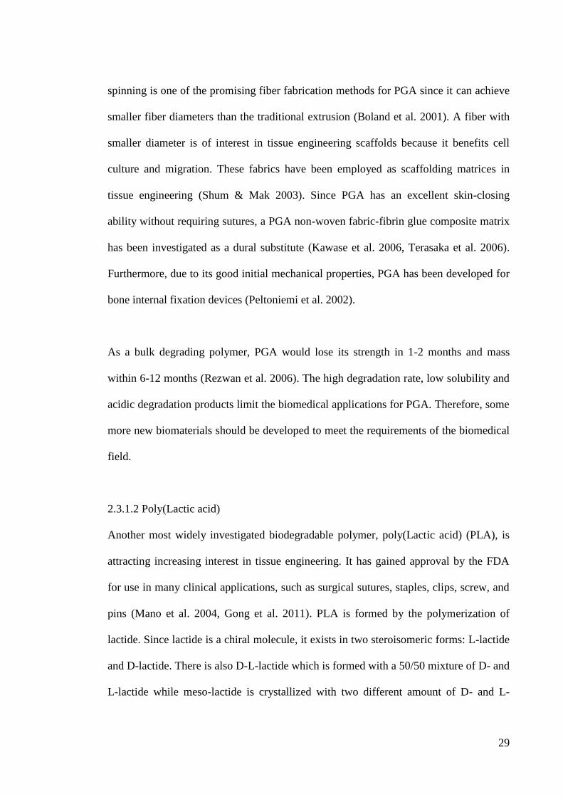
29
spinning is one of the promising fiber fabrication methods for PGA since it can achieve
smaller fiber diameters than the traditional extrusion (Boland et al. 2001). A fiber with
smaller diameter is of interest in tissue engineering scaffolds because it benefits cell
culture and migration. These fabrics have been employed as scaffolding matrices in
tissue engineering (Shum & Mak 2003). Since PGA has an excellent skin-closing
ability without requiring sutures, a PGA non-woven fabric-fibrin glue composite matrix
has been investigated as a dural substitute (Kawase et al. 2006, Terasaka et al. 2006).
Furthermore, due to its good initial mechanical properties, PGA has been developed for
bone internal fixation devices (Peltoniemi et al. 2002).
As a bulk degrading polymer, PGA would lose its strength in 1-2 months and mass
within 6-12 months (Rezwan et al. 2006). The high degradation rate, low solubility and
acidic degradation products limit the biomedical applications for PGA. Therefore, some
more new biomaterials should be developed to meet the requirements of the biomedical
field.
2.3.1.2 Poly(Lactic acid)
Another most widely investigated biodegradable polymer, poly(Lactic acid) (PLA), is
attracting increasing interest in tissue engineering. It has gained approval by the FDA
for use in many clinical applications, such as surgical sutures, staples, clips, screw, and
pins (Mano et al. 2004, Gong et al. 2011). PLA is formed by the polymerization of
lactide. Since lactide is a chiral molecule, it exists in two steroisomeric forms: L-lactide
and D-lactide. There is also D-L-lactide which is formed with a 50/50 mixture of D- and
L-lactide while meso-lactide is crystallized with two different amount of D- and L-
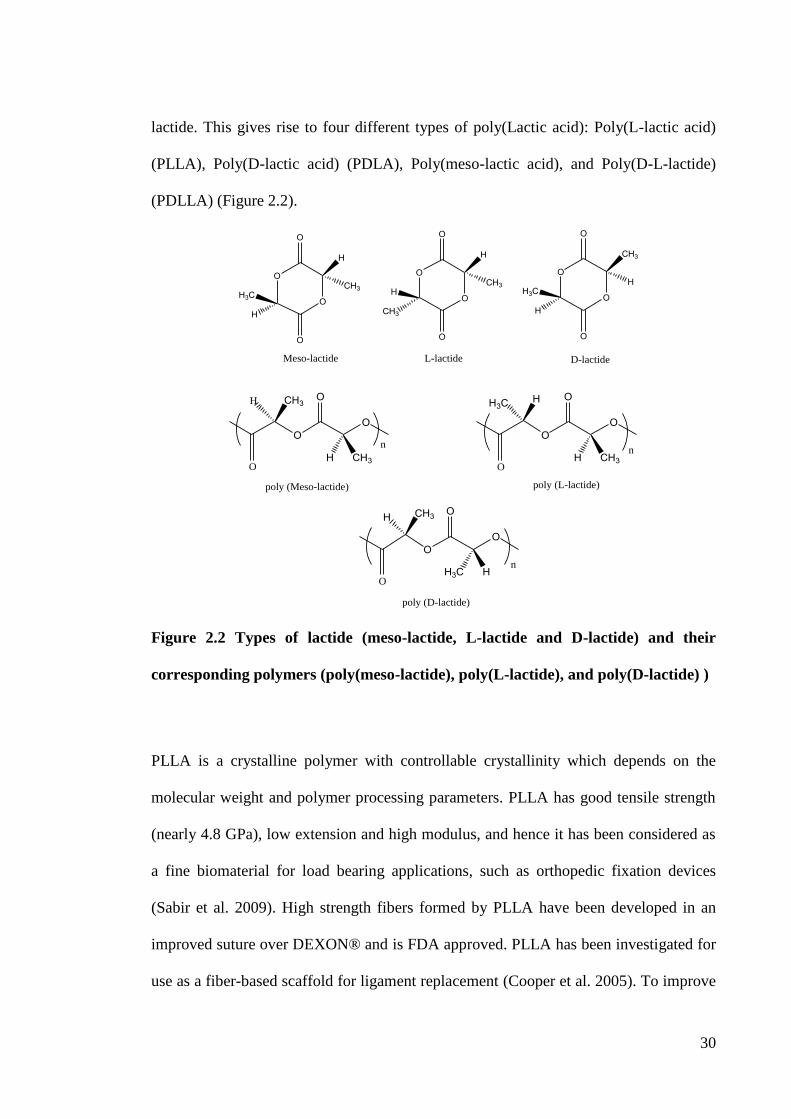
30
lactide. This gives rise to four different types of poly(Lactic acid): Poly(L-lactic acid)
(PLLA), Poly(D-lactic acid) (PDLA), Poly(meso-lactic acid), and Poly(D-L-lactide)
(PDLLA) (Figure 2.2).
O
O
H
CH3
H
H3C
O
O
O
O
H
CH3
CH3
H
O
O
O
O
CH3
H
H
H3C
O
O
Meso-lactide L-lactide D-lactide
OO
OCH3
CH3HO
H
nO
O
H
CH3
OH3C
HO
n
OO
CH3
H
OH
H3CO
n
poly (D-lactide)
poly (Meso-lactide) poly (L-lactide)
Figure 2.2 Types of lactide (meso-lactide, L-lactide and D-lactide) and their
corresponding polymers (poly(meso-lactide), poly(L-lactide), and poly(D-lactide) )
PLLA is a crystalline polymer with controllable crystallinity which depends on the
molecular weight and polymer processing parameters. PLLA has good tensile strength
(nearly 4.8 GPa), low extension and high modulus, and hence it has been considered as
a fine biomaterial for load bearing applications, such as orthopedic fixation devices
(Sabir et al. 2009). High strength fibers formed by PLLA have been developed in an
improved suture over DEXON® and is FDA approved. PLLA has been investigated for
use as a fiber-based scaffold for ligament replacement (Cooper et al. 2005). To improve
31
the mechanical properties of PLLA as a bone scaffold, researchers incorporated
hydroxyapatite into the biopolymer (Hong et al. 2005, Uskokovic et al. 2007,
Aleksendrić et al. 2010). PLLA has been fabricated into three-dimensional sponges and
hybridized with collagen and hydroxyapatite. Furthermore, scaffolds with drug-release
function have been developed using PLLA as the matrix (Huang et al. 2007, Uttarwar
2008).
PLA is much more hydrophobic than PGA due to the additional methyl group in the
structure of PLA. Therefore, the degradation rate of PLLA is much slower, and needs
more than 24 months to completely degrade (Middleton & Tipton 2000). The slow
degradation also limits the application of PLLA. Some co-polymers are under
investigation for improvement of their properties.
Poly(D-L-lactide) (PDLLA) is polymerized with random distributions of L- and D-
lactide. It is an amorphous polymer and shows a lower strength (approximately 1.9 GPa)
and faster degradation rate (12-16 months) compared to PLLA. It is therefore a good
candidate for developing drug delivery vehicles and non-bearing or low-bearing
scaffolds for tissue engineering. PDLLA has been widely used in the development of
exogenous matrices suitable for facilitating tissue regeneration (Huang et al. 2005,
Calandrelli et al. 2008, Wu et al. 2008b, Xu et al. 2009). It has been investigated as a
biomedical orthopedic coating material because it can optimize interaction at the
implant-tissue interface (Gollwitzer et al. 2005). In addition, different bioactive
particles such as hydroxyapatite (Deng et al. 2008), bioglass (Blaker et al. 2011),
wollastonite (Xu et al. 2009), CaSiO3 (Zhang et al. 2009) have been incorporated into
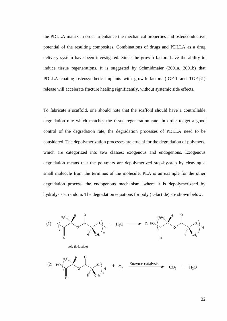
32
the PDLLA matrix in order to enhance the mechanical properties and osteoconductive
potential of the resulting composites. Combinations of drugs and PDLLA as a drug
delivery system have been investigated. Since the growth factors have the ability to
induce tissue regenerations, it is suggested by Schmidmaier (2001a, 2001b) that
PDLLA coating osteosynthetic implants with growth factors (IGF-1 and TGF-β1)
release will accelerate fracture healing significantly, without systemic side effects.
To fabricate a scaffold, one should note that the scaffold should have a controllable
degradation rate which matches the tissue regeneration rate. In order to get a good
control of the degradation rate, the degradation processes of PDLLA need to be
considered. The depolymerization processes are crucial for the degradation of polymers,
which are categorized into two classes: exogenous and endogenous. Exogenous
degradation means that the polymers are depolymerized step-by-step by cleaving a
small molecule from the terminus of the molecule. PLA is an example for the other
degradation process, the endogenous mechanism, where it is depolymerizaed by
hydrolysis at random. The degradation equations for poly (L-lactide) are shown below:
OO
H
CH3
OH3C
HO
n
poly (L-lactide)
+ H2O HOO
OH
H
CH3
OH3C
HO
n
HOO
OH
H
CH3
OH3C
HO
n
+ O2 CO2 H2O+
(1)
(2) Enzyme catalysis
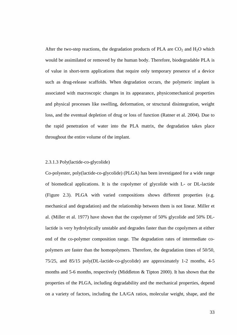
33
After the two-step reactions, the degradation products of PLA are CO2 and H2O which
would be assimilated or removed by the human body. Therefore, biodegradable PLA is
of value in short-term applications that require only temporary presence of a device
such as drug-release scaffolds. When degradation occurs, the polymeric implant is
associated with macroscopic changes in its appearance, physicomechanical properties
and physical processes like swelling, deformation, or structural disintegration, weight
loss, and the eventual depletion of drug or loss of function (Ratner et al. 2004). Due to
the rapid penetration of water into the PLA matrix, the degradation takes place
throughout the entire volume of the implant.
2.3.1.3 Poly(lactide-co-glycolide)
Co-polyester, poly(lactide-co-glycolide) (PLGA) has been investigated for a wide range
of biomedical applications. It is the copolymer of glycolide with L- or DL-lactide
(Figure 2.3). PLGA with varied compositions shows different properties (e.g.
mechanical and degradation) and the relationship between them is not linear. Miller et
al. (Miller et al. 1977) have shown that the copolymer of 50% glycolide and 50% DL-
lactide is very hydrolytically unstable and degrades faster than the copolymers at either
end of the co-polymer composition range. The degradation rates of intermediate co-
polymers are faster than the homopolymers. Therefore, the degradation times of 50/50,
75/25, and 85/15 poly(DL-lactide-co-glycolide) are approximately 1-2 months, 4-5
months and 5-6 months, respectively (Middleton & Tipton 2000). It has shown that the
properties of the PLGA, including degradability and the mechanical properties, depend
on a variety of factors, including the LA/GA ratios, molecular weight, shape, and the
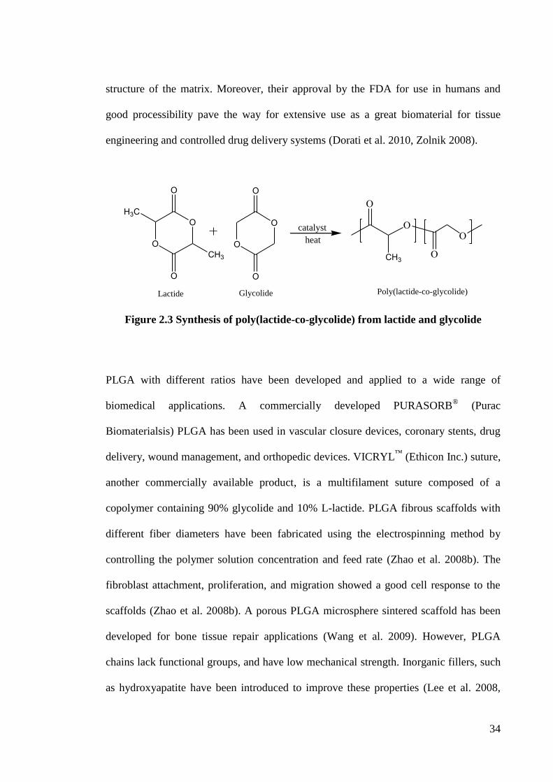
34
structure of the matrix. Moreover, their approval by the FDA for use in humans and
good processibility pave the way for extensive use as a great biomaterial for tissue
engineering and controlled drug delivery systems (Dorati et al. 2010, Zolnik 2008).
CH3
O
O
O
O
O
O
O
O
H3C
CH3
catalyst
heat
Lactide Glycolide Poly(lactide-co-glycolide)
Figure 2.3 Synthesis of poly(lactide-co-glycolide) from lactide and glycolide
PLGA with different ratios have been developed and applied to a wide range of
biomedical applications. A commercially developed PURASORB® (Purac
Biomaterialsis) PLGA has been used in vascular closure devices, coronary stents, drug
delivery, wound management, and orthopedic devices. VICRYL™
(Ethicon Inc.) suture,
another commercially available product, is a multifilament suture composed of a
copolymer containing 90% glycolide and 10% L-lactide. PLGA fibrous scaffolds with
different fiber diameters have been fabricated using the electrospinning method by
controlling the polymer solution concentration and feed rate (Zhao et al. 2008b). The
fibroblast attachment, proliferation, and migration showed a good cell response to the
scaffolds (Zhao et al. 2008b). A porous PLGA microsphere sintered scaffold has been
developed for bone tissue repair applications (Wang et al. 2009). However, PLGA
chains lack functional groups, and have low mechanical strength. Inorganic fillers, such
as hydroxyapatite have been introduced to improve these properties (Lee et al. 2008,
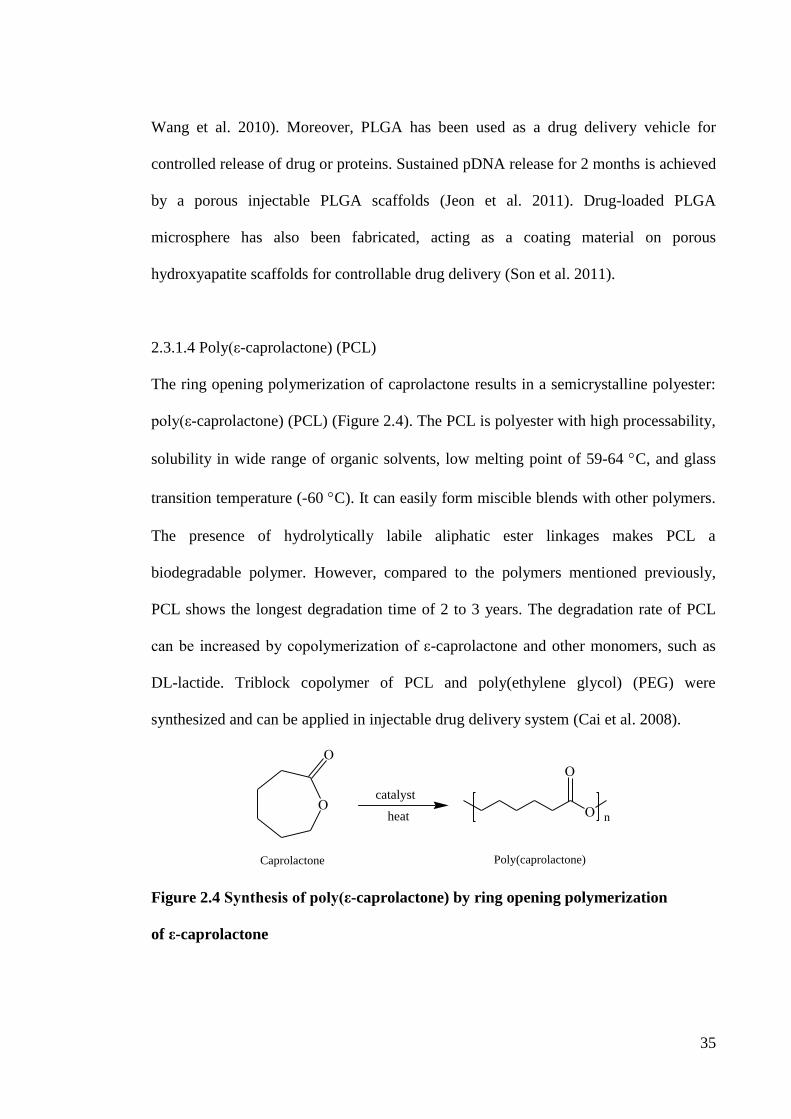
35
Wang et al. 2010). Moreover, PLGA has been used as a drug delivery vehicle for
controlled release of drug or proteins. Sustained pDNA release for 2 months is achieved
by a porous injectable PLGA scaffolds (Jeon et al. 2011). Drug-loaded PLGA
microsphere has also been fabricated, acting as a coating material on porous
hydroxyapatite scaffolds for controllable drug delivery (Son et al. 2011).
2.3.1.4 Poly(ε-caprolactone) (PCL)
The ring opening polymerization of caprolactone results in a semicrystalline polyester:
poly(ε-caprolactone) (PCL) (Figure 2.4). The PCL is polyester with high processability,
solubility in wide range of organic solvents, low melting point of 59-64 C, and glass
transition temperature (-60 C). It can easily form miscible blends with other polymers.
The presence of hydrolytically labile aliphatic ester linkages makes PCL a
biodegradable polymer. However, compared to the polymers mentioned previously,
PCL shows the longest degradation time of 2 to 3 years. The degradation rate of PCL
can be increased by copolymerization of ε-caprolactone and other monomers, such as
DL-lactide. Triblock copolymer of PCL and poly(ethylene glycol) (PEG) were
synthesized and can be applied in injectable drug delivery system (Cai et al. 2008).
n
Poly(caprolactone)Caprolactone
catalyst
heat
Figure 2.4 Synthesis of poly(ε-caprolactone) by ring opening polymerization
of ε-caprolactone
36
With the slow degradability, permeability to wide range of drugs, and biocompatibility,
PCL has been initially applied in a long term drug delivery system. One commercially
developed long-term contraceptive device, Capronor®, that releases levonorgestrel over
12-18 months, has been investigated and evaluated in humans in early 1989 (Darney et
al. 1989). Extensive research is ongoing to develop PCL drug carriers in the form of
micro- and nanoshpheres (Sinha et al. 2004). Taxol-loaded PCL microspheres has been
prepared and about 25% of the taxol were released from the microspheres by a 6 weeks
drug release study (Dordunoo et al. 1995). To increase the drug release rate (or polymer
degradation rate), PCL/PLGA (65:35) microsphere containing bovine serum albumin
has been developed (Yang et al. 2001). Apart from the drug delivery system, PCL
microspheres with bioactive apatite coated and macroporous morphology have been
fabricated for the application in augmentation and bone tissue engineering (Hong et al.
2009). Biphasic calcium phosphate reinforced PCL composite microspheres with
improved mechanical properties, good biocompatibility and the ability promoting cell
adhesion, growth, and proliferation have been investigated as bone scaffolds (Bao et al.
2011).
2.3.2 Research Trends of Advanced Scaffold Materials
Materials used for bone scaffold fabrication should be designed to stimulate specific
cell response at the molecular level. They should interact with cells and promote cell
adhesion, growth, differentiation, and extracellular matrix formation and organization.
The selection of biomaterials plays an important role in the success of bone tissue
engineering. The primary requirements of the biomaterials used in tissue regeneration
are biocompatibility, biodegradability and favorable mechanical properties.
37
Conventional single-phase materials cannot always provide all the necessary properties
for the bone tissue engineering and the characteristics of a true human bone. Though a
variety of biodegradable polymers have been developed and studied for tissue
engineering, no single biodegradable polymer can meet all the requirements for
biomedical scaffolds. For example, scaffolds derived from unmixed biodegradable
polymers often lack osteoconducting properties and mechanical strength. Therefore, the
design and fabrication of composite materials (a combination of two or more different
materials) in order to develop a multifunctional bone scaffold is of great research
interest. By incorporation of the bioactive particles to the polymers, it is possible to
control the mechanical properties, such as strength and modulus of the composites,
closer tonatural bone and improve the bioactivity of the composites. In particular,
Section 2.3.4 deals with a comprehensive literature review of polymer/bioactive
particles composites used in bone tissue engineering
2.3.3 Polymer/Bioactive Particles Composites
The use of particulate reinforcing fillers in biodegradable polymers is attracting
increasing attention for researchers in tissue engineering. Hydroxyapatite (HAp)
(Swetha et al. 2010), apatite-wollastonite (A/W) glass-ceramics (Xu et al. 2009), and
bioglass® (Blaker et al. 2011) have been widely used as the bioactive phase in the
biodegradable polymers reviewed previously for tissue engineering. The mechanical
properties of commonly used bioactive filler materials and human bones, are listed in
Table 2.1
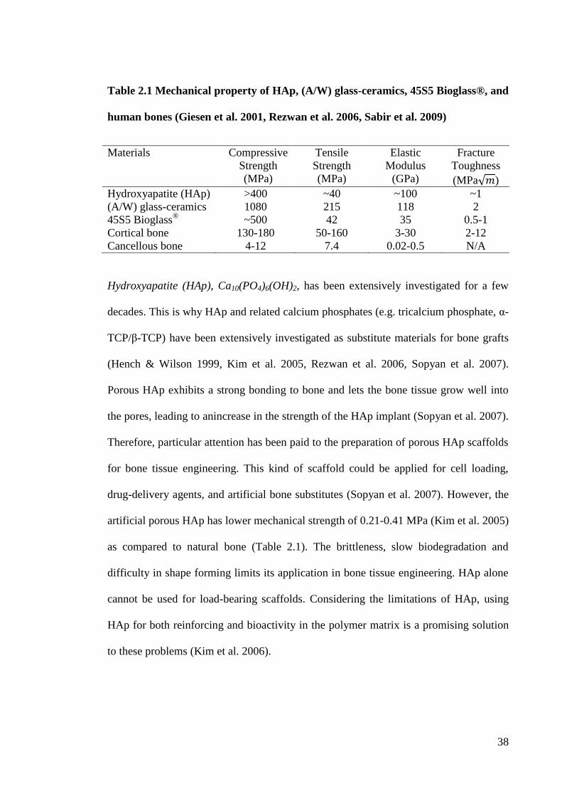
38
Table 2.1 Mechanical property of HAp, (A/W) glass-ceramics, 45S5 Bioglass®, and
human bones (Giesen et al. 2001, Rezwan et al. 2006, Sabir et al. 2009)
Materials Compressive
Strength
(MPa)
Tensile
Strength
(MPa)
Elastic
Modulus
(GPa)
Fracture
Toughness
(MPa )
Hydroxyapatite (HAp) >400 ~40 ~100 ~1
(A/W) glass-ceramics 1080 215 118 2
45S5 Bioglass®
~500 42 35 0.5-1
Cortical bone 130-180 50-160 3-30 2-12
Cancellous bone 4-12 7.4 0.02-0.5 N/A
Hydroxyapatite (HAp), Ca10(PO4)6(OH)2, has been extensively investigated for a few
decades. This is why HAp and related calcium phosphates (e.g. tricalcium phosphate, α-
TCP/β-TCP) have been extensively investigated as substitute materials for bone grafts
(Hench & Wilson 1999, Kim et al. 2005, Rezwan et al. 2006, Sopyan et al. 2007).
Porous HAp exhibits a strong bonding to bone and lets the bone tissue grow well into
the pores, leading to anincrease in the strength of the HAp implant (Sopyan et al. 2007).
Therefore, particular attention has been paid to the preparation of porous HAp scaffolds
for bone tissue engineering. This kind of scaffold could be applied for cell loading,
drug-delivery agents, and artificial bone substitutes (Sopyan et al. 2007). However, the
artificial porous HAp has lower mechanical strength of 0.21-0.41 MPa (Kim et al. 2005)
as compared to natural bone (Table 2.1). The brittleness, slow biodegradation and
difficulty in shape forming limits its application in bone tissue engineering. HAp alone
cannot be used for load-bearing scaffolds. Considering the limitations of HAp, using
HAp for both reinforcing and bioactivity in the polymer matrix is a promising solution
to these problems (Kim et al. 2006).
39
In the HAp filled polymeric composites, HAp acts as a reinforcing material to enhance
the mechanical properties, and meanwhile, it is also a bioactive component to improve
the osteoconductivity and biocompatible properties of the scaffolds (Swetha et al. 2010,
Zhao et al. 2008a). Therefore, development of composite scaffold is attractive.
Furthermore, addition of a bioactive HAp phase in biodegradable polymers could alter
the degradation rate by allowing rapid exchange of protons in alkaline water. It is
suggested providing a pH buffering effect to the polymer and modifying the acidic
polymer degradation (Li & Chang 2005). This would reduce the risk of inflammation.
The water absorption and wettability of the scaffolds would be improved with the
addition of HAp into the polymer matrix (Shikinami & Okuno 2001, Ren et al 2008). In
addition to being reinforcing and bioactive fillers, HAp showed another advantage in
the possibility of controling the biodegradation rate of the composite materials (Russias
et al. 2006, Chen et al. 2013). With high degree of mechanical properties, bioactivity,
and controllable degradability, polymer/HAp composites will be promising materials
for bone scaffold fabrication.
Bioactive glasses are the widely used bioactive fillers in tissue engineering applications.
In particular, 45S5 Bioglass® has drawn considerable interest because its dissolution
products can upregulate the gene expression and control osteogenesis (Xynos et al.
2000). 45S5 Bioglasss contains 45% SiO2, 24.5% Na2O, 24.4% CaO and 6% P2O5, in
weight percent. Among these constituents, SiO2 plays an essential role in bone
mineralization and gene activation. These commercial bioactive glasses have been
reported as class “A” bioactive materials and found to support enzyme activity and
40
vascularization (Jones, Hench 2001). 45S5 Bioglass® has been successfully used in
clinical treatments of periodontal disease and as a bone filler material (Hench 1998a).
The same as for HAp, bioglasses act as reinforcing fillers in the polymer matrix,
extending their applications in bone tissue engineering. The mechanical properties of
the porous poly(α-hydroxyacid)/Bioglass®
have been improved; the water absorption
and weight loss have been increased by the addition of Bioglass®
(Maquet et al. 2004,
Blaker et al. 2005). Moreover, the degradation kinetics of the composites can be
controlled by Bioglass® incorporation. This is another key reason that makes bioglass a
bioactive filler material for the bone scaffold. MG63 (human osteosarcoma cell line)
and A549 cells (human lung carcinoma cell line) have been seeded on the porous
PDLLA/Bioglass® composite scaffolds to investigate the effect of the Bioglass
® content
on the cell adhesion and growth (Verrier et al. 2004). The composite scaffolds show a
good biocampatibility and the increasing amount of Bioglass®
leads to enhanced MG63
cell attachment and proliferation. A549 cells grew better on PDLLA scaffold with 5 wt%
Bioglass®
when compared to the one with 40 wt% Bioglass®
(Verrier et al. 2004). This
implies that the appropriate concentration of Bioglass® particles in polymer-based
scaffolds may be different from cell to cell. One of the disadvantages of bioglasses is
that the crystallization of bioactive glasses decreases the level of bioactivity (Filho et al.
1996). This limits their application in bone tissue engineering since full crystallization
occurs prior to the densification by glass sintering. So improved sintering processes
need to be developed to prevent the full crystallization of bioglass and allow it achieve
the required mechanical properties.
41
Glass-ceramic apatite-wollastonite (A-W) is a bioactive ceramic which has an ability to
promote bone regeneration by providing strong interfacial bonding between the implant
and host tissue (Hench & Wilson 1999, Xu et al. 2009). Glass-ceramic A-W is a
ceramic containing crystalline apatite Ca10(PO4)6(O,F2) and wollastonite (CaOSiO2) in
a MgO-CaO-SiO2 glassy matrix. Compared with Bioglasss and HAp, glass-ceramic A-
W shows a greater modulus, compression and tensile strength. It has been used for the
fabrication of bone substitute because of its high bioactivity. The same as other
ceramics, A-W glass-ceramic has a relatively low fracture toughness which limits its
application in load-bearing applications. Therefore, it also becomes a filler material in
the polymer matrix, with the dual roles of improving bioactivity and mechanical
strength of the composite. A composite developed by A-W glass-ceramic incorporation
in a polyethylene (PE) matrix showed a faster apatite formation after immersion in SBF
as compared with that of HAPEX™
(a commercial available HAp/PE composite)
(Juhasz et al. 2003). A-W glass-ceramic filled poly(methylmethacrylate) (PMMA)
composites fabricated by Shinzati (2001) showed a higher osteoconductivity and better
mechanical properties with larger amount of A-W glass-ceramic filler.
The basic functional subunits of cells and tissues are defined at the nanoscale, hence
application of nanotechnology represents a new cutting edge in tissue engineering
research (Armentano et al. 2010). Nanotechnology enables the development of new
systems that mimic the complex, hierarchical structure of the native tissue. A
nanocomposite is a composite material in which at least one component is on the
nanometer-scale. It is considered as a new class of material because of its better
properties than those in microscale counterparts. Compared to the HAp in micron range,
42
the nano-HAp has a larger surface area, so the nano-composites exhibit enhanced
mechanical properties because of the strong interactions at the interface of the nano-
HAp and polymer (Kothapalli et al. 2005). Moreover, it has been reported that nano-
HAp could increase the initial calcium adsorption to the surface and hence enhance the
binding of vitronectin that subsequently promotes osteoblast adhesion and shortens the
bone regeneration rate (Webster et al. 2001). Three-dimensional biodegradable
PLGA/nano-HAp composites microsphere-based scaffolds with high compressive
strength, modulus and the appropriate pore structure for cell penetration have been
fabricated (Lv et al. 2009). Biodegradable polyphosphazene/nano-HAp composite
scaffolds for bone tissue engineering (Nukavarapu et al. 2008), biodegradable nano-
HAp/PLA porous scaffolds for non-load bearing tissue engineering applications were
also investigated (Kothapalli et al. 2005). Moreover, Yang and his coauthors (2004)
have developed a biomimetic nano-HAp/type I collagen scaffold for bone tissue
engineering. The results indicated the potential of this scaffold for exploitation of the
extacellular matrix in bone regeneration. The processing of nanocomposites raises the
possibility of realizing bone grafts with improved performance and is now a major
research trend in bone tissue engineering.
2.3.4 Multifunctional Bone Scaffolds
A multifunctional bone scaffold is a scaffold system designed with a combination of
different predefined functions, such as physical support, degradation, drug delivery
function, and shape memory effect (SME), and is used in bone tissue engineering for
assisting in bone regeneration. So far, the drug-eluting stent, including a shape memory
metal stent platform, polymer drug carrier and drug, is an example of multifunctional
43
devices (Ma et al. 2012). However, the stent needs to be removed after the treatment by
secondary surgery. A biodegradable composite with the SME and drug release function
without adverse effect on other functionalities is a promising material for bone tissue
engineering and involves minimally invasive surgery. Copolyester networks based on
semi-crystalline oligo [ -caprolactone)-co-glycolide] and amorphous oligo[(rac-
lactide)-co-glycolide] precursors were developed as multifunctional materials with SME,
degradability and drug release function (Neffe et al. 2009, Wischke et al. 2009).
Independence of the functionalities can be proven. As biodegradability has been
discussed in Section 2.2.4, a review on the shape memory polymers and drug delivery
scaffolds are presented in the following paragraphs.
The ability to recover from a plastic deformation to its original shape when stimulated
by a specific physical quantity is known as the shape memory effect (SME) (Huang et
al. 2010). Shape memory materials (SMMs) are materials featured by the SME. Shape
memory alloys (SMAs), shape memory ceramics (SMCs), and shape memory polymers
(SMPs) have been utilized in many fields, such as aerospace engineering and medical
devices. SMPs, a class of “actively moving” polymers, can recover from an initial
temporary shape to their permanent shapes by an external stimulus such as temperature,
light, pH, and magnetic field (Meng & Hu 2009). Compared to SMAs and SMCs, SMPs
have higher recoverable strain of over 100% for 10% of SMAs and 1% of SMCs,
respectively. Moreover, from the engineering aspect, polymers are cheaper and easier to
process than metals and ceramics. The shape change, with a change of temperature, is
called a thermally induced SME. This kind of polymer is an emerging class of smart
materials with potential applications in many areas of our lives, such as high-
44
performance textiles (Mondal & Hu 2006), packaging materials (Charlesby 1960),
intelligent medical devices (Small IV et al. 2009), and implants for minimally invasive
surgery (Lendlein & Langer 2002).
Polyurethane-based SMPs have been fabricated as endovascular interventions for the
treatment of vascular diseases (Langer & Tirrell 2004, Baer et al. 2007). A degradable
SMP has been fabricated as a suture and showed a good shape memory effect and high
potential in skin wound closure (Zheng et al. 2006). A degradable thermal SMP has
been fabricated as a suture for wound closure and was tested in a rat model (Zheng et al.
2006). The SMP suture showed a good shape memory effect and high potential in skin
wound closure. Moreover, the mechanical properties of shape memory polymer
composites (SMPCs) have been improved by incorporating reinforcing fillers such as
SiC particles (Liu et al. 2004), carbon powders (Yang et al. 2005), and hydroxyapatite
(Zheng et al. 2006). SMPs and their composites can be fabricated into scaffolding
devices with an interconnected porous structure, which is essential for cell growth,
proliferation and tissue regeneration. Porous SMPs and SMPCs have been rapidly
developed due to their advantages, e.g. greater volumetric expansion capabilities, wide
range of mechanical and physical properties (Hearon et al. 2013). Biodegradable SMPs
are considered as promising materials for designing multifunctional scaffolds for bone
tissue engineering, mainly because of their SME, low-density, low-cost, flexibility,
biodegradability, and biocompatibility.
In addition to the SME, creating a scaffold with a drug eluting function providing a
local release of bioactive molecules to influence the healing of bone is one of the
45
important approaches of bone tissue engineering. Such delivery systems use
biomaterials as drug carriers to deliver biologically active molecules at a desired rate for
a desired period, so as to improve the effectiveness of drug therapy, reduce toxicity and
promote cell proliferation (Langer 1998, Tessmar & Göpferich 2007). Degradable
polymeric scaffolds are therefore becoming promising delivery vehicles to deliver drug
or bone influencing proteins by covalently bonding the drug within the polymer and
releasing with polymer degradation, or mixing between the polymer chains or coating
on the polymer matrix to give an initial drug release. These drug delivery scaffolds for
bone tissue engineering can reduce the amount of drug administration for shortening the
therapeutic period and promoting cell adhesion, proliferation and tissue regeneration
(Ratner 2002).
Biodegradable polymers have become attractive candidates for drug delivery
applications. Among the biodegradable polymers for drug delivery systems, aliphatic
polyesters such as PLA, PGA, and their copolymers PLGA have been extensively
investigated due to their good biocompatibility and biodegradability (Olivier 2005,
Hulse et al. 2005, Patel et al. 2008). Early growth factors such as bone morphogenetic
proteins (BMP) were delivered by a semi-solid paste-like PLGA system (Smith et al.
1995). Such system used autologous blood or carboxymethylcelloulose (CMC) as the
binding agent. PGA non-woven fabric, of the disk type, has been investigated as a BMP
carrier for the ectopic bones (Yamamoto et al. 1996). A porous PLGA scaffold with
continuously releasing ascorbate-2-phosphate (AsAP) and dexamethasone (Dex) was
fabricated by the solvent casting/particulate leaching method (Kim et al. 2003). The
amount of mineralization was higher than the control when mesenchymal stem cells
46
were cultured in the scaffold containing AsAP and Dex. PLLA scaffolds with Dex
(Uttarwar 2008) and acetaminophen (Duarte et al. 2009) releasing functions have been
fabricated by emulsion-freeze-drying and the supercritical-assisted phase inversion
method, respectively. Moreover, some other degradable polymers such as poly(ε-
caprolactone) (PCL) (Russo et al. 2010), poly(ethylene glycol) (PEG) (Otsuka et al.
2003), PLA-PEG block copolymers (Tessmar et al. 2003) have also been investigated as
drug carriers.
Various methods have been used to fabricate drug/protein loaded polymeric scaffolds.
The easiest way to add drugs to scaffolds is the direct mixing of the drug with the
polymer matrix. This may lead to a drug loss or lower effectiveness of the drug during
fabrication process, especially for a scaffold fabricated by a particular leaching method
(Tessmar et al. 2006). In addition, if a high processing temperature is used, the drug
may be decomposed. The electrospinning process with an additional coaxial spinneret
was designed to provide a second channel for the protein solution and the incorporation
of protein to the polymer fibers (Zhang et al. 2006). Surface coating is another easily
achieved drug loading method by dipping the scaffolds in a protein solution (Sohier et
al. 2003). These numerous biodegradable polymers, in combination with innovative
processing methods, allow researchers to optimize controlled drug delivery systems for
faster bone regeneration.
2.4 Fabrication Techniques of Polymer Based Bone Scaffold
A variety of fabrication technologies have been developed for processing biodegradable
polymers into three-dimensional porous scaffolds. The conventional techniques include
47
fiber bonding, emulsion freeze drying, solvent casting/particulate leaching, gas
foaming/particulate leaching, phase separation, and high-pressure processing
(Hutmacher 2000, Liu et al. 2007). Latterly, solid freeform fabrication (SFF) has
emerged as a novel technology that enables the fabrication of custom-made devices
directly made from a computer aided design (CAD) data for developing a predefined
structure (Hollister 2005).
2.4.1 Solvent Casting/Particulate Leaching
Solvent casting/particulate leaching has probably been the best known and most widely
used method for the preparation of bone tissue engineering scaffolds. It involves the
dissolution of the polymer in an organic solvent, mixing with salt particles (e.g. sodium
chloride, sodium citrate, and saccharose), and casting the solution into a mould. After
the evaporation of the solvent, the particulates are leached away using water to form the
pores of the scaffold. This method has the advantages of independently controlling the
porosity and pore size of the scaffold by varying the number and pore size of the
leachable particles. The porosity of the scaffold can be adjusted from 30% to up to 90%
and the pore size of the scaffold from around 50 µm-100 µm (Chen et al. 2001).
Another advantage of this processing method is that only small amounts of polymers
are needed for scaffold fabrication compared to other complex machinery (Tessmar et al.
2006). Therefore, such salt leaching technique is especially useful for biomaterials in
the development stage, where only small quantities of polymers are available. Scaffolds
produced by the technique based on the salt leaching method have been used in some
studies for bone tissue engineering purpose, and have shown relatively good results (Xu
et al. 2009, Ochi et al. 2003).
48
Although this method can be easily carried out, it presents some disadvantages, such as
the possible retention of toxic solvent within the scaffold and the difficulty to leach out
all the particulates (Mikos & Temenoff 2000). Therefore, this original method has been
usually used for thin specimens or membranes. A modified method using lamination of
several thin sheets was used to produce multi-layered structures (Mikos et al. 1993).
The mechanical properties of the fabricated scaffolds are far from being ideal even
when compared to those from trabecular bone (Gomes et al. 2002). The pore
distribution within the scaffold is not uniform because the leachable solid particles with
higher density compared to liquid polymer solution tend to deposit different densities of
solid particles and liquid polymer solution during the salt leaching process (Guarino et
al. 2008). Hou and his co-workers (2003) reported an improved technique, involving
polymer coagulation, thermal compression molding and salt leaching, to better solve
this distribution problem. This improved method has been successfully applied in the
fabrication of three-dimensional porous scaffolds based on PDLLA, PCL, and
1000PEOT70PBT30, a segmented poly(ether ester) using polyethylene oxide (PEO)
and polybutylene terephthalate (PBT). The obtained scaffolds have homogeneous pore
morphologies.
2.4.2 Gas Foaming/Particulate Leaching
Gas foaming can be used to fabricate the highly porous polymer scaffolds without using
organic solvent. It involves exposing the polymer to high-pressure (e.g. 5.5 MPa)
carbon dioxide (CO2) which can saturate the polymer with the gas (Mooney et al. 1996).
The solubility of the gas in the polymer is rapidly decreased by reducing the CO2 gas
49
pressure to atmospheric levels. This is induced in the nucleation and growth of gas cells
within the polymer matrix. However, these gas foaming methods typically yielded a
closed pore structure, which could hinder cell growth and migration (Harris et al. 1998).
A novel technique combining high-pressure gas foaming and the particulate leaching
technique was developed (Harris et al. 1998). Firstly, a solid mixture of PLGA and
NaCl was fabricated through compression molding at 10 MPa, followed by exposure to
high pressure CO2 gas. After the gas foaming process, the NaCl particles were removed
by leaching the scaffold in the water.
Moreover, a method combining gas foaming and the particulate leaching was reported
by Nam (2000). Gas foaming/particulate leaching is another strategy for the fabrication
of the porous scaffolds using an effervescent salt as a gas foaming agent (Yoon et al.
2001). A polymer/solvent gel containing well dispersed effervescent salt particles (e.g.
ammonium bicarbonate) was firstly cast in a mold. Secondly, the molded mixture was
immersed in hot water, and the ammonium bicarbonate particles leached out from the
polymer matrix along with the evolution of ammonia and carbon dioxide gas. Afterward,
a scaffold with open cellular structure and high levels of pore connectivity was obtained
(Harris et al. 1998). This method can independently control the porosity and pore size of
the scaffold by varying the amount and the pore size of leachable particles. Compared
to the solvent casting/particulate leaching method, it can improve the interconnectivity
of the pores in the scaffold.
50
2.4.3 Phase Separation and Freeze Drying
Thermal induced phase separation (TIPS) has become a popular technology to produce
a high porosity scaffold due to the improved interconnectivity of the fabricated scaffold
compared with the that fabricated from the solvent casting/particulate leaching method
(Blaker et al. 2010, Mandoli et al. 2010). Briefly, a polymer solution is transferred to a
freezer for crystallizing the solvent and inducing a solid-liquid phase separation. After
freeze-drying the solvent, the resulting samples have a highly porous structure (porosity
of 97%) with anisotropic tubular morphology and extensive pore interconnectivity. The
structure of the scaffold is controlled by the quenching temperature, cooling method and
time, presence of surfactant, and polymer concentration. In addition, TIPS can be used
to create composite scaffolds by adding given amounts of bioactive powders into the
polymer solution before phase separation (Blaker et al. 2010). The main advantage of
TIPS is that a scaffold with larger thickness can be fabricated as compared to the
solvent casting/salt leaching method. Hence, the scaffold with an improved
interconnectivity and morphology similar to trabecular bone can be obtained by this
method (Holly et al. 2000).
Highly porous PDLLA/Bioglass®
composite scaffolds have been prepared by TIPS with
macropores of 100 μm and micropores of 10-50 μm in diameter by Maquet (2004).
Moreover, Ma and Zhang (2001) have reported an oriented microtubular scaffold
fabricated by a modified phase separation method with a uniaxial temperature gradient.
It is also reported that this technology may be used to generate porous scaffolds for
fibrillar tissues. However, this technique using organic solvents may cause the problem
of the solvent residual forming a potential source of toxicity for the cells.

51
2.4.4 Electrospinning
Electrospinning is a combination of electrospraying and spinning. A sufficiently high
electric field is applied to the liquid droplet (one electrode) to make the droplet charged
and come out from the tip of a die and be deposited onto a target substrate (the other
electrode). The diameter of the fibers fabricated by this technique ranges from several
microns to nanometers. Electrospinning has attracted great interest since it facilitates the
large scale production of fibrous non-woven micro/nano fabrics and simplifies the
fabrication process (Agarwal et al. 2008). The structural similarity to the tissue
extracellar matrix (ECM) is another reason that makes eletrospinning widely used in the
biomedical field (Bianco et al. 2009). Since the composition and structure of the ECM
has been found to affect cell behavior (Causa et al. 2007), the electrospun scaffolds
mimicking the architecture and biological function of ECM are considered as promising
substrates for tissue engineering.
Electrospun PCL/Ca-deficient HAp nanocomposites have been fabricated and used to
for embryonic stem cells culture and proliferation (Bianco et al. 2009). Aligning
nanofibrous arrays has been reported with the benefits of guiding cellular growth
(Cooper et al. 2011). Aligned chitosan/PCL fibers fabricated by electrospinning have
been found to promote the attachment and proliferation of Schwann cells, which are
important cells of the peripheral nervous system (Cooper et al. 2011). Aligned
nanofibrous PLGA/nano-HAp scaffolds were also developed for bone tissue
engineering (Jose et al. 2009). The fiber diameter and glass transition temperature of the
composite increased with increasing amounts of nano-HAp. Although the

52
electrospinning technique using polymer solution has been successful in nano/micro-
fibers fabrication, the potential clinical use of these fibers may be limited by the
residual organic solvent (Zhmayev et al. 2010). Therefore, solvent-free, gas-assisted
polymer melt electrospinning has been developed (Zhmayev et al. 2010). The limitation
of this method is that the high temperature process may cause structure damage of the
natural polymers or other bioactive inclusion (Li et al. 2012).
2.4.5 Solid Freeform Fabrication
With the rapid development of computer and materials processing technology, solid
freeform fabrication (SFF), also commonly known as rapid prototyping (RP), has
become available for scaffold fabrication in tissue engineering (Leong et al. 2003).
Based on computer controlled layer-by-layer manufacturing, a data file describing the
three-dimensional geometrical information of the target object is needed for SFF
technologies. This data file can be created by computer aided design (CAD) software or
converted from the images generated from computer tomography (CT) or magnetic
resonance imaging (MRI). Such techniques make it possible to create scaffolds with
precise porous architecture and allow the processing of a wide variety of powder
materials including polymers, metals and ceramics. SFF can be generally classified into
three-dimensional printing (3DP) (Sherwood et al. 2002a), fused deposition modeling
(FDM) (Hutmacher 2001), 3D plotting (Landers et al. 2002a, Landers et al. 2002b),
stereolithography (SLA) (Lee et al. 2007), and selective laser melting (SLM) (Lin et al.
2007)
53
3DP was developed at the Massachusetts Institute of Technology (MIT), and was the
first SFF technique for tissue engineering and biomedical purposes (Sachs et al. 1992).
There are also some commercially available 3DP machines (e.g. 3DP™
, Therics, Inc.,
Princeton, NJ) (Koegler et al. 2004). The technology was based on the printing of a
binder through a print head nozzle onto a powder bed. In contact with the binder,
powders would bond together in the print path. After one layer was built, the platform
moved downward a specific distance, and a fresh powder layer was coated onto it. The
process was repeated until the entire object was made. To date, 3DP is possibly the
most widely investigated SFF technique used for scaffold fabrication. Researchers have
fabricated scaffolds from a variety of materials such as PLLA (Huang et al. 2007),
PLGA (Sherwood et al. 2002), HAp (Ott & Irlinger 2009, Suwanprateeb et al. 2009),
TCP (Warnke et al. 2010) and so on. Huang (2007) fabricated levofloxacin loaded
PLLA implants with predefined microstructures. The drug-release profile implied that
the 3DP technique had the potential to control the drug release rate. It should be noted
that a drawback of SFF is the production of final scaffold products with low porosity
(Ravichandran et al. 2012). Kim et al. (1998) suggested that incorporation of the
particulate leaching technique in 3DP was an effective way to solve this problem. They
fabricated PLGA scaffolds with the addition of the salt particles. The obtained scaffolds
contained interconnected channels of size 800 µm and a total porosity of 60%. Large
numbers of hepatocytes were successful attached on the outer surface and internal
channels of the scaffolds. More recently, Inzana et al. (2014) utilized phosphoric acid as
the base binder solution for 3DP the calcium phosphate/collagen scaffolds for non-
loading bone defect regeneration. This method had high printing accuracy and it
enabled the creation of composites with enhanced mechanical properties and improved
54
bone healing. However, the residual binder, phosphoric acid, could pose issues for
biocampatibility.
For FDM, a moving heated nozzle was used to extrude a thermoplastic filament
material in a layered fashion to build a 3D scaffold (Hutmacher 2001). This technique
was successfully employed in the fabrication of honeycomb-like PCL scaffolds with
well controlled pore size and porosity (Hutmacher et al. 2001). These PCL scaffolds
showed good mechanical properties and excellent biocompatibility. Moreover,
composite scaffolds, PCL/HAp, were also developed by FDM technology (Zein et al.
2002). Kim (1998) fabricated PCL/PLGA scaffolds through a multi-head deposition
system (MHDS) based on FDM for tissue engineering (Kim & Cho 2009). The obtained
scaffolds with controllable structure and good compatibility implied that MHDS was a
good method for scaffold fabrication. A versatile system called 3D plotting was
reported by Lander (2002a). This technique can be applied to fabricate hydrogel
scaffolds with predefined internal and external structures. A number of factors, such as
the liquid medium, temperature, nozzle type and the density of the plotting material as
well as the rheological behavior, are of concern for 3D plotting. An agar scaffold with
porosity of 35%-40% was fabricated by Landers (2002a). SLA is another well
researched SFF technology, which uses a laser to crosslink photo-polymerizable
polymer and fabricate precisely designed 3D scaffolds layer by layer (Cooke et al.
2003). Poly(propylene fumarate) (PPF) is one of the promising cross-linkable and
degradable polymers that has been studied and fabricated into 3D scaffolds for critical-
sized defect repair (Fisher et al. 2002). Optimal resin composition and laser parameters
for constructing 3D scaffolds in a conventional SLA machine have been investigated by
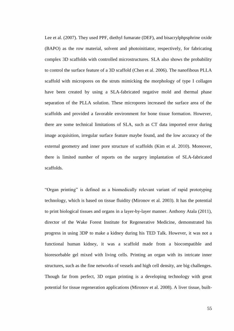
55
Lee et al. (2007). They used PPF, diethyl fumarate (DEF), and bisacrylphpsphrine oxide
(BAPO) as the row material, solvent and photoinitiator, respectively, for fabricating
complex 3D scaffolds with controlled microstructures. SLA also shows the probability
to control the surface feature of a 3D scaffold (Chen et al. 2006). The nanofibous PLLA
scaffold with micropores on the struts mimicking the morphology of type I collagen
have been created by using a SLA-fabricated negative mold and thermal phase
separation of the PLLA solution. These micropores increased the surface area of the
scaffolds and provided a favorable environment for bone tissue formation. However,
there are some technical limitations of SLA, such as CT data imported error during
image acquisition, irregular surface feature maybe found, and the low accuracy of the
external geometry and inner pore structure of scaffolds (Kim et al. 2010). Moreover,
there is limited number of reports on the surgery implantation of SLA-fabricated
scaffolds.
“Organ printing” is defined as a biomedically relevant variant of rapid prototyping
technology, which is based on tissue fluidity (Mironov et al. 2003). It has the potential
to print biological tissues and organs in a layer-by-layer manner. Anthony Atala (2011),
director of the Wake Forest Institute for Regenerative Medicine, demonstrated his
progress in using 3DP to make a kidney during his TED Talk. However, it was not a
functional human kidney, it was a scaffold made from a biocompatible and
bioresorbable gel mixed with living cells. Printing an organ with its intricate inner
structures, such as the fine networks of vessels and high cell density, are big challenges.
Though far from perfect, 3D organ printing is a developing technology with great
potential for tissue regeneration applications (Mironov et al. 2008). A liver tissue, built-
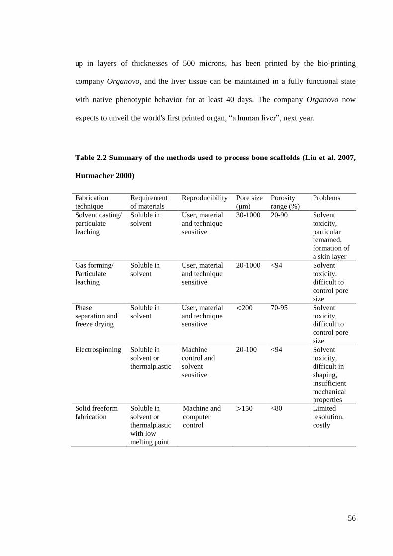
56
up in layers of thicknesses of 500 microns, has been printed by the bio-printing
company Organovo, and the liver tissue can be maintained in a fully functional state
with native phenotypic behavior for at least 40 days. The company Organovo now
expects to unveil the world's first printed organ, “a human liver”, next year.
Table 2.2 Summary of the methods used to process bone scaffolds (Liu et al. 2007,
Hutmacher 2000)
Fabrication
technique
Requirement
of materials
Reproducibility Pore size
(μm)
Porosity
range (%)
Problems
Solvent casting/
particulate
leaching
Soluble in
solvent
User, material
and technique
sensitive
30-1000 20-90 Solvent
toxicity,
particular
remained,
formation of
a skin layer
Gas forming/
Particulate
leaching
Soluble in
solvent
User, material
and technique
sensitive
20-1000 <94 Solvent
toxicity,
difficult to
control pore
size
Phase
separation and
freeze drying
Soluble in
solvent
User, material
and technique
sensitive
<200 70-95 Solvent
toxicity,
difficult to
control pore
size
Electrospinning Soluble in
solvent or
thermalplastic
Machine
control and
solvent
sensitive
20-100 <94 Solvent
toxicity,
difficult in
shaping,
insufficient
mechanical
properties
Solid freeform
fabrication
Soluble in
solvent or
thermalplastic
with low
melting point
Machine and
computer
control
>150 <80 Limited
resolution,
costly
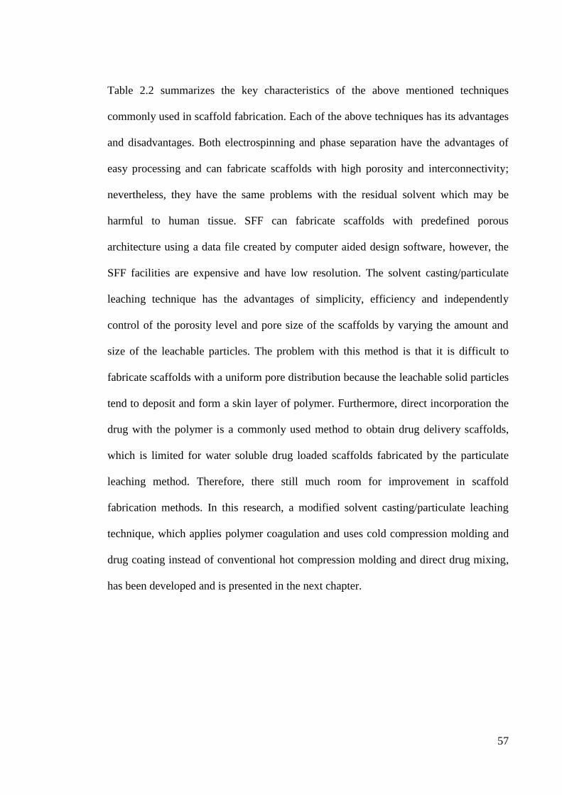
57
Table 2.2 summarizes the key characteristics of the above mentioned techniques
commonly used in scaffold fabrication. Each of the above techniques has its advantages
and disadvantages. Both electrospinning and phase separation have the advantages of
easy processing and can fabricate scaffolds with high porosity and interconnectivity;
nevertheless, they have the same problems with the residual solvent which may be
harmful to human tissue. SFF can fabricate scaffolds with predefined porous
architecture using a data file created by computer aided design software, however, the
SFF facilities are expensive and have low resolution. The solvent casting/particulate
leaching technique has the advantages of simplicity, efficiency and independently
control of the porosity level and pore size of the scaffolds by varying the amount and
size of the leachable particles. The problem with this method is that it is difficult to
fabricate scaffolds with a uniform pore distribution because the leachable solid particles
tend to deposit and form a skin layer of polymer. Furthermore, direct incorporation the
drug with the polymer is a commonly used method to obtain drug delivery scaffolds,
which is limited for water soluble drug loaded scaffolds fabricated by the particulate
leaching method. Therefore, there still much room for improvement in scaffold
fabrication methods. In this research, a modified solvent casting/particulate leaching
technique, which applies polymer coagulation and uses cold compression molding and
drug coating instead of conventional hot compression molding and direct drug mixing,
has been developed and is presented in the next chapter.
Table
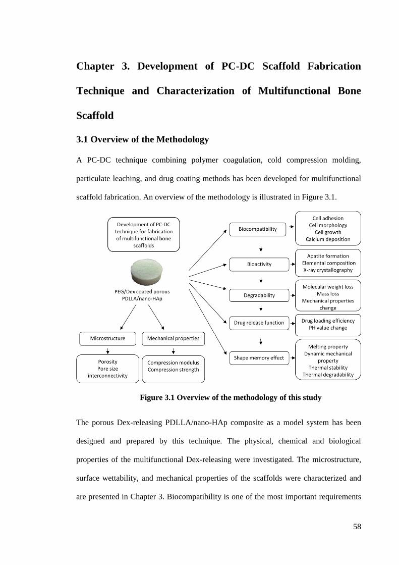
58
Chapter 3. Development of PC-DC Scaffold Fabrication
Technique and Characterization of Multifunctional Bone
Scaffold
3.1 Overview of the Methodology
A PC-DC technique combining polymer coagulation, cold compression molding,
particulate leaching, and drug coating methods has been developed for multifunctional
scaffold fabrication. An overview of the methodology is illustrated in Figure 3.1.
Figure 3.1 Overview of the methodology of this study
The porous Dex-releasing PDLLA/nano-HAp composite as a model system has been
designed and prepared by this technique. The physical, chemical and biological
properties of the multifunctional Dex-releasing were investigated. The microstructure,
surface wettability, and mechanical properties of the scaffolds were characterized and
are presented in Chapter 3. Biocompatibility is one of the most important requirements
59
for a scaffold. The ability of these scaffolds to support the osteoblast-like cell (MG63)
adhesion, proliferation and phenotypic expression was evaluated and is described in
Chapter 4. The bioactivity and degradability of these scaffolds were examined through
assessment of apatite deposition, molecular weight change, mass loss, and mechanical
properties change during incubation in simulated body fluids. Moreover, the drug
release behavior of the scaffolds was also investigated as presented in Chapter 5. The
shape memory effect was also studied and is described in Chapter 6. The effects of the
fabrication conditions, nano-HAp fractions and porosity values of the composites on
these properties were also investigated.
3.2 Introduction
Biodegradable polymers have been fabricated into scaffolds for various tissue
regeneration because they can be gradually degraded and replaced by the newly
regenerated tissues which can avoid secondary surgery to remove the implants after
tissue recovery or when the drug supply is depleted (Liu et al. 2007, Biondi et al. 2008).
Drug delivery technology has been attracting increasing interest in recent years for
improving human lives. Such delivery systems utilize biodegradable materials as the
drug vehicle to deliver biologically active molecules at a desired behavior, so as to
improve the effectiveness of the drug therapy and to reduce toxicity (Langer 1998).
Shape memory polymers (SMPs) have been considered as an intelligent material with
potential applications in implants for minimally invasive surgery since they can be
precisely positioned in the body in a temporary small shape and attain their application-
relevant shape after implantation which can highly reduce the risk of surgery.
Implantation of multifunctional porous scaffolds, with the function of biodegradability,
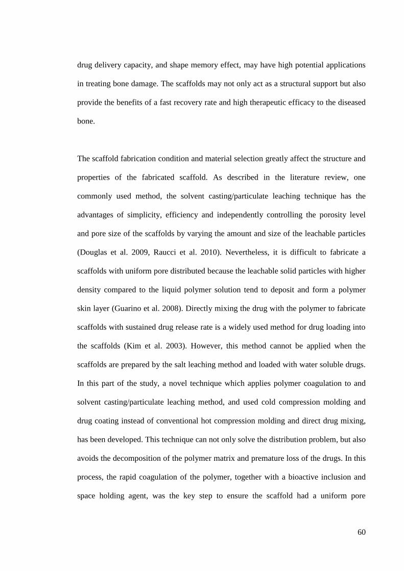
60
drug delivery capacity, and shape memory effect, may have high potential applications
in treating bone damage. The scaffolds may not only act as a structural support but also
provide the benefits of a fast recovery rate and high therapeutic efficacy to the diseased
bone.
The scaffold fabrication condition and material selection greatly affect the structure and
properties of the fabricated scaffold. As described in the literature review, one
commonly used method, the solvent casting/particulate leaching technique has the
advantages of simplicity, efficiency and independently controlling the porosity level
and pore size of the scaffolds by varying the amount and size of the leachable particles
(Douglas et al. 2009, Raucci et al. 2010). Nevertheless, it is difficult to fabricate a
scaffolds with uniform pore distributed because the leachable solid particles with higher
density compared to the liquid polymer solution tend to deposit and form a polymer
skin layer (Guarino et al. 2008). Directly mixing the drug with the polymer to fabricate
scaffolds with sustained drug release rate is a widely used method for drug loading into
the scaffolds (Kim et al. 2003). However, this method cannot be applied when the
scaffolds are prepared by the salt leaching method and loaded with water soluble drugs.
In this part of the study, a novel technique which applies polymer coagulation to and
solvent casting/particulate leaching method, and used cold compression molding and
drug coating instead of conventional hot compression molding and direct drug mixing,
has been developed. This technique can not only solve the distribution problem, but also
avoids the decomposition of the polymer matrix and premature loss of the drugs. In this
process, the rapid coagulation of the polymer, together with a bioactive inclusion and
space holding agent, was the key step to ensure the scaffold had a uniform pore
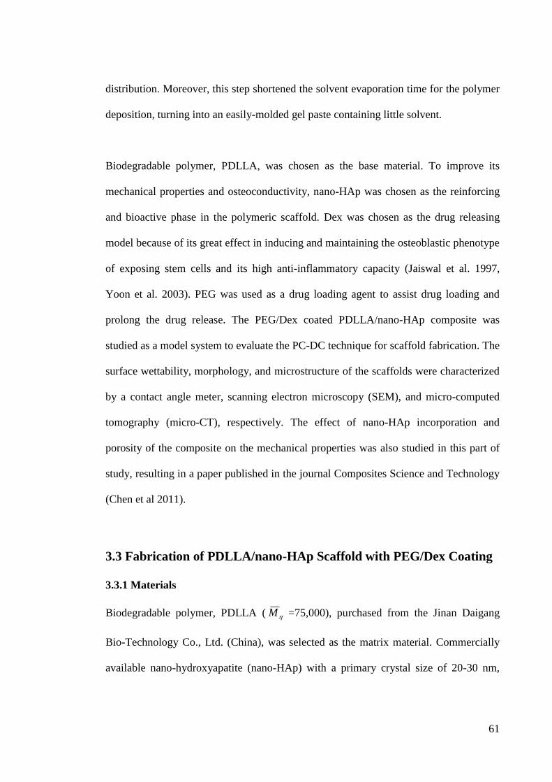
61
distribution. Moreover, this step shortened the solvent evaporation time for the polymer
deposition, turning into an easily-molded gel paste containing little solvent.
Biodegradable polymer, PDLLA, was chosen as the base material. To improve its
mechanical properties and osteoconductivity, nano-HAp was chosen as the reinforcing
and bioactive phase in the polymeric scaffold. Dex was chosen as the drug releasing
model because of its great effect in inducing and maintaining the osteoblastic phenotype
of exposing stem cells and its high anti-inflammatory capacity (Jaiswal et al. 1997,
Yoon et al. 2003). PEG was used as a drug loading agent to assist drug loading and
prolong the drug release. The PEG/Dex coated PDLLA/nano-HAp composite was
studied as a model system to evaluate the PC-DC technique for scaffold fabrication. The
surface wettability, morphology, and microstructure of the scaffolds were characterized
by a contact angle meter, scanning electron microscopy (SEM), and micro-computed
tomography (micro-CT), respectively. The effect of nano-HAp incorporation and
porosity of the composite on the mechanical properties was also studied in this part of
study, resulting in a paper published in the journal Composites Science and Technology
(Chen et al 2011).
3.3 Fabrication of PDLLA/nano-HAp Scaffold with PEG/Dex Coating
3.3.1 Materials
Biodegradable polymer, PDLLA ( M =75,000), purchased from the Jinan Daigang
Bio-Technology Co., Ltd. (China), was selected as the matrix material. Commercially
available nano-hydroxyapatite (nano-HAp) with a primary crystal size of 20-30 nm,
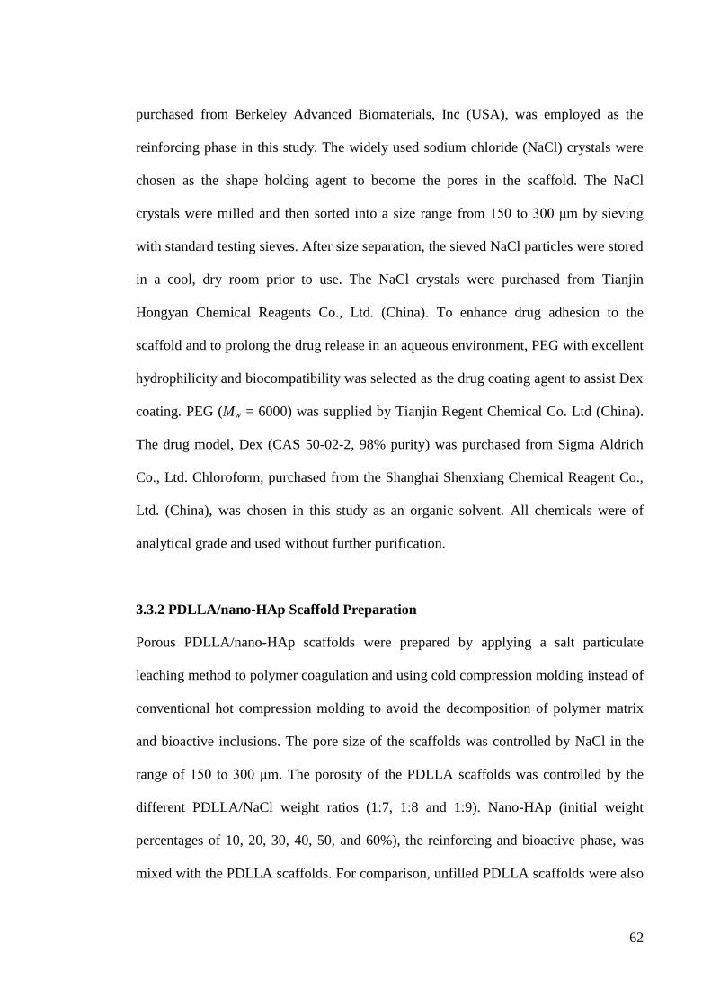
62
purchased from Berkeley Advanced Biomaterials, Inc (USA), was employed as the
reinforcing phase in this study. The widely used sodium chloride (NaCl) crystals were
chosen as the shape holding agent to become the pores in the scaffold. The NaCl
crystals were milled and then sorted into a size range from 150 to 300 μm by sieving
with standard testing sieves. After size separation, the sieved NaCl particles were stored
in a cool, dry room prior to use. The NaCl crystals were purchased from Tianjin
Hongyan Chemical Reagents Co., Ltd. (China). To enhance drug adhesion to the
scaffold and to prolong the drug release in an aqueous environment, PEG with excellent
hydrophilicity and biocompatibility was selected as the drug coating agent to assist Dex
coating. PEG (Mw = 6000) was supplied by Tianjin Regent Chemical Co. Ltd (China).
The drug model, Dex (CAS 50-02-2, 98% purity) was purchased from Sigma Aldrich
Co., Ltd. Chloroform, purchased from the Shanghai Shenxiang Chemical Reagent Co.,
Ltd. (China), was chosen in this study as an organic solvent. All chemicals were of
analytical grade and used without further purification.
3.3.2 PDLLA/nano-HAp Scaffold Preparation
Porous PDLLA/nano-HAp scaffolds were prepared by applying a salt particulate
leaching method to polymer coagulation and using cold compression molding instead of
conventional hot compression molding to avoid the decomposition of polymer matrix
and bioactive inclusions. The pore size of the scaffolds was controlled by NaCl in the
range of 150 to 300 μm. The porosity of the PDLLA scaffolds was controlled by the
different PDLLA/NaCl weight ratios (1:7, 1:8 and 1:9). Nano-HAp (initial weight
percentages of 10, 20, 30, 40, 50, and 60%), the reinforcing and bioactive phase, was
mixed with the PDLLA scaffolds. For comparison, unfilled PDLLA scaffolds were also
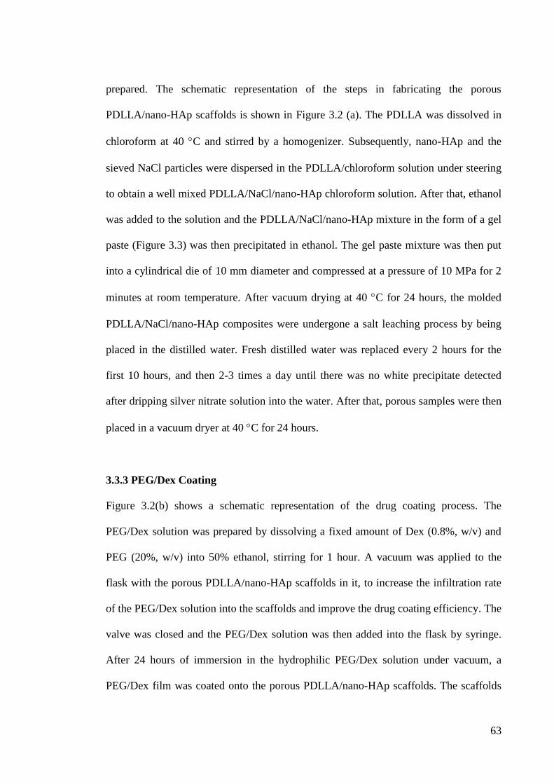
63
prepared. The schematic representation of the steps in fabricating the porous
PDLLA/nano-HAp scaffolds is shown in Figure 3.2 (a). The PDLLA was dissolved in
chloroform at 40 C and stirred by a homogenizer. Subsequently, nano-HAp and the
sieved NaCl particles were dispersed in the PDLLA/chloroform solution under steering
to obtain a well mixed PDLLA/NaCl/nano-HAp chloroform solution. After that, ethanol
was added to the solution and the PDLLA/NaCl/nano-HAp mixture in the form of a gel
paste (Figure 3.3) was then precipitated in ethanol. The gel paste mixture was then put
into a cylindrical die of 10 mm diameter and compressed at a pressure of 10 MPa for 2
minutes at room temperature. After vacuum drying at 40 C for 24 hours, the molded
PDLLA/NaCl/nano-HAp composites were undergone a salt leaching process by being
placed in the distilled water. Fresh distilled water was replaced every 2 hours for the
first 10 hours, and then 2-3 times a day until there was no white precipitate detected
after dripping silver nitrate solution into the water. After that, porous samples were then
placed in a vacuum dryer at 40 C for 24 hours.
3.3.3 PEG/Dex Coating
Figure 3.2(b) shows a schematic representation of the drug coating process. The
PEG/Dex solution was prepared by dissolving a fixed amount of Dex (0.8%, w/v) and
PEG (20%, w/v) into 50% ethanol, stirring for 1 hour. A vacuum was applied to the
flask with the porous PDLLA/nano-HAp scaffolds in it, to increase the infiltration rate
of the PEG/Dex solution into the scaffolds and improve the drug coating efficiency. The
valve was closed and the PEG/Dex solution was then added into the flask by syringe.
After 24 hours of immersion in the hydrophilic PEG/Dex solution under vacuum, a
PEG/Dex film was coated onto the porous PDLLA/nano-HAp scaffolds. The scaffolds
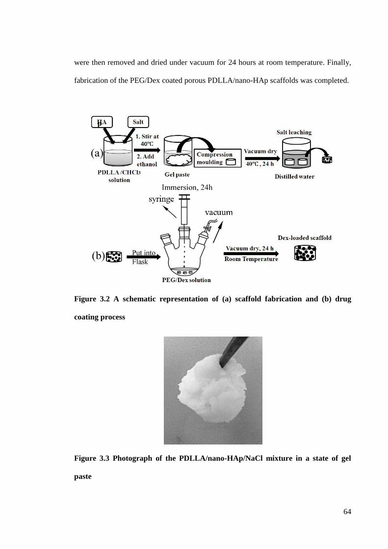
64
were then removed and dried under vacuum for 24 hours at room temperature. Finally,
fabrication of the PEG/Dex coated porous PDLLA/nano-HAp scaffolds was completed.
Figure 3.2 A schematic representation of (a) scaffold fabrication and (b) drug
coating process
Figure 3.3 Photograph of the PDLLA/nano-HAp/NaCl mixture in a state of gel
paste
p
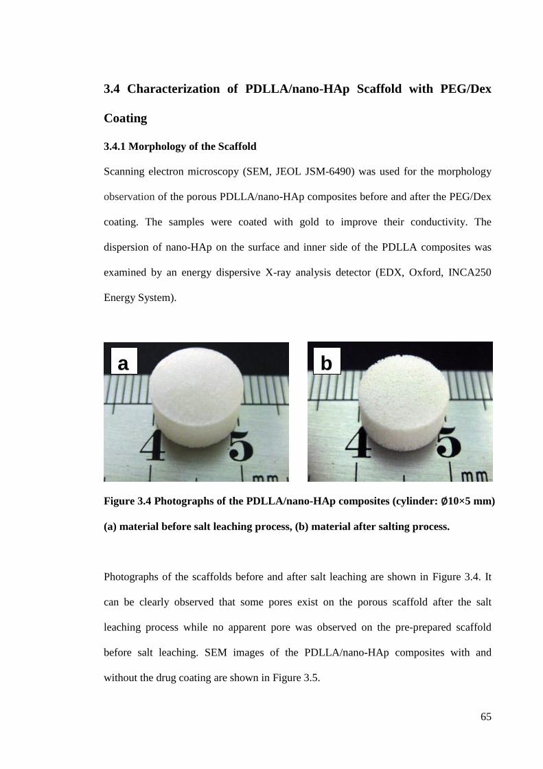
65
3.4 Characterization of PDLLA/nano-HAp Scaffold with PEG/Dex
Coating
3.4.1 Morphology of the Scaffold
Scanning electron microscopy (SEM, JEOL JSM-6490) was used for the morphology
observation of the porous PDLLA/nano-HAp composites before and after the PEG/Dex
coating. The samples were coated with gold to improve their conductivity. The
dispersion of nano-HAp on the surface and inner side of the PDLLA composites was
examined by an energy dispersive X-ray analysis detector (EDX, Oxford, INCA250
Energy System).
Figure 3.4 Photographs of the PDLLA/nano-HAp composites (cylinder: ∅10×5 mm)
(a) material before salt leaching process, (b) material after salting process.
Photographs of the scaffolds before and after salt leaching are shown in Figure 3.4. It
can be clearly observed that some pores exist on the porous scaffold after the salt
leaching process while no apparent pore was observed on the pre-prepared scaffold
before salt leaching. SEM images of the PDLLA/nano-HAp composites with and
without the drug coating are shown in Figure 3.5.
b a
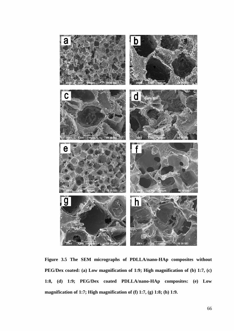
66
Figure 3.5 The SEM micrographs of PDLLA/nano-HAp composites without
PEG/Dex coated: (a) Low magnification of 1:9; High magnification of (b) 1:7, (c)
1:8, (d) 1:9; PEG/Dex coated PDLLA/nano-HAp composites: (e) Low
magnification of 1:7; High magnification of (f) 1:7, (g) 1:8; (h) 1:9.
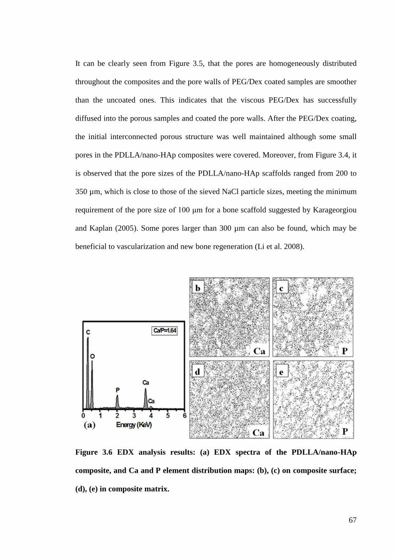
67
It can be clearly seen from Figure 3.5, that the pores are homogeneously distributed
throughout the composites and the pore walls of PEG/Dex coated samples are smoother
than the uncoated ones. This indicates that the viscous PEG/Dex has successfully
diffused into the porous samples and coated the pore walls. After the PEG/Dex coating,
the initial interconnected porous structure was well maintained although some small
pores in the PDLLA/nano-HAp composites were covered. Moreover, from Figure 3.4, it
is observed that the pore sizes of the PDLLA/nano-HAp scaffolds ranged from 200 to
350 µm, which is close to those of the sieved NaCl particle sizes, meeting the minimum
requirement of the pore size of 100 μm for a bone scaffold suggested by Karageorgiou
and Kaplan (2005). Some pores larger than 300 µm can also be found, which may be
beneficial to vascularization and new bone regeneration (Li et al. 2008).
Figure 3.6 EDX analysis results: (a) EDX spectra of the PDLLA/nano-HAp
composite, and Ca and P element distribution maps: (b), (c) on composite surface;
(d), (e) in composite matrix.
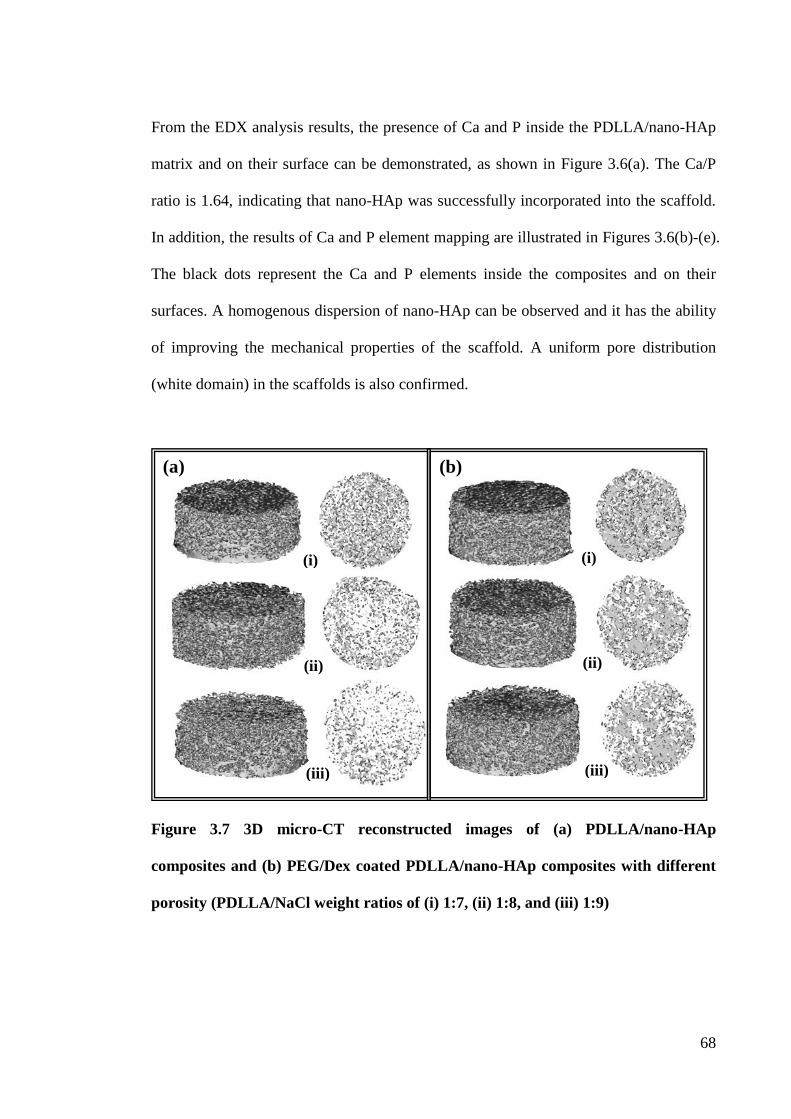
68
From the EDX analysis results, the presence of Ca and P inside the PDLLA/nano-HAp
matrix and on their surface can be demonstrated, as shown in Figure 3.6(a). The Ca/P
ratio is 1.64, indicating that nano-HAp was successfully incorporated into the scaffold.
In addition, the results of Ca and P element mapping are illustrated in Figures 3.6(b)-(e).
The black dots represent the Ca and P elements inside the composites and on their
surfaces. A homogenous dispersion of nano-HAp can be observed and it has the ability
of improving the mechanical properties of the scaffold. A uniform pore distribution
(white domain) in the scaffolds is also confirmed.
Figure 3.7 3D micro-CT reconstructed images of (a) PDLLA/nano-HAp
composites and (b) PEG/Dex coated PDLLA/nano-HAp composites with different
porosity (PDLLA/NaCl weight ratios of (i) 1:7, (ii) 1:8, and (iii) 1:9)
(i) (i)
(ii)
(iii)
(ii)
(iii)
(a) (b)
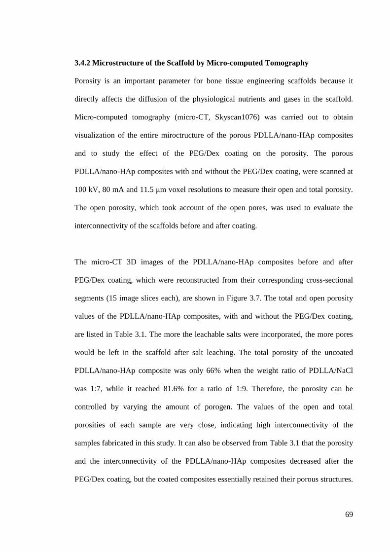
69
3.4.2 Microstructure of the Scaffold by Micro-computed Tomography
Porosity is an important parameter for bone tissue engineering scaffolds because it
directly affects the diffusion of the physiological nutrients and gases in the scaffold.
Micro-computed tomography (micro-CT, Skyscan1076) was carried out to obtain
visualization of the entire miroctructure of the porous PDLLA/nano-HAp composites
and to study the effect of the PEG/Dex coating on the porosity. The porous
PDLLA/nano-HAp composites with and without the PEG/Dex coating, were scanned at
100 kV, 80 mA and 11.5 μm voxel resolutions to measure their open and total porosity.
The open porosity, which took account of the open pores, was used to evaluate the
interconnectivity of the scaffolds before and after coating.
The micro-CT 3D images of the PDLLA/nano-HAp composites before and after
PEG/Dex coating, which were reconstructed from their corresponding cross-sectional
segments (15 image slices each), are shown in Figure 3.7. The total and open porosity
values of the PDLLA/nano-HAp composites, with and without the PEG/Dex coating,
are listed in Table 3.1. The more the leachable salts were incorporated, the more pores
would be left in the scaffold after salt leaching. The total porosity of the uncoated
PDLLA/nano-HAp composite was only 66% when the weight ratio of PDLLA/NaCl
was 1:7, while it reached 81.6% for a ratio of 1:9. Therefore, the porosity can be
controlled by varying the amount of porogen. The values of the open and total
porosities of each sample are very close, indicating high interconnectivity of the
samples fabricated in this study. It can also be observed from Table 3.1 that the porosity
and the interconnectivity of the PDLLA/nano-HAp composites decreased after the
PEG/Dex coating, but the coated composites essentially retained their porous structures.
70
This implies that the porosity and pore size can be independently controlled by varying
the amount and size of the NaCl particles when using the particulate leaching method.
The microstructure of the scaffolds may promote cell growth, migration and the
transport of nutrients and oxygen, as well as the removal of metabolic waste when
implanted for bone regeneration (Hutmacher 2000).
Table 3.1 Total and open porosity of PDLLA/nano-HAp composites before and
after PEG/Dex coating.
Before PEG/Dex coating After PEG/Dex coating
Total
porosity (%)
Open
porosity (%)
Total
porosity (%)
Open
porosity (%)
1:7 + 20 wt% nano-HAp 66.01 66.00 51.48 51.21
1:8 + 20 wt% nano-HAp 77.02 77.01 58.04 57.94
1:9 + 20 wt% nano-HAp 81.58 81.57 61.51 61.44
3.4.3 Wettability of the Scaffold
Surface hydrophilicity is one of the important aspects that affect the biocompatibility of
the materials in a biological environment (Xu & Wu 2009). Water contact angle
measurement was conducted to evaluate the surface hydrophilicity of the unfilled
PDLLA and the PDLLA/nano-HAp composites, with and without the PEG/Dex coating.
The water-in-air contact angle of the samples was measured by a digital contact angle
meter (Digidrop, Model R&D, GBX Company). The sample was placed onto the work
stage of the meter. The contact angle was measured within 10 seconds after a droplet of
distilled water (5μl) contacted the sample surface.
Porosity
(%) Scaffolds
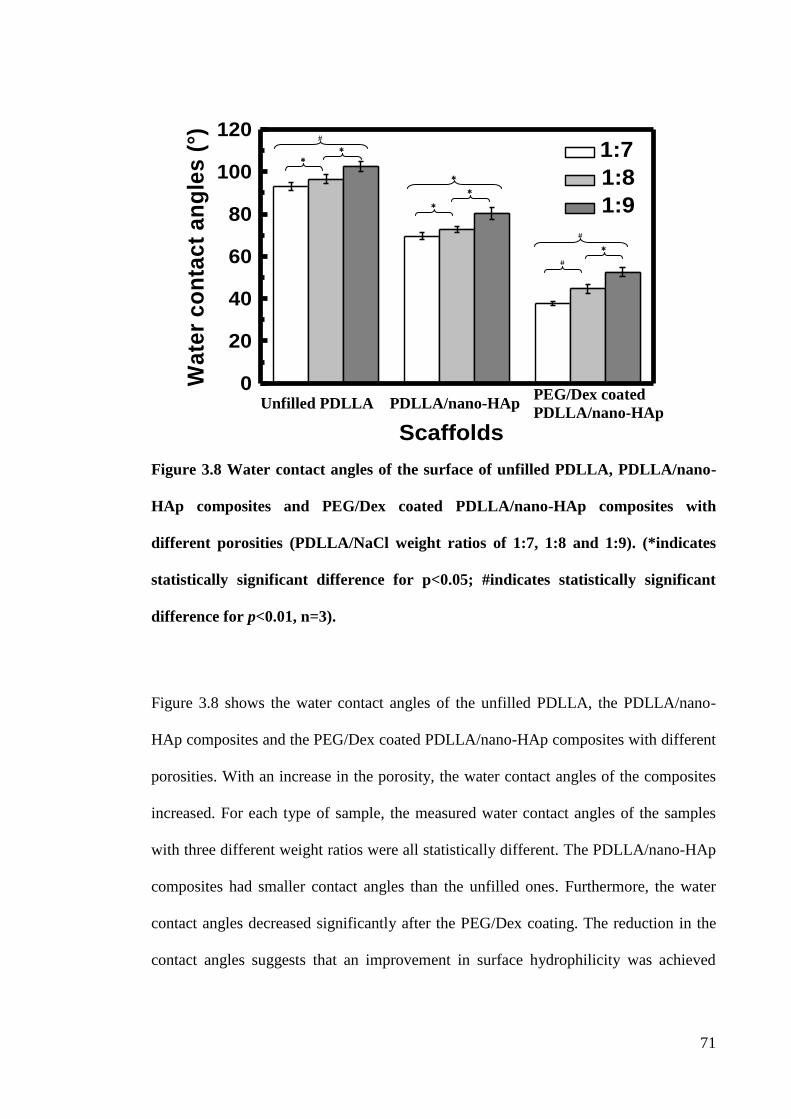
71
Figure 3.8 Water contact angles of the surface of unfilled PDLLA, PDLLA/nano-
HAp composites and PEG/Dex coated PDLLA/nano-HAp composites with
different porosities (PDLLA/NaCl weight ratios of 1:7, 1:8 and 1:9). (*indicates
statistically significant difference for p<0.05; #indicates statistically significant
difference for p<0.01, n=3).
Figure 3.8 shows the water contact angles of the unfilled PDLLA, the PDLLA/nano-
HAp composites and the PEG/Dex coated PDLLA/nano-HAp composites with different
porosities. With an increase in the porosity, the water contact angles of the composites
increased. For each type of sample, the measured water contact angles of the samples
with three different weight ratios were all statistically different. The PDLLA/nano-HAp
composites had smaller contact angles than the unfilled ones. Furthermore, the water
contact angles decreased significantly after the PEG/Dex coating. The reduction in the
contact angles suggests that an improvement in surface hydrophilicity was achieved
PDLLA PDLLA+HAp PDLLA+HAp+PEG
0
20
40
60
80
100
120
Wa
ter
co
nta
ct
an
gle
s (
°)
1:7
1:8
1:9
Scaffolds
*
#
*
*
*
#
*
*
#
PEG/Dex coated
PDLLA/nano-HAp
Unfilled PDLLA PDLLA/nano-HAp
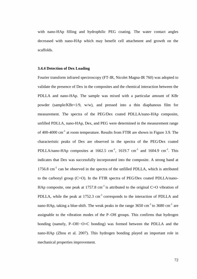
72
with nano-HAp filling and hydrophilic PEG coating. The water contact angles
decreased with nano-HAp which may benefit cell attachment and growth on the
scaffolds.
3.4.4 Detection of Dex Loading
Fourier transform infrared spectroscopy (FT-IR, Nicolet Magna-IR 760) was adopted to
validate the presence of Dex in the composites and the chemical interaction between the
PDLLA and nano-HAp. The sample was mixed with a particular amount of KBr
powder (sample/KBr=1/9, w/w), and pressed into a thin diaphanous film for
measurement. The spectra of the PEG/Dex coated PDLLA/nano-HAp composite,
unfilled PDLLA, nano-HAp, Dex, and PEG were determined in the measurement range
of 400-4000 cm-1
at room temperature. Results from FTIR are shown in Figure 3.9. The
characteristic peaks of Dex are observed in the spectra of the PEG/Dex coated
PDLLA/nano-HAp composites at 1662.5 cm-1
, 1619.7 cm-1
and 1604.9 cm-1
. This
indicates that Dex was successfully incorporated into the composite. A strong band at
1756.8 cm-1
can be observed in the spectra of the unfilled PDLLA, which is attributed
to the carbonyl group (C=O). In the FTIR spectra of PEG/Dex coated PDLLA/nano-
HAp composite, one peak at 1757.8 cm-1
is attributed to the original C=O vibration of
PDLLA, while the peak at 1752.3 cm-1
corresponds to the interaction of PDLLA and
nano-HAp, taking a blue-shift. The weak peaks in the range 3650 cm-1
to 3680 cm-1
are
assignable to the vibration modes of the P–OH groups. This confirms that hydrogen
bonding (namely, P–OH···O=C bonding) was formed between the PDLLA and the
nano-HAp (Zhou et al. 2007). This hydrogen bonding played an important role in
mechanical properties improvement.
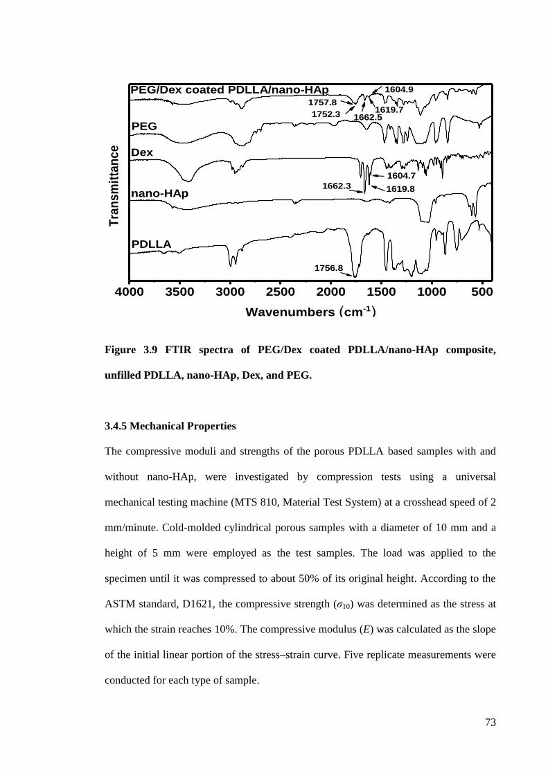
73
Figure 3.9 FTIR spectra of PEG/Dex coated PDLLA/nano-HAp composite,
unfilled PDLLA, nano-HAp, Dex, and PEG.
3.4.5 Mechanical Properties
The compressive moduli and strengths of the porous PDLLA based samples with and
without nano-HAp, were investigated by compression tests using a universal
mechanical testing machine (MTS 810, Material Test System) at a crosshead speed of 2
mm/minute. Cold-molded cylindrical porous samples with a diameter of 10 mm and a
height of 5 mm were employed as the test samples. The load was applied to the
specimen until it was compressed to about 50% of its original height. According to the
ASTM standard, D1621, the compressive strength (ζ10) was determined as the stress at
which the strain reaches 10%. The compressive modulus (E) was calculated as the slope
of the initial linear portion of the stress–strain curve. Five replicate measurements were
conducted for each type of sample.
4000 3500 3000 2500 2000 1500 1000 500
1604.9
1619.71662.51752.3
Tra
ns
mit
tan
ce
Wavenumbers (cm-1)
PEG/Dex coated PDLLA/nano-HAp
PEG
Dex
nano-HAp
PDLLA
1756.8
1757.8
1662.3
1604.7
1619.8
74
E and ζ10 of the samples are listed in Table 3.2. For the unfilled PDLLA, the
compressive modulus ranged from 25.3±1.2 MPa for a PDLLA/NaCl ratio of 1:9 to
64.8±2.7 MPa for a ratio of 1:7. The ζ10 ranged from 1.5±0.1 MPa to 2.9±0.2 MPa. This
is because the increase in the porosity of the unfilled PDLLA, which is associated with
increasing amounts of porogen, led to a decrease of E and ζ10. A balance can be reached
between porosity, pore size and mechanical properties. This phenomenon can be found
in the PDLLA/nano-HAp composites as well.
Table 3.2 Compressive modulus (E) and the compressive strength (σ10) of the
PEG/Dex coated porous PDLLA/nano-HAp composites and unfilled PDLLA.
Samples
(PDLLA/salt, w/w) E
(MPa)
σ10
(MPa)
1:7 64.8±2.7 2.9±0.2
1:8 56.1±2.7 1.8±0.1
1:9 25.3±1.2 1.5±0.1
1:7 + 20 wt% nano-HAp 97.9±2.3 3.1±0.1
1:8 + 20 wt% nano-HAp 78.4±1.7 3.0±0.1
1:9 + 20 wt% nano-HAp 38.7±2.5 2.4±0.3
The compressive moduli and compressive strengths of the PDLLA composites with the
addition of nano-HAp were increased by more than 50% and 20%, respectively, as
compared with the unfilled ones. This is because a closer packing of polymer chains
was formed when a high pressure applied to the PDLLA/nano-HAp composite in the
compression molding process (Teng et al. 2007). The enhancement of the mechanical
properties of the composite is likely due to the homogenous distribution of the nano-
HAp in the PDLLA and the hydrogen bonding formed between the nano-HAp and
PDLLA due to the high pressure during the compression molding (Zhou et al. 2007).
The mechanical properties of the 1:7 and 1:8 nano-HAp filled PDLLA scaffolds fell
75
within the normal range of modulus (50-250 MPa) and strength (1-10 MPa) values for
human cancellous bone (Lin et al. 2003). Therefore, the PEG/Dex coated PDLLA/nano-
HAp composites fabricated in this study may have the potential to be used in trabecular
bone regeneration.
3.5 Summary
Choosing an appropriate technique and biomaterials for scaffold fabrication is important
for the control of pore size and structure, drug release capacity, shape memory effect,
and mechanical properties of the scaffolds. In this part of study, a PC-DC technique was
developed. Multifunctional PEG/Dex coated porous PDLLA/nano-HAp composites as a
model system were successfully fabricated by this newly developed technique
combining coagulation, cold compression molding, salt leaching and drug coating. This
technique independently controlled the pore size and porosity of the scaffold by varying
the amount and size of the leachable particles (NaCl). The cold compression moulding
and drug coating method prevented decomposition of the polymer matrix and premature
loss of the drug. This technique reduced solvent residual by the polymer coagulation,
and particulate leaching/solvent casting steps. The gel-like polymer paste formed by
coagulation of the PDLLA in the non-solvent proved easy to mold. Moreover, applying
polymer coagulation to the solvent casting/particulate leaching method successfully
solved the non-uniform distribution of the pores within the scaffolds. A layer of viscous
PEG/Dex was observed on the pore walls of the scaffolds from the SEM images. The
FT-IR spectroscopy suggested that Dex was effectively loaded into the matrix.
PEG/Dex coating on the scaffold surface under vaccum is a promising way for drug
76
loading process. The technique is economical and can be applied to a wide range of
polymers and inorganic fillers and drugs for scaffold fabrication.
The pores were distributed homogeneously throughout the scaffolds and the pore sizes
were ranged from 200 to 350 µm. These pore sizes meet the requirement for a bone
scaffold and the porosities of the scaffolds ranged from 66% to 81.6%. PDLLA/nano-
HAp composite had higher compressive moduli and strengths than the unfilled ones.
The significant improvement of the wettability of the PDLLA composites was achieved
by the nano-HAp incorporating and the PEG/Dex film coating. The scaffolds may
promote cell growth, migration and the transport of nutrients and oxygen, as well as the
removal of metabolic waste when implanted for bone regeneration. In this chapter, the
PC-DC technique and basic characteristics of the composites was presented. The
biocompatibility is the most important requirement for a bone scaffold, the next step
was to investigate the interaction between the cell and the fabricated scaffolds, and the
results are presented in Chapter 4.
77
Chapter 4. In Vitro Human Osteoblast-like Cell Response to
the PDLLA/nano-HAp Composites
4.1 Introduction
Autograft is considered as the golden standard treatment for large bone defects, which
harvests the „donor‟ bone from a non-load-bearing site in the patient‟s body. It can
provide three essential element of bone regeneration: osteoconduction, osteoinduction,
and osteogenic properties. Nevertheless, autologous transplantation is severely
hampered by the limited amount of autograft and the associated complications at the
donor site (Giannoudis et al. 2005). Implanting bone scaffolds made from biomaterials
provides an alternative approach for bone defect repair and regeneration, avoiding the
limitations of autologous bone graft (Chen et al. 2002). A multifunctional bone graft
substitute/scaffold derived from biomedical materials with controllable structure,
mechanical and bioactive drug release kinetic for cell growth and tissue regeneration is
critical in bone tissue engineering. Design and fabrication of biocompatible scaffolds
mimicking the structural and biological functions of the natural extracellular matrix
(ECM) which provides cells with an optimized microenvironment is one of the major
challenges (Ma 2008). Understanding the cell-ECM interaction mechanism becomes
one of the focuses in the tissue engineering field, because it can provide valuable
information including the effects of the surface composition, porosity, pore size,
topography of the ECM on the cellular behavior, for fabricating a scaffold promoting
the cell adhesion, growth, differentiation and later bone regeneration (Van Dijk et al.
2013, Dicesare et al. 2013).
78
The ability of the scaffold to support cell adhesion, growth and differentiation highly
determines the degree of success of tissue engineering. Cultures of osteoblast-like cells
in this scaffold in vitro is a promising approach to investigate the biological activity of a
scaffold used as a cell carrier. In this part of the study, PDLLA/nano-HAp composites
were fabricated using the recently developed PC-DC technique (Chen et al. 2011) for in
vitro evaluation of osteoblast cellular responses. PDLLA/nano-HAp composites can be
considered as one type of ECM which may have important influences on cell behavior.
Therefore, in addition to the evaluation of microstructure, wettability, and mechanical
properties of the composites, the in vitro cell interaction with composites was
investigated and presented in this chapter. The potential application of the fabricated
composite scaffolds is for bone tissue engineering, thus osteoblasts-like cells, MG63,
was chosen in this research. MG63 is an osteosarcoma cell line with an osteoblastic
phenotype and has been widely used in research in bone tissue engineering (Verrier et al.
2004, Liu et al. 2004, Wang et al. 2003). To the best of my knowledge, this was the first
study of cellular response on PDLLA/nano-HAp composites using the MG63 cell
model.
Four types of PDLLA/nano-HAp composites were fabricated: unfilled PDLLA with
polymer/porogen ratios of 1:7 and 1:9 were used to study the effect of porosity on the
cellular response; unfilled 1:9 PDLLA and PDLLA with 50 wt% nano-HAp were
chosen to investigate the effect of nano-HAp incorporation; how the PEG/Dex coating
affects the cell behavior was also explored. 3-(4,5-dimethylthiazol-2-yl)-2,5-
diphenyltetrazolium bromide (MTT) assay, alkaline phosphatase activity (ALP) assay,
scanning electron microscope (SEM), Alizarin Red staining (ARS) and fluorescence
79
staining were adopted to investigate the interactions between the MG63 cells and the
samples. For comparison, unfilled PDLLA film fabricated by the solvent casting
method was also investigated. The results showed that the PDLLA based scaffolds
fabricated in this study are biocompatible and facilitate cell attachment and proliferation.
The pore size of the fabricated scaffolds ranged from 200 to 350 µm and the high
porosity of 60% to 80% allowed cells to host and penetrate inside the scaffolds. The
whole scaffolds covered by the MG63 cells were observed after a 7-day culture, in
which the number of cells on the PDLLA based substrates increased. Moreover,
scaffolds with higher porosity values were beneficial for cell attachment, growth,
migration and differentiation. Nano-HAp incorporation promoting calcium deposition
(cell mineralization/osteoconduction) was also determined in this part of study.
4.2 Cell Culture
The human osteoblast-like cell, the MG63 cell, is an osteosarcoma cell isolated from a
relative uncommon tumor. Though these cells possess abnormal growth characteristics,
MG63 cells express typical osteoblast phenotypes, such as osteoblastic genes,
synthesize bone matrix protein, and expression of ALP (Gartland et al. 2005). They
became an extremely useful osteoblast cell model in bone regeneration research
(Verrier et al. 2004, Srinivasan et al. 2012). In this study, MG63 cells (American Type
Culture Collection, ATCC, USA) were chosen and seeded onto 3D porous PDLLA
based scaffolds to study the effect of porosity, nano-HAp incorporation and PEG/Dex
coating on the response of cultured cells. The MG63 cells were cultured in a tissue
culture polystyrene (PS) flask (75 cm2) at 37 C in an incubator with 5% CO2 in an air
atmosphere. The optical microscope images of the MG63 cells well attached on the
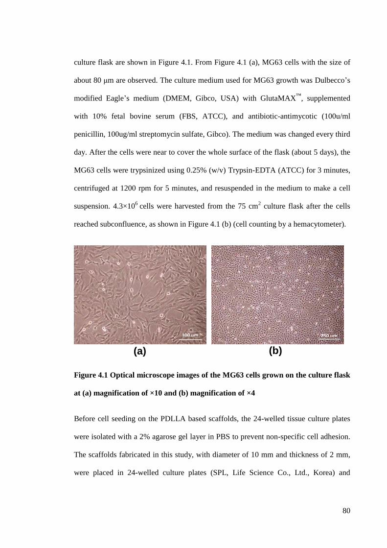
80
culture flask are shown in Figure 4.1. From Figure 4.1 (a), MG63 cells with the size of
about 80 μm are observed. The culture medium used for MG63 growth was Dulbecco‟s
modified Eagle‟s medium (DMEM, Gibco, USA) with GlutaMAX™
, supplemented
with 10% fetal bovine serum (FBS, ATCC), and antibiotic-antimycotic (100u/ml
penicillin, 100ug/ml streptomycin sulfate, Gibco). The medium was changed every third
day. After the cells were near to cover the whole surface of the flask (about 5 days), the
MG63 cells were trypsinized using 0.25% (w/v) Trypsin-EDTA (ATCC) for 3 minutes,
centrifuged at 1200 rpm for 5 minutes, and resuspended in the medium to make a cell
suspension. 4.3×10⁶
cells were harvested from the 75 cm2 culture flask after the cells
reached subconfluence, as shown in Figure 4.1 (b) (cell counting by a hemacytometer).
Figure 4.1 Optical microscope images of the MG63 cells grown on the culture flask
at (a) magnification of ×10 and (b) magnification of ×4
Before cell seeding on the PDLLA based scaffolds, the 24-welled tissue culture plates
were isolated with a 2% agarose gel layer in PBS to prevent non-specific cell adhesion.
The scaffolds fabricated in this study, with diameter of 10 mm and thickness of 2 mm,
were placed in 24-welled culture plates (SPL, Life Science Co., Ltd., Korea) and
(a)
100 μm
(b)
250 μm
81
sterilized by soaking in 70% ethanol for 4 hours and the scaffolds were left in PBS
overnight, then exposed to ultraviolet (UV) radiation (2 hours for each side) for further
sterilization and pre-wetting. After that, the scaffolds were presoaked in a complete cell
culture medium for 1 day. A drop of 50 μl of the cell suspension containing certain cell
numbers was added to the top of the scaffold and allowed to be absorbed by the porous
scaffolds for 2 hours. The same number of cells were seeded on the unfilled PDLLA
film as the control. Following a 2-hour cell absorption, 1 ml of additional complete
medium was added to each well to cover the whole scaffold. Cells were further
incubated at 37 C in a humidified atmosphere with 5% CO2 for various periods of time
(1, 3, and 7 days) and the culture mediums were changed twice a week.
Experiments were run in triplicate for each sample and repeated three times. All data
were expressed as mean±standard deviation (SD). A one-way analysis of variance
(ANOVA) and the Fisher LSD Test for means comparison were used to assess the
significant differences. p values < 0.05 were considered statistically significant (n=3).
4.3 Characterization of the Scaffolds with Cells
4.3.1 Cell Adhesion
Cell adhesion is known as an important early cellular process which happens within 3
hours after cell seeding, and directly influences later cellular processes such as cell
proliferation, migration and even tissue regeneration (Anselme 2000, Causa et al.
2006)). Although the same volumes of cell suspensions were injected onto the scaffolds
surface and control films, only a fraction of the cells were left on the substrates while
the remainder adhered to the cell culture plate. Fluorescence staining was conducted for
82
observation and evaluation of cell adhesion on the PDLLA based materials. A number
of cells, about 10
4, were seeded on the specimens before staining. After a 4 hours
culture, the cells were gently washed with PBS three times and fixed in 4%
formaldehyde/PBS for 30 min, then permeabilized with 0.1% Triton X-100 in PBS for 5
min at room temperature. The scaffold with attached cells was incubated with 500 μm
of 1μg/ml DAPI (Sigma) in PBS. The cell nuclei (DAPI, blue) of the cells were
observed with a fluorescence microscope (Eclipse 80i, Nikon, Japan).
Figure 4.2 shows typical fluorescence microscopy images of the PDLLA based
substrates and control film after a 4 hours culture. The cell nuclei (light blue dots) were
stained with DAPI. The numbers of cells on the scaffold and film in the selected images
were analyzed by a computer programme (Image J) and are shown in Figure 4.2. The
MG63 cells attached to all the samples can be observed. Although good cell
attachments were found on all the samples, more cells adhered to the 1:9 PDLLA
scaffolds when compared to the 1:7 samples and the control films. This suggests that
the higher porosity of the scaffold, the larger the space and surface area for cell
migration and proliferation. For the PEG/Dex coated scaffold, the number of MG63
cells attached was the lowest. This is mainly due to the PEG coating having the ability
of rejecting protein adsorption (Kingshott & Griesser 1999). Cell adhesion is highly
dependent on the adsorption of adhesion proteins on the surface of the scaffolds that are
recognized by the integrin receptors on the cells (Horbett 2004). Therefore, cell
adhesion was disturbed by the less amount of protein adsorbed on the scaffold surface.
This can be improved by incorporation or adsorption of RGD or fibronectin to the
PEG/Dex coating (Jeschke et al. 2002).
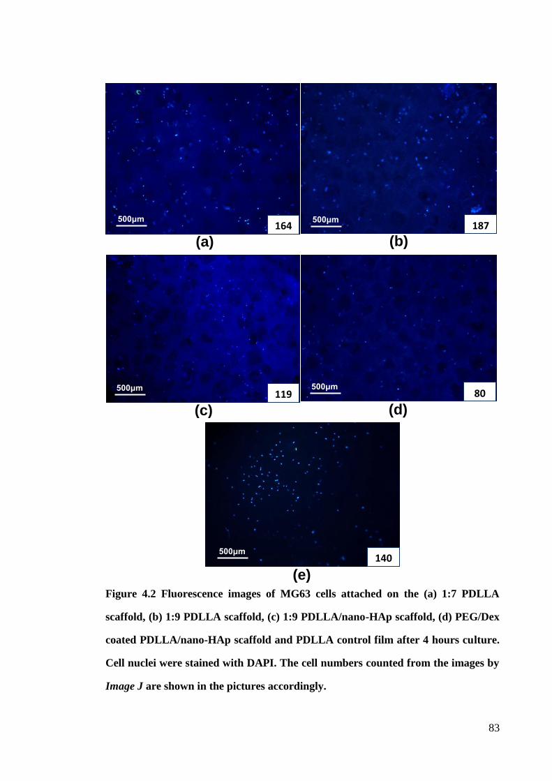
83
Figure 4.2 Fluorescence images of MG63 cells attached on the (a) 1:7 PDLLA
scaffold, (b) 1:9 PDLLA scaffold, (c) 1:9 PDLLA/nano-HAp scaffold, (d) PEG/Dex
coated PDLLA/nano-HAp scaffold and PDLLA control film after 4 hours culture.
Cell nuclei were stained with DAPI. The cell numbers counted from the images by
Image J are shown in the pictures accordingly.
(e)
140 500μm
(d)
80 500μm
(c)
119 500μm
(b) 187
500μm
(a) 164
500μm
84
4.3.2 MTT Test for Cell Proliferation
The proliferation of the cells was assessed quantitatively using the 3-(4,5-
dimethylthiazol-2-yl)-2,5-diphenyltetrazolium bromide (MTT, Sigma) assay. The MTT
assay relies on the ability of the viable cells to turn a yellow dye into a water-insoluble
purple formazan product. The formazan product can be dissolved in dimethyl sulfoxide
(DMSO) solution. The absorbance value of the DMSO solution in terms of optical
density (OD) is proportional to the cell number on the substrate. The cell number was
2×10⁴ for each sample. 5mg/ml of MTT in PBS solution was prepared and diluted by
DMEM solution without FBS (1:10 v/v). For each time interval (1, 3 and 7 days), the
cell seeded samples were washed by PBS twice. 1 ml of MTT DMEM solution was
added to each culture well and incubated at 37 C and 5% CO2 for 4 hours. The
supernatant of each well was then removed and 1ml of DMSO (Damao Chemical
Reagents Co. Ltd., China) was added to dissolve the formazon. The absorbance value of
the DMSO solution was measured at a wavelength of 570 nm with a microplate reader
(Infinite F200, TECAN). Three samples of each kind were tested for each incubation
period, and each test was performed three times. Background absorbance was obtained
by incubation of the MTT substrate on the sample without cell seeded and subtracted
from the results.
The results of MTT testing are shown in Figure 4.3. It is seen that the cell number on all
the substrates and control film increased with culture time, suggesting good
biocompatibility of PDLLA scaffolds fabricated in this study. Moreover, all the samples
showed a higher cell activity than the control film. This is because the scaffolds can
provide greater surface area for cell attachment and growth. More cells on the 1:9
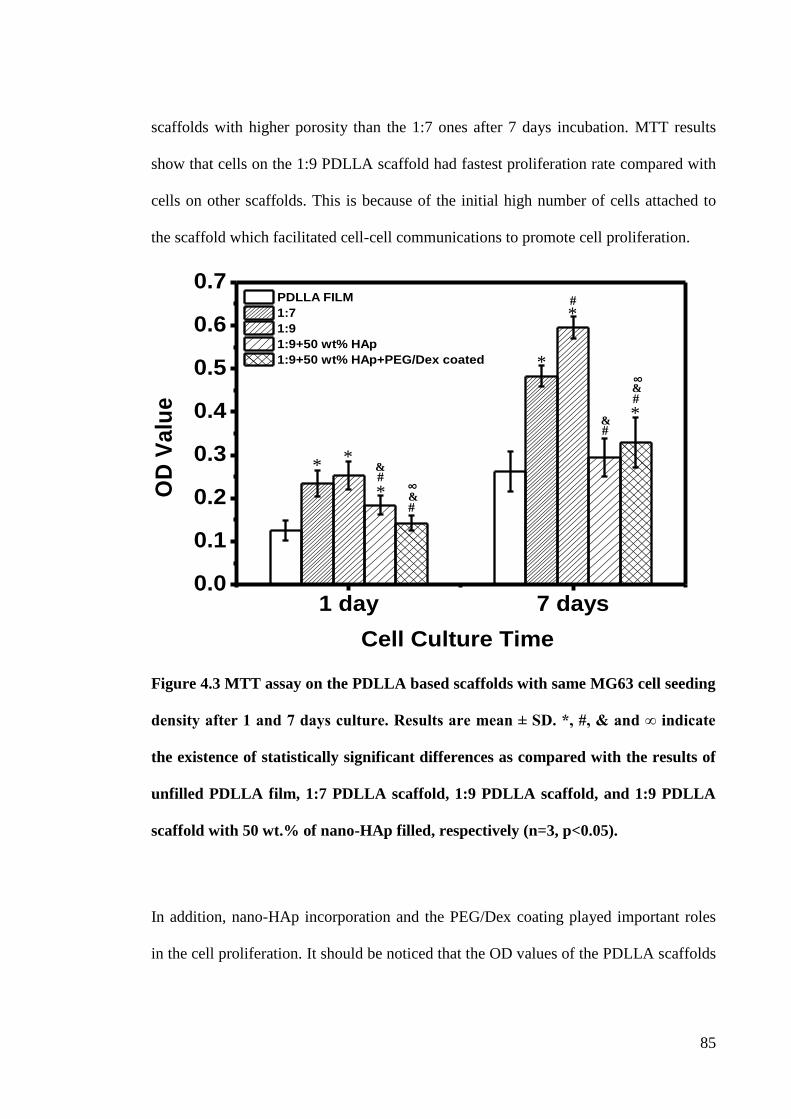
85
scaffolds with higher porosity than the 1:7 ones after 7 days incubation. MTT results
show that cells on the 1:9 PDLLA scaffold had fastest proliferation rate compared with
cells on other scaffolds. This is because of the initial high number of cells attached to
the scaffold which facilitated cell-cell communications to promote cell proliferation.
Figure 4.3 MTT assay on the PDLLA based scaffolds with same MG63 cell seeding
density after 1 and 7 days culture. Results are mean ± SD. *, #, & and ∞ indicate
the existence of statistically significant differences as compared with the results of
unfilled PDLLA film, 1:7 PDLLA scaffold, 1:9 PDLLA scaffold, and 1:9 PDLLA
scaffold with 50 wt.% of nano-HAp filled, respectively (n=3, p<0.05).
In addition, nano-HAp incorporation and the PEG/Dex coating played important roles
in the cell proliferation. It should be noticed that the OD values of the PDLLA scaffolds
1 day 7 days0.0
0.1
0.2
0.3
0.4
0.5
0.6
0.7
OD
Va
lue
Cell Culture Time
PDLLA FILM
1:7
1:9
1:9+50 wt% HAp
1:9+50 wt% HAp+PEG/Dex coated
* *
* #
# &
&
∞
§
*
* #
* #
&
∞
§
#
&
86
with 50 wt% of nano-HAp fill decreased when compared with the unfilled ones. One
reason may be the lower number of initial cell adhering on the nanocomposites
compared to the unfilled ones, leading to a lower cell proliferation rate (Causa et al.
2005). Another reason may be the excessive nano-HAp incorporation in the composites,
causing a faster degradation and destruction of the composite which appears to damage
the attached and proliferated cells (Lee et al. 2008). The cell activity of scaffolds with
PEG/Dex coating was lower than the uncoated one after a 1 day culture, but increased
after 7 days of culture. However, compared with the control, their OD values were
higher, which indicated that the PEG/Dex coated PDLLA/nano-HAp scaffolds provided
a biomimetic environment for cell attachment and growth.
4.3.3 Alkaline Phosphatase Assay
Alkaline phosphatase activity (ALP) is an important early osteogenic differentiation
marker of osteoblasts (Robey 1989). The osteoblastic phenotype expression was
measured by the ALP activity term. The specific activity of ALP was assayed in the
cells as the release of p-nitophenol from p-nitophenolphosphate, indicating the
osteoblast differentiation. The number of seeded cell was 2×10⁴ for each sample. At the
specified time scales (7 and 14 days), the cells attached to the PDLLA based samples
were rinsed three times with PBS and lysed with 1% triton X-100 (Sigma, Inc., USA)
for 15 minutes. The cell lysate was analyzed for alkaline phosphatase content using an
alkaline phosphatase assay kit (Abcam®, UK). The amount of p-nitophenol released was
determined by quantifying the optical density at 405 nm in a microplate reader (Infinite
F200, TECAN).
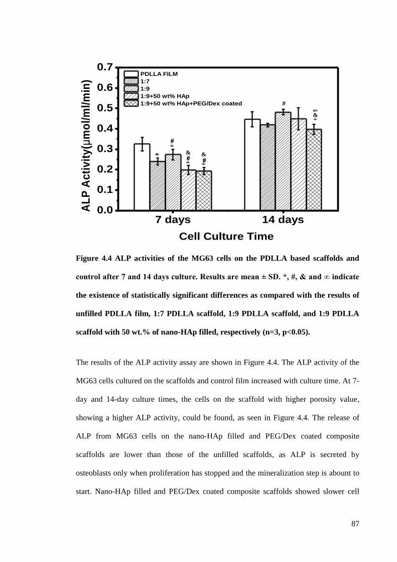
87
Figure 4.4 ALP activities of the MG63 cells on the PDLLA based scaffolds and
control after 7 and 14 days culture. Results are mean ± SD. *, #, & and ∞ indicate
the existence of statistically significant differences as compared with the results of
unfilled PDLLA film, 1:7 PDLLA scaffold, 1:9 PDLLA scaffold, and 1:9 PDLLA
scaffold with 50 wt.% of nano-HAp filled, respectively (n=3, p<0.05).
The results of the ALP activity assay are shown in Figure 4.4. The ALP activity of the
MG63 cells cultured on the scaffolds and control film increased with culture time. At 7-
day and 14-day culture times, the cells on the scaffold with higher porosity value,
showing a higher ALP activity, could be found, as seen in Figure 4.4. The release of
ALP from MG63 cells on the nano-HAp filled and PEG/Dex coated composite
scaffolds are lower than those of the unfilled scaffolds, as ALP is secreted by
osteoblasts only when proliferation has stopped and the mineralization step is abount to
start. Nano-HAp filled and PEG/Dex coated composite scaffolds showed slower cell
*
*
*
*
&
#
#
#
&
&
∞ #
*
*
*
#
#
#
7 days 14 days0.0
0.1
0.2
0.3
0.4
0.5
0.6
0.7
AL
P A
cti
vit
y(μ
mo
l/m
l/m
in)
Cell Culture Time
PDLLA FILM
1:7
1:9
1:9+50 wt% HAp
1:9+50 wt% HAp+PEG/Dex coated
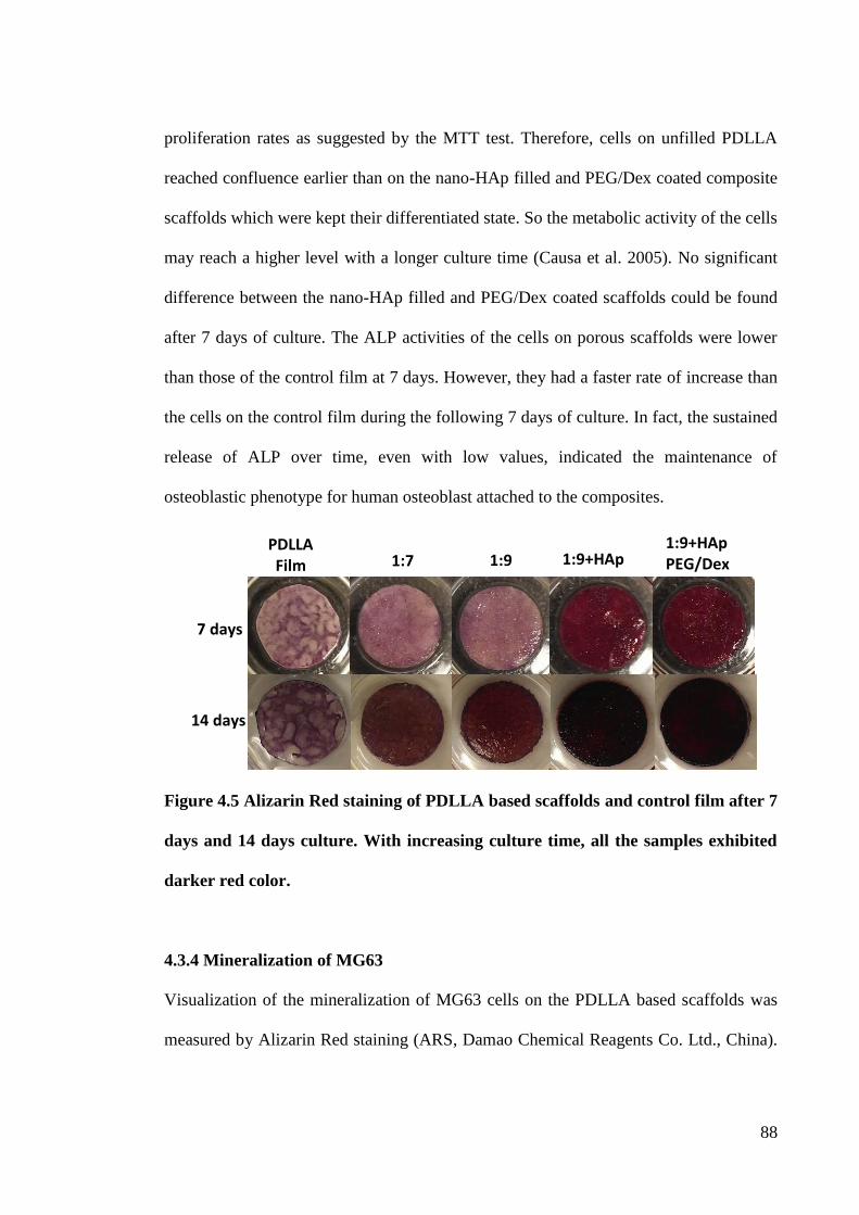
88
proliferation rates as suggested by the MTT test. Therefore, cells on unfilled PDLLA
reached confluence earlier than on the nano-HAp filled and PEG/Dex coated composite
scaffolds which were kept their differentiated state. So the metabolic activity of the cells
may reach a higher level with a longer culture time (Causa et al. 2005). No significant
difference between the nano-HAp filled and PEG/Dex coated scaffolds could be found
after 7 days of culture. The ALP activities of the cells on porous scaffolds were lower
than those of the control film at 7 days. However, they had a faster rate of increase than
the cells on the control film during the following 7 days of culture. In fact, the sustained
release of ALP over time, even with low values, indicated the maintenance of
osteoblastic phenotype for human osteoblast attached to the composites.
Figure 4.5 Alizarin Red staining of PDLLA based scaffolds and control film after 7
days and 14 days culture. With increasing culture time, all the samples exhibited
darker red color.
4.3.4 Mineralization of MG63
Visualization of the mineralization of MG63 cells on the PDLLA based scaffolds was
measured by Alizarin Red staining (ARS, Damao Chemical Reagents Co. Ltd., China).
PDLLA Film 1:7 1:9 1:9+HAp
1:9+HAp PEG/Dex
7 days
14 days
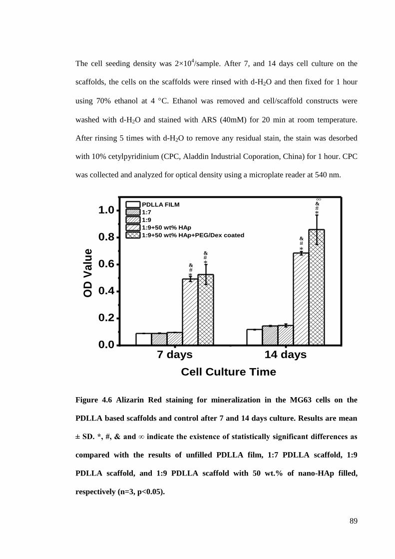
89
The cell seeding density was 2×10⁴/sample. After 7, and 14 days cell culture on the
scaffolds, the cells on the scaffolds were rinsed with d-H2O and then fixed for 1 hour
using 70% ethanol at 4 C. Ethanol was removed and cell/scaffold constructs were
washed with d-H2O and stained with ARS (40mM) for 20 min at room temperature.
After rinsing 5 times with d-H2O to remove any residual stain, the stain was desorbed
with 10% cetylpyridinium (CPC, Aladdin Industrial Coporation, China) for 1 hour. CPC
was collected and analyzed for optical density using a microplate reader at 540 nm.
Figure 4.6 Alizarin Red staining for mineralization in the MG63 cells on the
PDLLA based scaffolds and control after 7 and 14 days culture. Results are mean
± SD. *, #, & and ∞ indicate the existence of statistically significant differences as
compared with the results of unfilled PDLLA film, 1:7 PDLLA scaffold, 1:9
PDLLA scaffold, and 1:9 PDLLA scaffold with 50 wt.% of nano-HAp filled,
respectively (n=3, p<0.05).
*
*
*
*
#
&
#
&
&
&
#
#
∞
7 days 14 days0.0
0.2
0.4
0.6
0.8
1.0 PDLLA FILM
1:7
1:9
1:9+50 wt% HAp
1:9+50 wt% HAp+PEG/Dex coated
OD
Va
lue
Cell Culture Time
90
The images of the ARS stained PDLLA scaffolds and control films after 7 and 14 days
of culture are shown in Figure 4.5. The calcium salts could be stained into red by ARS
for detection of the mineralization of the cell. From Figure 4.5, scaffolds with nano-
HAp showed darker color compared to the unfilled ones and control film at the two
different time points. This is an indication of larger calcium deposition amounts on the
HAp filled composite. Moreover, the ARS staining proved the increasing mineralization
of the MG63 cells with the culture time (darker color). These results could be further
confirmed by the OD value of the cetylpyridinium solution which desorbed the ARS
from the samples (Figure 4.6). Nano-HAp filled scaffolds showed higher OD value than
the unfilled ones and the film and the OD value of all the scaffolds increased with time.
Additionally, the highest degree of mineralization was achieved by the MG63 cells on
PEG/Dex coated PDLLA/nano-HAp scaffold after 14 days. This suggested that
PEG/Dex coating and nano-HAp incorporation could promote bone formation, which is
of great importance for a scaffold in bone tissue applications.
4.3.5 Cell Morphology
Scanning electron microscopy (SEM) was used for cell morphology observation on the
scaffolds. The cell seeding density was 104/well for the SEM. the culture medium after
the samples had been cultured for a predetermined period (4 hours, 3 and 7 days) was
removed and discarded. The samples were washed with PBS three times and the cells
adhering to the samples were then fixed with 2.5% glutaraldehyde in PBS for 24 hours
at 4 C. After thorough washing with PBS, the cells on the samples were dehydrated
sequentially in an ethanol graded series (30%, 50%, 70%, 80%, 90%, and 100%) for 15
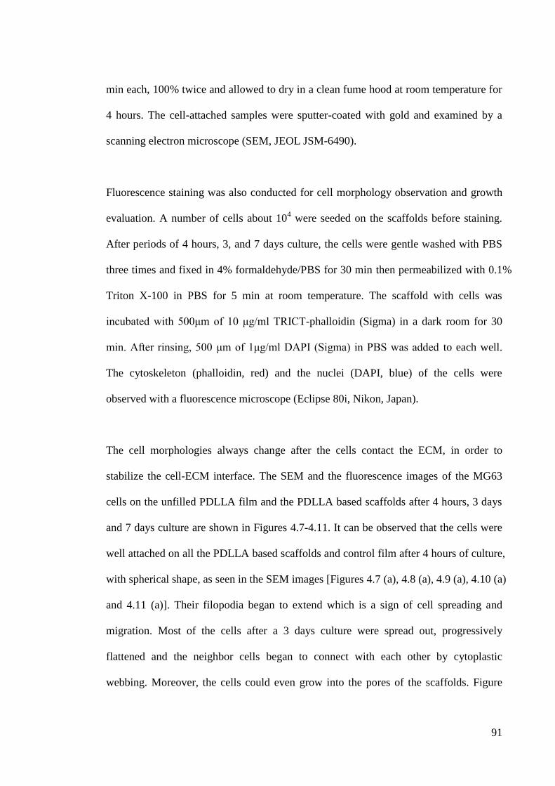
91
min each, 100% twice and allowed to dry in a clean fume hood at room temperature for
4 hours. The cell-attached samples were sputter-coated with gold and examined by a
scanning electron microscope (SEM, JEOL JSM-6490).
Fluorescence staining was also conducted for cell morphology observation and growth
evaluation. A number of cells about 104 were seeded on the scaffolds before staining.
After periods of 4 hours, 3, and 7 days culture, the cells were gentle washed with PBS
three times and fixed in 4% formaldehyde/PBS for 30 min then permeabilized with 0.1%
Triton X-100 in PBS for 5 min at room temperature. The scaffold with cells was
incubated with 500μm of 10 μg/ml TRICT-phalloidin (Sigma) in a dark room for 30
min. After rinsing, 500 μm of 1μg/ml DAPI (Sigma) in PBS was added to each well.
The cytoskeleton (phalloidin, red) and the nuclei (DAPI, blue) of the cells were
observed with a fluorescence microscope (Eclipse 80i, Nikon, Japan).
The cell morphologies always change after the cells contact the ECM, in order to
stabilize the cell-ECM interface. The SEM and the fluorescence images of the MG63
cells on the unfilled PDLLA film and the PDLLA based scaffolds after 4 hours, 3 days
and 7 days culture are shown in Figures 4.7-4.11. It can be observed that the cells were
well attached on all the PDLLA based scaffolds and control film after 4 hours of culture,
with spherical shape, as seen in the SEM images [Figures 4.7 (a), 4.8 (a), 4.9 (a), 4.10 (a)
and 4.11 (a)]. Their filopodia began to extend which is a sign of cell spreading and
migration. Most of the cells after a 3 days culture were spread out, progressively
flattened and the neighbor cells began to connect with each other by cytoplastic
webbing. Moreover, the cells could even grow into the pores of the scaffolds. Figure
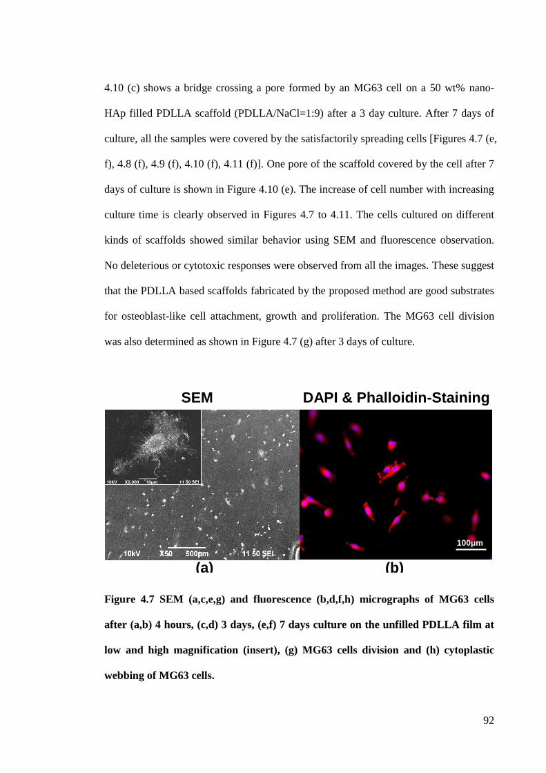
92
4.10 (c) shows a bridge crossing a pore formed by an MG63 cell on a 50 wt% nano-
HAp filled PDLLA scaffold (PDLLA/NaCl=1:9) after a 3 day culture. After 7 days of
culture, all the samples were covered by the satisfactorily spreading cells [Figures 4.7 (e,
f), 4.8 (f), 4.9 (f), 4.10 (f), 4.11 (f)]. One pore of the scaffold covered by the cell after 7
days of culture is shown in Figure 4.10 (e). The increase of cell number with increasing
culture time is clearly observed in Figures 4.7 to 4.11. The cells cultured on different
kinds of scaffolds showed similar behavior using SEM and fluorescence observation.
No deleterious or cytotoxic responses were observed from all the images. These suggest
that the PDLLA based scaffolds fabricated by the proposed method are good substrates
for osteoblast-like cell attachment, growth and proliferation. The MG63 cell division
was also determined as shown in Figure 4.7 (g) after 3 days of culture.
Figure 4.7 SEM (a,c,e,g) and fluorescence (b,d,f,h) micrographs of MG63 cells
after (a,b) 4 hours, (c,d) 3 days, (e,f) 7 days culture on the unfilled PDLLA film at
low and high magnification (insert), (g) MG63 cells division and (h) cytoplastic
webbing of MG63 cells.
SEM DAPI & Phalloidin-Staining
(b) (a)
100μm
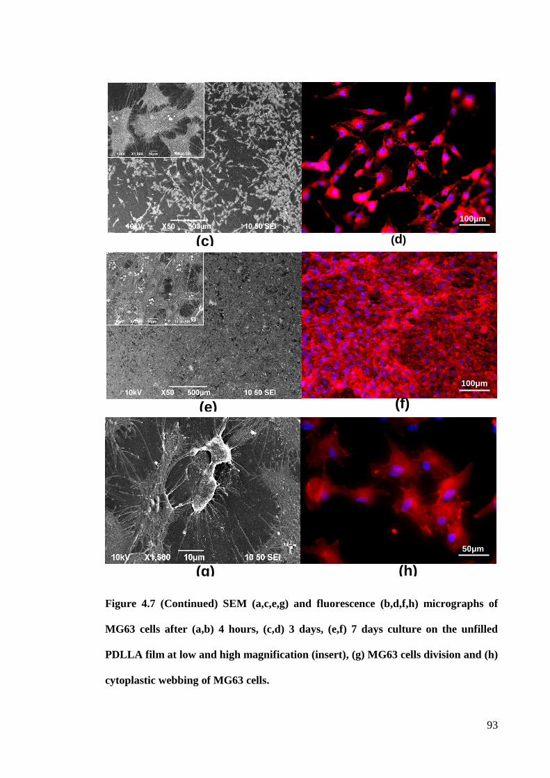
93
Figure 4.7 (Continued) SEM (a,c,e,g) and fluorescence (b,d,f,h) micrographs of
MG63 cells after (a,b) 4 hours, (c,d) 3 days, (e,f) 7 days culture on the unfilled
PDLLA film at low and high magnification (insert), (g) MG63 cells division and (h)
cytoplastic webbing of MG63 cells.
(h) (g)
(f) (e)
(d) (c)
50μm
100μm
100μm
94
Figure 4.8 SEM (a,c,e) and fluorescence (b,d,f) images of MG63 cells after 4 hours
(a,b), 3 days (c,d), and 7 days (e,f) culture on the unfilled PDLLA with
polymer/porogen ratio of 1:7.
(f) (e)
(d) (c)
SEM DAPI & Phalloidin-Staining
(b) (a)
100μm
100μm
100μm
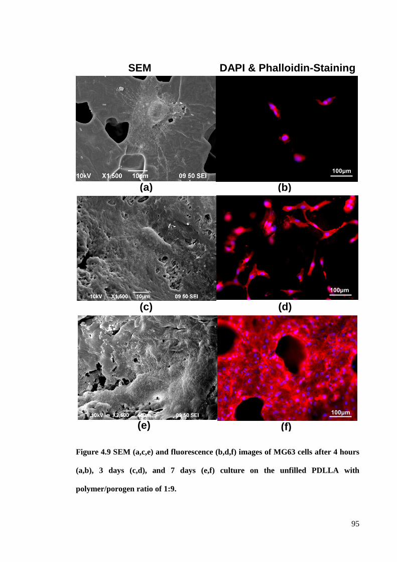
95
Figure 4.9 SEM (a,c,e) and fluorescence (b,d,f) images of MG63 cells after 4 hours
(a,b), 3 days (c,d), and 7 days (e,f) culture on the unfilled PDLLA with
polymer/porogen ratio of 1:9.
(f) (e)
(d) (c)
SEM DAPI & Phalloidin-Staining
(b) (a)
100μm
100μm
100μm
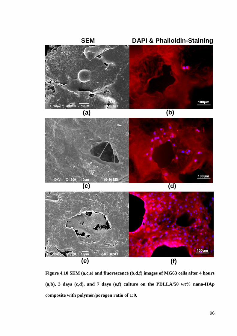
96
Figure 4.10 SEM (a,c,e) and fluorescence (b,d,f) images of MG63 cells after 4 hours
(a,b), 3 days (c,d), and 7 days (e,f) culture on the PDLLA/50 wt% nano-HAp
composite with polymer/porogen ratio of 1:9.
(f) (e)
(d) (c)
SEM DAPI & Phalloidin-Staining
(b) (a)
100μm
100μm
100μm
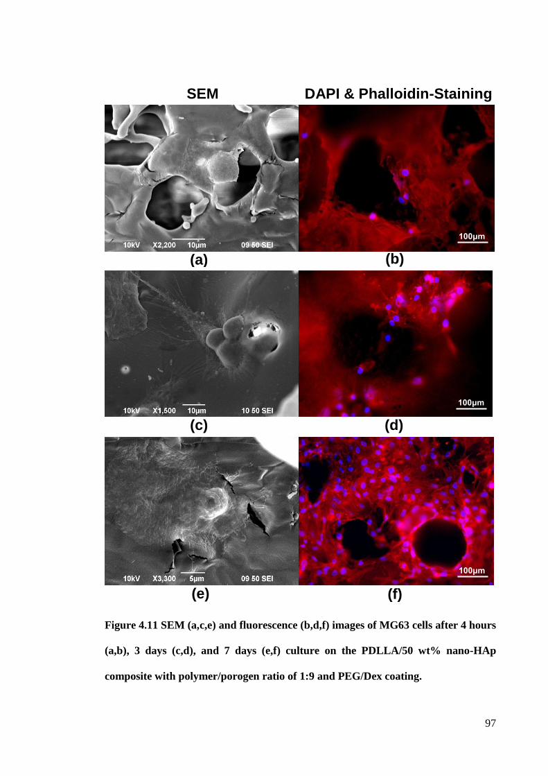
97
Figure 4.11 SEM (a,c,e) and fluorescence (b,d,f) images of MG63 cells after 4 hours
(a,b), 3 days (c,d), and 7 days (e,f) culture on the PDLLA/50 wt% nano-HAp
composite with polymer/porogen ratio of 1:9 and PEG/Dex coating.
(f) (e)
(d) (c)
SEM DAPI & Phalloidin-Staining
(b) (a)
100μm
100μm
100μm
98
4.4 Summary
In this part of the study, human osteoblast-like cells, MG63, cultured on the PDLLA
based composites shows that the cell adhesion, proliferation and differentiation were
highly influenced by the chemical composition and microstructure including porosity
and pore size of the scaffolds. The SEM and fluorescence images show that the pore
size of the fabricated scaffolds ranged from 200 to 350 µm, and high porosity of 60% to
80% allow not only the fluid passage through the scaffolds structure, but also the cells
hosting and penetrating inside the scaffolds. The whole scaffolds covered by the MG63
cells were observed from the fluorescence images after 7 days culture. The cell
morphology changed from spherical shape to a well spread-out flat shape with time, and
no deleterious or cytotoxic responses were observed. MTT results showed that the
higher the porosity of the scaffold, the more cell adhesion and the faster proliferation
rate of cells on the scaffold. The results from the ARS and ALP assay prove that the
scaffold with higher porosity and the addition of nano-HAp facilitated osteogenic
differentiation of the osteoblast progenitor MG63 cells. All these confirm that the
PDLLA based scaffolds fabricated in this study are biocompatible, osteoconductive and
facilitate cell attachment and proliferation. After confirming the biocompatibility of the
PDG/Dex coated PDLLA/nano-HAp composites, the other important properties of a
bone scaffolds, such as, bioactivity, degradability, and drug release capacity are
discussed in the following chapter.
99
Chapter 5. In Vitro Bioactivity, Degradation and Drug
Release Capacity of PDLLA/nano-HAp Composites
5.1 Introduction
Degradation behavior of composite bone scaffolds is of crucial importance in tissue
engineering, because the degradation rate is essentially linked to cell growth, host
response and tissue regeneration (Babensee et al. 1998). The scaffolds should have a
controllable degradation rate that matches the regeneration rate of new bone tissue. The
degradation kinetics of composite scaffolds can be affected by their composition,
microstructure, chemical and physical properties, and molecular weight, as well as the
environmental conditions (Wu & Ding 2004, Söntjens et al. 2012). Therefore,
controlling and evaluating the degradation behavior of composite scaffolds are of great
importance for the success of a biodegradable scaffold for bone tissue engineering. In
previous chapters, a novel PC-DC technique was developed for fabrication of PEG/Dex
coated PDLLA/nano-HAp composites, with independently controllable pore size and
porosity. The improved wettability, compressive moduli and strengths of the
PDLLA/nano-HAp composites, without incubation in simulated body fluids (SBF),
were well confirmed. The degradability of the PDLLA/nano-HAp composite was
investigated by measuring their mass and molecular weight changes during incubation
in a SBF in this part of study. An interesting phenomenon in which that nano-HAp has a
great effect of increasing the degradation rate of the composites in the first 7 days
incubation, but little effect for the following 21 days was found. Therefore, nano-HAp
could be used to control the degradation rate of composites to enable the applications in
bone tissue engineering.
100
The formation of a biomimetic apatite layer on the surface of a bone scaffold after being
implanted in the body is the critical requirement for bonding to living bone. It was
reported by Oyane (2003) that apatite formation in a SBF could be reproduced when the
artificial biomaterial was implanted in a living body. Therefore, the SBF can be applied
to estimate in vitro bioactivity of the artificial biomaterial by the apatite deposition on
the implant. The bonelike apatite coated on artificial biomaterials has been confirmed
with an improvement in the biological properties of these materials (Song et al. 2004).
Since the deposition of bonelike apatite on an implant material surface has been
confirmed as beneficial for cell adhesion and proliferation, a large amount of research
studies have focused on the mechanism and time requirement for apatite formation in
the SBF, as well as modification to the formulation or pH value of the SBF for
accelerating apatite formation (Chou et al. 2004, Chen et al. 2005). The question of how
the SBF incubation (e.g. incubation time and apatite formation) affects the mechanical
properties and the degradability of the bioactive scaffold may be raised. So far, very
limited studies have been reported on the changes of the mechanical properties of
biodegradable porous PDLLA/nano-HAp composites during incubation in a SBF. In
this chapter, in vitro evaluation of the bioactivity, degradability, and change of
mechanical properties of porous Dex-releasing PDLLA based scaffold materials, with
different amounts of nano-HAp, was undertaken. The bioactivity of the samples was
validated from the biomimetic apatite formation on the surface of the samples after
incubation in a SBF. The relationship between the SBF immersion time and the
compressive modulus and strength of the PDLLA/nano-HAp composites was monitored
in detail. An improvement in bioactivity (apatite formation ability) through the addition
101
of the nano-HAp content in the composites was found. Nano-HAp incorporation and
apatite formation made a positive impact on the mechanical properties of the
composites; however, plasticization and degradation of PDLLA had a negative impact.
Moreover, the PDLLA/nano-HAp composite with pH-compensation effect of reducing
the risk of chronic inflammation complications was confirmed.
Drug delivery technology, utilizing biodegradable materials as the drug vehicle to
deliver biologically active molecules at a desired behavior, is attracting increasing
interest in recent years for improving human live (Langer 1998). Such delivery systems
can improve the effectiveness of the drug therapy and to reduce toxicity. The PEG/Dex
coated PDLLA/nano-HAp composites fabricated in this study were equipped with a
drug release function. In this part of study, an in vitro drug release study was carried out
to investigate the drug release behavior. The influences of porosity and nano-HAp
fraction on the Dex release behavior of the PDLLA based scaffold materials were also
investigated. Some results from this part of study have been published in the Journal of
the Mechanical Behavior of Biomedical Materials (Chen et al. 2013).
5.2 Preparation of SBF
The SBF was prepared by dissolving reagent grade sodium chloride (NaCl), sodium
hydrogen carbonate (NaHCO3), potassium chloride (KCl), di-potassium hydrogen
phosphate trihydrate (K2HPO4·3H2O), magnesium chloride hexahydrate (MgCl∙6H2O),
calcium chloride (CaCl2), sodium sulfate (Na2SO4), and tris-hydroxymethyl
aminomethane (Tris, (CH2OH)3CNH2) into deionized water (Kokubo & Takadama
2006). To prepare 1000 ml of SBF, a beaker with 700 ml of deionized water was heated
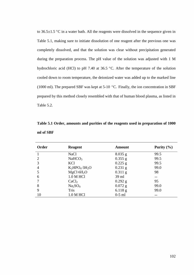
102
to 36.5±1.5 C in a water bath. All the reagents were dissolved in the sequence given in
Table 5.1, making sure to initiate dissolution of one reagent after the previous one was
completely dissolved, and that the solution was clear without precipitation generated
during the preparation process. The pH value of the solution was adjusted with 1 M
hydrochloric acid (HCl) to pH 7.40 at 36.5 C. After the temperature of the solution
cooled down to room temperature, the deionized water was added up to the marked line
(1000 ml). The prepared SBF was kept at 5-10 C. Finally, the ion concentration in SBF
prepared by this method closely resembled with that of human blood plasma, as listed in
Table 5.2.
Table 5.1 Order, amounts and purities of the reagents used in preparation of 1000
ml of SBF
Order Reagent Amount Purity (%)
1 NaCl 8.035 g 99.5
2 NaHCO3 0.355 g 99.5
3 KCl 0.225 g 99.5
4 K2HPO4·3H2O 0.231 g 99.0
5 MgCl∙6H2O 0.311 g 98
6 1.0 M HCl 39 ml ― 7 CaCl2 0.292 g 95
8 Na2SO4 0.072 g 99.0
9 Tris 6.118 g 99.0
10 1.0 M HCl 0-5 ml ―
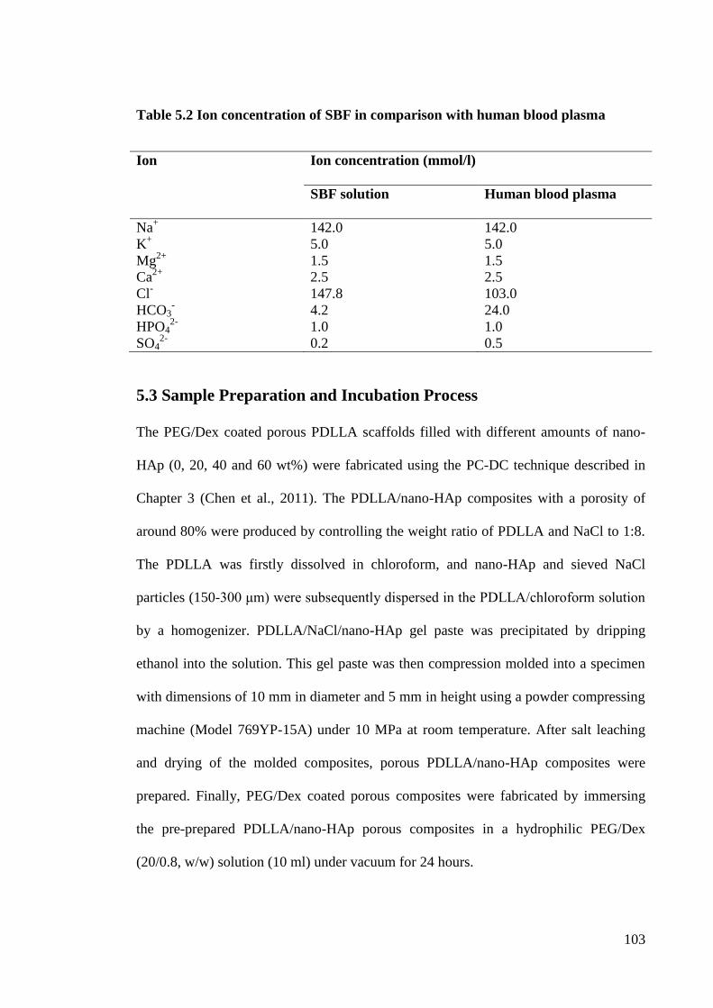
103
Table 5.2 Ion concentration of SBF in comparison with human blood plasma
Ion Ion concentration (mmol/l)
SBF solution Human blood plasma
Na+ 142.0 142.0
K+ 5.0 5.0
Mg2+
1.5 1.5
Ca2+
2.5 2.5
Cl- 147.8 103.0
HCO3- 4.2 24.0
HPO42-
1.0 1.0
SO42-
0.2 0.5
5.3 Sample Preparation and Incubation Process
The PEG/Dex coated porous PDLLA scaffolds filled with different amounts of nano-
HAp (0, 20, 40 and 60 wt%) were fabricated using the PC-DC technique described in
Chapter 3 (Chen et al., 2011). The PDLLA/nano-HAp composites with a porosity of
around 80% were produced by controlling the weight ratio of PDLLA and NaCl to 1:8.
The PDLLA was firstly dissolved in chloroform, and nano-HAp and sieved NaCl
particles (150-300 μm) were subsequently dispersed in the PDLLA/chloroform solution
by a homogenizer. PDLLA/NaCl/nano-HAp gel paste was precipitated by dripping
ethanol into the solution. This gel paste was then compression molded into a specimen
with dimensions of 10 mm in diameter and 5 mm in height using a powder compressing
machine (Model 769YP-15A) under 10 MPa at room temperature. After salt leaching
and drying of the molded composites, porous PDLLA/nano-HAp composites were
prepared. Finally, PEG/Dex coated porous composites were fabricated by immersing
the pre-prepared PDLLA/nano-HAp porous composites in a hydrophilic PEG/Dex
(20/0.8, w/w) solution (10 ml) under vacuum for 24 hours.
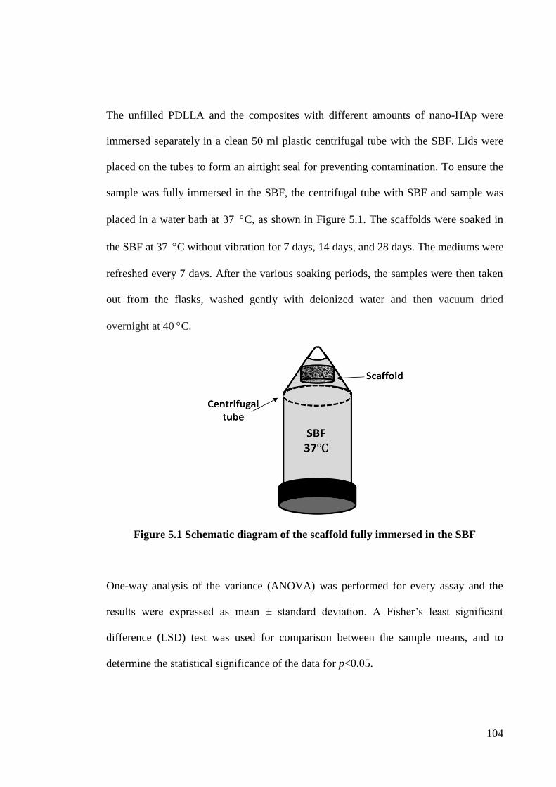
104
The unfilled PDLLA and the composites with different amounts of nano-HAp were
immersed separately in a clean 50 ml plastic centrifugal tube with the SBF. Lids were
placed on the tubes to form an airtight seal for preventing contamination. To ensure the
sample was fully immersed in the SBF, the centrifugal tube with SBF and sample was
placed in a water bath at 37 C, as shown in Figure 5.1. The scaffolds were soaked in
the SBF at 37 C without vibration for 7 days, 14 days, and 28 days. The mediums were
refreshed every 7 days. After the various soaking periods, the samples were then taken
out from the flasks, washed gently with deionized water and then vacuum dried
overnight at 40 C.
Figure 5.1 Schematic diagram of the scaffold fully immersed in the SBF
One-way analysis of the variance (ANOVA) was performed for every assay and the
results were expressed as mean ± standard deviation. A Fisher‟s least significant
difference (LSD) test was used for comparison between the sample means, and to
determine the statistical significance of the data for p<0.05.
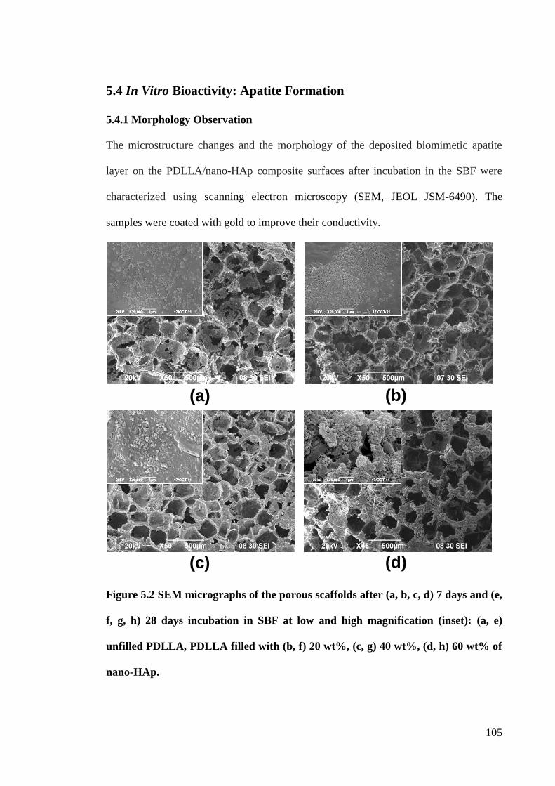
105
5.4 In Vitro Bioactivity: Apatite Formation
5.4.1 Morphology Observation
The microstructure changes and the morphology of the deposited biomimetic apatite
layer on the PDLLA/nano-HAp composite surfaces after incubation in the SBF were
characterized using scanning electron microscopy (SEM, JEOL JSM-6490). The
samples were coated with gold to improve their conductivity.
Figure 5.2 SEM micrographs of the porous scaffolds after (a, b, c, d) 7 days and (e,
f, g, h) 28 days incubation in SBF at low and high magnification (inset): (a, e)
unfilled PDLLA, PDLLA filled with (b, f) 20 wt%, (c, g) 40 wt%, (d, h) 60 wt% of
nano-HAp.
(c) (d)
(c) (d)
(a) (b)
(b)
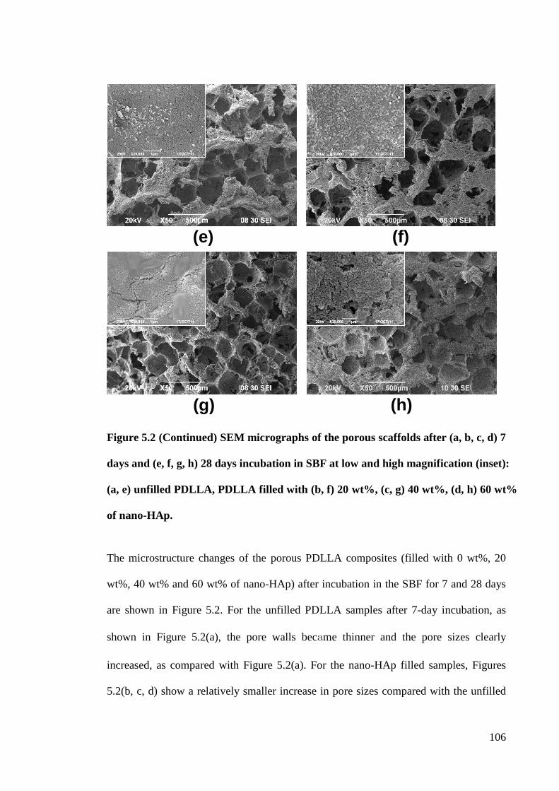
106
Figure 5.2 (Continued) SEM micrographs of the porous scaffolds after (a, b, c, d) 7
days and (e, f, g, h) 28 days incubation in SBF at low and high magnification (inset):
(a, e) unfilled PDLLA, PDLLA filled with (b, f) 20 wt%, (c, g) 40 wt%, (d, h) 60 wt%
of nano-HAp.
The microstructure changes of the porous PDLLA composites (filled with 0 wt%, 20
wt%, 40 wt% and 60 wt% of nano-HAp) after incubation in the SBF for 7 and 28 days
are shown in Figure 5.2. For the unfilled PDLLA samples after 7-day incubation, as
shown in Figure 5.2(a), the pore walls became thinner and the pore sizes clearly
increased, as compared with Figure 5.2(a). For the nano-HAp filled samples, Figures
5.2(b, c, d) show a relatively smaller increase in pore sizes compared with the unfilled
(g) (h)
(g) (h)
(e) (f)
(e) (f)
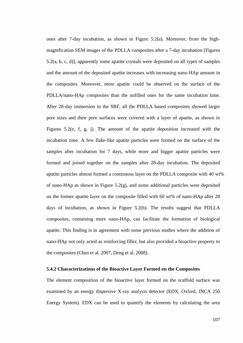
107
ones after 7-day incubation, as shown in Figure 5.2(a). Moreover, from the high-
magnification SEM images of the PDLLA composites after a 7-day incubation [Figures
5.2(a, b, c, d)], apparently some apatite crystals were deposited on all types of samples
and the amount of the deposited apatite increases with increasing nano-HAp amount in
the composites. Moreover, more apatite could be observed on the surface of the
PDLLA/nano-HAp composites than the unfilled ones for the same incubation time.
After 28-day immersion in the SBF, all the PDLLA based composites showed larger
pore sizes and their pore surfaces were covered with a layer of apatite, as shown in
Figures 5.2(e, f, g, j). The amount of the apatite deposition increased with the
incubation time. A few flake-like apatite particles were formed on the surface of the
samples after incubation for 7 days, while more and bigger apatite particles were
formed and joined together on the samples after 28-day incubation. The deposited
apatite particles almost formed a continuous layer on the PDLLA composite with 40 wt%
of nano-HAp as shown in Figure 5.2(g), and some additional particles were deposited
on the former apatite layer on the composite filled with 60 wt% of nano-HAp after 28
days of incubation, as shown in Figure 5.2(h). The results suggest that PDLLA
composites, containing more nano-HAp, can facilitate the formation of biological
apatite. This finding is in agreement with some previous studies where the addition of
nano-HAp not only acted as reinforcing filler, but also provided a bioactive property to
the composites (Chen et al. 2007, Deng et al. 2008).
5.4.2 Characterizations of the Bioactive Layer Formed on the Composites
The element composition of the bioactive layer formed on the scaffold surface was
examined by an energy dispersive X-ray analysis detector (EDX, Oxford, INCA 250
Energy System). EDX can be used to quantify the elements by calculating the area
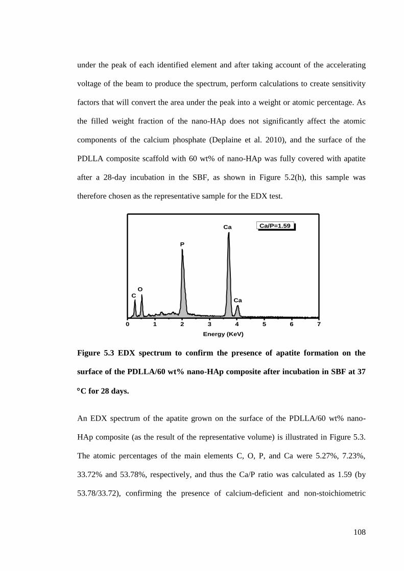
108
under the peak of each identified element and after taking account of the accelerating
voltage of the beam to produce the spectrum, perform calculations to create sensitivity
factors that will convert the area under the peak into a weight or atomic percentage. As
the filled weight fraction of the nano-HAp does not significantly affect the atomic
components of the calcium phosphate (Deplaine et al. 2010), and the surface of the
PDLLA composite scaffold with 60 wt% of nano-HAp was fully covered with apatite
after a 28-day incubation in the SBF, as shown in Figure 5.2(h), this sample was
therefore chosen as the representative sample for the EDX test.
Figure 5.3 EDX spectrum to confirm the presence of apatite formation on the
surface of the PDLLA/60 wt% nano-HAp composite after incubation in SBF at 37
C for 28 days.
An EDX spectrum of the apatite grown on the surface of the PDLLA/60 wt% nano-
HAp composite (as the result of the representative volume) is illustrated in Figure 5.3.
The atomic percentages of the main elements C, O, P, and Ca were 5.27%, 7.23%,
33.72% and 53.78%, respectively, and thus the Ca/P ratio was calculated as 1.59 (by
53.78/33.72), confirming the presence of calcium-deficient and non-stoichiometric
0 1 2 3 4 5 6 7
Energy (KeV)
Ca/P=1.59
Ca
Ca
P
OC
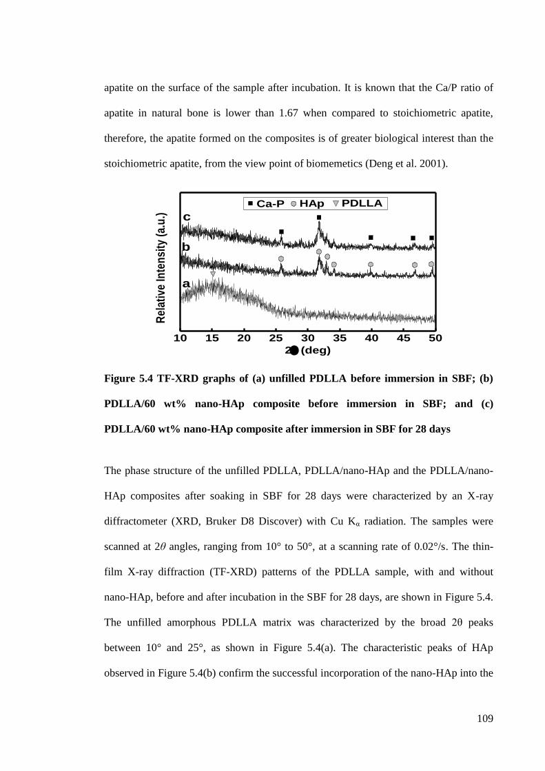
109
apatite on the surface of the sample after incubation. It is known that the Ca/P ratio of
apatite in natural bone is lower than 1.67 when compared to stoichiometric apatite,
therefore, the apatite formed on the composites is of greater biological interest than the
stoichiometric apatite, from the view point of biomemetics (Deng et al. 2001).
Figure 5.4 TF-XRD graphs of (a) unfilled PDLLA before immersion in SBF; (b)
PDLLA/60 wt% nano-HAp composite before immersion in SBF; and (c)
PDLLA/60 wt% nano-HAp composite after immersion in SBF for 28 days
The phase structure of the unfilled PDLLA, PDLLA/nano-HAp and the PDLLA/nano-
HAp composites after soaking in SBF for 28 days were characterized by an X-ray
diffractometer (XRD, Bruker D8 Discover) with Cu Kα radiation. The samples were
scanned at 2θ angles, ranging from 10° to 50°, at a scanning rate of 0.02°/s. The thin-
film X-ray diffraction (TF-XRD) patterns of the PDLLA sample, with and without
nano-HAp, before and after incubation in the SBF for 28 days, are shown in Figure 5.4.
The unfilled amorphous PDLLA matrix was characterized by the broad 2θ peaks
between 10° and 25°, as shown in Figure 5.4(a). The characteristic peaks of HAp
observed in Figure 5.4(b) confirm the successful incorporation of the nano-HAp into the
10 15 20 25 30 35 40 45 50
HAp
Rel
ativ
e In
ten
sity
(a.
u.)
2(deg)
a
b
cCa-P PDLLA
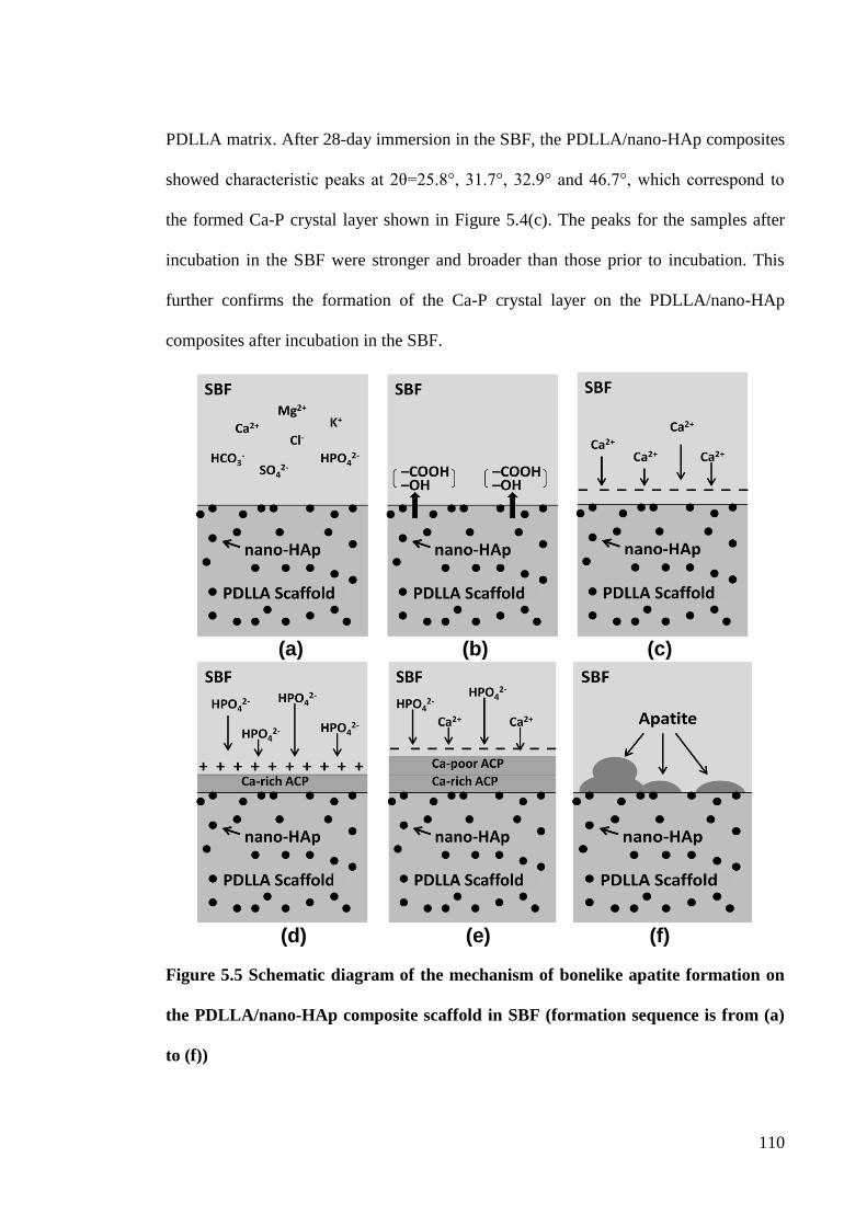
110
PDLLA matrix. After 28-day immersion in the SBF, the PDLLA/nano-HAp composites
showed characteristic peaks at 2θ=25.8°, 31.7°, 32.9° and 46.7°, which correspond to
the formed Ca-P crystal layer shown in Figure 5.4(c). The peaks for the samples after
incubation in the SBF were stronger and broader than those prior to incubation. This
further confirms the formation of the Ca-P crystal layer on the PDLLA/nano-HAp
composites after incubation in the SBF.
(a) (b) (c)
(d) (e) (f)
Figure 5.5 Schematic diagram of the mechanism of bonelike apatite formation on
the PDLLA/nano-HAp composite scaffold in SBF (formation sequence is from (a)
to (f))
111
The process and kinetics of apatite formation on the scaffolds can be affected by the
surface composition and structure of the scaffolds (Kim et al. 2005). The hydrolysis of
PDLLA and the nano-HAp filling place an important role in apatite forming (Zhang &
Ma 1999). The mechanism of bonelike apatite formation on the PDLLA/nano-HAp
scaffolds is schematically illustrated in Figure 5.5. In the SBF, –COOH, and –OH
groups can be generated from the hydrolysis of PDLLA [Figures 5.5(a-b)]. The weak
acidic group –COOH may dissociate in SBF and further change to –COO-, which leads
to the PDLLA surface being negatively charged. Moreover, the nano-HAp revealed
negative surface potential (Kim et al. 2005). The –COOH, –COO-, –OH groups and
nano-HAp located at the surface of PDLLA scaffolds may interact with positive Ca2+
in
the SBF to form a Ca-rich amorphous calcium phosphate (ACP) [Figure 5.5(c)]. The
surface potential can increase and gain positive charge. The Ca-rich ACP on the
PDLLA then interacts with the negative HPO42-
in the fluid to form the Ca-poor ACP
[Figure 5.5(d)]. The Ca-poor ACP appears to be stable by gradually crystallizing into
bonelike apatite with a low solubility in the SBF [Figures 5.5(e-f)]. Therefore,
increasing the nano-HAp fraction gives way to the increasing nano-HAp to the SBF,
which helps to provide a more negatively charged surface on the scaffold that attracts
positive Ca2+
and stimulates apatite nucleation. The bonelike apatite formed on the
scaffold can provide better cell interaction and osteoconductivity, and is the reason why
scaffolds with higher fractions of nano-HAp show better bioactivity.
112
5.5 Degradation of the Composites
5.5.1 Molecular Weight Change
The degradability of the PDLLA/nano-HAp composites with different nano-HAp
amounts was investigated by a gel permeation chromatography (GPC, Waters
Associates, Inc.) system to determine their molecular weight loss. The measurements
were carried out at 30 C at a flow rate of 1ml/min using tetrahydrofuran (THF) as an
eluent. A set of monodisperse linear polystyrene standards (Polysciences, Inc.) were
used to obtain a calibration curve. The weight average ( ) molecular weights of the
samples were estimated. Three replicate measurements were conducted for each type of
sample.
The results of GPC are shown in Figure 5.6. The of all the samples decreased with
time after incubation in the SBF, resulting from the degradation of the PDLLA. After a
28-day incubation, the unfilled PDLLA had the lowest compared to the filled ones.
However, for the first 7 days of incubation, the degradation rate increased when the
PDLLA were filled with larger amounts of nano-HAp, because the PDLLA with higher
nano-HAp content could increase the hydrophilicity of the composite and hence
promote the penetration rate of the SBF. Therefore, the of the PDLLA with 60 wt %
of nano-HAp decreased faster than the other filled samples, as shown in Figure 5.6. In
all, incorporation of the nano-HAp can be used to control the degradation rate of the
PDLLA/nano-HAp composites.
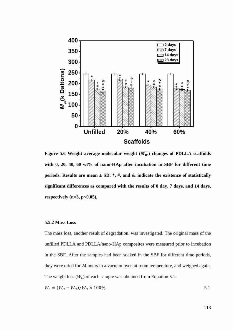
113
Figure 5.6 Weight average molecular weight ( ) changes of PDLLA scaffolds
with 0, 20, 40, 60 wt% of nano-HAp after incubation in SBF for different time
periods. Results are mean ± SD. *, #, and & indicate the existence of statistically
significant differences as compared with the results of 0 day, 7 days, and 14 days,
respectively (n=3, p<0.05).
5.5.2 Mass Loss
The mass loss, another result of degradation, was investigated. The original mass of the
unfilled PDLLA and PDLLA/nano-HAp composites were measured prior to incubation
in the SBF. After the samples had been soaked in the SBF for different time periods,
they were dried for 24 hours in a vacuum oven at room temperature, and weighed again.
The weight loss ( ) of each sample was obtained from Equation 5.1.
5.1
*
* *
# #
*
* * * * * * * *
#
# # # #
#
& &
& &
Unfilled 20% 40% 60%0
50
100
150
200
250
300
350
400
Mw(k
Da
lto
ns
)
Scaffolds
0 days
7 days
14 days
28 days
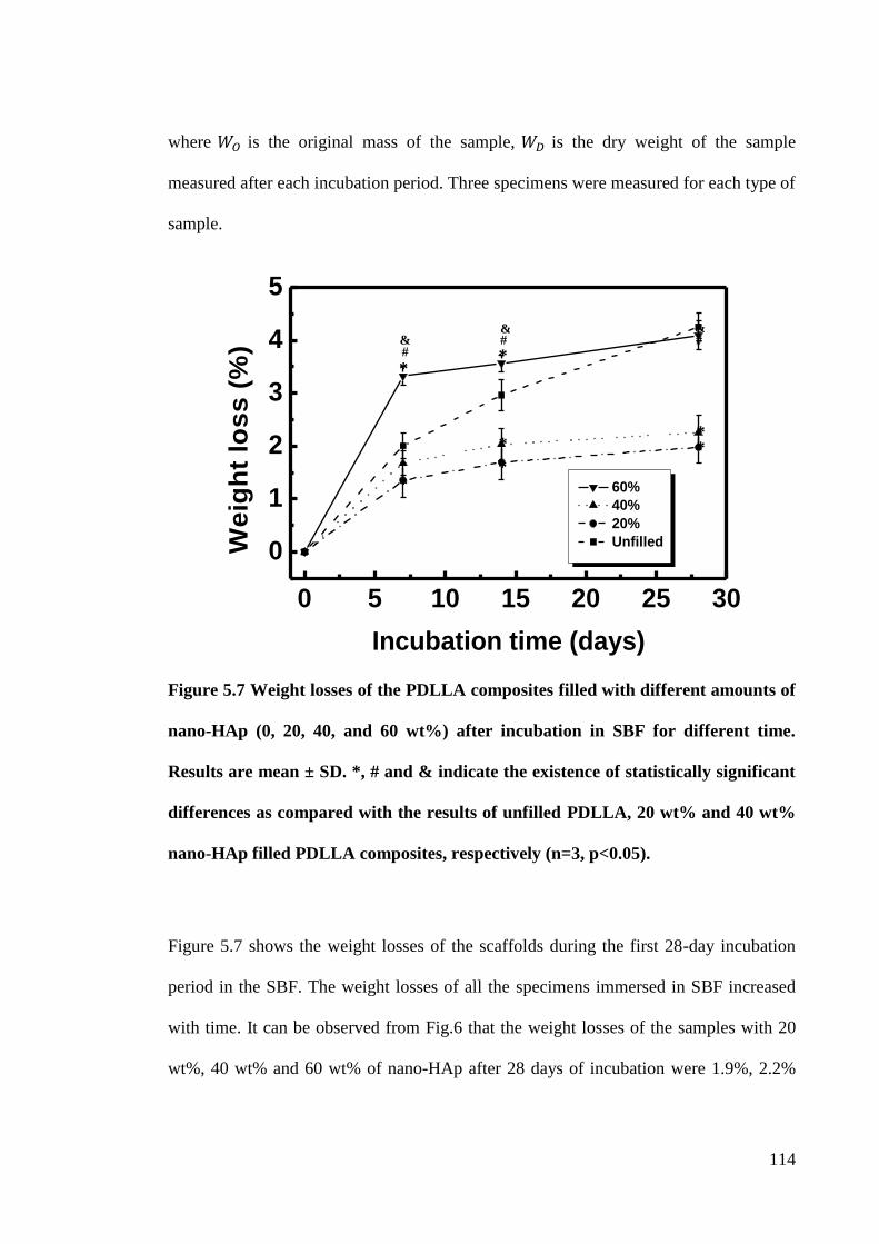
114
where is the original mass of the sample,
is the dry weight of the sample
measured after each incubation period. Three specimens were measured for each type of
sample.
Figure 5.7 Weight losses of the PDLLA composites filled with different amounts of
nano-HAp (0, 20, 40, and 60 wt%) after incubation in SBF for different time.
Results are mean ± SD. *, # and & indicate the existence of statistically significant
differences as compared with the results of unfilled PDLLA, 20 wt% and 40 wt%
nano-HAp filled PDLLA composites, respectively (n=3, p<0.05).
Figure 5.7 shows the weight losses of the scaffolds during the first 28-day incubation
period in the SBF. The weight losses of all the specimens immersed in SBF increased
with time. It can be observed from Fig.6 that the weight losses of the samples with 20
wt%, 40 wt% and 60 wt% of nano-HAp after 28 days of incubation were 1.9%, 2.2%
0 5 10 15 20 25 30
0
1
2
3
4
5
W
eig
ht
los
s (
%)
Incubation time (days)
60%
40%
20%
Unfilled
*
*
*
# &
*
* # # &
* *
&
115
and 4.2%, respectively. This suggests that the PDLLA/60 wt % nano-HAp have the
highest weight losses during their 28 days of incubation in the SBF as compared with
the others. However, there is no statistically significant difference in the weight losses
between the composites with 20 wt% and 40 wt% of nano-HAp during the incubation
period. The reason of the rapid weight losses of the composites are the dissolution of the
nano-HAp particles, and degradation and fragmentation of the polymer matrices in the
composites. In addition, incorporation of nano-HAp increases the overall hydrophilicity
of the composites which resulted in increasing their hydrolysis rates. Therefore, the
composites with large amounts of nano-HAp, for example, 60 wt%, had a faster weight
loss rate especially during the first 7-day incubation (Ang et al. 2007). However, the
weight loss of the unfilled PDLLA was higher than that of the composites with 20 wt%
and 40 wt% of nano-HAp but lower than that of the composite with 60 wt% after 7 days.
It is because the higher amounts of bonelike apatite deposition on the samples with 20
wt% and 40 wt% of nano-HAp than on the unfilled one, and can retard the weight loss
of the composites. As the apatite formation rates of the unfilled PDLLA was slower
than that of the nano-HAp filled composites during incubation in the SBF, the unfilled
PDLLA had a higher weight loss of nearly 4.4% after 28 days of immersion, as
compared with the composites. However, there are no statistically significant
differences between the unfilled PDLLA and the PDLLA/60 wt% nano-HAp composite
after a 28-day immersion.
5.6 Mechanical Properties Evaluation after Incubation in SBF
In order to investigate the effects of apatite deposition and polymer degradation on the
mechanical properties of the porous multifunctional PDLLA based composites, uniaxial
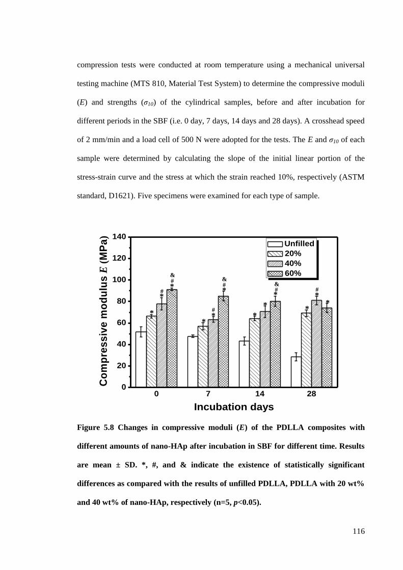
116
compression tests were conducted at room temperature using a mechanical universal
testing machine (MTS 810, Material Test System) to determine the compressive moduli
(E) and strengths (ζ10) of the cylindrical samples, before and after incubation for
different periods in the SBF (i.e. 0 day, 7 days, 14 days and 28 days). A crosshead speed
of 2 mm/min and a load cell of 500 N were adopted for the tests. The E and ζ10 of each
sample were determined by calculating the slope of the initial linear portion of the
stress-strain curve and the stress at which the strain reached 10%, respectively (ASTM
standard, D1621). Five specimens were examined for each type of sample.
Figure 5.8 Changes in compressive moduli (E) of the PDLLA composites with
different amounts of nano-HAp after incubation in SBF for different time. Results
are mean ± SD. *, #, and & indicate the existence of statistically significant
differences as compared with the results of unfilled PDLLA, PDLLA with 20 wt%
and 40 wt% of nano-HAp, respectively (n=5, p<0.05).
0 7 14 280
20
40
60
80
100
120
140
Co
mp
res
siv
e m
od
ulu
s E
(M
Pa
)
Incubation days
Unfilled
20%
40%
60%
* *
* *
#
*
*
*
*
* *
# #
#
#
#
&
* *
& &
117
The E and ζ10 of the PDLLA composites with different amounts of nano-HAp before
incubation in the SBF are shown in Figures 5.8 and 5.9, noted as 0-day incubation.
Before incubation, PDLLA/nano-HAp composites had higher E and ζ10 than the
unfilled PDLLA ones. E and ζ10 of the PDLLA with 60 wt% of nano-HAp increased to
91.3±1.2 MPa and 2.5±0.2 MPa, respectively, which were close to the data for human
cancellous bone (Rezwan et al. 2006). The increase in E for the PDLLA/nano-HAp
composites is mainly attributed to the addition of the rigid nano-HAp filler (Thomas et
al. 2006). Nano-sized HAp with a high total surface area can enhance stress transfer
between the matrix and the fillers. Therefore, the addition of nano-HAp led to the
increase in ζ10 through an efficient stress transfer mechanism (Fu et al. 2008). In
addition to the high surface area of the nano-HAp, the interfacial adhesion between the
fillers and the polymer matrix is of great importance for ζ10 (Kovačević et al. 2008). The
hydrogen bonding formed between the uniformly distributed nano-HAp and the PDLLA
matrix under compression molding also increased the interfacial adhesion and hence the
enhancement of the ζ10 of the PDLLA/nano-HAp composites is due to the better load
transfer (Zhou et al. 2007, Fu et al. 2008).
The changes in the E and ζ10 of the porous PDLLA based nanocomposites after
incubation in the SBF for different times are shown in Figures 5.8 and 5.9. After
soaking in the SBF for 7 days, the E value of all the PDLLA based samples decreased,
as shown in Figure 5.8. The main reasons for this decrease are the pore size increase of
the samples, the decrease in the molecular weight of the PDLLA, and the loss of the
118
nano-HAp particles. In addition, the reduction of E can also be attributed to the
plasticizing effect because the terminal carboxylic acid functional group on the PDLLA
chains facilitates water penetration (Blaker et al. 2011). From the 7-day to the 28-day
incubation, it could be observed that the E value of the PDLLA composites with 20 wt%
and 40 wt% of nano-HAp increased with incubation time. The increase of E can be
mainly attributed to the apatite deposition on the composites and the improvement of
the interfacial adhesion between the deposited apatite and the PDLLA matrix. From
Figure 5.8, the E values of the unfilled PDLLA reduced from 51.6±4.7 MPa to 28.6±3.9
MPa after a 28-day incubation, because of the continuous degradation of the PDLLA
and relatively little apatite being deposited during the incubation. Nano-HAp particles
filled in the PDLLA composite tended to fall off and interact with the SBF because of
their good hydrophilicity. The loss of nano-HAp particles led to the formation of some
voids within the PDLLA matrix, and hence more surfaces of the PDLLA were exposed
to hydrolytic attack, accelerating the degradation of the PDLLA and weakening its
overall structure. Therefore, when the PDLLA composite was filled with a higher
content of hydrophilic nano-HAp, this phenomenon was more obvious and resulted in
the E reduction of the scaffolds with 60 wt% of nano-HAp, within the 28-day
incubation. However, the scaffolds with 40 wt% nano-HAp after a 28-day incubation
could reach an E value of 80.9±4.1 MPa, which was much larger than that of an unfilled
PDLLA. In general, the incorporation of the nano-HAp into the PDLLA can promote
apatite deposition on the composites and enhance their compressive moduli.
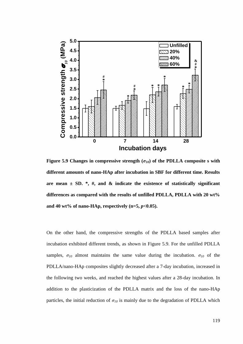
119
Figure 5.9 Changes in compressive strength (σ10) of the PDLLA composite s with
different amounts of nano-HAp after incubation in SBF for different time. Results
are mean ± SD. *, #, and & indicate the existence of statistically significant
differences as compared with the results of unfilled PDLLA, PDLLA with 20 wt%
and 40 wt% of nano-HAp, respectively (n=5, p<0.05).
On the other hand, the compressive strengths of the PDLLA based samples after
incubation exhibited different trends, as shown in Figure 5.9. For the unfilled PDLLA
samples, ζ10 almost maintains the same value during the incubation. ζ10 of the
PDLLA/nano-HAp composites slightly decreased after a 7-day incubation, increased in
the following two weeks, and reached the highest values after a 28-day incubation. In
addition to the plasticization of the PDLLA matrix and the loss of the nano-HAp
particles, the initial reduction of ζ10 is mainly due to the degradation of PDLLA which
*
*
*
*
*
* * * #
# &
#
*
0 7 14 280.0
0.5
1.0
1.5
2.0
2.5
3.0
3.5
4.0
4.5
5.0
Co
mp
res
siv
e s
tre
ng
th
10 (
MP
a)
Incubation days
Unfilled
20%
40%
60%
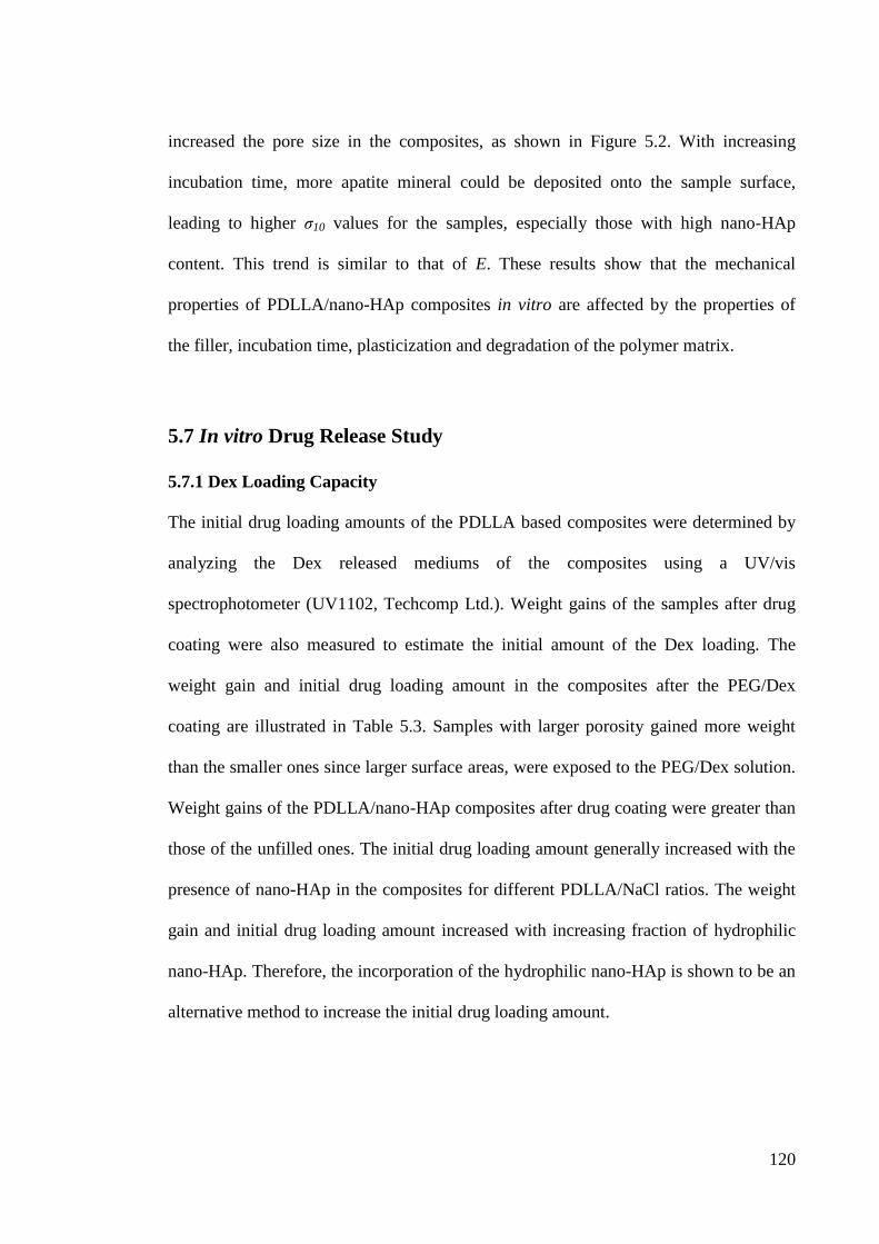
120
increased the pore size in the composites, as shown in Figure 5.2. With increasing
incubation time, more apatite mineral could be deposited onto the sample surface,
leading to higher ζ10 values for the samples, especially those with high nano-HAp
content. This trend is similar to that of E. These results show that the mechanical
properties of PDLLA/nano-HAp composites in vitro are affected by the properties of
the filler, incubation time, plasticization and degradation of the polymer matrix.
5.7 In vitro Drug Release Study
5.7.1 Dex Loading Capacity
The initial drug loading amounts of the PDLLA based composites were determined by
analyzing the Dex released mediums of the composites using a UV/vis
spectrophotometer (UV1102, Techcomp Ltd.). Weight gains of the samples after drug
coating were also measured to estimate the initial amount of the Dex loading. The
weight gain and initial drug loading amount in the composites after the PEG/Dex
coating are illustrated in Table 5.3. Samples with larger porosity gained more weight
than the smaller ones since larger surface areas, were exposed to the PEG/Dex solution.
Weight gains of the PDLLA/nano-HAp composites after drug coating were greater than
those of the unfilled ones. The initial drug loading amount generally increased with the
presence of nano-HAp in the composites for different PDLLA/NaCl ratios. The weight
gain and initial drug loading amount increased with increasing fraction of hydrophilic
nano-HAp. Therefore, the incorporation of the hydrophilic nano-HAp is shown to be an
alternative method to increase the initial drug loading amount.
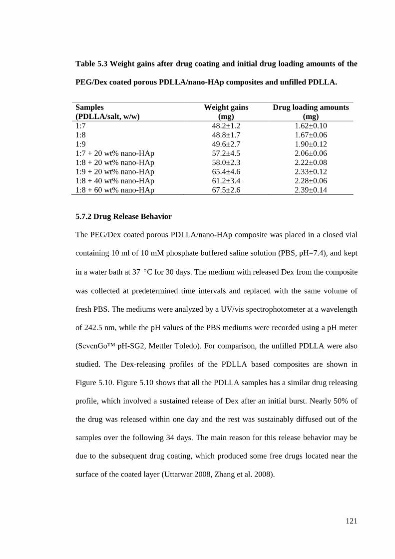
121
Table 5.3 Weight gains after drug coating and initial drug loading amounts of the
PEG/Dex coated porous PDLLA/nano-HAp composites and unfilled PDLLA.
Samples
(PDLLA/salt, w/w)
Weight gains
(mg)
Drug loading amounts
(mg)
1:7 48.2±1.2 1.62±0.10
1:8 48.8±1.7 1.67±0.06
1:9 49.6±2.7 1.90±0.12
1:7 + 20 wt% nano-HAp 57.2±4.5 2.06±0.06
1:8 + 20 wt% nano-HAp 58.0±2.3 2.22±0.08
1:9 + 20 wt% nano-HAp 65.4±4.6 2.33±0.12
1:8 + 40 wt% nano-HAp 61.2±3.4 2.28±0.06
1:8 + 60 wt% nano-HAp 67.5±2.6 2.39±0.14
5.7.2 Drug Release Behavior
The PEG/Dex coated porous PDLLA/nano-HAp composite was placed in a closed vial
containing 10 ml of 10 mM phosphate buffered saline solution (PBS, pH=7.4), and kept
in a water bath at 37 C for 30 days. The medium with released Dex from the composite
was collected at predetermined time intervals and replaced with the same volume of
fresh PBS. The mediums were analyzed by a UV/vis spectrophotometer at a wavelength
of 242.5 nm, while the pH values of the PBS mediums were recorded using a pH meter
(SevenGo™ pH-SG2, Mettler Toledo). For comparison, the unfilled PDLLA were also
studied. The Dex-releasing profiles of the PDLLA based composites are shown in
Figure 5.10. Figure 5.10 shows that all the PDLLA samples has a similar drug releasing
profile, which involved a sustained release of Dex after an initial burst. Nearly 50% of
the drug was released within one day and the rest was sustainably diffused out of the
samples over the following 34 days. The main reason for this release behavior may be
due to the subsequent drug coating, which produced some free drugs located near the
surface of the coated layer (Uttarwar 2008, Zhang et al. 2008).
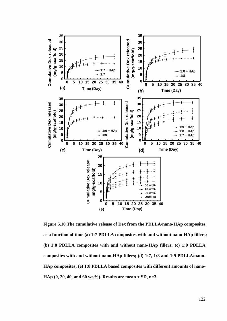
122
Figure 5.10 The cumulative release of Dex from the PDLLA/nano-HAp composites
as a function of time (a) 1:7 PDLLA composites with and without nano-HAp fillers;
(b) 1:8 PDLLA composites with and without nano-HAp fillers; (c) 1:9 PDLLA
composites with and without nano-HAp fillers; (d) 1:7, 1:8 and 1:9 PDLLA/nano-
HAp composites; (e) 1:8 PDLLA based composites with different amounts of nano-
HAp (0, 20, 40, and 60 wt.%). Results are mean ± SD, n=3.
0 5 10 15 20 25 30 35 400
5
10
15
20
25
30
35
Time (Day)
Cu
mu
lati
ve
De
x r
ele
as
ed
(mg
/g-s
ca
ffo
ld)
1:7 + HAp
1:7
(a)0 5 10 15 20 25 30 35 40
0
5
10
15
20
25
30
35
(b)
Cu
mu
lati
ve
De
x r
ele
as
ed
(mg
/g-s
ca
ffo
ld)
Time (Day)
1:8 + HAp
1:8
0 5 10 15 20 25 30 35 400
5
10
15
20
25
30
35
(c) Time (Day)
Cu
mu
lati
ve
De
x r
ele
as
ed
(mg
/g-s
ca
ffo
ld)
1:9 + HAp
1:9
0 5 10 15 20 25 30 35 400
5
10
15
20
25
30
35
Cu
mu
lati
ve
De
x r
ele
as
ed
(mg
/g-s
ca
ffo
ld)
Time (Day)
1:9 + HAp
1:8 + HAp
1:7 + HAp
(d)
0 5 10 15 20 25 30 35 400
5
10
15
20
25
Cu
mu
lati
ve
De
x r
ele
as
e
(mg
/g-s
ca
ffo
ld)
Time (Day)
60 wt%
40 wt%
20 wt%
Unfilled
(e)
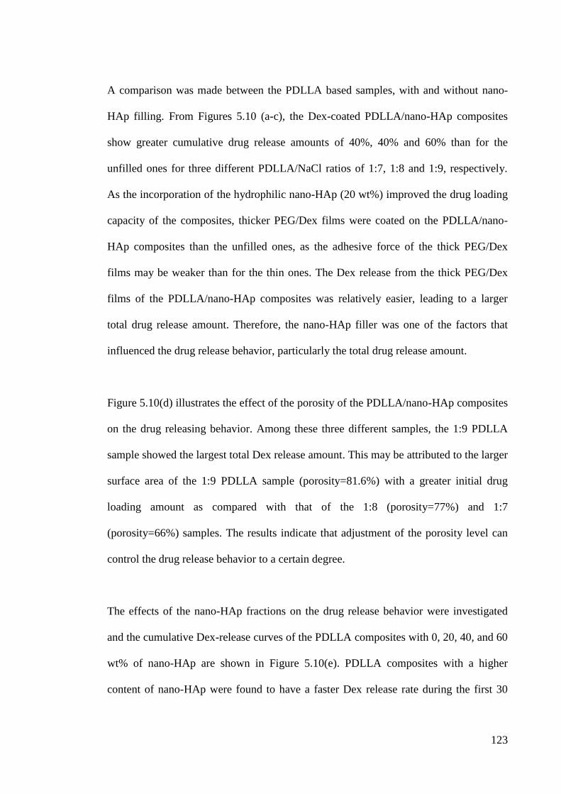
123
A comparison was made between the PDLLA based samples, with and without nano-
HAp filling. From Figures 5.10 (a-c), the Dex-coated PDLLA/nano-HAp composites
show greater cumulative drug release amounts of 40%, 40% and 60% than for the
unfilled ones for three different PDLLA/NaCl ratios of 1:7, 1:8 and 1:9, respectively.
As the incorporation of the hydrophilic nano-HAp (20 wt%) improved the drug loading
capacity of the composites, thicker PEG/Dex films were coated on the PDLLA/nano-
HAp composites than the unfilled ones, as the adhesive force of the thick PEG/Dex
films may be weaker than for the thin ones. The Dex release from the thick PEG/Dex
films of the PDLLA/nano-HAp composites was relatively easier, leading to a larger
total drug release amount. Therefore, the nano-HAp filler was one of the factors that
influenced the drug release behavior, particularly the total drug release amount.
Figure 5.10(d) illustrates the effect of the porosity of the PDLLA/nano-HAp composites
on the drug releasing behavior. Among these three different samples, the 1:9 PDLLA
sample showed the largest total Dex release amount. This may be attributed to the larger
surface area of the 1:9 PDLLA sample (porosity=81.6%) with a greater initial drug
loading amount as compared with that of the 1:8 (porosity=77%) and 1:7
(porosity=66%) samples. The results indicate that adjustment of the porosity level can
control the drug release behavior to a certain degree.
The effects of the nano-HAp fractions on the drug release behavior were investigated
and the cumulative Dex-release curves of the PDLLA composites with 0, 20, 40, and 60
wt% of nano-HAp are shown in Figure 5.10(e). PDLLA composites with a higher
content of nano-HAp were found to have a faster Dex release rate during the first 30
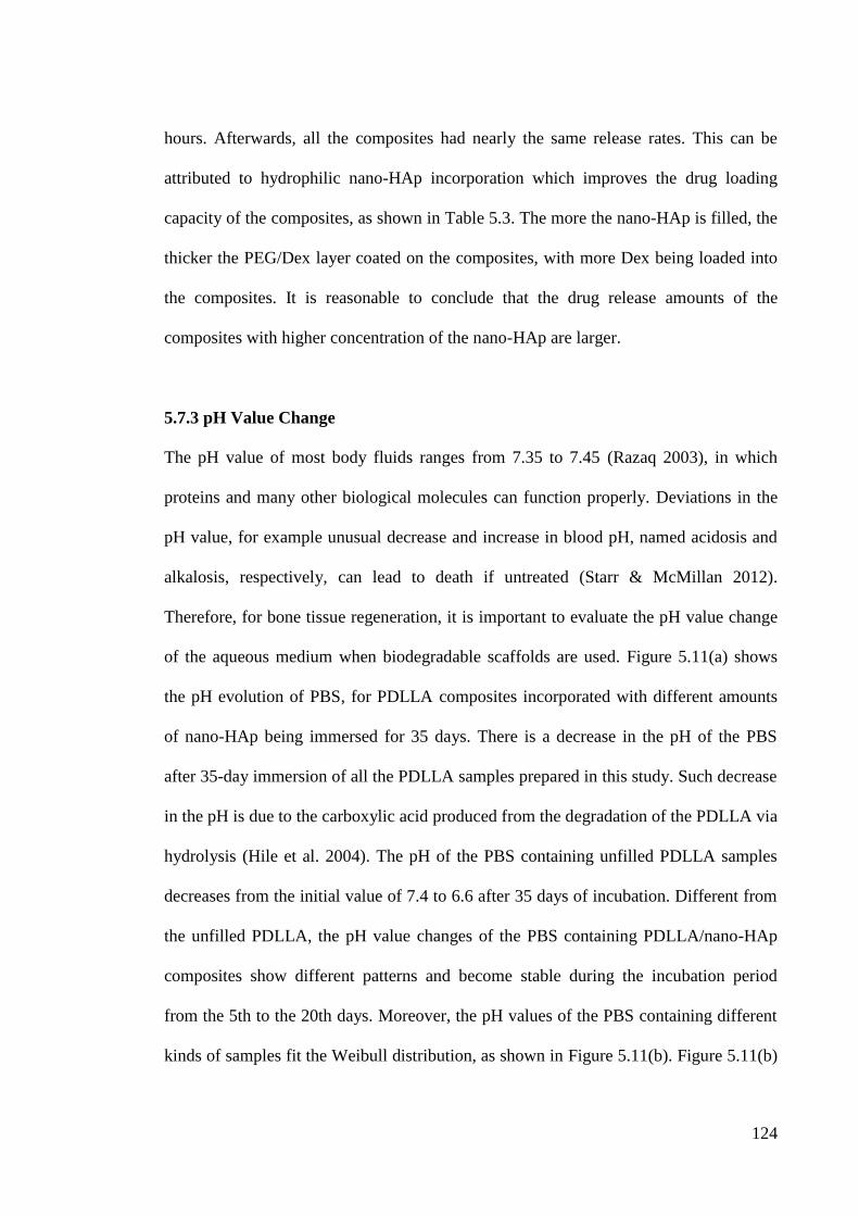
124
hours. Afterwards, all the composites had nearly the same release rates. This can be
attributed to hydrophilic nano-HAp incorporation which improves the drug loading
capacity of the composites, as shown in Table 5.3. The more the nano-HAp is filled, the
thicker the PEG/Dex layer coated on the composites, with more Dex being loaded into
the composites. It is reasonable to conclude that the drug release amounts of the
composites with higher concentration of the nano-HAp are larger.
5.7.3 pH Value Change
The pH value of most body fluids ranges from 7.35 to 7.45 (Razaq 2003), in which
proteins and many other biological molecules can function properly. Deviations in the
pH value, for example unusual decrease and increase in blood pH, named acidosis and
alkalosis, respectively, can lead to death if untreated (Starr & McMillan 2012).
Therefore, for bone tissue regeneration, it is important to evaluate the pH value change
of the aqueous medium when biodegradable scaffolds are used. Figure 5.11(a) shows
the pH evolution of PBS, for PDLLA composites incorporated with different amounts
of nano-HAp being immersed for 35 days. There is a decrease in the pH of the PBS
after 35-day immersion of all the PDLLA samples prepared in this study. Such decrease
in the pH is due to the carboxylic acid produced from the degradation of the PDLLA via
hydrolysis (Hile et al. 2004). The pH of the PBS containing unfilled PDLLA samples
decreases from the initial value of 7.4 to 6.6 after 35 days of incubation. Different from
the unfilled PDLLA, the pH value changes of the PBS containing PDLLA/nano-HAp
composites show different patterns and become stable during the incubation period
from the 5th to the 20th days. Moreover, the pH values of the PBS containing different
kinds of samples fit the Weibull distribution, as shown in Figure 5.11(b). Figure 5.11(b)
125
shows decreasing trends in the pH value of all PDLLA samples, and PDLLA/nano-HAp
composites show a slower decreasing rate in the pH value than unfilled ones. This
suggests that the nano-HAp has the effect of reducing the acidity of the medium and
serves as a buffer system (Bucholz 2002, Schiller & Epple 2003). Moreover, the more
the nano-HAp is filled, the less the pH of the neighboring environment decreases.
Figure 5.11 The pH changes of PBS after immersion of porous PDLLA with 0, 20,
40, 60 wt.% of nano-HAp, (a) pH versus incubation time and (b) The Weibull
probability plot of pH value data.
5.8 Summary
PEG/Dex coated porous PDLLA/nano-HAp composites, fabricated by the PC-DC
technique showed improved bioactivity, due to apatite formation, and enhanced
mechanical properties close to human cancellous bone. The effects of the nano-HAp
fillers on the molecular weight, mass loss and drug release behavior of the PDLLA
composites, as well as the pH value changes of PBS, were thoroughly investigated in
vitro. The PDLLA/nano-HAp composites had a fast degradation rate during the first 7
days incubation and the degradation rate slowed down in the following 21 days.
0 5 10 15 20 25 30 356.50
6.75
7.00
7.25
7.50
pH
Va
lue
Incubation Time (days)
60%
40%
20%
Unfilled
7.4 7.2 7 6.8 6.6 6.41
5
10
50
90
9999.9
Unfilled
Unfilled Reference Line
pH Value
We
ibu
ll P
erc
en
tile
s 20%
20% Reference Line
60%
60% Reference Line
40%
40% Reference Line
(b) (a)
126
Mechanical properties of the PDLLA/nano-HAp composites after incubation in the SBF
can be greatly affected by the weight loss, degradation and apatite formation of the
samples. Results from the mechanical properties investigation show that the weight loss,
polymer plasticization, and degradation of the PDLLA/nano-HAp composites resulted
in a reduction of their compressive moduli and strengths. On the other hand,
compressive moduli and strengths of the scaffolds were improved due to the rigid
apatite formation on them after the SBF incubation. 35-day Dex-releasing was observed
from the drug release study. The initial drug loading amounts and total drug release
amounts increased with the fraction of nano-HAp and porosity of the scaffolds. A larger
total drug release amount of scaffold provides with longer drug release times and higher
drug concentration around the damaged bone tissue, which may improve the efficiency
of treatment. Moreover, the PDLLA/nano-HAp composites had a pH buffering effect
during a 35-day incubation in the PBS, so that the risk of chronic inflammation
complications can be reduced. The PDLLA/nano-HAp scaffolds fabricated in this study
have been confirmed with great biocompatibility, interconnected porous structure,
similar mechanical properties as human cancellous bone, drug delivery ability and
degradability. The shape memory effects, as well as their thermal mechanical properties
are described in the next chapter.
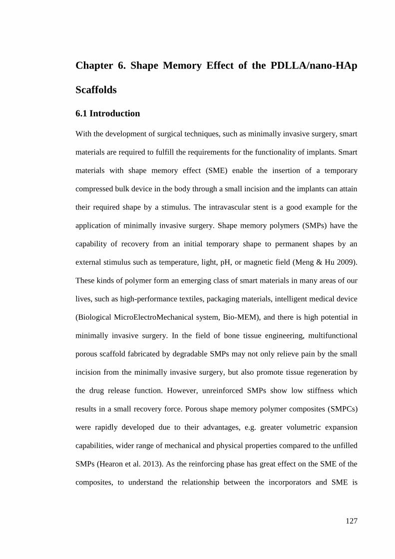
127
Chapter 6. Shape Memory Effect of the PDLLA/nano-HAp
Scaffolds
6.1 Introduction
With the development of surgical techniques, such as minimally invasive surgery, smart
materials are required to fulfill the requirements for the functionality of implants. Smart
materials with shape memory effect (SME) enable the insertion of a temporary
compressed bulk device in the body through a small incision and the implants can attain
their required shape by a stimulus. The intravascular stent is a good example for the
application of minimally invasive surgery. Shape memory polymers (SMPs) have the
capability of recovery from an initial temporary shape to permanent shapes by an
external stimulus such as temperature, light, pH, or magnetic field (Meng & Hu 2009).
These kinds of polymer form an emerging class of smart materials in many areas of our
lives, such as high-performance textiles, packaging materials, intelligent medical device
(Biological MicroElectroMechanical system, Bio-MEM), and there is high potential in
minimally invasive surgery. In the field of bone tissue engineering, multifunctional
porous scaffold fabricated by degradable SMPs may not only relieve pain by the small
incision from the minimally invasive surgery, but also promote tissue regeneration by
the drug release function. However, unreinforced SMPs show low stiffness which
results in a small recovery force. Porous shape memory polymer composites (SMPCs)
were rapidly developed due to their advantages, e.g. greater volumetric expansion
capabilities, wider range of mechanical and physical properties compared to the unfilled
SMPs (Hearon et al. 2013). As the reinforcing phase has great effect on the SME of the
composites, to understand the relationship between the incorporators and SME is
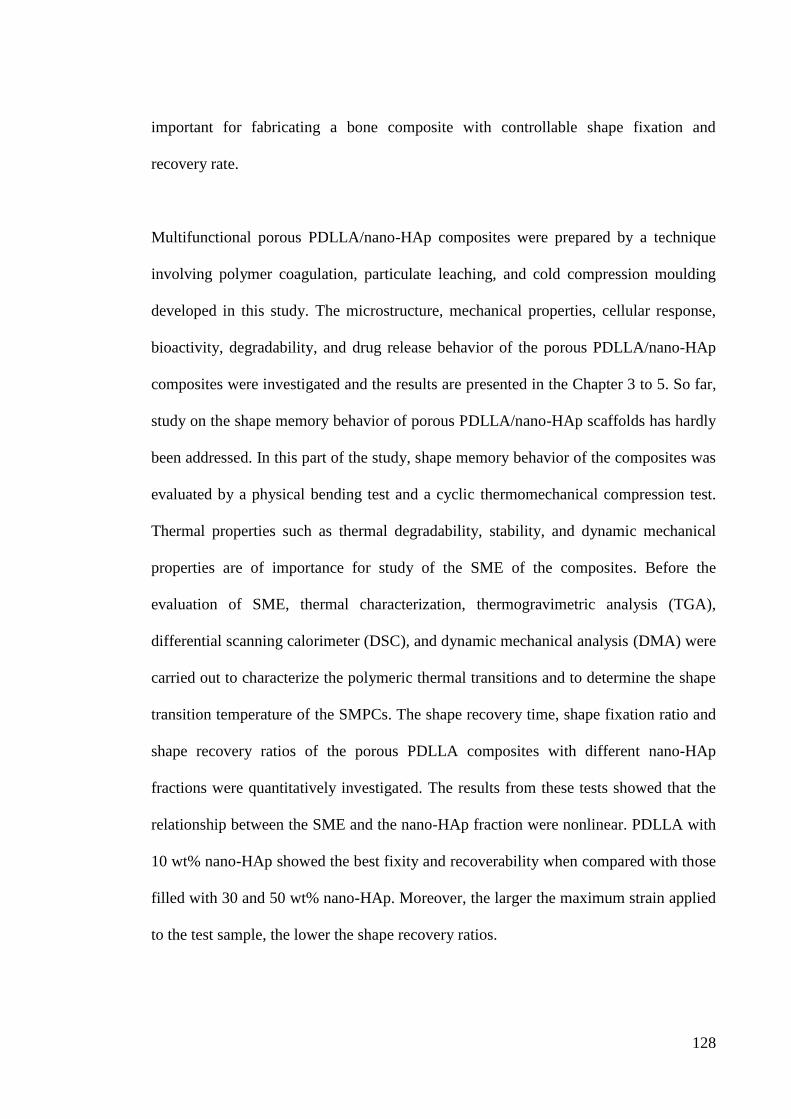
128
important for fabricating a bone composite with controllable shape fixation and
recovery rate.
Multifunctional porous PDLLA/nano-HAp composites were prepared by a technique
involving polymer coagulation, particulate leaching, and cold compression moulding
developed in this study. The microstructure, mechanical properties, cellular response,
bioactivity, degradability, and drug release behavior of the porous PDLLA/nano-HAp
composites were investigated and the results are presented in the Chapter 3 to 5. So far,
study on the shape memory behavior of porous PDLLA/nano-HAp scaffolds has hardly
been addressed. In this part of the study, shape memory behavior of the composites was
evaluated by a physical bending test and a cyclic thermomechanical compression test.
Thermal properties such as thermal degradability, stability, and dynamic mechanical
properties are of importance for study of the SME of the composites. Before the
evaluation of SME, thermal characterization, thermogravimetric analysis (TGA),
differential scanning calorimeter (DSC), and dynamic mechanical analysis (DMA) were
carried out to characterize the polymeric thermal transitions and to determine the shape
transition temperature of the SMPCs. The shape recovery time, shape fixation ratio and
shape recovery ratios of the porous PDLLA composites with different nano-HAp
fractions were quantitatively investigated. The results from these tests showed that the
relationship between the SME and the nano-HAp fraction were nonlinear. PDLLA with
10 wt% nano-HAp showed the best fixity and recoverability when compared with those
filled with 30 and 50 wt% nano-HAp. Moreover, the larger the maximum strain applied
to the test sample, the lower the shape recovery ratios.
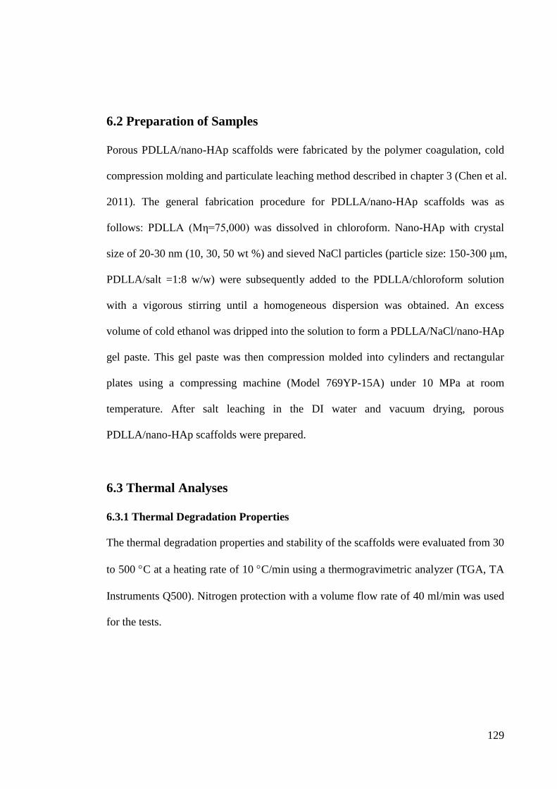
129
6.2 Preparation of Samples
Porous PDLLA/nano-HAp scaffolds were fabricated by the polymer coagulation, cold
compression molding and particulate leaching method described in chapter 3 (Chen et al.
2011). The general fabrication procedure for PDLLA/nano-HAp scaffolds was as
follows: PDLLA (Mη=75,000) was dissolved in chloroform. Nano-HAp with crystal
size of 20-30 nm (10, 30, 50 wt %) and sieved NaCl particles (particle size: 150-300 μm,
PDLLA/salt =1:8 w/w) were subsequently added to the PDLLA/chloroform solution
with a vigorous stirring until a homogeneous dispersion was obtained. An excess
volume of cold ethanol was dripped into the solution to form a PDLLA/NaCl/nano-HAp
gel paste. This gel paste was then compression molded into cylinders and rectangular
plates using a compressing machine (Model 769YP-15A) under 10 MPa at room
temperature. After salt leaching in the DI water and vacuum drying, porous
PDLLA/nano-HAp scaffolds were prepared.
6.3 Thermal Analyses
6.3.1 Thermal Degradation Properties
The thermal degradation properties and stability of the scaffolds were evaluated from 30
to 500 C at a heating rate of 10 C/min using a thermogravimetric analyzer (TGA, TA
Instruments Q500). Nitrogen protection with a volume flow rate of 40 ml/min was used
for the tests.
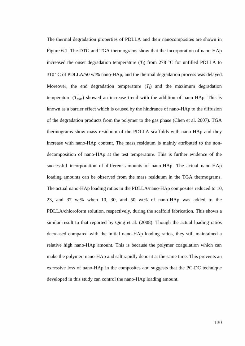
130
The thermal degradation properties of PDLLA and their nanocomposites are shown in
Figure 6.1. The DTG and TGA thermograms show that the incorporation of nano-HAp
increased the onset degradation temperature (Ti) from 278 C for unfilled PDLLA to
310 C of PDLLA/50 wt% nano-HAp, and the thermal degradation process was delayed.
Moreover, the end degradation temperature (Tf) and the maximum degradation
temperature (Tmax) showed an increase trend with the addition of nano-HAp. This is
known as a barrier effect which is caused by the hindrance of nano-HAp to the diffusion
of the degradation products from the polymer to the gas phase (Chen et al. 2007). TGA
thermograms show mass residuum of the PDLLA scaffolds with nano-HAp and they
increase with nano-HAp content. The mass residuum is mainly attributed to the non-
decomposition of nano-HAp at the test temperature. This is further evidence of the
successful incorporation of different amounts of nano-HAp. The actual nano-HAp
loading amounts can be observed from the mass residuum in the TGA thermograms.
The actual nano-HAp loading ratios in the PDLLA/nano-HAp composites reduced to 10,
23, and 37 wt% when 10, 30, and 50 wt% of nano-HAp was added to the
PDLLA/chloroform solution, respectively, during the scaffold fabrication. This shows a
similar result to that reported by Qing et al. (2008). Though the actual loading ratios
decreased compared with the initial nano-HAp loading ratios, they still maintained a
relative high nano-HAp amount. This is because the polymer coagulation which can
make the polymer, nano-HAp and salt rapidly deposit at the same time. This prevents an
excessive loss of nano-HAp in the composites and suggests that the PC-DC technique
developed in this study can control the nano-HAp loading amount.
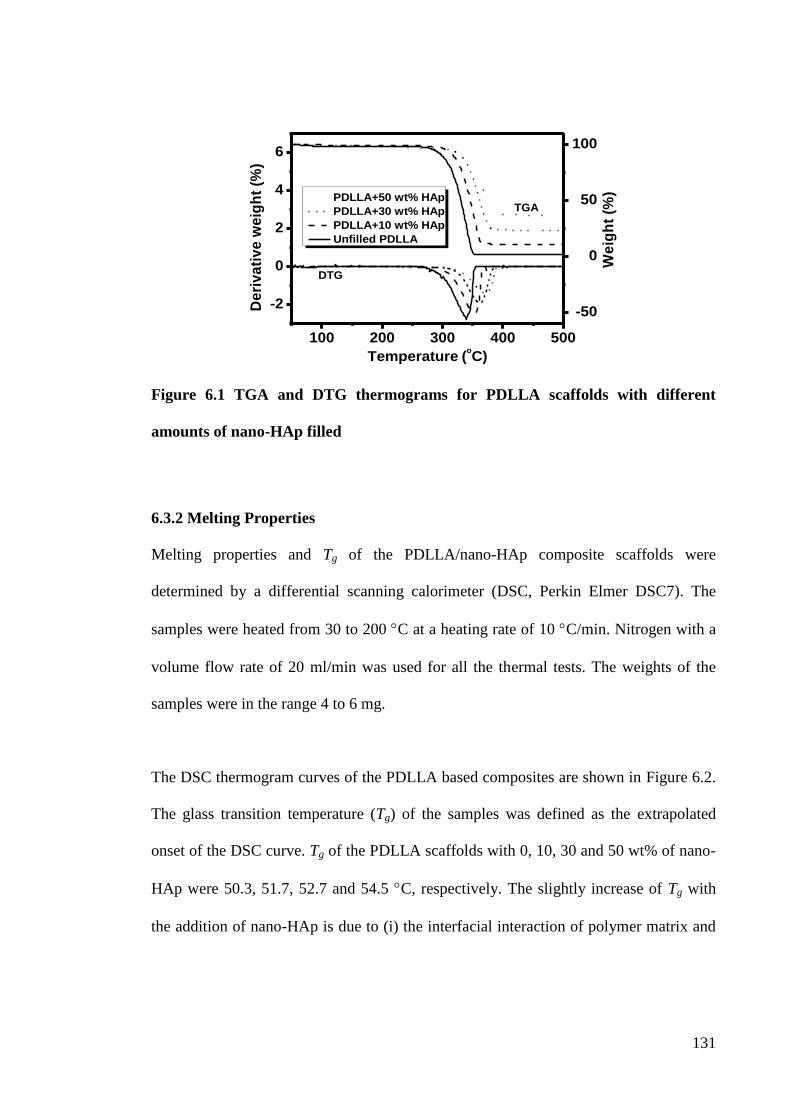
131
Figure 6.1 TGA and DTG thermograms for PDLLA scaffolds with different
amounts of nano-HAp filled
6.3.2 Melting Properties
Melting properties and Tg of the PDLLA/nano-HAp composite scaffolds were
determined by a differential scanning calorimeter (DSC, Perkin Elmer DSC7). The
samples were heated from 30 to 200 C at a heating rate of 10 C/min. Nitrogen with a
volume flow rate of 20 ml/min was used for all the thermal tests. The weights of the
samples were in the range 4 to 6 mg.
The DSC thermogram curves of the PDLLA based composites are shown in Figure 6.2.
The glass transition temperature (Tg) of the samples was defined as the extrapolated
onset of the DSC curve. Tg of the PDLLA scaffolds with 0, 10, 30 and 50 wt% of nano-
HAp were 50.3, 51.7, 52.7 and 54.5 C, respectively. The slightly increase of Tg with
the addition of nano-HAp is due to (i) the interfacial interaction of polymer matrix and
100 200 300 400 500
-50
0
50
100
-2
0
2
4
6
We
igh
t (%
)
Temperature (oC)
PDLLA+50 wt% HAp
PDLLA+30 wt% HAp
PDLLA+10 wt% HAp
Unfilled PDLLA
De
riv
ati
ve
we
igh
t (%
)
DTG
TGA
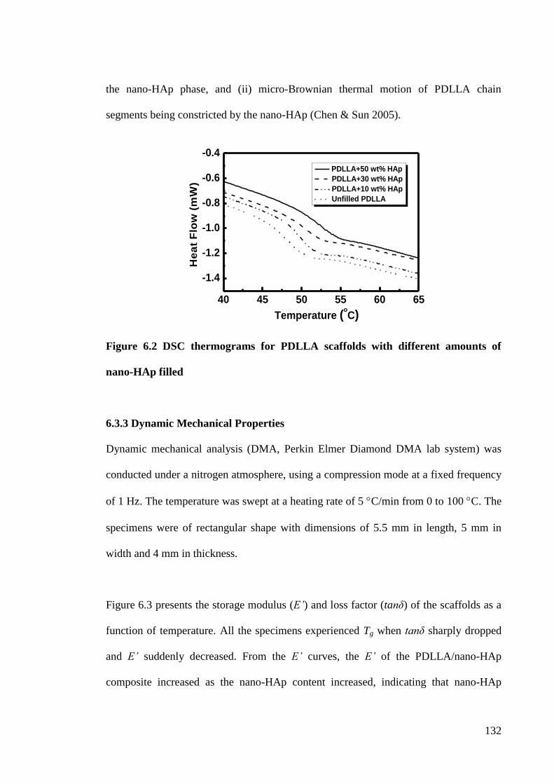
132
the nano-HAp phase, and (ii) micro-Brownian thermal motion of PDLLA chain
segments being constricted by the nano-HAp (Chen & Sun 2005).
Figure 6.2 DSC thermograms for PDLLA scaffolds with different amounts of
nano-HAp filled
6.3.3 Dynamic Mechanical Properties
Dynamic mechanical analysis (DMA, Perkin Elmer Diamond DMA lab system) was
conducted under a nitrogen atmosphere, using a compression mode at a fixed frequency
of 1 Hz. The temperature was swept at a heating rate of 5 C/min from 0 to 100 C. The
specimens were of rectangular shape with dimensions of 5.5 mm in length, 5 mm in
width and 4 mm in thickness.
Figure 6.3 presents the storage modulus (E’) and loss factor (tanδ) of the scaffolds as a
function of temperature. All the specimens experienced Tg when tanδ sharply dropped
and E’ suddenly decreased. From the E’ curves, the E’ of the PDLLA/nano-HAp
composite increased as the nano-HAp content increased, indicating that nano-HAp
40 45 50 55 60 65
-1.4
-1.2
-1.0
-0.8
-0.6
-0.4
PDLLA+50 wt% HAp
PDLLA+30 wt% HAp
PDLLA+10 wt% HAp
Unfilled PDLLA
He
at
Flo
w (
mW
)
Temperature (oC)
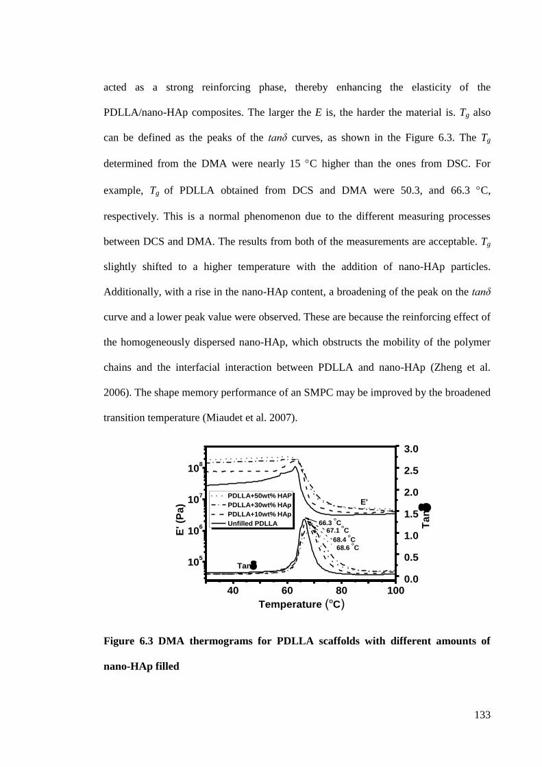
133
acted as a strong reinforcing phase, thereby enhancing the elasticity of the
PDLLA/nano-HAp composites. The larger the E is, the harder the material is. Tg also
can be defined as the peaks of the tanδ curves, as shown in the Figure 6.3. The Tg
determined from the DMA were nearly 15 C higher than the ones from DSC. For
example, Tg of PDLLA obtained from DCS and DMA were 50.3, and 66.3 C,
respectively. This is a normal phenomenon due to the different measuring processes
between DCS and DMA. The results from both of the measurements are acceptable. Tg
slightly shifted to a higher temperature with the addition of nano-HAp particles.
Additionally, with a rise in the nano-HAp content, a broadening of the peak on the tanδ
curve and a lower peak value were observed. These are because the reinforcing effect of
the homogeneously dispersed nano-HAp, which obstructs the mobility of the polymer
chains and the interfacial interaction between PDLLA and nano-HAp (Zheng et al.
2006). The shape memory performance of an SMPC may be improved by the broadened
transition temperature (Miaudet et al. 2007).
Figure 6.3 DMA thermograms for PDLLA scaffolds with different amounts of
nano-HAp filled
105
106
107
108
40 60 80 1000.0
0.5
1.0
1.5
2.0
2.5
3.0
E'
Temperature (OC)
E' (P
a)
Tan
68.6 O
C
68.4 O
C
67.1 O
C66.3
O
C Ta
n
PDLLA+50wt% HAP
PDLLA+30wt% HAp
PDLLA+10wt% HAp
Unfilled PDLLA
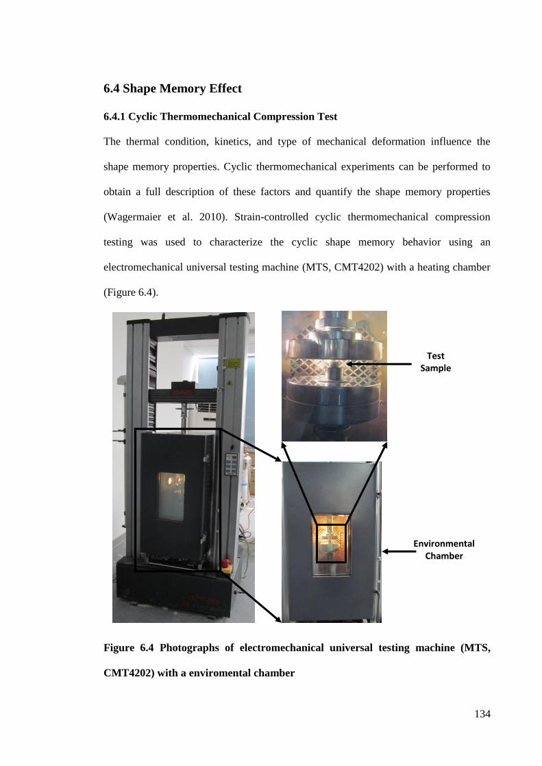
134
6.4 Shape Memory Effect
6.4.1 Cyclic Thermomechanical Compression Test
The thermal condition, kinetics, and type of mechanical deformation influence the
shape memory properties. Cyclic thermomechanical experiments can be performed to
obtain a full description of these factors and quantify the shape memory properties
(Wagermaier et al. 2010). Strain-controlled cyclic thermomechanical compression
testing was used to characterize the cyclic shape memory behavior using an
electromechanical universal testing machine (MTS, CMT4202) with a heating chamber
(Figure 6.4).
Figure 6.4 Photographs of electromechanical universal testing machine (MTS,
CMT4202) with a enviromental chamber
Environmental Chamber
Test Sample
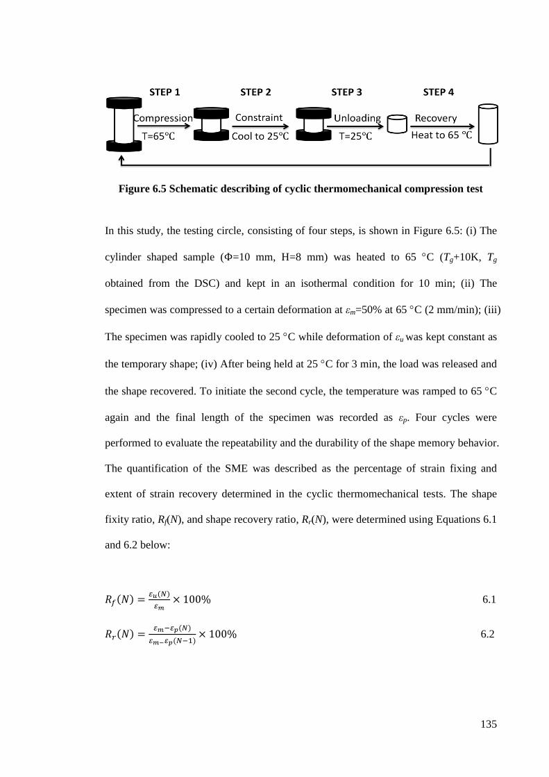
135
Figure 6.5 Schematic describing of cyclic thermomechanical compression test
In this study, the testing circle, consisting of four steps, is shown in Figure 6.5: (i) The
cylinder shaped sample (Φ=10 mm, H=8 mm) was heated to 65 C (Tg+10K, Tg
obtained from the DSC) and kept in an isothermal condition for 10 min; (ii) The
specimen was compressed to a certain deformation at εm=50% at 65 C (2 mm/min); (iii)
The specimen was rapidly cooled to 25 C while deformation of εu was kept constant as
the temporary shape; (iv) After being held at 25 C for 3 min, the load was released and
the shape recovered. To initiate the second cycle, the temperature was ramped to 65 C
again and the final length of the specimen was recorded as εp. Four cycles were
performed to evaluate the repeatability and the durability of the shape memory behavior.
The quantification of the SME was described as the percentage of strain fixing and
extent of strain recovery determined in the cyclic thermomechanical tests. The shape
fixity ratio, Rf(N), and shape recovery ratio, Rr(N), were determined using Equations 6.1
and 6.2 below:
6.1
6.2
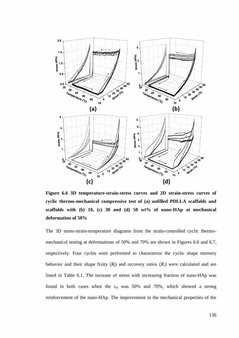
136
(a) (b)
(c) (d)
Figure 6.6 3D temperature-strain-stress curves and 2D strain-stress curves of
cyclic thermo-mechanical compressive test of (a) unfilled PDLLA scaffolds and
scaffolds with (b) 10, (c) 30 and (d) 50 wt% of nano-HAp at mechanical
deformation of 50%
The 3D stress-strain-temperature diagrams from the strain-controlled cyclic thermo-
mechanical testing at deformations of 50% and 70% are shown in Figures 6.6 and 6.7,
respectively. Four cycles were performed to characterize the cyclic shape memory
behavior and their shape fixity (Rf) and recovery ratios (Rr) were calculated and are
listed in Table 6.1. The increase of stress with increasing fraction of nano-HAp was
found in both cases when the εm was 50% and 70%, which showed a strong
reinforcement of the nano-HAp. The improvement in the mechanical properties of the
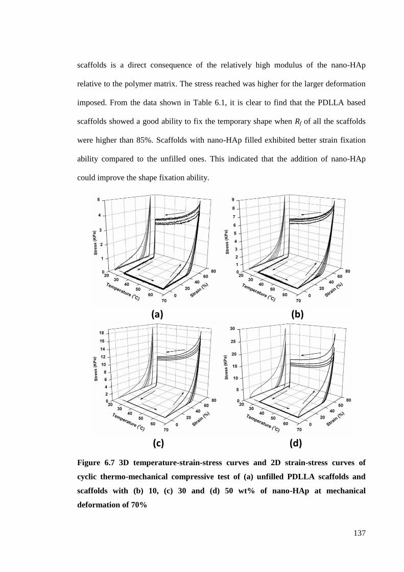
137
scaffolds is a direct consequence of the relatively high modulus of the nano-HAp
relative to the polymer matrix. The stress reached was higher for the larger deformation
imposed. From the data shown in Table 6.1, it is clear to find that the PDLLA based
scaffolds showed a good ability to fix the temporary shape when Rf of all the scaffolds
were higher than 85%. Scaffolds with nano-HAp filled exhibited better strain fixation
ability compared to the unfilled ones. This indicated that the addition of nano-HAp
could improve the shape fixation ability.
(a) (b)
(c) (d)
Figure 6.7 3D temperature-strain-stress curves and 2D strain-stress curves of
cyclic thermo-mechanical compressive test of (a) unfilled PDLLA scaffolds and
scaffolds with (b) 10, (c) 30 and (d) 50 wt% of nano-HAp at mechanical
deformation of 70%
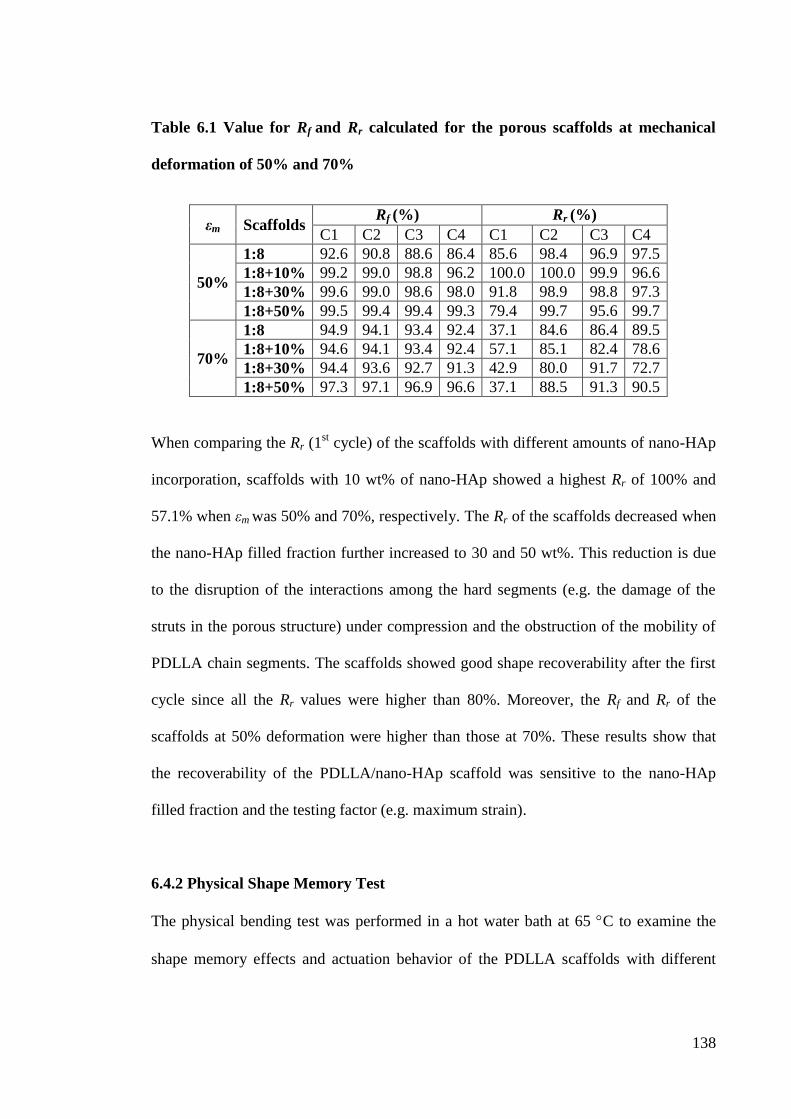
138
Table 6.1 Value for Rf and Rr calculated for the porous scaffolds at mechanical
deformation of 50% and 70%
εm Scaffolds Rf (%) Rr (%)
C1 C2 C3 C4 C1 C2 C3 C4
50%
1:8 92.6 90.8 88.6 86.4 85.6 98.4 96.9 97.5
1:8+10% 99.2 99.0 98.8 96.2 100.0 100.0 99.9 96.6
1:8+30% 99.6 99.0 98.6 98.0 91.8 98.9 98.8 97.3
1:8+50% 99.5 99.4 99.4 99.3 79.4 99.7 95.6 99.7
70%
1:8 94.9 94.1 93.4 92.4 37.1 84.6 86.4 89.5
1:8+10% 94.6 94.1 93.4 92.4 57.1 85.1 82.4 78.6
1:8+30% 94.4 93.6 92.7 91.3 42.9 80.0 91.7 72.7
1:8+50% 97.3 97.1 96.9 96.6 37.1 88.5 91.3 90.5
When comparing the Rr (1st cycle) of the scaffolds with different amounts of nano-HAp
incorporation, scaffolds with 10 wt% of nano-HAp showed a highest Rr of 100% and
57.1% when εm was 50% and 70%, respectively. The Rr of the scaffolds decreased when
the nano-HAp filled fraction further increased to 30 and 50 wt%. This reduction is due
to the disruption of the interactions among the hard segments (e.g. the damage of the
struts in the porous structure) under compression and the obstruction of the mobility of
PDLLA chain segments. The scaffolds showed good shape recoverability after the first
cycle since all the Rr values were higher than 80%. Moreover, the Rf and Rr of the
scaffolds at 50% deformation were higher than those at 70%. These results show that
the recoverability of the PDLLA/nano-HAp scaffold was sensitive to the nano-HAp
filled fraction and the testing factor (e.g. maximum strain).
6.4.2 Physical Shape Memory Test
The physical bending test was performed in a hot water bath at 65 C to examine the
shape memory effects and actuation behavior of the PDLLA scaffolds with different
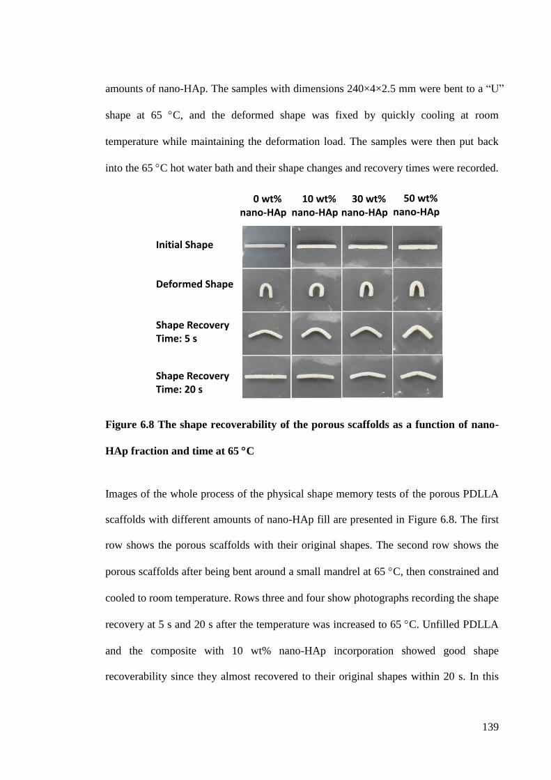
139
amounts of nano-HAp. The samples with dimensions 240×4×2.5 mm were bent to a “U”
shape at 65 C, and the deformed shape was fixed by quickly cooling at room
temperature while maintaining the deformation load. The samples were then put back
into the 65 C hot water bath and their shape changes and recovery times were recorded.
Figure 6.8 The shape recoverability of the porous scaffolds as a function of nano-
HAp fraction and time at 65 C
Images of the whole process of the physical shape memory tests of the porous PDLLA
scaffolds with different amounts of nano-HAp fill are presented in Figure 6.8. The first
row shows the porous scaffolds with their original shapes. The second row shows the
porous scaffolds after being bent around a small mandrel at 65 C, then constrained and
cooled to room temperature. Rows three and four show photographs recording the shape
recovery at 5 s and 20 s after the temperature was increased to 65 C. Unfilled PDLLA
and the composite with 10 wt% nano-HAp incorporation showed good shape
recoverability since they almost recovered to their original shapes within 20 s. In this
0 wt% nano-HAp
10 wt% nano-HAp
30 wt% nano-HAp
50 wt% nano-HAp
Initial Shape
Deformed Shape
Shape Recovery Time: 5 s
Shape Recovery Time: 20 s
140
PDLLA/nano-HAp composite system, the nano-HAp and the temporary entanglements
of the polymer chains acted as the physical netpoints which could be used for the
fixation of the permanent shape. The amorphous PDLLA (switching segments) became
flexible when the temperature increase to 65 C and the composite could be deformed
elastically. And the deformed shape could be fixed when the temperature cooled back to
room temperature. The permanent shape recovered when the composite was heated up
again. However, the scaffolds with 30 and 50 wt% of nano-HAp filled showed
irreversible deformations. This is because the larger amount of nano-HAp (>10 wt%)
restricted the movement of the amorphous PDLLA chain segments and the damage of
the scaffolds under compression. This phenomenon is in a good agreement with the
cyclic thermomechanical tests.
Table 6.2 Shape recovery time of the porous scaffolds recovered from “U” shape to
their original “I” shape at 65 C
Scaffolds 1:8 1:8 +10% 1:8 +30% 1:8 +50%
Shape Recovery Time (s) 8 4 14 20
The shape recovery times of the scaffolds are shown in Table 6.2. The scaffolds with 10
wt% of nano-HAp recovered faster than the unfilled ones and the scaffolds with higher
fraction of nano-HAp. Small amount of nano-HAp (e.g. 10 wt%) act as reinforcing
fillers and netpoints in the shape memory composites, not only shortening the shape
recovery time but also improving the mechanical properties, shape recoverability and
shape fixation ability. Though the transition temperature is 65°C for the PDLLA/nano-
HAp, the scaffold still showed a potential as a bone scaffold in bone tissue engineering
for minimally invasive surgery. Co-polymers or block-polymers derived from PDLLA
141
and other polymers may be developed with a lower transition temperature in the next
stage of this study.
6.5 Summary
The effect of nano-HAp on the thermal properties and shape memory effect of the
porous PDLLA composite were investigated. The Ti and Tg increased with the
increasing amounts of nano-HAp particles, which directly affected the mechanical
properties and shape memory behavior of the composites by increasing the shape
transition temperature. Porous PDLLA/nano-HAp composites fabricated in this study
showed desirable shape memory effects from the cyclic thermomechanical test and
physical shape memory test. The sample still showed good shape recoverability after 4
cycles. PDLLA with 10 wt% nano-HAp possessed the best shape fixity, recoverability
and shortest shape recovery time of only 4 seconds. The addition of nano-HAp (30 and
50 wt%) restricted the movement of the PDLLA chain segments and reduced the
crystallinity of the PDLLA chains, which resulted in the residual deformation and slow
recovery time. PDLLA/nano-HAp showed a better shape memory effect when the
applied maximum strain was 50% than that at 70%. The degree of the SMPCs
compressed also is another factor that influences the ability of shape fixation and
recovery. The multifunctional PEG/Dex coated porous PDLLA/nano-HAp scaffolds
fabricated were confirmed to have controllable interconnected porous structure,
improved mechanical properties, good cellular response, bioactivity, degradability, drug
release function and great shape memory properties. Therefore, PEG/Dex coated
PDLLA/nano-HAp scaffolds have a high potential in bone tissue engineering
applications.
142
Chapter 7. Conclusions and Statement of Originality
7.1 Overall Conclusions
A new technique was developed for bone scaffold fabrication. This technique can
independently control the pore size and porosity of the scaffolds by means of particle
sizes and the amounts of NaCl, whereas the present gas forming/particulate leaching
method has difficulty in controlling the pore size. The solvent residual can be greatly
reduced by the polymer coagulation and vacuum drying processes when compared with
the conventional solvent casting/particulate leaching method. Moreover, the gel-like
polymer based paste formed by coagulation of the polymer in the non-solvent proved
easy to mold and produced a more uniform NaCl distribution in the polymer matrix
without the formation of a skin layer. Scaffolds having a highly interconnected pore
structure and uniform pore distribution can be fabricated using this technique. Unlike
the thermal electrospinning and compression molding method, the PC-DC technique is
a low temperature approach which can be applied to a wide range of polymer based
materials. Compared with SFF, the PC-DC technique can be easily carried out and it
does not need costly equipment. Dex is effectively loaded into the matrix, confirming
that the subsequent PEG/Dex coating process is a suitable alternative method for drug
loading, especially for water soluble drugs. This process avoids a premature drug loss
when the drug is incorporated into the scaffold during the scaffold fabrication process.
Using this technique, PEG/Dex coated porous PDLLA/nano-HAp porous scaffolds with
similar mechanical properties to human cancellous bone, shape memory effect and drug
release function were successfully fabricated.
143
The cellular response on the PDLLA based scaffolds showed that the cell adhesion,
proliferation and differentiation were highly influenced by the chemical composition
and microstructure of the scaffolds. The PDLLA based composites fabricated by the
proposed method, with pore sizes from 200 to 350 µm and porosity from 60% to 80%,
demonstrated good ability for supporting the cell attachment and ingrowth. All the
samples were covered by a monolayer of cells after 7 days of culture and no cytotoxic
responses were observed. Higher porosity of the scaffold, more cell adhesion and faster
proliferation rate of the cells on the scaffold were found. Addition of nano-HAp in the
PDLLA was proven to facilitate osteogenic differentiation of the osteoblast progenitor
MG63 cells. Moreover, bonelike apatite formation on the scaffolds after SBF incubation
showed an improved bioactivity by nano-HAp incorporation which could assist bone
bonding. Incorporation of nano-HAp slows down the polymer degradation and mass
loss of the nano-HAp filled scaffolds. In vitro evaluation of the mechanical properties of
the scaffolds showed that the weight loss, polymer plasticization, and degradation of the
PDLLA/nano-HAp scaffolds resulted in a reduction of their compressive moduli and
strengths. On the other hand, compressive moduli and the strengths of the scaffolds
were improved due to the rigid apatite formation on them after the SBF incubation.
Moreover, the PDLLA/nano-HAp scaffolds had a pH buffering effect during incubation
in the SBF, which reduces the risk of chronic inflammation complications.
The influences of the nano-HAp fraction on the multifunctionalities of the composites
have been investigated. PDLLA based composites with larger nano-HAp content had
higher initial drug loading amounts and larger total drug release amounts. Higher
compressive moduli and strengths could be achieved by the larger amounts of nano-
144
HAp incorporation. The significant improvement of the wettability of the composites
was achieved by the incorporation of nano-HAp particles and the coating of the
PEG/Dex films. Desirable shape memory effects of the porous PDLLA/nano-HAp
scaffolds fabricated in this study were confirmed by the cyclic thermomechanical and
physical shape memory tests. Nano-HAp with 10 wt% in PDLLA was shown to be the
best fraction since the scaffold produced possessed the best shape fixity, recoverability
and shortest shape recovery time. The addition of nano-HAp (30 and 50 wt%) restricted
the movement of PDLLA chain segments and reduced the crystallinity of the PDLLA
chains, which resulted in the residual deformation. These indicated that the shape
memory effects of the PDLLA scaffolds can be controlled by the nano-HAp filled
fraction. All these demonstrate that multifunctional porous PDLLA/nano-HAp
composites have a high potential for minimally invasive surgery and bone tissue
engineering applications. The PC-DC technique described in this study provides a new
route for the fabrication of drug loaded scaffolds for bone tissue engineering.
7.2 Originality and Contributions of the Research Work
This study provides a novel way for the fabrication of multifunctional scaffold material
with drug releasing function, shape memory effect and controllable biodegradation rate.
The PC-DC technique solves the problem of non-uniform pore distribution within a
composite matrix fabricated by the conventional solvent casting/particulate leaching
method. Moreover, it shortens the solvent evaporation time and reduces solvent
residuals by polymer coagulation and vacuum drying steps. This low temperature
fabrication technique prevents the thermal decomposition of drug inclusion and the
145
polymer contributes to a wider range of choices of drug loadings and polymer matrices
for bone scaffold fabrication.
This proposed technique provides the ability to develop multifunctional porous
scaffolds which have a similar structure and mechanical properties as human bone.
Porous PDLLA/nano-HAp composites were first coated with PEG/Dex film as a model
system for bone tissue scaffold materials. The PEG/Dex coated PDLLA/nano-HAp
composites then fabricated have many advantages: (a) controllable biodegradability of
the scaffold will avoid secondary surgery to take out the implants after tissue recovery;
(b) controllable mechanical properties of the scaffold will be in phase with the tissue
regeneration; (c) adjustable total drug release amounts provide relatively high
therapeutic efficacy to the diseased tissue to obtain a faster recover rate; (d) excellent
bioactivity (bone like apatite formation) and biocompatibility of the scaffold facilitates
cell adhesion, growth, and differentiation; (e) scaffolds with shape memory effect have
the ability to be precisely positioned in the body by minimal invasive techniques in a
temporary small shape and then gain their application-relevant shape after implantation;
(f) scaffolds have an active cellular response and high potential in bone tissue
engineering.
This is the first time the change of the mechanical properties of the porous
PDLLA/nano-HAp composite scaffolds during incubation in a SBF has been evaluated.
Mechanical properties of bone scaffolds change after implantation by the complex
interactions between the scaffolds and the body fluid (e.g. degradation of the scaffolds,
bonelike apatite formation on the scaffolds, and plasticization of the polymer matrix).
146
To evaluate and control the mechanical property changes are of crucial important for
the success of a scaffold for bone tissue engineering. The results showed that nano-HAp
incorporation and apatite formation made a positive impact on the mechanical
properties of the scaffolds; however, plasticization and degradation of PDLLA had a
negative impact. An interesting phenomenon was found in that the PDLLA/nano-HAp
composites had a fast degradation rate during the first 7 days incubation and the
degradation rate slowed down in the following 21 days. This information could
facilitate the design and fabrication of porous biodegradable scaffolds for different bone
defects.
It is amongst the first attempts to determine the shape memory behavior of the porous
PDLLA/nano-HAp composites. Controlling the nano-HAp filling fraction of the
scaffold could affect the shape memory effects of the scaffolds. PDLLA with 10 wt%
nano-HAp showed the best fixity and recoverability when compared with those filled
with 30 and 50 wt% nano-HAp. Moreover, the larger the maximum strain applied to the
test sample, the lower the shape recovery ratios. The addition of nano-HAp (30 and 50
wt%) restricted the movement of PDLLA chain segments and reduced the crystallinity
of the PDLLA chains, which resulted in the residual deformation.
This research is the first in regard to the cellular response of the PDLLA/nano-HAp
composites using a MG63 cell model. The results showed that the PDLLA based
scaffolds fabricated in this study are biocompatible and facilitate cell attachment and
proliferation. The pore size of the fabricated scaffolds ranged from 200 to 350 µm and
the high porosity of 60% to 80% allowed cells to host and penetrate inside the scaffolds.
147
The whole scaffolds covered by the MG63 cells were observed after 7 days of culture,
during which time, the number of cells on the PDLLA based substrates increased.
Moreover, the scaffolds with higher porosity were beneficial for cell attachment, growth,
migration and differentiation. Nano-HAp incorporation that promoted calcium
deposition (cell mineralization/osteoconduction) was also found.
7.3 Research Outputs
Some findings from this study gave rise to the following research outputs:
1. Chen L., Tang C.Y., Chen D.Z., Wong C.T., Tsui C.P., Fabrication and
characterization of poly-D-L-lactide/nano-hydroxyapatite composite scaffolds with
poly(ethylene glycol) coating and dexamethasone releasing, Composites Science and
Technology, 2011;71(16):1842-1849.
2. Chen L., Tang C.Y., Tsui C.P., Chen D.Z., Mechanical properties and in vitro
evaluation of bioactivity of bioactivity, degradation of dexamethasone-releasing poly-
D-L-lactide/nano-hydroxyapatite composite scaffolds, Journal of the Mechanical
Behavior of Biomedical Materials, 2013;22:41-50.
3. Chen L., Tang C.Y., Ku H.S.L., Tsui C.P., Chen X., Microwave sintering and
characterization of polypropylene/multi-walled carbon nanotube/hydroxyapatite
composites, Composite Part B, 2014;56:504-511.
4. Chen L., Tang C.Y., H.S. Ku, Chen D.Z., Tsui C.P., Surface coating of poly-D-L-
lactide/nano- hydroxyapatite composite scaffolds for dexamethasone-releasing function
148
and wettability enhancement, presented at the Materials Science & Technology 2012
Conference & Exhibition, October 7-11, 2012, Pittsburgh, Pennsylvania, USA.
5. Chen L., Tang C.Y., Tsui C.P., Chen D.Z., Fabrication and characterization of nano-
HAp reinforced poly-D-L-lactide scaffolds with poly (ethylene glycol)/dexamethasone
coatings, presented at the 6th Asia-Europe Symposium on Processing and Properties of
Reinforced Polymers, June 2-6, 2013, Wuhan, China
6. Chen L., Tang C.Y., Tsui C.P., Chen D.Z., Thermal characterization and shape
memory effect of porous poly-D-L-lactide/nano-hydroxyapatite composite scaffolds,
under preparation.
7. Chen L., Tang C.Y., Tsui C.P., Chen D.Z., Human osteoblast-like cells (MG63)
proliferation on poly-D-L-lactide/nano-hydroxyapatite composite scaffolds with
poly(ethylene glycol) coating and dexamethasone releasing, under preparation.
7.4 Suggestions for Future Work
In this study, PEG/Dex coated PDLLA/nano-HAp composite scaffolds have been
fabricated using the newly developed technique. Despite the microstructures,
degradability, drug release behavior, mechanical properties, shape memory effects,
bioactivity and in vitro cellular investigation of the scaffolds being extensively studied,
there are some related issues worthy of further investigation, as follows:
i. Surface modification of the PEG/Dex coating by incorporating some other bioactive
149
organic components, such as collagen, Arg-Gly-Asp tripeptide, and growth factors,
may be undertaken to further improve the cell adhesion and osteoconductivity of the
scaffolds and promote cellular adhesion, growth and differentiation.
ii. Microcapsules with bioactive drugs inside may be developed and coated on the
scaffolds for a controllable drug delivery system. The drug release rate can be
controlled by the thickness and degradation rate of the microcapsules.
iii. Co-polymers or block-polymers derived from PDLLA with controllable shape
memory effect and transition temperatures close to the human body temperature
may be developed, and may show a higher potential for bone tissue engineering.
iv. Preclinical studies, such as animal model experiments, may be conducted in vivo to
further evaluate and ascertain the performance of the PDLLA/nano-HAp scaffolds
with PEG/Dex coatings. The experiments should begin with small animals and
proceed to larger animals after some positive results emerge.
v. Based on the relationship between the shape memory behavior, drug release profile,
degradation, apatite formation and mechanical properties changes of the produced
composites, a finite element model can be developed to simulate the behavior of the
materials implanted into a human body. This finite element model may facilitate the
design and manufacture of multifunctional composite bone scaffolds and provide a
powerful tool to develop the material and meet patient-specific functional
requirements.
150
References
Adams S.B.J., Shamji M.F., Nettles D.L., Hwang P., Setton L.A. 2009, "Sustained
Release of Antibiotics From Injectable and Thermally Responsive Polypeptide
Depots", Journal of Biomedical Materials Research Part B-Applied biomaterials,
vol. 90B, no. 1, pp. 67-74.
Agarwal, S., Wendorff, J.H., Greiner, A. 2008, "Use of electrospinning technique for
biomedical applications", Polymer, vol. 49, no. 26, pp. 5603-5621.
Agrawal, C.M., Ray, R.B. 2001, "Biodegradable polymeric scaffolds for
musculoskeletal tissue engineering", Journal of Biomedical Materials Research, vol.
55, no. 2, pp. 141-150.
Albrektsson, T., Johansson, C. 2001, "Osteoinduction, osteoconduction and
osseointegration", European Spine Journal, vol. 10, no. 2, pp. 96-101.
Aleksendrić, D., Balac, I., Tang C,Y,, Tsui, C.P., Ushokovic, P.S., Ushokovic, D.P.
2010, "Surface characterisation of PLLA polymer in HAp/PLLA biocomposite
material by means of nanoindentation and artificial neural networks". Advances in
Applied Aeramics, vol. 109, no. 2, pp.65-70.
Ang, K.C., Leong, K.F., Chua, C.K., Chandrasekaran, M. 2007, "Compressive
properties and degradability of poly(e-caprolatone)/ hydroxyapatite composites
under accelerated hydrolytic degradation", Journal of Biomedical Materials
Research - Part A, vol. 80, no. 3, pp. 655-660.
Anklekar, R.M., Agrawal, D.K., Roy, R.B. 2001, "Microwave sintering and mechanical
properties of PM copper steel", Powder Metallurgy, vol. 44, no. 4, pp. 355-362.
Anselme, K. 2000, "Osteoblast adhesion on biomaterials", Biomaterials, vol. 21, no. 7,
pp. 667-681.
Anselme, K., Ponche, A., Bigerelle, M. 2010, "Relative influence of surface topography
and surface chemistry on cell response to bone implant materials. Part 2: Biological
aspects", Proceedings of the Institution of Mechanical Engineers, Part H: Journal
of Engineering in Medicine, vol. 224, no. 12, pp. 1487-1507.
Anthony Atala, "Printing a human kidney", Filmed on March 2011 at TED2011,
http://www.ted.com/talks/anthony_atala_printing_a_human_kidney
Armentano, I., Dottori, M., Fortunati, E., Mattioli, S., Kenny, J.M. 2010,
"Biodegradable polymer matrix nanocomposites for tissue engineering: A review",
Polymer Degradation and Stability, vol. 95, no. 11, pp. 2126-2146.
151
Assouline, E., Wachtel, E., Grigull, S., Lustiger, A., Wagner, H.D., Marom, G. 2001,
"Lamellar twisting in a isotactic polypropylene transcrystallinity investigated by
synchrotron microbeam X-ray diffraction", Polymer, vol. 42, no. 14, pp. 6231-6237.
Babensee, J.E., Anderson, J.M., McIntire, L.V., Mikos, A.G. 1998, "Host response to
tissue engineered devices", Advanced Drug Delivery Reviews, vol. 33, no. 1-2, pp.
111-139.
Baer, G.M., Small IV, W., Wilson, T.S., Benett, W.J., Matthews, D.L., Hartman, J.,
Maitland, D.J. 2007, "Fabrication and in vitro deployment of a laser-activated shape
memory polymer vascular stent", BioMedical Engineering Online, vol. 6. art. no. 43.
Bao, T.Q., Franco, R.A., Lee, B.T. 2011, "Preparation and characterization of novel
poly(e-caprolactone)/ biphasic calcium phosphate hybrid composite microspheres",
Journal of Biomedical Materials Research - Part B Applied Biomaterials, vol. 98 B,
no. 2, pp. 272-279.
Behrend, M., Kluge, E., Schüttler, W. 2006, "On the use of unsealed polypropylene
mesh as tracheal replacement", ASAIO Journal, vol. 52, no. 3, pp. 328-333.
Bhattacharyya, A.R., Sreekumar, T.V., Liu, T., Kumar, S., Ericson, L.M., Hauge, R.H.,
Smalley, R.E. 2003, "Crystallization and orientation studies in polypropylene/single
wall carbon nanotube composite", Polymer, vol. 44, no. 8, pp. 2373-2377.
Bianco, A., Di Federico, E., Moscatelli, I., Camaioni, A., Armentano, I., Campagnolo,
L., Dottori, M., Kenny, J.M., Siracusa, G., Gusmano, G. 2009, "Electrospun poly(e-
caprolactone)/Ca-deficient hydroxyapatite nanohybrids: Microstructure, mechanical
properties and cell response by murine embryonic stem cells", Materials Science
and Engineering C, vol. 29, no. 6, pp. 2063-2071.
Biondi M., Ungaro F., Quaglia F., Netti P.A. 2008, "Controlled drug delivery in tissue
engineering", Advanced Drug Delivery Reviews, vol. 60, pp. 229-242.
Black, J., Hastings, G. 1998, Handbook of Biomaterial Properties, Chapman & Hall,
London.
Blaker, J.J., Bismarck, A., Boccaccini, A.R., Young, A.M., Nazhat, S.N. 2010,
"Premature degradation of poly(α-hydroxyesters) during thermal processing of
Bioglass®-containing composites", Acta Biomaterialia, vol. 6, no. 3, pp. 756-762.
Blaker, J.J., Maquet, V., Jérôme, R., Boccaccini, A.R., Nazhat, S.N. 2005, "Mechanical
properties of highly porous PDLLA/Bioglass® composite foams as scaffolds for
bone tissue engineering", Acta Biomaterialia, vol. 1, no. 6, pp. 643-652.
Blaker, J.J., Nazhat, S.N., Maquet, V., Boccaccini, A.R. 2011, "Long-term in vitro
degradation of PDLLA/Bioglass® bone scaffolds in acellular simulated body fluid",
Acta Biomaterialia, vol. 7, no. 2, pp. 829-840.
152
Blau, H.M., Brazelton, T.R., Weimann, J.M. 2001, "The evolving concept of a stem cell:
Entity or function?", Cell, vol. 105, no. 7, pp. 829-841.
Boccaccini, A.R., Blaker, J.J., Maquet, V., Day, R.M., Jéróme, R. 2005, "Preparation
and characterisation of poly(lactide-co-grycolide) (PLGA) and PLGA/Bioglass®
composite tubular foam scaffolds for tissue engineering applications", Materials
Science and Engineering C, vol. 25, no. 1, pp. 23-31.
Boland, E., Wnek, G., Simpson, D., Pawlowski, K., Bowlin, G. 2001, "Tailoring tissue
engineering scaffolds using electrostatic processing techniques: A study of
poly(glycolic acid) electrospinning", Journal of Macromolecular Science-pure and
Applied Chemisry, vol. 38, no. 12, pp. 1231-1243.
Bose, S., Roy, M., Bandyopadhyay, A. 2012, "Recent advances in bone tissue
engineering scaffolds", Trends in Biotechnology, vol. 30, no. 10, pp. 546-554.
Bucholz, R. 2002, "Nonallograft osteoconductive bone graft substitutes", Clinical
Orthopaedics and Related Research, vol. 395, pp. 44-52.
Burr, D.B. & Akkus, O. 2014, "Bone Morphology and Organization" in Basic and
Applied Bone Biology, eds. D.B. Burr & M.R. Allen, Academic Press, San Diego,
pp. 3-25.
Buttery, L.D.K., Bourne, S., Xynos, J.D., Wood, H., Hughes, F.J., Hughes, S.P.F.,
Episkopou, V., Polak, J.M. 2001, "Differentiation of osteoblasts and in Vitro bone
formation from murine embryonic stem cells", Tissue Engineering, vol. 7, no. 1, pp.
89-99.
Cai, B.L., Chang, Y.G., Mei, J.H., Ji, W.W., Yi, F.P., Yang, D.Z., Guo, Z.L., Ma, L.G.,
Wang, K., Ming, J.T., Yu, Q.W., Qian, Z.Y. 2008, "Thermoreversible gel-sol
behavior of biodegradable PCL-PEG-PCL triblock copolymer in aqueous solutions",
Journal of Biomedical Materials Research - Part B Applied Biomaterials, vol. 84,
no. 1, pp. 165-175.
Calandrelli, L., Calarco, A., Laurienzo, P., Malinconico, M., Petillo, O., Peluso, G.
2008, "Compatibilized polymer blends based on PDLLA and PCL for application in
bioartificial liver", Biomacromolecules, vol. 9, no. 6, pp. 1527-1534.
Campanelli, G., Catena, F. & Ansaloni, L. 2008, "Prosthetic abdominal wall hernia
repair in emergency surgery: From polypropylene to biological meshes", World
Journal of Emergency Surgery, vol. 3, no. 1.
Caplan, A.I. 1994, "The mesengenic process", Clinics in Plastic Surgery, vol. 21, no. 3,
pp. 429-435.
Cartmell, S., Huynh, K., Lin, A., Nagaraja, S., Guldberg, R. 2004, "Quantitative
microcomputed tomography analysis of mineralization within three-dimensional
153
scaffolds in vitro". Journal of Biomedical Materials Research-Part A, vol. 69, no. 1,
pp. 97-104.
Causa, F., Netti, P.A., Ambrosio, L., Ciapetti, G., Baldini, N., Pagani, S., Martini, D.,
Giunti, A. 2006, "Poly-e-caprolactone/hydroxyapatite composites for bone
regeneration: In vitro characterization and human osteoblast response", Journal of
Biomedical Materials Research - Part A, vol. 76, no. 1, pp. 151-162.
Causa, F., Netti, P.A., Ambrosio, L. 2007, "A multi-functional scaffold for tissue
regeneration: The need to engineer a tissue analogue", Biomaterials, vol. 28, no. 34,
pp. 5093-5099.
Charlesby, A. (ed) 1960, Atomic Radiation and Polymers, Pergamon Press, New York.
Chen, G.P., Ushida, T., Tateishi, T. 2001, "Development of biodegradable porous
scaffolds for tissue engineering", Materials Science and Engineering C, vol. 17, no.
1-2, pp. 63-69.
Chen, D.Z., Tang, C.Y., Chan, K.C., Tsui, C.P., Yu, P.H.F., Leung, M.C.P., Uskokovic,
P.S. 2007, "Dynamic mechanical properties and in vitro bioactivity of PHBHV/HA
nanocomposite", Composites Science and Technology, vol. 67, no. 7-8, pp. 1617-
1626.
Chen, Y., Mak, A., Li, J., Wang, M., Shum, A. 2005, "Formation of apatite on
poly(alpha-hydroxy acid) in an accelerated biomimetic process", Journal of
Biomedical Materials Research,Part B, vol. 73B, no. 1, pp. 9-9.
Chen, B., Sun, K. 2005, "Poly (e-caprolactone)/hydroxyapatite composites: Effects of
particle size, molecular weight distribution and irradiation on interfacial interaction
and properties", Polymer Testing, vol. 24, no. 1, pp. 64-70.
Chen, G., Ushida, T., Tateishi, T. 2002, "Scaffold design for tissue engineering",
Macromolecular Bioscience, vol. 2, no. 2, pp. 67-77.
Chen, J., Mwenifumbo, S., Langhammer, C., McGovern, J.P., Li, M., Beye, A.,
Soboyejo, W.O. 2007, "Cell/surface interactions and adhesion on Ti-6Al-4V:
Effects of surface texture", Journal of Biomedical Materials Research, Part B, vol.
82B, no. 2, pp. 360-373.
Chen, L., Tang, C.Y., Chen, D.Z., Wong, C.T., Tsui, C.P. 2011, "Fabrication and
characterization of poly-D-L-lactide/nano-hydroxyapatite composite scaffolds with
poly (ethylene glycol) coating and dexamethasone releasing", Composites Science
and Technology, vol. 71, no. 16, pp. 1842-1849.
Chen, L., Tang, C.Y., Tsui, C.P., Chen, D.Z. 2013, "Mechanical properties and in vitro
evaluation of bioactivity and degradation of dexamethasone-releasing poly-D-L-
154
lactide/nano-hydroxyapatite composite scaffolds", Journal of the Mechanical
Behavior of Biomedical Materials, vol. 22, pp. 41-50.
Chen, V.J., Smith, L.A., and Ma, P.X. 2006, "Bone regeneration on computer-designed
nano-fibrous scaffolds", Biomaterials, vol. 27, no. 21, pp. 3973-3979.
Chou, Y., Chiou, W., Xu, Y., Dunn, J., Wu, B. 2004, "The effect of pH on the structural
evolution of accelerated biomimetic apatite", Biomaterials, vol. 25, no. 22, pp. 9-9.
Chuenjitkuntaworn, B., Inrung, W., Damrongsri, D., Mekaapiruk, K., Supaphol, P.,
Pavasant, P. 2010, "Polycaprolactone/Hydroxyapatite composite scaffolds:
Preparation, characterization, and in vitro and in vivo biological responses of human
primary bone cells", Journal of Biomedical Materials Research part A, vol. 94A, no.
1, pp. 241-251.
Chung, H.J., Park, T.G. 2007, "Surface engineered and drug releasing pre-fabricated
scaffolds for tissue engineering", Advanced Drug Delivery Reviews, vol. 59, no. 4-5,
pp. 249-262.
Coleman, J.N., Khan, U., Gun'ko, Y.K. 2006, "Mechanical reinforcement of polymers
using carbon nanotubes", Advanced Materials, vol. 18, no. 6, pp. 689-706.
Cooke, M.N., Fisher, J.P., Dean, D., Rimnac, C., Mikos, A.G. 2003, "Use of
stereolithography to manufacture critical-sized 3D biodegradable scaffolds for bone
ingrowth", Journal of Biomedical Materials Research Part B, vol. 64, no. 2, pp. 65-
69.
Cooper, A., Bhattarai, N., Zhang, M. 2011, "Fabrication and cellular compatibility of
aligned chitosan-PCL fibers for nerve tissue regeneration", Carbohydrate Polymers,
vol. 85, no. 1, pp. 149-156.
Cooper, J.A., Lu, H.H., Ko, F.K., Freeman, J.W., Laurencin, C.T. 2005, "Fiber-based
tissue-engineered scaffold for ligament replacement: Design considerations and in
vitro evaluation", Biomaterials, vol. 26, no. 13, pp. 1523-1532.
Cosson, M., Debodinance, P., Boukerrou, M., Chauvet, M.P., Lobry, P., Crépin, G.,
Ego, A. 2003, "Mechanical properties of synthetic implants used in the repair of
prolapse and urinary incontinence in women: Which is the ideal material?",
International Urogynecology Journal and Pelvic Floor Dysfunction, vol. 14, no. 3,
pp. 169-178.
Cowins, S.C. 1989, "Mechanics of Materials" in Bone Mechanics, ed. S.C. Cowins, pp.
15-42.
Currey, J.D. 2002, Bones: Structure and Mechanics, 1st edn, Princeton University Press,
Princeton, USA.
155
Darney, P.D., Monroe, S.E., Klaisle, C.M., Alvarado, A. 1989, "Clinical evaluation of
the Capronor contraceptive implant: Preliminary report", American Journal of
Obstetrics and Gynecology, vol. 160, no. 5, pp. 1292-1295.
De Tayrac, R., Devoldere, G., Renaudie, J., Villard, P., Guilbaud, O., Eglin, G., Bénifla,
J., Demaria, F., Blanchot, J., Broux, P., Cosson, M., Lucot, J., Delporte, P.,
Fernandez, H., Frayret, C., Hacquin, F., Maaliki, K., Marpeau, L., Sergent, F. 2007,
"Prolapse repair by vaginal route using a new protected low-weight polypropylene
mesh: 1-year functional and anatomical outcome in a prospective multicentre study",
International Urogynecology Journal and Pelvic Floor Dysfunction, vol. 18, no. 3,
pp. 251-256.
Deng, X.M., Hao, J.Y., Wang, C.S. 2001, "Preparation and mechanical properties of
nanocomposites of poly(D,L-lactide) with Ca-deficient hydroxyapatite
nanocrystals", Biomaterials, vol. 22, no. 21, pp. 2867-2873.
Deng, C., Weng, J., Lu, X., Zhou, S.B., Wan, J.X., Qu, S.X., Feng, B. & Li, X.H. 2008,
"Preparation and in vitro bioactivity of poly(d,l-lactide) composite containing
hydroxyapatite nanocrystals", Materials Science and Engineering: C, vol. 28, no. 8,
pp. 1304-1310.
Deplaine, H., Ribelles, J.L.G., Ferrer, G.G. 2010, "Effect of the content of
hydroxyapatite nanoparticles on the properties and bioactivity of poly(l-lactide)-
Hybrid membranes", Composites Science and Technology, vol. 70, no. 13, pp.
1805-1812.
Dicesare, P., Fox, W.M., Hill, M.J., Krishnan, G.R., Yang, S., Sarkar, D. 2013, "Cell-
material interactions on biphasic polyurethane matrix", Journal of Biomedical
Materials Research , Part A, vol. 101 A, no. 8, pp. 2151-2163.
Donaruma, L.G. 1988, "Definitions in biomaterials, D. F. Williams, Ed., Elsevier,
Amsterdam, 1987, 72 pp.", Journal of Polymer Science, Part C, vol. 26, no. 9, pp.
414-414.
Dorati, R., Colonna, C., Genta, I., Modena, T., Conti, B. 2010, "Effect of porogen on
the physico-chemical properties and degradation performance of PLGA scaffolds",
Polymer Degradation and Stability, vol. 95, no. 4, pp. 694-701.
Dordunoo, S.K., Jackson, J.K., Arsenault, L.A., Oktaba, A.M.C., Hunter, W.L., Burt,
H.M. 1995, "Taxol encapsulation in poly(e-caprolactone) microspheres", Cancer
Chemotherapy and Pharmacology, vol. 36, no. 4, pp. 279-282.
Dos Santos, E.A., Farina, M., Soares, G.A.,Anselme, K. 2009, "Chemical and
topographical influence of hydroxyapatite and β-tricalcium phosphate surfaces on
human osteoblastic cell behavior", Journal of Biomedical Materials Research, Part
A, vol. 89, no. 2, pp. 510-520.
156
Douglas, T., Pamula, E., Hauk, D., Wiltfang, J., Sivananthan, S., Sherry, E., Warnke,
P.H. 2009, "Porous polymer/hydroxyapatite scaffolds: Characterization and
biocompatibility investigations", Journal of Materials Science: Materials in
Medicine, vol. 20, no. 9, pp. 1909-1915.
Du, C., Cui, F.Z., Zhu, X.D., De Groot, K. 1999, "Three-dimensional nano-
HAp/collagen matrix loading with osteogenic cells in organ culture", Journal of
Biomedical Materials Research, vol. 44, no. 4, pp. 407-415.
Duarte, A.R.C., Mano, J.F., Reis, R.L. 2009, "Dexamethasone-loaded scaffolds
prepared by supercritical-assisted phase inversion", Acta Biomaterialia, vol. 5, no. 6,
pp. 2054-2062.
Esawi, A.M.K., Salem, H.G., Hussein, H.M., Ramadan, A.R. 2010, "Effect of
processing technique on the dispersion of carbon nanotubes within polypropylene
carbon nanotube-composites and its effect on their mechanical properties", Polymer
Composites, vol. 31, no. 5, pp. 772-780.
Filho, O.P., La Torre, G.P., Hench, L.L. 1996, "Effect of crystallization on apatite-layer
formation of bioactive glass 45S5", Journal of Biomedical Materials Research, vol.
30, no. 4, pp. 509-514.
Fisher, J.P., Dean, D., Mikos, A.G. 2002, "Photocrosslinking characteristics and
mechanical properties of diethyl fumarate/poly(propylene fumarate) biomaterials",
Biomaterials, vol. 23, no. 22, pp. 4333-4343,
Fu, S.Y., Feng, X.Q., Lauke, B., Mai, Y.W. 2008, "Effects of particle size,
particle/matrix interface adhesion and particle loading on mechanical properties of
particulate-polymer composites", Composites, Part B, vol. 39, no. 6, pp. 933-961.
Fung, Y.C. 1993, Biomechanics mechanical properties of living tissue, 2nd edn,
Springer-Verlag New York, Inc., New York, USA.
Galli, R., Borello, U., Gritti, A., Minasi, M.G., Bjornson, C., Coletta, M., Mora, M.,
Cusella De Angelis, M.G., Fiocco, R., Cossu, G., Vescovi, A.L. 2000, "Skeletal
myogenic potential of human and mouse neural stem cells", Nature Neuroscience,
vol. 3, no. 10, pp. 986-991.
Gartland, A., Buckley, K.A., Dillon, J.P., Curran, J.M., Hunt, J.A., Gallagher, J.A. 2005,
"Isolation and culture of human osteoblasts" in Human Cell Culture Protocols, ed. J.
Picot, 2nd edn, Humana Press, Totowa, New Jersey, pp. 29-54.
Gartland, A., Buckley, K.A., Dillon, J.P., Curran, J.M., Hunt, J.A., & Gallagher, J.A.
2005, "Isolation and culture of human osteoblasts" in Human cell culture protocols,
ed. J. Picot, 2nd edn, Humana Press, Totowa, New Jersey, pp. 29-54.
157
Geethamma, V.G., Kalaprasad, G., Groeninckx, G., Thomas, S. 2005, "Dynamic
mechanical behavior of short coir fiber reinforced natural rubber composites",
Composites Part A: Applied Science and Manufacturing, vol. 36, no. 11, pp. 1499-
1506.
German R.M. 1989, Particle packing characteristics, Metal powder industries
federation, Princeton, NJ.
Giannoudis, P.V., Dinopoulos, H., Tsiridis, E. 2005, "Bone substitutes: an update.",
Injury, vol. 36 Suppl 3, pp. S20-27.
Giesen, E.B.W., Ding, M., Dalstra, M., Van Eijden, T.M.G.J. 2001, "Mechanical
properties of cancellous bone in the human mandibular condyle are anisotropic",
Journal of Biomechanics, vol. 34, no. 6, pp. 799-803.
Gollwitzer, H., Thomas, P., Diehl, P., Steinhauser, E., Summer, B., Barnstorf, S.,
Gerdesmeyer, L., Mittelmeier, W., Stemberger, A. 2005, "Biomechanical and
allergological characteristics of a biodegradable poly(D, L-lactic acid) coating for
orthopaedic implants", Journal of Orthopaedic Research, vol. 23, no. 4, pp. 802-
809.
Gomes, M.E., Salgado A.J., Reis, R.L. 2002, Polymer Based Systems on Tissue
Engineering, Replacement and Regeneration, 1st edition, Kluwer, Dordrecht,
Netherlands.
Gong, X.H., Tang, C.Y., Wong, C.T., LU, W.W.J., Zhang, Y.G., Lam, W.M., Wu, S.P.,
Liu, J.N. 2010, "Fabrication of poly(lactic acid) scaffolds by a modified solvent
casting/porogen leaching method". E-Polymer, no. 113, pp.1-9.
Guarino, V., Causa, F., Salerno, A., Ambrosio, L., Netti, P.A. 2008, "Design and
manufacture of microporous polymeric materials with hierarchal complex structure
for biomedical application", Materials Science and Technology, vol. 24, no. 9, pp.
1111-1117.
Gunatillake, P., Mayadunne, R., Adhikari, R. 2006, "recent developments in
biodegradable synthetic polymers". Biotechnology Annual Review, vol. 12, pp. 301-
347.
Gunes, I.S., Jana, S.C. 2008, "Shape memory polymers and their nanocomposites: A
review of science and technology of new multifunctional materials", Journal of
Nanoscience and Nanotechnology, vol. 8, no. 4, pp. 1616-1637.
Harris, L.D., Kim, B.S., Mooney, D.J. 1998, "Open pore biodegradable matrices formed
with gas foaming", Journal of Biomedical Materials Research, vol. 42, no. 3, pp.
396-402.
158
He, W., Ma, Z.W., Yong, T., Teo, W.E., Ramakrishna, S. 2005, "Fabrication of
collagen-coated biodegradable polymer nanofiber mesh and its potential for
endothelial cells growth", Biomaterials, vol. 26, no. 36, pp. 7606-7615.
Hearon, K., Singhal, P., Horn, J., Small, W., Olsovsky, C., Maitland, K.C., Wilson, T.S.,
Maitland, D.J. 2013, "Porous shape-memory polymers", Polymer Reviews, vol. 53,
no. 1, pp. 41-75.
Heath, C.A. 2000, "Cells for tissue engineering", Trends in biotechnology, vol. 18, no. 1,
pp. 17-19.
Hench, L.L. 1998, "Bioceramics", Journal of the American Ceramic Society, vol. 81, no.
7, pp. 1705-1728.
Hench LL, Wilson J, editors. An introduction to bioceramics. 2nd
ed. London: Word
Scientific; 1999.
Hile, D.D., Doherty, S.A., Trantolo, D.J. 2004, "Prediction of resorption rates for
composite polylactide/hydroxylapatite internal fixation devices based on initial
degradation profiles", Journal of Biomedical Materials Research, vol. 71B, no. 1,
pp. 201-205.
Holland, T.A., Mikos, A.G. 2006, "Review: Biodegradable polymeric scaffolds.
Improvements in bone tissue engineering through controlled drug delivery." Tissue
Engineering I: Advances in Biochemical Engineering/Biotechnology, vol. 102, pp.
161-185.
Hollister, S.J. 2005, "Porous scaffold design for tissue engineering", Nature Materials,
vol. 4, no. 7, pp. 518-524.
Holly, C.E., Schoichet, M.S., Davies, J.E. 2000, "Engineering three-dimensional bone
tissue in vitro using biodegradable scaffolds: Investigating initial cell-seeding
density and culture period", Journal of Biomedical Materials Research, vol. 51, pp.
376-382.
Hong, S.J., Yu, H.S., Kim, H.W. 2009, "Tissue engineering polymeric microcarriers
with macroporous morphology and bone-bioactive surface", Macromolecular
Bioscience, vol. 9, no. 7, pp. 639-645.
Hong, Z., Zhang, P., He, C., Qiu, X., Liu, A., Chen, L., Chen, X., Jing, X. 2005, "Nano-
composite of poly(L-lactide) and surface grafted hydroxyapatite: Mechanical
properties and biocompatibility", Biomaterials, vol. 26, no. 32, pp. 6296-6304.
Horbett, T.A. 2004, "The role of adsorbed proteins in tissue response to biomaterials" in
Biomaterials science, an introduction to materials in medicine, ed. Ratner BD,
Hoffman, A.S., Schoen, F.J., Lemons, J.E., 2nd edn, pp. 237-246.
159
Hou Q.P., Grijpma, D.W., Feijen, J. 2003, "Porous polymeric structures for tissue
engineering prepared by a coagulation, compression moulding and salt leaching
technique", Biomaterials, vol. 24, no. 11, pp. 1937-1947.
Huang, W.D., Zheng, Q.X., Sun, W.Q., Xu, H., Yang, X. 2007, "Levofloxacin implants
with predefined microstructure fabricated by three-dimensional printing technique",
International journal of pharmaceutics, vol. 339, no. 1-2, pp. 33-38.
Huang, W.D., Sun, W.Q., Zheng, Q.X., Guo, X.D., Yu, D.G., Liu, W., Xu, H.B., Yang,
X.L. 2005, "Design and fabrication of drug delivery devices with complex
architectures based onthree-dimensional printing technique", Journal of Wuhan
University of Technology (Materials science edition), vol. 21, no. z1.
Huang, W.M., Ding, Z., Wang, C.C., Wei, J., Zhao, Y., Purnawali, H. 2010, "Shape
memory materials", Materials Today, vol. 13, no. 7-8, pp. 54-61.
Hulse, G.K., Stalenberg, V., McCallum, D., Smit, W., O'Neil, G., Morris, N., Tait, R.J.
2005, "Histological changes over time around the site of sustained release
naltrexone-poly(DL-lactide) implants in humans", Journal of Controlled Release,
vol. 108, no. 1, pp. 43-55.
Hutmacher DW 2000, "Scaffolds in tissue engineering bone and cartilage",
Biomaterials, vol. 21, no. 24, pp. 2529-2543.
Hutmacher, D.W. 2001, "Scaffold design and fabrication technologies for engineering
tissues - State of the art and future perspectives", Journal of Biomaterials Science,
Polymer Edition, vol. 12, no. 1, pp. 107-124.
Hutmacher, D.W., Schantz, J.T., Lam, C.X., Tan, K.C., Lim, T.C. 2007, "State of the
art and future directions of scaffold-based bone engineering from a biomaterials
perspective.", Journal of Tissue Engineering and Regenerative Medicine, vol. 1, no.
4, pp. 245-260.
Hutmacher, D.W., Schantz, J.T., Zein, I., Ng, K.W., Teoh, S.H.,Tan, K.C. 2001,
"Mechanical properties and cell cultural response of polycaprolactone scaffolds
designed and fabricated via fused deposition modeling", Journal of Biomedical
Materials Research, vol. 55, no. 2, pp. 203-216.
Ichioka, N., Inaba, M., Kushida, T., Esumi, T., Takahara, K., Inaba, K., Ogawa, R., Iida,
H., Ikehara, S. 2002, "Prevention of senile osteoporosis in SAMP6 mice by
intrabone marrow injection of allogeneic bone marrow cells", Stem Cells, vol. 20,
no. 6, pp. 542-551.
Inzana, J.A., Olvera, D., Fuller, S.M., Kelly, J.P., Graeve, O.A., Schwarz, E.M., Kates,
S.L., Awad, H.A., 2014 "3D printing of composite calcium phosphate and collagen
scaffolds for bone regeneration", Biomaterials, vol.35, no.13, , pp.4026-4034,
160
Lee, J.B, Lee, S.H., Yu, S.M., Park, J.C., Choi, J.B., Kim, J.K. 2008, "PLGA scaffold
incorporated with hydroxyapatite for cartilage regeneration", Surface & Coating
Technology, vol.202, pp. 5757-5761.
Li, J.l., Stocum, D.L. 2014, "Fracture Healing" in Basic and Applied Bone Biology, eds.
D.B. Burr , M.R. Allen, Academic Press, San Diego, pp. 205-223.
Iolascon, G., Di Pietro, G., Capaldo, A., Gioia, C., Gatto, S., Gimigliano, F. 2010,
"Periprosthetic bone density as outcome of therapeutic response", Clinical Cases in
Mineral and Bone Metabolism, vol. 7, no. 1, pp. 27-31.
Ismail, F.S.M., Rohanizadeh, R., Atwa, S., Mason, R.S., Ruys, A.J., Martin, P.J.,
Bendavid, A. 2007, "The influence of surface chemistry and topography on the
contact guidance of MG63 osteoblast cells", Journal of Materials Science:
Materials in Medicine, vol. 18, no. 5, pp. 705-714.
Jaiswal, N., Haynesworth, S.E., Caplan, A.I., Bruder, S.P. 1997, "Osteogenic
differentiation of purified, culture-expanded human mesenchymal stem cells in
vitro", Journal of Cellular Biochemistry, vol. 64, no. 2, pp. 295-312.
Jeon, O., Krebs, M., Alsberg, E. 2011, "Controlled and sustained gene delivery from
injectable, porous PLGA scaffolds", Journal of Biomedical Materials Research -
Part A, vol. 98 A, no. 1, pp. 72-79.
Jeschke, B., Meyer, J., Jonczyk, A., Kessler, H., Adamietz, P., Meenen, N.M.,
Kantlehner, M., Goepfert, C. & Nies, B. 2002, "RGD-peptides for tissue
engineering of articular cartilage", Biomaterials, vol. 23, no. 16, pp. 3455-3463.
Jones, J.R., Hench, L.L. 2001, "Biomedical materials for new millennium: Perspective
on the future", Materials Science and Technology, vol. 17, no. 8, pp. 891-900.
Jose, M.V., Thomas, V., Johnson, K.T., Dean, D.R., Nyairo, E. 2009, "Aligned
PLGA/HA nanofibrous nanocomposite scaffolds for bone tissue engineering", Acta
Biomaterialia, vol. 5, no. 1, pp. 305-315.
Juhasz, J.A., Best, S.M., Bonfield, W., Kawashita, M., Miyata, N., Kokubo, T.
Nakamura, T. 2003, "Apatite-forming ability of glass-ceramic apatite-wollastonite -
Polyethylene composites: Effect of filler content", Journal of Materials Science:
Materials in Medicine, vol. 14, no. 6, pp. 489-495.
Karageorgiou, V., Kaplan, D. 2005, "Porosity of 3D biornaterial scaffolds and
osteogenesis", Biomaterials, vol. 26, no. 27, pp. 5474-5491.
Kawase, T., Bruce, J.N., Kaye, A.H. 2006, "Fibrin glue and polyglycolic acid
nonwoven fabric as a biocompatible dural substitute: Commentary", Neurosurgery,
vol. 58, no. SUPPL. 1.
161
Kearns, J.C., Shambaugh, R.L. 2002, "Polypropylene fibers reinforced with carbon
nanotubes", Journal of Applied Polymer Science, vol. 86, no. 8, pp. 2079-2084.
Keselowsky, B.G., Collard, D.M., García, A.J. 2005, "Integrin binding specificity
regulates biomaterial surface chemistry effects on cell differentiation", Proceedings
of the National Academy of Sciences of the United States of America, vol. 102, no.
17, pp. 5953-5957.
Khademhosseini, A., Langer, R. 2007, "Microengineered hydrogels for tissue
engineering", Biomaterials, vol. 28, no. 34, pp. 5087-5092.
Kim, B., Mooney, D.J. 1998, "Development of biocompatible synthetic extracellular
matrices for tissue engineering", Trends in Biotechnology, vol. 16, no. 5, pp. 224-
230.
Kim, H., Kim, H.W., Suh, H. 2003, "Sustained release of ascorbate-2-phosphate and
dexamethasone from porous PLGA scaffolds for bone tissue engineering using
mesenchymal stem cells", Biomaterials, vol. 24, no. 25, pp. 4671-4679.
Kim, H.D., Valentini, R.F. 2002, "Retention and activity of BMP-2 in hyaluronic acid-
based scaffolds in vitro". Journal of Biomedical Materials Research, vol.59, no.3,
pp. 573–84.
Kim, H.W, Knowles, JC, Kim, H.E. 2004, "Hydroxyapatite/poly(epsilon-caprolactone)
composite coatings on hydroxyapatite porous bone scaffold for drug delivery",
Biomaterials, vol. 25, no. 7-8, pp. 1279-1287.
Kim, H.W., Himeno, T., Kokubo, T., Nakamura, T. 2005, "Process and kinetics of
bonelike apatite formation on sintered hydroxyapatite in a simulated body fluid",
Biomaterials, vol. 26, no. 21, pp. 4366-4373.
Kim, H.W., Knowles, J.C., Kim, H.E. 2005, "Hydroxyapatite porous scaffold
engineered with biological polymer hybrid coating for antibiotic Vancomycin
release", Journal of Materials Science: Materials in Medicine, vol. 16, no. 3, pp.
189-195.
Kim, J.Y., Cho, D. 2009, "Blended PCL/PLGA scaffold fabrication using multi-head
deposition system", Microelectronic Engineering, vol. 86, no. 4-6, pp. 1447-1450.
Kim, K., Yeatts, A., Dean, D., Fisher, J.P, 2010, "Stereolithographic bone scaffold
design parameters: osteogenic differentiation and signal expression". Tissue
Engineering Part B Review. vol. 16, no. 5, pp. 523-539.
Kim, K., Yu, M., Zong, X.H., Chiu, J., Fang, D.F., Seo, Y.S., Hsiao, B.S., Chu, B.,
Hadjiargyrou, M. 2003, "Control of degradation rate and hydrophilicity in
electrospun non-woven poly(D,L-lactide) nanofiber scaffolds for biomedical
applications", Biomaterials, vol. 24, no. 27, pp. 4977-4985.
162
Kim, S.S., Park, M. S., Jeon, O., Choi,Y.C., Kim, B.S. 2006, "Poly(lactide-co-
glycolide)/hydroxyapatite composite scaffolds for bone tissue engineering",
Biomaterials, vol. 27, no. 8, pp. 1399-1409.
Kim, S.S., Utsunomiya, H., Koski, J.A., Wu, B.M., Cima, M.J., Sohn, J., Mukai, K.,
Griffith, L.G., Vacanti, J.P. 1998, "Survival and function of hepatocytes on a novel
three-dimensional synthetic biodegradable polymer scaffold with an intrinsic
network of channels", Annals of Surgery, vol. 228, no. 1, pp. 8-13.
Kingshott, P., Griesser, H.J. 1999, "Surfaces that resist bioadhesion", Current Opinion
in Solid State and Materials Science, vol. 4, no. 4, pp. 403-412.
Kissel W., Han J., Meyer J. 1999, "Polypropylene: structure, properties, manufacturing
processes, and applications" in Handbook of Polypropylene and Polypropylene
Composites, ed. Karian H., Marcel Dekker, Inc, pp. 15-38.
Koegler, W.S., Griffith, L.G. 2004, "Osteoblast response to PLGA tissue engineering
scaffolds with PEO modified surface chemistries and demonstration of patterned
cell response", Biomaterials vol. 25, no. 14, pp. 2819-2830.
Kokubo, T., Takadama, H. 2006, "How useful is SBF in predicting in vivo bone
bioactivity?", Biomaterials, vol. 27, no. 15, pp. 2907-2915.
Kokubo, T. 1991, "Bioactive glass ceramics: properties and applications", Biomaterials,
vol. 12, no. 2, pp. 155-163.
Kolan, K.C.R., Leu, M.C., Hilmas, G.E., Velez, M. 2012, "Effect of material, process
parameters, and simulated body fluids on mechanical properties of 13-93 bioactive
glass porous constructs made by selective laser sintering", Journal of the
Mechanical Behavior of Biomedical Materials, vol. 13, no. 0, pp. 14-24.
Kothapalli, C.R., Shaw, M.T., Wei, M. 2005, "Biodegradable HA-PLA 3-D porous
scaffolds: Effect of nano-sized filler content on scaffold properties", Acta
Biomaterialia, vol. 1, no. 6, pp. 653-662.
Kovačević, V., Vrsaljko, D., Blagojević, S.L., Leskovac, M. 2008, "Adhesion
parameters at the interface in nanoparticulate filled polymer systems", Polymer
Engineering and Science, vol. 48, no. 10, pp. 1994-2002.
Kruyt, M.C., De Bruijn, J.D., Wilson, C.E., Oner, F.C., Van Blitterswijk, C.A., Verbout,
A.J., Dhert, W.J.A. 2003, "Viable osteogenic cells are obligatory for tissue-
engineered ectopic bone formation in goats", Tissue Engineering, vol. 9, no. 2, pp.
327-336.
Kuboki, Y., Takita, H., Kobayashi, D., Tsuruga, E., Inoue, M., Murata, M., Nagai, N.,
Dohi, Y., Ohgushi, H. 1998, "BMP-induced osteogenesis on the surface of
hydroxyapatite with geometrically feasible and nonfeasible structures: Topology of
163
osteogenesis", Journal of Biomedical Materials Research, vol. 39, no. 2, pp. 190-
199.
Landers, R., Hübner, U., Schmelzeisen, R., Mülhaupt, R. 2002a, "Rapid prototyping of
scaffolds derived from thermoreversible hydrogels and tailored for applications in
tissue engineering", Biomaterials, vol. 23, no. 23, pp. 4437-4447.
Landers, R., Pfister, A., Hübner, U., John, H., Schmelzeisen, R., Mülhaupt, R. 2002b,
"Fabrication of soft tissue engineering scaffolds by means of rapid prototyping
techniques", Journal of Materials Science, vol. 37, no. 15, pp. 3107-3116.
Langer, R. 1998, "Drug delivery and targeting", Nature, vol. 392, no. 6679 SUPPL., pp.
5-10.
Langer, R., Tirrell, D.A. 2004, "Designing materials for biology and medicine", Nature,
vol. 428, no. 6982, pp. 487-492.
Langer, R., Vacanti, J.P. 1993, "Tissue Engineering", Science, vol. 260, no. 5110, pp.
920-926.
Laurencin, C.T., Ambrosio, A.M.A., Borden, M.D., Cooper Jr., J.A. 1999, Tissue
Engineering: Orthopedic Applications.
Lee, J.B., Lee, S.H., Yu, S.M., Park, J.-., Choi, J.B. & Kim, J.K. 2008, "PLGA scaffold
incorporated with hydroxyapatite for cartilage regeneration", Surface and Coatings
Technology, vol. 202, no. 22-23, pp. 5757-5761.
Lee, K.W., Wang, S., Fox, B.C., Ritman, E.L., Yaszemski, M.J., and Lu, L. 2007,
"Poly(propylene fumarate) bone tissue engineering scaffold fabrication using
stereolithography: effects of resin formulations and laser parameters",
Biomacromolecules, vol. 8, no. 4, pp. 1077-1084.
Lendlein, A., Langer, R. 2002, "Biodegradable, elastic shape-memory polymers for
potential biomedical applications", Science, vol. 296, no. 5573, pp. 1673-1676.
Leng, J., Lan, X., Liu, Y., Du, S. 2011, "Shape-memory polymers and their composites:
Stimulus methods and applications", Progress in Materials Science, vol. 56, no. 7,
pp. 1077-1135.
Leong, K.F., Cheah, C.M., Chua, C.K. 2003, "Solid freeform fabrication of three-
dimensional scaffolds for engineering replacement tissues and organs",
Biomaterials, vol. 24, no. 13, pp. 2363-2378.
Li, H., Chang, J. 2005, "pH-compensation effect of bioactive inorganic fillers on the
degradation of PLGA", Composites Science and Technology, vol. 65, no. 14, pp.
2226-2232.
164
Li, W.J., Jiang, Y.J., Tuan, R.S. 2008, "Cell-nanofiber-based cartilage tissue
engineering using improved cell seeding, growth factor, and bioreactor
technologies", Tissue Engineering - Part A., vol. 14, no. 5, pp. 639-648.
Li, X., Liu, H., Wang, J., Li, C. 2012, "Preparation and characterization of poly(e-
caprolactone) nonwoven mats via melt electrospinning", Polymer, vol. 53, no. 1, pp.
248-253.
Li, Y., Han, R., Wang, F. 2006, "Application of mesenchymal stem cells in bone and
cartilage tissue engineering", Chinese Journal of Clinical Rehabilitation, vol. 10, no.
5, pp. 121-124.
Liang, J.Z., Yang, J., Tang, C.Y. 2010, "Die-swell behavior of PP/Al(OH)3/Mg(OH)2
flame retardent composite melts". Polymer Testing, vol. 29, pp.624-628 (2010)
Liao, S., Wang, W., Uo, M., Ohkawa, S., Aksaka, T., Tamura, K., Cui, F., Watari, F.
2005, "A three-layered nano-carbonated hydroxyapatite/collagen/PLGA composite
membrane for guided tissue regeneration", Biomaterials, vol. 26, no. 36, pp. 7564.
Lin, A.S.P., Barrows, T.H., Cartmell, S.H., Guldberg, R.E. 2003, "Microarchitectural
and mechanical characterization of oriented porous polymer scaffolds",
Biomaterials, vol. 24, no. 3, pp. 481-489.
Lin, C.Y., Wirtz, T., LaMarca, F., Hollister, S.J. 2007, "Structural and mechanical
evaluations of a topology optimized titanium interbody fusion cage fabricated by
selective laser melting process", Journal of Biomedical Materials Research Part A,
vol.83, no. 2, pp. 272-279.
Lincks, J., Boyan, B.D., Blanchard, C.R., Lohmann, C.H., Liu, Y., Cochran, D.L., Dean,
D.D., Schwartz, Z. 1998, "Response of MG63 osteoblast-like cells to titanium and
titanium alloy is dependent on surface roughness and composition", Biomaterials,
vol. 19, no. 23, pp. 2219-2232.
Liu, C., Xia, Z., Czernuszka, J.T. 2007, "Design and development of three-dimensional
scaffolds for tissue engineering", Chemical Engineering Research and Design, vol.
85, no. A7, pp. 1051-1064.
Liu, H., Lee, I., Wang, J., Yang, S., Young, T. 2004, "Preparation of PLLA membranes
with different morphologies for culture of MG-63 Cells", Biomaterials, vol. 25, no.
18, pp. 4047-4056.
Liu, Y., Gall, K., Dunn, M. L., McCluskey, P. 2004, "Thermomechanics of shape
memory polymer nanocomposites," Mechanics of Materials, vol. 36, pp. 929-940.
Liu, Y.G., Zhou, C.R. & Sun, Y. 2012, "A biomimetic strategy for controllable
degradation of chitosan scaffolds", Journal of Materials Research, vol. 27, no. 14,
pp. 1859-1868.
165
Lv, Q., Nair, L., Laurencin, C.T. 2009, "Fabrication, characterization, and in vitro
evaluation of poly(lactic acid glycolic acid)/nano-hydroxyapatite composite
microsphere-based scaffolds for bone tissue engineering in rotating bioreactors.",
Journal of Biomedical Materials Research - Part A, vol. 91A, no. 3, pp. 679-691.
Ma, P.X., Zhang, R. 2001, "Microtubular architecture of biodegradable polymer
scaffolds", Journal of Biomedical Materials Research, vol. 56, no. 4, pp. 469-477.
Ma, P.X. 2008, "Biomimetic materials for tissue engineering", Advanced Drug Delivery
Reviews, vol. 60, no. 2, pp. 184-198.
Ma, X., Wu, T., Robich, M.P. 2012, "Drug-eluting stent coatings", Interventional
Cardiology, vol. 4, no. 1, pp. 73-83.
Mandoli, C., Mecheri, B., Forte, G., Pagliari, F., Pagliari, S., Carotenuto, F.,
Fiaccavento, R., Rinaldi, A., Di Nardo, P., Licoccia, S., Traversa, E. 2010, "Thick
soft tissue reconstruction on highly perfusive biodegradable scaffolds",
Macromolecular Bioscience, vol. 10, no. 2, pp. 127-138.
Mano, J.f., Sousa, R.A., Boesel, L.F., Neves, N.M., Reis, R.L. 2004, "Bioinert,
biodegradable and injectable polymeric matrix composites for hard tissue
replacement: State of the art and recent developments", Composites Science and
Technology, vol. 64, no. 6, pp. 789-817.
Maquet, V., Boccaccini, A.R., Pravata, L., Notingher, I., Jérôme, R. 2004, "Porous
poly(α-hydroxyacid)/Bioglass® composite scaffolds for bone tissue engineering. I:
Preparation and in vitro characterisation", Biomaterials, vol. 25, no. 18, pp. 4185-
4194.
Marie, P.J. 2008, "Transcription factors controlling osteoblastogenesis", Archives of
Biochemistry and Biophysics, vol. 473, no. 2, pp. 98-105.
Maspero, F.A., Ruffieux, K., Muller, B., Wintermantel, E. 2002, "Resorbabledefect
analog PLGA scaffolds using CO2 as solvent:structural characterization". Journal of
Biomedical Materials Research, vol. 62, no.1, pp.89-98.
Matsuo, K., Irie, N. 2008, "Osteoclast-osteoblast communication", Archives of
Biochemistry and Biophysics, vol. 473, no. 2, pp. 201-209.
Medtronic Inc., 2013, "Orthobiologics Market (Allografts, Viscosupplements, DBM,
BMP, Stem Cell and Others) is Expected to Reach USD 5,519.9 Million Globally in
2019" Transparency Market Research, NY, PR Newswire Association LLC
Meng, Q., Hu, J. 2009, "A review of shape memory polymer composites and blends",
Composites Part A: Applied Science and Manufacturing, vol. 40, no. 11, pp. 1661-
1672.
166
Meyer, U.,Wiesmann, H.P. 2006, "Bone and cartilage" in Bone and cartilage
engineering, eds. U. Meyer, H.P. Wiesmann, Springer, Germany, pp. 7-46.
Miaudet, P., Derré, A., Maugey, M., Zakri, C., Piccione, P.M., Inoubli, R., Poulin, P.
2007, "Shape and temperature memory of nanocomposites with broadened glass
transition", Science, vol. 318, no. 5854, pp. 1294-1296.
Middleton, J.C., Tipton, A.J. 2000, "Synthetic biodegradable polymers as orthopedic
devices", Biomaterials, vol. 21, no. 23, pp. 2335-2346.
Mikos, A.G., Sarakinos, G., Leite, S.M., Vacanti, J.P., Langer, R. 1993, "Laminated
three-dimensional biodegradable foams for use in tissue engineering", Biomaterials,
vol. 14, no. 5, pp. 323-330.
Mikos, A.G., Temenoff, J.S. 2000, "Formation of highly porous biodegradable scaffolds
for tissue engineering", Electronic Journal of Biotechnology, vol. 3, no. 2, pp. 114-
119.
Miller, R.A., Brady, J.M., Cutright, D.E. 1977, "Degradation rates of oral resorbable
implants (polylactates and polyglycolates): Rate modification with changes in
PLA/PGA copolymer ratios", Journal of Biomedical Materials Research, vol. 11,
no. 5, pp. 711-719.
Mironov, V., Boland, T., Trusk, T., Forgacs, G., Markwald, R.R. 2003,"Organ printing:
computer-aided jet-based 3D tissue engineering", Trends in Biotechnology, vol. 21,
no. 4, pp. 157-161.
Mironov, V., Kasyanov, V., Drake, C., Markwald, R.R. 2008, "Organ printing:
promises and challenges", Regenative Medicine, vol. 3, no.1, pp.93-103.
Mondal, S., Hu, J.L. 2006, "Temperature stimulating shape memory polyurethane for
smart clothing", Indian Journal of Fibre and Textile Research, vol. 31, no. 1, pp.
66-71.
Mooney, D.J., Baldwin, D.F., Suh, N.P., Vacanti, J.P., Langer, R. 1996, "Novel
approach to fabricate porous sponges of poly(D,L-lactic-co-glycolic acid) without
the use of organic solvents", Biomaterials, vol. 17, no. 14, pp. 1417-1422.
Mourino, V., Boccaccini, A.R. 2010, "Bone tissue engineering therapeutics: controlled
drug delivery in three-dimensional scaffolds", Journal of The Royal Society
Interface, vol. 7, no. 43, pp. 209-227.
Nair, L.S., Laurencin, C.T. 2007, "Biodegradable polymers as biomaterials", Progress
in Polymer Science (Oxford), vol. 32, no. 8-9, pp. 762-798.
167
Nam, Y.S., Yoon, J.J., Park, T.G. 2000, "A novel fabrication method of macroporous
biodegradable polymer scaffolds using gas foaming salt as a porogen additive",
Journal of Biomedical Materials Research, vol. 53, no. 1, pp. 1-7.
Nazarov, R., Jin, H.J., Kaplan, D.L. 2004, "Porous 3-D scaffolds from regenerated silk
fibroin". Biomacromolecules, vol.5, no.3, pp. 718-26.
Neffe, A.T., Hanh, B.D., Steuer, S., Lendlein, A. 2009, "Polymer networks combining
controlled drug release, biodegradation, and shape memory capability", Advanced
Materials, vol.22, pp. 3394–3398.
Ngabonziza, Y., Li, J., Barry, C.F. 2011, "Electrical conductivity and mechanical
properties of multiwalled carbon nanotube-reinforced polypropylene
nanocomposites", Acta Mechanica, vol. 220, no. 1-4, pp. 289-298.
Nikpour, M.R., Rabiee, S.M., Jahanshahi, M. 2012, "Synthesis and characterization of
hydroxyapatite/chitosan nanocomposite materials for medical engineering
applications", Composites Part B, vol. 43, no. 4, pp. 1881-1886.
Nukavarapu, S.P., Kumbar, S.G., Brown, J.L., Krogman, N.R., Weikel, A.L.,
Hindenlang, M.D., Nair, L.S., Allcock, H.R., Laurencin, C.T. 2008,
"Polyphosphazene/nano-hydroxyapatite composite microsphere scaffolds for bone
tissue engineering", Biomacromolecules, vol. 9, no. 7, pp. 1818-1825.
Ochi, K., Chen, G., Ushida, T., Gojo, S., Segawa, K., Tai, H., Ueno, K., Ohkawa, H.,
Mori, T., 2003, "Use of isolated mature osteoblasts in abundance acts as desired-
shaped bone regeneration in combination with a modified poly-DL-lactic-Co-
glycolic acid (PLGA)-collagen sponge", Journal of Cellular Physiology, vol. 194,
no. 1, pp. 45-53.
Olevsky, E.A. 1998, "Theory of sintering: From discrete to continuum", Materials
Science and Engineering R: Reports, vol. 23, no. 2, pp. 41-100.
Olivier, J.C. 2005, "Drug transport to brain with targeted nanoparticles", NeuroRx, vol.
2, no. 1, pp. 108-119.
Orthopedic Biomaterials, The World Market, PR Newswire, New York, 03 Nov 2011.
Otsuka, H., Nagasaki, Y., Kataoka, K. 2003, "PEGylated nanoparticles for biological
and pharmaceutical applications", Advanced Drug Delivery Reviews, vol. 55, no. 3,
pp. 403-419.
Ott, A., Irlinger, F. 2009, "Hydroxyapatite powder used for rapid prototyping in medical
engineering", International Journal of Computer Applications in Technology, vol.
36, no. 1, pp. 32-37.
168
Oyane, A., Kim, H.M., Furuya, T., Kokubo, T., Miyazaki, T., Nakamura, T. 2003,
"Preparation and assessment of revised simulated body fluids", Journal of
Biomedical Materials Research Part A, vol. 65A, no. 2, pp. 188-195.
Park, J.B., Lakes, R.S. 1992, Biomaterials: an introduction, Plenum Press, New York.
Patel, Z.S., Ueda, H., Yamamoto, M, Tabata, Y., Mikos, A.G. 2008, "In vitro and in
vivo release of vascular endothelial growth factor from gelatin microparticles and
biodegradable composite scaffolds", Pharmaceutical Research, vol. 25, no. 10, pp.
2370-2378.
Patel, P., Mundargi, R.C., Babu, V.R., Jain, D., Rangaswamy, V., Aminabhavi, T.M.
2008, "Microencapsulation of doxycycline into poly(lactide-co-glycolide) by spray
drying technique: Effect of polymer molecular weight on process parameters",
Journal of Applied Polymer Science, vol. 108, no. 6, pp. 4038-4046.
Peltoniemi, H., Ashammakhi, N., Kontio, R., Waris, T., Salo, A., Lindqvist, C., Grätz,
K., Suuronen, R. 2002, "The use of bioabsorbable osteofixation devices in
craniomaxillofacial surgery", Oral surgery, Oral Medicine, Oral Pathology, Oral
Radiology, and Endodontics, vol. 94, no. 1, pp. 5-14.
Peter, S.J., Miller, M.J., Yasko, A.W., Yaszemski, M.J., Mikos, A.G. 1998, "Polymer
concepts in tissue engineering", Journal of Biomedical Materials Research, vol. 43,
no. 4, pp. 422-427.
Pizzoferrato, A., Arciola, C.R., Cenni, E. 1995, "Biocampatibility of implant materials:
a quantitative approach." in Encyclopedic Handbook of Biomaterials and
Bioengineering, ed. D.L. Wise, Marcel Dekker, , pp. 329-370.
Platt, J.L. 1996, "The immunological barriers to xenotransplantation", Critical Reviews
in Immunology, vol. 16, no. 4, pp. 331-358.
Pompe, W., Worch, H., Epple ,M.,Friess, W., Gelinsky, M., Greil, P., Hempele, U.,
Scharnwebera, D., Schulte, K. 2003, "Functionally graded materials for biomedical
applications", Materials Science and Engineering A, vol. 362, no. 1-2, pp. 40-60.
Prabaharan, M., Rodriguez-Perez, M.A., De Saja, J.A., Mano, J.F. 2007, "Preparation
and characterization of poly(L-lactic acid)-chitosan hybrid scaffolds with drug
release capability", Journal of Biomedical Materials Research, Part B, vol. 81, no.
2, pp. 427-434.
Preston, S.L., Alison, M.R., Forbes, S.J., Direkze, N.C., Poulsom, R., Wright, N.A.
2003, "The new stem cell biology: Something for everyone", Journal of Clinical
Pathology - Molecular Pathology, vol. 56, no. 2, pp. 86-96.
Qing L., Lakshmi N., Cato T.L., 2009, "Fabrication, characterization, and in vitro
evaluation of poly(lactic acid glycolic acid)/nano-hydroxyapatite composite
169
microsphere-based scaffolds for bone tissue engineering in rotating bioreactors",
Joural of Biomedical Materials Research Part A, vol 91, no. 3, pp. 679-691.
Ratner, B.D., Hoffman AS, Schoen FJ, Lemons JE 2004, Biomaterials Science, ,
London.
Ratner, B.D. 2002, "Reducing capsular thickness and enhancing angiogenesis around
implant drug release systems", Journal of Controlled Release, vol. 78, no. 1-3, pp.
211-218.
Raucci, M.G., Guarino, V. & Ambrosio, L. 2010, "Hybrid composite scaffolds prepared
by sol-gel method for bone regeneration", Composites Science and Technology, vol.
70, no. 13, pp. 1861-1868.
Rauwendaal, C. 2001, Polymer Extrusion, 4th edn, Carl Hanser Verlag, Munich.
Ravichandran, R., Sundarrajan, S., Venugopal, J.R., Mukherjee, S., Ramakrishna, S.
2012, "Advances in polymeric systems for tissue engineering and biomedical
applications", Macromolecular Bioscience, vol. 12, no. 3, pp. 286-311.
Razaq, S. 2003, "The effect of extracellular pH on matrix turnover by cells of the
bovine nucleus pulposus", European Spine Journal, vol. 12, no. 4, pp. 341-349.
Ren, J., Zhao, P., Ren, T.B., Gu, S.Y., Pan, K.F. 2008, "Poly (D,L-lactide)/nano-
hydroxyapatite composite scaffolds for bone tissue engineering and
biocompatibility evaluation", Journal of Materials Science-Materials in Medicine,
vol. 19, no. 3, pp. 1075-1082.
Rezwan, K., Chen, Q.Z., Blaker, J.J., Boccaccini, A.R. 2006, "Biodegradable and
bioactive porous polymer/inorganic composite scaffolds for bone tissue
engineering", Biomaterials, vol. 27, no. 18, pp. 3413-3431.
Robey, P.G. 1989, "The biochemistry of bone", Endocrinology and Metabolism Clinics
of North America, vol. 18, no. 4, pp. 859-902.
Rose, F.R.A.J., Oreffo, R.O.C. 2002, "Bone tissue engineering: Hope vs hype",
Biochemical and Biophysical Research Communications, vol. 292, no. 1, pp. 1-7.
Roy, T.D., Simon, J.L., Ricci, J.L., Rekow, E.D., Thompson, V.P., Parsons, J.R. 2003,
"Performance of hydroxyapatite bone repair scaffolds created via three-dimensional
fabrication techniques", Journal of Biomedical Materials Research, Part A, vol. 67,
no. 4, pp. 1228-1237.
Russias, J., Saiz, E., Nalla, R.K., Gryn, K., Ritchie, R.O., Tomsia, A.P. 2006,
"Fabrication and mechanical properties of PLA/HA composites: A study of in vitro
degradation", Materials Science and Engineering C, vol. 26, no. 8, pp. 1289-1295.
170
Russo, G., Vittoria, V., Lamberti, G., Titomanlio, G. 2010, "Electrospinning of drug-
loaded polymer systems: Preparation, characterization and drug release", Journal of
Applied Polymer Science, vol. 122, no. 6, pp. 316.
Ryan, G., Pandit, A., Apatsidis, D.P. 2006, "Fabrication methods of porous metals for
use in orthopaedic applications", Biomaterials, vol. 27, no. 13, pp. 2651-2670.
Sabir, M.I., Xu, X.X., Li, L. 2009, "A review on biodegradable polymeric materials for
bone tissue engineering applications", Journal of Materials Science, vol. 44, no. 21,
pp. 5713-5724.
Sachs, E., Cima, M., Williams, P., Brancazio, D., Cornie, J. 1992, "Three dimensional
printing. Rapid Tooling and prototypes directly from a CAD model", Journal of
Engineering for Industry, vol. 114, no. 4, pp. 481-488.
Salerno, A., Zeppetelli, S., Oliviero, M., Battista, E., Di Maio, E., Iannace, S., Netti,
P.A. 2012, "Microstructure, degradation and in vitro MG63 cells interactions of a
new poly(e-caprolactone), zein, and hydroxyapatite composite for bone tissue
engineering", Journal of Bioactive and Compatible Polymers, vol. 27, no. 3, pp.
210-226.
Salgado, A.J., Coutinho, O.P., Reis, R.L. 2004, "Bone tissue engineering: State of the
art and future trends", Macromolecular Bioscience, vol. 4, no. 8, pp. 743-765.
Schiller, C., Rasche, C., Wehmöller, M., Beckmann, F., Eufinger, H., Epple, M., Weihe,
S. 2004, "Geometrically structured implants for cranial reconstruction made of
biodegradable polyesters and calcium phosphate/calcium carbonate", Biomaterials,
vol. 25, no. 7-8, pp. 1239-1247.
Schiller, C., Siedler, M., Peters, F., Epple, M. 2001, "Functionally graded materials of
biodegradable polyesters and bone-like calcium phosphates for bone replacement ",
Ceramic Transactions (USA), vol. 114, pp. 97-108.
Schiller, C., Epple, M. 2003, "Carbonated calcium phosphates are suitable pH-
stabilising fillers for biodegradable polyesters", Biomaterials, vol. 24, no. 12, pp.
2037-2043.
Schmidmaier, G., Wildemann, B., Bail, H., Lucke, M., Fuchs, T., Stemberger, A.,
Flyvbjerg, A., Haas, N.P., Raschke, M. 2001a, "Local application of growth factors
(insulin-like growth factor-1 and transforming growth factor-β1) from a
biodegradable poly(D,L-lactide) coating of osteosynthetic implants accelerates
fracture healing in rats", Bone, vol. 28, no. 4, pp. 341-350.
Schmidmaier, G., Wildemann, B., Stemberger, A., Haas, N.P., Raschke, M. 2001b,
"Biodegradable poly(D,L-lactide) coating of implants for continuous release of
growth factors", Journal of Biomedical Materials Research, vol. 58, no. 4, pp. 449-
455.
171
Setzer, B., Bächle, M., Metzger, M.C., Kohal, R.J. 2009, "The gene-expression and
phenotypic response of hFOB 1.19 osteoblasts to surface-modified titanium and
zirconia", Biomaterials, vol. 30, no. 6, pp. 979-990.
Shalaby, W.S., Karen, J.L.B. 2003, Absorbable and Biodegradable Polymers,
Series:Advances in Polymeric Biomaterials edn, CRC Press.
Shang, Q., Wang, Z., Liu, W., Shi, Y., Cui, L., Cao, Y. 2001, "Tissue-engineered bone
repair of sheep cranial defects with autologous bone marrow stromal cells", Journal
of Craniofacial Surgery, vol. 12, no. 6, pp. 586-593.
Sheridan, M.H., Shea, L.D., Peters, M.C., Mooney, D.J. 2000, "Bioadsorbable polymer
scaffolds for tissue engineering capable of sustained growth factor delivery",
Journal of Controlled Release, vol. 64, no. 1-3, pp. 91-102.
Sherwood, J.K., Riley, S.L., Palazzolo, R., Brown, S.C., Monkhouse, D.C., Coates, M.,
Griffith, L.G., Landeen, L.K., Ratcliffe, A. 2002, "A three-dimensional
osteochondral composite scaffold for articular cartilage repair", Biomaterials, vol.
23, no. 24, pp. 4739-4751.
Shikinami, Y. 2001, Shape-Memory, Biodegradeble and absorbable material, 523/105
edn, A61L 17/12, US.
Shikinami, Y., Okuno, M. 2001, "Bioresorbable devices made of forged composites of
hydroxyapatite (HA) particles and poly L-lactide (PLLA). Part II: Practical
properties of miniscrews and miniplates", Biomaterials, vol. 22, no. 23, pp. 3197-
3211.
Shin, H., Jo, S., Mikios, A.G. 2003, "Biomimetic materials for tissue engineering",
Biomaterials, vol. 24, pp. 4353-4363
Shum, A.W.T., Mak, A.F.T. 2003, "Morphological and biomechanical characterization
of poly(glycolic acid) scaffolds after in vitro degradation", Polymer Degradation
and Stability, vol. 81, no. 1, pp. 141-149.
Sinha, V.R., Bansal, K., Kaushik, R., Kumria, R., Trehan, A. 2004, "Poly-e-
caprolactone microspheres and nanospheres: An overview", International Journal
of Pharmaceutics, vol. 278, no. 1, pp. 1-23.
Shinzato, S., Nakamura, T., Ando, K., Kokubo, T. & Kitamura, Y. 2002, "Mechanical
properties and osteoconductivity of new bioactive composites consisting of partially
crystallized glass beads and poly(methyl methacrylate)", Journal of Biomedical
Materials Research, vol. 60, no. 4, pp. 556-563.
Small IV, W., Gjersing, E., Herberg, J.L., Wilson, T.S., Maitland, D.J. 2009, "Magnetic
resonance flow velocity and temperature mapping of a shape memory polymer foam
device", BioMedical Engineering Online, vol. 8.
172
Smith, J.L., Jin, L., Parsons, T., Turek, T., Ron, E., Philbrook, C.M., Kenley, R.A.,
Marden, L., Hollinger, J., Bostrom, M.P.G., Tomin, E., Lane, J.M. 1995, "Osseous
regeneration in preclinical models using bioabsorbable delivery technology for
recombinant human bone morphogenetic protein 2 (rhBMP-2)", Journal of
Controlled Release, vol. 36, no. 1-2, pp. 183-195.
Sohier, J., Haan, R.E., de Groot, K., Bezemer, J.M., 2003, "A novel method to obtain
protein release from porous polymer scaffolds: emulsion coating", Journal of
Controlled Release, vol.87, pp. 57–68.
Sommerfeldt, D.W., Robin, C.T. 2001, "Biology of bone and how it orchestrates the
form and function of the skeleton", European Spine Journal, vol. 10, pp. S86-S95.
Son, J.S., Appleford, M., Ong, J.L., Wenke, J.C., Kim, J.M., Choi, S.H., Oh, D.S. 2011,
"Porous hydroxyapatite scaffold with three-dimensional localized drug delivery
system using biodegradable microspheres", Journal of Controlled Release, vol. 153,
no. 2, pp. 133-140.
Song, W., Jun, Y., Han, Y., Hong, S. 2004, "Biomimetic apatite coatings on micro-arc
oxidized titania", Biomaterials, vol. 25, no. 17, pp. 3341-3349.
Söntjens, S.H.M., Engels, T.A.P., Smit, T.H., Govaert, L.E. 2012, "Time-dependent
failure of amorphous poly-d,l-lactide: Influence of molecular weight", Journal of
the Mechanical Behavior of Biomedical Materials, vol. 13, no. 0, pp. 69-77.
Sopyan, I., Mel, M., Ramesh, S., Khalid, K.A. 2007, "Porous hydroxyapatite for
artificial bone applications", Science and Technology of Advanced Materials, vol. 8,
no. 1-2, pp. 116-123.
Srinivasan, S., Jayasree, R., Chennazhi, K.P., Nair, S.V. Jayakumar, R. 2012,
"Biocompatible alginate/nano bioactive glass ceramic composite scaffolds for
periodontal tissue regeneration", Carbohydrate Polymers, vol. 87, no. 1, pp. 274-
283.
Starr, C., McMillan, B. 2012, Human biology, 9th edn, Yolanda Cossio.
Suwanprateeb, J., Sanngam, R., Suvannapruk, W., Panyathanmaporn, T. 2009,
"Mechanical and in vitro performance of apatite-wollastonite glass ceramic
reinforced hydroxyapatite composite fabricated by 3D-printing", Journal of
Materials Science: Materials in Medicine, vol. 20, no. 6, pp. 1281-1289.
Swetha, M., Sahithi, K., Moorthi, A., Srinivasan, N., Ramasamy, K., Selvamurugan, N.
2010, "Biocomposites containing natural polymers and hydroxyapatite for bone
tissue engineering", International Journal of Biological Macromolecules, vol. 47,
no. 1, pp. 1-4.
173
Tanahashi, M., Yao, T., Kokubo, T., Minoda, M., Miyamoto, T. 1994, "Apatite coating
on organic polymers by a biomimetic process", Journal of the American Ceramic
Society, vol. 77, no. 11, pp. 2805-2808.
Teng, X.R., Ren, J., Gu, S.Y. 2007, "Preparation and characterization of porous
PDLLA/HA composite foams by supercritical carbon dioxide technology", Journal
of Biomedical Materials Research part B, vol. 81B, no. 1, pp. 185-193.
Terasaka, S., Iwasaki, Y., Shinya, N., Uchida, T. 2006, "Fibrin glue and polyglycolic
acid nonwoven fabric as a biocompatible dural substitute", Neurosurgery, vol. 58,
no. SUPPL. 1.
Tessmar, J., Holland, T.A., Mikos, A.G. 2006, "Salt leaching for polymer scaffolds:
laboratory scale manufacture of cell carriers" in Scaffolding in tissue engineering,
eds. P.X. Ma & J. Elisseeff, 1st edn, CRC Press, Boca Raton, FL, USA, pp. 111-123.
Tessmar, J., Mikos, A., Göpferich, A. 2003, "The use of poly(ethylene glycol)-block-
poly(lactic acid) derived copolymers for the rapid creation of biomimetic surfaces",
Biomaterials, vol. 24, no. 24, pp. 4475-4486.
Tessmar, J., Göpferich, A. 2007, "Matrices and scaffolds for protein delivery in tissue
engineering", Advanced Drug Delivery Reviews, vol. 59, no. 4-5, pp. 274-291.
Thomas S., Y.W. 2009, Advances in polymer processing: from macro-to nano-scales,
Woodhead Publishing.
Thomas, V., Jagani, S., Johnson, K., Jose, M.V., Dean, D.R., Vohra, Y.K., Nyairo, E.
2006, "Electrospun bioactive nanocomposite scaffolds of polycaprolactone and
nanohydroxyapatite for bone tissue engineering", Journal of Nanoscience and
Nanotechnology, vol. 6, no. 2, pp. 487-493.
Thridandapani, R.R., Folgar, C.E., Kulp, A., Folz, D.C., Clark, D.E. 2011, "Effect of
direct microwave sintering on structure and properties of 8 mol% Y2O 3-ZrO 2",
International Journal of Applied Ceramic Technology, vol. 8, no. 5, pp. 1229-1236.
Toma, J.G., Akhavan, M., Fernandes, K.J.L., Barnabé-Heider, F., Sadikot, A., Kaplan,
D.R., Miller, F.D. 2001, "Isolation of multipotent adult stem cells from the dermis
of mammalian skin", Nature Cell Biology, vol. 3, no. 9, pp. 778-784.
Uskokovic, P.S., Tang, C.Y., Tsui, C.P., Ignjatovic, N., Uskokovic, D.P. 2007,
"Micromechanical properties of a hydroxyapatite/poly-l-lactide biocomposite using
nanoindentation and modulus mapping", Journal of the European Ceramic Society,
vol. 27, no. 2-3, pp. 1559-1564.
Uttarwar M, A.P. 2008, "Fabrication of porous, drug-releasing, biodegradable, polymer
scaffolds for sustained drug release", Journal of Biomedical Materials Research,
Part B, vol. 87B, no. 1, pp. 121-131.
174
Väänänen, H.K., Laitala-Leinonen, T. 2008, "Osteoclast lineage and function", Archives
of Biochemistry and Biophysics, vol. 473, no. 2, pp. 132-138.
Valentini, L., Biagiotti, J., Kenny, J.M., Santucci, S. 2003, "Effects of single-walled
carbon nanotubes on the crystallization behavior of polypropylene", Journal of
Applied Polymer Science, vol. 87, no. 4, pp. 708-713.
Van Dijk, M., Göransson, S.A., Strömblad, S. 2013, "Cell to extracellular matrix
interactions and their reciprocal nature in cancer", Experimental Cell Research, vol.
319, no. 11, pp. 1663-1670.
Verrier, S., Blaker, J.J., Maquet, V., Hench, L.L., Boccaccini, A.R. 2004,
"PDLLA/Bioglass® composites for soft-tissue and hard-tissue engineering: an in
vitro cell biology assessment", Biomaterials, vol. 25, no. 15, pp. 3013-3021.
Wadhawan, A., Garrett, D., Perez, J.M. 2003, "Nanoparticle-assisted microwave
absorption by single-wall carbon nanotubes", Applied Physics Letters, vol. 83, no.
13, pp. 2683-2685.
Wagermaier, W., Kratz, K., Heuchel, M., Lendlein, A. 2010, Characterization methods
for shape-memory polymers. Advances in Polymer Science, vol. 226, pp. 97-145.
Wan, Y., Qu, X., Lu, J., Zhu, C., Wan, L., Yang, J., Bei, J., Wang, S. 2004,
"Characterization ofsurface property of poly(lactide-co-glycolide) after oxygen
plasma treatment", Biomaterials, vol. 25, no. 23, pp. 4777-4783.
Wang, X.Q., Wenk, E., Zhang, X.H., Meinel, L., Vunjak-Novakovic, G., Kaplan, D.L.
2009, "Growth factor gradients via microsphere delivery in biopolymer scaffolds for
osteochondral tissue engineering", Journal of Controlled Release, vol. 134, no. 2,
pp. 81-90.
Wang, C., Chen, T., Chang, S., Cheng, S., Chin, T. 2007, "Strong carbon-nanotube-
polymer bonding by microwave irradiation", Advanced Functional Materials, vol.
17, no. 12, pp. 1979-1983.
Wang, J.H., Wei, C.W., Liu, H.C., Young, T.H. 2003, "Behavior of MG-63 cells on
nylon/chitosan-blended membranes", Journal of Biomedical Materials Research -
Part A, vol. 64, no. 4, pp. 606-615.
Wang, M., Ladizesky, N.H., Tanner, K.E., Ward, I.M.,Bonfield, W. 2000,
"Hydrostatically extruded HAPEXTM", Journal of Materials Science, vol. 35, no. 4,
pp. 1023-1030.
Wang, Y., Chen, F.B., Li, Y.C., Wu, K.C. 2004, "Melt processing of
polypropylene/clay nanocomposites modified with maleated polypropylene
compatibilizers", Composites Part B: Engineering, vol. 35, no. 2, pp. 111-124.
175
Wang, Y.J, Shi, X.T., Ren, L., Wang, C.M., Wang, D.A. 2009, "Porous poly (lactic-co-
glycolide) microsphere sintered scaffolds for tissue repair applications", Materials
Science and Engineering C, vol. 29, no. 8, pp. 2502-2507.
Wang, Y.J, Shi, X.T, Ren, L., Yao, Y., Zhang, F., Wang, D.A. 2010, "Poly(lactide-co-
glycolide)/titania composite microsphere-sintered scaffolds for bone tissue
engineering applications", Journal of Biomedical Materials Research , Part B, vol.
93, no. 1, pp. 84-92.
Warnke, P.H., Seitz, H., Warnke, F., Becker, S.T., Sivananthan, S., Sherry, E., Liu, Q.,
Wiltfang, J., Douglas, T. 2010, "Ceramic scaffolds produced by computer-assisted
3D printing and sintering: Characterization and biocompatibility investigations",
Journal of Biomedical Materials Research, Part B, vol. 93, no. 1, pp. 212-217.
Watari, F., Yokoyama, A., Omori, M., Hirai, T., Kondo, H., Uo, M., Kawasaki, T. 2004,
"Biocompatibility of materials and development to functionally graded implant for
bio-medical application", Composites Science and Technology, vol. 64, no. 6, pp.
893-908.
Webster TJ, Schadler LS, Siegel RW, Bizios R. 2001, "Mechanisms of enhanced
osteoblast adhesionon nanophase alumina involve vitronectin", Tissue Engineering,
vol. 7, no.3, pp.291-301.
Williams, D.F. 2008, "On the mechanisms of biocompatibility", Biomaterials, vol. 29,
no. 20, pp. 2941-2953.
Wischke, C., Lendlein, A. 2010, "Shape-memory polymers as drug carriers-a
multifunctional system", Pharmaceutical research, vol. 27, no. 4, pp. 527-529.
Wischke, C., Neffe, A.T., Steuer, S. Lendlein A. 2009, "Evaluation of a degradable
shape-memory polymer network as matrix for controlled drug release", Journal of
Control Release vol. 138, pp. 243–250.
Wu, L.B., Ding, J.D. 2004, "In vitro degradation of three-dimensional porous poly(D,L-
lactide-co-glycolide) scaffolds for tissue engineering", Biomaterials, vol. 25, no. 27,
pp. 5821-5830.
Wu, D., Sun, Y., Wu, L., Zhang, M. 2008a, "Linear viscoelastic properties and
crystallization behavior of multi-walled carbon nanotube/ polypropylene
composites", Journal of Applied Polymer Science, vol. 108, no. 3, pp. 1506-1513.
Wu, H., Wan, Y., Cao, X., Wu, Q. 2008b, "Proliferation of chondrocytes on porous
poly(dl-lactide)/chitosan scaffolds", Acta Biomaterialia, vol. 4, no. 1, pp. 76-87.
Xu, L., Xiong, Z.C., Yang, D.J., Zhang, L.F., Chang, J., Xiong, C.D. 2009, "Preparation
and in vitro degradation of novel bioactive polylactide/wollastonite scaffolds",
Journal of Applied Polymer Science, vol. 114, no. 6, pp. 3396-3406.
176
Xu, Z.H., Wu, Q.Y. 2009, "Effect of lecithin content blend with poly (L-lactic acid) on
viability and proliferation of mesenchymal stem cells", Materials science and
engineering C, vol. 29, no. 5, pp. 1593-1598.
Xynos, I.D., Edgar, A.J., Buttery, L.D.K., Hench, L.L., Polak, J.M. 2000, "Ionic
products of bioactive glass dissolution increase proliferation of human osteoblasts
and induce insulin-like growth factor II mRNA expression and protein synthesis",
Biochemical and BIophysical Research Communications, vol. 276, no. 2, pp. 461-
465.
Yamamoto, M., Kato, K., Ikada, Y. 1996, "Effect of the structure of bone
morphogenetic protein carriers on ectopic bone regeneration", Tissue Engineering,
vol. 2, no. 4, pp. 315-326.
Yang, B., Huang, W.M., Li C., Chor, J.H. 2005, "Effects of moisture on the glass
transition temperature of polyurethane shape memory polymer filled with nano-
carbon powder," European Polymer Journal, vol. 41, pp. 1123-1128.
Yang, X.B., Bhatnagar, R.S., Li, S., Oreffo, R.O. 2004. "Biomimetic collagen scaffolds
for human bone cell growth and differentiation". Tissue Engineering, vol. 10, pp.
1148–59.
Yang, Y.Y., Chung, T.S., Ng, N.P. 2001, "Morphology, drug distribution, and in vitro
release profiles of biodegradable polymeric microspheres containing protein
fabricated by double-emulsion solvent extraction/evaporation method",
Biomaterials, vol. 22, no. 3, pp. 231-241.
Yoon, J.J., Kim, J.H., Park, T.G. 2003, "Dexamethasone-releasing biodegradable
polymer scaffolds fabricated by a gas-foaming/salt-leaching method", Biomaterials,
vol. 24, no. 13, pp. 2323-2329.
Yoon, J.J., Park, T.G. 2001, "Degradation behaviors of biodegradable macroporous
scaffolds prepared by gas foaming of effervescent salts", Journal of Biomedical
Materials Research, vol. 55, no. 3, pp. 401-408.
Zaidi, M. 2007, "Skeletal remodeling in health and disease.", Nature Medicine, vol. 13,
pp. 791-801.
Zein, I., Hutmacher, D.W., Tan, K.C., Teoh, S.H. 2002, "Fused deposition modeling of
novel scaffold architectures for tissue engineering applications", Biomaterials, vol.
23, no. 4, pp. 1169-1185.
Zeltinger, J., Sherwood, J.K., Graham, D.A. 2001, "Effect of pore size and void fraction
on cellular adhesion, proliferation, and matrix deposition", Tissue engineering, vol.
7, no. 5, pp. 557-572.
177
Zelzer, M., Majani, R., Bradley, J.W., Rose, F.R.A.J., Davies, M.C., Alexander, M.R.
2008, "Investigation of cell-surface interactions using chemical gradients formed
from plasma polymers", Biomaterials, vol. 29, no. 2, pp. 172-184.
Zhang, D.J., Zhang, L.F., Xiong, Z.C., Bai, W., Xiong, C.D. 2009, "Preparation and
characterization of biodegradable poly(d,l-lactide) and surface-modified bioactive
glass composites as bone repair materials", Journal of Materials Science-Materials
in Medicine, vol. 20, no. 10, pp. 1971-1978.
Zhang, L.F., Yang, D.J., Chen, H.C., Sun, R., Xu, L., Xiong, Z.C., Govender, T., Xiong,
C.D. 2008, "An ionically crosslinked hydrogel containing vancomycin coating on a
porous scaffold for drug delivery and cell culture", International Journal of
Pharmaceutics, vol. 353, no. 1-2, pp. 74-87.
Zhang, L.F., Sun, R., Xu, L., Du, J., Xiong, Z.C., Chen, H.C., Xiong, C.D. 2008,
"Hydrophilic poly (ethylene glycol) coating on PDLLA/BCP bone scaffold for drug
delivery and cell culture", Materials science and engineering C, vol. 28, no. 1, pp.
141-149.
Zhang, M.Q. 2004, "Biocompatibility of materials" in Biomaterials and Tissue
Engineering, ed. D. Shi, 2004 edition edn, pp. 83-143.
Zhang, C., Hu, Y.Y., Zhang, S.M., Xiong, Z., Yan, Y.N., Cui, F.Z. 2004, "Application
of 3-dimensional porous hydroxyapatite/collagen-poly(L-lactic acid) in tissue
engineering of bone: (I)fabrication of 3-dimensional porous
hydroxyapatite/collagen-poly(L-lactic acid)", Chinese Journal of Biomedical
Engineering, vol. 23, no. 5.
Zhang, M., Ye, L., Gao, Y., Lv, X., Chang, J. 2009, "Effects of hydrolysis on dodecyl
alcohol modified β-CaSiO3 particles and PDLLA/modified β-CaSiO3 composite
films", Composites Science and Technology, vol. 69, no. 15-16, pp. 2547-2553.
Zhang, R., Ma, P.X. 1999, "Porous poly(L-lactic acid)/apatite composites created by
biomimetic process", Journal of Biomedical Materials Research, vol. 45, no. 4, pp.
285-293.
Zhang, Y.Z., Wang, X., Feng, Y., Li, J., Lim, C.T., Ramakrishna, S. 2006. "Coaxial
electrospinning of (fluorescein isothiocyanate-conjugated bovine serum albumin)-
encapsulated poly(e-caprolactone) nanofibers for sustained release",
Biomacromolecules, vol. 7, pp. 1049-1057.
Zhao, J., Guo, L.Y., Yang, X.B., Weng, J. 2008a, "Preparation of bioactive porous
HA/PCL composite scaffolds", Applied Surface Science, vol. 255, no. 5 Part 2, pp.
2942-2946.
Zhao, L., He, C., Gao, Y., Cen, L., Cui, L., Cao, Y. 2008b, "Preparation and
cytocompatibility of PLGA scaffolds with controllable fiber morphology and
178
diameter using electrospinning method", Journal of Biomedical Materials Research,
Part B, vol. 87, no. 1, pp. 26-34.
Zheng, X., Zhou, S., Li, X., Weng, J. 2006, "Shape memory properties of poly(d,l-
lactide)/hydroxyapatite composites", Biomaterials, vol. 27, no. 24, pp. 4288-4295.
Zhmayev, E., Cho, D., Joo, Y.L. 2010, "Nanofibers from gas-assisted polymer melt
electrospinning", Polymer, vol. 51, no. 18, pp. 4140-4144.
Zhou, S.B., Zheng, X.T., Yu, X.J., Wang, J.X., Weng, J., Li, X.H., Feng, B., Yin, M.
2007, "Hydrogen bonding interaction of poly(D,L-lactide)/hydroxyapatite
nanocomposites", Chemistry of Materials, vol. 19, no. 2, pp. 247-253.
Zolnik, B.S., Burgess, D.J. 2008, "Evaluation of in vivo-in vitro release of
dexamethasone from PLGA microspheres", Journal of Controlled Release, vol. 127,
no. 2, pp. 137-145.
179
Appendix: Feasibility Study on Fabrication of Polymer
Biocomposite by Microwave Sintering
A1. Introduction
Biodegradable Dex-releasing PDLLA/nano-HAp scaffolds were successfully fabricated
by the newly developed PC-DC technique in the previous parts of this study. This
technique has the advantage of independently controlling the pore size and porosity
level of the scaffold, avoiding decomposition of the polymer matrix and bioactive
inclusions, low cost and easy operation. However, the processing time of this technique
is about one week including complete salt leaching and vacuum drying. Therefore, a
study to explore a faster processing way for polymer composite fabrication was carried
out in this part of study.
Nowadays, microwave processing has attracted great research interest for composite
fabrication because it can greatly reduce energy consumption and processing time
(Thomas 2009), and avoids dissolving problems by direct coupling with materials,
under two conditions, i.e., direct (Thridandapani et al. 2011) and hybrid sintering
(Anklekar et al. 2001). Direct microwave sintering takes the materials themselves as
the only susceptors, while hybrid sintering uses both the fabricating materials and
external susceptors with high dielectric loss to absorb the microwave energy. In
addition to the time and energy saving, microwave sintering can enhance the bonding
strength of the fabricated composite, because of the soldering effect between the filler
and the matrix of different dielectric constants (Wang et al. 2007). However, this
method is limited to polymeric materials which generally have low dielectrics (Esawi et
180
al. 2010). Polypropylene (PP) is a type of non-degradable polymer that has been
commonly used in the construction, automotive and fiber manufacturing industries
because of its dimensional stability, low density, and high chemical resistance (Kissel et
al. 1999, Liang et al. 2010). In addition to these industrial applications, PP also has
some biomedical applications, such as abdominal wall hernia repair, urinary
incontinence, prolapsed repair and tracheal replacement (Behrend et al. 2006, De
Tayrac et al. 2007, Campanelli et al. 2008). The advantages of using PP for tissue
replacement include non-biodegradability, durability, high dimensional stability as well
as good chemical resistivity (Cosson et al. 2003). In this part of the study, PP was
chosen as a model material for the feasibility study on microwave sintering of
biocomposites due to its low dielectrics and low microwave absorption rate. Therefore,
a microwave susceptor should be selected and incorporated into PP to assist microwave
sintering of low dielectrics PP. Carbon nanotubes (CNTs) with superior electromagnetic
absorbing properties and extraordinary mechanical properties (Wadhawan et al. 2003)
could be a promising candidate to act as susceptors to assist microwave sintering.
Bhattacharyya et al. (2003) pointed out that CNTs acted as nucleating sites for PP
crystallization, and an addition of 0.8 wt% of single-walled carbon nanotubes
(SWCNTs) increased the PP crystallization rate by more than an order of magnitude.
Another study conducted by Valentini et al. (2003) showed that there was an increasing
nucleating effect with increasing SWCNTs loading, which was followed by saturation.
In addition to the nucleating effects in the PP/CNTs composites, mechanical
enhancement of the composites was also observed after incorporation of CNTs into PP
(Kearns & Shambaugh 2002). HAp, a typical bioactive ceramic, has attracted great
research interest due to its chemical similarity to the mineral component of bone and its
181
good ability in generating strong bonding with growing tissue (Nikpour et al. 2012). It
also has the ability to stiffen composites and increase their elastic moduli (Wang et al.
2000). Therefore, a composite combining PP, MWCNTs, and HAp with improved
mechanical and bioactivity was selected as a model system for the feasibility study on
microwave sintering of polymer based composite for potential biomedical applications.
In this part of the study, microwave sintering was conducted to fabricate
PP/MWCNT/HAp composites. To overcome the main barrier in microwave sintering of
microwave transparent PP, dielectric MWCNTs were chosen as the microwave
susceptors and incorporated in the PP matrix. Moreover, MWCNTs also act as the
reinforcing inclusions for the PP matrix. Meanwhile, the addition of HAp particles is
expected to provide good bioactivity, and enhance the stiffness of the composites. XRD,
SEM, and EDX were used to characterize the sintered PP/MWCNT/HAp composites in
terms of chemical compositions, HAp distributions, and microstructures of the
composites. The results of this work have been accepted for publication in the journal
Composites Part B.
A2. Sample Preparation
The PP powders were obtained from Goonvean Fibres (Devon, UK), with a density of
0.9 g/cm3 and particle sizes ranging from 50 to 80 μm. The melting point of the PP was
in the range of 150-170 C. The MWCNTs (catalog no. S-MWNT-1020) were
purchased from the Shenzhen Nano-Tech Port Co., Ltd. (Shenzhen, China), with purity >
95%. The outer diameter and the length of MWCNTs were 10-20 nm and 5-15 μm,
respectively. The as-received MWCNTs were treated with 38% hydrochloric acid for
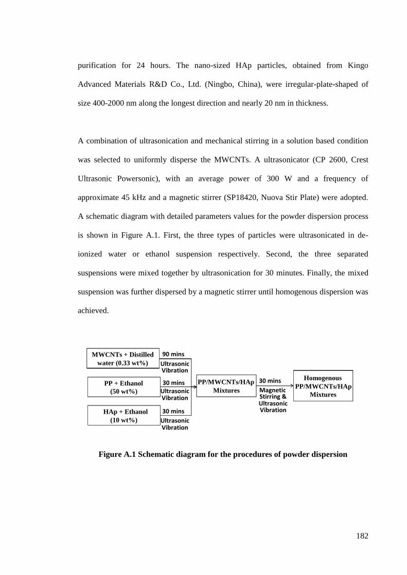
182
purification for 24 hours. The nano-sized HAp particles, obtained from Kingo
Advanced Materials R&D Co., Ltd. (Ningbo, China), were irregular-plate-shaped of
size 400-2000 nm along the longest direction and nearly 20 nm in thickness.
A combination of ultrasonication and mechanical stirring in a solution based condition
was selected to uniformly disperse the MWCNTs. A ultrasonicator (CP 2600, Crest
Ultrasonic Powersonic), with an average power of 300 W and a frequency of
approximate 45 kHz and a magnetic stirrer (SP18420, Nuova Stir Plate) were adopted.
A schematic diagram with detailed parameters values for the powder dispersion process
is shown in Figure A.1. First, the three types of particles were ultrasonicated in de-
ionized water or ethanol suspension respectively. Second, the three separated
suspensions were mixed together by ultrasonication for 30 minutes. Finally, the mixed
suspension was further dispersed by a magnetic stirrer until homogenous dispersion was
achieved.
Figure A.1 Schematic diagram for the procedures of powder dispersion
MWCNTs + Distilled water (0.33 wt%)
PP + Ethanol (50 wt%)
HAp + Ethanol (10 wt%)
Ultrasonic Vibration
90 mins
30 mins
30 mins
PP/MWCNTs/HAp Mixtures
30 mins
Ultrasonic Vibration
Ultrasonic Vibration
Magnetic Stirring & Ultrasonic Vibration
Homogenous PP/MWCNTs/HAp
Mixtures
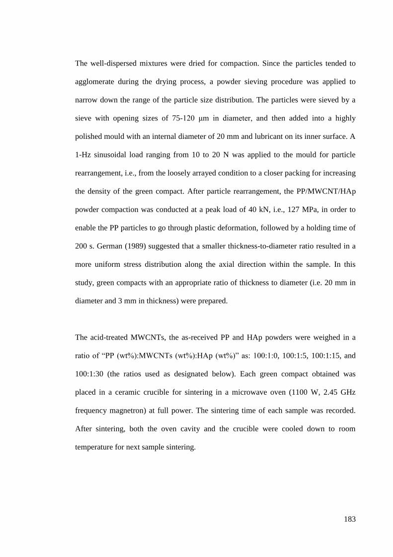
183
The well-dispersed mixtures were dried for compaction. Since the particles tended to
agglomerate during the drying process, a powder sieving procedure was applied to
narrow down the range of the particle size distribution. The particles were sieved by a
sieve with opening sizes of 75-120 μm in diameter, and then added into a highly
polished mould with an internal diameter of 20 mm and lubricant on its inner surface. A
1-Hz sinusoidal load ranging from 10 to 20 N was applied to the mould for particle
rearrangement, i.e., from the loosely arrayed condition to a closer packing for increasing
the density of the green compact. After particle rearrangement, the PP/MWCNT/HAp
powder compaction was conducted at a peak load of 40 kN, i.e., 127 MPa, in order to
enable the PP particles to go through plastic deformation, followed by a holding time of
200 s. German (1989) suggested that a smaller thickness-to-diameter ratio resulted in a
more uniform stress distribution along the axial direction within the sample. In this
study, green compacts with an appropriate ratio of thickness to diameter (i.e. 20 mm in
diameter and 3 mm in thickness) were prepared.
The acid-treated MWCNTs, the as-received PP and HAp powders were weighed in a
ratio of “PP (wt%):MWCNTs (wt%):HAp (wt%)” as: 100:1:0, 100:1:5, 100:1:15, and
100:1:30 (the ratios used as designated below). Each green compact obtained was
placed in a ceramic crucible for sintering in a microwave oven (1100 W, 2.45 GHz
frequency magnetron) at full power. The sintering time of each sample was recorded.
After sintering, both the oven cavity and the crucible were cooled down to room
temperature for next sample sintering.
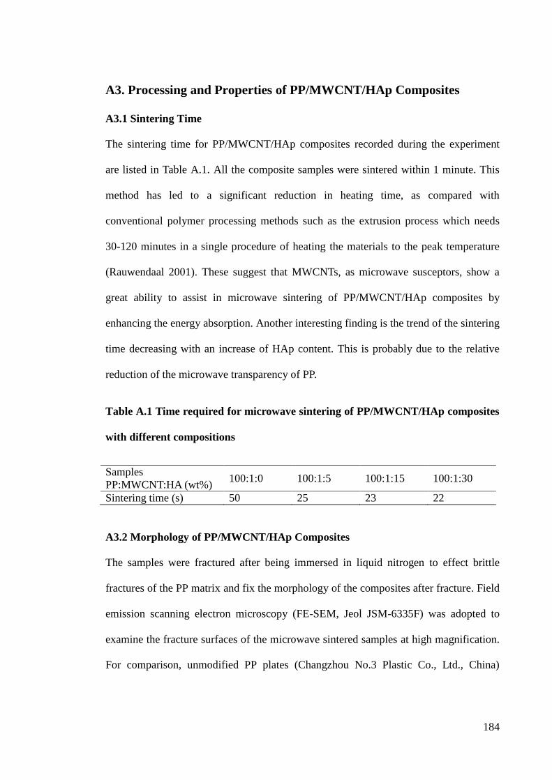
184
A3. Processing and Properties of PP/MWCNT/HAp Composites
A3.1 Sintering Time
The sintering time for PP/MWCNT/HAp composites recorded during the experiment
are listed in Table A.1. All the composite samples were sintered within 1 minute. This
method has led to a significant reduction in heating time, as compared with
conventional polymer processing methods such as the extrusion process which needs
30-120 minutes in a single procedure of heating the materials to the peak temperature
(Rauwendaal 2001). These suggest that MWCNTs, as microwave susceptors, show a
great ability to assist in microwave sintering of PP/MWCNT/HAp composites by
enhancing the energy absorption. Another interesting finding is the trend of the sintering
time decreasing with an increase of HAp content. This is probably due to the relative
reduction of the microwave transparency of PP.
Table A.1 Time required for microwave sintering of PP/MWCNT/HAp composites
with different compositions
Samples
PP:MWCNT:HA (wt%) 100:1:0 100:1:5 100:1:15 100:1:30
Sintering time (s) 50 25 23 22
A3.2 Morphology of PP/MWCNT/HAp Composites
The samples were fractured after being immersed in liquid nitrogen to effect brittle
fractures of the PP matrix and fix the morphology of the composites after fracture. Field
emission scanning electron microscopy (FE-SEM, Jeol JSM-6335F) was adopted to
examine the fracture surfaces of the microwave sintered samples at high magnification.
For comparison, unmodified PP plates (Changzhou No.3 Plastic Co., Ltd., China)
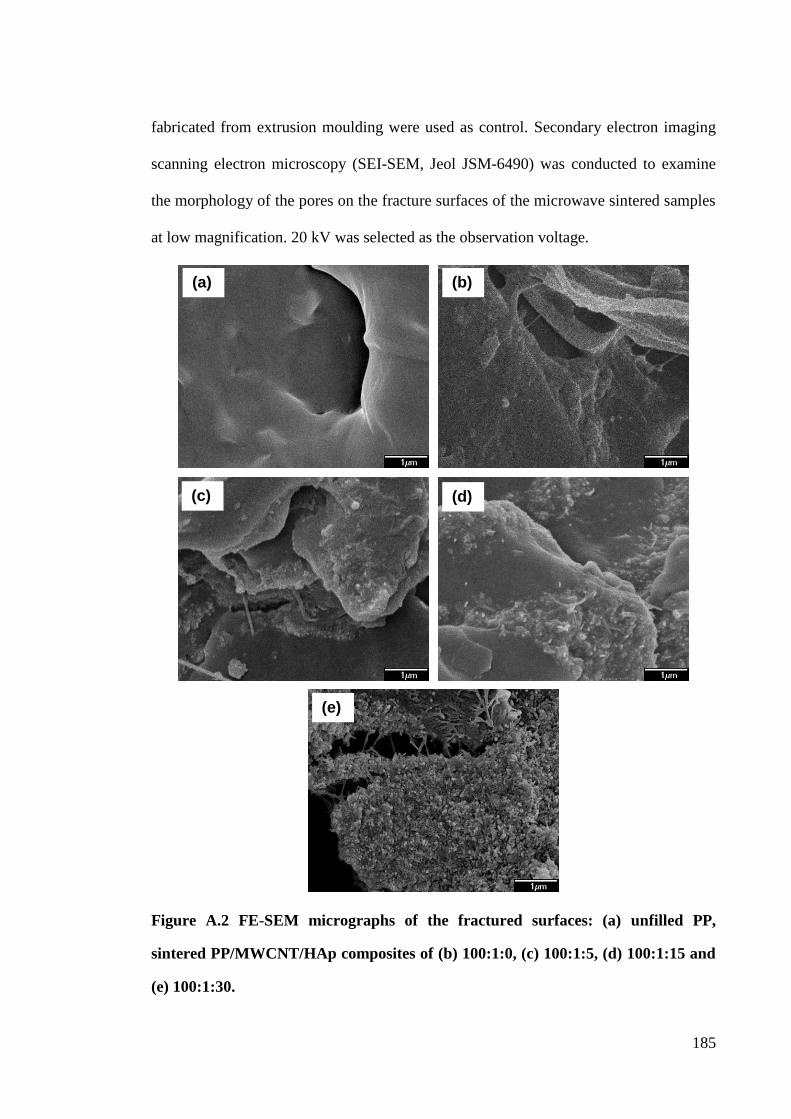
185
fabricated from extrusion moulding were used as control. Secondary electron imaging
scanning electron microscopy (SEI-SEM, Jeol JSM-6490) was conducted to examine
the morphology of the pores on the fracture surfaces of the microwave sintered samples
at low magnification. 20 kV was selected as the observation voltage.
Figure A.2 FE-SEM micrographs of the fractured surfaces: (a) unfilled PP,
sintered PP/MWCNT/HAp composites of (b) 100:1:0, (c) 100:1:5, (d) 100:1:15 and
(e) 100:1:30.
(a) (b)
(c) (d)
(e)
186
Figure A.2 shows the FE-SEM images of the unfilled and CNTs/HAp filled PP samples.
Unfilled PP shows a smoother surface compared to the composites. Crack bridging
effect is observed at the fracture sites for all the PP/MWCNT/HAp composites. The
bridging elements are probably due to the breakage of the sintered PP and the stretched
MWCNTs. This bridging effect can improve the mechanical properties such as elastic
modulus, yield and tensile strengths. Similar images and results can be found in the
literature (Coleman et al. 2006). The roughness extent of the fracture surfaces increases
with increasing amount of HAp, as shown in Figures A.2(b)-(e).
A porous structure is one of the key factors that influence the mechanical and biological
performance of a biomedical component (Olevsky 1998). SEI-SEM images are
presented in Figure A.3. It can be seen that pores were generated in all the sintered
PP/MWCNT/HAp samples, while there were no pores observed in the unfilled PP plate.
From Figure A.3(b) to (e), the average diameter of the pores firstly reaches a peak of
nearly 100 μm at 100:1:0 then decreases to 50 μm at 100:1:5 and 10 μm at 100:1:15,
and finally increases to 20 μm at 100:1:30. The initial cavities of the green compact
may be the reason for pore generation in the sintered PP/MWCNT/HAp composites,
because some cavities may exist among PP particles even when they were tightly
compacted. As the PP particle size is much bigger than that of the MWCNT and HAp,
the MWCNT and HAp powders tended to appear in these cavities and attach to the
surface of the PP powders after compaction. The cavities in the composites without the
HAp addition are bigger in size than those of the PP/MWCNT/HAp composites,
resulting in bigger pores, with diameters of 100 μm generated in the sintered 100:1:0
composites. The diameter of the pores decreased with the increase of HAp content and
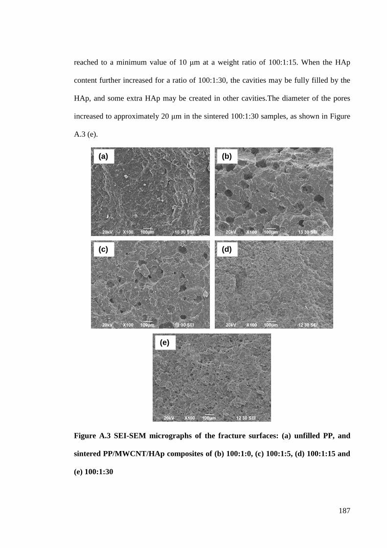
187
reached to a minimum value of 10 μm at a weight ratio of 100:1:15. When the HAp
content further increased for a ratio of 100:1:30, the cavities may be fully filled by the
HAp, and some extra HAp may be created in other cavities.The diameter of the pores
increased to approximately 20 μm in the sintered 100:1:30 samples, as shown in Figure
A.3 (e).
Figure A.3 SEI-SEM micrographs of the fracture surfaces: (a) unfilled PP, and
sintered PP/MWCNT/HAp composites of (b) 100:1:0, (c) 100:1:5, (d) 100:1:15 and
(e) 100:1:30
(a) (b)
(c) (d)
(e)
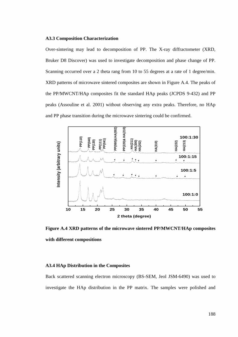
188
A3.3 Composition Characterization
Over-sintering may lead to decomposition of PP. The X-ray diffractometer (XRD,
Bruker D8 Discover) was used to investigate decomposition and phase change of PP.
Scanning occurred over a 2 theta rang from 10 to 55 degrees at a rate of 1 degree/min.
XRD patterns of microwave sintered composites are shown in Figure A.4. The peaks of
the PP/MWCNT/HAp composites fit the standard HAp peaks (JCPDS 9-432) and PP
peaks (Assouline et al. 2001) without observing any extra peaks. Therefore, no HAp
and PP phase transition during the microwave sintering could be confirmed.
Figure A.4 XRD patterns of the microwave sintered PP/MWCNT/HAp composites
with different compositions
A3.4 HAp Distribution in the Composites
Back scattered scanning electron microscopy (BS-SEM, Jeol JSM-6490) was used to
investigate the HAp distribution in the PP matrix. The samples were polished and
10 15 20 25 30 35 40 45 50 55
HA
(31
0)
HA
(21
3)
HA
(22
2)
HA
(20
2)
HA
(30
0)
HA
(21
1)
PP
(22
0)&
HA
(21
0)
HA
(00
2)
PP
(06
0)&
PP
(04
1)
PP
(11
1)
PP
(13
0)
PP
(04
0)
PP
(11
0)
100:1:0
Inte
ns
ity
(a
rbit
rary
un
its
)
2 theta (degree)
100:1:5
100:1:15
100:1:30
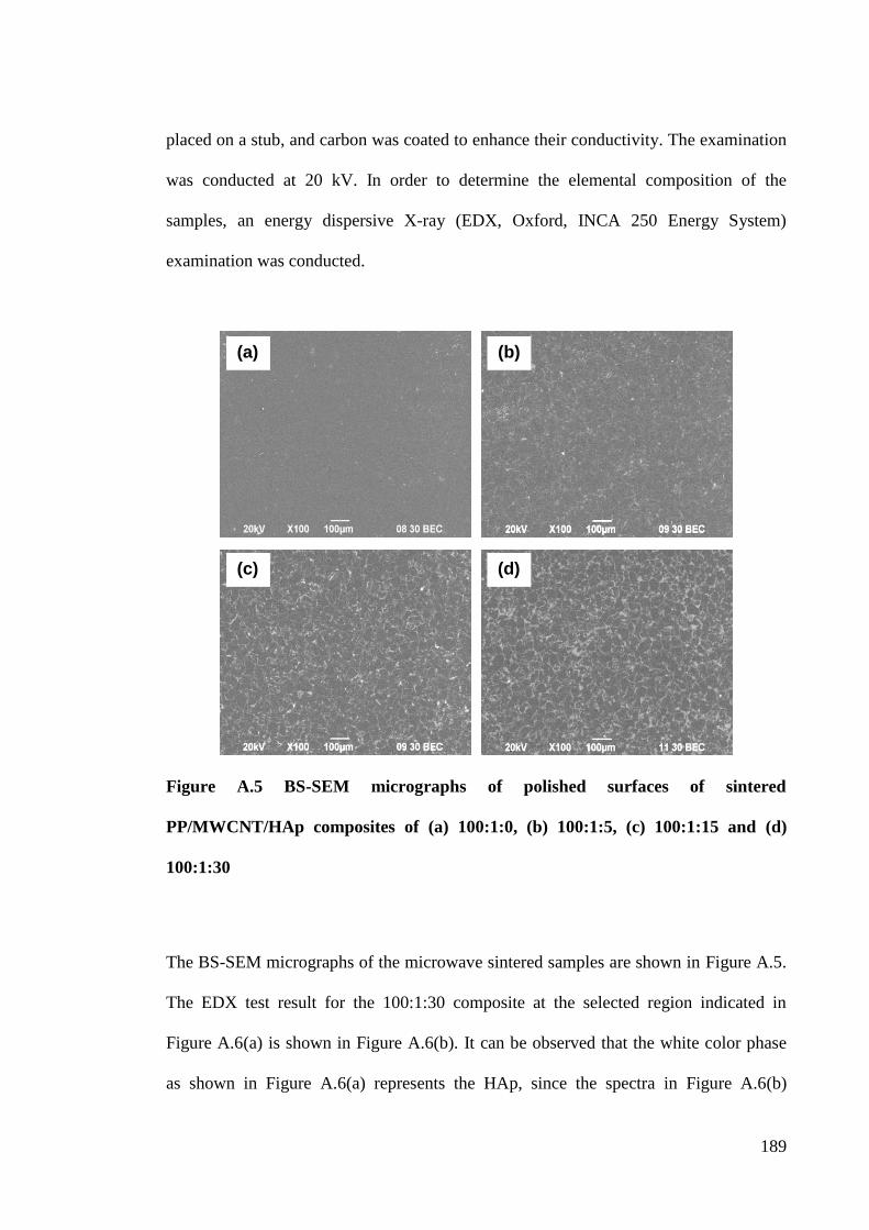
189
placed on a stub, and carbon was coated to enhance their conductivity. The examination
was conducted at 20 kV. In order to determine the elemental composition of the
samples, an energy dispersive X-ray (EDX, Oxford, INCA 250 Energy System)
examination was conducted.
Figure A.5 BS-SEM micrographs of polished surfaces of sintered
PP/MWCNT/HAp composites of (a) 100:1:0, (b) 100:1:5, (c) 100:1:15 and (d)
100:1:30
The BS-SEM micrographs of the microwave sintered samples are shown in Figure A.5.
The EDX test result for the 100:1:30 composite at the selected region indicated in
Figure A.6(a) is shown in Figure A.6(b). It can be observed that the white color phase
as shown in Figure A.6(a) represents the HAp, since the spectra in Figure A.6(b)
(a) (b)
(c) (d)
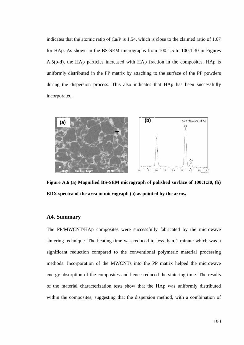
190
indicates that the atomic ratio of Ca/P is 1.54, which is close to the claimed ratio of 1.67
for HAp. As shown in the BS-SEM micrographs from 100:1:5 to 100:1:30 in Figures
A.5(b-d), the HAp particles increased with HAp fraction in the composites. HAp is
uniformly distributed in the PP matrix by attaching to the surface of the PP powders
during the dispersion process. This also indicates that HAp has been successfully
incorporated.
Figure A.6 (a) Magnified BS-SEM micrograph of polished surface of 100:1:30, (b)
EDX spectra of the area in micrograph (a) as pointed by the arrow
A4. Summary
The PP/MWCNT/HAp composites were successfully fabricated by the microwave
sintering technique. The heating time was reduced to less than 1 minute which was a
significant reduction compared to the conventional polymeric material processing
methods. Incorporation of the MWCNTs into the PP matrix helped the microwave
energy absorption of the composites and hence reduced the sintering time. The results
of the material characterization tests show that the HAp was uniformly distributed
within the composites, suggesting that the dispersion method, with a combination of
(a) (b)
191
ultrasonication and mechanical stirring, was effective. Pores were generated in the
sintered PP/MWCNT/HAp composites and the smallest pores occurred at 100:1:15. The
outcomes of the study have demonstrated the feasibility of fabricating the PP based
composite, with the addition of HAp and MWCNTs fillers as a model system, by
microwave sintering. This method has high potential applications in fabricating similar
polymer based composite materials.
Further research may be conducted to investigate the application of the microwave
sintering technique in fabricating porous polymer based composites. Surface treatments
maybe carried out to improve the interfacial bonding between the particles and matrix
and hence improve the mechanical properties of the composites. Moreover, in vitro and
in vivo tests are also suggested for further investigation of their bioactivity.


