Embed Size (px)
Citation preview
University of Louisville University of Louisville
ThinkIR: The University of Louisville's Institutional Repository ThinkIR: The University of Louisville's Institutional Repository
Electronic Theses and Dissertations
12-2018
Association between cigarette smoking and ovarian reserve Association between cigarette smoking and ovarian reserve
among women seeking fertility treatment with effect modification among women seeking fertility treatment with effect modification
by race and NAT2 genotype. by race and NAT2 genotype.
Islamiat A. Oladipupo University of Louisville
Follow this and additional works at: https://ir.library.louisville.edu/etd
Part of the Epidemiology Commons, and the Women's Health Commons
Recommended Citation Recommended Citation Oladipupo, Islamiat A., "Association between cigarette smoking and ovarian reserve among women seeking fertility treatment with effect modification by race and NAT2 genotype." (2018). Electronic Theses and Dissertations. Paper 3123. https://doi.org/10.18297/etd/3123
This Doctoral Dissertation is brought to you for free and open access by ThinkIR: The University of Louisville's Institutional Repository. It has been accepted for inclusion in Electronic Theses and Dissertations by an authorized administrator of ThinkIR: The University of Louisville's Institutional Repository. This title appears here courtesy of the author, who has retained all other copyrights. For more information, please contact [email protected].
ASSOCIATION BETWEEN CIGARETTE SMOKING AND OVARIAN RESERVE
AMONG WOMEN SEEKING FERTILITY TREATMENT WITH EFFECT
MODIFICATION BY RACE AND NAT2 GENOTYPE
By
Islamiat A. Oladipupo
MPH, University of Louisville, 2014
MBBS, Olabisi Onabanjo University, 2006
A Dissertation
Submitted to the Faculty of the
School of Public Health and Information Sciences
of the University of Louisville
in Partial Fulfillment of the Requirements
for the Degree of
Doctor of Philosophy in Public Health Sciences
Department of Epidemiology and Population Health
University of Louisville
Louisville, Kentucky
December 2018

ii
ASSOCIATION BETWEEN CIGARETTE SMOKING AND OVARIAN RESERVE
AMONG WOMEN SEEKING FERTILITY TREATMENT WITH EFFECT
MODIFICATION BY RACE AND NAT2 GENOTYPE
By
Islamiat A. Oladipupo
MPH, University of Louisville, 2014
MBBS, Olabisi Onabanjo University, 2006
A Dissertation Approved on
November 9, 2018
by the following Dissertation Committee:
________________________________
Dr. Kira Taylor, PhD, MS
________________________________
Dr. Kathy Baumgartner, PhD
________________________________
Dr. David W. Hein, PhD
________________________________
Dr. Anne Wallis, PhD
________________________________
Dr. Bakeerathan Gunaratnam, PhD

iii
DEDICATION
To my Late Dad, my number one Cheerleader, Alhaji Abdul-Hakeem Atunrase, I wish
you were here to celebrate this great accomplishment with me. You will always be in my
heart.
iv
ACKNOWLEDGEMENTS
I would like to express my deepest gratitude to my mentor and dissertation
committee chair, Dr. Kira Taylor. The opportunity you gave me to join your research
team, your patient guidance, enthusiastic encouragement and useful critique of my
dissertation is very much appreciated. I could not have imagined having a better advisor
and mentor. I would like to thank my committee members: Dr. Kathy Baumgartner, Dr.
David Hein, Dr. Anne Wallis and Dr. Bakeerathan Gunaratnam. I thank you for your
time, professional guidance and valuable support. I would also like to thank the
Louisville Tobacco Smoke Exposure, Genetic Susceptibility and Infertility Study
(LOUSSI) student research staff: Emily Steinmetz, T’shura Ali and Sashia Torres; Mark
Doll for helping with the NAT2 genotyping and analysis.
My profound gratitude to Prof. Richard Baumgartner for your never-ending
support right from my MPH days at the department of Epidemiology. Thank you to my
mum, Alhaja Risikat Atunrase and my sister, Basirat Bashua for their unflinching support
and constant words of encouragement. To my husband, Abolaji Oladipupo, I could not
have achieved this without your support, love, patience and understanding. Your belief in
me kept me going. To my children, Haleema and Farooq, thank you for your
unconditional love.
Finally, I would like to acknowledge the co-investigators of the LOUSSI study, Fellows
and the staff of the University of Louisville Fertility Clinic and the funding source that

v
made this research possible: National Institutes of Health (NIH), Eunice Kennedy Shriver
National Institute of Child Health and Human Development (NICHD), “The Interaction
between NAT2 Acetylator Status and Exposure to Tobacco Smoke on Ovarian Reserve
and In Vitro Fertilization Outcomes” (Grant: 1R15HD087911-01 Principal Investigator
(PI): Kira Taylor).
vi
ABSTRACT
ASSOCIATION BETWEEN CIGARETTE SMOKING AND OVARIAN RESERVE
AMONG WOMEN SEEKING FERTILITY TREATMENT WITH EFFECT
MODIFICATION BY RACE AND NAT2 GENOTYPE
Islamiat A. Oladipupo
November 9, 2018
Cigarette smoking in women has been associated with adverse reproductive
outcomes such as reduced ovarian reserve, poorer in vitro fertilization (IVF) outcomes
and increased adverse pregnancy outcomes. This study examined the association of
smoking with ovarian reserve in a cross-sectional study of women seeking fertility
treatment, and potential effect modification by race and NAT2 acetylator phenotype.
Data from 265 women from the Louisville Tobacco Smoke, Genetic
Susceptibility, and Infertility (LOUSSI) Study were analyzed. A total of 265 women
were recruited through a single infertility clinic between September 2016 and June 2018.
Information on current smoking status was assessed using a structured questionnaire and
confirmed by cotinine assay. Single nucleotide polymorphisms in NAT2 were genotyped
to determine acetylator status and serum anti-Müllerian hormone (AMH) level was used
to assess ovarian reserve. The association of smoking with ovarian reserve was assessed
using linear and logistic regression models with adjustment for potential confounders.
vii
Effect modification by race and NAT2 phenotype were assessed by including interaction
terms in the regression models.
Overall, smoking was not significantly associated with ovarian reserve. Results
suggest that heavy smoking and higher pack-years of exposure may decrease ovarian
reserve. Although most associations were not statistically significant, the effect of
smoking on ovarian reserve was more pronounced among non-Hispanic Black women
and slow NAT2 acetylators. These results are based on a small clinical population and
require replication in a larger and more representative study population.
viii
TABLE OF CONTENTS
PAGE
DEDICATION ................................................................................................................... iii
ACKNOWLEDGEMENTS ............................................................................................... iv
ABSTRACT ....................................................................................................................... vi
LIST OF TABLES .............................................................................................................. x
A. INTRODUCTION ....................................................................................................... 1
B. OBJECTIVE AND SPECIFIC AIMS ......................................................................... 3
C. BACKGROUND AND SIGNIFICANCE................................................................... 5
Epidemiology .................................................................................................................. 5
Causes of infertility ......................................................................................................... 7
Ovarian reserve ............................................................................................................... 8
Antimullerian Hormone (AMH) .................................................................................. 9
Antral Follicle Count (AFC)...................................................................................... 10
Predictors/ Determinants of Ovarian reserve ............................................................. 11
Active smoking ............................................................................................................. 14
Biomarkers of Active smoking .................................................................................. 15
Smoking and Ovarian reserve ....................................................................................... 18
N-acetyltransferase2 (NAT2) ........................................................................................ 25
Summary and Gap in Current Knowledge .................................................................... 28
D. RESEARCH DESIGN AND METHODS ................................................................. 30
Study Design ................................................................................................................. 30
Study Population ........................................................................................................... 30
Subject Recruitment Methods ....................................................................................... 32
Exposure Assessment .................................................................................................... 32
Outcome Measurement ................................................................................................. 33
Covariates ...................................................................................................................... 34
Data Collection and Study Procedures .......................................................................... 34
Statistical Analyses ....................................................................................................... 38
ix
Sample Size / Power calculation ................................................................................... 42
E. RESULTS .................................................................................................................. 45
F. DISCUSSION ............................................................................................................ 57
Effect Modification by Race ...................................................................................... 59
Effect Modification by NAT2 .................................................................................... 60
Strengths and Limitations.............................................................................................. 61
Conclusion ..................................................................................................................... 63
REFERENCES ................................................................................................................. 64
Appendix 1 ........................................................................................................................ 83
Appendix 2 ........................................................................................................................ 91
Appendix 3 ........................................................................................................................ 93
CURRICULUM VITAE ................................................................................................. 102
x
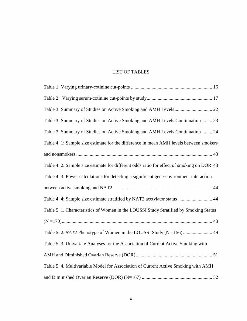
LIST OF TABLES
Table 1: Varying urinary-cotinine cut-points ................................................................... 16
Table 2: Varying serum-cotinine cut-points by study...................................................... 17
Table 3: Summary of Studies on Active Smoking and AMH Levels ............................... 22
Table 3: Summary of Studies on Active Smoking and AMH Levels Continuation ......... 23
Table 3: Summary of Studies on Active Smoking and AMH Levels Continuation ......... 24
Table 4. 1: Sample size estimate for the difference in mean AMH levels between smokers
and nonsmokers ................................................................................................................ 43
Table 4. 2: Sample size estimate for different odds ratio for effect of smoking on DOR 43
Table 4. 3: Power calculations for detecting a significant gene-environment interaction
between active smoking and NAT2 .................................................................................. 44
Table 4. 4: Sample size estimate stratified by NAT2 acetylator status ............................ 44
Table 5. 1. Characteristics of Women in the LOUSSI Study Stratified by Smoking Status
(N =170) ............................................................................................................................ 48
Table 5. 2. NAT2 Phenotype of Women in the LOUSSI Study (N =156) ........................ 49
Table 5. 3. Univariate Analyses for the Association of Current Active Smoking with
AMH and Diminished Ovarian Reserve (DOR) ............................................................... 51
Table 5. 4. Multivariable Model for Association of Current Active Smoking with AMH
and Diminished Ovarian Reserve (DOR) (N=167) .......................................................... 52
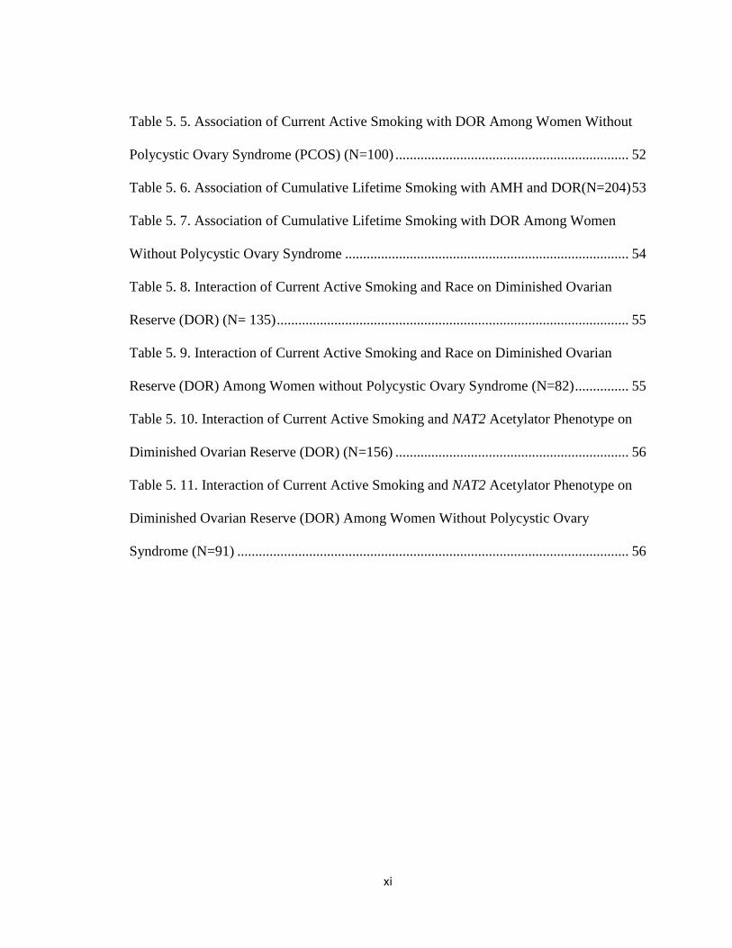
xi
Table 5. 5. Association of Current Active Smoking with DOR Among Women Without
Polycystic Ovary Syndrome (PCOS) (N=100) ................................................................. 52
Table 5. 6. Association of Cumulative Lifetime Smoking with AMH and DOR(N=204) 53
Table 5. 7. Association of Cumulative Lifetime Smoking with DOR Among Women
Without Polycystic Ovary Syndrome ............................................................................... 54
Table 5. 8. Interaction of Current Active Smoking and Race on Diminished Ovarian
Reserve (DOR) (N= 135) .................................................................................................. 55
Table 5. 9. Interaction of Current Active Smoking and Race on Diminished Ovarian
Reserve (DOR) Among Women without Polycystic Ovary Syndrome (N=82) ............... 55
Table 5. 10. Interaction of Current Active Smoking and NAT2 Acetylator Phenotype on
Diminished Ovarian Reserve (DOR) (N=156) ................................................................. 56
Table 5. 11. Interaction of Current Active Smoking and NAT2 Acetylator Phenotype on
Diminished Ovarian Reserve (DOR) Among Women Without Polycystic Ovary
Syndrome (N=91) ............................................................................................................. 56
1
A. INTRODUCTION
Infertility and impaired fecundity among women of reproductive age remain an
important public health issue. It is estimated that 10.9% of women between the ages of
15-44 have impaired fecundity [1]. Among married women 15-44 years of age, 6.0% are
infertile and non-Hispanic black women are more likely to be infertile than non-Hispanic
white (NHW) women [1]. Among women aged 15-44, 12% had ever used infertility
services with use highest among older women, NHW women and women with higher
educational levels and income [1, 2]. The median per-person costs of infertility treatment
ranged from $1,182 for medications only, to $24,373 and $38,015 for in-vitro fertilization
(IVF) and IVF with donor egg groups, respectively. Eighty- five percent of IVF costs are
paid out of pocket [3, 4]. In addition to the financial burden, the diagnosis and treatment
of infertility is associated with psychological and emotional consequences such as
anxiety and depression [5, 6].
Causes of infertility include ovulatory disorders, tubal damage, uterine or
peritoneal problems and male factors, and in 15% to 30% of cases, etiology is unknown
[7, 8]. Ovulatory dysfunction or ovarian failure accounts for 20% of cases of infertility
[8]. Diminished ovarian reserve (DOR) defined as a condition where the response to
ovarian stimulation or fecundity is reduced compared with women of comparable age,
2
is present in about 27.5% of women undergoing IVF [9]. Markers of ovarian reserve
include anti-Müllerian hormone (AMH) serum levels and antral follicle count (AFC) as
assessed by transvaginal ultrasound [10, 11]. AMH levels and AFC are important
predictors of IVF success with lower levels reflecting smaller follicular pool and ovarian
reserve [10-15]. Major predictors of ovarian reserve include age, age at menarche, parity,
obesity and race/ethnicity [16].
Cigarette smoking in women has been associated with adverse reproductive
outcomes such as reduced ovarian reserve, poorer IVF outcome and increased adverse
pregnancy outcomes [17-22]. Studies of the association of active smoking with ovarian
reserve have been inconsistent. No epidemiological studies have been conducted to
explore potential gene-environment interaction as a possible cause of inconsistency in
results [23, 24].
N-acetyltransferase2 (NAT2) is an important enzyme in the conjugation of certain
drugs and other xenobiotics such as tobacco smoke, caffeine and pesticides [25, 26]. The
effects of NAT2 polymorphisms and their interaction with smoking on different cancer
risks have been established [27-29]. However, only one epidemiological study has been
conducted on the effects of NAT2 polymorphisms and possible interaction with smoking
on fertility and related outcomes among women [24]. No studies have explored the
potential interaction between smoking and NAT2 phenotypes on ovarian reserve.
3
B. OBJECTIVE AND SPECIFIC AIMS
The primary objective of the study is to examine the relationship between active
cigarette smoking and ovarian reserve as measured by AMH levels in females seeking
fertility treatment. Racial disparity by ethnicity (African-American, NHW) will be
explored by comparing the association in African-American (AA) with NHW women. In
addition, potential effect modification by NAT2 acetylator status on ovarian reserve will
be evaluated.
The specific aims of the study are as follows;
1(a). To determine if current active smoking, as measured by urinary cotinine
levels and questionnaire, is associated with ovarian reserve (as measured by AMH)
among women seeking fertility treatment after controlling for potential confounders such
as age, body mass index (BMI), age at menarche, parity and PCOS status.
Hypothesis: Smoking is associated with ovarian reserve with smokers having decreased
ovarian reserve compared to nonsmokers.
1(b). To determine if a dose-response relationship exists between current active
smoking (as measured by number of cigarettes smoked per day) and ovarian reserve. The
dose of exposure will be assessed based on response to the smoking questionnaire.

4
Hypothesis: A dose-response relationship exists between active smoking and ovarian
reserve with a higher level of exposure being associated with decreased ovarian reserve.
1(c). To determine if a dose-response relationship exists between cumulative
lifetime smoking (as measured by pack-years reported on the questionnaire) and ovarian
reserve.
Hypothesis: A dose-response relationship exists between cumulative lifetime exposure to
smoking and ovarian reserve with longer pack-years of exposure being associated with
decreased ovarian reserve.
2. To determine if the association of current active smoking with ovarian reserve
differ by race, comparing African-American with Caucasians (NHW) women.
Hypothesis: The associations between active smoking and ovarian reserve will be
different when comparing AA to NHW women (i.e. there is a significant interaction
between smoking and race when examining their joint effects on ovarian reserve).
3. To determine if there is an interaction between current active smoking and
NAT2 acetylator status on ovarian reserve.
Hypothesis: There is a significant interaction between smoking and NAT2 acetylator
status when examining their joint effects on ovarian reserve, with slow acetylators having
lower ovarian reserve compared to intermediate and rapid acetylators.
5
C. BACKGROUND AND SIGNIFICANCE
Infertility is defined as failure to achieve clinical pregnancy after 12 months or
more of regular unprotected sexual intercourse [30]. A diagnosis of infertility is made
based on detailed medical history and physical examination followed by diagnostic
evaluations to identify the underlying etiology [11]. Diagnostic evaluation for infertility
includes tests for ovarian function, uterine abnormalities, cervical factors, tubal patency,
peritoneal factors and semen analysis [11, 31, 32].
Infertility is divided into two major subtypes of primary and secondary infertility
[30]. Secondary infertility refers to the inability of a woman to get pregnant or carry a
pregnancy to live birth following a previous pregnancy or live birth. Diagnosis of primary
infertility is made in women with no prior history of conception or live birth [30].
Epidemiology
Globally, infertility affects 48.5 million couples. Of these, 19.2 million have
primary infertility and 29.3 million have secondary infertility [33]. The prevalence of
infertility has increased globally [33]. In 2010, the prevalence of secondary infertility
increased with age, from 2.6% in women aged 20–24 to 27.1% in women aged 40–44
years [33]. The trends were reversed for primary infertility with prevalence higher among
6
women aged 20-24 (2.7%) compared to women aged 25–29 (2.0%) and women aged 30–
44 (1.6%) [33].
In the United States, the percentage of all married women 15-44 years of age who
are infertile was 6.0% between 2006 to 2010 and increased to 6.7% in 2015 [1, 34]. The
prevalence of infertility increased with age. Among currently married women 15-44 years
with no prior live birth, the prevalence was 8.7% among women aged 15-29,11.0% in
women 30-34, 23.0% in women 35-39 and 26.2% in women 40-44 years [34].
An estimated 7.3 million women 15-44 years have ever received any infertility
services between 2011 to 2015 [2, 34]. The use of infertility services varied by
socioeconomic status and race/ethnicity. Among women 25-44 years of age, ever use of
fertility services ranged from 9.5% in women with no high school diploma to 22.6 % in
women with a master’s degree or higher [2]. Use of fertility service was higher among
women 300-399 % the above poverty level (21.2%) compared to women less than 100%
below poverty level (12.9%) [2]. Among the racial/ethnic groups, Non-Hispanic black
(African-Americans) had lower ever use of fertility services (11.0%) than non-Hispanic
white (19.1%), or Hispanic (13.2%) women [2].
Some of the disparities in use of infertility services have been linked to the
significant costs of medical services and the lack of adequate health insurance to cover
necessary diagnostic tests or treatments [2, 35, 36]. The estimated median cost per IVF
cycle rose from $9226 in 2001 to about $12,513 in 2006 [37, 38]. The median per-person
cost of infertility treatment ranged from $1,182 for medications only, to $24,373 and
$38,015 for invitro fertilization (IVF) and IVF with donor egg groups, respectively [3]. In
7
the United States, only 25% of health care plans cover infertility treatment with 85% of
IVF costs being paid out of pocket [4, 36].
Aside from the financial burden associated with infertility, the condition is
associated with psychological and emotional consequences such as anxiety and
depression [5, 6]. In a cross-sectional survey of infertile women referred for infertility
treatment, 40.8% had depression and 86.8% had anxiety [5]. The duration of infertility
predicted 18% of anxiety and 25% of depression among infertile women [6].
Causes of infertility
Conception requires the timely release of a matured oocyte; adequate number of
progressively motile and normal spermatozoa capable of reaching and fertilizing the
oocyte; patent fallopian tubes for free passage of the sperm to reach the oocyte for
fertilization and migration of zygote/embryo to the uterus; and a well primed
endometrium that allows for implantation [11, 31, 32]. Biological, anatomical or
functional defects at any of these stages may lead to infertility. The probable causes of
infertility are classified into tubal factor, uterine or peritoneal problems, male factor and
ovulatory dysfunction [7, 8]. An estimated 40% of couples with infertility have a
combination of factors while 15% may not display any objective alterations in fertility
function [8].
Tubal factors account for 14% of cases of infertility and include conditions that
causes tubal obstruction and /or peritoneal adhesions [7]. Conditions that affect the
integrity of the uterine cavity and peritoneum such as endometriosis, uterine fibroids,
uterine synechiae may impact fertility [7, 8, 11, 32]. The male factors include conditions
8
that affects the concentration, motility, vitality and morphology of sperm cells [7, 8, 31,
32].
Ovulatory Dysfunction. Ovulation normally occurs when an ovary releases a
single mature oocyte every month and women who ovulate typically have regular and
consistent menstrual duration and flow [7]. Ovulatory dysfunction commonly results in
menstrual disturbances and account for 40% of infertility in women [11, 39]. Causes of
ovulatory dysfunction include polycystic ovary syndrome (PCOS), obesity, weight gain
or loss, strenuous exercise, thyroid dysfunction, and hyperprolactinemia [7, 8, 11].
Methods of assessing ovulatory dysfunction include detailed menstrual history, physical
and gynecological examination, basal temperature measurements, ultrasound, serum
progesterone level and endometrial biopsy [11].
Ovarian reserve
Ovarian reserve describes reproductive potential as a function of the number and
quality of oocytes [10]. It reflects the number of oocytes remaining in the ovaries that
influences the probability of getting pregnant [7]. Decreased or diminished ovarian
reserve (DOR) describes women of reproductive age having regular menses whose
response to ovarian stimulation or fecundity is reduced compared with those women of
comparable age [7].
Diagnostic criteria for DOR is poorly defined as standardized definition is lacking
[10, 40]. Definition and diagnostics criteria of DOR varies across fertility clinics and
studies [40]. Evidence suggest an upward trend in the prevalence of DOR among assisted
reproductive technology (ART) patients [41, 42]. Devin et al. reported an increase in the
prevalence of DOR from 19 to 26% from 2004 to 2011 in a retrospective study of
9
181,536 ART cycles reported to Society for Assisted Reproductive Technologies (SART)
by United States clinics [41]. In a cross-sectional study using ART cycles between 2004–
2007, a significant secular trend of increased odds of DOR diagnosis was observed [42].
The adjusted odds ratio of DOR per year studied was 1.11 (95% CI: 1.09–1.13), which
translates to a 23% higher odd of DOR in 2005 compared to 2004, 37% higher odds of
DOR in 2006 compared to 2004, and 52% higher odds of DOR in 2007 compared to
2004 [42].
Measures of the ovarian reserve have been used to predict DOR and counsel
infertile couples on choice of treatment [7, 10]. Tests of ovarian reserve include early
follicular phase antral follicle count (AFC) using transvaginal ultrasonography and
biochemical tests [7, 10, 11]. The biochemical tests include basal measurements of
follicle-stimulating hormone (FSH), estradiol, inhibin B, and antimullerian hormone
(AMH), and the clomiphene citrate challenge test (CCCT) [7, 10, 11]. Antral follicle
count and serum AMH have good predictive value and are the preferred methods for
assessing ovarian reserve [43-45].
Antimullerian Hormone (AMH)
Anti-Müllerian hormone (AMH), also called Müllerian inhibiting substance
(MIS), is a glycoprotein hormone belonging to the large family of transforming growth
factors-β (TGF- β) [10]. In women, AMH is expressed uniquely by the ovary in the
granulosa cells, primarily secreted by primary, preantral and antral follicles and are direct
measures of the follicular pool [10, 11]. As the number of ovarian follicles declines with
age, AMH concentrations decline [16]. Serum concentrations of AMH are gonadotropin-
independent and therefore remain relatively constant within and between menstrual
10
cycles in both normal, young, ovulating women and in women with infertility, making it
a valuable and reliable marker of ovarian function [10, 14, 46-48].
The following are considered the lower limit of age-appropriate serum AMH
values for these 5-year age intervals: 0.5ng/ML for 45 years, 1ng/mL for 40 years,
1.5ng/mL for 35 years, 2.5ng/mL for 30 years and 3.0ng/mL for 25 years [44]. Thus,
AMH level ≥ 1.0ng/mL but ≤3.5ng/mL if age appropriate is consistent with normal
ovarian response to ovarian stimulation. Lower serum AMH levels(<1ng/mL) have been
associated with poor responses to ovarian stimulation, poor embryo quality and poor
pregnancy outcomes in IVF patients [13, 15, 49-51].
Varying levels of serum AMH have been used to predict ovarian response to
stimulation. Serum cut-points ranging from 0.2 to 0.7ng/ml have been associated with
poor ovarian response with sensitivity and specificity ranging from 40-97% and 78-92%,
respectively [10]. In a retrospective study of one hundred and eight IVF patients, AMH
with a cutoff of 0.2 ng/mL predicted poor response to ovarian stimulation with a
sensitivity of 87% and specificity of 64% [51]. In a prospective study of one hundred
thirty-five women undergoing the first cycle of assisted reproduction treatment (ART),
serum AMH predicted poor ovarian response with a sensitivity of 100% and specificity
of 73% at an optimum cutoff value of ≤0.99ng/mL [45]. In a meta-analysis that included
28 studies, AMH was found to have good predictive value for poor ovarian response with
an area under the curve (AUC) of 0.78 [52].
Antral Follicle Count (AFC)
Antral follicle count (AFC) is the sum of antral follicles in both ovaries, as
observed with transvaginal ultrasonography during early follicular phase (day 2-4) of the
11
menstrual cycle [11, 44]. Antral follicles are defined as those measuring 2-10mm in mean
diameter in the greatest two-dimensional plane [11, 44]. The AFC correlates with
histologically determined number of primordial follicles and capacity of the ovary to
produce oocytes in either an assisted reproduction setting or through natural
folliculogenesis [53]. In experienced centers, AFC has good inter-cycle reliability and
inter-observer reliability [11, 44].
Low AFC, considered to be less than or equal 6 total antral follicles, is associated
with poor response to ovarian stimulation during IVF [11, 44, 54]. AFC ≤10 predicted
poor ovarian response with sensitivity of 93% and specificity of 88% with an AUC of
0.935 in prospective study of one hundred thirty-five women undergoing the first cycle
ART [45]. AUC for AFC in predicting poor ovarian response was found to be 0.76 in a
meta-analysis of 28 studies on IVF [52].
Studies have shown that AFC is highly correlated with AMH level [55, 56].
Antral follicles measuring <6mm express the greatest amount of AMH, and levels decline
as antral follicles increase in size [57]. Both AFC and AMH have strong and similar
linear relationship with the size of the primordial follicle pool and ovarian reserve [43-45,
58]. The limited intra- and inter-cycle variation, objectivity and potential standardization
of AMH assays, makes it the preferred biomarker of ovarian reserve in women.
Predictors/ Determinants of Ovarian reserve
Current age and Age at menarche. Older women and women with earlier age at
menarche have been shown to have lower levels of AMH reflecting a decline in the
ovarian pool [12, 16]. In a cross-sectional study of premenopausal women conducted by
Jung et al., women 40 years and older had significantly lower AMH concentrations
12
(0.73ng/ml) compared to women less than 35 years of age (2.52ng/ml) [12]. AMH
concentrations were also significantly lower among women with age at menarche less
than 12years (0.90 ng/mL) compared to women with age at menarche ≥14 years (1.12
ng/mL) [12].
Race/ethnicity. The association of race/ethnicity with AMH levels is
inconsistent. In the multi-ethnic cohort study of ovarian aging (OVA), mean AMH levels
were significantly lower in AA women (22.8±1.7pM) compared to Caucasians (30.1±
1.5pM) [16]. The rate of decrease in level of AMH also varied by race. In Caucasian
women, AMH decreased by 2 pM per year, while in African American women it
decreased by 0.84 pM per year [16]. Studies that reported no association between race
and AMH levels were cross-sectional and majorly of single ethnic group (Asian and
Caucasian) with under-representation of other races [12, 59, 60].
Parity. High parity has been postulated to have a protective effect on ovarian
reserve by reducing follicular recruitment [61]. In cross-sectional study of 2320 women
aged 20 to 59 years, high parity was found to be associated with higher age-specific
AMH levels(p=0.02) [59]. Not all studies found a positive association between parity and
AMH levels. Bragg et al. found a statistically significant inverse association between
parity and AMH: women with 2 (p< 0.005) or 3 or more(p<0.01) children had lower
AMH levels than nulliparous women [62]. Bleil et al. also found having had one or more
live births versus being nulliparous was associated with a 31.1% (95% CI: 21.6%, 39.5%)
reduction in AMH [61].
Oral contraceptive pill (OCP) use. Current use of OCP has been linked with
lower levels of AMH [12, 59]. Concentrations of AMH were significantly lower among
13
current users of oral contraceptives (0.36ng/ml) compared with never or former users
(1.15 ng/mL) [12].
Polycystic Ovarian Syndrome (PCOS). Multiple epidemiological investigations
have consistently demonstrated that women with diagnosis of PCOS have significantly
elevated serum AMH levels. In a hospital-based case-control study, mean serum AMH
level was markedly increased in the PCOS group (47.1 +/- 22.9 compared to. 20.8 +/-
11.6 pmol/liter in controls; p < 0.0001) [63]. In a retrospective study of women evaluated
for infertility, 97% of women with AMH >10ng/ml had PCOS [64].
Past Ovarian Surgery. Ovarian surgery is associated with decreased ovarian
reserve possibly due to decrease in ovarian volume [65, 66]. In women with regular
cycle, serum AMH was undetectable after bilateral oophorectomy [67]. Similarly, serum
AMH levels were significantly decreased after 1-week post-cystectomy (median =0.67;
range= 0.02–1.93; 95% CI: 0.44–1.70 ng/mL), and subsequently increased to about 65%
of the preoperative level after 3 months (1.50; range=0.58–3.27; 95% CI: 0.58–3.26
ng/mL) [65].
Obesity. The effect of obesity, body mass index (BMI) of at least 30kg/m2, on
ovarian reserve is inconsistent among studies. In a population-based cohort of one
hundred and twenty-two late reproductive-age (35-47 years) women, a significant inverse
association of AMH levels with BMI was observed [68]. Obese women had mean AMH
levels that were 65% lower than non-obese women (geometric mean ratio (GMR) = 0.35;
95% CI: 0.13, 0.92; p=0.034) [68]. Among ovulatory women ages 18 to 35 years, AMH
levels were 34% lower in the obese group (2.9±2.1 ng/mL) compared to normal group
(4.4±1.8ng/ml), p<0.05 [69]. Buyuk et al. found a similar inverse association between
14
BMI and AMH but only among women with DOR [70]. Among women with DOR,
overweight and obese women had 33% lower AMH levels than those with a normal BMI
(0.4 ± 0.3 vs. 0.6 ± 0.5, respectively; p=0.0001); no association was found between BMI
and AMH among women with normal ovarian reserve [70]. The negative association
between BMI and AMH was not replicated in other studies [59, 71-73].
Active smoking
Exposure to tobacco smoke has been associated with harmful reproduction
ranging from decreased ovarian reserve and infertility to poor pregnancy outcome [17-
19]. Some of the constituents of tobacco smoke known to have toxic effects on
reproductive health include, carbon monoxide, nicotine, cadmium and polycyclic
aromatic hydrocarbons (PAHs) such as benzo[a]pyrene (B[a]P) [74, 75].
Most of the evidence on probable biological mechanism on how exposure to
tobacco smoke may influence ovarian reserve and reproduction is based on experimental
studies in animals. Some of the proposed biological mechanism through which tobacco
smoke influences ovarian reserve includes inhibition of follicular development;
premature luteinization of the preovulatory follicle; reduction of oocyte vascularization
and maturation; atresia of oocytes in primordial and small primary follicles; impaired
steroidogenesis; increased chromosomal errors; and cytotoxicity [75, 76].
Benzo[a]pyrene (B[a]P), present in cigarette smoke, has been shown to significantly affect
the phase I enzymes and cell death genes during preantral/antral and preovulatory growth
leading to apoptosis and decreased viability [77]. Benzo[a]pyrene exposure decreased
AMH output overall during preantral and antral follicle development further confirming
the adverse effects of cigarette smoking on follicular development and survival [78].
15
Cadmium, found in tobacco smoke, accumulates in ovarian follicles and inhibits
the expression of P450scc leading to impaired ovarian steroidogenesis [79-81]. Cadmium
was also shown to increase ovarian oxidative stress [82]. Increased lipid peroxidation,
reduced glutathione contents, increased catalase activity and decreased superoxide
dismutase activity were observed in ovaries exposed to cadmium [82].
Smokers have been shown to secrete significantly higher amounts of vascular
endothelial growth factor (VEGF) soluble receptor 1, which may result in decreased
availability of VEGF and impaired angiogenesis and oocyte maturation [83].
Based on data from the National Health Interview Survey (NHIS), 13.6% of U.S.
women 18 years and above were a current cigarette smoker in 2015 [84]. The prevalence
of cigarette smoking was higher among non-Hispanic white (16.0%) followed by African
–Americans (13.3%) and Hispanics (7.1%) [84]. In Kentucky, 24% of women 18 years
and older were a current smoker in 2016 [85].
Biomarkers of Active smoking
Cotinine is a major proximate metabolite of nicotine and reflects exposure to other
constituents of tobacco smoke. It is measured in plasma, saliva and urine with a half-life
ranging from 7-40 hours in adults, reflecting up to 3-5 days of exposure to tobacco smoke
[86-88]. Urinary cotinine is a widely used biomarker of exposure to tobacco smoke. The
advantages of urinary cotinine as biomarker of tobacco smoke exposure are that cotinine
concentrations and other metabolites are higher than in other biological fluids; it represents
relatively acute exposure; and collection is non-invasive [87]. The urinary cut-off of greater
than or equal to 2.47ng/ml have been shown to detect active smoking with the highest
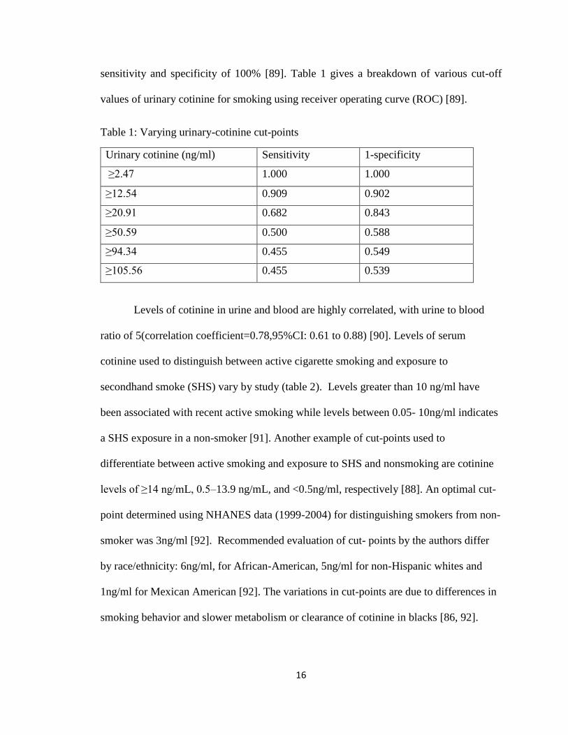
16
sensitivity and specificity of 100% [89]. Table 1 gives a breakdown of various cut-off
values of urinary cotinine for smoking using receiver operating curve (ROC) [89].
Table 1: Varying urinary-cotinine cut-points
Urinary cotinine (ng/ml) Sensitivity 1-specificity
≥2.47 1.000 1.000
≥12.54 0.909 0.902
≥20.91 0.682 0.843
≥50.59 0.500 0.588
≥94.34 0.455 0.549
≥105.56 0.455 0.539
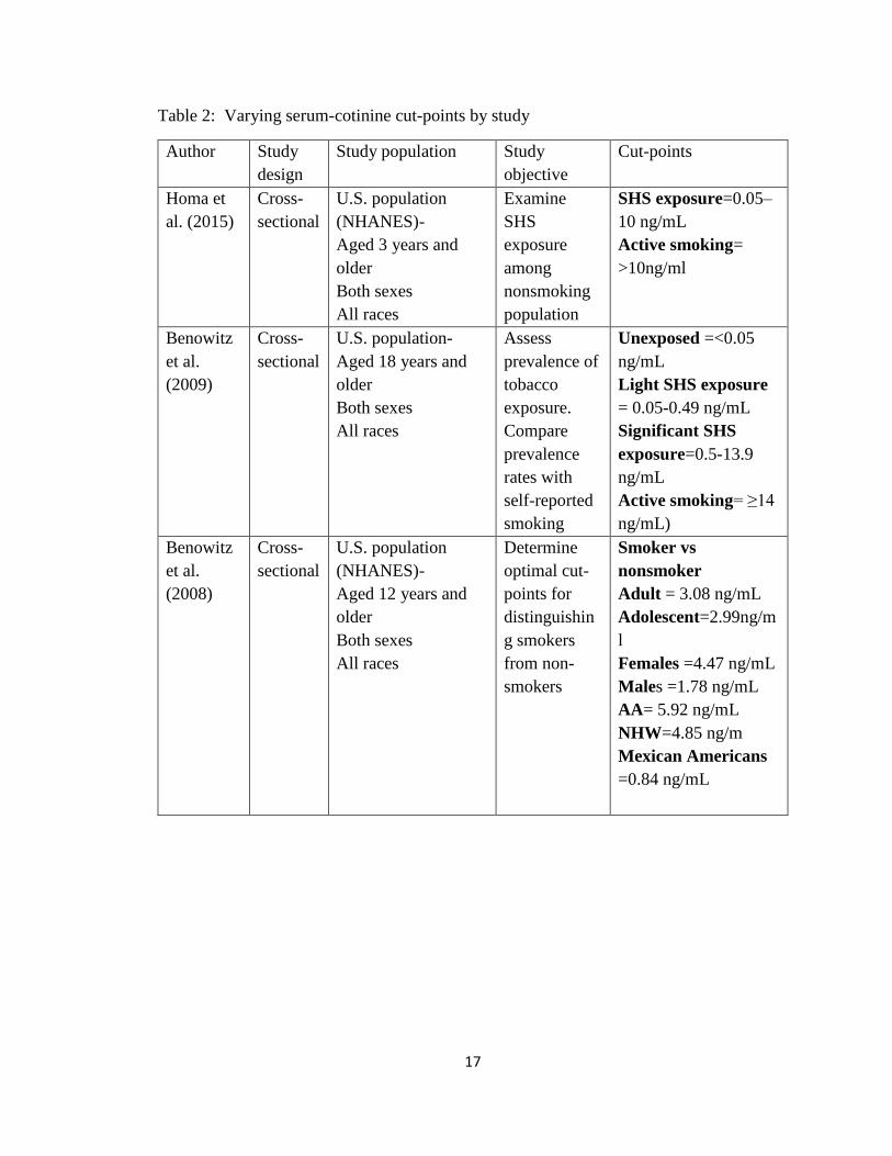
Levels of cotinine in urine and blood are highly correlated, with urine to blood
ratio of 5(correlation coefficient=0.78,95%CI: 0.61 to 0.88) [90]. Levels of serum
cotinine used to distinguish between active cigarette smoking and exposure to
secondhand smoke (SHS) vary by study (table 2). Levels greater than 10 ng/ml have
been associated with recent active smoking while levels between 0.05- 10ng/ml indicates
a SHS exposure in a non-smoker [91]. Another example of cut-points used to
differentiate between active smoking and exposure to SHS and nonsmoking are cotinine
levels of ≥14 ng/mL, 0.5–13.9 ng/mL, and <0.5ng/ml, respectively [88]. An optimal cut-
point determined using NHANES data (1999-2004) for distinguishing smokers from non-
smoker was 3ng/ml [92]. Recommended evaluation of cut- points by the authors differ
by race/ethnicity: 6ng/ml, for African-American, 5ng/ml for non-Hispanic whites and
1ng/ml for Mexican American [92]. The variations in cut-points are due to differences in
smoking behavior and slower metabolism or clearance of cotinine in blacks [86, 92].
17
Table 2: Varying serum-cotinine cut-points by study
Author Study
design
Study population Study
objective
Cut-points
Homa et
al. (2015)
Cross-
sectional
U.S. population
(NHANES)-
Aged 3 years and
older
Both sexes
All races
Examine
SHS
exposure
among
nonsmoking
population
SHS exposure=0.05–
10 ng/mL
Active smoking=
>10ng/ml
Benowitz
et al.
(2009)
Cross-
sectional
U.S. population-
Aged 18 years and
older
Both sexes
All races
Assess
prevalence of
tobacco
exposure.
Compare
prevalence
rates with
self-reported
smoking
Unexposed =<0.05
ng/mL
Light SHS exposure
= 0.05-0.49 ng/mL
Significant SHS
exposure=0.5-13.9
ng/mL
Active smoking= ≥14
ng/mL)
Benowitz
et al.
(2008)
Cross-
sectional
U.S. population
(NHANES)-
Aged 12 years and
older
Both sexes
All races
Determine
optimal cut-
points for
distinguishin
g smokers
from non-
smokers
Smoker vs
nonsmoker
Adult = 3.08 ng/mL
Adolescent=2.99ng/m
l
Females =4.47 ng/mL
Males =1.78 ng/mL
AA= 5.92 ng/mL
NHW=4.85 ng/m
Mexican Americans
=0.84 ng/mL
18
Smoking and Ovarian reserve
The association of smoking with ovarian reserve have been largely inconsistent.
Of the thirteen epidemiological studies that have explored the association of smoking
with ovarian reserve, only eight found a significant association. Of the thirteen studies,
only one used the biomarker cotinine to validate smoking status among participants [20].
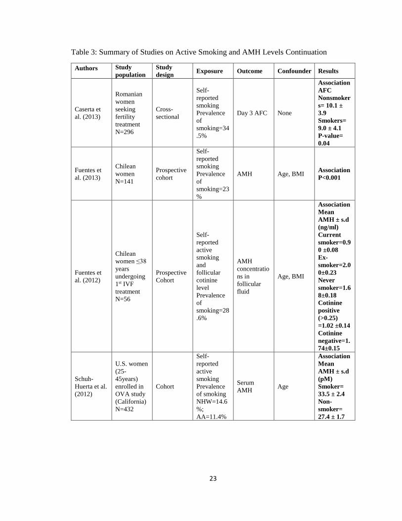
Fuentes et al. in a prospective cohort study examined the association between
cigarette smoking and AMH concentrations in follicular fluid of fifty-six women
undergoing first ART cycle [20]. Smoking status was based on follicular cotinine level
and self-report. Women with cotinine concentrations >0.25 ng/mL in the follicular fluid
were considered cotinine positive otherwise subjects were classified as cotinine negative.
Participants were also categorized into current smokers, ex-smokers, and never smokers
based on response to the smoking questionnaire. The mean concentration of AMH in
follicular fluid was significantly decreased in the cotinine positive group compared to the
cotinine negative group (1.02 ± 0.14 and 1.74 ± 0.15, respectively, p < 0.05) [20].
Similarly, current smokers had significantly lower mean AMH level (0.90 ± 0.08 ng/ml)
compared to ex-smokers (2.00 ± 0.23 ng/ml) and never smokers (1.68 ±0.18 ng/ml) [20].
A similar association was also found by the author in a follow up study of 141 infertile
women [93]. Active smoking was significantly associated with a 2.29ng/mL decrease in
plasma AMH levels (p<0.001) [93].
Another prospective study of 277 women undergoing IVF found decreased levels
of basal serum AMH among smokers compared to non-smokers (3.57 ± 1.74 vs. 4.34 ±
1.91). In addition, among women that received antagonist ovarian stimulation protocol,
smokers had significantly lower AMH levels (2.86 ± 1.32µg/l) compared to non-smokers
19
(3.74 ± 1.69 µg/l) [21]. Smoking status was based on self-report and women that quit for
more than a year were considered non-smokers.
Schuh-Huerta et al. prospectively examined the effects of environmental or
lifestyle factors including self-reported smoking status on AMH levels among multi-
ethnic cohort of women aged 25 – 45 years [16]. An inverse association between
smoking and AMH levels was found. After controlling for age, serum AMH levels were
significantly higher among smoking women (33.5 ± 2.4) compared to non-smoking
women (27.4 ± 1.7 pM) (p = 0.038) [16]. The mean AMH level was found to be
significantly lower (p<0.001) in African-American women compared to Caucasians.
Mean AMH level was 30.1 ± 1.5 pM in Caucasians and 22.8 ± 1.7 pM in African -
Americans [16].
Self-reported active smoking was found to be associated with a lower serum
AMH concentrations in a retrospective analysis of IVF patients. Serum AMH
concentrations were significantly lower in the smoker group (3.06 +/- 1.68 µg/l)
compared to non-smokers (3.86 +/- 1.92 µg/l) after controlling for age and BMI [17]. In
addition, serum AMH level was negatively correlated with the number of cigarettes
smoked daily (r = −0.36, p < 0.001) [17].
In a cross-sectional study of 284 women aged 38 to 50 years an association
between current smoking and lower AMH levels was observed [94]. Based of
questionnaire response, women were classified as current smokers; past smokers-reported
a history of smoking, with cessation occurring at least 2 years before the study; passive
smokers-currently living with someone who smoked in their home or never smokers.
Only current smoking was associated with lower AMH values. Current smokers had 44%
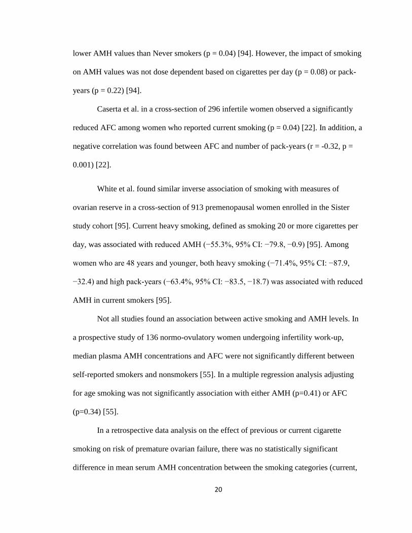
20
lower AMH values than Never smokers (p = 0.04) [94]. However, the impact of smoking
on AMH values was not dose dependent based on cigarettes per day (p = 0.08) or pack-
years (p = 0.22) [94].
Caserta et al. in a cross-section of 296 infertile women observed a significantly
reduced AFC among women who reported current smoking (p = 0.04) [22]. In addition, a
negative correlation was found between AFC and number of pack-years (r = -0.32, p =
0.001) [22].
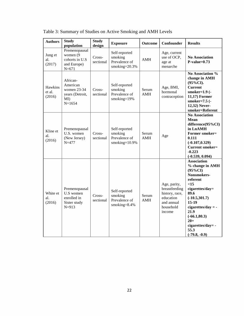
White et al. found similar inverse association of smoking with measures of
ovarian reserve in a cross-section of 913 premenopausal women enrolled in the Sister
study cohort [95]. Current heavy smoking, defined as smoking 20 or more cigarettes per
day, was associated with reduced AMH (−55.3%, 95% CI: −79.8, −0.9) [95]. Among
women who are 48 years and younger, both heavy smoking (−71.4%, 95% CI: −87.9,
−32.4) and high pack-years (−63.4%, 95% CI: −83.5, −18.7) was associated with reduced
AMH in current smokers [95].
Not all studies found an association between active smoking and AMH levels. In
a prospective study of 136 normo-ovulatory women undergoing infertility work-up,
median plasma AMH concentrations and AFC were not significantly different between
self-reported smokers and nonsmokers [55]. In a multiple regression analysis adjusting
for age smoking was not significantly association with either AMH (p=0.41) or AFC
(p=0.34) [55].
In a retrospective data analysis on the effect of previous or current cigarette
smoking on risk of premature ovarian failure, there was no statistically significant
difference in mean serum AMH concentration between the smoking categories (current,
21
ex-smoker and non-smoker) after adjustment for age (p-value 0.895) [96]. Hawkins
Bressler et al. in cross-sectional study of 1654 African-American women aged 23-34
found no association between self-reported active smoking and serum AMH
concentrations [97]. In two other cross-sectional studies of premenopausal women,
smoking was not significantly associated with measures of ovarian reserve [12, 98].
22
Table 3: Summary of Studies on Active Smoking and AMH Levels
Authors Study
population
Study
design Exposure Outcome Confounder Results
Jung et
al.
(2017)
Premenopausal
women (9
cohorts in U.S
and Europe)
N=671
Cross-
sectional
Self-reported
smoking
Prevalence of
smoking=20.3%
AMH
Age, current
use of OCP,
age at
menarche
No Association
P-value=0.73
Hawkins
et al.
(2016)
African-
American
women 23-34
years (Detroit,
MI)
N=1654
Cross-
sectional
Self-reported
smoking
Prevalence of
smoking=19%
Serum
AMH
Age, BMI,
hormonal
contraception
No Association %
change in AMH
(95%CI).
Current
smoker=1.9 (-
11,17) Former
smoker=7.5 (-
12,32) Never-
smoker=Referent
Kline et
al.
(2016)
Premenopausal
U.S. women
(New Jersey)
N=477
Cross-
sectional
Self-reported
smoking
Prevalence of
smoking=10.9%
Serum
AMH Age
No Association
Mean
difference(95%CI)
in LnAMH
Former smoker=
0.111
(-0.107,0.329)
Current smoker=
-0.223
(-0.539, 0.094)
White et
al.
(2016)
Premenopausal
U.S women
enrolled in
Sister study
N=913
Cross-
sectional
Self-reported
smoking
Prevalence of
smoking=8.4%
Serum
AMH
Age, parity,
breastfeeding
history, race,
education
and annual
household
income
Association
% change in AMH
(95%CI)
Nonsmokers-
referent
<15
cigarettes/day=
89.6
(-10.5,301.7)
15-19
cigarettes/day = -
21.9
(-66.1,80.3)
20+
cigarettes/day= -
55.3
(-79.8, -0.9)
23
Table 3: Summary of Studies on Active Smoking and AMH Levels Continuation
Authors Study
population
Study
design Exposure Outcome Confounder Results
Caserta et
al. (2013)
Romanian
women
seeking
fertility
treatment
N=296
Cross-
sectional
Self-
reported
smoking
Prevalence
of
smoking=34
.5%
Day 3 AFC None
Association
AFC
Nonsmoker
s= 10.1 ±
3.9
Smokers=
9.0 ± 4.1
P-value=
0.04
Fuentes et
al. (2013)
Chilean
women
N=141
Prospective
cohort
Self-
reported
smoking
Prevalence
of
smoking=23
%
AMH Age, BMI Association
P<0.001
Fuentes et
al. (2012)
Chilean
women ≤38
years
undergoing
1st IVF
treatment
N=56
Prospective
Cohort
Self-
reported
active
smoking
and
follicular
cotinine
level
Prevalence
of
smoking=28
.6%
AMH
concentratio
ns in
follicular
fluid
Age, BMI
Association
Mean
AMH ± s.d
(ng/ml)
Current
smoker=0.9
0 ±0.08
Ex-
smoker=2.0
0±0.23
Never
smoker=1.6
8±0.18
Cotinine
positive
(>0.25)
=1.02 ±0.14
Cotinine
negative=1.
74±0.15
Schuh-
Huerta et al.
(2012)
U.S. women
(25-
45years)
enrolled in
OVA study
(California)
N=432
Cohort
Self-
reported
active
smoking
Prevalence
of smoking
NHW=14.6
%;
AA=11.4%
Serum
AMH Age
Association
Mean
AMH ± s.d
(pM)
Smoker=
33.5 ± 2.4
Non-
smoker=
27.4 ± 1.7
24
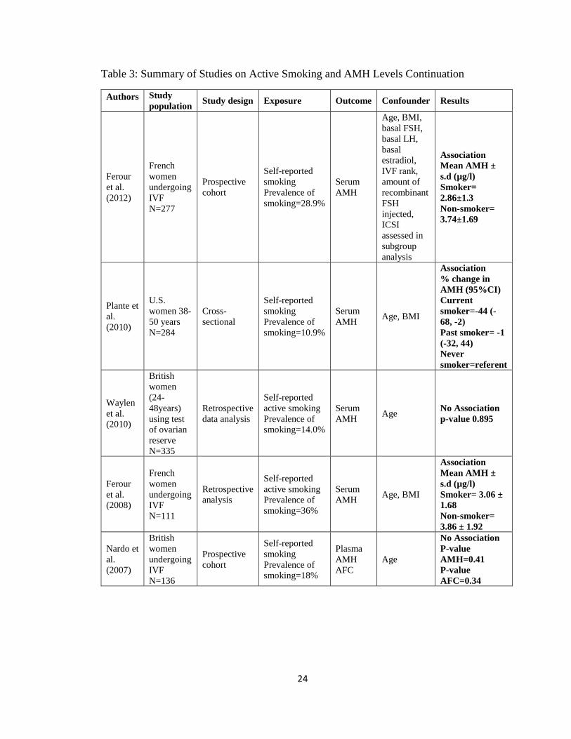
Table 3: Summary of Studies on Active Smoking and AMH Levels Continuation
Authors Study
population Study design Exposure Outcome Confounder Results
Ferour
et al.
(2012)
French
women
undergoing
IVF
N=277
Prospective
cohort
Self-reported
smoking
Prevalence of
smoking=28.9%
Serum
AMH
Age, BMI,
basal FSH,
basal LH,
basal
estradiol,
IVF rank,
amount of
recombinant
FSH
injected,
ICSI
assessed in
subgroup
analysis
Association
Mean AMH ±
s.d (µg/l)
Smoker=
2.86±1.3
Non-smoker=
3.74±1.69
Plante et
al.
(2010)
U.S.
women 38-
50 years
N=284
Cross-
sectional
Self-reported
smoking
Prevalence of
smoking=10.9%
Serum
AMH Age, BMI
Association
% change in
AMH (95%CI)
Current
smoker=-44 (-
68, -2)
Past smoker= -1
(-32, 44)
Never
smoker=referent
Waylen
et al.
(2010)
British
women
(24-
48years)
using test
of ovarian
reserve
N=335
Retrospective
data analysis
Self-reported
active smoking
Prevalence of
smoking=14.0%
Serum
AMH Age
No Association
p-value 0.895
Ferour
et al.
(2008)
French
women
undergoing
IVF
N=111
Retrospective
analysis
Self-reported
active smoking
Prevalence of
smoking=36%
Serum
AMH Age, BMI
Association
Mean AMH ±
s.d (µg/l)
Smoker= 3.06 ±
1.68
Non-smoker=
3.86 ± 1.92
Nardo et
al.
(2007)
British
women
undergoing
IVF
N=136
Prospective
cohort
Self-reported
smoking
Prevalence of
smoking=18%
Plasma
AMH
AFC
Age
No Association
P-value
AMH=0.41
P-value
AFC=0.34
25
A strong association between active smoking and ovarian reserve has not been
established due to inconsistency in available data. Prior studies have been limited by
small sample size [20], adjustment for a limited number of possible confounders [16, 17,
20, 22, 55, 93, 94, 96, 98] and use of self-report to assess exposure [12, 16, 17, 21, 22,
55, 93-98]. The categorization of smoking status also varied across the studies. Definition
of smoking status was not explicitly stated [12, 16, 17, 21, 22, 55, 96] and assessment of
smoking status was based on three [12, 20, 94-98] or two categories [16, 17, 21, 22, 55,
93] (e.g., never/past/current or never/ever) which did not consider the duration and
quantity of smoking. The few studies that explored dose-response relationship between
smoking and ovarian reserve assessed dose using number of cigarettes smoked [59, 95,
97], duration of smoking in years [95, 97] and pack-years of smoking [22, 59, 95].
N-acetyltransferase 2 (NAT2)
N-acetyltransferase 2 (NAT2) is a polymorphic phase 2 metabolic enzyme that
conjugates hydrazine derivatives and aromatic amine drugs with acetyl-groups [25, 99].
NAT2 is expressed primarily in the liver and gastrointestinal tract and involved in the
acetylation and metabolism of some procarcinogens and xenobiotics [25, 26, 29, 100,
101]. The NAT2 enzyme plays important roles in the metabolic activation and
deactivation of carcinogenic aromatic amine, 4-aminobiphenyl (ABP), found in cigarette
smoke [25, 102]. Studies of human 4-ABP exposure reported higher levels of 4-ABP-
DNA adducts that initiate mutagenesis and carcinogenesis [103], in smokers than
nonsmoker in tissues such as larynx [104], liver [105], bladder [106, 107], breast [108-
111] and sputum [112]. The role of 4-ABP-DNA adducts and their relationship with
tobacco exposure and ovarian reserve is yet to be evaluated. DNA adducts are a function
26
of the environmental exposure and polymorphisms in genes involved in carcinogen
metabolism [113].
Polymorphisms in the NAT2 gene results in biological differences in acetylating
capacity and metabolism that form the basis for slow, intermediate, and rapid acetylator
phenotypes [99-101, 114]. Various combinations of single nucleotide polymorphisms
(SNPs) on the NAT2 gene are identified as NAT2 alleles or haplotypes and many NAT2
alleles have been identified in human populations [100, 115]. The NAT2*4 allele
considered the “wild-type” allele due to absence of SNPs, is associated with rapid
acetylation activity [26, 29, 114]. The NAT2 alleles containing the rs1801279 (191G>A),
rs1801280 (341T>C), rs1799930 (590G>A), and rs1799931 (857G>A) SNPs are
associated with slow acetylation [29, 114, 116]. The most common slow acetylator alleles
in human populations contain one or more of these polymorphisms, identified as
NAT2*5, NAT2*6, NAT2*7, and NAT2*14 and their subtypes [26]. Individuals
homozygous for “wild-type” NAT2 acetylator alleles are deduced as rapid acetylators,
individuals homozygous for slow acetylator NAT2 alleles are deduced as slow
acetylators, and individuals possessing one rapid and one slow NAT2 allele are deduced
as intermediate acetylators [29, 114, 117].
Ethnic differences in the frequency of rapid and slow acetylator NAT2 alleles or
haplotype have been reported [24, 26, 29, 118-120]. The wild-type allele occurs at
somewhat higher frequency (41-65%) in African Americans than in non-Hispanic whites
(20-46%) [24, 26, 121]. The slow acetylator alleles occur in about 40-60% of non-
Hispanic white population and African Americans [24, 26, 118, 120, 121]. The ethnic
27
disparity in NAT2 acetylator status is attributed to ethnic differences in the frequencies of
SNPs and genotypes [29].
Variable reductions in catalytic activity, substrate affinity, and stability of NAT2
protein expressed in human liver from individuals with slow acetylator phenotype have
been reported [29, 100, 101, 114, 122]. Slow NAT2 acetylators have been shown to have
higher serum concentrations of drugs and other xenobiotics, and increased susceptibility
to adverse drug reactions and effects of toxins including those found in tobacco smoke
[25, 26, 100, 101, 114].
Epidemiological studies have explored the association of NAT2 polymorphism
with risk of various cancers such as urinary bladder [27, 29, 123-125], colorectal [126],
lung [127], cervical [28], breast [128, 129] and ovarian [130]. NAT2 acetylator status also
modified the association of smoking with risk of bladder and breast cancer, and colorectal
adenoma [27, 125, 126, 128]. The presence of slow acetylation increased the bladder
cancer risk in smokers (OR, 1.51; 95% CI, 1.34-1.70; p < .00001) in a fixed-effect model
including 9 studies (3412 cases (663 nonsmokers and 2749 smokers) and 3507 controls
(1413 nonsmokers and 2094 smokers)) [27]. In postmenopausal slow acetylators, current
and past smoking increased breast cancer risk in a dose-dependent manner [128]. The
adjusted odds ratio for smoking ≤15, 16 to 20 and more than 20 cigarettes in past 2 years
were 0.8 (95% CI:0.3-2.5), 3.2 (95% CI:1.3-7.8),4.4 (95% CI:1.3-14.8), respectively (p-
trend, <0.01) [128]. Additionally, packs per average year significantly elevated breast
cancer risk among postmenopausal slow acetylators (OR,2.8; 95% CI: 1.4-5.5) [128].
Only a handful of studies have assessed the effects of NAT2 acetylators on
fertility and related outcomes in women. Mendola et al. in a case-control study (29 cases
28
and 72 controls) investigated the effect of smoking, NAT2 genotype and their interaction
on recurrent spontaneous abortion in women. The odds ratio for recurrent spontaneous
abortion was 1.34 (95% CI: 0.63 - 2.86) among smokers and 1.34 (95% CI: 0.56 - 2.54)
among slow acetylators [131]. No significant interaction was observed. A positive
association between slow acetylator status and endometriosis was reported in case-control
study (29 cases and 72 controls) conducted in France [132]. In a prospective study of 319
women office workers followed for an average of 8 menstrual cycles, NAT2 acetylator
status modified the effect of smoking on fecundability [24]. Current smoking was
significantly associated with reduced fecundability among slow acetylators, with an
adjusted fecundability odds ratio of 0.34 (95% CI: 0.22 – 0.90) [24].
Summary and Gap in Current Knowledge
Genetic heterogeneity is one factor that may explain the inconsistent results of
previous studies on the association of smoking with ovarian reserve. NAT2 acetylator
status have been shown to modify the association of smoking with different disease
outcomes, with slow acetylators being more susceptible [23, 24, 27, 125, 126, 131].
Taylor et al. reported reduced fecundability among current smokers who are slow
acetylators [24]; however, it remains to be established if the interaction of NAT2
genotype and smoking on fecundability is modulated through reduction in ovarian
reserve. No studies have assessed the effect of NAT2 acetylator status or its’ potential
interaction with smoking on ovarian reserve.
African- American women have lower ovarian reserve as measured by AMH levels,
than NHW women, a finding that has been attributed primarily to genetic variations [16].
However, life style factor such as smoking appears to play a significant role, but to date
29
there have been no well-designed studies to address this issue. It is unclear to what extent
ethnicity may interact with smoking to affect ovarian reserve. None of the studies on the
association of smoking with ovarian reserve evaluated racial differences; most of the
studies were restricted to a single ethnic group. Only a handful included NHW and AA
participants; however, racial disparity in association of smoking with AMH were not
evaluated.
This study will not only add to the body of evidence on the relationship between
smoking and ovarian reserve, it will provide evidence-based information about the
magnitude and reliability of associations between active smoking and ovarian reserve with
better validation of exposure using cotinine measurement.
Findings from this study will contribute to the understanding of the racial disparity
in ovarian reserve and how environmental/behavioral risk factors and genetic susceptibility
may interact to reduce a woman’s ovarian reserve, impacting her reproductive potential.
Findings will also provide evidence to support smoking cessation and policies to reduce
exposure to tobacco smoke.
30
D. RESEARCH DESIGN AND METHODS
Study Design
Given the uncertainty as to the relationship of smoking with ovarian reserve and
the possibility for a long latency period, a cross-sectional study nested within the ongoing
Louisville Tobacco Smoke Exposure, Genetic Susceptibility & Infertility Study (LOUSSI
study) was used. This study design allowed comparison of the prevalence of smoking
among women with diminished ovarian and women with normal ovarian reserve,
estimation of the association of smoking with ovarian reserve and evaluation of the
interaction of smoking with race/ethnicity and NAT2 genotype. Other possible risk factors
were also evaluated in relation to ovarian reserve.
The cross-sectional study design offered the advantage of a shorter study duration,
no loss to follow and reduce cost. This is especially important, as little is known about the
time frame from exposure to tobacco smoke and the development of DOR.
Study Population
The Louisville Tobacco Smoke Exposure, Genetic Susceptibility & Infertility
Study (LOUSSI study) is an observational study that is focused on the effects of
31
tobacco exposure and NAT2 acetylator status on ovarian reserve and IVF outcomes
(Granting institution- Eunice Kennedy Shriver National Institute of Child Health and
Human Development; grant number- 1R15HD087911-01; principal investigator-Dr. Kira
Taylor). Institutional Review Board approval for this study was obtained from the
University of Louisville, IRB number:16.0063.
Participants were recruited from the infertility clinic at the University of
Louisville Reproductive Endocrinology and Infertility (REI) clinic. These participants are
women of reproductive age seeking infertility treatment and were mostly from the
Louisville and southern Indiana area. Based on medical record data and personal
communication with Dr. Bohler (Co-investigator on LOUSSI study), about 350 female
patients were referred to the University of Louisville REI clinic for fertility treatment in
2017 and approximately 55% are Non-Hispanic White, 27% Non-Hispanic Black and
10% are Hispanic. Data from CDC Assisted Reproductive Technology (ART) report
showed that 60% of women who used ART at the University of Louisville Fertility center
in 2015 were less than 35 years of age [133].
Inclusion Criteria: All women 21 years and above seeking infertility treatment at
the University of Louisville Reproductive Endocrinology and Infertility (REI) clinic were
eligible for enrollment into the study. Both new and existing patients planning various
types of fertility treatments were eligible for recruitment into the study.
Excluded were women with ongoing pregnancy and patients who cannot
communicate in English or are unable to understand and complete the informed consent
and questionnaire.
32
Subject Recruitment Methods
All clinic patients were mailed a participation letter and a brochure describing the
study, inviting them to contact the Principal investigator if they wish to participate. In
addition, women 21 years and older seeking fertility treatment at the REI clinic were
approached during their visit by the attending physician to determine interest and
eligibility for the study. Patients who express interest were referred to a research assistant
who explains the benefits and risks of the study as well as the study procedures. Informed
consent process (IRB number: 16.0063) was initiated for patients that agreed to
participate and a gift card ($25) offered, to thank them for their participation. For this
dissertation, 265 women were recruited from the REI clinic between September 2016 and
June 2018.
Exposure Assessment
The primary exposure, current active smoking, was assessed based on response to
supplemental smoking questionnaire and urinary cotinine levels. The urinary cut-point
for discriminating smokers from nonsmokers was set at ≥100ng/mL and validity of self-
reported smoking was tested using a Kappa statistic.
• Current smoker: - defined as a person that self-identifies as currently smoking at
least 1 cigarette/week or quit less than 1 month ago; AND has positive cotinine
levels (≥100 ng/mL).
• Former smoker: - defined as a non-current smoker with history of smoking at
least 1 cigarette/week or quit more than 1 month ago AND has cotinine levels <14
ng/mL.
33
• Nonsmoker: - never smoked more than 1 cigarette/week AND has cotinine levels
<14 ng/mL.
• Cumulative lifetime smoking: Pack-years smoked was calculated for current and
former smokers by multiplying the number of packs smoked per day by the years
of smoking.
Outcome Measurement
Assessment of ovarian reserve was based on baseline serum AMH levels. This
measure of ovarian reserve was extracted from patients’ medical record. Women
routinely provide a serum sample for analysis of reproductive hormones, including AMH
as part of the initial infertility workup, and have transvaginal ultrasound performed to
assess follicular development and count antral follicles. The AMH assays were performed
by Quest Diagnostics using chemiluminescence with lower limit of detection being
0.03ng/mL. AMH value below the lower limit of detection was assigned a value of
0.03ng/mL.
The cut-points of AMH for diagnosis of DOR range from 0.1 to over 2.5ng/ml
[10, 48, 49]. For this study, diminished ovarian reserve (DOR) was defined as having
baseline serum AMH level less than 1ng/mL [97, 134].
NAT2 Acetylator Status
Four SNPs, rs1801279 (191G>A), rs1801280 (341T>C), rs1799930 (590G>A),
and rs1799931 (857G>A) in the NAT2 coding region and their corresponding alleles and
haplotypes were determined [29]. NAT2 variants for these SNPs are associated with slow
acetylation [29]. The NAT2*4 allele is associated with rapid acetylation activity [29].
34
• Rapid acetylators: defined as possessing two of the NAT2 alleles associated
with rapid acetylation activity (NAT2*4)
• Intermediate acetylators: defined as possessing one of the alleles associated
with rapid acetylation activity and one allele associated with slow acetylation
(NAT2*5, NAT2*6, NAT2*7, and NAT2*14).
• Slow acetylators: individuals that possessed two slow acetylation alleles
(NAT2*5, NAT2*6, NAT2*7, and NAT2*14) were classified as slow acetylators.
Covariates
The covariates assessed include but are not limited to sociodemographic
characteristics (age, ethnicity); marital status; weight and height at enrollment for
calculation of body mass index (BMI); age at menarche, parity and PCOS status. Age,
ethnicity, marital status, age at menarche and parity was based on self-report. PCOS
status was based on self-report or physician diagnosis.
BMI at enrollment, calculated as the body weight in kilograms divided by the
square of height in meters (kg/m2) was used to classify participants as normal weight
(17.5 – 24.9), overweight (25.0 – 29.9), obese (30.0 – 34.9), or morbidly obese (≥35)
based on WHO classification [135].
Data Collection and Study Procedures
Exposure to tobacco smoke was assessed using a combination of a self-
administered smoking questionnaire (SSQ; appendix 1) and cotinine levels in urine. The
SSQ was administered during subject’s clinic visit. Urine sample was also collected for
cotinine assay and DNA extraction, after informed consent was signed. Information on
35
outcome measures and covariates were extracted from medical record (data collection
form; appendix 2).
Medical record excerpts, biological specimens (urine and DNA samples) and
questionnaires were labeled with an identification number randomly assigned to each
participant. The newly assigned identification number was used to create and populate an
electronic database with data from medical records, SSQ, cotinine and NAT2 assays.
1. Instrument: Self-administered Smoking Questionnaire (SSQ)
Once a participant has agreed to partake in the study and signed informed consent
form, a self-administered SSQ designed to assess active and passive exposure to tobacco
smoke was given to subject.
Quality control: Questions from the smoking questionnaire (SSQ) were adapted from
smoking questions from National Health and Nutrition Examination Survey III
(NHANES III) and National Health interview survey [136, 137].
2. Medical Record Extraction
Information on the outcome measure and important covariates were extracted
from the patient history form (appendix 3) and medical record using data collection
sheets labeled with the patient’s randomly assigned identification number.
The patient history form is a survey completed by patients at first clinic visit. The
survey is designed to collect information on demographic characteristics, reproductive
and social history. Data extracted were selected based on the relationship to fertility
outcomes, tobacco smoke exposure, or both. Information assessed from the patient
history form include but are not limited to, date of visit; sociodemographic characteristics
(age, ethnicity); menstrual history (age at first period, menstrual cycle length and
36
regularity, ovulation); number of previous pregnancies; sexually transmitted infections;
social history (occupation, marital status and length of time with current partner);
physical exercise and diet history; alcohol and caffeine use; and smoking history.
The racial categories noted on the patient history form include American Indian;
Asian; White; Alaska Native; Black or African American; Native Hawaiian or other
Pacific Islander; and other. For ethnicity, patient can self-identify as Hispanic/ Latino or
not Hispanic/Latino.
Information on AMH which is routinely done as part of infertility workup was
extracted from the medical record as well as PCOS diagnosis. Results of AMH assay
done after first clinic visit (for new patients) and within six months of enrollment (for
existing patients) was extracted. Physical measurements (weight, height, BMI and blood
pressure) taken at time of enrollment was also assessed from the medical record.
Quality control: Medical records were extracted in duplicate and updated as study
progressed.
3. Cotinine Assays
Cotinine, a biomarker of tobacco use, is a major metabolite of nicotine and can be
detected in urine and other bodily fluids for up to 72 hours following exposure [88].
Urine was collected at enrollment (during the patient’s office visit) into sterile
vials and labeled with the patient’s randomly assigned identification number. Urine
samples were divided into two; one for the cotinine assays and the other for DNA
extraction. The samples were stored at 4 degrees Celsius until time of assay. DNA
samples were frozen at -20 degrees Celsius until genotyping.
37
Cotinine ELISA assays (Calbiotech, Spring Valley, CA) was used be used to
estimate the level of cotinine in the urine of participants. The Calbiotech Cotinine kit is a
solid phase competitive ELISA that contain standards and controls. Cotinine in the
samples competes with a cotinine enzyme (HRP) conjugate for binding sites. The
intensity of color generated at the end of assay procedure is inversely proportional to the
concentration of cotinine in the samples. A standard curve was then prepared relating
color intensity to the concentration of the cotinine. The maximum detectable level of
cotinine for the Calbiotech assay is 100ng/mL. Urinary cotinine levels were estimated to
the nearest 0.50ng/mL.
Quality control: All cotinine assays were done in triplicate and mean value was used.
Samples with relative standard deviation greater than 0.10 were assayed in triplicate a
second time in a subsequent assay.
4. NAT2 Assays
Genomic DNA was isolated from urine sample using the ZR Urine DNA Isolation
Kit™ (Zymo Research, Irvine, CA. USA) according to manufacturer’s instructions. A
NAT2 four-SNP genotype panel of rs1801279 (191G>A), rs1801280 (341T>C),
rs1799930 (590G>A) and rs1799931 (857G>A) was used to infer NAT2 acetylator
phenotype. The accuracy of the four-SNP panel in determining NAT2 acetylator status is
98.4% and comparable to the seven-SNP panel [138]. The assay uses SNP-specific PCR
primers and fluorogenic probes designed using Primer Express™ (Applied Biosystems,
CA, USA). The fluorogenic probes are labeled with a reporter dye (either
carboxyfluorescein or VIC®, Applied Biosystems) and are specific for one of the two
38
possible bases identified at the four SNPs rs1801279 (191G>A), rs1801280 (341T>C),
rs1799930 (590G>A) and rs1799931 (857G>A) in the NAT2 coding region [139].
The presence of the four SNPs was determined using a predeveloped TaqMan®
SNP Genotyping Assays (Applied Biosystems) according to manufacturer’s instructions.
Briefly the 20× primer and probe mix was used at a 1× concentration with a half volume
of 2× TaqMan Universal PCR Master Mix and 12.0 ng of genomic sample per well [138,
140]. Assays were designed and optimized to work with TaqMan® Universal PCR
Master Mix using the same thermal cycling conditions, which are 60°C for 30 s, 95°C for
10 min followed by 40 cycles of 95°C for 15 s and 60°C for 1 min. The PCR
amplification and end-point reading were performed on Step One Plus™ real-time
instrument (Applied Biosystems). Major advantages of the TaqMan® SNP Genotyping
Assays are that they do not require post-PCR processing (such as enzyme digestion) or
the use of radioactivity [140].
Quality control: Controls (no DNA template) were ran to ensure that there is no
amplification of contaminating DNA.
Statistical Analyses
Distribution of variables were examined; frequencies and percentages for
categorical variables; mean and standard deviation for normally distributed continuous
variable; median with interquartile ranges for non-normally distributed continuous
variable. Differences in demographics and covariates between smokers and nonsmokers
were tested using chi-square for categorical variables, Student’s T-test for continuously
normally distributed variable and Wilcoxon rank-sum for continuous variable not
normally distributed.
39
The outcome measure, baseline AMH level was not normally distributed. Two
regression models, linear regression and logistic regression, was used to examine the
relationship between active exposure to tobacco smoke and ovarian reserve.
Linear regression models with natural logarithm(ln) of AMH as the dependent
variable was used to generate adjusted estimates of β associated with each exposure of
interest. Percent differences and 95% confidence intervals (CIs) in serum AMH between
exposure groups was calculated from the βs as follows; ([exp(β) −1] *100) and presented
with corresponding 95% confidence intervals (CIs) and p-values [97]. Multivariable-
adjusted logistic regression was used to assess odds of diminished ovarian reserve
(DOR), defined as AMH <1ng/mL, and 95% CIs.
Confounders were evaluated using a combination of theory and data-based
approach. Adjustment were made for known covariates such as age, race/ethnicity, BMI,
age at menarche parity and PCOS status in multivariable regression analyses while other
variables that change the beta estimate for exposure by more than 10 % were considered
as confounder. In addition, regression analyses were stratified by PCOS status.
All analyses were two-sided and conducted using Statistical Analysis Software
version 9.4 (SAS Institute, Cary, NC). Statistical significance was assumed for p-value
<0.05.
Treatment of exposure variables for each of the specific aims:
Specific aim 1(a) Current active smoking (dichotomous; yes/no): The effect of
current active smoking on ovarian reserve was assessed by comparing current active
smokers with non-current smokers (Former smoker or never-smoker with cotinine levels
<14 ng/mL).
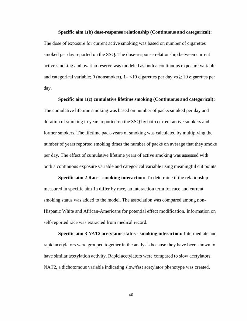
40
Specific aim 1(b) dose-response relationship (Continuous and categorical):
The dose of exposure for current active smoking was based on number of cigarettes
smoked per day reported on the SSQ. The dose-response relationship between current
active smoking and ovarian reserve was modeled as both a continuous exposure variable
and categorical variable; 0 (nonsmoker), 1– <10 cigarettes per day vs ≥ 10 cigarettes per
day.
Specific aim 1(c) cumulative lifetime smoking (Continuous and categorical):
The cumulative lifetime smoking was based on number of packs smoked per day and
duration of smoking in years reported on the SSQ by both current active smokers and
former smokers. The lifetime pack-years of smoking was calculated by multiplying the
number of years reported smoking times the number of packs on average that they smoke
per day. The effect of cumulative lifetime years of active smoking was assessed with
both a continuous exposure variable and categorical variable using meaningful cut points.
Specific aim 2 Race - smoking interaction: To determine if the relationship
measured in specific aim 1a differ by race, an interaction term for race and current
smoking status was added to the model. The association was compared among non-
Hispanic White and African-Americans for potential effect modification. Information on
self-reported race was extracted from medical record.
Specific aim 3 NAT2 acetylator status - smoking interaction: Intermediate and
rapid acetylators were grouped together in the analysis because they have been shown to
have similar acetylation activity. Rapid acetylators were compared to slow acetylators.
NAT2, a dichotomous variable indicating slow/fast acetylator phenotype was created.
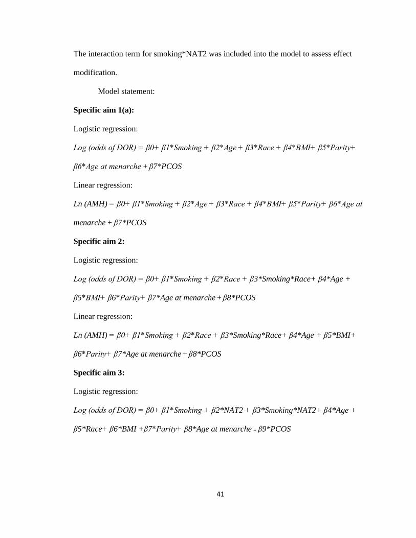
41
The interaction term for smoking*NAT2 was included into the model to assess effect
modification.
Model statement:
Specific aim 1(a):
Logistic regression:
Log (odds of DOR) = β0+ β1*Smoking + β2*Age + β3*Race + β4*BMI+ β5*Parity+
β6*Age at menarche + β7*PCOS
Linear regression:
Ln (AMH) = β0+ β1*Smoking + β2*Age + β3*Race + β4*BMI+ β5*Parity+ β6*Age at
menarche + β7*PCOS
Specific aim 2:
Logistic regression:
Log (odds of DOR) = β0+ β1*Smoking + β2*Race + β3*Smoking*Race+ β4*Age +
β5*BMI+ β6*Parity+ β7*Age at menarche + β8*PCOS
Linear regression:
Ln (AMH) = β0+ β1*Smoking + β2*Race + β3*Smoking*Race+ β4*Age + β5*BMI+
β6*Parity+ β7*Age at menarche + β8*PCOS
Specific aim 3:
Logistic regression:
Log (odds of DOR) = β0+ β1*Smoking + β2*NAT2 + β3*Smoking*NAT2+ β4*Age +
β5*Race+ β6*BMI +β7*Parity+ β8*Age at menarche + β9*PCOS
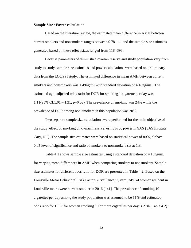
42
Sample Size / Power calculation
Based on the literature review, the estimated mean difference in AMH between
current smokers and nonsmokers ranges between 0.78- 1.1 and the sample size estimates
generated based on these effect sizes ranged from 118 -398.
Because parameters of diminished ovarian reserve and study population vary from
study to study, sample size estimates and power calculations were based on preliminary
data from the LOUSSI study. The estimated difference in mean AMH between current
smokers and nonsmokers was 1.49ng/ml with standard deviation of 4.18ng/mL. The
estimated age- adjusted odds ratio for DOR for smoking 1 cigarette per day was
1.11(95% CI:1.01 – 1.21, p=0.03). The prevalence of smoking was 24% while the
prevalence of DOR among non-smokers in this population was 30%.
Two separate sample size calculations were performed for the main objective of
the study, effect of smoking on ovarian reserve, using Proc power in SAS (SAS Institute,
Cary, NC). The sample size estimates were based on statistical power of 80%, alpha=
0.05 level of significance and ratio of smokers to nonsmokers set at 1:3.
Table 4.1 shows sample size estimates using a standard deviation of 4.18ng/mL
for varying mean differences in AMH when comparing smokers to nonsmokers. Sample
size estimates for different odds ratio for DOR are presented in Table 4.2. Based on the
Louisville Metro Behavioral Risk Factor Surveillance System, 24% of women resident in
Louisville metro were current smoker in 2016 [141]. The prevalence of smoking 10
cigarettes per day among the study population was assumed to be 11% and estimated
odds ratio for DOR for women smoking 10 or more cigarettes per day is 2.84 (Table 4.2).
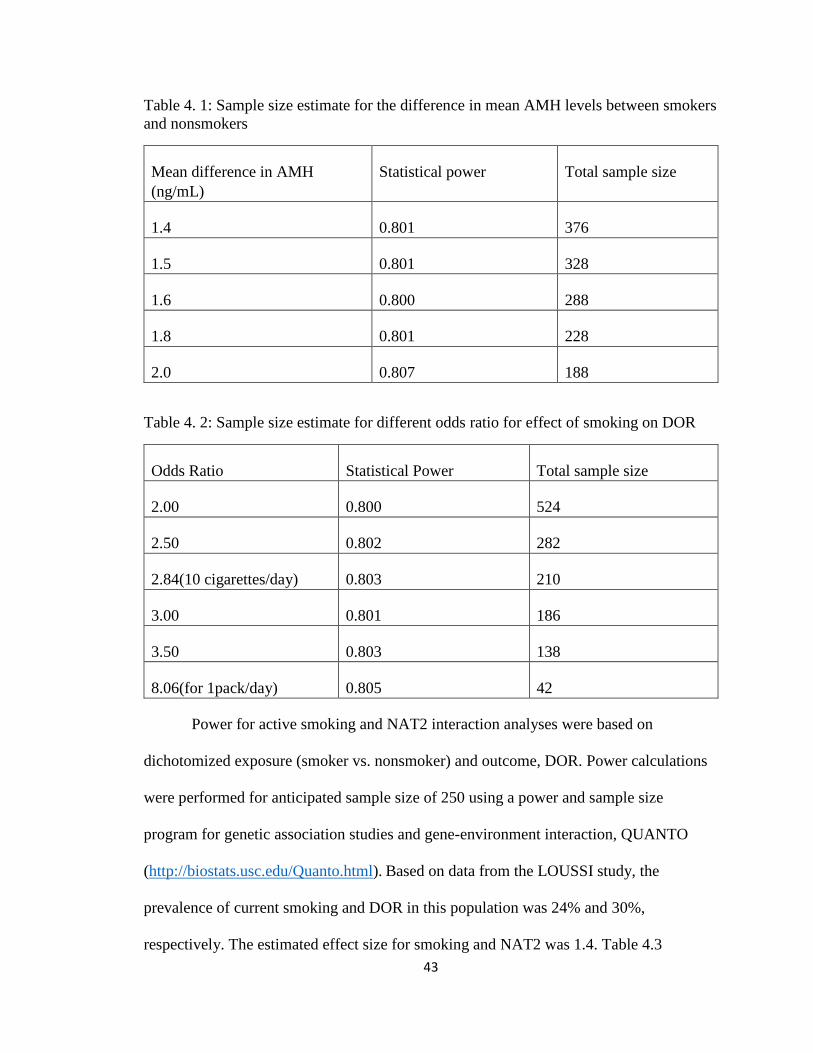
43
Table 4. 1: Sample size estimate for the difference in mean AMH levels between smokers
and nonsmokers
Mean difference in AMH
(ng/mL)
Statistical power Total sample size
1.4 0.801 376
1.5 0.801 328
1.6 0.800 288
1.8 0.801 228
2.0 0.807 188
Table 4. 2: Sample size estimate for different odds ratio for effect of smoking on DOR
Odds Ratio Statistical Power Total sample size
2.00 0.800 524
2.50 0.802 282
2.84(10 cigarettes/day) 0.803 210
3.00 0.801 186
3.50 0.803 138
8.06(for 1pack/day) 0.805 42
Power for active smoking and NAT2 interaction analyses were based on
dichotomized exposure (smoker vs. nonsmoker) and outcome, DOR. Power calculations
were performed for anticipated sample size of 250 using a power and sample size
program for genetic association studies and gene-environment interaction, QUANTO
(http://biostats.usc.edu/Quanto.html). Based on data from the LOUSSI study, the
prevalence of current smoking and DOR in this population was 24% and 30%,
respectively. The estimated effect size for smoking and NAT2 was 1.4. Table 4.3
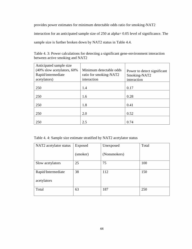
44
provides power estimates for minimum detectable odds ratio for smoking-NAT2
interaction for an anticipated sample size of 250 at alpha= 0.05 level of significance. The
sample size is further broken down by NAT2 status in Table 4.4.
Table 4. 3: Power calculations for detecting a significant gene-environment interaction
between active smoking and NAT2
Anticipated sample size
(40% slow acetylators, 60%
Rapid/intermediate
acetylators)
Minimum detectable odds
ratio for smoking-NAT2
interaction
Power to detect significant
Smoking-NAT2
interaction
250 1.4 0.17
250 1.6 0.28
250 1.8 0.41
250 2.0 0.52
250 2.5 0.74
Table 4. 4: Sample size estimate stratified by NAT2 acetylator status
NAT2 acetylator status Exposed
(smoker)
Unexposed
(Nonsmokers)
Total
Slow acetylators 25 75 100
Rapid/Intermediate
acetylators
38 112 150
Total 63 187 250
45
E. RESULTS
Out of 265 women who were enrolled into the LOUSSI study, 59 (22.3%) were
excluded for the following reasons: missing medical record data on serum AMH levels
(n=44); or they did not provide urine for cotinine assays (n=15). An additional 36 women
were excluded from analyses due to lack of correlation between self-reported smoking
status (never smoker or former smoker for more than 1 month) and increased cotinine
level indicating either a high tobacco exposure (≥100ng/mL, n=16) and or significant
passive exposure (14-99ng/mL, n=20) (Figure 1). The overall Kappa statistics of self-
reported smoking status was 0.81 (95% CI: 0.72 – 0.89; p<0.001) (data not shown).
The remaining 170 women included 31 (18%) current active smokers and 139
(82%) nonsmokers. Among the current smokers, 53% reported smoking 10 or more
cigarettes per day, 21% reported smoking between 5 to 9 cigarettes per day while 26%
reported smoking less than 5 cigarettes per day.
Table 5.1 presents the characteristics of the women in the LOUSSI study stratified
by smoking status. Nonsmokers had higher AMH levels, AFC and prevalence of DOR
than smokers, but the differences were not significant. In general, smokers tended to be
46
younger (P=0.04), of self-reported White race (p=0.01) and were more likely to be single
(71% vs. 23%, p= <0.001). Smokers and nonsmokers were comparable in terms of age at
menarche, BMI, parity and prevalence of PCOS.
The NAT2 acetylator status of the participants is presented in Table 5.2. DNA
extraction and/or NAT2 genotyping were unsuccessful in 14 participants. A total of 11
NAT2 genotypes were identified and assigned to rapid, intermediate and slow. About
39% of women in the study were slow acetylators, 44% intermediate and 17% rapid
acetylators. Rapid and intermediate acetylators were merged for analysis. The distribution
of NAT2 acetylator status did not differ by smoking status or self-reported race (Table
5.2).
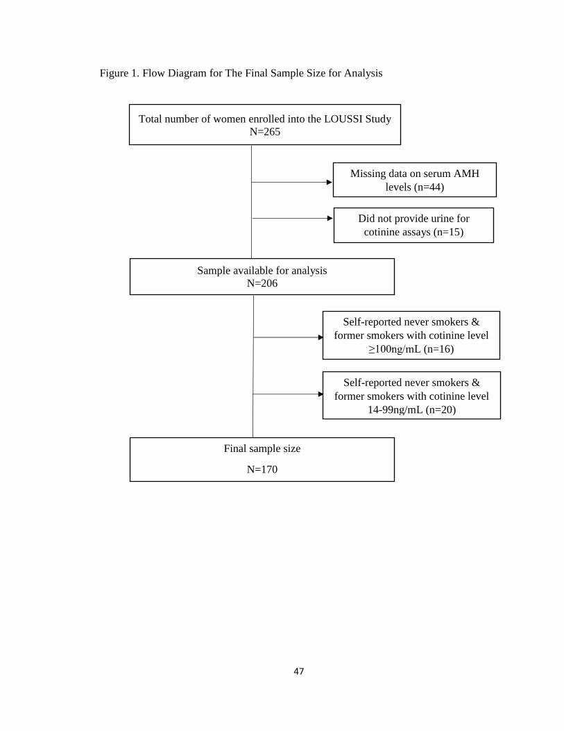
47
Figure 1. Flow Diagram for The Final Sample Size for Analysis
Total number of women enrolled into the LOUSSI Study
N=265
Missing data on serum AMH
levels (n=44)
Sample available for analysis
N=206
Final sample size
N=170
Did not provide urine for
cotinine assays (n=15)
Self-reported never smokers &
former smokers with cotinine level
≥100ng/mL (n=16)
Self-reported never smokers &
former smokers with cotinine level
14-99ng/mL (n=20)

48
Table 5. 1. Characteristics of Women in the Study Stratified by Smoking Status (N
=170)
Nonsmokers
N (%)
(n=139)
Smokers
N (%)
(n=31)
P-value
Age (years) 0.04
21-25 16 (11.5) 9 (30.0)
26-30 26 (18.7) 7 (23.3)
31-35 50 (36.0) 8 (26.7)
36-45 47 (33.8) 6 (20.0)
Race 0.01
White 73 (52.9) 17 (54.9)
Black 32 (23.2) 13 (41.9)
Other 33 (23.9) 1 (3.2)
Age at menarche(years) mean (s.d) 12.5 (1.9) 11.9 (1.5) 0.10
Body Mass Index 0.17
17.5-24.9 36 (26.3) 8 (25.8)
25-29.9 44 (32.1) 6 (19.4)
30-34.9 25 (18.2) 11 (35.4)
≥ 35 32 (23.4) 6 (19.4)
Poly Cystic Ovary Syndrome 0.80
No 83 (60.6) 18 (58.1)
Yes 54 (39.4) 13 (41.9))
Parity
None 60 (43.8) 9 (29.0) 0.30
One 31 (22.6) 8 (25.8)
Two or more 46 (33.6) 14 (45.2)
Marital Status <0.001
Single 31 (23.7) 20 (71.4)
Married 100 (76.3) 8 (28.6)
Serum AMH (ng/mL) median
(interquartile range) 2.33 (4.0) 2.07 (2.0) 0.48
Diminished ovarian reserve (DOR) 0.59
No 101 (72.7) 24 (77.4)
Yes 38 (27.3) 7 (22.6)
Antral Follicle Count (AFC)
median (interquartile range) 21 (17) 17(11) 0.48
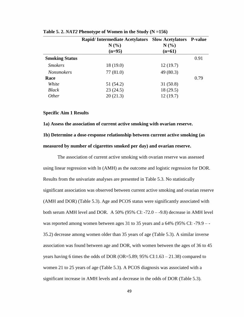
49
Table 5. 2. NAT2 Phenotype of Women in the Study (N =156)
Specific Aim 1 Results
1a) Assess the association of current active smoking with ovarian reserve.
1b) Determine a dose-response relationship between current active smoking (as
measured by number of cigarettes smoked per day) and ovarian reserve.
The association of current active smoking with ovarian reserve was assessed
using linear regression with ln (AMH) as the outcome and logistic regression for DOR.
Results from the univariate analyses are presented in Table 5.3. No statistically
significant association was observed between current active smoking and ovarian reserve
(AMH and DOR) (Table 5.3). Age and PCOS status were significantly associated with
both serum AMH level and DOR. A 50% (95% CI: -72.0 – -9.8) decrease in AMH level
was reported among women between ages 31 to 35 years and a 64% (95% CI: -79.9 – -
35.2) decrease among women older than 35 years of age (Table 5.3). A similar inverse
association was found between age and DOR, with women between the ages of 36 to 45
years having 6 times the odds of DOR (OR=5.89; 95% CI:1.63 – 21.38) compared to
women 21 to 25 years of age (Table 5.3). A PCOS diagnosis was associated with a
significant increase in AMH levels and a decrease in the odds of DOR (Table 5.3).
Rapid/ Intermediate Acetylators
N (%)
(n=95)
Slow Acetylators
N (%)
(n=61)
P-value
Smoking Status 0.91
Smokers 18 (19.0) 12 (19.7)
Nonsmokers 77 (81.0) 49 (80.3)
Race 0.79
White 51 (54.2) 31 (50.8)
Black 23 (24.5) 18 (29.5)
Other 20 (21.3) 12 (19.7)
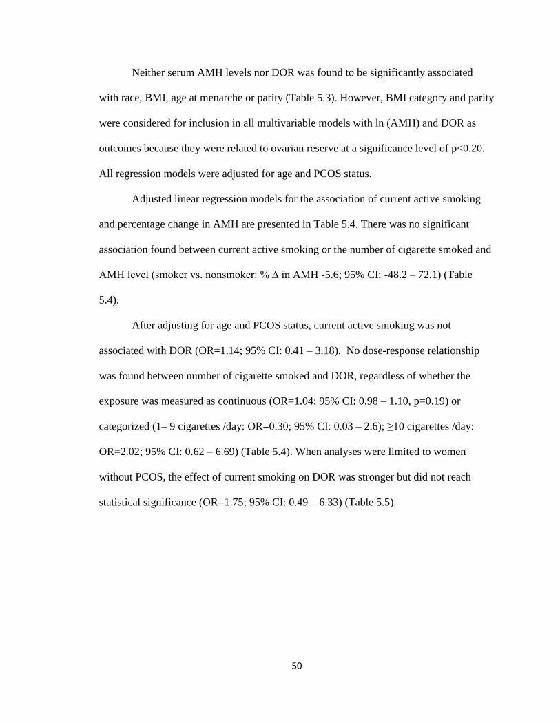
50
Neither serum AMH levels nor DOR was found to be significantly associated
with race, BMI, age at menarche or parity (Table 5.3). However, BMI category and parity
were considered for inclusion in all multivariable models with ln (AMH) and DOR as
outcomes because they were related to ovarian reserve at a significance level of p<0.20.
All regression models were adjusted for age and PCOS status.
Adjusted linear regression models for the association of current active smoking
and percentage change in AMH are presented in Table 5.4. There was no significant
association found between current active smoking or the number of cigarette smoked and
AMH level (smoker vs. nonsmoker: % ∆ in AMH -5.6; 95% CI: -48.2 – 72.1) (Table
5.4).
After adjusting for age and PCOS status, current active smoking was not
associated with DOR (OR=1.14; 95% CI: 0.41 – 3.18). No dose-response relationship
was found between number of cigarette smoked and DOR, regardless of whether the
exposure was measured as continuous (OR=1.04; 95% CI: 0.98 – 1.10, p=0.19) or
categorized (1– 9 cigarettes /day: OR=0.30; 95% CI: 0.03 – 2.6); ≥10 cigarettes /day:
OR=2.02; 95% CI: 0.62 – 6.69) (Table 5.4). When analyses were limited to women
without PCOS, the effect of current smoking on DOR was stronger but did not reach
statistical significance (OR=1.75; 95% CI: 0.49 – 6.33) (Table 5.5).
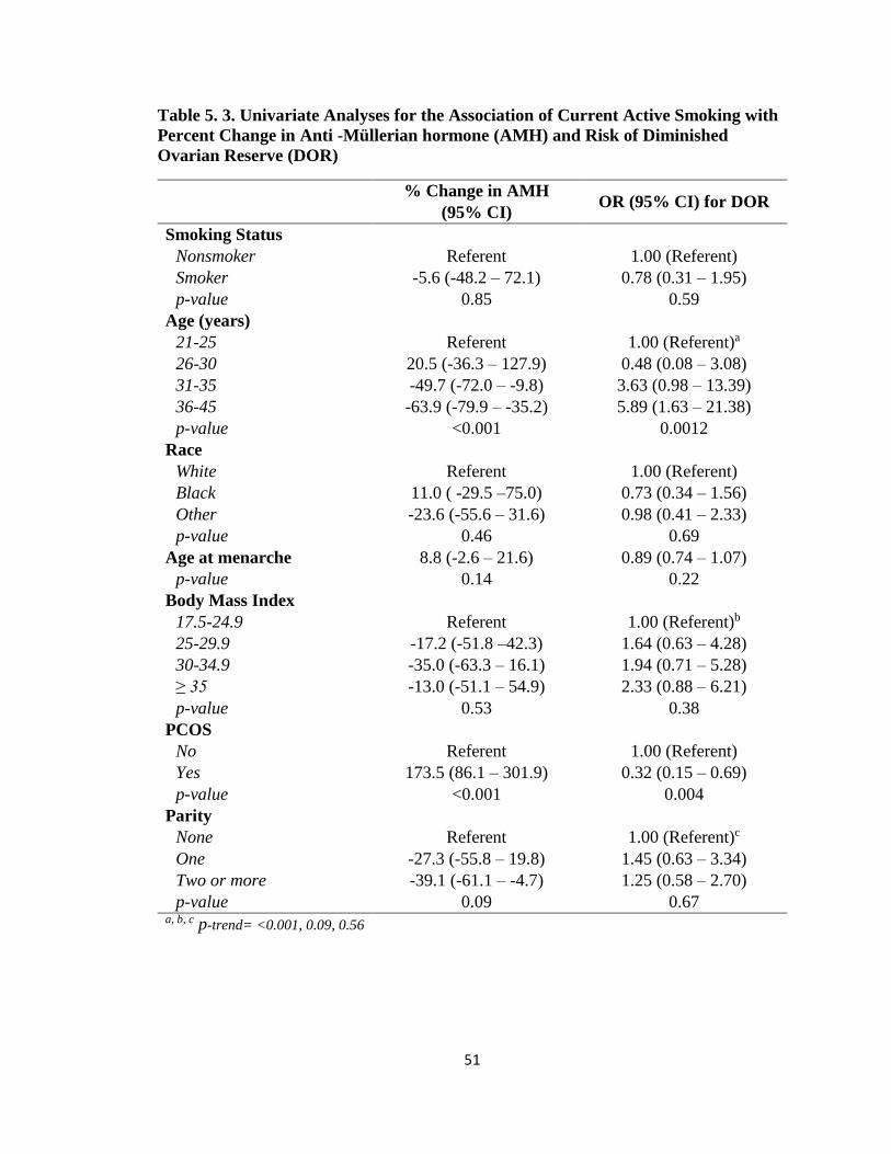
51
Table 5. 3. Univariate Analyses for the Association of Current Active Smoking with
Percent Change in Anti -Müllerian hormone (AMH) and Risk of Diminished
Ovarian Reserve (DOR)
% Change in AMH
(95% CI) OR (95% CI) for DOR
Smoking Status
Nonsmoker Referent 1.00 (Referent)
Smoker -5.6 (-48.2 – 72.1) 0.78 (0.31 – 1.95)
p-value 0.85 0.59
Age (years)
21-25 Referent 1.00 (Referent)a
26-30 20.5 (-36.3 – 127.9) 0.48 (0.08 – 3.08)
31-35 -49.7 (-72.0 – -9.8) 3.63 (0.98 – 13.39)
36-45 -63.9 (-79.9 – -35.2) 5.89 (1.63 – 21.38)
p-value <0.001 0.0012
Race
White Referent 1.00 (Referent)
Black 11.0 ( -29.5 –75.0) 0.73 (0.34 – 1.56)
Other -23.6 (-55.6 – 31.6) 0.98 (0.41 – 2.33)
p-value 0.46 0.69
Age at menarche 8.8 (-2.6 – 21.6) 0.89 (0.74 – 1.07)
p-value 0.14 0.22
Body Mass Index
17.5-24.9 Referent 1.00 (Referent)b
25-29.9 -17.2 (-51.8 –42.3) 1.64 (0.63 – 4.28)
30-34.9 -35.0 (-63.3 – 16.1) 1.94 (0.71 – 5.28)
≥ 35 -13.0 (-51.1 – 54.9) 2.33 (0.88 – 6.21)
p-value 0.53 0.38
PCOS
No Referent 1.00 (Referent)
Yes 173.5 (86.1 – 301.9) 0.32 (0.15 – 0.69)
p-value <0.001 0.004
Parity
None Referent 1.00 (Referent)c
One -27.3 (-55.8 – 19.8) 1.45 (0.63 – 3.34)
Two or more -39.1 (-61.1 – -4.7) 1.25 (0.58 – 2.70)
p-value 0.09 0.67 a, b, c p-trend= <0.001, 0.09, 0.56
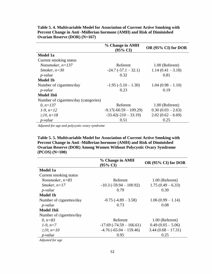
52
Table 5. 4. Multivariable Model for Association of Current Active Smoking with
Percent Change in Anti -Müllerian hormone (AMH) and Risk of Diminished
Ovarian Reserve (DOR) (N=167)
% Change in AMH
(95% CI) OR (95% CI) for DOR
Model 1a
Current smoking status
Nonsmoker, n=137 Referent 1.00 (Referent)
Smoker, n=30 -24.7 (-57.1 – 32.1) 1.14 (0.41 – 3.18)
p-value 0.32 0.81
Model 1b
Number of cigarettes/day -1.95 (-5.10 – 1.30) 1.04 (0.98 – 1.10)
p-value 0.23 0.19
Model 1bii
Number of cigarettes/day (categories)
0, n=137 Referent 1.00 (Referent)
1-9, n=12 -9.17(-60.59 – 109.29) 0.30 (0.03 – 2.63)
≥10, n=18 -33.42(-210 – 33.19) 2.02 (0.62 – 6.69)
p-value 0.51 0.25
Adjusted for age and polycystic ovary syndrome
Table 5. 5. Multivariable Model for Association of Current Active Smoking with
Percent Change in Anti -Müllerian hormone (AMH) and Risk of Diminished
Ovarian Reserve (DOR) Among Women Without Polycystic Ovary Syndrome
(PCOS) (N=100)
% Change in AMH
(95% CI) OR (95% CI) for DOR
Model 1a
Current smoking status
Nonsmoker, n=83 Referent 1.00 (Referent)
Smoker, n=17 -10.3 (-59.94 – 100.92) 1.75 (0.49 – 6.33)
p-value 0.79 0.39
Model 1b
Number of cigarettes/day -0.75 (-4.89 – 3.58) 1.06 (0.99 – 1.14)
p-value 0.73 0.08
Model 1bii
Number of cigarettes/day
0, n=83 Referent 1.00 (Referent)
1-9, n=7 -17.69 (-74.59 – 166.61) 0.49 (0.05 – 5.06)
≥10, n=10 -4.76 (-65.04 – 159.46) 3.44 (0.68 – 17.31)
p-value 0.95 0.25
Adjusted for age
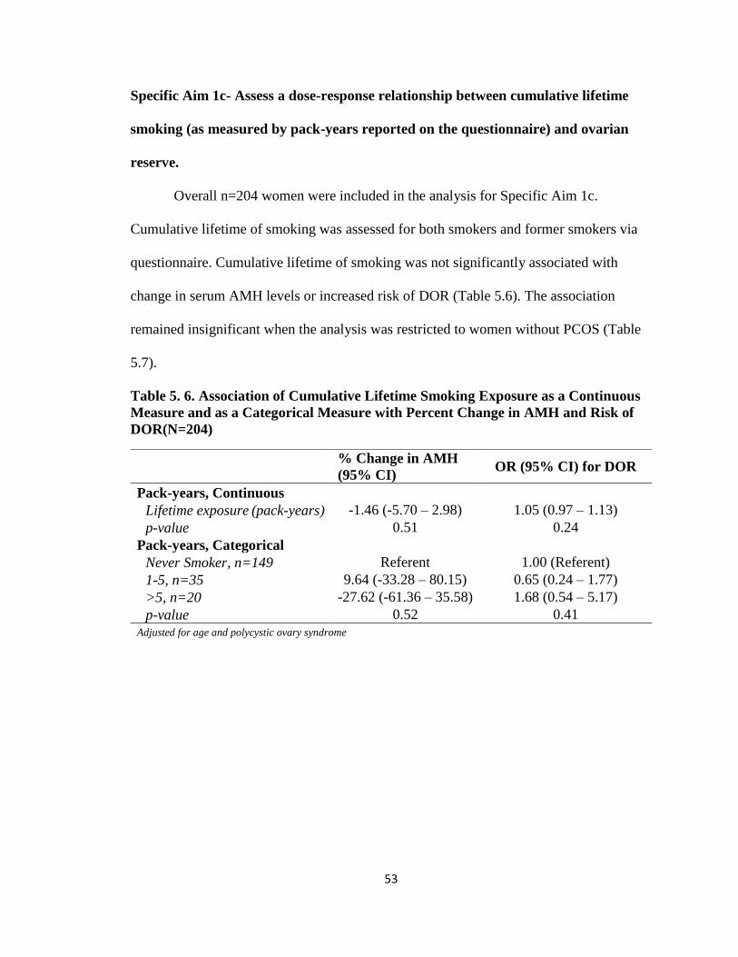
53
Specific Aim 1c- Assess a dose-response relationship between cumulative lifetime
smoking (as measured by pack-years reported on the questionnaire) and ovarian
reserve.
Overall n=204 women were included in the analysis for Specific Aim 1c.
Cumulative lifetime of smoking was assessed for both smokers and former smokers via
questionnaire. Cumulative lifetime of smoking was not significantly associated with
change in serum AMH levels or increased risk of DOR (Table 5.6). The association
remained insignificant when the analysis was restricted to women without PCOS (Table
5.7).
Table 5. 6. Association of Cumulative Lifetime Smoking Exposure as a Continuous
Measure and as a Categorical Measure with Percent Change in AMH and Risk of
DOR(N=204)
% Change in AMH
(95% CI) OR (95% CI) for DOR
Pack-years, Continuous
Lifetime exposure (pack-years) -1.46 (-5.70 – 2.98) 1.05 (0.97 – 1.13)
p-value 0.51 0.24
Pack-years, Categorical
Never Smoker, n=149 Referent 1.00 (Referent)
1-5, n=35 9.64 (-33.28 – 80.15) 0.65 (0.24 – 1.77)
>5, n=20 -27.62 (-61.36 – 35.58) 1.68 (0.54 – 5.17)
p-value 0.52 0.41
Adjusted for age and polycystic ovary syndrome
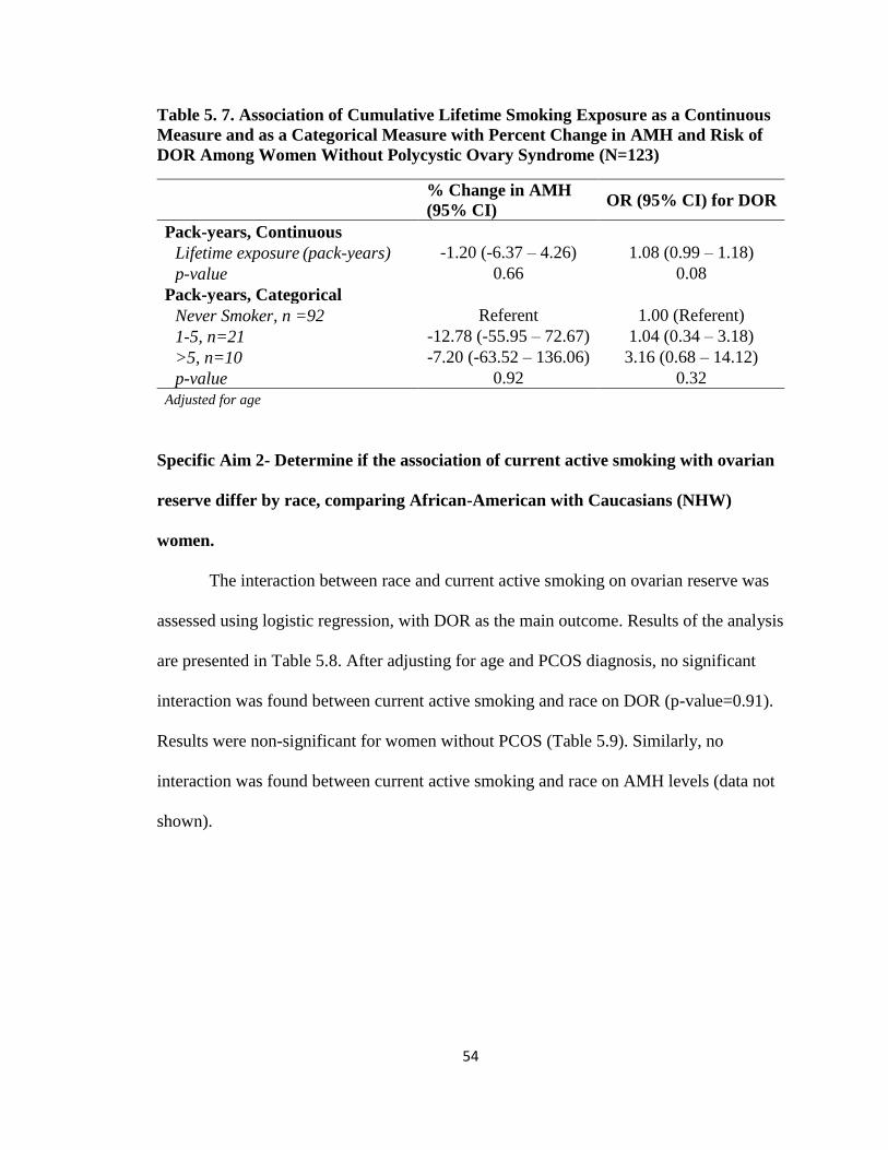
54
Table 5. 7. Association of Cumulative Lifetime Smoking Exposure as a Continuous
Measure and as a Categorical Measure with Percent Change in AMH and Risk of
DOR Among Women Without Polycystic Ovary Syndrome (N=123)
% Change in AMH
(95% CI) OR (95% CI) for DOR
Pack-years, Continuous
Lifetime exposure (pack-years) -1.20 (-6.37 – 4.26) 1.08 (0.99 – 1.18)
p-value 0.66 0.08
Pack-years, Categorical
Never Smoker, n =92 Referent 1.00 (Referent)
1-5, n=21 -12.78 (-55.95 – 72.67) 1.04 (0.34 – 3.18)
>5, n=10 -7.20 (-63.52 – 136.06) 3.16 (0.68 – 14.12)
p-value 0.92 0.32
Adjusted for age
Specific Aim 2- Determine if the association of current active smoking with ovarian
reserve differ by race, comparing African-American with Caucasians (NHW)
women.
The interaction between race and current active smoking on ovarian reserve was
assessed using logistic regression, with DOR as the main outcome. Results of the analysis
are presented in Table 5.8. After adjusting for age and PCOS diagnosis, no significant
interaction was found between current active smoking and race on DOR (p-value=0.91).
Results were non-significant for women without PCOS (Table 5.9). Similarly, no
interaction was found between current active smoking and race on AMH levels (data not
shown).
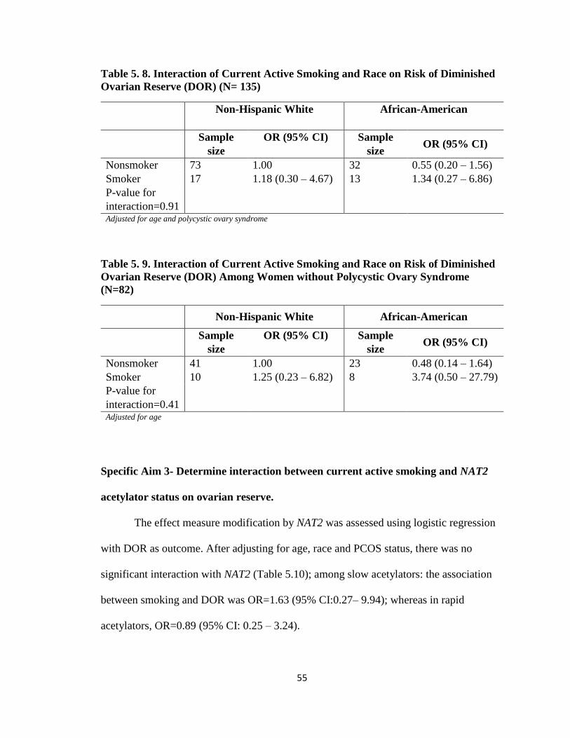
55
Table 5. 8. Interaction of Current Active Smoking and Race on Risk of Diminished
Ovarian Reserve (DOR) (N= 135)
Non-Hispanic White
African-American
Sample
size
OR (95% CI) Sample
size OR (95% CI)
Nonsmoker 73 1.00 32 0.55 (0.20 – 1.56)
Smoker 17 1.18 (0.30 – 4.67) 13 1.34 (0.27 – 6.86)
P-value for
interaction=0.91
Adjusted for age and polycystic ovary syndrome
Table 5. 9. Interaction of Current Active Smoking and Race on Risk of Diminished
Ovarian Reserve (DOR) Among Women without Polycystic Ovary Syndrome
(N=82)
Non-Hispanic White African-American
Sample
size
OR (95% CI) Sample
size OR (95% CI)
Nonsmoker 41 1.00 23 0.48 (0.14 – 1.64)
Smoker 10 1.25 (0.23 – 6.82) 8 3.74 (0.50 – 27.79)
P-value for
interaction=0.41
Adjusted for age
Specific Aim 3- Determine interaction between current active smoking and NAT2
acetylator status on ovarian reserve.
The effect measure modification by NAT2 was assessed using logistic regression
with DOR as outcome. After adjusting for age, race and PCOS status, there was no
significant interaction with NAT2 (Table 5.10); among slow acetylators: the association
between smoking and DOR was OR=1.63 (95% CI:0.27– 9.94); whereas in rapid
acetylators, OR=0.89 (95% CI: 0.25 – 3.24).
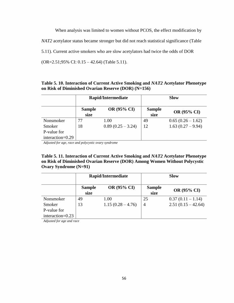
56
When analysis was limited to women without PCOS, the effect modification by
NAT2 acetylator status became stronger but did not reach statistical significance (Table
5.11). Current active smokers who are slow acetylators had twice the odds of DOR
(OR=2.51;95% CI: 0.15 – 42.64) (Table 5.11).
Table 5. 10. Interaction of Current Active Smoking and NAT2 Acetylator Phenotype
on Risk of Diminished Ovarian Reserve (DOR) (N=156)
Rapid/Intermediate
Slow
Sample
size
OR (95% CI) Sample
size OR (95% CI)
Nonsmoker 77 1.00 49 0.65 (0.26 – 1.62)
Smoker 18 0.89 (0.25 – 3.24) 12 1.63 (0.27 – 9.94)
P-value for
interaction=0.29
Adjusted for age, race and polycystic ovary syndrome
Table 5. 11. Interaction of Current Active Smoking and NAT2 Acetylator Phenotype
on Risk of Diminished Ovarian Reserve (DOR) Among Women Without Polycystic
Ovary Syndrome (N=91)
Rapid/Intermediate
Slow
Sample
size
OR (95% CI) Sample
size OR (95% CI)
Nonsmoker 49 1.00 25 0.37 (0.11 – 1.14)
Smoker 13 1.15 (0.28 – 4.76) 4 2.51 (0.15 – 42.64)
P-value for
interaction=0.23
Adjusted for age and race
57
F. DISCUSSION
This study assessed the association of smoking with ovarian reserve among
women of reproductive age. The existing evidence on smoking and ovarian reserve as
measured by AMH has been inconclusive [12, 16, 17, 20, 21, 55, 93-98]. This is the
first study to explore effect modification by race and NAT2 phenotype as a possible
cause of inconsistency in previous studies, and the first study of smoking and ovarian
reserve to validate self-reported exposure status with cotinine.
The findings from this cross-sectional analysis are consistent with previous
prospective [55], retrospective [96] and cross-sectional studies [12, 97, 98] that found
no significant association between smoking and ovarian reserve. However, other
studies did find a significant association between smoking and ovarian reserve [16,
17, 20, 21, 93-95]. If an association truly exists, then nonsignificant results from
previous studies may be due to a crude assessment of smoking exposure status based
on self-report and not accounting for duration and quantity of smoking. Further,
previous results are difficult to reconcile because of differences in the definition of
exposure status and adjustment factors across studies.
58
Only a few studies have explored a dose-response relationship between smoking
and ovarian reserve [22, 59, 95, 96]. This study provided a detailed assessment of
smoking status, validated current exposure status with cotinine, assessed a dose-response
relationship, and estimated cumulative lifetime exposure using the quantity and duration
of smoking. While effect sizes did not reach statistical significance, the magnitude and
direction of associations suggest that smoking may reduce ovarian reserve especially
among heavy smokers. White et al. (2016) in a cross-sectional study of 913
premenopausal women, found a significant association between self-reported smoking
and serum AMH among women who smoked 20 or more cigarettes /day (−56.2%,
95%CI: −80.3, −2.8%) [95]. Similar findings with number of pack-years smoked were
reported by Dolleman et al. where significant association between smoking and age-
specific serum AMH was found for 10-15 pack-years (β= -7.0, p=0.003) and 15-20 pack-
years (β= -8.5, p=0.001) pack-years but not for 5-10 pack-years (β= -3.1, p=0.15) [59].
Other studies that reported significant inverse associations between smoking and
ovarian reserve (as measured by AMH) reported moderate [16, 94, 95] to high [17, 20,
21, 93] prevalence of smoking in the study population. Considering the established
relationship between smoking and early age at menopause [142-146], it is plausible that
59
smoking is associated with ovarian aging and reserve, but the association was not
detected in this study due to low frequency of heavy smokers.
Studies on the association of smoking with ovarian reserve varied in the
covariates selected for adjustment. None of the studies controlled for PCOS, even though
several of the studies [17, 20-22, 55, 93] were conducted among infertile or sub-fertile
populations. Women with PCOS diagnosis have been shown to have significantly
elevated AMH levels [63, 64]. In analyses that excluded women with PCOS diagnosis,
effect sizes were generally stronger, suggesting PCOS is not only a confounder, but also
an effect modifier of the association of smoking with ovarian reserve. Therefore,
controlling for and stratifying on PCOS was a major improvement in the study design
compared to prior studies.
Timing of exposure may be a very important consideration for environmental
predictors of AMH. For example, exposure to toxins during early development may be
more influential on ovarian reserve than exposure as an adult. Experimental studies in
animals shows that smoking inhibits follicular development; causes premature
luteinization and atresia of the preovulatory follicle; reduces oocyte vascularization and
maturation; impairs steroidogenesis; and increases chromosomal errors; and cytotoxicity;
in a stage dependent manner [75-78]. Most studies on the association of smoking with
ovarian reserve including this study, did not account for the timing of exposure.
Effect Modification by Race
Racial disparity in the association of smoking with ovarian reserve is largely
unknown. The results of this study suggest that the risk of diminished ovarian reserve
may be increased in African-American smokers. Schuh-Huerta et al. (2011) in a
60
genome-wide association (GWA) analyses on hormonal markers of ovarian reserve
(FSH and AMH) identified genome-wide significant SNPs associated with FSH
(rs6543833, rs12465811, rs11255291) and AMH levels (rs9875589) in an African-
American population [16]. One SNP (rs12213875) identified in Caucasians was
associated with a mean decrease of 1.3+1.4 pM in AMH among GG genotypes
compared to an increase of 13.8+4.2 pM and 9.9+2.1 pM in the AA and AG
genotypes, respectively [16]. While these results provide evidence for underlying
genetic variations in ovarian reserve by race, disparities in environmental and
behavioral factors such as smoking, also need to be explored.
Effect Modification by NAT2
This study is the first to explore population differences in NAT2 genotype
frequencies as a possible reason for the inconsistency in the association of smoking
with ovarian reserve. Results from this study, though not statistically significant,
suggests that the risk of diminished ovarian reserve may be increased for current
smokers who are slow NAT2 acetylators. Polymorphisms in the NAT2 gene modify
susceptibility to harmful heterocyclic and aromatic amines constituents in tobacco
smoke [25, 26, 100, 101]. High levels of DNA adduct of 4-aminobiphenyl, a
carcinogenic aromatic amine in tobacco smoke, have been reported in tissues of
smokers [103, 104, 106-112, 147]. Additionally, it has been shown that slow NAT2
acetylators have variable reductions in catalytic activities that make them more
susceptible to the effect of toxins [26, 100, 101, 114]. Given the significantly reduced
fecundability reported among current smokers who are slow acetylators by Taylor et
al. (2011), it is plausible that the effect is modulated through a reduction in ovarian
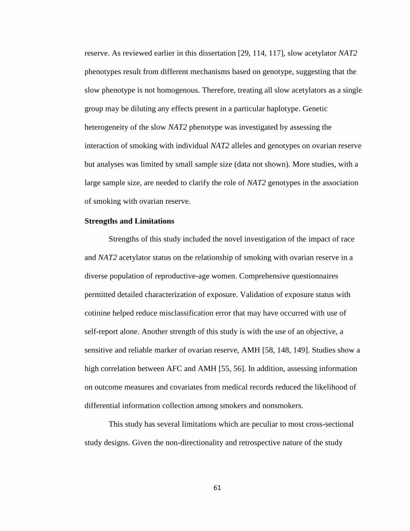
61
reserve. As reviewed earlier in this dissertation [29, 114, 117], slow acetylator NAT2
phenotypes result from different mechanisms based on genotype, suggesting that the
slow phenotype is not homogenous. Therefore, treating all slow acetylators as a single
group may be diluting any effects present in a particular haplotype. Genetic
heterogeneity of the slow NAT2 phenotype was investigated by assessing the
interaction of smoking with individual NAT2 alleles and genotypes on ovarian reserve
but analyses was limited by small sample size (data not shown). More studies, with a
large sample size, are needed to clarify the role of NAT2 genotypes in the association
of smoking with ovarian reserve.
Strengths and Limitations
Strengths of this study included the novel investigation of the impact of race
and NAT2 acetylator status on the relationship of smoking with ovarian reserve in a
diverse population of reproductive-age women. Comprehensive questionnaires
permitted detailed characterization of exposure. Validation of exposure status with
cotinine helped reduce misclassification error that may have occurred with use of
self-report alone. Another strength of this study is with the use of an objective, a
sensitive and reliable marker of ovarian reserve, AMH [58, 148, 149]. Studies show a
high correlation between AFC and AMH [55, 56]. In addition, assessing information
on outcome measures and covariates from medical records reduced the likelihood of
differential information collection among smokers and nonsmokers.
This study has several limitations which are peculiar to most cross-sectional
study designs. Given the non-directionality and retrospective nature of the study
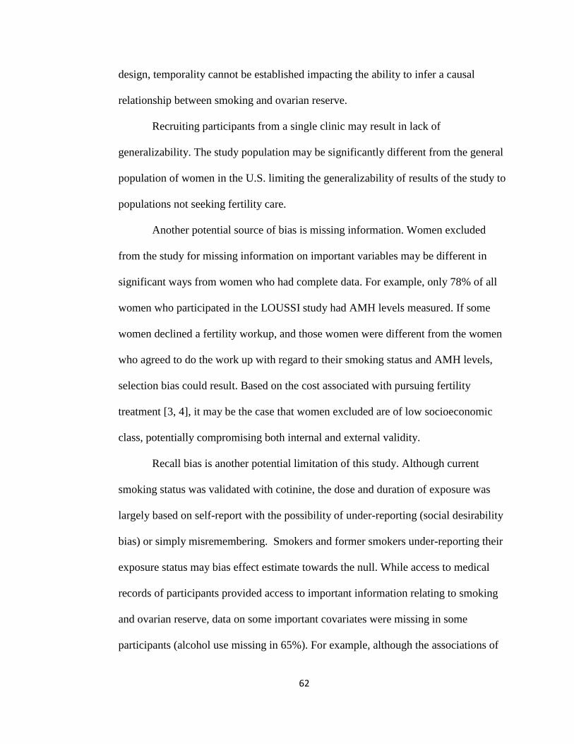
62
design, temporality cannot be established impacting the ability to infer a causal
relationship between smoking and ovarian reserve.
Recruiting participants from a single clinic may result in lack of
generalizability. The study population may be significantly different from the general
population of women in the U.S. limiting the generalizability of results of the study to
populations not seeking fertility care.
Another potential source of bias is missing information. Women excluded
from the study for missing information on important variables may be different in
significant ways from women who had complete data. For example, only 78% of all
women who participated in the LOUSSI study had AMH levels measured. If some
women declined a fertility workup, and those women were different from the women
who agreed to do the work up with regard to their smoking status and AMH levels,
selection bias could result. Based on the cost associated with pursuing fertility
treatment [3, 4], it may be the case that women excluded are of low socioeconomic
class, potentially compromising both internal and external validity.
Recall bias is another potential limitation of this study. Although current
smoking status was validated with cotinine, the dose and duration of exposure was
largely based on self-report with the possibility of under-reporting (social desirability
bias) or simply misremembering. Smokers and former smokers under-reporting their
exposure status may bias effect estimate towards the null. While access to medical
records of participants provided access to important information relating to smoking
and ovarian reserve, data on some important covariates were missing in some
participants (alcohol use missing in 65%). For example, although the associations of
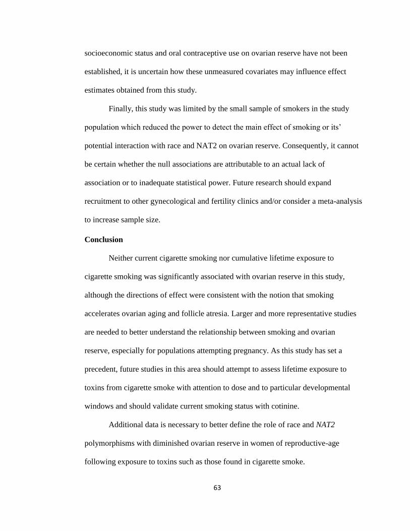
63
socioeconomic status and oral contraceptive use on ovarian reserve have not been
established, it is uncertain how these unmeasured covariates may influence effect
estimates obtained from this study.
Finally, this study was limited by the small sample of smokers in the study
population which reduced the power to detect the main effect of smoking or its’
potential interaction with race and NAT2 on ovarian reserve. Consequently, it cannot
be certain whether the null associations are attributable to an actual lack of
association or to inadequate statistical power. Future research should expand
recruitment to other gynecological and fertility clinics and/or consider a meta-analysis
to increase sample size.
Conclusion
Neither current cigarette smoking nor cumulative lifetime exposure to
cigarette smoking was significantly associated with ovarian reserve in this study,
although the directions of effect were consistent with the notion that smoking
accelerates ovarian aging and follicle atresia. Larger and more representative studies
are needed to better understand the relationship between smoking and ovarian
reserve, especially for populations attempting pregnancy. As this study has set a
precedent, future studies in this area should attempt to assess lifetime exposure to
toxins from cigarette smoke with attention to dose and to particular developmental
windows and should validate current smoking status with cotinine.
Additional data is necessary to better define the role of race and NAT2
polymorphisms with diminished ovarian reserve in women of reproductive-age
following exposure to toxins such as those found in cigarette smoke.
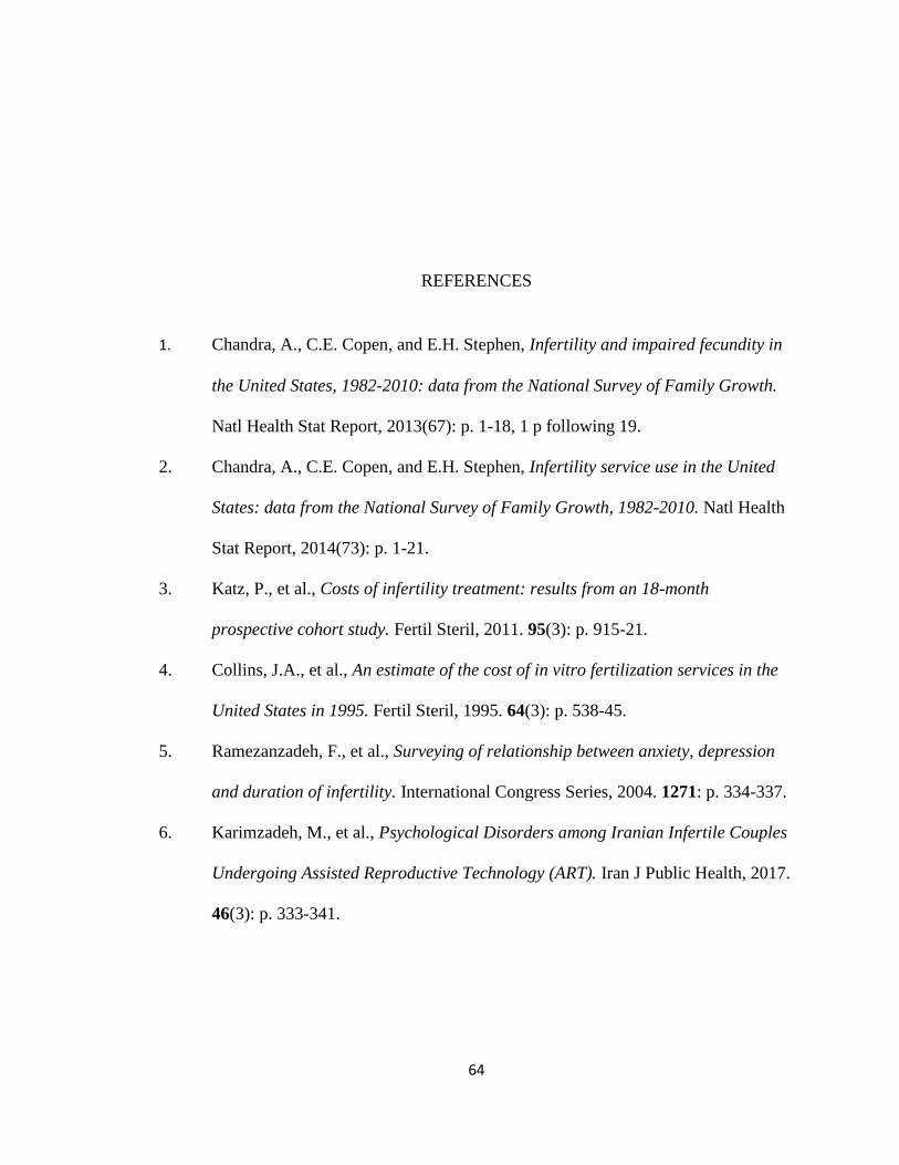
64
REFERENCES
1. Chandra, A., C.E. Copen, and E.H. Stephen, Infertility and impaired fecundity in
the United States, 1982-2010: data from the National Survey of Family Growth.
Natl Health Stat Report, 2013(67): p. 1-18, 1 p following 19.
2. Chandra, A., C.E. Copen, and E.H. Stephen, Infertility service use in the United
States: data from the National Survey of Family Growth, 1982-2010. Natl Health
Stat Report, 2014(73): p. 1-21.
3. Katz, P., et al., Costs of infertility treatment: results from an 18-month
prospective cohort study. Fertil Steril, 2011. 95(3): p. 915-21.
4. Collins, J.A., et al., An estimate of the cost of in vitro fertilization services in the
United States in 1995. Fertil Steril, 1995. 64(3): p. 538-45.
5. Ramezanzadeh, F., et al., Surveying of relationship between anxiety, depression
and duration of infertility. International Congress Series, 2004. 1271: p. 334-337.
6. Karimzadeh, M., et al., Psychological Disorders among Iranian Infertile Couples
Undergoing Assisted Reproductive Technology (ART). Iran J Public Health, 2017.
46(3): p. 333-341.
65
7. Gelbaya, T.A., et al., Definition and epidemiology of unexplained infertility.
Obstet Gynecol Surv, 2014. 69(2): p. 109-15.
8. Brugo-Olmedo, S., C. Chillik, and S. Kopelman, Definition and causes of
infertility. Reprod Biomed Online, 2001. 2(1): p. 41-53.
9. Center for Disease Control and Prevention, American Society for Reproductive
Medicine, and Society for Assisted Reproductive Technology, 2012 Assisted
Reproductive Technology National Summary Report., U.D.o.H.a.H. Services,
Editor. 2014.
10. Practice Committee of the American Society for Reproductive, M., Testing and
interpreting measures of ovarian reserve: a committee opinion. Fertil Steril, 2015.
103(3): p. e9-e17.
11. Practice Committee of the American Society for Reproductive, M., Diagnostic
evaluation of the infertile female: a committee opinion. Fertil Steril, 2015. 103(6):
p. e44-50.
12. Jung, S., et al., Demographic, lifestyle, and other factors in relation to
antimullerian hormone levels in mostly late premenopausal women. Fertil Steril,
2017. 107(4): p. 1012-1022.e2.
13. Hazout, A., et al., Serum antimullerian hormone/mullerian-inhibiting substance
appears to be a more discriminatory marker of assisted reproductive technology
outcome than follicle-stimulating hormone, inhibin B, or estradiol. Fertil Steril,
2004. 82(5): p. 1323-9.
66
14. Seifer, D.B. and D.T. Maclaughlin, Mullerian Inhibiting Substance is an ovarian
growth factor of emerging clinical significance. Fertil Steril, 2007. 88(3): p. 539-
46.
15. Broer, S.L., et al., The role of antimullerian hormone in prediction of outcome
after IVF: comparison with the antral follicle count. Fertil Steril, 2009. 91(3): p.
705-14.
16. Schuh-Huerta, S.M., et al., Genetic variants and environmental factors associated
with hormonal markers of ovarian reserve in Caucasian and African American
women. Hum Reprod, 2012. 27(2): p. 594-608.
17. Freour, T., et al., Active smoking compromises IVF outcome and affects ovarian
reserve. Reprod Biomed Online, 2008. 16(1): p. 96-102.
18. El-Nemr, A., et al., Effect of smoking on ovarian reserve and ovarian stimulation
in in-vitro fertilization and embryo transfer. Hum Reprod, 1998. 13(8): p. 2192-8.
19. Radin, R.G., et al., Active and passive smoking and fecundability in Danish
pregnancy planners. Fertil Steril, 2014. 102(1): p. 183-191.e2.
20. Fuentes, A., et al., Decreased anti-Mullerian hormone concentration in follicular
fluid of female smokers undergoing artificial reproductive techniques.
Chemosphere, 2012. 88(4): p. 403-6.
21. Freour, T., et al., Ovarian reserve and in vitro fertilization cycles outcome
according to women smoking status and stimulation regimen. Arch Gynecol
Obstet, 2012. 285(4): p. 1177-82.
22. Caserta, D., et al., The influence of cigarette smoking on a population of infertile
men and women. Arch Gynecol Obstet, 2013. 287(4): p. 813-8.
67
23. Dubinskaia, E.D., et al., [Role of the genetic factors, detoxication systems and
oxidative stress in the pathogenesis of endometriosis and infertility (review)].
Vestn Ross Akad Med Nauk, 2013(8): p. 14-9.
24. Taylor, K.C., et al., Alcohol, smoking, and caffeine in relation to fecundability,
with effect modification by NAT2. Ann Epidemiol, 2011. 21(11): p. 864-72.
25. Weber, W.W. and D.W. Hein, N-acetylation pharmacogenetics. Pharmacol Rev,
1985. 37(1): p. 25-79.
26. Walker, K., et al., Genetic polymorphism in N-Acetyltransferase (NAT):
Population distribution of NAT1 and NAT2 activity. J Toxicol Environ Health B
Crit Rev, 2009. 12(5-6): p. 440-72.
27. Wu, H., et al., Association Between N-acetyltransferase 2 Polymorphism and
Bladder Cancer Risk: Results From Studies of the Past Decade and a Meta-
Analysis. Clin Genitourin Cancer, 2016. 14(2): p. 122-9.
28. Costa, S., et al., A slow acetylator genotype associated with an increased risk of
advanced cervical cancer. J Cancer Res Clin Oncol, 2002. 128(12): p. 678-82.
29. Hein, D.W., N-acetyltransferase 2 genetic polymorphism: effects of carcinogen
and haplotype on urinary bladder cancer risk. Oncogene, 2006. 25(11): p. 1649-
58.
30. World Health Organization. Sexual and Reproductive Health. Infertility
definitions and Terminology. [cited 2017 June 22]; Available from:
http://www.who.int/reproductivehealth/topics/infertility/definitions/en/.
68
31. Practice Committee of the American Society for Reproductive Medicine,
Diagnostic evaluation of the infertile male: a committee opinion. Fertil Steril,
2015. 103(3): p. e18-25.
32. Lashen, H., Investigations for infertility. Obstetrics, Gynaecology & Reproductive
Medicine, 2007. 17(7): p. 211-216.
33. Mascarenhas, M.N., et al., National, regional, and global trends in infertility
prevalence since 1990: a systematic analysis of 277 health surveys. PLoS Med,
2012. 9(12): p. e1001356.
34. Centers for Disease Control and Prevention and National Center for Health
Statistics. National Survey of Family Growth: Key Statistics from the National
Survey of Family and Growth-I Listing. [cited 2017 July 25]; Available from:
https://www.cdc.gov/nchs/nsfg/key_statistics/i.htm#infertility
35. Smith, J.F., et al., Socioeconomic disparities in the use and success of fertility
treatments: analysis of data from a prospective cohort in the United States. Fertil
Steril, 2011. 96(1): p. 95-101.
36. Bitler, M.P. and L. Schmidt, Utilization of infertility treatments: the effects of
insurance mandates. Demography, 2012. 49(1): p. 125-49.
37. Collins, J., Cost-effectiveness of in vitro fertilization. Semin Reprod Med, 2001.
19(3): p. 279-89.
38. Chambers, G.M., et al., The economic impact of assisted reproductive technology:
a review of selected developed countries. Fertil Steril, 2009. 91(6): p. 2281-94.
39. Mosher, W.D. and W.F. Pratt, Fecundity and infertility in the United States:
incidence and trends. Fertil Steril, 1991. 56(2): p. 192-3.
69
40. Cohen, J., N. Chabbert-Buffet, and E. Darai, Diminished ovarian reserve,
premature ovarian failure, poor ovarian responder--a plea for universal
definitions. J Assist Reprod Genet, 2015. 32(12): p. 1709-12.
41. Devine, K., et al., Diminished ovarian reserve in the United States assisted
reproductive technology population: diagnostic trends among 181,536 cycles
from the Society for Assisted Reproductive Technology Clinic Outcomes
Reporting System. Fertil Steril, 2015. 104(3): p. 612-19.e3.
42. Butts, S.F., et al., Diagnosis and treatment of diminished ovarian reserve in
assisted reproductive technology cycles of women up to age 40 years: the role of
insurance mandates. Fertil Steril, 2013. 99(2): p. 382-8.
43. Fleming, R., et al., Assessing ovarian response: antral follicle count versus anti-
Mullerian hormone. Reprod Biomed Online, 2015. 31(4): p. 486-96.
44. Tal, R. and D.B. Seifer, Ovarian reserve testing: a user's guide. Am J Obstet
Gynecol, 2017. 217(2): p. 129-140.
45. Jayaprakasan, K., et al., A prospective, comparative analysis of anti-Mullerian
hormone, inhibin-B, and three-dimensional ultrasound determinants of ovarian
reserve in the prediction of poor response to controlled ovarian stimulation. Fertil
Steril, 2010. 93(3): p. 855-64.
46. Hehenkamp, W.J., et al., Anti-Mullerian hormone levels in the spontaneous
menstrual cycle do not show substantial fluctuation. J Clin Endocrinol Metab,
2006. 91(10): p. 4057-63.
70
47. Tsepelidis, S., et al., Stable serum levels of anti-Mullerian hormone during the
menstrual cycle: a prospective study in normo-ovulatory women. Hum Reprod,
2007. 22(7): p. 1837-40.
48. Xu, H., et al., Retrospective cohort study: AMH is the best ovarian reserve
markers in predicting ovarian response but has unfavorable value in predicting
clinical pregnancy in GnRH antagonist protocol. Arch Gynecol Obstet, 2017.
295(3): p. 763-770.
49. Silberstein, T., et al., Mullerian inhibiting substance levels at the time of HCG
administration in IVF cycles predict both ovarian reserve and embryo
morphology. Hum Reprod, 2006. 21(1): p. 159-63.
50. Ebner, T., et al., Basal level of anti-Mullerian hormone is associated with oocyte
quality in stimulated cycles. Hum Reprod, 2006. 21(8): p. 2022-6.
51. Muttukrishna, S., et al., Antral follicle count, anti-mullerian hormone and inhibin
B: predictors of ovarian response in assisted reproductive technology? Bjog,
2005. 112(10): p. 1384-90.
52. Broer, S.L., et al., Added value of ovarian reserve testing on patient
characteristics in the prediction of ovarian response and ongoing pregnancy: an
individual patient data approach. Hum Reprod Update, 2013. 19(1): p. 26-36.
53. Hansen, K.R., et al., Correlation of ovarian reserve tests with histologically
determined primordial follicle number. Fertility and Sterility. 95(1): p. 170-175.
54. Hendriks, D.J., et al., Antral follicle count in the prediction of poor ovarian
response and pregnancy after in vitro fertilization: a meta-analysis and
71
comparison with basal follicle-stimulating hormone level. Fertil Steril, 2005.
83(2): p. 291-301.
55. Nardo, L.G., et al., Anti-Mullerian hormone levels and antral follicle count in
women enrolled in in vitro fertilization cycles: relationship to lifestyle factors,
chronological age and reproductive history. Gynecol Endocrinol, 2007. 23(8): p.
486-93.
56. van Disseldorp, J., et al., Comparison of inter- and intra-cycle variability of anti-
Mullerian hormone and antral follicle counts. Hum Reprod, 2010. 25(1): p. 221-
7.
57. Weenen, C., Anti-Mullerian hormone expression pattern in the human ovary:
potential implications for initial and cyclic follicle recruitment. Molecular Human
Reproduction, 2004. 10(2): p. 77-83.
58. Nelson, S.M., Biomarkers of ovarian response: current and future applications.
Fertil Steril, 2013. 99(4): p. 963-9.
59. Dolleman, M., et al., Reproductive and lifestyle determinants of anti-Mullerian
hormone in a large population-based study. J Clin Endocrinol Metab, 2013.
98(5): p. 2106-15.
60. Bhide, P., et al., Serum anti-Mullerian hormone levels across different ethnic
groups: a cross-sectional study. BJOG, 2015. 122(12): p. 1625-9.
61. Bleil, M.E., et al., Race/ethnic disparities in reproductive age: an examination of
ovarian reserve estimates across four race/ethnic groups of healthy, regularly
cycling women. Fertil Steril, 2014. 101(1): p. 199-207.
72
62. Bragg, J.M., et al., Age at menarche and parity are independently associated with
Anti-Mullerian hormone, a marker of ovarian reserve, in Filipino young adult
women. Am J Hum Biol, 2012. 24(6): p. 739-45.
63. Pigny, P., et al., Elevated serum level of anti-mullerian hormone in patients with
polycystic ovary syndrome: relationship to the ovarian follicle excess and to the
follicular arrest. J Clin Endocrinol Metab, 2003. 88(12): p. 5957-62.
64. Tal, R., et al., Characterization of women with elevated antimullerian hormone
levels (AMH): correlation of AMH with polycystic ovarian syndrome phenotypes
and assisted reproductive technology outcomes. Am J Obstet Gynecol, 2014.
211(1): p. 59.e1-8.
65. Chang, H.J., et al., Impact of laparoscopic cystectomy on ovarian reserve: serial
changes of serum anti-Müllerian hormone levels. Fertility and Sterility.
94(1): p. 343-349.
66. Iwase, A., et al., Serum anti-Müllerian hormone level is a useful marker for
evaluating the impact of laparoscopic cystectomy on ovarian reserve. Fertility
and Sterility. 94(7): p. 2846-2849.
67. La Marca, A., et al., Anti-Mullerian hormone in premenopausal women and after
spontaneous or surgically induced menopause. J Soc Gynecol Investig, 2005.
12(7): p. 545-8.
68. Freeman, E.W., et al., Association of anti-mullerian hormone levels with obesity
in late reproductive-age women. Fertility and Sterility. 87(1): p. 101-106.
69. Steiner, A.Z., et al., Antimullerian hormone and obesity: insights in oral
contraceptive users. Contraception, 2010. 81(3): p. 245-8.
73
70. Buyuk, E., et al., Elevated body mass index is associated with lower serum anti-
mullerian hormone levels in infertile women with diminished ovarian reserve but
not with normal ovarian reserve. Fertil Steril, 2011. 95(7): p. 2364-8.
71. Shaw, C.M., et al., Serum antimüllerian hormone in healthy premenopausal
women. Fertility and Sterility. 95(8): p. 2718-2721.
72. Iliodromiti, S., R.A. Anderson, and S.M. Nelson, Technical and performance
characteristics of anti-Mullerian hormone and antral follicle count as biomarkers
of ovarian response. Hum Reprod Update, 2015. 21(6): p. 698-710.
73. Sahmay, S., et al., Is there any correlation between amh and obesity in
premenopausal women? Arch Gynecol Obstet, 2012. 286(3): p. 661-5.
74. Centers for Disease, C., et al., Publications and Reports of the Surgeon General,
in How Tobacco Smoke Causes Disease: The Biology and Behavioral Basis for
Smoking-Attributable Disease: A Report of the Surgeon General. 2010, Centers
for Disease Control and Prevention (US): Atlanta (GA).
75. Dechanet, C., et al., Effects of cigarette smoking on reproduction. Hum Reprod
Update, 2011. 17(1): p. 76-95.
76. Sadeu, J.C. and W.G. Foster, Cigarette smoke condensate exposure delays
follicular development and function in a stage-dependent manner. Fertil Steril,
2011. 95(7): p. 2410-7.
77. Sadeu, J.C. and W.G. Foster, The cigarette smoke constituent benzo[a]pyrene
disrupts metabolic enzyme, and apoptosis pathway member gene expression in
ovarian follicles. Reprod Toxicol, 2013. 40: p. 52-9.
74
78. Sadeu, J.C. and W.G. Foster, Effect of in vitro exposure to benzo[a]pyrene, a
component of cigarette smoke, on folliculogenesis, steroidogenesis and oocyte
nuclear maturation. Reprod Toxicol, 2011. 31(4): p. 402-8.
79. Vrsanska, S., et al., Components of cigarette smoke inhibit expansion of oocyte-
cumulus complexes from porcine follicles. Physiol Res, 2003. 52(3): p. 383-7.
80. Massanyi, P., et al., Female reproductive toxicology of cadmium. Acta Biol Hung,
2007. 58(3): p. 287-99.
81. Henson, M.C. and P.J. Chedrese, Endocrine disruption by cadmium, a common
environmental toxicant with paradoxical effects on reproduction. Exp Biol Med
(Maywood), 2004. 229(5): p. 383-92.
82. Nampoothiri, L.P., A. Agarwal, and S. Gupta, Effect of co-exposure to lead and
cadmium on antioxidant status in rat ovarian granulose cells. Arch Toxicol,
2007. 81(3): p. 145-50.
83. Motejlek, K., et al., Smoking impairs angiogenesis during maturation of human
oocytes. Fertil Steril, 2006. 86(1): p. 186-91.
84. Jamal, A., et al., Current Cigarette Smoking Among Adults - United States, 2005-
2015. MMWR Morb Mortal Wkly Rep, 2016. 65(44): p. 1205-1211.
85. Centers for Disease Control and Prevention and B.R.F.S. System. Behavioral Risk
Factor Surveillance System Prevalence and Trends Data. 2016 [cited 2018
March 11]; Available from:
https://www.cdc.gov/brfss/brfssprevalence/index.html.
86. Benowitz, N.L., Biomarkers of environmental tobacco smoke exposure. Environ
Health Perspect, 1999. 107 Suppl 2: p. 349-55.
75
87. Avila-Tang, E., et al., Assessing secondhand smoke using biological markers. Tob
Control, 2013. 22(3): p. 164-71.
88. Benowitz, N.L., et al., Prevalence of smoking assessed biochemically in an urban
public hospital: a rationale for routine cotinine screening. Am J Epidemiol, 2009.
170(7): p. 885-91.
89. Balhara, Y.P. and R. Jain, A receiver operated curve-based evaluation of change
in sensitivity and specificity of cotinine urinalysis for detecting active tobacco use.
J Cancer Res Ther, 2013. 9(1): p. 84-9.
90. Benowitz, N.L., et al., Urine nicotine metabolite concentrations in relation to
plasma cotinine during low-level nicotine exposure. Nicotine Tob Res, 2009.
11(8): p. 954-60.
91. Homa, D.M., et al., Vital signs: disparities in nonsmokers' exposure to
secondhand smoke--United States, 1999-2012. MMWR Morb Mortal Wkly Rep,
2015. 64(4): p. 103-8.
92. Benowitz, N.L., et al., Optimal serum cotinine levels for distinguishing cigarette
smokers and nonsmokers within different racial/ethnic groups in the United States
between 1999 and 2004. Am J Epidemiol, 2009. 169(2): p. 236-48.
93. Fuentes, A., et al., [Effects of smoking on plasma antimullerian hormone
concentrations among infertile women]. Rev Med Chil, 2013. 141(1): p. 23-7.
94. Plante, B.J., et al., The impact of smoking on antimullerian hormone levels in
women aged 38 to 50 years. Menopause, 2010. 17(3): p. 571-6.
95. White, A.J., et al., Antimullerian hormone in relation to tobacco and marijuana
use and sources of indoor heating/cooking. Fertil Steril, 2016. 106(3): p. 723-30.
76
96. Waylen, A.L., G.L. Jones, and W.L. Ledger, Effect of cigarette smoking upon
reproductive hormones in women of reproductive age: a retrospective analysis.
Reprod Biomed Online, 2010. 20(6): p. 861-5.
97. Hawkins Bressler, L., et al., Alcohol, cigarette smoking, and ovarian reserve in
reproductive-age African-American women. Am J Obstet Gynecol, 2016. 215(6):
p. 758 e1-758 e9.
98. Kline, J., A. Tang, and B. Levin, Smoking, alcohol and caffeine in relation to two
hormonal indicators of ovarian age during the reproductive years. Maturitas,
2016. 92: p. 115-122.
99. Hein, D.W., Acetylator genotype and arylamine-induced carcinogenesis. Biochim
Biophys Acta, 1988. 948(1): p. 37-66.
100. Hein, D.W., et al., Molecular genetics and epidemiology of the NAT1 and NAT2
acetylation polymorphisms. Cancer Epidemiol Biomarkers Prev, 2000. 9(1): p.
29-42.
101. Sim, E., et al., Arylamine N-acetyltransferases: structural and functional
implications of polymorphisms. Toxicology, 2008. 254(3): p. 170-83.
102. Hein, D.W., Molecular genetics and function of NAT1 and NAT2: role in
aromatic amine metabolism and carcinogenesis. Mutat Res, 2002. 506-507: p. 65-
77.
103. Hein, D.W., et al., Acetyltransferases and susceptibility to chemicals. Toxicol
Lett, 1992. 64-65 Spec No: p. 123-30.
77
104. Flamini, G., et al., 4-Aminobiphenyl-DNA adducts in laryngeal tissue and
smoking habits: an immunohistochemical study. Carcinogenesis, 1998. 19(2): p.
353-7.
105. Wang, L.Y., et al., 4-Aminobiphenyl DNA damage in liver tissue of hepatocellular
carcinoma patients and controls. Am J Epidemiol, 1998. 147(3): p. 315-23.
106. Curigliano, G., et al., Immunohistochemical quantitation of 4-aminobiphenyl-
DNA adducts and p53 nuclear overexpression in T1 bladder cancer of smokers
and nonsmokers. Carcinogenesis, 1996. 17(5): p. 911-6.
107. Airoldi, L., et al., Determinants of 4-aminobiphenyl-DNA adducts in bladder
cancer biopsies. Carcinogenesis, 2002. 23(5): p. 861-6.
108. Zhu, J., et al., Detection of 2-amino-1-methyl-6-phenylimidazo[4,5-b]-pyridine-
DNA adducts in normal breast tissues and risk of breast cancer. Cancer
Epidemiol Biomarkers Prev, 2003. 12(9): p. 830-7.
109. Gorlewska-Roberts, K., et al., Carcinogen-DNA adducts in human breast
epithelial cells. Environ Mol Mutagen, 2002. 39(2-3): p. 184-92.
110. Faraglia, B., et al., Evaluation of 4-aminobiphenyl-DNA adducts in human breast
cancer: the influence of tobacco smoke. Carcinogenesis, 2003. 24(4): p. 719-25.
111. Ambrosone, C.B., et al., Hair dye use, meat intake, and tobacco exposure and
presence of carcinogen-DNA adducts in exfoliated breast ductal epithelial cells.
Arch Biochem Biophys, 2007. 464(2): p. 169-75.
112. Besaratinia, A., et al., Immunoperoxidase detection of 4-aminobiphenyl- and
polycyclic aromatic hydrocarbons-DNA adducts in induced sputum of smokers
and non-smokers. Mutat Res, 2000. 468(2): p. 125-35.
78
113. Bendaly, J., et al., Differences between human slow N-acetyltransferase 2 alleles
in levels of 4-aminobiphenyl-induced DNA adducts and mutations. Mutat Res,
2009. 671(1-2): p. 13-9.
114. Walraven, J.M., et al., Structure/function evaluations of single nucleotide
polymorphisms in human N-acetyltransferase 2. Curr Drug Metab, 2008. 9(6): p.
471-86.
115. Vatsis, K.P., et al., Nomenclature for N-acetyltransferases. Pharmacogenetics,
1995. 5(1): p. 1-17.
116. Supplementary Table 2. Distribution and annotation of NAT genes in Humans.
[cited 2017 October 19]; Available from:
http://nat.mbg.duth.gr/Human%20NAT2%20alleles_2013.htm#_Footnotes.
117. Hein, D.W., N-acetyltransferase SNPs: emerging concepts serve as a paradigm
for understanding complexities of personalized medicine. Expert Opin Drug
Metab Toxicol, 2009. 5(4): p. 353-66.
118. Lin, H.J., et al., Slow acetylator mutations in the human polymorphic N-
acetyltransferase gene in 786 Asians, blacks, Hispanics, and whites: application
to metabolic epidemiology. Am J Hum Genet, 1993. 52(4): p. 827-34.
119. Garcia-Martin, E., Interethnic and intraethnic variability of NAT2 single
nucleotide polymorphisms. Curr Drug Metab, 2008. 9(6): p. 487-97.
120. Gross, M., et al., Distribution and concordance of N-acetyltransferase genotype
and phenotype in an American population. Cancer Epidemiol Biomarkers Prev,
1999. 8(8): p. 683-92.
79
121. Garte, S., et al., Metabolic gene polymorphism frequencies in control populations.
Cancer Epidemiol Biomarkers Prev, 2001. 10(12): p. 1239-48.
122. Zang, Y., et al., Functional characterization of single-nucleotide polymorphisms
and haplotypes of human N-acetyltransferase 2. Carcinogenesis, 2007. 28(8): p.
1665-71.
123. Marcus, P.M., et al., Cigarette smoking, N-acetyltransferase 2 acetylation status,
and bladder cancer risk: a case-series meta-analysis of a gene-environment
interaction. Cancer Epidemiol Biomarkers Prev, 2000. 9(5): p. 461-7.
124. Garcia-Closas, M., et al., NAT2 slow acetylation, GSTM1 null genotype, and risk
of bladder cancer: results from the Spanish Bladder Cancer Study and meta-
analyses. Lancet, 2005. 366(9486): p. 649-59.
125. Risch, A., et al., Slow N-acetylation genotype is a susceptibility factor in
occupational and smoking related bladder cancer. Hum Mol Genet, 1995. 4(2): p.
231-6.
126. Moslehi, R., et al., Cigarette smoking, N-acetyltransferase genes and the risk of
advanced colorectal adenoma. Pharmacogenomics, 2006. 7(6): p. 819-29.
127. Wikman, H., et al., Relevance of N-acetyltransferase 1 and 2 (NAT1, NAT2)
genetic polymorphisms in non-small cell lung cancer susceptibility.
Pharmacogenetics, 2001. 11(2): p. 157-68.
128. Ambrosone, C.B., et al., Cigarette smoking, N-acetyltransferase 2 genetic
polymorphisms, and breast cancer risk. Jama, 1996. 276(18): p. 1494-501.
129. Hunter, D.J., et al., A prospective study of NAT2 acetylation genotype, cigarette
smoking, and risk of breast cancer. Carcinogenesis, 1997. 18(11): p. 2127-32.
80
130. Goode, E.L., et al., Xenobiotic-Metabolizing gene polymorphisms and ovarian
cancer risk. Mol Carcinog, 2011. 50(5): p. 397-402.
131. Mendola, P., et al., Risk of recurrent spontaneous abortion, cigarette smoking,
and genetic polymorphisms in NAT2 and GSTM1. Epidemiology, 1998. 9(6): p.
666-8.
132. Baranova, H., et al., Possible involvement of arylamine N-acetyltransferase 2,
glutathione S-transferases M1 and T1 genes in the development of endometriosis.
Mol Hum Reprod, 1999. 5(7): p. 636-41.
133. Centers for Disease Control and Prevention, et al., Assisted Reproductive
Technology Fertility Clinic Success Rates Report Atlanta (GA): US Dept of
Health and Human Services; 2015. 2015: p. 236.
134. Kara, M., et al., Does dehydroepiandrosterone supplementation really affect IVF-
ICSI outcome in women with poor ovarian reserve? Eur J Obstet Gynecol Reprod
Biol, 2014. 173: p. 63-5.
135. World Health Organization. Global Database on Body Mass Index; BMI
classification. . 05/02/2018 [cited 2018 February 5]; Available from:
http://apps.who.int/bmi/index.jsp?introPage=intro_3.html.
136. Centers for Disease Control and Prevention. National Center for Health Statistics:
NHANES. Questionnaires, Datasets, and Related Documentation. October 30,
2015 [cited 2018 February 8]; Available from:
https://www.cdc.gov/nchs/nhanes/nhanes_questionnaires.htm.
137. Centers for Disease Control and Prevention. National Center for Health Statistics:
National Health Interview Survey. Questionnaires, Datasets, and Related
81
Documentation. June 26, 2017 [cited 2018 February 8]; Available from:
https://www.cdc.gov/nchs/nhis/nhis_questionnaires.htm.
138. Hein, D.W. and M.A. Doll, Accuracy of various human NAT2 SNP genotyping
panels to infer rapid, intermediate and slow acetylator phenotypes.
Pharmacogenomics, 2012. 13(1): p. 31-41.
139. Doll, M.A. and D.W. Hein, Comprehensive human NAT2 genotype method using
single nucleotide polymorphism-specific polymerase chain reaction primers and
fluorogenic probes. Anal Biochem, 2001. 288(1): p. 106-8.
140. Hein, D.W. and M.A. Doll, TaqMan real time-polymerase chain reaction methods
for determination of nucleotide polymorphisms in human N-acetyltransferase-1
(NAT1) and -2 (NAT2). Curr Protoc Toxicol, 2005. Chapter 4: p. Unit4.15.
141. Pallam, H., et al., Louisville Metro Behavioral Risk Factor Surveillance Survey
Results, L.D.o.P.H.a. Wellness, Editor. 2016.
142. Oboni, J.B., et al., Impact of smoking on fertility and age of menopause: a
population-based assessment. BMJ Open, 2016. 6(11): p. e012015.
143. Schoenaker, D.A., et al., Socioeconomic position, lifestyle factors and age at
natural menopause: a systematic review and meta-analyses of studies across six
continents. Int J Epidemiol, 2014. 43(5): p. 1542-62.
144. Sun, L., et al., Meta-analysis suggests that smoking is associated with an
increased risk of early natural menopause. Menopause, 2012. 19(2): p. 126-32.
145. Parente, R.C., et al., The relationship between smoking and age at the menopause:
A systematic review. Maturitas, 2008. 61(4): p. 287-98.
82
146. Midgette, A.S. and J.A. Baron, Cigarette smoking and the risk of natural
menopause. Epidemiology, 1990. 1(6): p. 474-80.
147. Wang, S., et al., Influence of arylamine N-acetyltransferase, sex, and age on 4-
aminobiphenyl-induced in vivo mutant frequencies and spectra in mouse liver.
Environ Mol Mutagen, 2012. 53(5): p. 350-7.
148. Welsh, P., K. Smith, and S.M. Nelson, A single-centre evaluation of two new anti-
Mullerian hormone assays and comparison with the current clinical standard
assay. Hum Reprod, 2014. 29(5): p. 1035-41.
149. Burks, H.R., et al., Can highly sensitive antimullerian hormone testing predict
failed response to ovarian stimulation? Fertil Steril, 2015. 104(3): p. 643-8.
83
Appendix 1
Supplemental Smoking and Hypertension Questionnaire
Sample of questionnaire designed to assess smoking history and Secondhand smoke
exposure
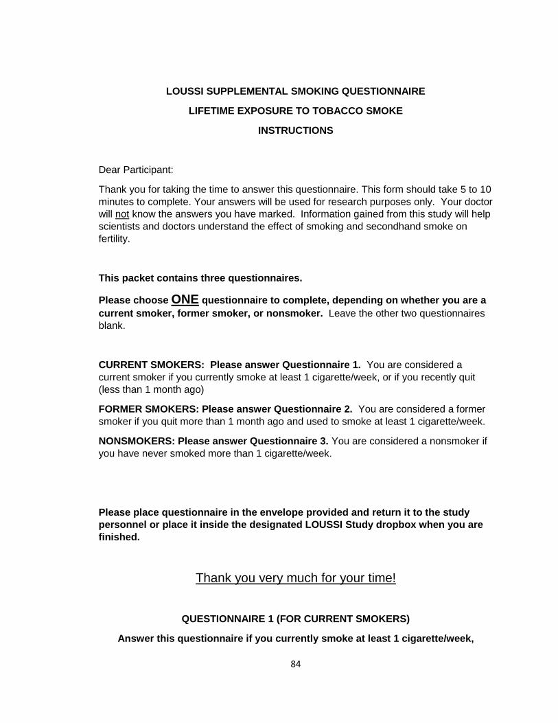
84
LOUSSI SUPPLEMENTAL SMOKING QUESTIONNAIRE
LIFETIME EXPOSURE TO TOBACCO SMOKE
INSTRUCTIONS
Dear Participant:
Thank you for taking the time to answer this questionnaire. This form should take 5 to 10
minutes to complete. Your answers will be used for research purposes only. Your doctor
will not know the answers you have marked. Information gained from this study will help
scientists and doctors understand the effect of smoking and secondhand smoke on
fertility.
This packet contains three questionnaires.
Please choose ONE questionnaire to complete, depending on whether you are a
current smoker, former smoker, or nonsmoker. Leave the other two questionnaires
blank.
CURRENT SMOKERS: Please answer Questionnaire 1. You are considered a
current smoker if you currently smoke at least 1 cigarette/week, or if you recently quit
(less than 1 month ago)
FORMER SMOKERS: Please answer Questionnaire 2. You are considered a former
smoker if you quit more than 1 month ago and used to smoke at least 1 cigarette/week.
NONSMOKERS: Please answer Questionnaire 3. You are considered a nonsmoker if
you have never smoked more than 1 cigarette/week.
Please place questionnaire in the envelope provided and return it to the study
personnel or place it inside the designated LOUSSI Study dropbox when you are
finished.
Thank you very much for your time!
QUESTIONNAIRE 1 (FOR CURRENT SMOKERS)
Answer this questionnaire if you currently smoke at least 1 cigarette/week,
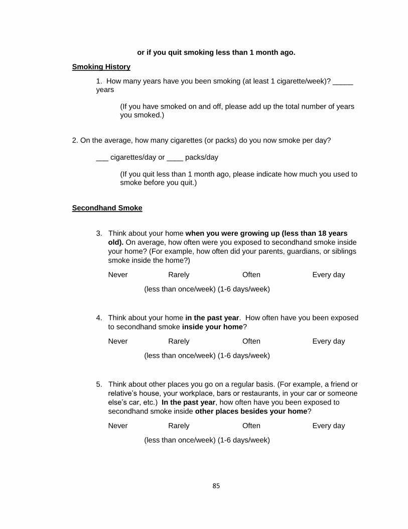
85
or if you quit smoking less than 1 month ago.
Smoking History
1. How many years have you been smoking (at least 1 cigarette/week)? _____ years
(If you have smoked on and off, please add up the total number of years you smoked.)
2. On the average, how many cigarettes (or packs) do you now smoke per day?
___ cigarettes/day or ____ packs/day
(If you quit less than 1 month ago, please indicate how much you used to smoke before you quit.)
Secondhand Smoke
3. Think about your home when you were growing up (less than 18 years
old). On average, how often were you exposed to secondhand smoke inside
your home? (For example, how often did your parents, guardians, or siblings
smoke inside the home?)
Never Rarely Often Every day
(less than once/week) (1-6 days/week)
4. Think about your home in the past year. How often have you been exposed
to secondhand smoke inside your home?
Never Rarely Often Every day
(less than once/week) (1-6 days/week)
5. Think about other places you go on a regular basis. (For example, a friend or
relative’s house, your workplace, bars or restaurants, in your car or someone
else’s car, etc.) In the past year, how often have you been exposed to
secondhand smoke inside other places besides your home?
Never Rarely Often Every day
(less than once/week) (1-6 days/week)

86
Please see
back →
6. Does anyone else living in your home smoke cigarettes, cigars or a pipe?
☐ Yes
☐ No
If you answered “Yes”, how many other people living in your home smoke these
tobacco products?
____Enter number
What is your relationship with the smoker(s)? (Check all that apply)
____Spouse or partner
____Other
Other sources of nicotine 7. Please check whether you have used any of the following in the past week:
_____ Nicotine patches _____ Nicorette gum or similar _____ E-cigarettes _____ Smokeless tobacco or chewing tobacco _____ Other source of nicotine: ______________

87
QUESTIONNAIRE 2 (FOR FORMER SMOKERS)
Answer this questionnaire if you used to smoke at least 1 cigarette/week and quit
at least 1 month ago.
Past Smoking 1. When is the last time you smoked a cigarette? (Month, Year) ____, ______
2. How many years did you smoke at least 1 cigarette/week? ____years
(If you smoked on and off, please add up the total number of years you smoked.) 3. On the average, how many cigarettes (or packs) did you used to smoke per
day? ___ cigarettes/day or ____ packs/day
Secondhand Smoke
4. Think about your home when you were growing up (less than 18 years
old). On average, how often were you exposed to secondhand smoke inside
your home? (For example, how often did your parents, guardians, or siblings
smoke inside the home?)
Never Rarely Often Every day
(less than once/week) (1-6 days/week)
5. Think about your home in the past year. How often have you been exposed
to secondhand smoke inside your home?
Never Rarely Often Every day
(less than once/week) (1-6 days/week)
6. Think about other places you go on a regular basis. (For example, a friend or
relative’s house, your workplace, bars or restaurants, in your car or someone
else’s car, etc.) In the past year, how often have you been exposed to
secondhand smoke inside other places besides your home?
Never Rarely Often Every day
(less than once/week) (1-6 days/week)

88
Please see
back →
7. Does anyone living in your home smoke cigarettes, cigars or a pipe?
☐ Yes
☐ No
If you answered “Yes”, how many people living in your home smoke these tobacco
products?
____Enter number
What is your relationship with the smoker(s)? (check all that apply)
____Spouse or partner
____Other
Other sources of nicotine 8. Please check whether you have used any of the following in the past week:
_____ Nicotine patches _____ Nicorette gum or similar _____ E-cigarettes _____ Smokeless tobacco or chewing tobacco _____ Other source of nicotine: ______________
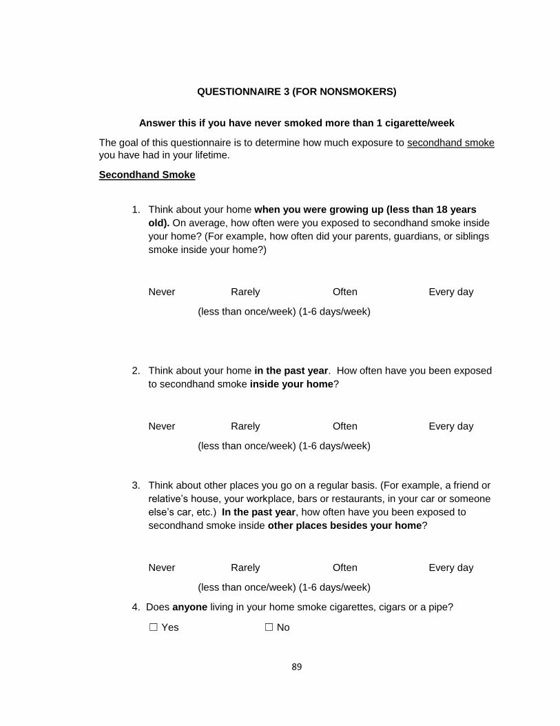
89
QUESTIONNAIRE 3 (FOR NONSMOKERS)
Answer this if you have never smoked more than 1 cigarette/week
The goal of this questionnaire is to determine how much exposure to secondhand smoke
you have had in your lifetime.
Secondhand Smoke
1. Think about your home when you were growing up (less than 18 years
old). On average, how often were you exposed to secondhand smoke inside
your home? (For example, how often did your parents, guardians, or siblings
smoke inside your home?)
Never Rarely Often Every day
(less than once/week) (1-6 days/week)
2. Think about your home in the past year. How often have you been exposed
to secondhand smoke inside your home?
Never Rarely Often Every day
(less than once/week) (1-6 days/week)
3. Think about other places you go on a regular basis. (For example, a friend or
relative’s house, your workplace, bars or restaurants, in your car or someone
else’s car, etc.) In the past year, how often have you been exposed to
secondhand smoke inside other places besides your home?
Never Rarely Often Every day
(less than once/week) (1-6 days/week)
4. Does anyone living in your home smoke cigarettes, cigars or a pipe?
☐ Yes ☐ No

90
If you answered “Yes”, how many people living in your home smoke these
tobacco products?
____Enter number
What is your relationship with the smoker(s)? (check all that apply)
____Spouse or partner
____Other Please see
back→
Other sources of nicotine
5. Please check whether you have used any of the following in the past week:
_____ Nicotine patches _____ Nicorette gum or similar _____ E-cigarettes _____ Smokeless tobacco or chewing tobacco _____ Other source of nicotine: ______________

91
Appendix 2
Data Collection Forms
Sample of data entry form for past medical and social history; anthropometric measures;
blood pressure and ovarian reserve measures

92
DATA COLLECTION FORM Subject Identification Number: __________________
1. Self-reported data From most recent Patient History Form (New patient info) Date of first visit (MM/YY):________________ Date of study enrollment (MM/YY):________ Age (years) at enrollment: ________________ Height: (feet, inches): ______feet_____inches Weight: _________pounds or __________ kg Body mass index: _________________kg/m2 Total number of previous pregnancies: _____ Regular periods (yes/no): ________________ Average menstrual cycle length: _______days Age at first period: _________________years Sexually transmitted infections: _____(yes/no) If yes, list:___________________________ “Social History” Section: Occupation: __________________________ Married/single: _______________________ Partner/no partner? ____________________ Length of time with current partner: _______ Routine exercise (yes/no): ____________ Routine exposure to chemicals(Yes/no):______ If yes, list here: ________________________ Diet restrictions: ____________________ Meals/day: ________________________
Current tobacco use (yes/no): ________ Packs/day ________________________ for how many years? ___________ Ever smoked 100 cigarettes? ________ Alcohol (yes/no) ________________ Alcoholic drinks/week___________ Caffeine (yes/no) __________________ Caffeinated drinks/day__________ Drugs (e.g. marijuana) (yes/no) _______ Ancestry or Race _________________ Consistent ovulation (yes/no)________
93
Appendix 3
Patient History Forms
Sample of survey filled by patients during first clinic visit
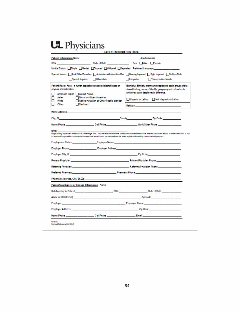
94
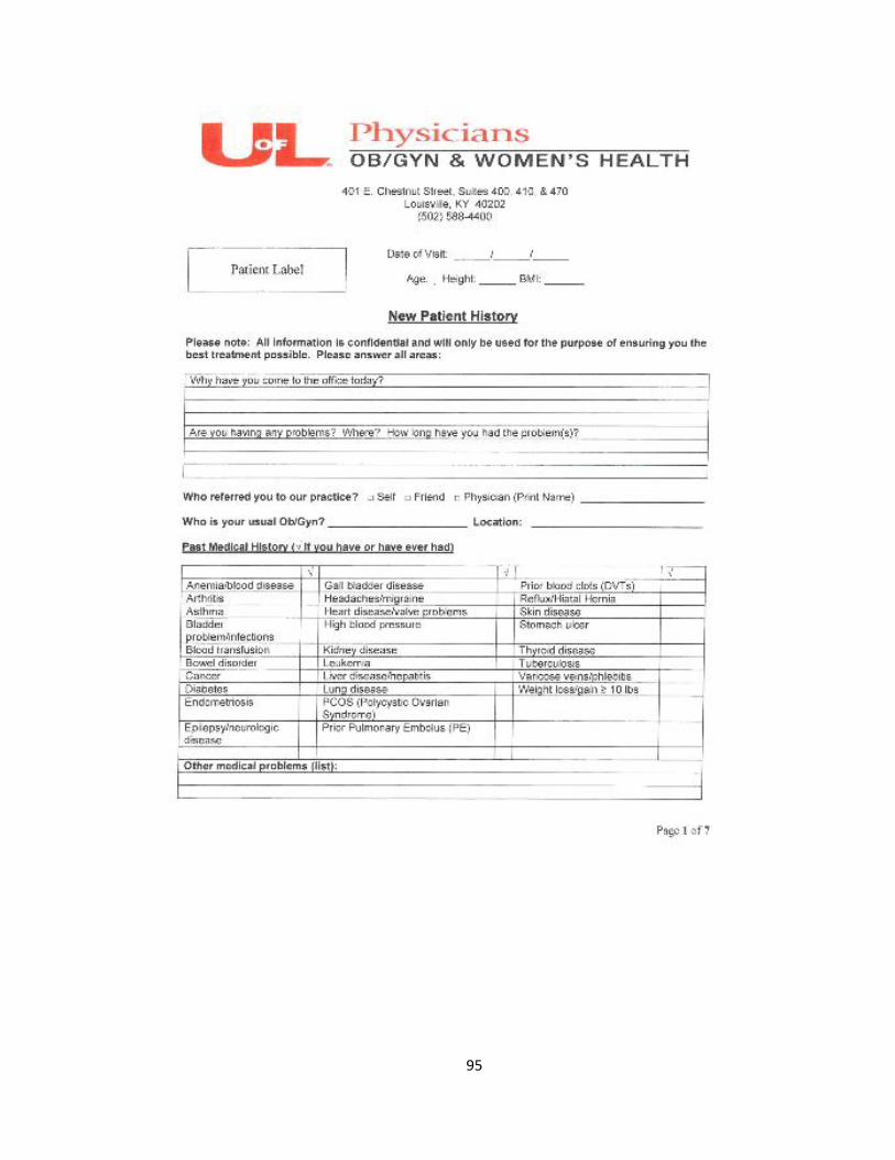
95
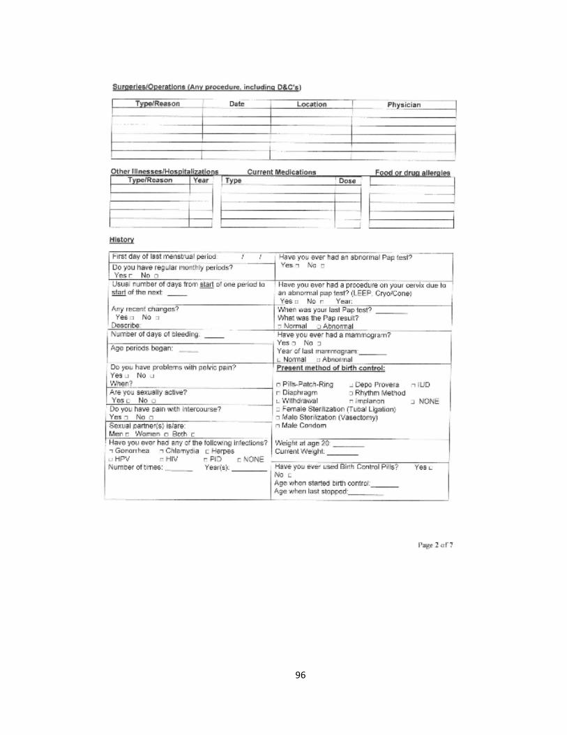
96
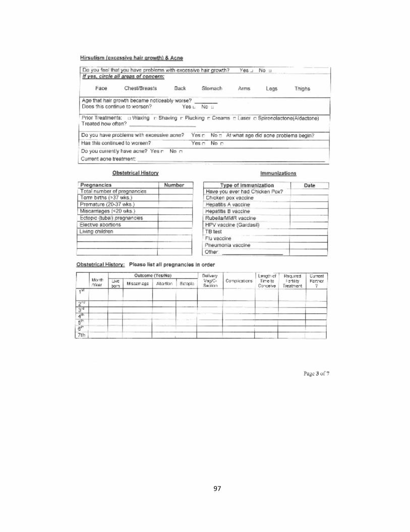
97
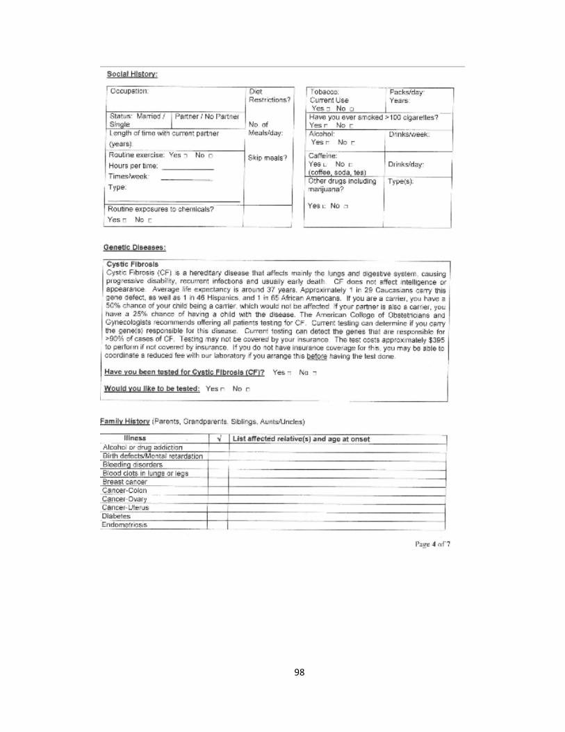
98
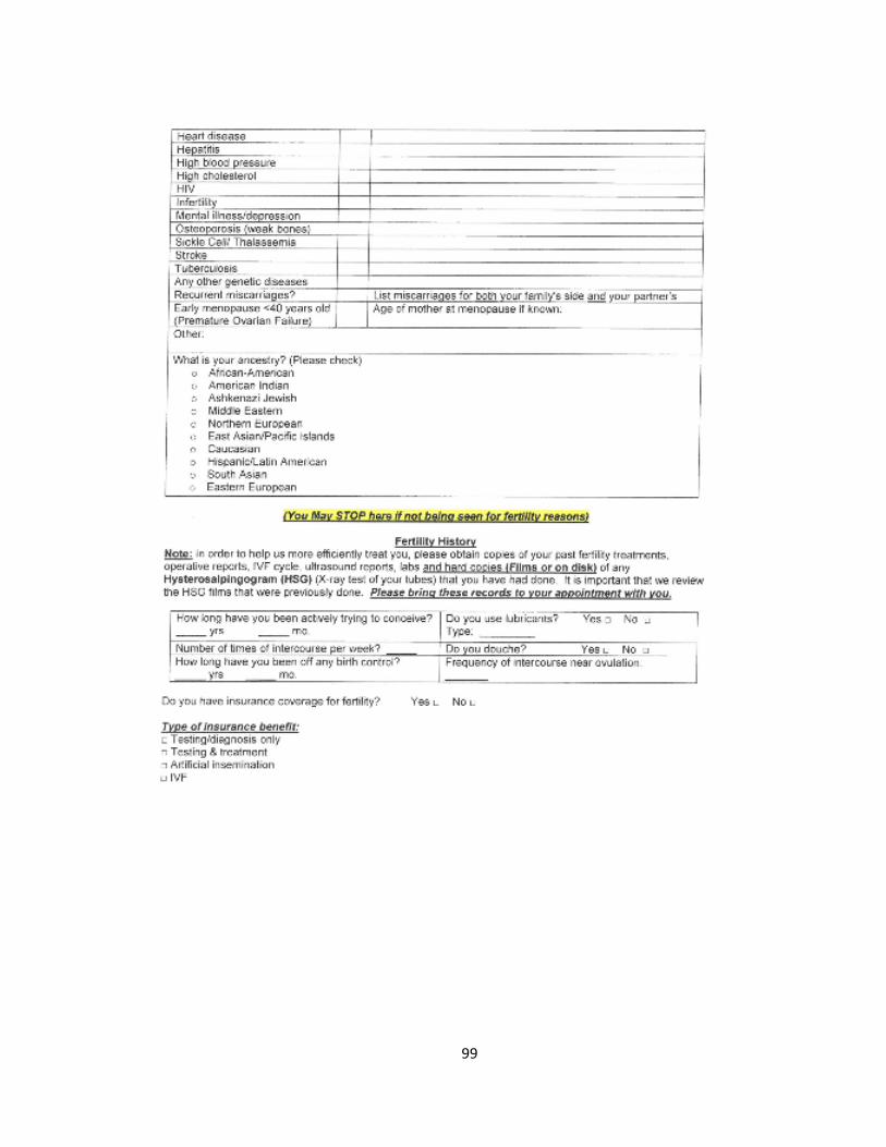
99
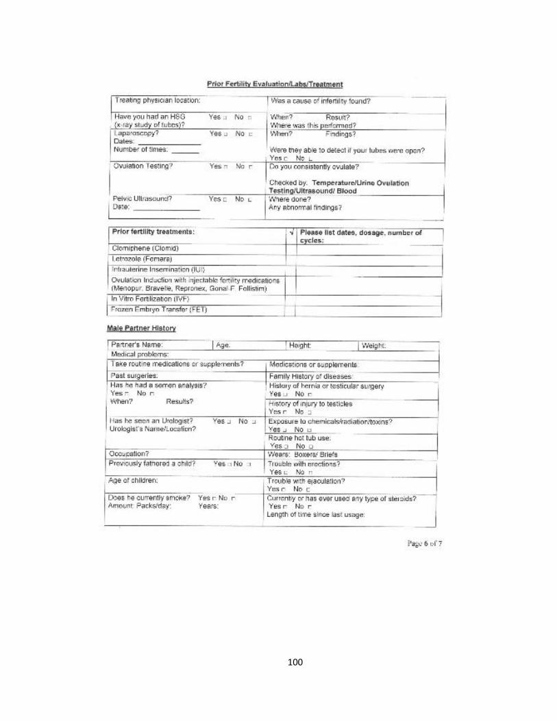
100
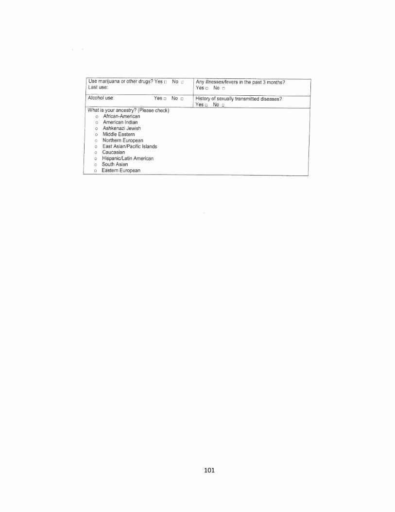
101
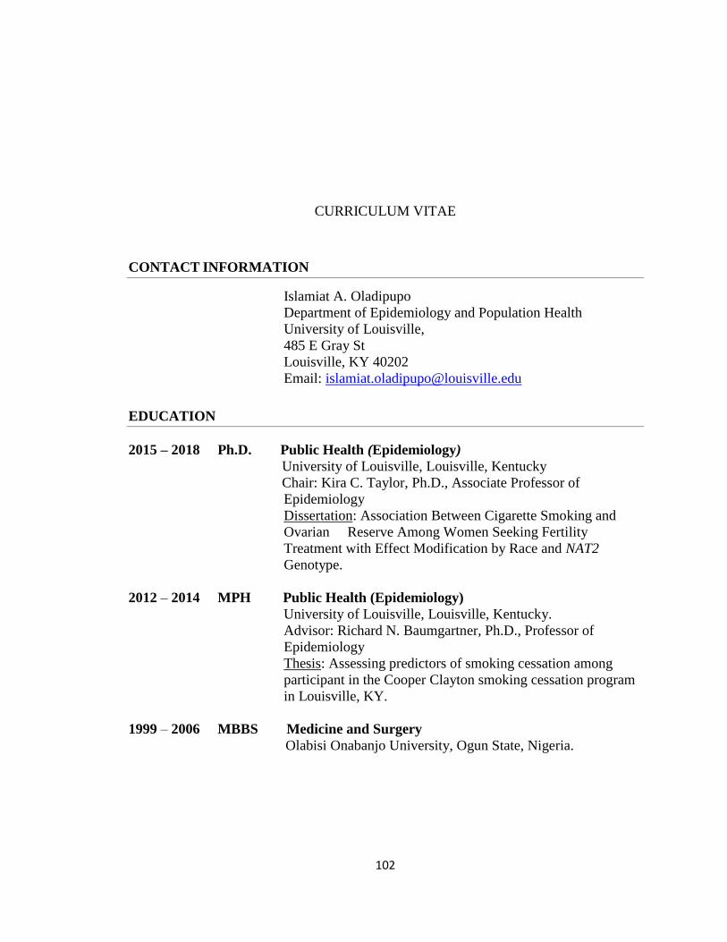
102
CURRICULUM VITAE
CONTACT INFORMATION
Islamiat A. Oladipupo
Department of Epidemiology and Population Health
University of Louisville,
485 E Gray St Louisville, KY 40202
Email: [email protected]
EDUCATION
2015 – 2018 Ph.D. Public Health (Epidemiology)
University of Louisville, Louisville, Kentucky
Chair: Kira C. Taylor, Ph.D., Associate Professor of
Epidemiology
Dissertation: Association Between Cigarette Smoking and
Ovarian Reserve Among Women Seeking Fertility
Treatment with Effect Modification by Race and NAT2
Genotype.
2012 – 2014 MPH Public Health (Epidemiology)
University of Louisville, Louisville, Kentucky.
Advisor: Richard N. Baumgartner, Ph.D., Professor of
Epidemiology
Thesis: Assessing predictors of smoking cessation among
participant in the Cooper Clayton smoking cessation program
in Louisville, KY.
1999 – 2006 MBBS Medicine and Surgery
Olabisi Onabanjo University, Ogun State, Nigeria.
103
RESEARCH EXPERIENCE
2016 – Present Graduate Research Assistant, Louisville Tobacco Smoke
Exposure,
Genetic Susceptibility, & Infertility Study, University of Louisville,
Louisville, KY.
Description: Clinic-based research evaluating the interaction
between NAT2 acetylator status and exposure to tobacco smoke on
ovarian reserve and in- vitro fertilization outcomes. Research
supported by a $ 0.45 million grant from the Eunice Kennedy
Shriver National Institute of Child Health and Human Development
(NICHD), through the National Institute of Health (NIH) R15 parent
program.
Projects: Association Between Active Smoking and Ovarian
Reserve Among Females Seeking Fertility Treatment, and
Interaction With N- Acetyltransferase 2(NAT2) Genotype.
Prevalence and Validity of Self-Reported Smoking Among Women
Seeking Fertility Care.
2013 – 2014 Epidemiology Intern, Department of Community Health and
Prevention,
Louisville Metro Department of Public Health and Wellness,
Louisville, Kentucky.
Description: Evaluated the Cooper Clayton smoking cessation
program, a comprehensive behavioral smoking cessation program
started over 25 years ago that utilizes education, skills training and
social support.
TEACHING EXPERIENCE
2018 – Present Teaching Assistant, Department of Health Management and System
Sciences, University of Louisville, Louisville, KY.
PHMS 301- Introduction to the U.S. Healthcare Delivery Systems
Description: A graduate level course that explores different aspects
of health care delivery in the United States with emphasis on role
health professionals working in different types of health service
organizations. Grade all quizzes, homework, assignment, and exams.
PHPH 431- Contemporary Issues in Public Health
104
Description: An Undergraduate level course with an average of 80
students per semester. Developed course work, quizzes, and in-class
activities. Grade all quizzes, homework, assignment, and exams.
PHUN 101- Introduction to Public Health
Description: An Undergraduate level course with an average of 80
students per semester. Grade all quizzes, homework, assignment, and
exams. Met with students to answer any questions and mentor them.
CLINICAL EXPERIENCE
2010 – 2012 Medical Officer, Department of Obstetrics and Gynecology, Harvey
Road Health Center, Lagos Nigeria
Description: Provided standard clinical care to patients daily
(approximately 30 patients per day), consulted in emergency,
gynecology, ante -natal and post-natal clinics and performed
procedures such as manual vacuum aspiration, episioraphy,
instrumental deliveries, normal deliveries and repair of laceration.
2008 – 2010 Medical Officer, Department of Obstetrics and Gynecology, Lagos
Island Maternity Hospital, Lagos, Nigeria
Description: Managed and treated life-threatening cases and
emergencies in obstetrics and gynecology. Provided reproductive
health and HIV/AIDS counseling to patient and participated in
Prevention of Maternal to Child Transmission of HIV /AIDS.
Worked with the AIDS Prevention Initiative of Nigeria (APIN) in
collection, analysis and reporting of data on HIV /AIDS patients.
2006– 2007 House Officer, General Hospital, Lagos, Nigeria.
Description: Managed several cases in pediatrics, medicine, surgery,
obstetrics and Gynecology under the supervision of consultants and
specialists (approximately 40 per day).
HONORS AND AWARDS
2018 School of Interdisciplinary and Graduate Studies Certificate of Excellence,
University of Louisville
2018 School of Public Health and Information Science Conference Travel Award,
University of Louisville
2017 Graduate School Council Conference Travel Award, University of Louisville
2017 School of Interdisciplinary and Graduate Studies Fellowship Award,
University of Louisville
105
2016 University Fellowship, University of Louisville, KY
2014 Delta Omega National Public Health Honorary Award
2006 Best Graduating Medical Student (out of 150), Olabisi Onabanjo University
2002 Ogun State Scholarship Award for Academic Excellence
2002 Federal Government Scholarship Award for Academic Excellence
PEER-REVIEWED PRESENTATIONS
1. Oladipupo I, Ali T, Bohler H, Pagidas K, Torres S, Mann M, Gentry A, Dondik
Y, Chiang J, Taylor K. Prevalence and Validity of Self-Reported Smoking
Among Women Seeking Fertility Care. [Poster Presentation]. American College
of Epidemiology (Annals of Epidemiology- Manuscript number- AEP_2018_288)
Annual Meeting, September 2018.
2. Ali T, Oladipupo I, Bohler H, Pagidas K, Hein D, Mann M, Gentry A, Dondik Y,
Chiang J, Taylor K. Childhood Secondhand Tobacco Smoke Exposure and
Ovarian Reserve Among Females Seeking Fertility Care, and Interaction with N-
Acetyltransferase 2(NAT2) Genotype [Poster Presentation]. American College of
Epidemiology (Annals of Epidemiology) Annual Meeting,2018.
3. Oladipupo I, Ali T, Bohler H, Pagidas K, Hein D, Mann M, Gentry A, Dondik Y,
Chiang J, Taylor K. Association between Smoking and Ovarian Reserve Among
Females Seeking Fertility Treatment, and Interaction with N- Acetyltransferase
2(NAT2) Genotype [Oral Presentation]. Society for Pediatric and Perinatal
Epidemiologic Research Annual Meeting, Baltimore, Maryland, June 2018.
NON-PEER REVIEWED PRESENTATIONS
1. Oladipupo I. Assessing the predictors of smoking cessation among participants
of the Cooper Clayton Smoking Cessation Program in Louisville, KY. Louisville
Metro Department of Public Health and Wellness, Louisville, Kentucky April
2014.
2. Oladipupo I, Hickey C, Horlander L. Jones K, Smart A. Availability, Price, and
Quality of Fresh Fruits and Vegetables: A Comparison of Select Stores in
Louisville, Kentucky. School of Public Health and Information Sciences,
University of Louisville, Louisville, Kentucky, April 2013.
3. Hickey C, Oladipupo I, Horlander L, Jones K, Smart A. Impact of Lead
abatement policy in Kentucky on the incidence of Lead related illness in children
of West Louisville, Kentucky. School of Public Health and Information Sciences,
University of Louisville, Louisville, Kentucky, February 2013.

106
4. Oladipupo I. Assessment of the land use types in relation to risk of pollution of
three public parks in Jefferson county, Kentucky. Department of Geography,
University of Louisville, Louisville, Kentucky, December 2013.
5. Oladipupo I. Community Assessment of Clark County, Kentucky. School of
Public Health and Information Sciences, University of Louisville, Louisville,
Kentucky, December 2012.
MANUSCRIPTS IN PROGRESS
1. Oladipupo I, Baumgartner KB, Hein DW, Wallis A, Gunaratnam B, and Taylor
KC. Association Between Cigarette Smoking and Ovarian Reserve Among
Women Seeking Fertility Treatment.
2. Oladipupo I, Baumgartner KB, Hein DW, Wallis A, Gunaratnam B J, and Taylor
KC. Interaction Between NAT2 Acetylator Status and Active Exposure to
Tobacco Smoke on Ovarian Reserve Among Women Seeking Fertility Care.
3. Oladipupo I, Baumgartner KB, Hein DW, Wallis A, Gunaratnam B, and Taylor
KC. Racial Disparity in the Association of Cigarette Smoking with Ovarian
Reserve Among Women Seeking Fertility Treatment.
PROFESSIONAL AFFILIATIONS
American College of Epidemiology (ACE) – Current
Journal: Annals of Epidemiology
Society for Epidemiologic Research (SER) – Current
Journals: American Journal of Epidemiology, Epidemiologic Reviews
Society for Pediatric and Perinatal Epidemiologic Research (SPER) – Current
Journal: Pediatric and Perinatal Epidemiology
American Public Health Association (APHA) - Current
Kentucky Pubic Health association (KPHA) - Current
Nigerian Medical Association (NMA) - Current
Medical Guild Lagos – Past
PROFESSIONAL WORKSHOPS
• Principles of Data Management in Clinical and Epidemiologic Research,
American College of Epidemiology (ACE), Annual Meeting Workshop,
September 2018.
• Advanced Statistical Method workshop, Society for Pediatric and Perinatal
Epidemiologic Research (SPER), Annual Meeting Workshop, June 2017.


