Embed Size (px)
Citation preview
COMPREHENSIVE INVITED REVIEW
From Molecular Mechanisms to Clinical Managementof Antineoplastic Drug-Induced Cardiovascular Toxicity:A Translational Overview
Carlo Gabriele Tocchetti,1 Christian Cadeddu,2 Daniela Di Lisi,3 Saveria Femmino,4 Rosalinda Madonna,5,6
Donato Mele,7 Ines Monte,8 Giuseppina Novo,3 Claudia Penna,4 Alessia Pepe,9 Paolo Spallarossa,10
Gilda Varricchi,1,11 Concetta Zito,12 Pasquale Pagliaro,4,* and Giuseppe Mercuro2,*
Abstract
Significance: Antineoplastic therapies have significantly improved the prognosis of oncology patients. However, thesetreatments can bring to a higher incidence of side-effects, including the worrying cardiovascular toxicity (CTX).Recent Advances: Substantial evidence indicates multiple mechanisms of CTX, with redox mechanismsplaying a key role. Recent data singled out mitochondria as key targets for antineoplastic drug-induced CTX;understanding the underlying mechanisms is, therefore, crucial for effective cardioprotection, without com-promising the efficacy of anti-cancer treatments.Critical Issues: CTX can occur within a few days or many years after treatment. Type I CTX is associated withirreversible cardiac cell injury, and it is typically caused by anthracyclines and traditional chemotherapeutics.Type II CTX is generally caused by novel biologics and more targeted drugs, and it is associated with reversiblemyocardial dysfunction. Therefore, patients undergoing anti-cancer treatments should be closely monitored,and patients at risk of CTX should be identified before beginning treatment to reduce CTX-related morbidity.Future Directions: Genetic profiling of clinical risk factors and an integrated approach using molecular, imaging,and clinical data may allow the recognition of patients who are at a high risk of developing chemotherapy-relatedCTX, and it may suggest methodologies to limit damage in a wider range of patients. The involvement of redoxmechanisms in cancer biology and anticancer treatments is a very active field of research. Further investigationswill be necessary to uncover the hallmarks of cancer from a redox perspective and to develop more efficaciousantineoplastic therapies that also spare the cardiovascular system. Antioxid. Redox Signal. 30, 2110–2153.
Keywords: chemotherapy, ErbB2 inhibitors, vascular endothelial growth factor, tyrosine kinase inhibitors,oxidative/nitrosative stress, cancer immunotherapy
1Department of Translational Medical Sciences, Federico II University, Naples, Italy.2Department of Medical Sciences and Public Health, University of Cagliari, Cagliari, Italy.3Biomedical Department of Internal Medicine, University of Palermo, Palermo, Italy.4Department of Clinical and Biological Sciences, University of Turin, Turin, Italy.5Center of Aging Sciences and Translational Medicine - CESI-MeT, ‘‘G. d’Annunzio’’ University, Chieti, Italy.6Department of Internal Medicine, The Texas Heart Institute and Center for Cardiovascular Biology and Atherosclerosis Research, The
University of Texas Health Science Center at Houston, Houston, Texas.7Cardiology Unit, Emergency Department, University Hospital of Ferrara, Ferrara, Italy.8Department of General Surgery and Medical-Surgery Specialities, University of Catania, Catania, Italy.9U.O.C. Magnetic Resonance Imaging, Fondazione Toscana G. Monasterio C.N.R., Pisa, Italy.
10Clinic of Cardiovascular Diseases, IRCCS San Martino IST, Genova, Italy.11Center for Basic and Clinical Immunology Research (CISI) - Federico II University, Naples, Italy.12Division of Cardiology, Clinical and Experimental Department of Medicine and Pharmacology, Policlinico ‘‘G. Martino’’ University
of Messina, Messina, Italy.*These authors share senior authorship.
Reviewing Editors: Gregory Bellot, Jozef Dulak, Hasan Mukhtar, Pasquale Pignatelli, Des Richardson, Martin Sterba, Yuchiro Suzuki, and OrenTirosh
ANTIOXIDANTS & REDOX SIGNALINGVolume 30, Number 18, 2019ª Mary Ann Liebert, Inc.DOI: 10.1089/ars.2016.6930
2110
Table of Contents
I. Introduction: The Clinical Problem of Antineoplastic Drug Cardiovascular Toxicity 2112II. Anthracyclines 2113
A. Adverse effects 2113B. Mechanisms of adverse effects 2113C. Ways to reduce CV toxicity 2114
III. ErbB2 Inhibitors 2114A. Adverse effects 2115B. Mechanisms of adverse effects 2115C. Ways to reduce CV toxicity 2116
IV. VEGF Inhibitors and Multi-Targeted Kinase Inhibitors 2116A. Adverse events 2116B. Mechanisms of adverse effects 2117
V. TKIs and Anti-BCR-abl Agents 2118A. Imatinib adverse events 2119B. Mechanisms of imatinib-induced adverse events 2119C. Nilotinib adverse events 2119D. Mechanisms of nilotinib-induced adverse events 2119E. Dasatinib adverse events 2120F. Mechanisms of dasatinib-induced adverse events 2120G. Bosutinib adverse events 2120H. Ponatinib adverse events 2120
VI. Taxanes 2121A. Adverse effects 2121B. Mechanisms of adverse effects 2121C. Ways to reduce CV toxicity 2121
VII. Cancer Immunotherapy 2121A. Immune checkpoint inhibitors 2121B. CTLA-4 2121C. PD-1/PD-L1 pathway 2121D. Combination of checkpoint inhibitors 2122E. Checkpoint inhibitors and immune-related adverse events 2122
VIII. Antimetabolites 2122A. Adverse effects 2122B. Mechanism of adverse effects 2123
IX. Proteasome Inhibitors 2123A. Adverse effects 2123B. Mechanisms of adverse effects 2124C. Ways to reduce CV toxicity 2124
X. The Importance of Mechanisms Such as Oxidative and Nitrosative Stress in CV Toxicity: An Overview 2124A. Drugs with antioxidant properties 2126
1. b blockers: Anti-adrenergic and antioxidant drugs 21272. The redox role of renin-angiotensin-aldosterone system antagonists: ACE-Is and ARBs 2128
B. Novel strategies in cardioprotection against ANT-induced CTX 2129C. Nonpharmacologic strategies 2130D. Biomarkers of oxidative/nitrosative CTX 2130
XI. Early Detection, Monitoring, and Management of Heart and Vascular Toxicities 2130A. Cardioncology: an expanding science for an expanding problem 2130B. Diagnostic tools for CTX detection 2130C. LVEF and emerging modalities for assessing myocardial damage: some clarifying points 2131D. Cardiac biomarkers 2132E. Detection of vascular complications: old and new strategies 2132F. Timing of clinical evaluation and follow-up: the importance of risk factors assessment 2133G. Drug-specific timing for CTX monitoring and treatment 2133
1. ANTs 21332. Anti-ErbB2 21343. VEGF inhibitors 2134
XII. Conclusions and Perspectives 2134
ANTINEOPLASTIC DRUG-INDUCED CARDIOTOXICITY 2111
I. Introduction: The Clinical Problemof Antineoplastic Drug Cardiovascular Toxicity
During the previous years, the prognosis of cancer wasgreatly enhanced by advancements in antitumoral thera-
peutic protocols; many types of malignancies can now be curedor maintained in remission for a long time, allowing patients tolive the rest of their lives in remission from cancer (21, 132,173, 266, 392). Unfortunately, antitumoral treatments exertsome adverse side-effects (Table 1). The cardiovascular (CV)system can be negatively affected by such therapies, and this isespecially true in the so-called long-term cancer survivors,since the likelihood that cardiac side-effects of antitumoraltreatments become the main health problem after tumor elim-ination increases with survival (250, 287, 429, 432).
The most common CV complications of antineoplastictherapies include vasospastic and thromboembolic ischemia,
arterial hypertension, dysrhythmia, and left ventricular (LV)dysfunction, leading to heart failure (HF) (25, 204, 376, 429,432). Cardiac dysfunction caused by anthracyclines (ANTs)has long been known as the main form of anti-cancer drug-induced cardiotoxicity (CTX) (91–94), with production ofreactive oxygen species (ROS) and reactive nitrogen species(RNS) being considered main cytotoxic mechanisms (seesection X for details). In the past decades, new biologic anti-cancer drugs, such as intracellular signaling inhibitors, wereincreasingly used. These molecules may also be cardiotoxic,since they block pathways that are major modulators ofmyocardial function, especially under conditions of cardiacstress, such as hypertension or hypertrophy (376), withmechanisms of action that often involve redox signaling aswell. As an example, drugs that target the human epidermalgrowth factor receptor 2 (HER/ErbB2) and the vascular en-dothelial growth factor (VEGF) exert a considerable adverse
Table 1. Drugs: Indications and Associated Types of Cardiovascular Toxicity
Class/drug Indication Toxicity
AnthracyclinesDoxorubicin, daunorubicin, epirubicin Breast cancer, leukemia, lymphoma,
ovarian cancer, sarcomaLV dysfunction/HF
(2%–48% incidence)
Anti-ErbB2Trastuzumab, lapatinib, pertuzumab Breast cancer, gastric cancer LV dysfunction
(0.2%–20.1% incidence)Vascular endothelial growth factor
inhibitors and multi-targetedkinase inhibitorsBevacizumab, sunitinib, sorafenib,
regorafenib, pazopanib, axitinib,vandetanib
Gastrointestinal cancer, hepatocellularcarcinoma, renal cell carcinoma,
LV dysfunction(2.7%–19% incidence),Hypertension (15%–44%incidence), Vasculardamage (1.4%–3.8%incidence)
Tyrosine kinase inhibitors and anti-BCR-abl agentsImatinib, nilotinib, dasatinib,
bosutinib, ponatinibChronic myeloid leukemia Edema, cardiac dysfunction
(0.2%–4% incidence), QTcprolongation (2%–40.5%incidence)
TaxanesDocetaxel, paclitaxel Breast cancer, colorectal cancer,
nonsmall cell lung cancer, andovarian cancers
Bradycardia, LV dysfunction,ischemia (0.3%–1.7%incidence)
Cancer ImmunotherapiesIpilimumab, nivolumab,
pembrolizumab, atezolizumabMetastatic melanoma Immune myocarditis
(0.01%–0.27% incidence)Metastatic nonsmall cell lungcancer (NSCLC)
Advanced renal cell carcinomaClassical Hodgkin lymphomaAdvanced urothelial carcinoma
AntimetabolitesFluorouracil (5-FU), capecitabine,
gemcitabineGastrointestinal, breast, head,
neck, and pancreatic cancerCoronary spasms/ischemia
(7%–18% incidence)
Proteasome inhibitorsBortezomib; carfilzomib, ixazomib,
delanzomib, oprozomib, and marizomibMultiple myeloma and other
hematologic conditions(amyloidosis, non-Hodgkinlymphoma)
LV dysfunction (2%–25%incidence)
Modified from Suter and Ewer (376) and Zamorano et al. (432).ErbB2, human epidermal growth factor receptor 2 (HER2); HF, heart failure; LV, left ventricular.
2112 TOCCHETTI ET AL.
effect on myocardial function via different mechanisms,based on the role of the proteins inhibited. The toxicityproduced by biologic drugs seems to be due to mechanismsother than cardiomyocyte disruption, is most often reversiblewith discontinuation of the drugs, and has been classified astype II CTX (93, 94). On the other hand, ANTs produce aform of cardiac dysfunction that is typically irreversible,termed type I CTX, and that is characterized by evident ul-trastructural myocardial abnormalities (93, 94). Of note,these two CTX paradigms may overlap. One paradigmaticexample is the ErbB2 receptor inhibitor trastuzumab, whichcan cause irreversible LV dysfunction in patients previouslytreated with ANTs (376, 432), with the neuregulin/ErbB2pathway that seems to modulate the increase in ROS-causedANTs (390).
In this article, we address the main cellular and molecularmechanisms and pathophysiologic and clinical characteris-tics of antineoplastic drug-related CTX, since only a com-prehensive assessment of this phenomenon can provideimportant hints to predict, treat, and prevent it. Special em-phasis is placed on LV dysfunction and HF, in considerationof their clinical and social burden (25, 125), with updatedinsights regarding the role of oxidative damage, a mechanismthat appears to have a major role in antineoplastic drug-induced CTX (412, 432). Whenever appropriate, we dividedthe various sections into three categories (i) adverse effects,(ii) mechanisms of adverse effects, and (iii) ways to reduceCV toxicity.
II. Anthracyclines
Among the drugs with a greater cardiotoxic potential,ANTs are good representatives of the type I CTX paradigm.
ANTs are widely used and effective antineoplastic drugs,which are indicated for the therapy of many kinds of cancers,including lymphomas, leukemias, and sarcomas, and for bothearly and advanced breast cancer. However, these drugs havebeen recognized as cardiotoxic since the 1960s (384).
A. Adverse effects
ANT-induced CTX can manifest as a sort of cardiomy-opathy, referred to as ANT-induced cardiomyopathy, leadingto HF, which limits the usability of the drugs, with importantconsequences for managing malignancies. ANT-related CTXrepresents a significant clinical burden, producing LV dys-function in a maximum of 9% of cases in a recent largeprospective study (35).
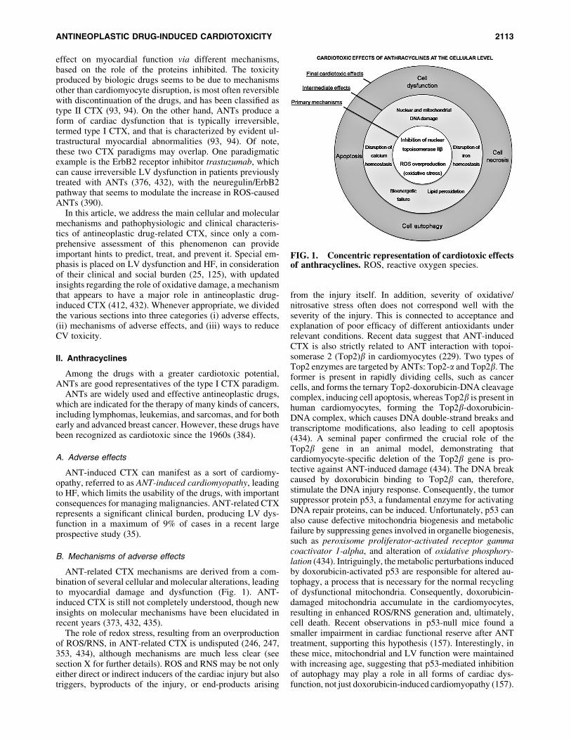
B. Mechanisms of adverse effects
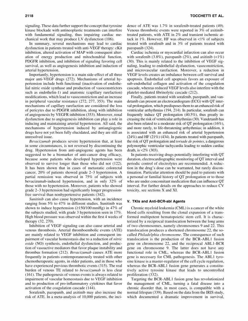
ANT-related CTX mechanisms are derived from a com-bination of several cellular and molecular alterations, leadingto myocardial damage and dysfunction (Fig. 1). ANT-induced CTX is still not completely understood, though newinsights on molecular mechanisms have been elucidated inrecent years (373, 432, 435).
The role of redox stress, resulting from an overproductionof ROS/RNS, in ANT-related CTX is undisputed (246, 247,353, 434), although mechanisms are much less clear (seesection X for further details). ROS and RNS may be not onlyeither direct or indirect inducers of the cardiac injury but alsotriggers, byproducts of the injury, or end-products arising
from the injury itself. In addition, severity of oxidative/nitrosative stress often does not correspond well with theseverity of the injury. This is connected to acceptance andexplanation of poor efficacy of different antioxidants underrelevant conditions. Recent data suggest that ANT-inducedCTX is also strictly related to ANT interaction with topoi-somerase 2 (Top2)b in cardiomyocytes (229). Two types ofTop2 enzymes are targeted by ANTs: Top2-a and Top2b. Theformer is present in rapidly dividing cells, such as cancercells, and forms the ternary Top2-doxorubicin-DNA cleavagecomplex, inducing cell apoptosis, whereas Top2b is present inhuman cardiomyocytes, forming the Top2b-doxorubicin-DNA complex, which causes DNA double-strand breaks andtranscriptome modifications, also leading to cell apoptosis(434). A seminal paper confirmed the crucial role of theTop2b gene in an animal model, demonstrating thatcardiomyocyte-specific deletion of the Top2b gene is pro-tective against ANT-induced damage (434). The DNA breakcaused by doxorubicin binding to Top2b can, therefore,stimulate the DNA injury response. Consequently, the tumorsuppressor protein p53, a fundamental enzyme for activatingDNA repair proteins, can be induced. Unfortunately, p53 canalso cause defective mitochondria biogenesis and metabolicfailure by suppressing genes involved in organelle biogenesis,such as peroxisome proliferator-activated receptor gammacoactivator 1-alpha, and alteration of oxidative phosphory-lation (434). Intriguingly, the metabolic perturbations inducedby doxorubicin-activated p53 are responsible for altered au-tophagy, a process that is necessary for the normal recyclingof dysfunctional mitochondria. Consequently, doxorubicin-damaged mitochondria accumulate in the cardiomyocytes,resulting in enhanced ROS/RNS generation and, ultimately,cell death. Recent observations in p53-null mice found asmaller impairment in cardiac functional reserve after ANTtreatment, supporting this hypothesis (157). Interestingly, inthese mice, mitochondrial and LV function were maintainedwith increasing age, suggesting that p53-mediated inhibitionof autophagy may play a role in all forms of cardiac dys-function, not just doxorubicin-induced cardiomyopathy (157).
FIG. 1. Concentric representation of cardiotoxic effectsof anthracyclines. ROS, reactive oxygen species.
ANTINEOPLASTIC DRUG-INDUCED CARDIOTOXICITY 2113
Apart from p53, doxorubicin may also induce the mitogen-activated protein kinase (MAPK) pathway via ROS- andCa2+-dependent mechanisms (437). Importantly, extracellu-lar signal-regulated kinases (ERKs), members of the MAPKfamily, may protect myocytes from apoptosis, whereas p38MAPK induces death of cardiomyocytes (437). More studiesare needed to elucidate the role of such kinases and of otherless-characterized signaling pathways in ANT-induced cardi-otoxicity. However, these data confirm that oxidative reactions,at the basis of ANT-induced LV dysfunction, are involved inmost types of HF. Therefore, timely innovative pharmacolog-ical strategies that interfere with specific molecules involved inheart dysfunction (e.g., p53) may represent a potential commonapproach in limiting HF occurrence (250, 341).
ANT-alcohol metabolites also play a pivotal role in in-ducing cellular injury and CTX via iron-dependent and -in-dependent mechanisms. In fact, these metabolites disrupt ironand calcium homeostasis and, ultimately, lead to intracellularCa2+ overload. Calcium overload has also been related toincreased calpain proteolytic activity, which leads to cellulardisarray and sarcomere disruption, resulting in sarcopenia(220). In addition, the interaction of ANTs with critical sig-naling pathways and with the activity of transcription factorsmay also explain sarcopenia, which derives from the limi-tation of sarcomere protein synthesis (165).
Mitochondrial activity has a central role in ANT-inducedCTX (257, 258). The presence of doxorubicin in the mito-chondrion, due to a high affinity for the mitochondrial phos-pholipid cardiolipin, negatively affects its function, stimulatingROS/RNS production, inhibiting oxidative phosphorylation,and causing mitochondrial DNA damage (300), with a conse-quent progressive reduction of energy production, leading tocell dysfunction (210). ANTs also appear to be responsible formitochondrial calcium accumulation (300), leading to mito-chondrial membrane injury. The mitochondrial pathway is anadditional mechanism that is responsible for cellular intrinsicapoptosis. This involves elements of the outer mitochondrialmembrane, including Bax and Bak, and activates cytochrome Cand caspase, among others (257).
Some researchers have hypothesized that the loss of ironhomeostasis and the Ca2+ overload caused by ANT alcoholmetabolites, impairing cardiomyocyte energy and redox bal-ance, could be sufficient to induce significant myocardial
dysfunction, and that, in addition, cardiac injury could be in-creased by an apoptotic loss of cardiomyocytes triggered byROS and Fe2+ (257, 258). ANTs can also affect cardiac pro-genitor cells, hampering the regeneration capabilities ofcardiac tissues after myocardial damage (158, 289). The con-troversy on the role of cardiac stem cells is discussed in thesection XII.
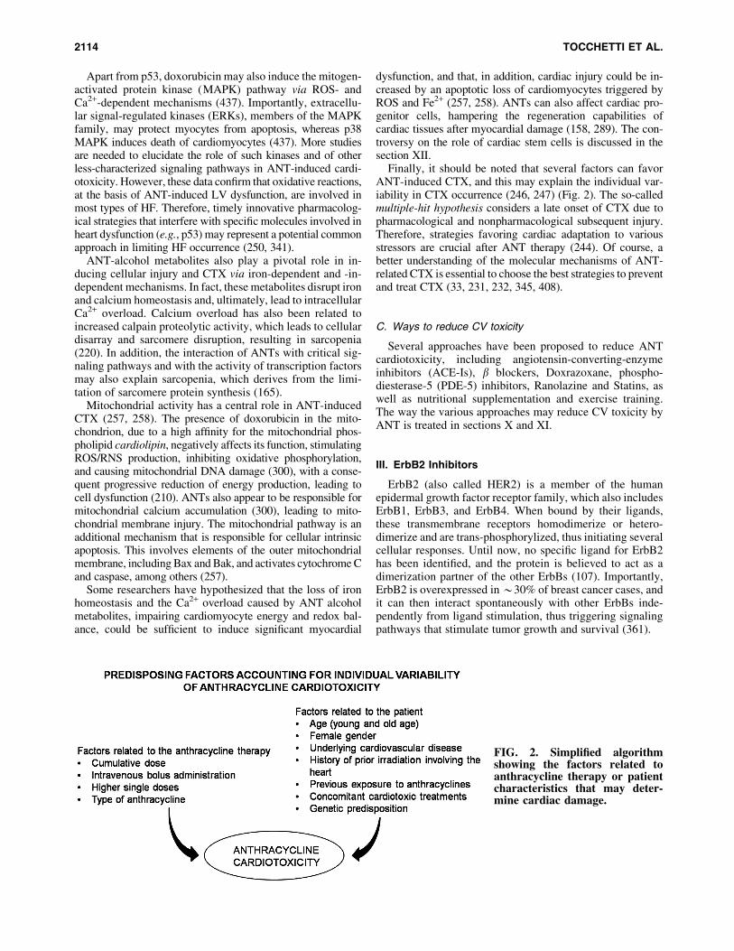
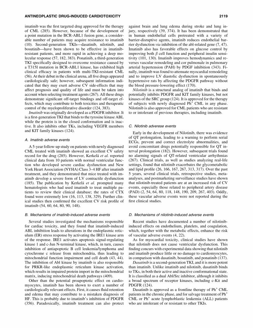
Finally, it should be noted that several factors can favorANT-induced CTX, and this may explain the individual var-iability in CTX occurrence (246, 247) (Fig. 2). The so-calledmultiple-hit hypothesis considers a late onset of CTX due topharmacological and nonpharmacological subsequent injury.Therefore, strategies favoring cardiac adaptation to variousstressors are crucial after ANT therapy (244). Of course, abetter understanding of the molecular mechanisms of ANT-related CTX is essential to choose the best strategies to preventand treat CTX (33, 231, 232, 345, 408).
C. Ways to reduce CV toxicity
Several approaches have been proposed to reduce ANTcardiotoxicity, including angiotensin-converting-enzymeinhibitors (ACE-Is), b blockers, Doxrazoxane, phospho-diesterase-5 (PDE-5) inhibitors, Ranolazine and Statins, aswell as nutritional supplementation and exercise training.The way the various approaches may reduce CV toxicity byANT is treated in sections X and XI.
III. ErbB2 Inhibitors
ErbB2 (also called HER2) is a member of the humanepidermal growth factor receptor family, which also includesErbB1, ErbB3, and ErbB4. When bound by their ligands,these transmembrane receptors homodimerize or hetero-dimerize and are trans-phosphorylized, thus initiating severalcellular responses. Until now, no specific ligand for ErbB2has been identified, and the protein is believed to act as adimerization partner of the other ErbBs (107). Importantly,ErbB2 is overexpressed in *30% of breast cancer cases, andit can then interact spontaneously with other ErbBs inde-pendently from ligand stimulation, thus triggering signalingpathways that stimulate tumor growth and survival (361).
FIG. 2. Simplified algorithmshowing the factors related toanthracycline therapy or patientcharacteristics that may deter-mine cardiac damage.
2114 TOCCHETTI ET AL.
A. Adverse effects
Trastuzumab is a humanized monoclonal antibody thatbinds the extracellular domain IV of HER/ErbB2 (107, 377). Itis the prototypical anti-ErbB2 agent, and the first developedand most widely used type II cardiotoxic drug. Trastuzumab isparticularly useful in treating ErbB2+ breast and gastric can-cers. Unfortunately, it can also cause CTX in a substantialnumber of patients, peaking at 28% with concomitant admin-istration of trastuzumab and ANTs (262, 362, 377). Indeed,as said earlier, ANTs are responsible for type I CTX, withpermanent cardiac damage. Therefore, reduced left ventricleejection fraction (LVEF) results from the association of tras-tuzumab and doxorubicin: Trastuzumab enhances or even in-duces doxorubicin toxicity. Once anti-ErbB2 agents block theprotective mechanisms of ErbB2, the oxidative damage in-duced by doxorubicin increases (91). This co-administration isnow avoided. As a class II cardiac dysfunction (93, 94), tras-tuzumab CTX appears to be elicited by the impairment ofcontractility rather than the loss of cardiomyocytes, and pre-vious chemotherapy seems to be responsible for the troponinrelease observed in sequential treatment (91).
Pertuzumab is a more recent anti-HER2 antibody that bindsthe receptor’s domain II. Lapatinib is a different anti-ErbB2agent, a small-molecule inhibitor of the intracellular tyrosinekinase domain of ErbB2. Of note, trastuzumab only disruptsligand-independent ErbB2 signaling, whereas pertuzumab in-terferes with the formation of ligand-induced ErbB2 hetero-dimers. In contrast, lapatinib inhibits both ligand-induced andligand-independent ErbB2 signaling (69). Interestingly, lapa-tinib seems to be less toxic than trastuzumab. Data regardingthe toxicity of pertuzumab are more limited (262).
B. Mechanisms of adverse effects
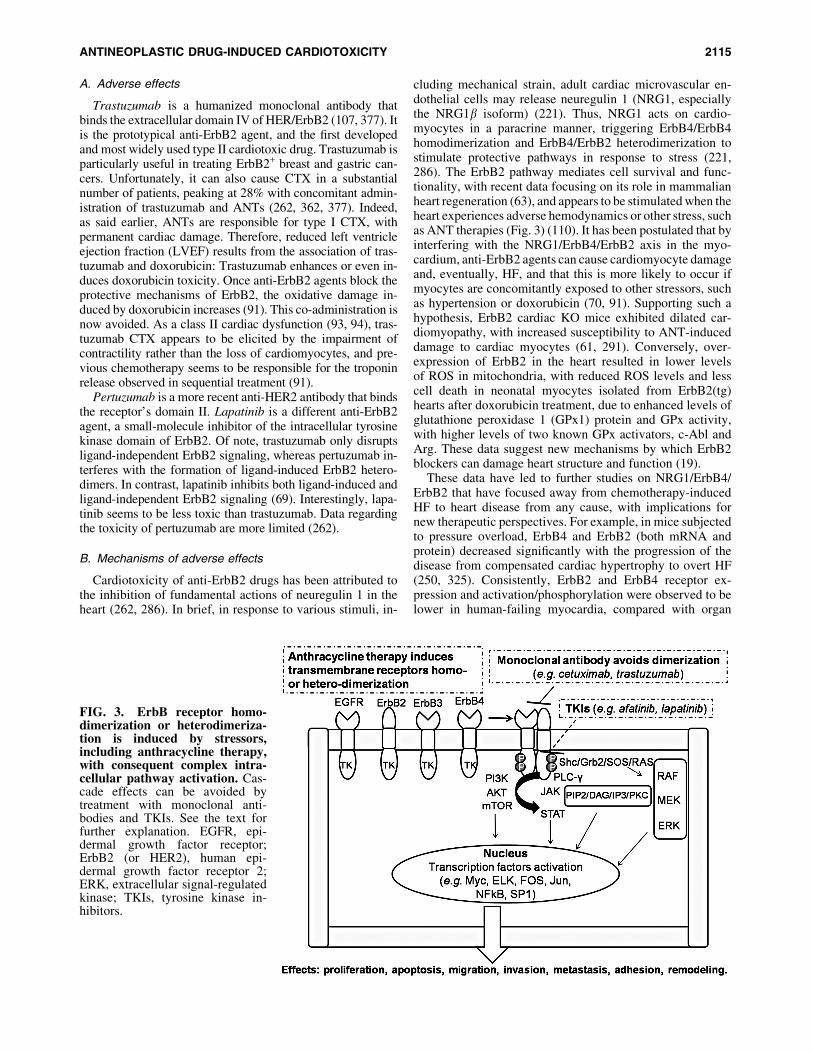
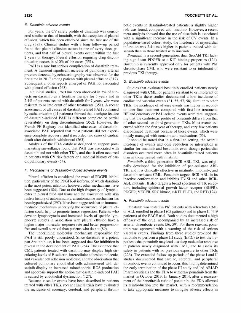
Cardiotoxicity of anti-ErbB2 drugs has been attributed tothe inhibition of fundamental actions of neuregulin 1 in theheart (262, 286). In brief, in response to various stimuli, in-
cluding mechanical strain, adult cardiac microvascular en-dothelial cells may release neuregulin 1 (NRG1, especiallythe NRG1b isoform) (221). Thus, NRG1 acts on cardio-myocytes in a paracrine manner, triggering ErbB4/ErbB4homodimerization and ErbB4/ErbB2 heterodimerization tostimulate protective pathways in response to stress (221,286). The ErbB2 pathway mediates cell survival and func-tionality, with recent data focusing on its role in mammalianheart regeneration (63), and appears to be stimulated when theheart experiences adverse hemodynamics or other stress, suchas ANT therapies (Fig. 3) (110). It has been postulated that byinterfering with the NRG1/ErbB4/ErbB2 axis in the myo-cardium, anti-ErbB2 agents can cause cardiomyocyte damageand, eventually, HF, and that this is more likely to occur ifmyocytes are concomitantly exposed to other stressors, suchas hypertension or doxorubicin (70, 91). Supporting such ahypothesis, ErbB2 cardiac KO mice exhibited dilated car-diomyopathy, with increased susceptibility to ANT-induceddamage to cardiac myocytes (61, 291). Conversely, over-expression of ErbB2 in the heart resulted in lower levelsof ROS in mitochondria, with reduced ROS levels and lesscell death in neonatal myocytes isolated from ErbB2(tg)hearts after doxorubicin treatment, due to enhanced levels ofglutathione peroxidase 1 (GPx1) protein and GPx activity,with higher levels of two known GPx activators, c-Abl andArg. These data suggest new mechanisms by which ErbB2blockers can damage heart structure and function (19).
These data have led to further studies on NRG1/ErbB4/ErbB2 that have focused away from chemotherapy-inducedHF to heart disease from any cause, with implications fornew therapeutic perspectives. For example, in mice subjectedto pressure overload, ErbB4 and ErbB2 (both mRNA andprotein) decreased significantly with the progression of thedisease from compensated cardiac hypertrophy to overt HF(250, 325). Consistently, ErbB2 and ErbB4 receptor ex-pression and activation/phosphorylation were observed to belower in human-failing myocardia, compared with organ
FIG. 3. ErbB receptor homo-dimerization or heterodimeriza-tion is induced by stressors,including anthracycline therapy,with consequent complex intra-cellular pathway activation. Cas-cade effects can be avoided bytreatment with monoclonal anti-bodies and TKIs. See the text forfurther explanation. EGFR, epi-dermal growth factor receptor;ErbB2 (or HER2), human epi-dermal growth factor receptor 2;ERK, extracellular signal-regulatedkinase; TKIs, tyrosine kinase in-hibitors.
ANTINEOPLASTIC DRUG-INDUCED CARDIOTOXICITY 2115
donors (326). Interestingly, LV unloading by implantation ofan LV assist device restored the levels of ErbB4 and ErbB2(326, 400). In an apparent contrast with these results, dogswith pacing-induced HF showed increased phosphorylationof ErbB4 and ErbB2 (78). Inactivation of the intracellulardownstream effectors of ErbB4 and ErbB2, ERK1/2 andAkt, was observed, suggesting a disabled NRG1/ErbB4/ErbB2 signaling. Actually, NRG1 expression is increased inHF compared with control conditions in most studies (78,250, 326). All in all, these data hint that deranged NRG1/ErbB4/ErbB2 activity is involved in the pathophysiology ofHF in at least two manners: (i) HF may derive from the use ofanti-ErbB2 drugs, such as trastuzumab; (ii) ErbB4/ErbB2 isdownregulated and/or uncoupled from intracellular signal-ing despite normal or increased NRG1, possibly leading tocardiac decompensation (250). Furthermore, novel observa-tions suggest that levels of catecholamines, which usuallyincrease with the occurrence of LV dysfunction and withANT administration (176, 250, 282), can stimulate ErbB2expression in myocytes, thus making these cells particularlysusceptible to the effects of trastuzumab, resulting in myo-cardial toxicity (382).
C. Ways to reduce CV toxicity
The aforementioned experimental results may support theuse of b blockers in the prevention of trastuzumab CTX (seesection X.A.1) (281), in line with a retrospective study thatfound that continuous use of b blockers was associated with alower risk of new HF events in subjects on trastuzumab, ANTs,or both (347). Prevention with b blockers is currently beingassessed in clinical trials (180, 281, 347, 349) with bisoprolol(MANTICORE 101-Breast) (305), NCT01009918 (carvedi-lol), and NCT01434134/NCT00806390 (metoprolol) to cure orprevent trastuzumab-induced LV dysfunction (250, 281). In-terestingly, from the recent PRADA (prevention of cardiacdysfunction during adjuvant breast cancer therapy) trial, we caninfer that blocking only b1 with metoprolol may not produce anadequate and sufficient cardioprotection (128), thus supportingthe use of nonselective b1 and b2 blockers (382). In the clinicalsetting of trastuzumab-induced cardiac dysfunction, whentrastuzumab is discontinued, normal ErbB2 signaling is re-stored, and the reduced LVEF can increase back to normallevels. Indeed, trastuzumab re-administration after discontin-uation is considered relatively safe on LVEF recovery (93, 94).
Animal studies have demonstrated that NRG1 regulatesdoxorubicin injury in rat myocytes (390). Owing to theaforementioned cardioprotective properties of NRG1 viaErbB4/ErbB2, and as the activity of these receptors is alteredin HF, the axis neuregulin-ERB is now being intensivelyinvestigated in clinical trials for HF treatment (111, 112,221). It has been hypothesized that NRG1 and NRG1 analogscan be used as therapeutic agents in HF. Intravenous ad-ministration of recombinant human NRG1 and of the glialgrowth factor 2 isoform of NRG1b enhanced heart functionand reduced LV dimensions in experimental failing hearts(111, 112, 218, 227, 250). Since NRG1 exerted positive ef-fects in animal models of ischemia-induced HF even whenadministered after acute myocardial infarction, it can bespeculated that it is able to produce beneficial reverse re-modeling of the damaged heart, and it does not simply limitcardiac dilation (111, 112, 227). Also, it appears that NRG1
exerts an antifibrotic effect, directly inhibiting cardiac fi-broblasts and, thus, preventing fibrosis (111, 112).
Clinical studies have demonstrated that recombinant hu-man NRG1 is well tolerated by patients, and it amelioratescardiac dimensions and LVEF until a maximum of 3 monthsafter treatment (116, 169). Nevertheless, NRG1 may beconsidered a growth factor for tumor cells, particularly whenadministered systemically. Hopefully, additional experi-mental and clinical studies can assess this fundamental safetyconcern, producing novel data regarding the effects of NRG1in HF (221, 250).
IV. VEGF Inhibitors and Multi-TargetedKinase Inhibitors
A. Adverse events
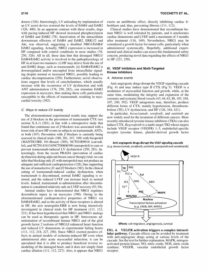
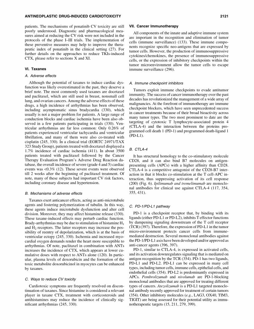
Anti-angiogenic drugs disrupt the VEGF signaling cascade(Fig. 4) and may induce type II CTX (Fig 5). VEGF is amodulator of myocardial function and growth, while, at thesame time, modulating the integrity and expansion of thecoronary and systemic blood vessels (43, 44, 62, 88, 103, 104,107, 240, 392). VEGF antagonists may, therefore, producedifferent forms of CTX, mainly hypertension, thromboem-bolism (TE), LV dysfunction, and HF (126, 344, 421).
In particular, bevacizumab, sorafenib, and sunitinib arenow widely used for the treatment of different cancers. Morerecently introduced tyrosine kinase inhibitors (TKIs) can alsoinduce CTX. Regorafenib is a multi-target TKI whose targetsinclude VEGF receptor (VEGFR) 1–3, endothelial-specificreceptor tyrosine kinase, platelet-derived growth factor
FIG. 4. VEGFR activation triggers a complex intracel-lular pathway. Cascade effects can be avoided by treatmentwith anti-angiogenic drugs acting at various levels of thecascade. See the text for further explanation. MAPK, mitogen-activated protein kinase; NO, nitric oxide; NOS, nitric oxidesynthase; VEGFR, vascular endothelial growth factorreceptor.
2116 TOCCHETTI ET AL.
receptor (PDGFR), fibroblast growth factor receptor, cKIT,RE arranged during transfection (RET), and rapidly accel-erated fibrosarcoma kinase. Regorafenib is used in thetreatment of colorectal tumors and gastrointestinal tumors(28). It may be responsible for arterial hypertension; lessfrequently, it can produce cardiac ischemia and myocardialinfarction (30).
Pazopanib and axitinib, used in the treatment of metastaticrenal tumors, are also associated with a high rate of arterialhypertension. Pazopanib is an orally administered multi-targeted TKI, targeting VEGFR 1–3, PDGFA and PDGFBreceptors, and c-KIT. In a recent study, the frequency ofpazopanib-associated hypertension varied between 36% and46% (268). Axitinib is a third-generation VEGFR inhibitorused in metastatic renal cancer after failure of previoustreatments, and it is very selective. In a study comparing ax-itinib and sorafenib, the frequency of hypertension was 29%for sorafenib and 40% for axitinib (162). New anti-angiogenicdrugs not yet approved for clinical use are vatalanib andnintedanib. Preliminary evidence indicates a potential risk ofarterial hypertension and congestive HF and, for vatalanib, ofpulmonary embolism, although more rarely (318, 407).
B. Mechanisms of adverse effects
The myocardium requires appropriate perfusion to func-tion properly (43, 44, 62, 88, 103, 104, 107, 240, 392), and itdepends on HIF-1 and VEGF pathways, similar to tumors. Ofnote, inhibition of HIF-1 by p53 may produce HF duringchronic pressure overload (336). Moreover, conditional ex-pression of a VEGF scavenger may cause myocardial hi-bernation and microvessel rarefaction, which can be reversedby suppressing the expression of the scavenger, even monthsafter its induction (244, 417). Such findings show that themyocardium is very sensitive to anti-angiogenic treatments,especially with hypertension-related pressure overload.
The antibody bevacizumab binds circulating VEGF-A,which triggers signaling in endothelial cells, and is used as atherapy for advanced lung, breast, and colon/rectum cancers(160, 334). It has been observed that bevacizumab can inducecardiac dysfunction in 1% of chemotherapy-naıve patientsand in 3% of patients with previous chemotherapy (256).Sunitinib and sorafenib are small-molecule TKIs, and they areapproved for treating metastatic renal cancer and imatinib-resistant gastrointestinal stromal tumors (45, 126). Impor-tantly, they are not highly selective, and they can inhibitkinases other than VEGF (43). In particular, sunitinib inter-feres with >30 other tyrosine kinases, including PDGFR alpha
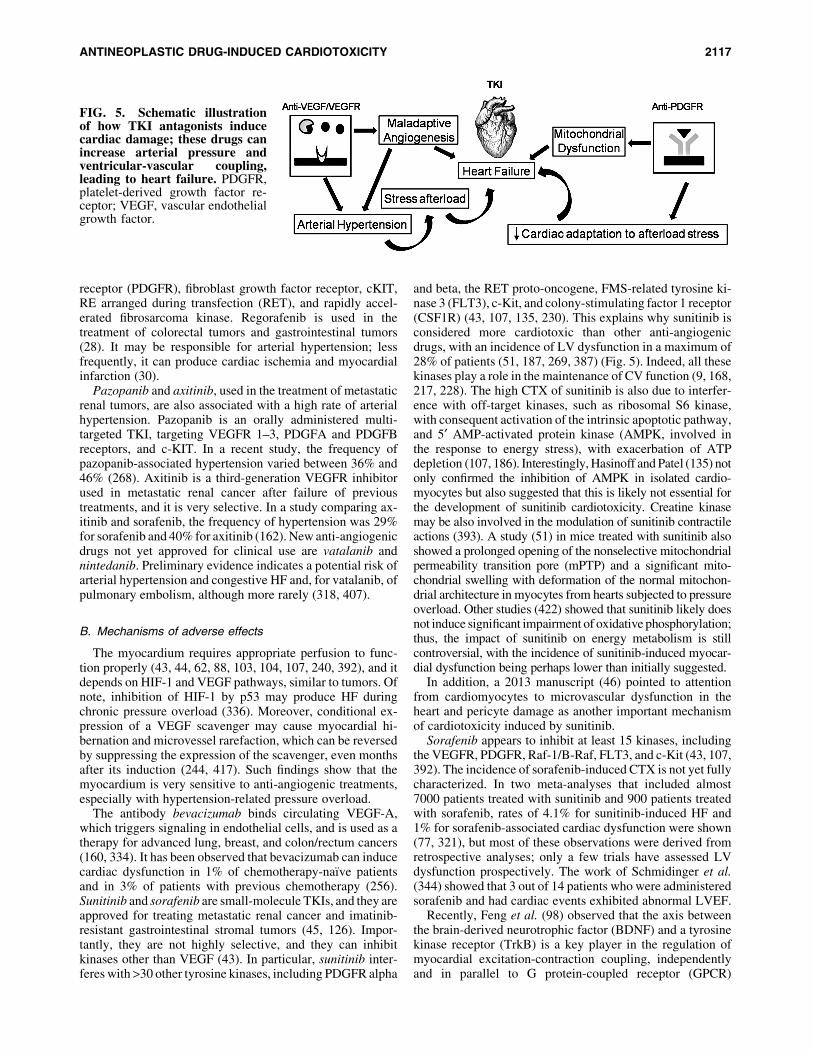
and beta, the RET proto-oncogene, FMS-related tyrosine ki-nase 3 (FLT3), c-Kit, and colony-stimulating factor 1 receptor(CSF1R) (43, 107, 135, 230). This explains why sunitinib isconsidered more cardiotoxic than other anti-angiogenicdrugs, with an incidence of LV dysfunction in a maximum of28% of patients (51, 187, 269, 387) (Fig. 5). Indeed, all thesekinases play a role in the maintenance of CV function (9, 168,217, 228). The high CTX of sunitinib is also due to interfer-ence with off-target kinases, such as ribosomal S6 kinase,with consequent activation of the intrinsic apoptotic pathway,and 5¢ AMP-activated protein kinase (AMPK, involved inthe response to energy stress), with exacerbation of ATPdepletion (107, 186). Interestingly, Hasinoff and Patel (135) notonly confirmed the inhibition of AMPK in isolated cardio-myocytes but also suggested that this is likely not essential forthe development of sunitinib cardiotoxicity. Creatine kinasemay be also involved in the modulation of sunitinib contractileactions (393). A study (51) in mice treated with sunitinib alsoshowed a prolonged opening of the nonselective mitochondrialpermeability transition pore (mPTP) and a significant mito-chondrial swelling with deformation of the normal mitochon-drial architecture in myocytes from hearts subjected to pressureoverload. Other studies (422) showed that sunitinib likely doesnot induce significant impairment of oxidative phosphorylation;thus, the impact of sunitinib on energy metabolism is stillcontroversial, with the incidence of sunitinib-induced myocar-dial dysfunction being perhaps lower than initially suggested.
In addition, a 2013 manuscript (46) pointed to attentionfrom cardiomyocytes to microvascular dysfunction in theheart and pericyte damage as another important mechanismof cardiotoxicity induced by sunitinib.
Sorafenib appears to inhibit at least 15 kinases, includingthe VEGFR, PDGFR, Raf-1/B-Raf, FLT3, and c-Kit (43, 107,392). The incidence of sorafenib-induced CTX is not yet fullycharacterized. In two meta-analyses that included almost7000 patients treated with sunitinib and 900 patients treatedwith sorafenib, rates of 4.1% for sunitinib-induced HF and1% for sorafenib-associated cardiac dysfunction were shown(77, 321), but most of these observations were derived fromretrospective analyses; only a few trials have assessed LVdysfunction prospectively. The work of Schmidinger et al.(344) showed that 3 out of 14 patients who were administeredsorafenib and had cardiac events exhibited abnormal LVEF.
Recently, Feng et al. (98) observed that the axis betweenthe brain-derived neurotrophic factor (BDNF) and a tyrosinekinase receptor (TrkB) is a key player in the regulation ofmyocardial excitation-contraction coupling, independentlyand in parallel to G protein-coupled receptor (GPCR)
FIG. 5. Schematic illustrationof how TKI antagonists inducecardiac damage; these drugs canincrease arterial pressure andventricular-vascular coupling,leading to heart failure. PDGFR,platelet-derived growth factor re-ceptor; VEGF, vascular endothelialgrowth factor.
ANTINEOPLASTIC DRUG-INDUCED CARDIOTOXICITY 2117
signaling. These data further support the concept that tyrosinekinase blockade with antineoplastic treatments can interferewith fundamental signaling, thus impairing cardiac me-chanical work that may produce LV dysfunction (106).
In summary, several mechanisms may lead to cardiacdysfunction in patients treated with anti-VEGF therapy: cKitinhibition, altered activation of MAP with consequent alter-ation of energy production and mitochondrial function,PDGFR inhibition, and inhibition of signaling favoring cellsurvival, as well as angiogenesis inhibition and induction ofarterial hypertension.
Importantly, hypertension is a main side-effect of all threemajor anti-VEGF drugs (272). Mechanisms of arterial hy-pertension include both functional (inactivation of endothe-lial nitric oxide synthase and production of vasoconstrictorssuch as endothelin-1) and anatomic (capillary rarefaction)modifications, which lead to vasoconstriction and an increasein peripheral vascular resistance (272, 277, 353). The mainmechanisms of capillary rarefaction are considered the lossof pericytes due to PDGFR inhibition, along with inhibitionof angiogenesis by VEGFR inhibition (353). Moreover, renaldysfunction due to angiogenesis inhibition can play a role ininducing and maintaining arterial hypertension. All said, themechanisms of hypertension induced by antiangiogenicdrugs have not yet been fully elucidated, and they are still anunresolved issue.
Bevacizumab produces severe hypertension that, at leastin some circumstances, is not reversed by discontinuing thedrug. Hypertension from anti-angiogenic agents has beensuggested to be a biomarker of anti-cancer drug efficacy,because some patients who developed hypertension wereobserved to survive longer than those who did not (122).It has been shown that in cases of metastatic colorectalcancer, 20% of patients showed grade 2–3 hypertension. Apartial remission was observed in 75% of subjects withbevacizumab-induced hypertension, and in only 32% ofthose with no hypertension. Moreover, patients who showedgrade 2–3 hypertension had significantly longer progression-free survival than nonhypertensive patients (342).
Sunitinib can also cause hypertension, with an incidenceranging from 5% to 47% in different studies. Sunitinib wasshown to induce hypertension (‡150/100 mmHg) in 47% ofthe subjects studied, with grade 3 hypertension seen in 17%.High blood pressure was observed within the first 4 weeks oftherapy (32, 270).
Inhibition of VEGF signaling can also cause arterial andvenous thrombosis. Arterial thromboembolic events (ATE)are mainly related to VEGF inhibition and consequent im-pairment of vascular homeostasis due to a reduction of nitricoxide (NO) synthesis, endothelial dysfunction, and produc-tion of vasoactive mediators that favor plaque instability andthrombus formation (212). Bevacizumab causes ATE morefrequently in patients contemporaneously treated with otherchemotherapeutic agents, in older patients, and in those whohave experienced previous thrombotic events (315). The realburden of venous TE related to bevacizumab is less clear(161). The pathogenesis of venous events is always related toimpairment of vascular homeostasis due to VEGF inhibitionand to production of pro-inflammatory cytokines that favoractivation of the coagulation cascade (144).
Sorafenib, pazopanib, and axitinib may also increase therisk of ATE. In a meta-analysis of 10,000 patients, the inci-
dence of ATE was 1.7% in sorafenib-treated patients (49).Venous thrombotic events were reported in 3% of axitinib-treated patients, with ATE in 2% and transient ischemic at-tack in 1%. However, HF was observed in <1% of subjectstreated with sorafenib and in 3% of patients treated withpazopanib (324).
Cardiac ischemia or myocardial infarction can also occurwith sorafenib (3.8%), pazopanib (2%), and axitinib (<1%)(30). This is mainly related to the inhibition of VEGF sig-naling, leading to endothelial dysfunction, vasoconstriction,and microvascular rarefaction. Moreover, a reduction inVEGF levels creates an imbalance between cell survival andapoptosis. Endothelial cell apoptosis favors an exposure ofsub-endothelial collagen and activation of the coagulationcascade, whereas reduced VEGF levels also interfere with theplatelet-mediated fibrinolytic cascade (212).
Finally, patients treated with sorafenib, pazopanib, and van-detanib can present an electrocardiogram (ECG) with QT inter-val prolongation, which predisposes them to an enhanced risk ofventricular arrhythmias (194, 214). In particular, sorafenib canfrequently induce QT prolongation (40.5%), thus greatly in-creasing the risk of ventricular arrhythmias (30). Vandetanib hasalso been related to a moderate risk of QT prolongation (16%),and more rarely, to life-threatening arrhythmias; in addition, itis associated with an enhanced risk of arterial hypertension(24%) and HF (21%) (434). In patients treated with pazopanib,the risk of QT prolongation and torsade de pointes, a dangerouspolymorphic ventricular tachycardia leading to sudden cardiacdeath, is <2% (303).
In patients receiving drugs potentially interfering with QTduration, electrocardiographic monitoring of QT interval andperiodic control of electrolytes are recommended. A reduc-tion in the drug’s dose can be warranted, but rarely discon-tinuation. Particular attention should be paid to patients witha personal or familial history of QT prolongation or to thosewho are under concomitant medication that can influence QTinterval. For further details on the approaches to reduce CVtoxicity, see sections X and XI.
V. TKIs and Anti-BCR-abl Agents
Chronic myeloid leukemia (CML) is a cancer of the whiteblood cells resulting from the clonal expansion of a trans-formed multipotent hematopoietic stem cell. It is charac-terized by a reciprocal translocation between the long armsof two chromosomes, namely chromosomes 9 and 22. Thistranslocation produces a shortened chromosome 22, the so-called Philadelphia chromosome. The consequence of suchtranslocation is the production of the BCR-ABL1 fusiongene on chromosome 22, and the reciprocal ABL1-BCRgene on chromosome 9. The latter does not have anyfunctional role in CML, whereas the BCR-ABL1 fusiongene is necessary for CML pathogenesis. The ABL1 tyro-sine kinase is a master-regulator of the cell cycle regulation,whereas the BCR-ABL1 fusion gene generates a constitu-tively active tyrosine kinase that leads to uncontrolledproliferation (332).
Targeting the BCR-ABL1 fusion gene has revolutionizedthe management of CML, turning a fatal disease into achronic disorder that, in most cases, is compatible with anormal lifespan (339). Based on the data from the IRIS study,which documented a dramatic improvement in survival,
2118 TOCCHETTI ET AL.
imatinib was the first targeted drug approved for the therapyof CML (285). However, because of the development ofa point mutation in the BCR-ABL1 fusion gene, a consider-able number of patients may acquire resistance to imatinib(10). Second-generation TKIs—dasatinib, nilotinib, andbosutinib—have been shown to be effective in imatinib-resistant patients, and more rapid in achieving a deep mo-lecular response (57, 182, 383). Ponatinib, a third-generationTKI specifically designed to overcome resistance caused bya T315I mutation in BCR-ABL1 kinase, has exhibited highclinical efficacy in patients with multi-TKI-resistant CML(56). At their debut in the clinical arena, all five drugs appearedcardiologically safe; however, subsequent information indi-cated that they may exert adverse CV side-effects that mayaffect prognosis and quality of life and must be taken intoaccount when selecting treatment agents (267). All these drugsdemonstrate significant off-target bindings and off-target ef-fects, which may contribute to both toxicities and therapeuticcontrol of the myeloproliferative disorder (124, 383).
Imatinib was originally developed as a PDGFR inhibitor. Itis a first-generation TKI that binds to the tyrosine kinase ABLwhile the protein is in the closed conformation and is inac-tive. It also inhibits other TKs, including VEGFR membersand KIT family kinases (124).
A. Imatinib adverse events
A 5-year follow-up study on patients with newly diagnosedCML treated with imatinib showed an excellent CV safetyrecord for the drug (285). However, Kerkela et al. reportedclinical data from 10 patients with normal ventricular func-tion who developed severe cardiac dysfunction and NewYork Heart Association (NYHA) Class 3–4 HF after imatinibtreatment, and they demonstrated that mice treated with im-atinib develop a severe form of LV contractile dysfunction(185). The publication by Kerkela et al. prompted manyhematologists who had used imatinib to treat multiple pa-tients to review their clinical database; the rates of CTXfound were extremely low (16, 113, 138, 329). Further clin-ical studies then confirmed the excellent CV risk profile ofimatinib (54, 60, 64, 80, 90, 148).
B. Mechanisms of imatinib-induced adverse events
Several studies investigated the mechanisms responsiblefor cardiac toxicity, and they found that imatinib-inducedABL inhibition leads to alterations in the endoplasmic retic-ulum (ER) stress response by activating the IRE1 kinase armof the response. IRE1 activates apoptosis signal-regulatingkinase 1 and c-Jun N-terminal kinase, which, in turn, causesinhibition of antiapoptotic B cell leukemia/lymphoma andcytochrome c release from mitochondria, thus leading tomitochondrial function impairment and cell death (43, 44).The inhibition of Abl kinase by imatinib is also responsiblefor PRKR-like endoplasmic reticulum kinase activation,which results in impaired protein import in the mitochondrialmatrix, inducing mitochondrial death pathways (409).
Other than the potential proapoptotic effect on cardio-myocytes, imatinib has been shown to exert a number ofcardiologically relevant effects. First, it causes fluid retentionand edema that may contribute to a mistaken diagnosis ofHF. This is probably due to imatinib’s inhibition of PDGFR(356). Paradoxically, imatinib treatment can also protect
against brain and lung edema during stroke and lung in-jury, respectively (39, 374). It has been demonstrated thatin human endothelial cells pretreated with a variety ofbarrier-disruptive agents, imatinib reduces endothelial bar-rier dysfunction via inhibition of the abl-related gene (7, 47).Imatinib also has favorable effects on glucose control byimproving both b cell function and peripheral insulin sensi-tivity (101, 130). Imatinib improves hemodynamics and re-verses vascular remodeling and cor pulmonale in pulmonaryarterial hypertension (PAH) by PDGF inhibition (343). Fi-nally, imatinib was found to attenuate myocardial remodelingand to improve LV diastolic dysfunction in spontaneouslyhypertensive rats by affecting the PDGFR pathway withoutthe blood pressure-lowering effect (170).
Nilotinib is a structural analog of imatinib that binds andpotentially inhibits PDGFR and KIT family kinases, but notkinases of the SRC group (124). It is approved for the therapyof subjects with newly diagnosed Ph+ CML in any phase.Nilotinib is also approved for CML patients who are resistantto or intolerant of previous therapies, including imatinib.
C. Nilotinib adverese events
Early in the development of Nilotinib, there was evidenceof QT prolongation, leading to a warning to perform serialECGs, prevent and correct electrolyte abnormalities, andavoid concomitant drugs potentially responsible for QT in-terval prolongation (182). However, subsequent trials foundno alarming signals of QT-related ventricular arrhythmias(267). Clinical trials, as well as studies analyzing real-lifesettings, found that nilotinib exacerbates the glycometabolicand lipid profile (26, 166, 167, 267, 313, 317). Over the past5 years, several clinical trials, retrospective studies, meta-analyses, and postmarketing surveillance studies have shownthat nilotinib-treated patients are at an increased risk of CVevents, especially those related to peripheral artery disease(PAD) (2, 54, 64, 80, 118, 148, 190, 209, 267, 403). Oddly,these vascular adverse events were not reported during thefirst clinical studies.
D. Mechanisms of nilotinib-induced adverse events
Recent studies have documented a number of nilotinib-induced effects on endothelium, platelets, and coagulation,which, together with the metabolic effects, enhance the riskof vascular adverse events (4, 22).
As for myocardial toxicity, clinical studies have shownthat nilotinib does not cause ventricular dysfunction. Thisfinding concurs with experimental data showing that nilotiniband imatinib produce little or no damage to cardiomyocytes,in comparison with dasatinib, bosutinib, and ponatinib (137).
Dasatinib is a second-generation TKI, and it is more potentthan imatinib. Unlike imatinib and nilotinib, dasatinib bindsto TKs, in both their active and inactive conformational state.It is classified as a dual Abl/Src inhibitor, although it inhibitsa broad spectrum of receptor kinases, including c-Kit andPDGFR (124).
Dasatinib is approved as a frontline therapy of Ph+ CMLpatients in the chronic phase, and for salvage treatment of Ph+
CML or Ph+ acute lymphoblastic leukemia (ALL) patientswho are intolerant of or resistant to other TKIs.
ANTINEOPLASTIC DRUG-INDUCED CARDIOTOXICITY 2119
E. Dasatinib adverse events
For years, the CV safety profile of dasatinib was consid-ered similar to that of imatinib, with the exception of pleuraleffusion, which has been observed since the first use of thedrug (383). Clinical studies with a long follow-up periodfound that pleural effusion occurs in one of every three pa-tients, and that half of pleural events occur within the first2 years of therapy. Pleural effusion requiring drug discon-tinuation occurs in <10% of the cases (351).
PAH is a rare but serious complication of dasatinib treat-ment. A transient significant increase of pulmonary arterialpressure detected by echocardiography was observed for thefirst time in 2017 among patients with pleural effusion (312).Subsequently, other reports emerged of PAH not associatedwith pleural effusion (263).
In clinical studies, PAH has been observed in 5% of sub-jects on dasatinib as a frontline therapy for 5 years and in2.4% of patients treated with dasatinib for 7 years, who wereresistant to or intolerant of other treatments (351). A recentassessment of all causes of dasatinib-related PAH confirmedby catheterization (41 patients) showed that a unique featureof dasatinib-induced PAH is different complete or partialreversibility on drug discontinuation (352). However, theFrench PH Registry that identified nine cases of dasatinib-associated PAH reported that most patients did not experi-ence complete recovery, and it recorded two cases of cardiacdeath after dasatinib withdrawal (263).
Analysis of the FDA database designed to support post-marketing surveillance found that PAH was associated withdasatinib and not with other TKIs, and that it often occurredin patients with CV risk factors or a medical history of car-diopulmonary events (54).
F. Mechanisms of dasatinib-induced adverse events
Pleural effusion is considered the result of PDGFR inhibi-tion, particularly of the PDGFR-b isoform, of which dasatinibis the most potent inhibitor; however, other mechanisms havebeen suggested (184). Due to the high frequency of lympho-cytes in pleural fluid and tissue and the association with skinrash or history of autoimmunity, an autoimunne mechanism hasbeen hypothesized (297). It has been suggested that an immune-mediated mechanism underlying the occurrence of pleural ef-fusion could help to promote tumor regression. Patients whodevelop lymphocytosis and increased levels of specific lym-phocyte subsets in association with pleural effusion have ahigher major molecular response rate and better progression-free and overall survival than patients who do not (89).
The underlining molecular mechanism responsible forPAH is still poorly understood. Since dasatinib is a potentpan-Src inhibitor, it has been suggested that Src inhibition ispivotal in the development of PAH (264). The evidence thatCML patients treated with dasatinib may display high cir-culating levels of E-selectin, intercellular adhesion molecule,and vascular cell adhesion molecule, and the observation thatcultured pulmonary endothelial cells administered with da-satinib display an increased mitochondrial ROS productionand apoptosis support the notion that dasatinib-induced PAHis caused by endothelial dysfunction (127).
Because vascular events have been described in patientstreated with other TKIs, recent clinical trials have evaluatedthe incidence of coronary, cerebral, and peripheral throm-
botic events in dasatinib-treated patients; a slightly higherrisk was found, compared with imatinib. However, a recentmeta-analysis showed that the use of dasatinib is associatedwith a significant increase in the risk of CV events. In apopulation-based cohort study, the incidence of myocardialinfarction was 2.4 times higher in patients treated with da-satinib than in those treated with imatinib.
Bosutinib is a second-generation, dual Src/Abl TKI lack-ing significant PDGFR or c-KIT binding properties (124).Bosutinib is currently approved only for patients with Ph1chronic-phase CML who were resistant to or intolerant ofprevious TKI therapy.
G. Bosutinib adverse events
Studies that evaluated bosutinib enrolled patients newlydiagnosed with CML, or patients resistant to or intolerant ofother TKIs; these studies documented a low incidence ofcardiac and vascular events (31, 55, 57, 58). Similar to otherTKIs, the incidence of adverse events was higher in second-or later-line treatment compared with first-line treatment.HF and coronary or PAD-related events were rare, suggest-ing that the cardiotoxic profile of bosutinib differs from thatof other second- or third-generation TKIs. Most events oc-curred within the first year of therapy, and very few patientsdiscontinued treatment because of these events, which weremostly managed with concomitant medications (55).
It should be noted that in a first-line setting, the overallincidence of events and dose reduction or interruption issimilar for imatinib and bosutinib, even though pericardialdisorders occurred more often in bosutinib-treated patientsthan in those treated with imatinib.
Ponatinib, a third-generation BCR-ABL TKI, was origi-nally developed for the inhibition of pan-resistant ABLTK, and it is clinically effective in imatinib-, nilotinib-, anddasatinib-resistant CML. Ponatinib targets BCR-ABL in itsinactive conformation and inhibits T315I and other BCR-ABL mutants. It also targets a broad spectrum of TK recep-tors, including epidermal growth factor receptor (EGFR),PDGFR, VEGFR, SRC kinase, c-KIT, FLT3, and RET (124).
H. Ponatinib adverse events
Ponatinib was tested in Ph+ patients with refractory CMLor ALL enrolled in phase I (65 patients) and in phase II (449patients) of the PACE trial. Both studies documented a highefficacy of the drug, accompanied by an increased risk ofarterial thrombotic events (56, 59). For these reasons, pona-tinib was approved with a warning of the risk of seriousvascular events. Findings from these studies provided therationale to perform a phase III study (EPIC) to test the hy-pothesis that ponatinib may lead to a deep molecular responsein patients newly diagnosed with CML, and to assess itssafety in patients with no previous exposure to other TKIs(226). The extended follow-up periods of the phase I and IIstudies documented that cardiac, cerebral, and peripheralthrombotic events continued to occur; this finding determinedthe early termination of the phase III study and led ARIADPharmaceuticals and the FDA to withdraw ponatinib from themarket in October 2013. In January 2014, after a reassess-ment of the benefit/risk ratio of ponatinib, the FDA allowedits reintroduction into the market, with a recommendationto take appropriate measures to mitigate adverse effects in
2120 TOCCHETTI ET AL.
patients. The mechanisms of ponatinib CV toxicity are stillpoorly understood. Diagnostic and pharmacological mea-sures aimed at reducing the CV risk were not included in theprotocols of the phase I–III trials. The implementation ofthese preventive measures may help to improve the thera-peutic index of ponatinib in the clinical setting (27). Forfurther details on the approaches to reduce TKIs-inducedCTX, please refer to sections X and XI.
VI. Taxanes
A. Adverse effects
Although the potential of taxanes to induce cardiac dys-function was likely overestimated in the past, they deserve abrief note. The most commonly used taxanes are docetaxeland paclitaxel, which are still used in breast, nonsmall celllung, and ovarian cancers. Among the adverse effects of thesedrugs, a high incidence of arrhythmias has been observed,including asymptomatic sinus bradycardia (330), whichusually is not a major problem for patients. A large range ofconduction blocks and cardiac ischemia have been also ob-served in a few patients participating in trials (330). Ven-tricular arrhythmias are far less common: Only 0.26% ofpatients experienced ventricular tachycardia and ventricularfibrillation, and many of them were also co-treated withcisplatin (245, 330). In a clinical trial (EORTC 24971/TAX323 Study Group), patients treated with docetaxel displayed a1.7% incidence of cardiac ischemia (411). In about 3500patients treated with paclitaxel followed by the CancerTherapy Evaluation Program’s Adverse Drug Reaction da-tabase, the overall incidence of severe (grade 4 and 5) cardiacevents was <0.3% (12). These severe events were observedtill 2 weeks after the beginning of paclitaxel treatment. Ofnote, many of these subjects had important CV risk factors,including coronary disease and hypertension.
B. Mechanisms of adverse effects
Taxanes exert anticancer effects, acting as anti-microtubuleagents and fostering polymerization of tubulin. In this way,these agents induce microtubule dysfunction and alter celldivision. Moreover, they may affect histamine release (330).These taxane-induced effects may perturb cardiac function.Brady-arrhythmias may be due to stimulation of histamine H1
and H2 receptors. The latter receptors may increase the pos-sibility of reentry of depolarization, which is at the basis ofventricular ectopy (245, 330). Ischemia and increased myo-cardial oxygen demands render the heart more susceptible toarrhythmias. Of note, paclitaxel in combination with ANTsincreases the incidence of CTX, which appears at lower cu-mulative doses with respect to ANTs alone (120). In partic-ular, plasma levels of doxorubicin and the formation of thetoxic metabolite doxorubicinol in myocytes can be enhancedby taxanes.
C. Ways to reduce CV toxicity
Cardiotoxic symptoms are frequently resolved on discon-tinuation of taxanes. Since histamine is considered a relevantplayer in taxane CTX, treatment with corticosteroids andantihistamines may reduce the incidence of clinically sig-nificant arrhythmias (245, 330).
VII. Cancer Immunotherapy
All components of the innate and adaptive immune systemare important in the recognition and elimination of tumorcells (immune surveillance) (133). These immune compo-nents recognize specific neo-antigens that are expressed bytumor cells. However, the production of immunosuppressivecytokines/chemokines, the presence of immunosuppressivecells, or the expression of inhibitory checkpoints within thetumor microenvironment allow the tumor cells to escapeimmune surveillance (296).
A. Immune checkpoint inhibitors
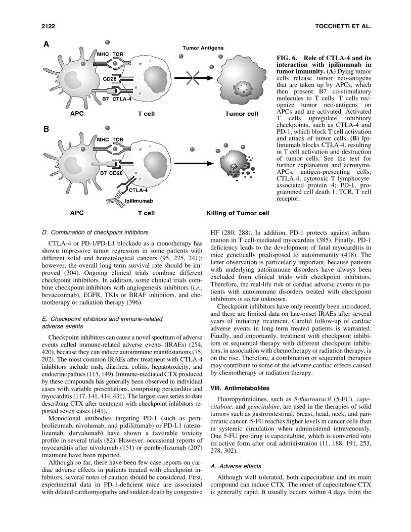
Tumors exploit immune checkpoints to evade antitumorimmunity. The success of cancer immunotherapy over the pastdecades has revolutionized the management of a wide array ofmalignancies. At the forefront of immunotherapy are immunecheckpoint blockers, which have seen unprecedented successin cancer treatments because of their broad bioactivity acrossmany tumor types. The two most prominent to date are thetargeting of cytotoxic T lymphocyte-associated protein 4(CTLA-4) and the interaction between the proteins pro-grammed cell death 1 (PD-1) and programmed death-ligand 1(PD-L1).
B. CTLA-4
It has structural homology to the co-stimulatory moleculeCD28, and it can also bind B7 molecules on antigen-presenting cells (APCs) with a higher affinity than CD28.CTLA-4 is a competitive antagonist of the CD28-B7 inter-action in that it blocks co-stimulation at the T cell-APC in-teraction, thus suppressing activation of its cell response(200) (Fig. 6). Ipilimumab and tremelimumab are monoclo-nal antibodies for clinical use against CTLA-4 (117, 354,355, 431).
C. PD-1/PD-L1 pathway
PD-1 is a checkpoint receptor that, by binding with itsligands (either PD-L1 or PD-L2), inhibits T effector functionsby dampening signaling downstream of the T cell receptor(TCR) (397). Therefore, the expression of PD-L1 in the tumormicro-environment protects cancer cells from immune-mediated destruction. Several monoclonal antibodies againstthe PD-1/PD-L1 axis have been developed and/or approved asanti-cancer agents (396, 397).
PD-1, similar to CTLA-4, is expressed in activated cells,and its activation downregulates signaling that is mediated onantigen recognition by the TCR (354). PD-1 has two ligands,PD-L1 and PD-L2. PD-L1 can be expressed in many celltypes, including tumor cells, immune cells, epithelial cells, andendothelial cells (354). PD-L2 is predominantly expressed inAPCs. Pembrolizumab and nivolumab are PD-1-blockingmonoclonal antibodies that are approved for treating differenttypes of cancers. Atezolizumab is a PD-L1 targeted monoclo-nal antibody recently approved for treatment of certain tumors(354). Other inhibitory molecules (e.g., LAG3, OX40, TIM3,TIGIT) are being assessed for their potential utility as immu-notherapeutic targets (15, 211, 279, 399).
ANTINEOPLASTIC DRUG-INDUCED CARDIOTOXICITY 2121
D. Combination of checkpoint inhibitors
CTLA-4 or PD-1/PD-L1 blockade as a monotherapy hasshown impressive tumor regression in some patients withdifferent solid and hematological cancers (95, 225, 241);however, the overall long-term survival rate should be im-proved (304). Ongoing clinical trials combine differentcheckpoint inhibitors. In addition, some clinical trials com-bine checkpoint inhibitors with angiogenesis inhibitors (i.e.,bevacizumab), EGFR, TKIs or BRAF inhibitors, and che-motherapy or radiation therapy (396).
E. Checkpoint inhibitors and immune-relatedadverse events
Checkpoint inhibitors can cause a novel spectrum of adverseevents called immune-related adverse events (IRAEs) (254,420), because they can induce autoimmune manifestations (75,202). The most common IRAEs after treatment with CTLA-4inhibitors include rash, diarrhea, colitis, hepatotoxicity, andendocrinopathies (115, 149). Immune-mediated CTX producedby these compounds has generally been observed in individualcases with variable presentations, comprising pericarditis andmyocarditis (117, 141, 414, 431). The largest case series to datedescribing CTX after treatment with checkpoint inhibitors re-ported seven cases (141).
Monoclonal antibodies targeting PD-1 (such as pem-brolizumab, nivolumab, and pidilizumab) or PD-L1 (atezo-lizumab, durvalumab) have shown a favorable toxicityprofile in several trials (82). However, occasional reports ofmyocarditis after nivolumab (151) or pembrolizumab (207)treatment have been reported.
Although so far, there have been few case reports on car-diac adverse effects in patients treated with checkpoint in-hibitors, several notes of caution should be considered. First,experimental data in PD-1-deficient mice are associatedwith dilated cardiomyopathy and sudden death by congestive
HF (280, 288). In addition, PD-1 protects against inflam-mation in T cell-mediated myocarditis (385). Finally, PD-1deficiency leads to the development of fatal myocarditis inmice genetically predisposed to autoimmunity (418). Thelatter observation is particularly important, because patientswith underlying autoimmune disorders have always beenexcluded from clinical trials with checkpoint inhibitors.Therefore, the real-life risk of cardiac adverse events in pa-tients with autoimmune disorders treated with checkpointinhibitors is so far unknown.
Checkpoint inhibitors have only recently been introduced,and there are limited data on late-onset IRAEs after severalyears of initiating treatment. Careful follow-up of cardiacadverse events in long-term treated patients is warranted.Finally, and importantly, treatment with checkpoint inhibi-tors or sequential therapy with different checkpoint inhibi-tors, in association with chemotherapy or radiation therapy, ison the rise. Therefore, a combination or sequential therapiesmay contribute to some of the adverse cardiac effects causedby chemotherapy or radiation therapy.
VIII. Antimetabolites
Fluoropyrimidines, such as 5-fluorouracil (5-FU), cape-citabine, and gemcitabine, are used in the therapies of solidtumors such as gastrointestinal, breast, head, neck, and pan-creatic cancer. 5-FU reaches higher levels in cancer cells thanin systemic circulation when administered intravenously.One 5-FU pro-drug is capecitabine, which is converted intoits active form after oral administration (11, 188, 191, 253,278, 302).
A. Adverse effects
Although well tolerated, both capecitabine and its maincompound can induce CTX. The onset of capecitabine CTXis generally rapid. It usually occurs within 4 days from the
FIG. 6. Role of CTLA-4 and itsinteraction with ipilimumab intumor immunity. (A) Dying tumorcells release tumor neo-antigensthat are taken up by APCs, whichthen present B7 co-stimulatorymolecules to T cells. T cells rec-ognize tumor neo-antigens onAPCs and are activated. ActivatedT cells upregulate inhibitorycheckpoints, such as CTLA-4 andPD-1, which block T cell activationand attack of tumor cells. (B) Ipi-limumab blocks CTLA-4, resultingin T cell activation and destructionof tumor cells. See the text forfurther explanation and acronyms.APCs, antigen-presenting cells;CTLA-4, cytotoxic T lymphocyte-associated protein 4; PD-1, pro-grammed cell death 1; TCR, T cellreceptor.
2122 TOCCHETTI ET AL.
start of the first cycle of chemotherapy (174, 175). Pro-spective and retrospective studies have shown that previousevents of cardiac ischemia are the strongest risk factorsfor fluoropyrimidine-induced CTX (195, 309, 310). ECGabnormalities have been found in patients treated with a24-h 5-FU infusion in combination with capecitabine. Pal-pitations and chest pain are the most common clinicalmanifestations, accompanied by new-onset ECG alterations,such as ST segment deviation, sinus tachycardia, QT pro-longation, and signs of myocardial ischemia and acutecoronary syndromes. Less frequent are arterial hypertension,ventricular arrhythmias, LV dysfunction, HF, and cardio-genic shock. Cardiac arrest and sudden death are very rareevents, and they account for <1% of clinical manifestations(5, 198, 309, 408).
The incidence of CTX produced by 5-FU varies between0% and 35%, with a mortality rate ranging between 2.2%and 13.3%, depending on the dose, cardiac comorbidities,and route of administration (198, 309, 310). Larger trials andmeta-analyses on breast and colorectal cancer patients, how-ever, have reported an incidence of symptomatic CTX of1.2%–4.3% during treatment (205, 407). A recent studyfound silent ischemia due to the cardiac stress test in 6%–7% of 5-FU-treated patients (216). Unfortunately, patientswithout overt CV risk factors are often treated as outpatients.As a result, symptomatic and asymptomatic cardiac eventsremain undiagnosed because of inadequate cardiac moni-toring. This can lead to a discrepancy between observed andactual CTX, in terms of both symptomatic and asymptomaticpatients.
B. Mechanism of adverse effects
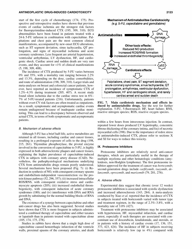
Although 5-FU has a brief half-life, active metabolites areretained in all tissues, including cardiac and cancer tissues,resulting in a prolonged exposure of cells to the drug (198,215, 261). Thymidine phosphorylase, the pivotal enzymeinvolved in the conversion of capecitabine to 5-FU, is highlyexpressed in both atherosclerotic plaques and cancer tissues,explaining the higher prevalence of capecitabine-inducedCTX in subjects with coronary artery disease (CAD). Ne-vetheless, the pathophysiological mechanisms underlyingCTX from antimetabolite drugs remain poorly understood.Several mechanisms have been proposed (Fig. 7): (i) Re-duction in synthesis of NO, with consequent coronary spasmsand endothelium-independent vasoconstriction via the pro-tein kinase pathway (52, 296, 357); (ii) increased intracellularROS/RNS, with consequent oxidative stress and cardio-myocyte apoptosis (205); (iii) increased endothelial throm-bogenicity, with consequent induction of acute coronarysyndromes (180); and (iv) interference with DNA and RNAgrowth by substituting for the normal building blocks of RNAand DNA.
The existence of a synergy between capecitabine and otheranti-cancer drugs has also been suggested. Several studieshave shown that CTX is more frequent in subjects adminis-tered a combined therapy of capecitabine and either taxanesor lapatinib than in patients treated with capecitabine alone(105, 174, 175, 278).
Studies in rabbits have shown that a single high dose ofcapecitabine caused hemorrhagic infarction of the ventriclewalls, proximal spasms of the coronary arteries, and death
within a few hours from intravenous injection. In contrast,repeated lower doses produced LV hypertrophy, concentricfibrous thickening of the coronary intima, and foci of necroticmyocardial cells (398). Due to the importance of redox stressin antimetabolite-induced CTX, please refer to sections Xand XI for ways to reduce this toxicity.
IX. Proteasome Inhibitors
Proteasome inhibitors are relatively novel anti-cancertherapies, which are particularly useful in the therapy ofmultiple myeloma and other hematologic conditions (amy-loidosis, non-Hodgkin lymphoma). The first proteasome in-hibitor approved for the treatment of cancer was bortezomib;second-generation drugs include carfilzomib, ixazomib, de-lanzomib, oprozomib, and marizomib (79, 201, 276).
A. Adverse effects
Experimental data suggest that chronic (over 12 weeks)proteasome inhibition is associated with systolic dysfunctionand increased atherosclerosis (143, 284). A meta-analysisconducted by Xiao et al. reported that the incidence of CTXin subjects treated with bortezomib varied with tumor typeand treatment regimen, in the range of 2.3%–3.8%, with amortality rate of 3.0% (427).
Treatment with proteasome inhibitors may be associatedwith hypertension, HF, myocardial infarction, and cardiacarrest, especially if such therapies are associated with con-comitant use of doxorubicin, lenalidomide, and steroids, orin patients with a history of cardiac events (17, 136, 151,373, 423, 424). The incidence of HF in subjects receivingbortezomib is relatively low (up to 4%) compared with
FIG. 7. Main cardiotoxic mechanism and effects in-duced by antimetabolite drugs. See the text for furtherexplanation. 5-FU, 5-fluorouracil; HF, heart failure; RNS,reactive nitrogen species; ROS, reactive oxygen species.
ANTINEOPLASTIC DRUG-INDUCED CARDIOTOXICITY 2123
carfilzomib (up to 25%), a more potent and irreversibleproteasomal inhibitor; patients treated with higher doses(‡36 mg/m2) of carfilzomib have a higher risk of cardiactoxicity (65, 328, 331).
B. Mechanisms of adverse effects
Proteasome inhibitors are compounds that block the ac-tivity of proteasomes, protein complexes that play a key rolein degrading dysfunctional or unneeded proteins; these cel-lular complexes that break down proteins are particularlyimportant for the functional maintenance of cardiomyocytes.Therefore, cardiac dysfunction may be expected if the func-tion of these complexes is impaired.
Many mechanisms of chemotherapy-induced cardiotoxi-city still remain to be clarified. It has been suggested (284)that bortezomib alters the function of cardiomyocytes throughthe impairment of mitochondrial energetics. The cardio-myocytes are contractile cells with a very high demand forATP and may be particularly sensitive to agents that disruptmitochondrial activity, such as the proteasome inhibitorbortezomib. Furthermore, a reduced synthesis of ATP couldtrigger the capillary tunneling, as revealed by a histopatho-logical examination of heart sections of rats treated withbortezomib (284). Chronic uptake inhibitor of the proteasomeis associated with increased oxidative stress at the level of theintima of the epicardial coronary arteries, resulting in thick-ening of the vessel wall, which can trigger premature ath-erosclerosis (143).
The addition of other chemotherapeutic agents such asANT with proteasome inhibitors improves the effectivenessof antineoplastic therapy; however, this combination of drugscan cause cardiotoxicity. Spur et al. (368) have analyzed thefunction of the proteasome in primary cardiomyocytes trea-ted with doxorubicin in the presence of proteasome inhibi-tors. Interestingly, the authors concluded that, contrary tocarfilzomib, which targets both the b5 standard proteasome andthe LMP7 immunoproteasome subunit, immunoproteasome-specific inhibitors with known anti-tumor capabilities forspecific cancer cells, such as multiple myeloma, may be ad-vantageous to reduce the mortality of cardiomyocytes, whenthere is a combination therapy, and, therefore, may be envi-sioned as a way to reduce CV toxicity, when compared withtraditional proteasome inhibitors.
C. Ways to reduce CV toxicity
The initial step in CV management of subjects treatedwith proteasome inhibitors is to assess their baseline risk forCTX, by taking a clinical history and conducting an exami-nation. More frequent surveillance may be warranted forsubjects with higher baseline clinical risk or abnormal car-diac baseline function.
CTX induced by proteasome inhibitors may be reversiblein some patients with prompt cessation of these therapies andinitiation of traditional HF treatments (123). ACE-Is or an-giotensin II receptor blockers (ARBs) in combination withb blockers are recommended in patients with symptomaticHF, similar to the general HF population and in asymptom-atic cardiac dysfunction, to prevent further cardiac dysfunc-tion or the development of symptomatic HF in patients at ahigh risk (432).
Single-center experiences have shown a decrease in theoccurrence of cardiac events when infusion time of carfil-zomib was settled to 30 min and a cardioprotective activity ofdexrazoxane was exhibited (17, 136).
In some cases, re-administration of carfilzomib with dosemodification is possible. Long-term surveillance should beconsidered for patients who developed CTX during thesetherapies and for those in whom cardioprotective treatmentshave been started, to confirm recovery or to detect irrevers-ible cardiac dysfunction.
X. The Importance of Mechanisms Such as Oxidativeand Nitrosative Stress in CV Toxicity: An Overview
As reported earlier, the cardiotoxic mechanisms of severalanticancer agents involve an unbalanced generation of ROSand RNS, leading to the so-called oxidative/nitrosative stress.ROS/RNS imbalance may derive from increased produc-tion or the inactivation of endogenous antioxidant enzymesby antineoplastic drugs, overwhelming the body’s defenses.Moreover, antioxidant resources (especially catalase) arelower in the cardiac tissue compared with other organs (e.g.,the liver), making the heart more vulnerable to ROS/RNSinjury (257, 258).
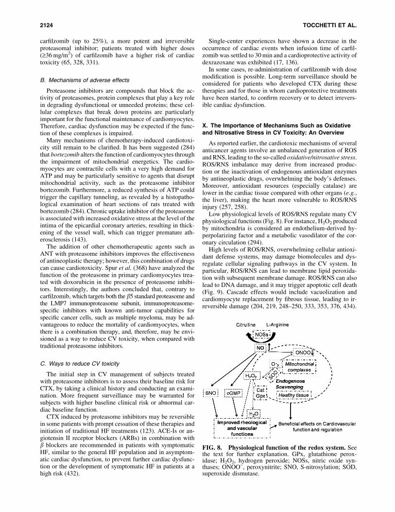
Low physiological levels of ROS/RNS regulate many CVphysiological functions (Fig. 8). For instance, H2O2 producedby mitochondria is considered an endothelium-derived hy-perpolarizing factor and a metabolic vasodilator of the cor-onary circulation (294).
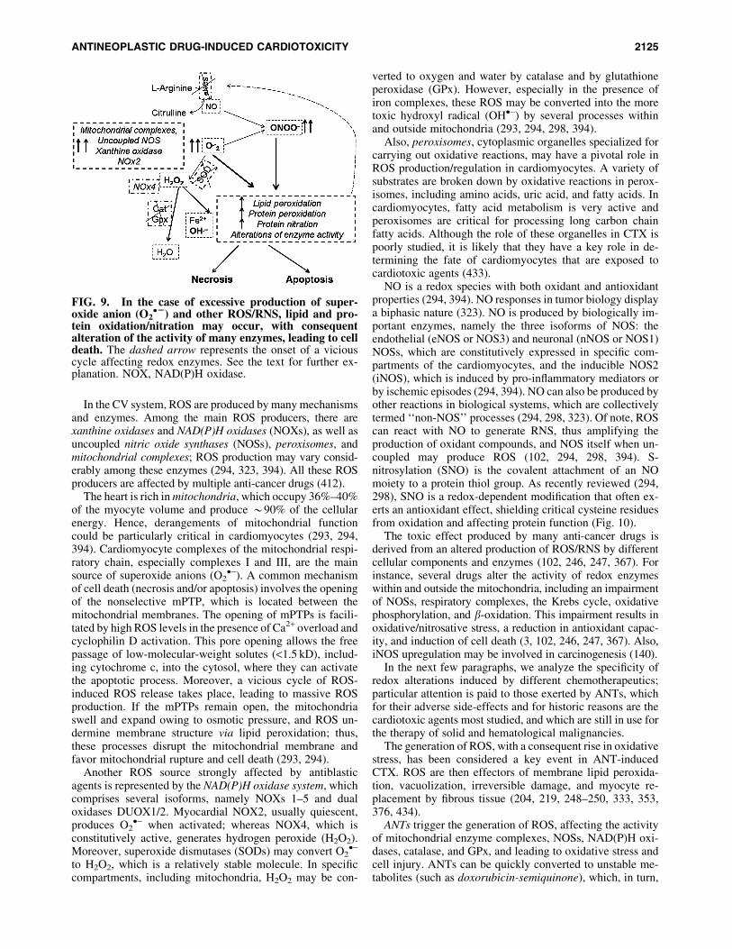
High levels of ROS/RNS, overwhelming cellular antioxi-dant defense systems, may damage biomolecules and dys-regulate cellular signaling pathways in the CV system. Inparticular, ROS/RNS can lead to membrane lipid peroxida-tion with subsequent membrane damage. ROS/RNS can alsolead to DNA damage, and it may trigger apoptotic cell death(Fig. 9). Cascade effects would include vacuolization andcardiomyocyte replacement by fibrous tissue, leading to ir-reversible damage (204, 219, 248–250, 333, 353, 376, 434).
FIG. 8. Physiological function of the redox system. Seethe text for further explanation. GPx, glutathione perox-idase; H2O2, hydrogen peroxide; NOSs, nitric oxide syn-thases; ONOO-, peroxynitrite; SNO, S-nitrosylation; SOD,superoxide dismutase.
2124 TOCCHETTI ET AL.
In the CV system, ROS are produced by many mechanismsand enzymes. Among the main ROS producers, there arexanthine oxidases and NAD(P)H oxidases (NOXs), as well asuncoupled nitric oxide synthases (NOSs), peroxisomes, andmitochondrial complexes; ROS production may vary consid-erably among these enzymes (294, 323, 394). All these ROSproducers are affected by multiple anti-cancer drugs (412).
The heart is rich in mitochondria, which occupy 36%–40%of the myocyte volume and produce *90% of the cellularenergy. Hence, derangements of mitochondrial functioncould be particularly critical in cardiomyocytes (293, 294,394). Cardiomyocyte complexes of the mitochondrial respi-ratory chain, especially complexes I and III, are the mainsource of superoxide anions (O2
�-). A common mechanismof cell death (necrosis and/or apoptosis) involves the openingof the nonselective mPTP, which is located between themitochondrial membranes. The opening of mPTPs is facili-tated by high ROS levels in the presence of Ca2+ overload andcyclophilin D activation. This pore opening allows the freepassage of low-molecular-weight solutes (<1.5 kD), includ-ing cytochrome c, into the cytosol, where they can activatethe apoptotic process. Moreover, a vicious cycle of ROS-induced ROS release takes place, leading to massive ROSproduction. If the mPTPs remain open, the mitochondriaswell and expand owing to osmotic pressure, and ROS un-dermine membrane structure via lipid peroxidation; thus,these processes disrupt the mitochondrial membrane andfavor mitochondrial rupture and cell death (293, 294).
Another ROS source strongly affected by antiblasticagents is represented by the NAD(P)H oxidase system, whichcomprises several isoforms, namely NOXs 1–5 and dualoxidases DUOX1/2. Myocardial NOX2, usually quiescent,produces O2
�- when activated; whereas NOX4, which isconstitutively active, generates hydrogen peroxide (H2O2).Moreover, superoxide dismutases (SODs) may convert O2
�-
to H2O2, which is a relatively stable molecule. In specificcompartments, including mitochondria, H2O2 may be con-
verted to oxygen and water by catalase and by glutathioneperoxidase (GPx). However, especially in the presence ofiron complexes, these ROS may be converted into the moretoxic hydroxyl radical (OH�-) by several processes withinand outside mitochondria (293, 294, 298, 394).
Also, peroxisomes, cytoplasmic organelles specialized forcarrying out oxidative reactions, may have a pivotal role inROS production/regulation in cardiomyocytes. A variety ofsubstrates are broken down by oxidative reactions in perox-isomes, including amino acids, uric acid, and fatty acids. Incardiomyocytes, fatty acid metabolism is very active andperoxisomes are critical for processing long carbon chainfatty acids. Although the role of these organelles in CTX ispoorly studied, it is likely that they have a key role in de-termining the fate of cardiomyocytes that are exposed tocardiotoxic agents (433).
NO is a redox species with both oxidant and antioxidantproperties (294, 394). NO responses in tumor biology displaya biphasic nature (323). NO is produced by biologically im-portant enzymes, namely the three isoforms of NOS: theendothelial (eNOS or NOS3) and neuronal (nNOS or NOS1)NOSs, which are constitutively expressed in specific com-partments of the cardiomyocytes, and the inducible NOS2(iNOS), which is induced by pro-inflammatory mediators orby ischemic episodes (294, 394). NO can also be produced byother reactions in biological systems, which are collectivelytermed ‘‘non-NOS’’ processes (294, 298, 323). Of note, ROScan react with NO to generate RNS, thus amplifying theproduction of oxidant compounds, and NOS itself when un-coupled may produce ROS (102, 294, 298, 394). S-nitrosylation (SNO) is the covalent attachment of an NOmoiety to a protein thiol group. As recently reviewed (294,298), SNO is a redox-dependent modification that often ex-erts an antioxidant effect, shielding critical cysteine residuesfrom oxidation and affecting protein function (Fig. 10).
The toxic effect produced by many anti-cancer drugs isderived from an altered production of ROS/RNS by differentcellular components and enzymes (102, 246, 247, 367). Forinstance, several drugs alter the activity of redox enzymeswithin and outside the mitochondria, including an impairmentof NOSs, respiratory complexes, the Krebs cycle, oxidativephosphorylation, and b-oxidation. This impairment results inoxidative/nitrosative stress, a reduction in antioxidant capac-ity, and induction of cell death (3, 102, 246, 247, 367). Also,iNOS upregulation may be involved in carcinogenesis (140).
In the next few paragraphs, we analyze the specificity ofredox alterations induced by different chemotherapeutics;particular attention is paid to those exerted by ANTs, whichfor their adverse side-effects and for historic reasons are thecardiotoxic agents most studied, and which are still in use forthe therapy of solid and hematological malignancies.
The generation of ROS, with a consequent rise in oxidativestress, has been considered a key event in ANT-inducedCTX. ROS are then effectors of membrane lipid peroxida-tion, vacuolization, irreversible damage, and myocyte re-placement by fibrous tissue (204, 219, 248–250, 333, 353,376, 434).
ANTs trigger the generation of ROS, affecting the activityof mitochondrial enzyme complexes, NOSs, NAD(P)H oxi-dases, catalase, and GPx, and leading to oxidative stress andcell injury. ANTs can be quickly converted to unstable me-tabolites (such as doxorubicin-semiquinone), which, in turn,
FIG. 9. In the case of excessive production of super-oxide anion (O2
�2) and other ROS/RNS, lipid and pro-tein oxidation/nitration may occur, with consequentalteration of the activity of many enzymes, leading to celldeath. The dashed arrow represents the onset of a viciouscycle affecting redox enzymes. See the text for further ex-planation. NOX, NAD(P)H oxidase.
ANTINEOPLASTIC DRUG-INDUCED CARDIOTOXICITY 2125
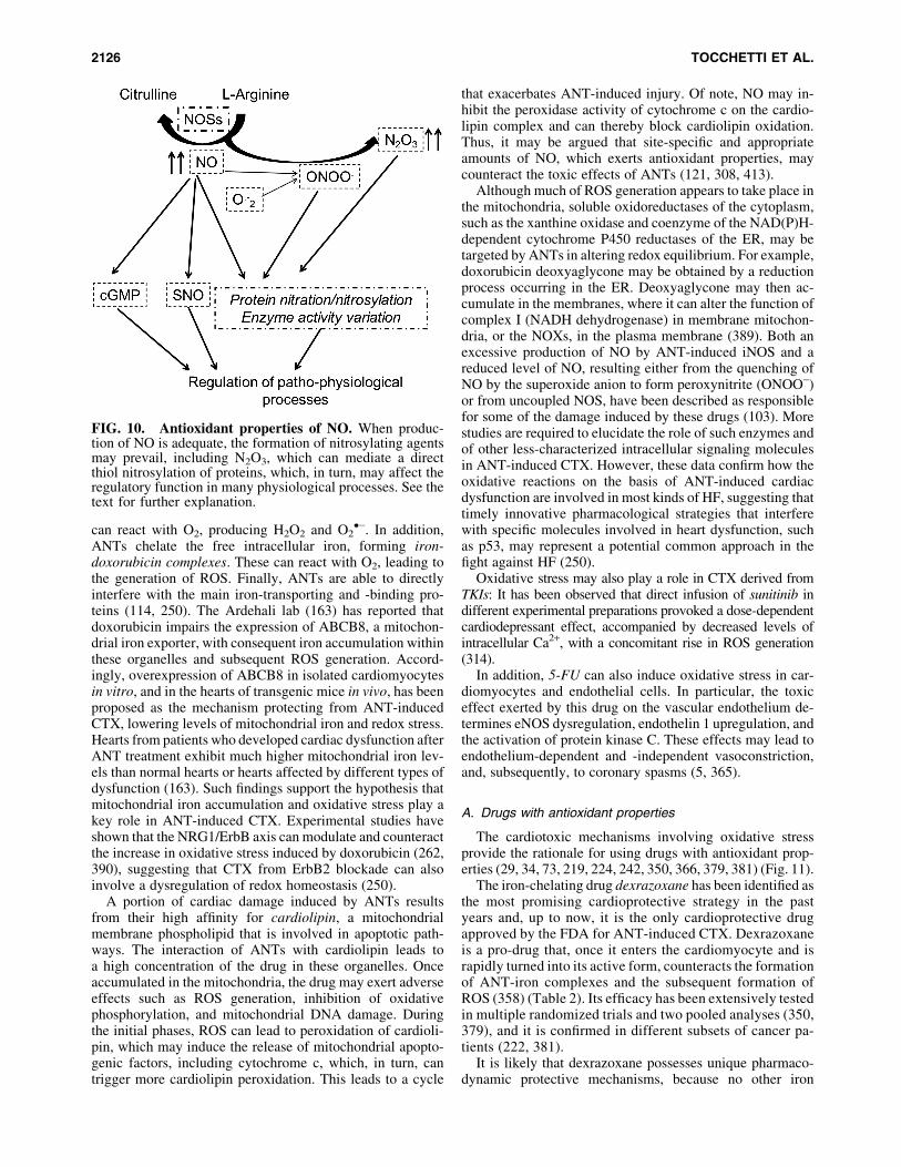
can react with O2, producing H2O2 and O2�-. In addition,
ANTs chelate the free intracellular iron, forming iron-doxorubicin complexes. These can react with O2, leading tothe generation of ROS. Finally, ANTs are able to directlyinterfere with the main iron-transporting and -binding pro-teins (114, 250). The Ardehali lab (163) has reported thatdoxorubicin impairs the expression of ABCB8, a mitochon-drial iron exporter, with consequent iron accumulation withinthese organelles and subsequent ROS generation. Accord-ingly, overexpression of ABCB8 in isolated cardiomyocytesin vitro, and in the hearts of transgenic mice in vivo, has beenproposed as the mechanism protecting from ANT-inducedCTX, lowering levels of mitochondrial iron and redox stress.Hearts from patients who developed cardiac dysfunction afterANT treatment exhibit much higher mitochondrial iron lev-els than normal hearts or hearts affected by different types ofdysfunction (163). Such findings support the hypothesis thatmitochondrial iron accumulation and oxidative stress play akey role in ANT-induced CTX. Experimental studies haveshown that the NRG1/ErbB axis can modulate and counteractthe increase in oxidative stress induced by doxorubicin (262,390), suggesting that CTX from ErbB2 blockade can alsoinvolve a dysregulation of redox homeostasis (250).
A portion of cardiac damage induced by ANTs resultsfrom their high affinity for cardiolipin, a mitochondrialmembrane phospholipid that is involved in apoptotic path-ways. The interaction of ANTs with cardiolipin leads toa high concentration of the drug in these organelles. Onceaccumulated in the mitochondria, the drug may exert adverseeffects such as ROS generation, inhibition of oxidativephosphorylation, and mitochondrial DNA damage. Duringthe initial phases, ROS can lead to peroxidation of cardioli-pin, which may induce the release of mitochondrial apopto-genic factors, including cytochrome c, which, in turn, cantrigger more cardiolipin peroxidation. This leads to a cycle
that exacerbates ANT-induced injury. Of note, NO may in-hibit the peroxidase activity of cytochrome c on the cardio-lipin complex and can thereby block cardiolipin oxidation.Thus, it may be argued that site-specific and appropriateamounts of NO, which exerts antioxidant properties, maycounteract the toxic effects of ANTs (121, 308, 413).
Although much of ROS generation appears to take place inthe mitochondria, soluble oxidoreductases of the cytoplasm,such as the xanthine oxidase and coenzyme of the NAD(P)H-dependent cytochrome P450 reductases of the ER, may betargeted by ANTs in altering redox equilibrium. For example,doxorubicin deoxyaglycone may be obtained by a reductionprocess occurring in the ER. Deoxyaglycone may then ac-cumulate in the membranes, where it can alter the function ofcomplex I (NADH dehydrogenase) in membrane mitochon-dria, or the NOXs, in the plasma membrane (389). Both anexcessive production of NO by ANT-induced iNOS and areduced level of NO, resulting either from the quenching ofNO by the superoxide anion to form peroxynitrite (ONOO-)or from uncoupled NOS, have been described as responsiblefor some of the damage induced by these drugs (103). Morestudies are required to elucidate the role of such enzymes andof other less-characterized intracellular signaling moleculesin ANT-induced CTX. However, these data confirm how theoxidative reactions on the basis of ANT-induced cardiacdysfunction are involved in most kinds of HF, suggesting thattimely innovative pharmacological strategies that interferewith specific molecules involved in heart dysfunction, suchas p53, may represent a potential common approach in thefight against HF (250).
Oxidative stress may also play a role in CTX derived fromTKIs: It has been observed that direct infusion of sunitinib indifferent experimental preparations provoked a dose-dependentcardiodepressant effect, accompanied by decreased levels ofintracellular Ca2+, with a concomitant rise in ROS generation(314).
In addition, 5-FU can also induce oxidative stress in car-diomyocytes and endothelial cells. In particular, the toxiceffect exerted by this drug on the vascular endothelium de-termines eNOS dysregulation, endothelin 1 upregulation, andthe activation of protein kinase C. These effects may lead toendothelium-dependent and -independent vasoconstriction,and, subsequently, to coronary spasms (5, 365).
A. Drugs with antioxidant properties
The cardiotoxic mechanisms involving oxidative stressprovide the rationale for using drugs with antioxidant prop-erties (29, 34, 73, 219, 224, 242, 350, 366, 379, 381) (Fig. 11).
The iron-chelating drug dexrazoxane has been identified asthe most promising cardioprotective strategy in the pastyears and, up to now, it is the only cardioprotective drugapproved by the FDA for ANT-induced CTX. Dexrazoxaneis a pro-drug that, once it enters the cardiomyocyte and israpidly turned into its active form, counteracts the formationof ANT-iron complexes and the subsequent formation ofROS (358) (Table 2). Its efficacy has been extensively testedin multiple randomized trials and two pooled analyses (350,379), and it is confirmed in different subsets of cancer pa-tients (222, 381).
It is likely that dexrazoxane possesses unique pharmaco-dynamic protective mechanisms, because no other iron
FIG. 10. Antioxidant properties of NO. When produc-tion of NO is adequate, the formation of nitrosylating agentsmay prevail, including N2O3, which can mediate a directthiol nitrosylation of proteins, which, in turn, may affect theregulatory function in many physiological processes. See thetext for further explanation.
2126 TOCCHETTI ET AL.
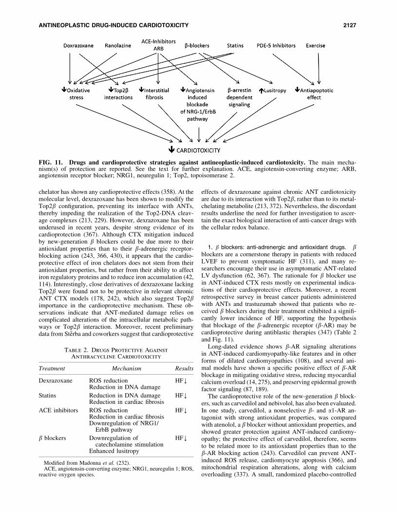
chelator has shown any cardioprotective effects (358). At themolecular level, dexrazoxane has been shown to modify theTop2b configuration, preventing its interface with ANTs,thereby impeding the realization of the Top2-DNA cleav-age complexes (213, 229). However, dexrazoxane has beenunderused in recent years, despite strong evidence of itscardioprotection (367). Although CTX mitigation inducedby new-generation b blockers could be due more to theirantioxidant properties than to their b-adrenergic receptor-blocking action (243, 366, 430), it appears that the cardio-protective effect of iron chelators does not stem from theirantioxidant properties, but rather from their ability to affectiron regulatory proteins and to reduce iron accumulation (42,114). Interestingly, close derivatives of dexrazoxane lackingTop2b were found not to be protective in relevant chronicANT CTX models (178, 242), which also suggest Top2bimportance in the cardioprotective mechanism. These ob-servations indicate that ANT-mediated damage relies oncomplicated alterations of the intracellular metabolic path-ways or Top2b interaction. Moreover, recent preliminarydata from Sterba and coworkers suggest that cardioprotective
effects of dexrazoxane against chronic ANT cardiotoxicityare due to its interaction with Top2b, rather than to its metal-chelating metabolite (213, 372). Nevertheless, the discordantresults underline the need for further investigation to ascer-tain the exact biological interaction of anti-cancer drugs withthe cellular redox balance.
1. b blockers: anti-adrenergic and antioxidant drugs. bblockers are a cornerstone therapy in patients with reducedLVEF to prevent symptomatic HF (311), and many re-searchers encourage their use in asymptomatic ANT-relatedLV dysfunction (62, 367). The rationale for b blocker usein ANT-induced CTX rests mostly on experimental indica-tions of their cardioprotective effects. Moreover, a recentretrospective survey in breast cancer patients administeredwith ANTs and trastuzumab showed that patients who re-ceived b blockers during their treatment exhibited a signifi-cantly lower incidence of HF, supporting the hypothesisthat blockage of the b-adrenergic receptor (b-AR) may becardioprotective during antiblastic therapies (347) (Table 2and Fig. 11).
Long-dated evidence shows b-AR signaling alterationsin ANT-induced cardiomyopathy-like features and in otherforms of dilated cardiomyopathies (108), and several ani-mal models have shown a specific positive effect of b-ARblockage in mitigating oxidative stress, reducing myocardialcalcium overload (14, 275), and preserving epidermal growthfactor signaling (87, 189).
The cardioprotective role of the new-generation b block-ers, such as carvedilol and nebivolol, has also been evaluated.In one study, carvedilol, a nonselective b- and a1-AR an-tagonist with strong antioxidant properties, was comparedwith atenolol, a b blocker without antioxidant properties, andshowed greater protection against ANT-induced cardiomy-opathy; the protective effect of carvedilol, therefore, seemsto be related more to its antioxidant properties than to theb-AR blocking action (243). Carvedilol can prevent ANT-induced ROS release, cardiomyocyte apoptosis (366), andmitochondrial respiration alterations, along with calciumoverloading (337). A small, randomized placebo-controlled
FIG. 11. Drugs and cardioprotective strategies against antineoplastic-induced cardiotoxicity. The main mecha-nism(s) of protection are reported. See the text for further explanation. ACE, angiotensin-converting enzyme; ARB,angiotensin receptor blocker; NRG1, neuregulin 1; Top2, topoisomerase 2.
Table 2. Drugs Protective Against
Anthracycline Cardiotoxicity
Treatment Mechanism Results
Dexrazoxane ROS reduction HFYReduction in DNA damage
Statins Reduction in DNA damage HFYReduction in cardiac fibrosis
ACE inhibitors ROS reduction HFYReduction in cardiac fibrosisDownregulation of NRG1/
ErbB pathway
b blockers Downregulation ofcatecholamine stimulation
HFY
Enhanced lusitropy
Modified from Madonna et al. (232).ACE, angiotensin-converting enzyme; NRG1, neuregulin 1; ROS,
reactive oxygen species.
ANTINEOPLASTIC DRUG-INDUCED CARDIOTOXICITY 2127
clinical trial on the cardioprotective effect of prophylactic-use carvedilol in patients undergoing ANT chemotherapyshowed a reduced incidence of LV systolic and diastolicdysfunction in the carvedilol group (181). However, strongclinical evidence of the cardioprotective effect of carvedilolis still lacking.
Nebivolol is a cardio-selective b blocker with limitedvasodilating properties related to its interaction with the l-arginine/NO pathway. In an experimental model of ANT-induced CTX, treatment with nebivolol, and to a lesser extentwith carvedilol, led to an improvement in LV function as-sociated with increased NO levels and a significant reduc-tion in oxidative stress (71, 393, 394). A small, randomizedplacebo-controlled study on the prophylactic use of nebivololin patients undergoing ANT chemotherapy showed, at the6-month follow-up, a reduced incidence of LV dilatationand systolic dysfunction in the nebivolol group comparedwith the placebo group (183). In addition, by enhancingprosurvival ERK signaling after ErbB2 inhibition (87), bblockers can also prevent trastuzumab-related CTX (Ta-ble 3); b blockers are now studied in a series of clinical trials
such as the MANTICORE 101 and the SAFE trials (33). Assaid, recent data from the PRADA trial indicated that the b1blocker metoprolol was not cardioprotective (128), thussupporting the use of unbiased b1 and b2 blockers (382).
In summary, different b blockers are not similarly effectivein treating CTX. The molecular and cellular mechanismsby which b blockers may confer cardioprotection are notcompletely clear. To date, a cardioprotective treatment withb blockers should still demonstrate their efficacy in largeclinical trials. In addition, in clinical practice, use of bblockers is hampered by their side-effects in an already-fragile patient, suggesting their use only in patients with ahigh CTX risk.
2. The redox role of renin-angiotensin-aldosterone systemantagonists: ACE-Is and ARBs. Many studies have founda key role for the renin-angiotensin-aldosterone system(RAAS) in modulating ANT-induced CTX (13). ACE-Is andARBs have already been shown to exert positive effects inreducing the progression of LV dysfunction and in preventingHF in asymptomatic high-risk patients (311).
Table 3. Prevention Strategies for Cardiac Dysfunction Induced by the Main Classes
of Antineoplastic Drugs
Anticancer agents Prevention strategy Rationale/mechanisms of action
Anthracyclines Lowering of lifetime cumulative doses Dose-dependent cardiotoxicityUsage of epirubicin, less cardiotoxic
than doxorubicinAvoiding bolus, preferring infusional
schedulesLiposomal formulationsDexrazoxane Lowering oxidative stress, preventing the
interactions between anthracyclinesand Top2b
ACE-1, ARBs Lowering oxidative stress, reducinginterstitial fibrosis, enhancing intracellularcalcium handling, cardiomyocyte metabolism,and mitochondrial function
b blockers Preserving EGF signaling via a b-arrestin-dependent mechanism, lowering oxidativestress, improving lusitropy
HMG-CoA reductase inhibitors Inhibiting Top2b-mediated DNA damage,antioxidant, and anti-inflammatory effects
Ranolazine Lowering oxidative stressPDE-5 inhibitors Anti-apoptotic effectsPhysical exercise Lowering oxidative stress and pro-apoptotic
signaling, enhancing calcium handling, andenhancing myocardial energetics
Anti-ErbB2 ACE-1, ARBs Decrease in angiotensin-induced blockade ofNRG1/ErbB pathway
b-blockers Stimulating b-arrestin-dependent signaling, withsubsequest activation of ErbB2-mediatedcardioprotective pathways
Anti-angiogenicdrugs (sorafeniband sunitinib)
Anti-hypertensive treatment (with ACE-1and ARBs, b blockers, dihydropyridinecalcium channel blockers, diuretics)
Counteracting hypertension-dependent cardiacdamage
Metformin Enhancing myocardial energetics via activationof cAMP-kinase
Modified from Cadeddu et al. (33).ARBs, angiotensin II receptor blockers; cAMP, cyclic adenosine monophosphate; EGF, epidermal growth factor; HMG-CoA,
hydroxymethylglutaryl-coenzyme A; PDE-5, phosphodiesterase-5; Top2, topoisomerase 2.
2128 TOCCHETTI ET AL.
The rationale for using RAAS blockade drugs in thissetting is based on the high risk of developing myocardialdysfunction after ANT treatment. Experimental studieshave shown the efficacy of ACE-Is in countering ANT-induced CTX, both acute high dose (1) and single low dose(24). The underlying mechanisms of their positive effectagainst ANT-related CTX contribute to neutralizing ROSdamage, reducing interstitial fibrosis, avoiding intracellularcalcium overload, and improving mitochondrial respirationand cardiomyocyte metabolism (1, 24) (Table 2 andFig. 11). Indeed, activation of the AT1R produces directadverse CV effects by stimulating NADPH oxidase, whichis involved in the genesis of oxidative stress, and initiatingMAPK, related to cell growth, inflammation, hypertrophy,and apoptosis (25).
Experimental animal models of acute and chronic ANT-induced cardiomyopathy showed a cardioprotective role forprophylactic administration of enalapril, captopril, and lisi-nopril (1). In a study of 115 patients treated with ANTs,enalapril was shown to significantly reduce the incidence ofLV dysfunction in comparison with a placebo (36).
Candesartan has been shown to treat experimental car-diotoxicity already induced by ANTs (364), whereas pre-and post-treatment with telmisartan protected againstacute doxorubicin-induced cardiotoxicity in rats (164).Telmisartan is the only ARB-modulating peroxisomeproliferator-activated receptor-c, and, therefore, affecting thebioavailability of NO, and inhibiting inflammatory moleculessuch as interleukin 6 (IL-6) and tumor necrosis factor (428).In a small prospective study, telmisartan was shown to miti-gate subclinical cardiotoxic effects (changes in strain rate)of epirubicin, with IL-6 and ROS increasing significantly inthe placebo arm but not in the telmisartan arm (34). Telmi-sartan could reverse acute (early) EPI-induced myocardialdysfunction and maintain a normal systolic function tillthe 18-month follow-up (73, 74). IL-6 and ROS increasedsignificantly in the placebo arm but did not change in thetelmisartan arm. In another study, valsartan showed a cardi-oprotective effect on acute cardiotoxicity in patients treatedwith high doses of ANTs (274).
Recent clinical trials have shown that a combination ofACE-Is and b blockers is beneficial in treating ANT-inducedCTX (23). Many other ongoing clinical trials are evaluatingthe role of ACE-Is and ARBs as cardiopreventive agents thatmay act by decreasing angiotensin-induced blockade of theneuregulin 1/ErbB pathway.
Finally, a recent meta-analysis showed that the prophy-lactic administration of ACE-Is and ARBs in patients onANT therapy was associated with a reduced relative risk ofdeveloping CTX compared with patients on a placebo (180).To date, however, cardioprotective treatment with ACE-Isor ARBs still requires a demonstration of efficacy in largeclinical trials.
In addition, in patients with anti-angiogenic drug-inducedhypertension, nondihydropyridine calcium channel blockersare not indicated, because of the pharmacokinetic interactionof sorafenib and sunitinib with CYP3A4 (33, 236). Thus,acknowledging the other mechanisms that are responsible forsunitinib-induced CTX, that is, disrupted mitochondrialfunction and impaired myocyte energy homeostasis, ACE-Isand b blockers (85, 106) could form an effective cardiopro-tective strategy (Table 3). Unfortunately, no clinical study to
date has shown a role for these drugs in preventing sunitinib-induced CTX (33).
B. Novel strategies in cardioprotectionagainst ANT-induced CTX
Several CV drugs have been tested as potentially effectivein counteracting ANT-induced CTX, and particularly statins,ranolazine, and phosphodiesterase-5 inhibitors.
Lovastatin and fluvastatin, hydroxymethylglutaryl-coenzymeA reductase inhibitors, showed a cardioprotective effect inexperimental studies (159, 320). Of note, these data derivefrom acute, subacute experimental cardiotoxicity studies andcell-based studies performed on proliferating H9c2 celllines, not cardiomyocytes. Therefore, effects of ANTs in thissetting deserve caution since these drugs possess strong anti-proliferative action besides cardiotoxic effects. The effects ofthe statins on the Top2b have not been directly demonstratedin the studies, just indirectly suggested to be Top2b related.Besides these caveats, LV systolic dysfunction induced bydoxorubicin was not affected by lovastatin (142). Only smallclinical studies have evaluated the effects of statins in patientstreated with ANTs (48, 348), reporting only minor positiveresults. Unfortunately, the study from the Marwick group(348) was a retrospective observational analysis, and evenChotenimitkhun et al. (48) do not appear to have conducted astandard controlled randomized trial. Therefore, larger studiesare, therefore, required to confirm these findings.
Late INa inhibition with ranolazine has been proposed as acardioprotective strategy in several experimental models ofcardiac dysfunction (316, 391, 394). Ranolazine was shownto preserve heart function in mice treated with ANTs by re-ducing oxidative stress. The inhibition of elevated [Na+]i
levels with ranolazine would prevent calcium overload andthe occurrence of oxidative damage by suppressing ROSgeneration, offering an advantage over conventional antioxi-dant strategies that counteract ROS after their production (196).The INTERACT study (259), designed to assess whether ra-nolazine could prevent ANT-induced diastolic dysfunction,indicated that ranolazine was a promising cardio-oncologicdrug, but more convincing data are still required.
Phosphodiesterase-5 inhibitors have also been tested ascardioprotective agents. Sildenafil demonstrated cardiopro-tective activity against ANT-induced LV dysfunction byopening mitochondrial KATP channels, preserving mito-chondrial membrane potential and myofibrillar integrity, andpreventing cardiomyocyte apoptosis (100). Tadalafil attenu-ated ANT-induced cardiomyopathy through NO-mediatedincreases in cGMP levels (177, 197).
Finally, redox mechanisms have also been proposed for theability of NRG1 to limit antiblastic drug-induced damage inadult rat cardiomyocytes (390).
Another redox compound attracting the interest of re-searchers and cardio-oncologists is hydrogen sulfide (H2S),whose role in cancer is still poorly understood. The dis-coveries that ROS-induced inhibition of cystathioninegamma-lyase, a key enzyme in the synthesis of H2S, is in-volved in ANT-induced CTX in H9c2 cardiomyocytes, andthat the administration of exogenous H2S can confer pro-tection against this CTX, have suggested a new tool withantioxidant properties in therapeutic strategies against an-tineoplastic drug-induced CTX (33, 247, 295). Further
ANTINEOPLASTIC DRUG-INDUCED CARDIOTOXICITY 2129
research in appropriate animal models is required, as well asmulti-centric randomized trials that compare the efficacy ofthese new compounds with that of already approved cardi-oprotective drugs.
Experimental studies have shown that sunitinib inhibitsAMPK activity, whereas restoration of this enzyme’s activityinhibits cell death (186). It has been speculated that promotingAMPK activity with metformin may reduce sunitinib-inducedCTX. Although metformin can counter heart dysfunction indifferent experimental models of cardiac injury (53, 129)(Table 3), an in vitro study that focused on sunitinib-relatedCTX did not find any beneficial effect of metformin (33). Asmentioned earlier, a recent study (98) demonstrated thatTrkB, a tyrosine kinase receptor, and its endogenous li-gand BDNF could directly modulate the cardiac excitation-contraction coupling, independently or in parallel withGPCR-mediated signaling, supporting the concept that inhi-bition of the tyrosine kinase receptor with antineoplastic drugscan interfere with fundamental pathways, damaging cardiaccontractility (106). At the same time, this study highlightsBDNF as an alternative potential therapeutic strategy for HF.
C. Nonpharmacologic strategies
Apart from pharmacologic interventions, we should alsoconsider that nutritional supplementation and exercisetraining may have important antioxidant properties (8, 139,192, 345, 346, 359, 369).
Dietary supplementation of antioxidants proved to be ableto alleviate doxorubicin cardiotoxicity in animal models (8,426). However, there is still scant evidence on both quality andsample size, suggesting that some antioxidant supplementa-tions may lower ANT-induced CTX (109). Nonpharmacologicstrategies for countering ANT-induced CTX include lifestyleinterventions and exercise ‘‘prehabilitation,’’ a type of pre-ventive exercise rehabilitation. It has been speculated thataerobic exercise can decrease ROS generation, inhibiting pro-apoptotic signaling, restoring proper calcium cycling, andstimulating the AMPK pathway, thus improving myocardialenergetics (345) (Table 3). In addition, exercise can have apositive impact on CV risk factors, such as hypertension, highcholesterol and lipids, overweight and obesity, and high bloodglucose or diabetes (192). In cancer survivors, a short period ofmixed aerobic and resistance exercise builds tolerance andflexibility in physical activity (369).
Although several studies have shown a role for aerobic ex-ercise in countering ANT (343)- and trastuzumab (139)-relatedCTX, additional work is needed to better characterize the ef-fects of training on antineoplastic drug-induced CTX (346).
D. Biomarkers of oxidative/nitrosative CTX
Since the importance of ROS/RNS production as earlymediators of chemotherapy-related CTX has been ascer-tained, we should identify useful circulating biomarkers ofoxidative/nitrosative CTX. Biomarkers with redox signifi-cance include the metabolomic identification of acetate andsuccinate (8). A decrease in NAD(P)H:quinone oxidoreduc-tase 1 activity and an increase in ROS generation byNAD(P)H oxidases have been considered early biomarkersof ANT-induced CTX (283). A significant variation in IL-6,IL-6R, and ROS has been observed together with a change inthe enzymatic activity of GPx, which was correlated with an
early variation in longitudinal systolic function in patientsafter the administration of epirubicin (73, 74).
Other potential CTX markers with redox importance underinvestigation in cardiology are high-sensitivity C-reactiveprotein, heart-type fatty acid-binding protein (H-FABP),glycogen phosphorylase BB (GPBB), and circulating mi-croRNAs, especially microRNA-146a (miR-146a). These areconsidered new potential markers for the diagnosis and riskstratification of acute coronary syndromes, and they may behelpful in the early detection of anti-cancer CTX; however, theclinical data available in the chemotherapy-induced CTX sce-nario remain insufficient (131, 152–155, 371, 419). For a dis-cussion on other biomarkers, see section XI.D.
XI. Early Detection, Monitoring, and Managementof Heart and Vascular Toxicities
A. Cardioncology: an expanding sciencefor an expanding problem
Assessing CTX from antineoplastic drugs is crucial.Screening for CTX at a preclinical stage is essential wheninvestigating the effects of TKIs. Many of these drugs causean increased risk of arrhythmia; thus, most of the preclinicalwork focuses on hERG (K+ channel). A major limitation ofin vitro work is that primary myocyte cultures of animal orhuman hearts dedifferentiate and die after a short while.Hence, assays based on stem cells and on the use of en-gineered heart tissue may better reflect in vivo settings. Suchexperimental studies could integrate assessment of severaleffects, including those on membrane action potential, cal-cium cycling, and myofilament function, as well as those ongene expression and cell survival (69, 88, 236, 376).
In clinical practice, most of the methods used to assessheart function are often inaccurate, and they may also mis-lead physicians to stop potentially lifesaving therapies. Ofnote, patients with advanced cancer may already exhibitpotential confounder symptoms mimicking anti-cancerdrugs’ side-effects. Moreover, complications such as myo-cardial ischemia, arterial hypertension, and arrhythmias arereadily recognized, but identifying mild LV dysfunction(especially at a subclinical stage) can be more challenging(376). In this context, a prompt and sensitive monitoring ofCV function plays a pivotal role.
In addition, the time point when CTX can become clinicallyovert varies substantially. Several antineoplastic drugs (i.e.,ANTs) can induce progressive cardiac remodeling and latecardiomyopathy, whereas others (i.e., trastuzumab) can producetransient LV dysfunction without known long-term conse-quences (376, 432). Furthermore, the inability to exactly predictthe long-term consequences of tumors leads to a false diagnosisof CV complications. Owing to all these concerns, cardioncol-ogy, a new branch of CV sciences, recently developed, with theaim of preventing, identifying, and treating CV complicationsof cancer therapy, optimizing clinical status for cardiac patientswho need potentially cardiotoxic treatment, facilitates in anyway cancer therapy in patients with CV complications andsurveilles for late side-effects in cancer survivors.
B. Diagnostic tools for CTX detection
The most common clinical diagnostic tools for thescreening and detection of CTX are biomarkers and cardiac
2130 TOCCHETTI ET AL.
imaging, including echocardiography, multiple-gated acqui-sition (MUGA) scan, and cardiac magnetic resonance (CMR)(Table 4). Nevertheless, an ECG is recommended for allsubjects before and during therapies, particularly for detect-ing arrhythmias and QT interval prolongation. Moreover,subjects on pyrimidine analogs should be monitored closelyfor cardiac ischemia by regular ECGs, and chemotherapeutictreatments should be withheld if ischemia is observed (432).
C. LVEF and emerging modalities for assessingmyocardial damage: some claryfing points
LVEF is the most frequently used parameter for assessingmyocardial function, and echocardiography and MUGA scansare largely employed in clinical practice for this purpose (40,41, 62, 171, 206, 234, 299, 307, 376, 432, 438). An LVEFdecrease (>10%) to a value <50% identifies patients with CTXin both imaging modalities (307, 432). Compared with echo-cardiography, MUGA provides the advantage of a high re-producibility, but it is hampered by significant disadvantages,such as cumulative radiation exposure and no data on othercardiac structures (299, 432). However, the choice of modal-ities for an early detection and monitoring of CTX depends onlocal expertise and availability, being careful to use the same
imaging modality with the best reproducibility for continuedscreening throughout the treatment period (432). Imagingtechniques that provide additional relevant clinical information(e.g., right ventricular function, pulmonary pressures, valvularfunction, pericardial evaluation) and high-quality radiation-freeimaging are preferred (307, 432). Confirming LVEF decreaseby repeated cardiac imaging (preferably by echocardiography)2–3 weeks after the baseline initial decrease in LVEF is stronglyrecommended, as well as categorizing LVEF reduction asasymptomatic or symptomatic, and repeating echocardiographymeasurements during follow-up (432). Repeated MUGA is notrecommended as a first-line option, and it should only be con-ducted if the radiation exposure is less than five mSv (432).
On the other hand, echocardiographic measurements,though simple, noninvasive, and readily available, are oftenvariable and of a low sensitivity (290), because of the seriousdamage and myocyte loss, leading to an observable reductionin LVEF (92, 416). On these grounds, novel indices of ven-tricular function are being introduced (37, 146, 150, 206,386). Speckle tracking echocardiography has shown thehighest sensitivity in detecting early alterations in LV func-tion associated with future development of cardiomyopathy(134, 146, 306, 307, 395, 438). In particular, a global longi-tudinal strain (GLS) >15% relative percentage reduction
Table 4. Assessement of Anti-Cancer Cardiotoxicity
Technique Diagnostic criteria Advantages Disadvantages
Echocardiography LVEF: >10% decrease with a value Availability Reproducibility1. LVEF <50% identify patients with
cardiotoxicitySafety Image quality
2. GLS GLS: >15% relative percentagereduction from baseline mayidentify patients at risk forcardiotoxicity
Comprehensive cardiacassessment
GLS: Intervendorvariability; low specificity
Ideal for serial evaluationsLow costGLS: Sensitivity
(early CTX)Nuclear cardiac
imaging (MUGA)>10% decrease in LVEF with a
value <50% identifies patientswith cardiotoxicity
Reproducibility Cumulative radiation exposureNo information on other
cardiac structures andpericardium
Not recommended forserial examinations
Cardiac magneticresonance
Useful if other techniques arenondiagnostic or to confirm thepresence of LV dysfunction inborderline LVEF value
Accuracy, reproducibility Limited availabilityRV and pericardium
assessmentPatient adaptation
Detection of edema,inflammation, andfibrosis (early andlate CTX)
General contraindicationsHigh costLow feasibility for serial
examinationsArterial stiffness
measurementCardiac biomarkers
Troponin I Any increase identifies pts whomay benefit from ACE-Is
Accuracy,reproducibility
Poor evidence for subtle rises
Hs Troponin I New elevation of Hs-Troponin Ipredicts LV dysfunction
Wide availability Variability with different assays
BNPa High sensitivity Unclear role for routinesurveillance
NT-proBNPa
Modified from Zamorano et al. (432).aMore useful for their negative predictive values.CTX, cardiotoxicity; BNP, B-type natriuretic peptide; GLS, global longitudinal strain; LVEF, left ventricle ejection fraction; MUGA,
multiple-gated acquisition; pts, patients.
ANTINEOPLASTIC DRUG-INDUCED CARDIOTOXICITY 2131
from baseline can identify patients who are at risk for CTX(97, 172, 307, 432).
Other imaging modalities, such as CMR, appear very prom-ising, although their availability is still limited (97, 238, 290, 299,388, 415). CMR is the gold-standard technique for LVEF esti-mation and can identify myocardial edema, inflammation, cap-illary leak, and fibrosis, enabling an accurate diagnosis of earlyand late CTX (239, 388, 432). Other recent strategies includemeasuring uptake of iodine-124-metaiodobenzylguanidine, aradiolabeled analog of norepinephrine, which is reduced aftermyocardial insults (145, 239, 415). In addition, antimyosinantibodies that detect myosin exposed after cardiac damagecould be useful (235, 239, 401, 402), whereas the uptake ofradiolabeled chemotherapeutics could be used as a predictor ofCTX (18, 239, 301).
D. Cardiac biomarkers
Troponins and natriuretic peptides can be correlated withfindings from cardiac imaging for the early detection of CTX(283, 292). Natriuretic peptide-level elevations were dem-onstrated to predict the development of future CTX in pa-tients with NHL and with breast cancer (99, 119). PersistentB-type natriuretic peptide (BNP) rises, after exposure toANTs, were more predictive of following LV dysfunctionthan transient elevations (327, 335). However, the utility ofNPs for predicting anticancer drug-induced CTX is stillcontroversial. Indeed, several studies did not confirm aprognostic value for BNP levels, especially when this bio-marker was used alone (66, 340, 378). It has been observedthat troponins I and T can predict late ANT-induced CTX inchildren (223) and in adult patients undergoing high-dosechemotherapy. Not only the increase but also the pattern ofthe elevation, and particularly a persistent increase after 1month since treatment, added prognostic information. Con-versely, persistently negative TnI identified low-risk patientswho did not need a strict cardiological follow-up (38). Pa-tients with troponin increase may benefit from early cardio-protective treatment with ACE-Is (36). In patients treatedwith trastuzumab, especially after previous exposure toANTs, troponin I elevation can identify those who will de-velop LV dysfunction and who will not recover in spite oftreatments for HF (37). High-sensitivity troponin I is moresensitive in the evaluation of subclinical depletion of cardiacphysiologic reserve, and a new elevation of this markerpredicted subsequent LV dysfunction in patients receivinga conventional dose of ANTs and/or trastuzumab (203). Thecombination of high-sensitivity troponin with GLS predictedthe risk to develop future cardiotoxicity with increased sen-sitivity (93%) and negative predictive value (91%) compared
with the evaluation of a single parameter in patients withbreast cancer (340). Therefore, ‘‘cardiac biomarkers pluscardiac imaging’’ should be encouraged to detect CTX early.There are still scant data supporting the use of biomarkers topredict future cardiac dysfunction due to immune and tar-geted cancer therapies. Few data indicated an increase oftroponin in subjects administered sunitinib and sorafenibfor metastatic renal cell carcinoma, who developed ventric-ular function impairment (344). Several other biomarkershave been studied or are under investigation to stratify therisk of cardiotoxicity. Myeloperoxidase, an index of oxida-tive stress, measured after ANT administration, seems tocorrelate with subsequent development of CTX (203).FABP and GPBB have also been proposed as novel bio-markers of chemotherapy-mediated cardiac damage, whichare even more precocious of troponin and BNP (86, 152,153). MicroRNAs have emerged as an attractive tool todetect myocardial injury. miR-146a may induce down-regulation of ErbB4 and subsequent stimulation of apoptoticpathways. Preclinical studies have shown increased levelsof miR-146a after doxorubicin administration (155). How-ever, more trials with long-term follow-up are needed tovalidate these data.
The use of cardiac biomarkers in the prediction of cardi-otoxicity has not only several advantages (accuracy, repro-ducibility, high sensitivity, wide availability, and relativelylow cost) but also limitations, for example, inter-assay vari-ations, insufficient evidence to establish specific cutoffs forenzyme elevations in this setting, and uncertainty in thetiming of blood sampling (432). Standardization in the as-sessment of cardiac toxicity using biomarkers is needed, andmulti-center trials are strongly required before establishingmore clear recommendation (376). Table 5 summarizes themain biomarkers that are evaluated for the detection of car-diac toxicity.
E. Detection of vascular complications:old and new strategies
Structural endothelial damage, endothelial dysfunction,acceleration of atherosclerosis, prothrombotic actions, andvascular inflammation are some of the underlying mechanismsof cancer therapy-related changes in vessel function. VEGFinhibitors, TKIs (such as nilotinib, ponatinib, dasanitib), cis-platin, 5-FU, and ANTs can induce hypertension, ischemia,and TE through their endothelial toxicity. CAD, stroke, PAD,PAH, arterial/venous TE disease, and nephrotoxicity are themain cinical presentations of vascular damage by cancertherapy. In particular, nuclear cellular actions by ANTs mayadversely modulate collagen turnover and vascular wall
Table 5. Cardiac Biomarkers to Detect Cardiac Toxicity
Cardiac biomarkers Typical mechanisms of release Anti-cancer drugs
Troponins (troponin I and T) Ischemia, inflammation, andoxidative stress
Anthracyclines; anthracycline-adjuvant trastuzumab
Natriuretic peptides (BNP pro-BNP) Increase in myocardial wallstress and LV filling pressure
Anthracyclines; various high-dose chemotherapy protocols
Fatty acid-binding protein (FABP) Ischemia and necrosis AnthracyclinesGlycogen phosphorylase BB (GPBB)Circulating microRNAs (miR-146a) Altered homeostasis Anthracyclines (preclinical studies)
2132 TOCCHETTI ET AL.
homeostasis, leading to acute damage (endothelial dysfunc-tion, increase in smooth muscle tone) and chronic damage(atherosclerosis, increased collagen synthesis), and resulting inan impaired CV coupling (81, 84, 273). Age and CV riskfactors may influence the extent of cardiac dysfunction(Fig. 12). VEGF inhibitors increase vascular tone due to adecrease in NO production, increased peripheral resistancefrom endothelial damage (sloughing) and dysfunction, andcapillary rarefaction (96). Cisplatin can induce myocardialischemia through an increased procoagulant status.
For an early detection of vascular complications, current re-ccomendations (432) simply suggest monitoring CV risk factors,assessing patient functional status, measuring blood pressurebefore cancer treatment, and optimizing anthypertensive therapyduring treatment with a goal of <140/90 mmHg (particularly inpatients treated with VEGF inhibitors). Moreover, the identifi-cation of patients with pre-existing CAD in those undergoing 5-FU is of paramount importance since this drug substantiallyincreases the risk of developing myocardial ischemia with avasospam-recognized mechanism. In addition, it should beconsidered that clinical factors (cancer-, treatment-, and patientrelated) could be related to an enhanced risk of venous TE insubjects administered TKIs. Nilotinib, ponatinib, and BCR-ABLTKIs used in treating CML are, instead, associated with PADand stroke. The evaluation of PAD risk at baseline (risk factorassessment, clinical examination, ankle-brachial index mea-surement) is, therefore, recommended in these patients (432).Moreover, cerebrovascular ultrasound screening is indicated insubjects who are irradiated for head and neck cancer or lym-phoma, especially beyond 5 years after irradiation (432).
However, to date, a common standard approach that isuseful in clinical practice for early detection of vascularcomplications such as atherosclerosis is missing.
The assessment of arterial stiffness, which increases earlyin the atherosclerotic process, to provide vasculotoxic pro-files of chemotherapeutic agents seems to be very proimising.These could be useful in the future in improving therapeuticstrategies (84, 179, 273, 425). Drafts et al. found, by mag-netic resonance imaging of the aorta, an early (*3 months)and abrupt increase of pulse wave velocity, a marker of ar-terial stiffness, which was dose-, age-, and CV risk factorindependent in patients treated with ANTs; furthermore, this
increase was accompanied by a subclinical reduction inLVEF and strain (81). This is one of the first demostrationsof early atherosclerosis with associated impaired ventricular-arterial coupling in cancer survivals. More recently, a markedstiffening of large elastic arteries was demonstrated by ap-planation tonometry also in patients undergoing VEGF in-hibitors (265). Accordingly, ACE-Is should be preferredbecause of their anti-stiffening properties in the aorta in hy-pertensive patients treated with ANTs and VEGF inhibitors.
F. Timing of clinical evaluation and follow-up:the importance of risk factors assessment
To date, no algorithm has been established for evaluationand follow-up of patients before, during, and after cancertherapies. However, a strong collaboration between cardi-ologists and oncologists is required to monitor and treat on-cology patients (3, 432). Of course, the cardio-oncology teammust be equipped appropriately to address the most commonquestions (i.e., CTX prediction, prevention strategies, high-riskpatient monitoring, management strategies) with an evidence-based approach (410, 425).
Long-term surveillance of cancer survivors with a poten-tial for late-onset CV complications should involve cardio-oncology teams. According to recent clinical guidelines oncancer treatment and CV toxicity, the first step in cardio-protection should be the identification of subjects at a higherrisk; this depends on several factors: cumulative dose, bodymass index >30 (especially for anti-HER2 compounds), age(elderly and pediatric population treated with ANTs) (199,376, 380), concomitant or previous radiation therapy, previ-ous cardiotoxic cancer treatment, pre-existing conditionssuch as cardiovascular disease (CVD), demographic andother CV risk factors, and lifestyle and genetic risk factorsthat may predispose patients to CTX at lower ANT doses (37,50, 432). Although recent experimental studies from Moulinand collaborators (271) seem to suggest the opposite, femalepatients appear to be more susceptible to doxorubicin damage(222–224).
Gender-specific pharmacokinetic differences or even a de-gree of protection conferred by androgens could be responsiblefor this phenomenon. It has been observed that testosterone canreduce ANT-induced damage in cardiac myocytes in vitro (6).
G. Drug-specific timing for CTX monitoringand treatment
The exact timing and frequency of imaging and/or bio-marker sampling will depend on the specific antineoplastictherapy, total cumulative dose, treatment protocol and dura-tion, and baseline CV risk (432).
1. ANTs. Doxorubicin and epirubicin can induce acuteCTX (including ECG changes, supraventricular and ven-tricular arrhythmias, or transient LV dysfunction), which isusually reversible, develops in <1% of patients immediatelyafter infusion of ANTs; early or subacute cardiotoxic ef-fects, occurring within the first year of treatment, and lateeffects typically are those manifesting after a median of7 years. Early or subacute CTX, the most frequent, requiresa baseline evaluation and further assessment at the end ofthe treatment, particularly in the presence of an enhanced riskfor CTX, or with consecutive treatment with potentially
FIG. 12. Simplified algorithm showing how cancertherapy alone or together with demographic and con-ventional risk factors may determine cardiac damage;both factors can increase myocardial and arterial stiffness,thus impairing ventricular-vascular coupling and, finally,leading to HF, with either decreased ejection fraction (EF)or preserved EF (HFPEF). CV, cardiovascular.
ANTINEOPLASTIC DRUG-INDUCED CARDIOTOXICITY 2133
cardiotoxic-targeted therapeutics. For higher-dose ANT-containing regimens, and in subjects with a high baselinerisk, earlier evaluation of heart function after a cumulativetotal doxorubicin (or equivalent) of 240 mg/m2 should beconsidered (432).
2. Anti-ErbB2. Since patients receiving anti-ErbB2therapeutics frequently also receive ANTs, a baseline eval-uation is always recommended. Usually, cardiac assessmentis performed every 3 months during and once after comple-tion of anti-ErbB2 therapies. An improvement in the earlydetection of cardiac dysfunction with troponins and GLSmeasurements every 3 months during adjuvant trastuzumabtreatment has been demonstrated. Troponin dosage with ev-ery cycle may be considered in patients with a high baselinerisk (variability in the timing of trastuzumab-induced cardiacdysfunction) (432).
3. VEGF inhibitors. The best timing of surveillancestrategies still needs to be clarified. There is high variabilityin the time of occurrence (early after treatment and delayedfor several months). Currently, it is reasonable to considerevaluations every 6 months initially until stability in valuesof LVEF is achieved. With a high baseline risk, it is betterto perform early clinical assessment in the first 2–4 weeksafter starting targeted therapies. There is limited evidenceto support any specific surveillance strategy. In addition,treatments with anti-VEGF inhibitors (236) pose a high risk(11%–45%) of inducing new hypertension or destabilizingpreviously controlled hypertension, including severe hy-pertension in 2%–20% of cases (255, 432). Monitoringblood pressure is, therefore, essential, and susceptible pa-tients should preferably be treated with ACE-Is, ARBs, or bblockers instead, considering the beneficial role of thesedrugs in HF (236, 370, 376).
With regards to therapeutic implications, beyond anthy-pertensive treatment, eventual standard HF therapy can beadministered (62, 376, 392), and with good life expectancy,aggressive treatments with LV assist devices can also beconsidered (322, 376). To prevent and treat vascular com-plications, such as PAD, antiplatelet therapy is recommended(432). Significant stenosis (e.g., those of carotid arteries) mayneed stenting or surgery. Targeted therapy for PAH can beused temporarily or permanently. Management of QT pro-longation is usually dependent on correction of the predis-posing factors (e.g., concomitant electrolyte abnormalities,QT-prolonging drugs). Commonly, an individualized ap-proach for the management of atrial fibrillation is needed, anddecisions on rate or rhythm control should be patient centredand symptom directed. Anticoagulation for venous TE pre-vention in patients with cancer should always be consideredaccording to the patient’s bleeding risk and life expectancy(432).
XII. Conclusions and Perspectives
Antineoplastic therapies have led to a prolonged life ex-pectancy for many patients, but CTX is a relatively commonadverse effect of these therapies. Apart from direct myocar-dial toxicity of single drugs, cardiac dysfunction and HF maybe exacerbated by drug-drug interactions, which are not onlydue to treatment with multiple anti-cancer drugs but also due
to the disparate drugs that are necessary to mitigate symp-toms. Even targeted therapies that were traditionally con-sidered less toxic may exert CTX. Moreover, drugs used toprevent CV complications have raised some concern aboutcancer induction (68). Improving the quality of patient carein the CTX setting is, indeed, a complex issue. Despite sev-eral proposed strategies to reduce anticancer drug-inducedCTX, LV systolic dysfunction and HF still occur in cancersurvivors. The scientific community has recognized thecomplexity of these toxicities, and it is developing multi-disciplinary strategies to prevent or minimize CTX and itslong-term effects (425). Understanding the mechanisms bywhich anti-cancer treatments affect the heart is critical forimplementing optimal drug design and proposing alternativetherapies. Multidisciplinary teams should work toward de-veloping methods to reduce the influence of risk factors andtoward detecting and treating potential CTX (3). Early de-tection requires awareness of chemotherapy-related cardiacdysfunction, as well as appropriate prevention, cardiacevaluation, and follow-up. These aspects are needed to pro-tect individuals who are predisposed to CVD. Very often,many of the patients developing adverse CV events havepreexisting known CVD or at least CV risk factors, for whichthey should receive appropriate treatment and adequate pre-vention counseling. Measures ‘‘that rest on common sense,’’such as exercise, are recommended for all patients receivingpotentially cardiotoxic therapy (367). These measures in-clude the management of preexisting comorbidities andrecommendations for a healthy lifestyle, and they should besuggested both before and after initiating cancer therapy. Inthe absence of specific cardiac monitoring guidelines forpotentially cardiotoxic agents, the evaluation and monitoringof LVEF must be considered. However, the prognostic valueof serial measurements of LVEF during treatment to detectand monitor CTX remains controversial. Several biomarkershave been proposed, which ideally should be simple tomeasure, widely available, of a low cost, and extensivelyused in treating other pathological conditions. For instance,troponin has been proposed for CTX monitoring, but itspractical use has yet to be shown definitively (292, 376, 432).
Hope for progress comes from the development of newpromising technologies, including the omics sciences, im-munotherapy, investigations of ROS-dependent pathways,and further work involving stem and progenitor cells (250)from both adult and embryonic sources, which so far havebeen investigated for their capacity to repair and regenerateCV tissues (233, 260). Since actively replicating cells are themain target of anti-cancer chemotherapy, stem and progeni-tor cells may be targeted by drugs that are involved in theonset and development of chemotherapy-induced CTX (20,260, 363). Specifically, cardiac-derived progenitor or stemcells (CPCs/CSCs) from the adult heart have recently at-tracted attention as a tool for studying chemotherapy-inducedcardiomyopathy and HF (67, 72). CPCs/CSCs that may existin specific architectural microenvironments known as ‘‘car-diac stem cell niches’’ have very recently entered the clini-cal trial arena (233, 338). Various cell subtypes have been(magnetically) isolated on the basis of specific antibodybinding, or preplating in fibronectin-coated culture dishes(yielding cell aggregates known as cardiospheres), thereforeproducing c-kit+/lineage- cells, Isl1 cells (193, 208, 436), andcardiosphere-derived c-Kit+/Sca-1+ cells. These cells are
2134 TOCCHETTI ET AL.
proposed as adult, multipotent stem cells, which are capableof self-renewal in culture, and of differentiating into severalcardiac lineages, such as endothelial cells, smooth musclecells, and cardiomyocytes.
CPCs/CSCs can participate in cardiac regeneration (193,208, 237, 252, 436) and cardioprotection by releasing para-crine factors such as insulin growth factor, granulocyte-colonystimulating factor, VEGF, and stem cell-derived factor-1a (20,260, 319, 363), although to date there are little data doc-umenting the extent and burden of this process in adults.CPCs/CSCs differentiation may be also responsible for thelate-onset ANT-induced CTX, which is an important issue inpediatric oncology (147, 158). CPCs/CSCs may be sensitiveto interference from a wide range of anti-cancer drugs, re-sulting in accumulation of oxidative DNA damage, growtharrest, inhibition of CPCs/CSCs proliferation, cell senes-cence, expression of p53, telomere attrition, and cell necrosisand apoptosis (67, 72). In particular, anti-cancer chemo-therapy may inhibit the expression of c-kit, resulting in im-pairment of CPCs/CSCs mobilization in the damaged area(107), and may impair several signaling pathways, includingAkt, VEGF, and Notch (251).
Therefore, the idea that autologous CPCs/CSCs can beobtained before antineoplastic drug treatment and used incancer patients who are sensitive to antineoplastic CTX, formanagement or prevention of HF, has been proposed (67,289). Repopulating the damaged heart by boosting endoge-nous CPCs/CSCs may represent an alternative strategy bywhich these cells can contribute to myocardial protection,neovascularization, and the reduction of cardiac fibrosis afterchemotherapy-induced CTX (76, 360). However, since theactual existence of CPCs/CSCs and their regenerative capacityhave been questioned and are still controversial [with specificreference to the c-kit cells (375, 404–406)], further studies arerequired to clarify several critical questions (e.g., the burdenof the endogenous cardiac regenerative process supported byCPCs/CSCs, identification of the molecular mechanisms bywhich protection, regeneration, and neovascularization mayoccur), before these approaches are introduced in clinicalpractice. Indeed, among stem cells, induced pluripotent stemcells are of great interest for research and application in re-generative medicine, disease modeling, drug discovery, andthe determination of drug toxicity (83). All these aspects needto be further investigated.
Considering all these perspectives, animal studies thatmodel risk factors for CVD and comorbidities are currentlyessential for adequate successive clinical translation. Basicand translational studies may help to identify novel andhighly effective therapeutic strategies to prevent CTX in-duced by antincancer therapeutics. The main aim of thesestudies must be the early identification of such CV alter-ations, thus preventing their CV side-effects, and the devel-opment of more personalized treatments. In this regard, theaddition of genetic profiling to clinical risk factor assessmentis expected to assist in identifying subjects who are at a highrisk of developing CTX in the future. There is an urgent needto close this gap in our knowledge to identify and establishoptimal strategies of prevention and management that bal-ance effectiveness of cancer therapy with long-term health(232, 425). This is a particularly delicate task, since redoxsignaling is involved in both mitogenic regulation and tumorsuppression (156). Also, on one side, experimental data
suggest that antioxidants may be effective in shielding theCV system from toxicity, but on the other side, clinical use ofcardioprotective antioxidants is controversial because of thepotential reduced cytotoxic efficacy against cancer. Furtherexperimental and clinical studies, in particular well-designedrandomized clinical trials, are, therefore, needed to assess thisimportant issue (412).
Acknowledgments
The authors would like to thank Prof. Donatella Gattullofor her invaluable support, and the Italian Society of Cardi-ovascular Research, and the Working Group on Drug Car-diotoxicity and Cardioprotection of the Italian Society ofCardiology for having made possible the meeting of scientistswho participated in the drafting of this article. The authors aresupported by the University of Cagliari (G.M. and C.C.), theUniversity of Torino (Meccasaric to P.P. and C.P.), RegioneCampania CISI-Lab, CREME Project, TIMING Project(C.G.T. and G.V.), and Ricerca di Ateneo to C.G.T.
References
1. Abd El-Aziz MA, Othman AI, Amer M, and El-MissiryMA. Potential protective role of angiotensin-convertingenzyme inhibitors captopril and enalapril againstadriamycin-induced acute cardiac and hepatic toxicity inrats. J Appl Toxicol 21: 469–473, 2001.
2. Aichberger KJ, Herndlhofer S, Schernthaner GH, Schil-linger M, Mitterbauer-Hohendanner G, Sillaber C, andValent P. Progressive peripheral arterial occlusive diseaseand other vascular events during nilotinib therapy inCML. Am J Hematol 86: 533–539, 2011.
3. Albini A, Pennesi G, Donatelli F, Cammarota R, De FloraS, and Noonan DM. Cardiotoxicity of anticancer drugs:the need for cardio-oncology and cardio-oncological pre-vention. J Natl Cancer Inst 102: 14–25, 2010.
4. Alhawiti N, Burbury KL, Kwa FA, O’Malley CJ, Shut-tleworth P, Alzard M, Hamadi A, Grigg AP, and JacksonDE. The tyrosine kinase inhibitor, nilotinib potentiates aprothrombotic state. Thromb Res 145: 54–64, 2016.
5. Alter P, Herzum M, Soufi M, Schaefer JR, and Maisch B.Cardiotoxicity of 5-fluorouracil. Cardiovasc HematolAgents Med Chem 4: 1–5, 2006.
6. Altieri P, Barisione C, Lazzarini E, Garuti A, Bezante GP,Canepa M, Spallarossa P, Tocchetti CG, Bollini S, BrunelliC, and Ameri P. Testosterone antagonizes doxorubicin-induced senescence of cardiomyocytes. J Am Heart Assoc.5: pii: e002383, 2016.
7. Aman J, van Bezu J, Damanafshan A, Huveneers S, EringaEC, Vogel SM, Groeneveld AB, Vonk Noordegraaf A, vanHinsbergh VW, and van Nieuw Amerongen GP. Effectivetreatment of edema and endothelial barrier dysfunction withimatinib. Circulation 126: 2728–2738, 2012.
8. Andreadou I, Papaefthimiou M, Zira A, Constantinou M,Sigala F, Skaltsounis AL, Tsantili-Kakoulidou A, Ilio-dromitis EK, Kremastinos DT, and Mikros E. Metabo-nomic identification of novel biomarkers in doxorubicincardiotoxicity and protective effect of the natural antiox-idant oleuropein. NMR Biomed 22: 585–592, 2009.
9. Anisimov A, Alitalo A, Korpisalo P, Soronen J, Kaija-lainen S, Leppanen VM, Jeltsch M, Yla-Herttuala S, andAlitalo K. Activated forms of VEGF-C and VEGF-D
ANTINEOPLASTIC DRUG-INDUCED CARDIOTOXICITY 2135
provide improved vascular function in skeletal muscle.Circ Res 104: 1302–1312, 2009.
10. Apperley JF. Chronic myeloid leukaemia. Lancet 385:1447–1459, 2015.
11. Aprile G, Mazzer M, Moroso S, and Puglisi F. Pharmacol-ogy and therapeutic efficacy of capecitabine: focus on breastand colorectal cancer. Anticancer Drugs 20: 217–229, 2009.
12. Arbuck SG, Strauss H, Rowinsky E, Christian M, SuffnessM, Adams J, Oakes M, McGuire W, Reed E, Gibbs H,Greenfield RA, and Montello M. A reassessment ofcardiac toxicity associated with Taxol. J Natl Cancer InstMonogr (15): 117–130, 1993.
13. Arnolda L, McGrath B, Cocks M, Sumithran E, andJohnston C. Adriamycin cardiomyopathy in the rabbit: ananimal model of low output cardiac failure with activationof vasoconstrictor mechanisms. Cardiovasc Res 19: 378–382, 1985.
14. Asanuma H, Minamino T, Sanada S, Takashima S, OgitaH, Ogai A, Asakura M, Liao Y, Asano Y, Shintani Y, KimJ, Shinozaki Y, Mori H, Node K, Kitamura S, Tomoike H,Hori M, and Kitakaze M. Beta-adrenoceptor blockercarvedilol provides cardioprotection via an adenosine-dependent mechanism in ischemic canine hearts. Circu-lation 109: 2773–2779, 2004.
15. Aspeslagh S, Postel-Vinay S, Rusakiewicz S, Soria JC,Zitvogel L, and Marabelle A. Rationale for anti-OX40cancer immunotherapy. Eur J Cancer 52: 50–66, 2016.
16. Atallah E, Kantarjian H, and Cortes J. In reply to ‘Car-diotoxicity of the cancer therapeutic agent imatinib me-sylate’. Nat Med 13: 14, 2007.
17. Atrash S, Tullos A, Panozzo S, Bhutani M, Van Rhee F,Barlogie B, and Usmani SZ. Cardiac complications inrelapsed and refractory multiple myeloma patients treatedwith carfilzomib. Blood Cancer J 5: e272, 2015.
18. Behr TM, Behe M, and Wormann B. Trastuzumab andbreast cancer. N Engl J Med 345: 995–998, 2001.
19. Belmonte F, Das S, Sysa-Shah P, Sivakumaran V, StanleyB, Guo X, Paolocci N, Aon MA, Nagane M, KuppusamyP, Steenbergen C, and Abrielson K. ErbB2 overexpressionupregulates antioxidant enzymes, reduces basal levels ofreactive oxygen species, and protects against doxorubicincardiotoxicity. Am J Physiol Heart Circ Physiol 309:H1271–H1280, 2015.
20. Beltrami AP, Barlucchi L, Torella D, Baker M, Limana F,Chimenti S, Kasahara H, Rota M, Musso E, Urbanek K,Leri A, Kajstura J, Nadal-Ginard B, and Anversa P. Adultcardiac stem cells are multipotent and support myocardialregeneration. Cell 114: 763–776, 2003.
21. Bloom MW, Hamo CE, Cardinale D, Ky B, Nohria A, BaerL, Skopicki H, Lenihan DJ, Gheorghiade M, Lyon AR, andButler J. Cancer therapy-related cardiac dysfunctionand heart failure: part 1: definitions, pathophysiology, riskfactors, and imaging. Circ Heart Fail 9: e002661, 2016.
22. Bocchia M, Galimberti S, Aprile L, Sicuranza A, GozziniA, Santilli F, Abruzzese E, Barate C, Scappini B, Fonta-nelli G, Trawinska MM, Defina M, Gozzetti A, Bosi A,Petrini M, and Puccetti L. Genetic predisposition and in-duced pro-inflammatory/pro-oxidative status may play arole in increased atherothrombotic events in nilotinibtreated chronic myeloid leukemia patients. Oncotarget 7:72311–72321, 2016.
23. Bosch X, Esteve J, Sitges M, de Caralt TM, DomenechA, Ortiz JT, Monzo M, Morales-Ruiz M, Perea RJ, andRovira M. Enalapril and carvedilol for preventing
chemotherapy-induced left ventricular systolic dysfunc-tion in patients with malignant hemopathies: the OVER-COME trial (preventiOn of left Ventricular dysfunctionwith Enalapril and caRvedilol in patients submitted tointensive ChemOtherapy for the treatment of MalignanthEmopathies). J Am Coll Cardiol 61: 2355–2362, 2013.
24. Boucek RJ, Steele A, Miracle A, and Atkinson J. Effectsof angiotensin-converting enzyme inhibitor on delayed-onset doxorubicin-induced cardiotoxicity. CardiovascToxicol 3: 319–329, 2003.
25. Braunwald E. The war against heart failure: the Lancetlecture. Lancet 385: 812–824, 2015.
26. Breccia M, Muscaritoli M, Gentilini F, Latagliata R,Carmosino I, Rossi Fanelli F, and Alimena G. Impairedfasting glucose level as metabolic side effect of nilotinibin non-diabetic chronic myeloid leukemia patients resis-tant to imatinib. Leuk Res 31: 1770–1772, 2007.
27. Breccia M, Pregno P, Spallarossa P, Arboscello E, CiceriF, Giorgi M, Grossi A, Mallardo M, Nodari S, Ottolini S,Sala C, Tortorella G, Rosti G, Pane F, Minotti G, andBaccarani M. Identification, prevention and managementof cardiovascular risk in chronic myeloid leukaemia pa-tients candidate to ponatinib: an expert opinion. Ann He-matol 96: 549–558, 2017.
28. Brinda BJ, Viganego F, Vo T, Dolan D, and Fradley MG.Anti-VEGF-induced hypertension: a review of patho-physiology and treatment options. Curr Treat OptionsCardiovasc Med 18: 33, 2016.
29. Broeyer FJ, Osanto S, Suzuki J, de Jongh F, van SlootenH, Tanis BC, Bruning T, Bax JJ, Ritsema van Eck HJ,de Kam ML, Cohen AF, Mituzhima Y, and Burggraaf J.Evaluation of lecithinized human recombinant super oxidedismutase as cardioprotectant in anthracycline-treatedbreast cancer patients. Br J Clin Pharmacol 78: 950–960,2014.
30. Bronte G, Bronte E, Novo G, Pernice G, Lo Vullo F,Musso E, Bronte F, Gulotta E, Rizzo S, Rolfo C, SilvestrisN, Bazan V, Novo S, and Russo A. Conquests and per-spectives of cardio-oncology in the field of tumorangiogenesis-targeting tyrosine kinase inhibitor-basedtherapy. Expert Opin Drug Saf 14: 253–267, 2015.
31. Brummendorf TH, Cortes JE, Khoury HJ, KantarjianHM, Kim DW, Schafhausen P, Conlan MG, Shapiro M,Turnbull K, Leip E, Gambacorti-Passerini C, and LiptonJH. Factors influencing long-term efficacy and tolerabilityof bosutinib in chronic phase chronic myeloid leukaemiaresistant or intolerant to imatinib. Br J Haematol 172: 97–110, 2016.
32. Burstein HJ, Elias AD, Rugo HS, Cobleigh MA, WolffAC, Eisenberg PD, Lehman M, Adams BJ, Bello CL,DePrimo SE, Baum CM, and Miller KD. Phase II study ofsunitinib malate, an oral multitargeted tyrosine kinaseinhibitor, in patients with metastatic breast cancer previ-ously treated with an anthracycline and a taxane. J ClinOncol 26: 1810–1816, 2008.
33. Cadeddu C, Mercurio V, Spallarossa P, Nodari S, TriggianiM, Monte I, Piras R, Madonna R, Pagliaro P, Tocchetti CG,and Mercuro G. Preventing antiblastic drug-related car-diomyopathy: old and new therapeutic strategies. J Cardi-ovasc Med (Hagerstown) 17: S64–S75, 2016.
34. Cadeddu C, Piras A, Mantovani G, Deidda M, Dessı M,Madeddu C, Massa E, and Mercuro G. Protective effectsof the angiotensin II receptor blocker telmisartan onepirubicin-induced inflammation, oxidative stress, and
2136 TOCCHETTI ET AL.
early ventricular impairment. Am Heart J 160: 487: e1–e7,2010.
35. Cardinale D, Colombo A, Bacchiani G, Tedeschi I, Mer-oni CA, Civelli M, Lamantia G, Colombo N, Cipolla CM,Veglia F, Fiorentini C, and Curigliano G. Early detectionof anthracycline cardiotoxicity and improvement withheart failure therapy. Circulation 131: 1981–1988, 2015.
36. Cardinale D, Colombo A, Sandri MT, Lamantia G, Co-lombo N, Civelli M, Martinelli G, Veglia F, Fiorentini C,and Cipolla CM. Prevention of high-dose chemotherapy-induced cardiotoxicity in high-risk patients by angiotensin-converting enzyme inhibition. Circulation 114: 2474–2481,2006.
37. Cardinale D, Colombo A, Torrisi R, Sandri MT, CivelliM, Salvatici M, Lamantia G, Colombo N, Cortinovis S,Dessanai MA, Nole F, Veglia F, and Cipolla CM.Trastuzumab-induced cardiotoxicity: clinical and prog-nostic implications of troponin I evaluation. J Clin Oncol28: 3910–3916, 2010.
38. Cardinale D, Sandri MT, Colombo A, Colombo N, BoeriM, Lamantia G, Civelli M, Peccatori F, Martinelli G,Fiorentini C, and Cipolla CM. Prognostic value of tro-ponin I in cardiac risk stratification of cancer patientsundergoing high-dose chemotherapy. Circulation 109:2749–2754, 2004.
39. Carnevale-Schianca F, Gallo S, Rota-Scalabrini D, San-giolo D, Fizzotti M, Caravelli D, Capaldi A, Anselmetti G,Palesandro E, D’Ambrosio L, Coha V, Obert R, Aglietta M,and Grignani G. Complete resolution of life-threateningbleomycin-induced pneumonitis after treatment with im-atinib mesylate in a patient with Hodgkin’s lymphoma:hope for severe chemotherapy-induced toxicity? J ClinOncol 29: e691–e693, 2011.
40. Carver JR, Schuster SJ, and Glick JH. Doxorubicin car-diotoxicity in the elderly: old drugs and new opportunities.J Clin Oncol 26: 3122–3124, 2008.
41. Carver JR, Shapiro CL, Ng A, Jacobs L, Schwartz C,Virgo KS, Hagerty KL, Somerfield MR, and Vaughn DJ;ASCO Cancer Survivorship Expert Panel. American So-ciety of Clinical Oncology clinical evidence review on theongoing care of adult cancer survivors: cardiac and pul-monary late effects. J Clin Oncol 25: 3991–4008, 2007.
42. Cascales A, Sanchez-Vega B, Navarro N, Pastor-QuiranteF, Corral J, Vicente V, and de la Pena FA. Clinical andgenetic determinants of anthracycline-induced cardiaciron accumulation. Int J Cardiol 154: 282–286, 2012.
43. Cheng H and Force T. Molecular mechanisms of cardio-vascular toxicity of targeted cancer therapeutics. Circ Res106: 21–34, 2010.
44. Cheng H and Force T. Why do kinase inhibitors causecardiotoxicity and what can be done about it? Prog Car-diovasc Dis 53: 114–120, 2010.
45. Chintalgattu V, Ai D, Langley RR, Zhang J, Bankson JA,Shih TL, Reddy AK, Coombes KR, Daher IN, Pati S,Patel SS, Pocius JS, Taffet GE, Buja LM, Entman ML,and Khakoo AY. Cardiomyocyte PDGFR-beta signaling isan essential component of the mouse cardiac response toload-induced stress. J Clin Invest 120: 472–484, 2010.
46. Chintalgattu V, Rees ML, Culver JC, Goel A, Jiffar T,Zhang J, Dunner K Jr., Pati S, Bankson JA, Pasqualini R,Arap W, Bryan NS, Taegtmeyer H, Langley RR, Yao H,Kupferman ME, Entman ML, Dickinson ME, and KhakooAY. Coronary microvascular pericytes are the cellular
target of sunitinib malate-induced cardiotoxicity. SciTransl Med 5: 187ra69, 2013.
47. Chislock EM and Pendergast AM. Abl family kinasesregulate endothelial barrier function in vitro and in mice.PLoS One 8: e85231, 2013.
48. Chotenimitkhun R, D’Agostino R Jr., Lawrence JA,Hamilton CA, Jordan JH, Vasu S, Lash TL, Yeboah J,Herrington DM, and Hundley WG. Chronic statin ad-ministration may attenuate early anthracycline-associateddeclines in left ventricular ejection function. Can J Car-diol 31: 302–307, 2015.
49. Choueiri TK, Schutz FA, Je Y, Rosenberg JE, and Bell-munt J. Risk of arterial thromboembolic events with su-nitinib and sorafenib: a systematic review and meta-analysis of clinical trials. J Clin Oncol 28: 2280–2285,2010.
50. Chow EJ, Chen Y, Kremer LC, Breslow NE, HudsonMM, Armstrong GT, Border WL, Feijen EA, Green DM,Meacham LR, Meeske KA, Mulrooney DA, Ness KK,Oeffinger KC, Sklar CA, Stovall M, van der Pal HJ,Weathers RE, Robison LL, and Yasui Y. Individual pre-diction of heart failure among childhood cancer survivors.J Clin Oncol 33: 394–402, 2015.
51. Chu TF, Rupnick MA, Kerkela R, Dallabrida SM, Zur-akowski D, Nguyen L, Woulfe K, Pravda E, Cassiola F,Desai J, George S, Morgan JA, Harris DM, Ismail NS,Chen JH, Schoen FJ, Van den Abbeele AD, Demetri GD,Force T, and Chen MH. Cardiotoxicity associated withthe tyrosine kinase inhibitor sunitinib. Lancet 370: 2011–2019, 2007.
52. Cianci G, Morelli MF, Cannita K, Morese R, RicevutoE, Di Rocco ZC, Porzio G, Lanfiuti Baldi P, and FicorellaC. Prophylactic options in patients with 5-fluorouracil-associated cardiotoxicity. Br J Cancer 88: 1507–1509,2003.
53. Cittadini A, Napoli R, Monti MG, Rea D, Longobardi S,Netti PA, Walser M, Sama M, Aimaretti G, Isgaard J, andSacca L. Metformin prevents the development of chronicheart failure in the SHHF rat model. Diabetes 61: 944–953, 2012.
54. Cortes J, Mauro M, Steegmann JL, Saglio G, Malhotra R,Ukropec JA, and Wallis NT. Cardiovascular and pulmo-nary adverse events in patients treated with BCR-ABLinhibitors: data from the FDA Adverse Event ReportingSystem. Am J Hematol 90: E66–E72, 2015.
55. Cortes JE, Jean Khoury H, Kantarjian H, BrummendorfTH, Mauro MJ, Matczak E, Pavlov D, Aguiar JM, FlyKD, Dimitrov S, Leip E, Shapiro M, Lipton JH, DurandJB, and Gambacorti-Passerini C. Long-term evaluation ofcardiac and vascular toxicity in patients with Philadelphiachromosome-positive leukemias treated with bosutinib.Am J Hematol 91: 606–616, 2016.
56. Cortes JE, Kantarjian H, Shah NP, Bixby D, Mauro MJ,Flinn I, O’Hare T, Hu S, Narasimhan NI, Rivera VM,Clackson T, Turner CD, Haluska FG, Druker BJ, Dei-ninger MW, and Talpaz M. Ponatinib in refractory Phi-ladelphia chromosome-positive leukemias. N Engl J Med367:2075–2088, 2012.
57. Cortes JE, Kantarjian HM, Brummendorf TH, Kim DW,Turkina AG, Shen ZX, Pasquini R, Khoury HJ, Arkin S,Volkert A, Besson N, Abbas R, Wang J, Leip E, andGambacorti-Passerini C. Safety and efficacy of bosutinib(SKI-606) in chronic phase Philadelphia chromosome-
ANTINEOPLASTIC DRUG-INDUCED CARDIOTOXICITY 2137
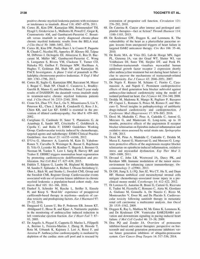
positive chronic myeloid leukemia patients with resistanceor intolerance to imatinib. Blood 118: 4567–4576, 2011.
58. Cortes JE, Kim DW, Kantarjian HM, Brummendorf TH,Dyagil I, Griskevicius L, Malhotra H, Powell C, Gogat K,Countouriotis AM, and Gambacorti-Passerini C. Bosuti-nib versus imatinib in newly diagnosed chronic-phasechronic myeloid leukemia: results from the BELA trial.J Clin Oncol 30: 3486–3492, 2012.
59. Cortes JE, Kim DW, Pinilla-Ibarz J, le Coutre P, PaquetteR, Chuah C, Nicolini FE, Apperley JF, Khoury HJ, TalpazM, DiPersio J, DeAngelo DJ, Abruzzese E, Rea D, Bac-carani M, Muller MC, Gambacorti-Passerini C, WongS, Lustgarten S, Rivera VM, Clackson T, Turner CD,Haluska FG, Guilhot F, Deininger MW, Hochhaus A,Hughes T, Goldman JM, Shah NP, and Kantarjian H;PACE Investigators. A phase 2 trial of ponatinib in Phi-ladelphia chromosome-positive leukemias. N Engl J Med369: 1783–1796, 2013.
60. Cortes JE, Saglio G, Kantarjian HM, Baccarani M, MayerJ, Boque C, Shah NP, Chuah C, Casanova L, Bradley-Garelik B, Manos G, and Hochhaus A. Final 5-year studyresults of DASISION: the dasatinib versus imatinib studyin treatment-naıve chronic myeloid leukemia patientstrial. J Clin Oncol 34: 2333–2340, 2016.
61. Crone SA, Zhao YY, Fan L, Gu Y, Minamisawa S, Liu Y,Peterson KL, Chen J, Kahn R, Condorelli G, Ross J Jr.,Chien KR, and Lee KF. ErbB2 is essential in the pre-vention of dilated cardiomyopathy. Nat Med 8: 459–465,2002.
62. Curigliano G, Cardinale D, Suter T, Plataniotis G, deAzambuja E, Sandri MT, Criscitiello C, Goldhirsch A,Cipolla C, and Roila F; ESMO Guidelines WorkingGroup. Cardiovascular toxicity induced by chemotherapy,targeted agents and radiotherapy: ESMO Clinical PracticeGuidelines. Ann Oncol 23: vii155–vii166, 2012.
63. D’Uva G, Aharonov A, Lauriola M, Kain D, Yahalom-Ronen Y, Carvalho S, Weisinger K, Bassat E, RajchmanD, Yifa O, Lysenko M, Konfino T, Hegesh J, Brenner O,Neeman M, Yarden Y, Leor J, Sarig R, Harvey RP, andTzahor E. ERBB2 triggers mammalian heart regenerationby promoting cardiomyocyte dedifferentiation and pro-liferation. Nat Cell Biol 17: 627–638, 2015.
64. Dahlen T, Edgren G, Lambe M, Hoglund M, BjorkholmM, Sandin F, Sjalander A, Richter J, Olsson-Stromberg U,Ohm L, Back M, and Stenke L; Swedish CML Group andthe Swedish CML Register Group. Cardiovascular eventsassociated with use of tyrosine kinase inhibitors in chronicmyeloid leukemia: a population-based cohort study. AnnIntern Med 165: 161–166, 2016.
65. Danhof S, Schreder M, Rasche L, Strifler S, EinseleH, and Knop S. ‘Real-life’ experience of preapprovalcarfilzomib-based therapy in myeloma—analysis of car-diac toxicity and predisposing factors. Eur J Haematol 97:25–32, 2016.
66. Daugaard G, Lassen U, Bie P, Pedersen EB, Jensen KT,Abildgaard U, Hesse B, and Kjaer A. Natriuretic peptidesin the monitoring of anthracycline induced reduction inleft ventricular ejection fraction. Eur J Heart Fail 7: 87–93, 2005.
67. De Angelis A, Piegari E, Cappetta D, Marino L, FilippelliA, Berrino L, Ferreira-Martins J, Zheng H, Hosoda T,Rota M, Urbanek K, Kajstura J, Leri A, Rossi F, andAnversa P. Anthracycline cardiomyopathy is mediated bydepletion of the cardiac stem cell pool and is rescued by
restoration of progenitor cell function. Circulation 121:276–292, 2010.
68. De Caterina R. Cancer after intense and prolonged anti-platelet therapies—fact or fiction? Thromb Haemost 114:1100–1103, 2015.
69. De Keulenaer GW, Doggen K, and Lemmens K. Thevulnerability of the heart as a pluricellular paracrine or-gan: lessons from unexpected triggers of heart failure intargeted ErbB2 anticancer therapy. Circ Res 106: 35–46,2010.
70. De Korte MA, de Vries EG, Lub-de Hooge MN, JagerPL, Gietema JA, van der Graaf WT, Sluiter WJ, vanVeldhuisen DJ, Suter TM, Sleijfer DT, and Perik PJ.111Indium-trastuzumab visualises myocardial humanepidermal growth factor receptor 2 expression shortlyafter anthracycline treatment but not during heart failure: aclue to uncover the mechanisms of trastuzumab-relatedcardiotoxicity. Eur J Cancer 43: 2046–2051, 2007.
71. De Nigris F, Rienzo M, Schiano C, Fiorito C, Casa-massimi A, and Napoli C. Prominent cardioprotectiveeffects of third generation beta blocker nebivolol againstanthracycline-induced cardiotoxicity using the model ofisolated perfused rat heart. Eur J Cancer 44: 334–340, 2008.
72. Deidda M, Madonna R, Mango R, Pagliaro P, BassareoPP, Cugusi L, Romano S, Penco M, Romeo F, and Mer-curo G. Novel insights in pathophysiology of antiblasticdrugs-induced cardiotoxicity and cardioprotection. JCardiovasc Med (Hagerstown) 17: S76–S83, 2016.
73. Dessı M, Madeddu C, Piras A, Cadeddu C, Antoni G,Mercuro G, and Mantovani G. Long-term, up to 18months, protective effects of the angiotensin II receptorblocker telmisartan on Epirubin-induced inflammation andoxidative stress assessed by serial strain rate. Springerplus2: 198, 2013.
74. Dessı M, Piras A, Madeddu C, Cadeddu C, Deidda M,Massa E, Antoni G, Mantovani G, and Mercuro G. Long-term protective effects of the angiotensin receptor blockertelmisartan on epirubicin-induced inflammation, oxidativestress and myocardial dysfunction. Exp Ther Med 2:1003–1009, 2011.
75. Devaud C, John LB, Westwood JA, Darcy PK, andKershaw MH. Immune modulation of the tumor micro-environment for enhancing cancer immunotherapy. On-coimmunology 2: e25961, 2013.
76. Di GH, Jiang S, Li FQ, Sun JZ, Wu CT, Hu X, and DuanHF. Human umbilical cord mesenchymal stromal cellsmitigate chemotherapy-associated tissue injury in a pre-clinical mouse model. Cytotherapy 14: 412–422, 2012.
77. Di Lorenzo G, Autorino R, Bruni G, Cartenı G, RicevutoE, Tudini M, Ficorella C, Romano C, Aieta M, GiordanoA, Giuliano M, Gonnella A, De Nunzio C, Rizzo M,Montesarchio V, Ewer M, and De Placido S. Cardiovas-cular toxicity following sunitinib therapy in metastaticrenal cell carcinoma: a multicenter analysis. Ann Oncol20: 1535–1542, 2009.
78. Doggen K, Ray L, Mathieu M, Mc Entee K, Lemmens K,and De Keulenaer GW. Ventricular ErbB2/ErbB4 acti-vation and downstream signaling in pacing-induced heartfailure. J Mol Cell Cardiol 46: 33–38, 2009.
79. Dou PQ and Zonder JA. Overview of proteasomeinhibitor-based anti-cancer therapies: perspective on bor-tezomib and second generation proteasome inhibitors ver-sus future generation inhibitors of ubiquitin-proteasomesystem. Curr Cancer Drug Targets 14: 517–536, 2014.
2138 TOCCHETTI ET AL.
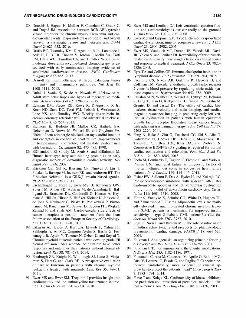
80. Douxfils J, Haguet H, Mullier F, Chatelain C, Graux C,and Dogne JM. Association between BCR-ABL tyrosinekinase inhibitors for chronic myeloid leukemia and car-diovascular events, major molecular response, and overallsurvival: a systematic review and meta-analysis. JAMAOncol 2: 625–632, 2016.
81. Drafts BC, Twomley KM, D’Agostino R Jr., Lawrence J,Avis N, Ellis LR, Thohan V, Jordan J, Melin SA, TortiFM, Little WC, Hamilton CA, and Hundley WG. Low tomoderate dose anthracycline-based chemotherapy is as-sociated with early noninvasive imaging evidence ofsubclinical cardiovascular disease. JACC CardiovascImaging 6: 877–885, 2013.
82. Dranoff G. Immunotherapy at large: balancing tumorimmunity and inflammatory pathology. Nat Med 19:1100–1111, 2013.
83. Dulak J, Szade K, Szade A, Nowak W, Jozkowicz A.Adult stem cells: hopes and hypes of regenerative medi-cine. Acta Biochim Pol 62: 329–337, 2015.
84. Eckman DM, Stacey RB, Rowe R, D’Agostino R Jr.,Kock ND, Sane DC, Torti FM, Yeboah J, Workman S,Lane KS, and Hundley WG. Weekly doxorubicin in-creases coronary arteriolar wall and adventitial thickness.PLoS One 8: e57554, 2013.
85. Eichhorn EJ, Bedotto JB, Malloy CR, Hatfield BA,Deitchman D, Brown M, Willard JE, and Grayburn PA.Effect of beta-adrenergic blockade on myocardial functionand energetics in congestive heart failure. Improvementsin hemodynamic, contractile, and diastolic performancewith bucindolol. Circulation 82: 473–483, 1990.
86. ElGhandour, El Sorady M, Azab S, and ElRahman M.Human heart-type fatty acid-binding protein as an earlydiagnostic marker of doxorubicin cardiac toxicity. He-matol Rev 1: e6, 2009.
87. Erickson CE, Gul R, Blessing CP, Nguyen J, Liu T,Pulakat L, Bastepe M, Jackson EK, and Andresen BT. Theb-blocker Nebivolol Is a GRK/b-arrestin biased agonist.PLoS One 8: e71980, 2013.
88. Eschenhagen T, Force T, Ewer MS, de Keulenaer GW,Suter TM, Anker SD, Avkiran M, de Azambuja E, Bal-ligand JL, Brutsaert DL, Condorelli G, Hansen A, Hey-mans S, Hill JA, Hirsch E, Hilfiker-Kleiner D, Janssens S,de Jong S, Neubauer G, Pieske B, Ponikowski P, Pirmo-hamed M, Rauchhaus M, Sawyer D, Sugden PH, Wojta J,Zannad F, and Shah AM. Cardiovascular side effects ofcancer therapies: a position statement from the heartfailure association of the European Society of Cardiology.Eur J Heart Fail 13: 1–10, 2011.
89. Eskazan AE, Eyice D, Kurt EA, Elverdi T, Yalniz FF,Salihoglu A, Ar MC, Ongoren Aydin S, Baslar Z, Fer-hanoglu B, Aydin Y, Tuzuner N, Ozbek U, and Soysal T.Chronic myeloid leukemia patients who develop grade I/IIpleural effusion under second-line dasatinib have betterresponses and outcomes than patients without pleural ef-fusion. Leuk Res 38: 781–787, 2014.
90. Estabragh ZR, Knight K, Watmough SJ, Lane S, Vinja-muri S, Hart G, and Clark RE. A prospective evaluationof cardiac function in patients with chronic myeloidleukaemia treated with imatinib. Leuk Res 35: 49–51,2011.
91. Ewer MS and Ewer SM. Troponin I provides insight intocardiotoxicity and the anthracycline-trastuzumab interac-tion. J Clin Oncol 28: 3901–3904, 2010.
92. Ewer MS and Lenihan DJ. Left ventricular ejection frac-tion and cardiotoxicity: is our ear really to the ground?J Clin Oncol 26: 1201–1203, 2008.
93. Ewer MS and Lippman SM. Type II chemotherapy-relatedcardiac dysfunction: time to recognize a new entity. J ClinOncol 23: 2900–2902, 2005.
94. Ewer MS, Vooletich MT, Durand JB, Woods ML, DavisJR, Valero V, and Lenihan DJ. Reversibility of trastuzumab-related cardiotoxicity: new insights based on clinical courseand response to medical treatment. J Clin Oncol 23: 7820–7826, 2005.
95. Eyre TA and Collins GP. Immune checkpoint inhibition inlymphoid disease. Br J Haematol 170: 291–304, 2015.
96. Facemire CS, Nixon AB, Griffiths R, Hurwitz H, andCoffman TM. Vascular endothelial growth factor receptor2 controls blood pressure by regulating nitric oxide syn-thase expression. Hypertension 54: 652–658, 2009.
97. Fallah-Rad N, Walker JR, Wassef A, Lytwyn M, BohonisS, Fang T, Tian G, Kirkpatrick ID, Singal PK, Krahn M,Grenier D, and Jassal DS. The utility of cardiac bio-markers, tissue velocity and strain imaging, and cardiacmagnetic resonance imaging in predicting early left ven-tricular dysfunction in patients with human epidermalgrowth factor receptor II-positive breast cancer treatedwith adjuvant trastuzumab therapy. J Am Coll Cardiol 57:2263–2270, 2011.
98. Feng N, Huke S, Zhu G, Tocchetti CG, Shi S, Aiba T,Kaludercic N, Hoover DB, Beck SE, Mankowski JL,Tomaselli GF, Bers DM, Kass DA, and Paolocci N.Constitutive BDNF/TrkB signaling is required for normalcardiac contraction and relaxation. Proc Natl Acad SciU S A 112: 1880–1885, 2015.
99. Feola M, Lombardo E, Taglieri C, Piccolo S, and Vado A.Plasma BNP and renal failure as prognostic factors ofmid-term clinical out come in congestive heart failurepatients. Int J Cardiol 149: 114–115, 2011.
100. Fisher PW, Salloum F, Das A, Hyder H, and Kukreja RC.Phosphodiesterase-5 inhibition with sildenafil attenuatescardiomyocyte apoptosis and left ventricular dysfunctionin a chronic model of doxorubicin cardiotoxicity. Circu-lation 111: 1601–1610, 2005.
101. Fitter S, Vandyke K, Schultz CG, White D, Hughes TP,and Zannettino AC. Plasma adiponectin levels are mark-edly elevated in imatinib-treated chronic myeloid leuke-mia (CML) patients: a mechanism for improved insulinsensitivity in type 2 diabetic CML patients? J Clin En-docrinol Metab 95: 3763–3767, 2010.
102. Fogli S, Nieri P, and Breschi MC. The role of nitric oxidein anthracycline toxicity and prospects for pharmacologicprevention of cardiac damage. FASEB J 18: 664–675,2004.
103. Folkman J. Angiogenesis: an organizing principle for drugdiscovery? Nat Rev Drug Discov 6: 273–286, 2007.
104. Folkman J. Tumor angiogenesis: therapeutic implications.N Engl J Med 285: 1182–1186, 1971.
105. Fontanella C, Aita M, Cinausero M, Aprile G, Baldin MG,Dusi V, Lestuzzi C, Fasola G, and Puglisi F. Capecitabine-induced cardiotoxicity: more evidence or clinical ap-proaches to protect the patients’ heart? Onco Targets Ther7; 1783–1791, 2014.
106. Force T and Kolaja KL. Cardiotoxicity of kinase inhibitors:the prediction and translation of preclinical models to clin-ical outcomes. Nat Rev Drug Discov 10: 111–126, 2011.
ANTINEOPLASTIC DRUG-INDUCED CARDIOTOXICITY 2139
107. Force T, Krause DS, and Van Etten RA. Molecularmechanisms of cardiotoxicity of tyrosine kinase inhibi-tion. Nat Rev Cancer 7: 332–344, 2007.
108. Fu M, Matoba M, Liang QM, Sjogren KG, and Hjalmar-son A. Properties of G-protein modulated receptor-adenylyl cyclase system in myocardium of spontaneouslyhypertensive rats treated with adriamycin. Int J Cardiol44: 9–18, 1994.
109. Fuchs-Tarlovsky V. Role of antioxidants in cancer ther-apy. Nutrition 29: 15–21, 2013.
110. Gabrielson K, Bedja D, Pin S, Tsao A, Gama L, Yuan B,and Muratore N. Heat shock protein 90 and erbB2 in thecardiac response to doxorubicin injury. Cancer Res 67:1436–1441, 2007.
111. Galindo CL, Kasasbeh E, Murphy A, Ryzhov S, LenihanS, Ahmad FA, Williams P, Nunnally A, Adcock J, Song Y,Harrell FE, Tran TL, Parry TJ, Iaci J, Ganguly A, Feok-tistov I, Stephenson MK, Caggiano AO, Sawyer DB, andCleator JH. Anti-remodeling and anti-fibrotic effects of theneuregulin-1b glial growth factor 2 in a large animal modelof heart failure. J Am Heart Assoc 3: e000773, 2014.
112. Galindo CL, Ryzhov S, and Sawyer DB. Neuregulin as aheart failure therapy and mediator of reverse remodeling.Curr Heart Fail Rep 11: 40–49, 2014.
113. Gambacorti-Passerini C, Tornaghi L, Franceschino A,Piazza R, Corneo G, and Pogliani E. In reply to ‘Cardi-otoxicity of the cancer therapeutic agent imatinib mesy-late’. Nat Med 13: 13–14, 2007.
114. Gammella E, Maccarinelli F, Buratti P, Recalcati S, andCairo G. The role of iron in anthracycline cardiotoxicity.Front Pharmacol 5: 25, 2014.
115. Gangadhar TC and Vonderheide RH. Mitigating the toxiceffects of anticancer immunotherapy. Nat Rev Clin Oncol11: 91–99, 2014.
116. Gao R, Zhang J, Cheng L, Wu X, Dong W, Yang X, Li T,Liu X, Xu Y, Li X, and Zhou M. A phase II, random-ized, double-blind, multicenter, based on standard ther-apy, placebo-controlled study of the efficacy and safetyof recombinant human neuregulin-1 in patients withchronic heart failure. J Am Coll Cardiol 55: 1907–1914,2010.
117. Geisler BP, Raad RA, Esaian D, Sharon E, andSchwartz DR. Apical ballooning and cardiomyopathy ina melanoma patient treated with ipilimumab: a case oftakotsubo-like syndrome. J Immunother Cancer 3: 4,2015.
118. Giles FJ, Mauro MJ, Hong F, Ortmann CE, McNeill C,Woodman RC, Hochhaus A, le Coutre PD, and Saglio G.Rates of peripheral arterial occlusive disease in patientswith chronic myeloid leukemia in the chronic phasetreated with imatinib, nilotinib, or non-tyrosine kinasetherapy: a retrospective cohort analysis. Leukemia 27:1310–1315, 2013.
119. Gimeno E, Gomez M, Gonzalez JR, Comın J, Alvarez-Larran A, Sanchez-Gonzalez B, Molina L, Domingo-Domenech E, Garcia-Pallarols F, Pedro C, Abella E,Vilaplana C, de Sanjose S, Besses C, and Salar A. NT-proBNP: a cardiac biomarker to assess prognosis in non-Hodgkin lymphoma. Leuk Res 35: 715–720, 2011.
120. Giordano SH, Booser DJ, Murray JL, Ibrahim NK, Rah-man ZU, Valero V, Theriault RL, Rosales MF, Rivera E,Frye D, Ewer M, Ordonez NG, Buzdar AU, and Horto-bagyi GN. A detailed evaluation of cardiac toxicity: aphase II study of doxorubicin and one- or three-hour-
infusion paclitaxel in patients with metastatic breast can-cer. Clin Cancer Res 8: 3360–3368, 2002.
121. Gonzalvez F and Gottlieb E. Cardiolipin: setting the beatof apoptosis. Apoptosis 12: 877–885, 2007.
122. Goodwin R, Ding K, Seymour L, LeMaıtre A, Arnold A,Shepherd FA, Dediu M, Ciuleanu T, Fenton D, Zukin M,Walde D, Laberge F, Vincent M, Ellis PM, and LaurieSA; NCIC Clinical Trials Group, Kingston, Ontario, Ca-nada. Treatment-emergent hypertension and outcomes inpatients with advanced non-small-cell lung cancer re-ceiving chemotherapy with or without the vascular en-dothelial growth factor receptor inhibitor cediranib: NCICClinical Trials Group Study BR24. Ann Oncol 21: 2220–2226, 2010.
123. Grandin EW, Ky B, Cornell RF, Carver J, and LenihanDJ. Patterns of cardiac toxicity associated with irrevers-ible proteasome inhibition in the treatment of multiplemyeloma. J Card Fail 21: 138–144, 2015.
124. Green MR, Newton MD, and Fancher KM. Off-targeteffects of BCR-ABL and JAK2 inhibitors. Am J ClinOncol 39: 76–84, 2016.
125. Greene SJ, Fonarow GC, Vaduganathan M, Khan SS,Butler J, and Gheorghiade M. The vulnerable phase afterhospitalization for heart failure. Nat Rev Cardiol 12: 220–229, 2015.
126. Gresset SM and Shah SR. Intricacies of bevacizumab-induced toxicities and their management. Ann Pharmac-other 43: 490–501, 2009.
127. Guignabert C, Phan C, Seferian A, Huertas A, Tu L,Thuillet R, Sattler C, Le Hiress M, Tamura Y, Jutant EM,Chaumais MC, Bouchet S, Maneglier B, Molimard M,Rousselot P, Sitbon O, Simonneau G, Montani D, andHumbert M. Dasatinib induces lung vascular toxicity andpredisposes to pulmonary hypertension. J Clin Invest 126:3207–3218, 2016.
128. Gulati G, Heck SL, Ree AH, Hoffmann P, Schulz-MengerJ, Fagerland MW, Gravdehaug B, von Knobelsdorff-Brenkenhoff F, Bratland A, Storas TH, Hagve TA, RøsjøH, Steine K, Geisler J, and Omland T. Prevention ofcardiac dysfunction during adjuvant breast cancer therapy(PRADA): a 2 · 2 factorial, randomized, placebo-controlled, double-blind clinical trial of candesartan andmetoprolol. Eur Heart J 37: 1671–1680, 2016.
129. Gundewar S, Calvert JW, Jha S, Toedt-Pingel I, Ji SY,Nunez D, Ramachandran A, Anaya-Cisneros M, Tian R,and Lefer DJ. Activation of AMP-activated protein kinaseby metformin improves left ventricular function and sur-vival in heart failure. Circ Res 104: 403–411, 2009.
130. Hagerkvist R, Sandler S, Mokhtari D, and Welsh NAmelioration of diabetes by imatinib mesylate (Gleevec):role of beta-cell NF-kappaB activation and anti-apoptoticpre-conditioning. FASEB J 21: 618–628, 2007.
131. Haghikia A, Podewski E, Libhaber E, Labidi S, Fischer D,Roentgen P, Tsikas D, Jordan J, Lichtinghagen R, vonKaisenberg CS, Struman I, Bovy N, Sliwa K, BauersachsJ, and Hilfiker-Kleiner D. Phenotyping and outcome oncontemporary management in a German cohort of patientswith peripartum cardiomyopathy. Basic Res Cardiol 108:366, 2013.
132. Hamo CE, Bloom MW, Cardinale D, Ky B, Nohria A, BaerL, Skopicki H, Lenihan DJ, Gheorghiade M, Lyon AR, andButler J. Cancer therapy-related cardiac dysfunction andheart failure: part 2: prevention, treatment, guidelines,and future directions. Circ Heart Fail 9: e002843, 2016.
2140 TOCCHETTI ET AL.
133. Hanahan D and Weinberg RA. Hallmarks of cancer: thenext generation. Cell 144: 646–674, 2011.
134. Hare JL, Brown JK, Leano R, Jenkins C, Woodward N, andMarwick TH. Use of myocardial deformation imaging todetect preclinical myocardial dysfunction before conven-tional measures in patients undergoing breast cancer treat-ment with trastuzumab. Am Heart J 158: 294–301, 2009.
135. Hasinoff BB and Patel D. The lack of target specificity ofsmall molecule anticancer kinase inhibitors is correlatedwith their ability to damage myocytes in vitro. ToxicolAppl Pharmacol 249: 132–139, 2010.
136. Hasinoff BB, Patel D, and Wu X. Molecular mechanismsof the cardiotoxicity of the proteasomal-targeted drugsbortezomib and carfilzomib. Cardiovasc Toxicol 2016[Epub ahead of print]; DOI: 10.1007/s12012-016-9378-7.
137. Hasinoff BB, Patel D, and Wu X. The myocyte-damagingeffects of the BCR-ABL1-targeted tyrosine kinase inhib-itors increase with potency and decrease with specificity.Cardiovasc Toxicol 2016 [Epub ahead of print]; DOI:10.1007/s12012-016-9386-7.
138. Hatfield A, Owen S, and Pilot PR. In reply to ‘Cardio-toxicity of the cancer therapeutic agent imatinib mesy-late’. Nat Med 13: 13, 2007.
139. Haykowsky MJ, Mackey JR, Thompson RB, Jones LW,and Paterson DI. Adjuvant trastuzumab induces ventric-ular remodeling despite aerobic exercise training. ClinCancer Res 15: 4963–4967, 2009.
140. Heinecke JL, Ridnour LA, Cheng RY, Switzer CH, Li-zardo MM, Khanna C, Glynn SA, Hussain SP, Young HA,Ambs S, and Wink DA. Tumor microenvironment-basedfeed-forward regulation of NOS2 in breast cancer pro-gression. Proc Natl Acad Sci U S A 111: 6323–6328,2014.
141. Heinzerling L, Ott PA, Hodi FS, Husain AN, Tajmir-RiahiA, Tawbi H, Pauschinger M, Gajewski TF, Lipson EJ, andLuke JJ. Cardiotoxicity associated with CTLA4 and PD1blocking immunotherapy. J Immunother Cancer 4: 50,2016.
142. Henninger C, Huelsenbeck S, Wenzel P, Brand M,Huelsenbeck J, Schad A, and Fritz G. Chronic heartdamage following doxorubicin treatment is alleviated bylovastatin. Pharmacol Res 91: 47–56, 2015.
143. Herrmann J, Saguner AM, Versari D, Peterson TE, ChadeA, Olson M, Lerman LO, and Lerman A. Chronic pro-teasome inhibition contributes to coronary atherosclerosis.Circ Res 101: 865–874, 2007.
144. Hesser BA, Liang XH, Camenisch G, Yang S, Lewin DA,Scheller R, Ferrara N, and Gerber HP. Down syndromecritical region protein 1 (DSCR1), a novel VEGF targetgene that regulates expression of inflammatory markers onactivated endothelial cells. Blood 104: 149–158, 2004.
145. Higuchi T and Schwaiger M. Imaging cardiac neuronalfunction and dysfunction. Curr Cardiol Rep 8: 131–138,2006.
146. Ho E, Brown A, Barrett P, Morgan RB, King G, KennedyMJ, and Murphy RT. Subclinical anthracycline- andtrastuzumab-induced cardiotoxicity in the long-termfollow-up of asymptomatic breast cancer survivors: aspeckle tracking echocardiographic study. Heart 96: 701–707, 2010.
147. Hoch M, Fischer P, Stapel B, Missol-Kolka E, Sekkali B,Scherr M, Favret F, Braun T, Eder M, Schuster-Gossier K,Gossier A, Hilfker A, Balligand JL, Drexler H, andHilfker-Kleiner D. Erythropoietin preserves the endothe-
lial differentiation capacity of cardiac progenitor cells andreduces heart failure during anticancer therapies. CellStem Cell 9: 131–143, 2011.
148. Hochhaus A, Saglio G, Hughes TP, Larson RA, Kim DW,Issaragrisil S, le Coutre PD, Etienne G, Dorlhiac-LlacerPE, Clark RE, Flinn IW, Nakamae H, Donohue B, DengW, Dalal D, Menssen HD, and Kantarjian HM. Long-termbenefits and risks of frontline nilotinib vs imatinib forchronic myeloid leukemia in chronic phase: 5-year updateof the randomized ENESTnd trial. Leukemia 30: 1044–1054, 2016.
149. Hodi FS, O’Day SJ, McDermott DF, Weber RW, SosmanJA, Haanen JB, Gonzalez R, Robert C, Schadendorf D,Hassel JC, Akerley W, van den Eertwegh AJ, Lutzky J,Lorigan P, Vaubel JM, Linette GP, Hogg D, OttensmeierCH, Lebbe C, Peschel C, Quirt I, Clark JI, Wolchok JD,Weber JS, Tian J, Yellin MJ, Nichol GM, Hoos A, andUrba WJ. Improved survival with ipilimumab in patientswith metastatic melanoma. N Engl J Med 363: 711–723,2010.
150. Hoffmann R, von Bardeleben S, ten Cate F, Borges AC,Kasprzak J, Firschke C, Lafitte S, Al-Saadi N, Kuntz-Hehner S, Engelhardt M, Becher H, and VanoverscheldeJL. Assessment of systolic left ventricular function: amulticenter comparison of cineventriculography, cardiacmagnetic resonance imaging, unenhanced and contrast-enhanced echocardiography. Eur Heart J 26: 607–616,2005.
151. Honton B, Despas F, Dumonteil N, Rouvellat C, RousselM, Carrie D, Galinier M, Montastruc JL, and Pathak A.Bortezomib and heart failure: case-report and review ofthe French Pharmacovigilance database. Fundam ClinPharmacol 28: 349–352, 2014.
152. Horacek JM, Tichy M, Pudil R, and Jebavy L. Glycogenphosphorylase BB could be a new circulating biomarkerfor detection of anthracycline cardiotoxicity. Ann Oncol19: 1656–1657, 2008.
153. Horacek JM, Tichy M, Jebavy L, Pudil R, Ulrychova M,and Maly J. Use of multiple biomarkers for evaluation ofanthracycline-induced cardiotoxicity in patients with acutmyeloid leukemia. Exp Oncol 30: 157–159, 2008.
154. Horacek JM, Vasatova M, Tichy M, Pudil R, Jebavy L,and Maly J. The use of cardiac biomarkers in detection ofcardiotoxicity associated with conventional and high-dosechemotherapy for acute leukemia. Exp Oncol 32: 97–99,2010.
155. Horie T, Ono K, Nishi H, Nagao K, Kinoshita M, Wata-nabe S, Kuwabara Y, Nakashima Y, Takanabe-Mori R,Nishi E, Hasegawa K, Kita T, and Kimura T. Acutedoxorubicin cardiotoxicity is associated with miR-146a-induced inhibition of the neuregulin-ErbB pathway. Car-diovasc Res 87: 656–664, 2010.
156. Hornsveld M and Dansen TB. The Hallmarks of cancerfrom a redox perspective. Antioxid Redox Signal 25: 300–325, 2016.
157. Hoshino A, Mita Y, Okawa Y, Ariyoshi M, Iwai-Kanai E,Ueyama T, Ikeda K, Ogata T, and Matoba S. Cytosolicp53 inhibits Parkin-mediated mitophagy and promotesmitochondrial dysfunction in the mouse heart. Nat Com-mun 4: 2308, 2013.
158. Huang C, Zhang X, Ramil JM, Rikka S, Kim L, Lee Y,Gude NA, Thistlethwaite PA, Sussman MA, Gottlieb RA,and Gustafsson AB. Juvenile exposure to anthracyclinesimpairs cardiac progenitor cell function and vasculariza-
ANTINEOPLASTIC DRUG-INDUCED CARDIOTOXICITY 2141
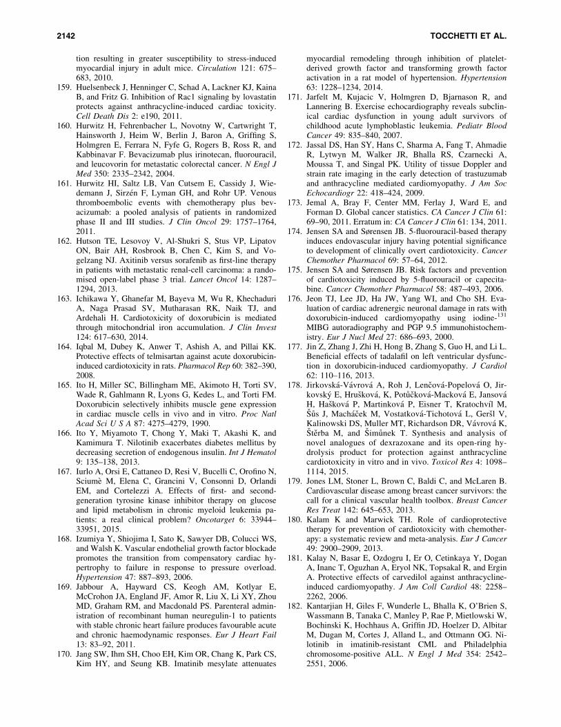
tion resulting in greater susceptibility to stress-inducedmyocardial injury in adult mice. Circulation 121: 675–683, 2010.
159. Huelsenbeck J, Henninger C, Schad A, Lackner KJ, KainaB, and Fritz G. Inhibition of Rac1 signaling by lovastatinprotects against anthracycline-induced cardiac toxicity.Cell Death Dis 2: e190, 2011.
160. Hurwitz H, Fehrenbacher L, Novotny W, Cartwright T,Hainsworth J, Heim W, Berlin J, Baron A, Griffing S,Holmgren E, Ferrara N, Fyfe G, Rogers B, Ross R, andKabbinavar F. Bevacizumab plus irinotecan, fluorouracil,and leucovorin for metastatic colorectal cancer. N Engl JMed 350: 2335–2342, 2004.
161. Hurwitz HI, Saltz LB, Van Cutsem E, Cassidy J, Wie-demann J, Sirzen F, Lyman GH, and Rohr UP. Venousthromboembolic events with chemotherapy plus bev-acizumab: a pooled analysis of patients in randomizedphase II and III studies. J Clin Oncol 29: 1757–1764,2011.
162. Hutson TE, Lesovoy V, Al-Shukri S, Stus VP, LipatovON, Bair AH, Rosbrook B, Chen C, Kim S, and Vo-gelzang NJ. Axitinib versus sorafenib as first-line therapyin patients with metastatic renal-cell carcinoma: a rando-mised open-label phase 3 trial. Lancet Oncol 14: 1287–1294, 2013.
163. Ichikawa Y, Ghanefar M, Bayeva M, Wu R, KhechaduriA, Naga Prasad SV, Mutharasan RK, Naik TJ, andArdehali H. Cardiotoxicity of doxorubicin is mediatedthrough mitochondrial iron accumulation. J Clin Invest124: 617–630, 2014.
164. Iqbal M, Dubey K, Anwer T, Ashish A, and Pillai KK.Protective effects of telmisartan against acute doxorubicin-induced cardiotoxicity in rats. Pharmacol Rep 60: 382–390,2008.
165. Ito H, Miller SC, Billingham ME, Akimoto H, Torti SV,Wade R, Gahlmann R, Lyons G, Kedes L, and Torti FM.Doxorubicin selectively inhibits muscle gene expressionin cardiac muscle cells in vivo and in vitro. Proc NatlAcad Sci U S A 87: 4275–4279, 1990.
166. Ito Y, Miyamoto T, Chong Y, Maki T, Akashi K, andKamimura T. Nilotinib exacerbates diabetes mellitus bydecreasing secretion of endogenous insulin. Int J Hematol9: 135–138, 2013.
167. Iurlo A, Orsi E, Cattaneo D, Resi V, Bucelli C, Orofino N,Sciume M, Elena C, Grancini V, Consonni D, OrlandiEM, and Cortelezzi A. Effects of first- and second-generation tyrosine kinase inhibitor therapy on glucoseand lipid metabolism in chronic myeloid leukemia pa-tients: a real clinical problem? Oncotarget 6: 33944–33951, 2015.
168. Izumiya Y, Shiojima I, Sato K, Sawyer DB, Colucci WS,and Walsh K. Vascular endothelial growth factor blockadepromotes the transition from compensatory cardiac hy-pertrophy to failure in response to pressure overload.Hypertension 47: 887–893, 2006.
169. Jabbour A, Hayward CS, Keogh AM, Kotlyar E,McCrohon JA, England JF, Amor R, Liu X, Li XY, ZhouMD, Graham RM, and Macdonald PS. Parenteral admin-istration of recombinant human neuregulin-1 to patientswith stable chronic heart failure produces favourable acuteand chronic haemodynamic responses. Eur J Heart Fail13: 83–92, 2011.
170. Jang SW, Ihm SH, Choo EH, Kim OR, Chang K, Park CS,Kim HY, and Seung KB. Imatinib mesylate attenuates
myocardial remodeling through inhibition of platelet-derived growth factor and transforming growth factoractivation in a rat model of hypertension. Hypertension63: 1228–1234, 2014.
171. Jarfelt M, Kujacic V, Holmgren D, Bjarnason R, andLannering B. Exercise echocardiography reveals subclin-ical cardiac dysfunction in young adult survivors ofchildhood acute lymphoblastic leukemia. Pediatr BloodCancer 49: 835–840, 2007.
172. Jassal DS, Han SY, Hans C, Sharma A, Fang T, AhmadieR, Lytwyn M, Walker JR, Bhalla RS, Czarnecki A,Moussa T, and Singal PK. Utility of tissue Doppler andstrain rate imaging in the early detection of trastuzumaband anthracycline mediated cardiomyopathy. J Am SocEchocardiogr 22: 418–424, 2009.
173. Jemal A, Bray F, Center MM, Ferlay J, Ward E, andForman D. Global cancer statistics. CA Cancer J Clin 61:69–90, 2011. Erratum in: CA Cancer J Clin 61: 134, 2011.
174. Jensen SA and Sørensen JB. 5-fluorouracil-based therapyinduces endovascular injury having potential significanceto development of clinically overt cardiotoxicity. CancerChemother Pharmacol 69: 57–64, 2012.
175. Jensen SA and Sørensen JB. Risk factors and preventionof cardiotoxicity induced by 5-fluorouracil or capecita-bine. Cancer Chemother Pharmacol 58: 487–493, 2006.
176. Jeon TJ, Lee JD, Ha JW, Yang WI, and Cho SH. Eva-luation of cardiac adrenergic neuronal damage in rats withdoxorubicin-induced cardiomyopathy using iodine-131
MIBG autoradiography and PGP 9.5 immunohistochem-istry. Eur J Nucl Med 27: 686–693, 2000.
177. Jin Z, Zhang J, Zhi H, Hong B, Zhang S, Guo H, and Li L.Beneficial effects of tadalafil on left ventricular dysfunc-tion in doxorubicin-induced cardiomyopathy. J Cardiol62: 110–116, 2013.
178. Jirkovska-Vavrova A, Roh J, Lencova-Popelova O, Jir-kovsky E, Hruskova, K, Potuckova-Mackova E, JansovaH, Haskova P, Martinkova P, Eisner T, Kratochvıl M,Sus J, Machacek M, Vostatkova-Tichotova L, Gersl V,Kalinowski DS, Muller MT, Richardson DR, Vavrova K,Sterba M, and Simunek T. Synthesis and analysis ofnovel analogues of dexrazoxane and its open-ring hy-drolysis product for protection against anthracyclinecardiotoxicity in vitro and in vivo. Toxicol Res 4: 1098–1114, 2015.
179. Jones LM, Stoner L, Brown C, Baldi C, and McLaren B.Cardiovascular disease among breast cancer survivors: thecall for a clinical vascular health toolbox. Breast CancerRes Treat 142: 645–653, 2013.
180. Kalam K and Marwick TH. Role of cardioprotectivetherapy for prevention of cardiotoxicity with chemother-apy: a systematic review and meta-analysis. Eur J Cancer49: 2900–2909, 2013.
181. Kalay N, Basar E, Ozdogru I, Er O, Cetinkaya Y, DoganA, Inanc T, Oguzhan A, Eryol NK, Topsakal R, and ErginA. Protective effects of carvedilol against anthracycline-induced cardiomyopathy. J Am Coll Cardiol 48: 2258–2262, 2006.
182. Kantarjian H, Giles F, Wunderle L, Bhalla K, O’Brien S,Wassmann B, Tanaka C, Manley P, Rae P, Mietlowski W,Bochinski K, Hochhaus A, Griffin JD, Hoelzer D, AlbitarM, Dugan M, Cortes J, Alland L, and Ottmann OG. Ni-lotinib in imatinib-resistant CML and Philadelphiachromosome-positive ALL. N Engl J Med 354: 2542–2551, 2006.
2142 TOCCHETTI ET AL.
183. Kaya MG, Ozkan M, Gunebakmaz O, Akkaya H, KayaEG, Akpek M, Kalay N, Dikilitas M, Yarlioglues M,Karaca H, Berk V, Ardic I, Ergin A, and Lam YY. Pro-tective effects of nebivolol against anthracycline-inducedcardiomyopathy: a randomized control study. Int J Car-diol 167: 2306–2310, 2013.
184. Kelly K, Swords R, Mahalingam D, Padmanabhan S, andGiles FJ. Serosal inflammation (pleural and pericardialeffusions) related to tyrosine kinase inhibitors. TargetOncol 4: 99–105, 2009.
185. Kerkela R, Grazette L, Yacobi R, Iliescu C, Patten R,Beahm C, Walters B, Shevtsov S, Pesant S, Clubb FJ,Rosenzweig A, Salomon RN, Van Etten RA, Alroy J,Durand JB, and Force T. Cardiotoxicity of the cancertherapeutic agent imatinib mesylate. Nat Med 12: 908–916, 2006.
186. Kerkela R, Woulfe KC, Durand JB, Vagnozzi R, KramerD, Chu TF, Beahm C, Chen MH, and Force T. Sunitinib-induced cardiotoxicity is mediated by off-target inhibitionof AMP-activated protein kinase. Clin Transl Sci 2: 15–25, 2009.
187. Khakoo AY, Kassiotis CM, Tannir N, Plana JC, HalushkaM, Bickford C, Trent J, 2nd, Champion JC, Durand JB,and Lenihan DJ. Heart failure associated with SunitinibMalate: a multitargeted receptor tyrosine kinase inhibitor.Cancer 112: 2500–2508, 2008.
188. Khan MF, Gottesman S, Boyella R, and Juneman E.Gemcitabine-induced cardiomyopathy: a case report andreview of the literature. J Medical Case Reports 8: 220–225, 2014.
189. Kim IM, Tilley DG, Chen J, Salazar NC, Whalen EJ,Violin JD, and Rockman HA. Beta-blockers alprenololand carvedilol stimulate beta-arrestin-mediated EGFRtransactivation. Proc Natl Acad Sci U S A 105: 14555–14560, 2008.
190. Kim TD, Rea D, Schwarz M, Grille P, Nicolini FE, RostiG, Levato L, Giles FJ, Dombret H, Mirault T, LabussiereH, Lindhorst R, Haverkamp W, Buschmann I, Dorken B,and le Coutre PD. Peripheral artery occlusive disease inchronic phase chronic myeloid leukemia patients treatedwith nilotinib or imatinib. Leukemia 27: 1316–1321, 2013.
191. Kinhult S, Albertsson M, Eskilsson J, and Cwikiel M.Antithrombotic treatment in protection against thrombo-genic effects of 5-fluorouracil on vascular endothelium: ascanning microscopy evaluation. Scanning 23: 1–8, 2001.
192. Kirkham AA and Davis MK. Exercise prevention of car-diovascular disease in breast cancer survivors. J Oncol2015: 917606, 2015.
193. Klaus A, Muller M, Schulz H, Saga Y, Martin JF, andBirchmeier W. Wnt/b-catenin and Bmp signals controldistinct sets of transcription factors in cardiac progenitorcells. Proc Natl Acad Sci U S A 109: 10921–10926, 2012.
194. Kloth JS, Pagani A, Verboom MC, Malovini A, Napoli-tano C, Kruit WH, Sleijfer S, Steeghs N, Zambelli A, andMathijssen RH. Incidence and relevance of QTc-intervalprolongation caused by tyrosine kinase inhibitors. Br JCancer 112: 1011–1016, 2015.
195. Koca D, Salman T, Unek IT, Oztop I, Ellidokuz H, ErenM, and Yilmaz U. Clinical and electrocardiographychanges in patients treated with capecitabine. Chemo-therapy 57: 381–387, 2011.
196. Kohlhaas M, Liu T, Knopp A, Zeller T, Ong MF, BohmM, O’Rourke B, and Maack C. Elevated cytosolic Na+increases mitochondrial formation of reactive oxygen
species in failing cardiac myocytes. Circulation 121:1606–1613, 2010.
197. Koka S, Das A, Zhu SG, Durrant D, Xi L, and KukrejaRC. Long-acting phosphodiesterase-5 inhibitor tadalafilattenuates doxorubicin-induced cardiomyopathy withoutinterfering with chemotherapeutic effect. J PharmacolExp Ther 334: 1023–1030, 2010.
198. Kosmas C, Kallistratos MS, Kopterides P, Syrios J, Sko-pelitis H, Mylonakis N, Karabelis A, and Tsavaris N.Cardiotoxicity of fluoropyrimidines in different schedulesof administration: a prospective study. J Cancer Res ClinOncol 134: 75–82, 2008.
199. Kremer LC, van Dalen EC, Offringa M, and Voute PA.Frequency and risk factors of anthracycline-inducedclinical heart failure in children: a systematic review. AnnOncol 13: 503–512, 2002.
200. Krummel MF and Allison JP. CD28 and CTLA-4 haveopposing effects on the response of T cells to stimulation.J Exp Med 182: 459–465, 1995.
201. Kubiczkova L, Pour L, Sedlarikova L, Hajek R, andSevcikova S. Proteasome inhibitors—molecular basis andcurrent perspectives in multiple myeloma. J Cell Mol Med18: 947–961, 2014.
202. Kuehn HS, Ouyang W, Lo B, Deenick EK, Niemela JE,Avery DT, Schickel JN, Tran DQ, Stoddard J, Zhang Y,Frucht DM, Dumitriu B, Scheinberg P, Folio LR, FreinCA, Price S, Koh C, Heller T, Seroogy CM, HuttenlocherA, Rao VK, Su HC, Kleiner D, Notarangelo LD, Ram-pertaap Y, Olivier KN, McElwee J, Hughes J, Pittaluga S,Oliveira JB, Meffre E, Fleisher TA, Holland SM, LenardoMJ, Tangye SG, and Uzel G. Immune dysregulation inhuman subjects with heterozygous germline mutations inCTLA4. Science 345: 1623–1627, 2014.
203. Ky B, Putt M, Sawaya H, French B, Januzzi JL Jr., SebagIA, Plana JC, Cohen V, Banchs J, Carver JR, Wiegers SE,Martin RP, Picard MH, Gerszten RE, Halpern EF, PasseriJ, Kuter I, and Scherrer-Crosbie M. Early increases inmultiple biomarkers predict subsequent cardiotoxicity inpatients with breast cancer treated with doxorubicin, tax-anes, and trastuzumab. J Am Coll Cardiol 63: 809–816,2014.
204. Ky B, Vejpongsa P, Yeh ET, Force T, and Moslehi JJ.Emerging paradigms in cardiomyopathies associated withcancer therapies. Circ Res 113: 754–764, 2013.
205. Lamberti M, Porto S, Zappavigna S, Addeo E, Marra M,Miraglia N, Sannolo N, Vanacore D, Stiuso P, andCaraglia M. A mechanistic study on the cardiotoxicity of5-fluorouracil in vitro and clinical and occupational per-spectives. Toxicol Lett 227: 151–156, 2014.
206. Lancellotti P, Anker SD, Donal E, Edvardsen T, PopescuBA, Farmakis D, Filippatos G, Habib G, Maggioni AP,Jerusalem G, and Galderisi M. EACVI/HFA CardiacOncology Toxicity Registry in breast cancer patients: ra-tionale, study design, and methodology (EACVI/HFACOT Registry)-EURObservational Research Program ofthe European Society of Cardiology. Eur Heart J Cardi-ovasc Imaging 16: 466–470, 2015.
207. Laubli H, Balmelli C, Bossard M, Pfister O, Glatz K, andZippelius A. Acute heart failure due to autoimmunemyocarditis under pembrolizumab treatment for meta-static melanoma. J Immunother Cancer 3: 11, 2015.
208. Laugwitz KL, Moretti A, Caron L, Nakano A, and ChienKR. Islet1 cardiovascular progenitors: a single source forheart lineages? Development 135: 193–205, 2008.
ANTINEOPLASTIC DRUG-INDUCED CARDIOTOXICITY 2143

209. Le Coutre P, Rea D, Abruzzese E, Dombret H, TrawinskaMM, Herndlhofer S, Dorken B, and Valent P. Severeperipheral arterial disease during nilotinib therapy. J NatlCancer Inst 103: 1347–1348, 2011.
210. Lebrecht D, Kokkori A, Ketelsen UP, Setzer B, andWalker UA. Tissue-specific mtDNA lesions and radical-associated mitochondrial dysfunction in human heartsexposed to doxorubicin. J Pathol 207: 436–444, 2005.
211. Lee CS, Cragg M, Glennie M, and Johnson P. Novelantibodies targeting immune regulatory checkpointsfor cancer therapy. Br J Clin Pharmacol 76: 233–247,2013.
212. Lee S, Chen TT, Barber CL, Jordan MC, Murdock J,Desai S, Ferrara N, Nagy A, Roos KP, and Iruela-ArispeML. Autocrine VEGF signaling is required for vascularhomeostasis. Cell 130: 691–703, 2007.
213. Lencova-Popelova O, Jirkovsky E, Jansova H, Jirkovska-Vavrova A, Vostatkova-Tichotova L, Mazurova Y,Adamcova M, Chladek J, Hroch M, Pokorna Z, Gersl V,Simunek T, and Sterba M. Cardioprotective effects ofinorganic nitrate/nitrite in chronic anthracycline cardio-toxicity: comparison with dexrazoxane. J Mol Cell Car-diol 91: 92–103, 2016.
214. Lenihan DJ and Kowey PR. Overview and management ofcardiac adverse events associated with tyrosine kinaseinhibitors. Oncologist 18: 900–908, 2013.
215. Lestuzzi C, Tartuferi L, and Corona G. Capecitabine (and5 fluorouracil) cardiotoxicity. Metabolic considerations.Breast J 17: 564–565, 2011.
216. Lestuzzi C, Vaccher E, Talamini R, Lleshi A, MeneguzzoN, Viel E, Scalone S, Tartuferi L, Buonadonna A, EjioforL, and Schmoll HJ. Effort myocardial ischemia duringchemotherapy with 5-fluorouracil: an underestimated risk.Ann Oncol 25: 1059–1064, 2014.
217. Levy BI. Microvascular plasticity and experimental heartfailure. Hypertension 47: 827–829, 2006.
218. Li B, Zheng Z, Wei Y, Wang M, Peng J, Kang T, HuangX, Xiao J, Li Y, and Li Z. Therapeutic effects ofneuregulin-1 in diabetic cardiomyopathy rats. CardiovascDiabetol 10: 69, 2011.
219. Li T and Singal PK. Adriamycin-induced early changes inmyocardial antioxidant enzymes and their modulation byprobucol. Circulation 102: 2105–2110, 2000.
220. Lim CC, Zuppinger C, Guo X, Kuster GM, Helmes M,Eppenberger HM, Suter TM, Liao R, and Sawyer DB.Anthracyclines induce calpain-dependent titin proteolysisand necrosis in cardiomyocytes. J Biol Chem 279: 8290–8299, 2004.
221. Lim SL, Lam CS, Segers VF, Brutsaert DL, and DeKeulenaer GW. Cardiac endothelium-myocyte interac-tion: clinical opportunities for new heart failure therapiesregardless of ejection fraction. Eur Heart J 36: 2050–2060, 2015.
222. Lipshultz SE, Miller TL, Lipsitz SR, Neuberg DS,Dahlberg SE, Colan SD, Silverman LB, Henkel JM,Franco VI, Cushman LL, Asselin BL, Clavell LA, AthaleU, Michon B, Laverdiere C, Schorin MA, Larsen E,Usmani N, and Sallan SE; Dana-Farber Cancer InstituteAcute Lymphoblastic Leukemia Consortium. Continuousversus bolus infusion of doxorubicin in children withALL: long-term cardiac outcomes. Pediatrics 130: 1003–1011, 2012.
223. Lipshultz SE, Rifai N, Sallan SE, Lipsitz SR, Dalton V,Sacks DB, and Ottlinger ME. Predictive value of cardiac
troponin T in pediatric patients at risk for myocardial in-jury. Circulation 96: 2641–2648, 1997.
224. Lipshultz SE, Scully RE, Lipsitz SR, Sallan SE, Silver-man LB, Miller TL, Barry EV, Asselin BL, Athale U,Clavell LA, Larsen E, Moghrabi A, Samson Y, MichonB, Schorin MA, Cohen HJ, Neuberg DS, Orav EJ, andColan SD. Assessment of dexrazoxane as a cardiopro-tectant in doxorubicin-treated children with high-riskacute lymphoblastic leukaemia: long-term follow-up of aprospective, randomised, multicentre trial. Lancet Oncol11: 950–961, 2010.
225. Lipson EJ, Bagnasco SM, Moore J Jr., Jang S, Patel MJ,Zachary AA, Pardoll DM, Taube JM, and Drake CG.Tumor regression and allograft rejection after adminis-tration of anti-PD-1. N Engl J Med 374: 896–898, 2016.
226. Lipton JH, Chuah C, Guerci-Bresler A, Rosti G, SimpsonD, Assouline S, Etienne G, Nicolini FE, le Coutre P, ClarkRE, Stenke L, Andorsky D, Oehler V, Lustgarten S,Rivera VM, Clackson T, Haluska FG, Baccarani M,Cortes JE, Guilhot F, Hochhaus A, Hughes T, KantarjianHM, Shah NP, Talpaz M, Deininger MW, and EPIC in-vestigators. Ponatinib versus imatinib for newly diagnosedchronic myeloid leukaemia: an international, randomised,open-label, phase 3 trial. Lancet Oncol 17: 612–621, 2016.
227. Liu X, Gu X, Li Z, Li X, Li H, Chang J, Chen P, Jin J, XiB, Chen D, Lai D, Graham RM, and Zhou M. Neuregulin-1/erbB-activation improves cardiac function and survivalin models of ischemic, dilated, and viral cardiomyopathy.J Am Coll Cardiol 48: 1438–1447, 2006.
228. Loges S, Roncal C, and Carmeliet P. Development oftargeted angiogenic medicine. J Thromb Haemost 7: 21–33, 2009.
229. Lyu YL, Kerrigan JE, Lin CP, Azarova AM, Tsai YC, BanY, and Liu LF. Topoisomerase IIbeta mediated DNAdouble-strand breaks: implications in doxorubicin cardi-otoxicity and prevention by dexrazoxane. Cancer Res 67:8839–8846, 2007.
230. Ma Y, Yamazaki T, Yang H, Kepp O, Galluzzi L, Zit-vogel L, Smyth MJ, and Kroemer G. Tumor necrosisfactor is dispensable for the success of immunogenic an-ticancer chemotherapy. Oncoimmunology 2: e24786, 2013.
231. Madonna R, Cadeddu C, Deidda M, Giricz Z, Madeddu C,Mele D, Monte I, Novo G, Pagliaro P, Pepe A, SpallarossaP, Tocchetti CG, Varga ZV, Zito C, Geng YJ, Mercuro G,and Ferdinandy P. Cardioprotection by gene therapy: areview paper on behalf of the Working Group on DrugCardiotoxicity and Cardioprotection of the Italian Societyof Cardiology. Int J Cardiol 191: 203–210, 2015.
232. Madonna R, Cadeddu C, Deidda M, Mele D, Monte I,Novo G, Pagliaro P, Pepe A, Spallarossa P, Tocchetti CG,Zito C, and Mercuro G. Improving the preclinical modelsfor the study of chemotherapy-induced cardiotoxicity: aPosition Paper of the Italian Working Group on DrugCardiotoxicity and Cardioprotection. Heart Fail Rev 20:621–631, 2015.
233. Madonna R, Van Laake LW, Davidson SM, Engel FB,Hausenloy DJ, Lecour S, Leor J, Perrino C, Schulz R,Ytrehus K, Landmesser U, Mummery CL, Janssens S,Willerson J, Eschenhagen T, Ferdinandy P, and SluijterJP. Position Paper of the European Society of CardiologyWorking Group Cellular Biology of the Heart: cell-basedtherapies for myocardial repair and regeneration in is-chemic heart disease and heart failure. Eur Heart J 37:1789–1798, 2016.
2144 TOCCHETTI ET AL.
234. Maiello M, Sharma RK, Ciccone MM, Reddy HK, andPalmiero P. Early diagnosis of cardiac toxicity relatedto antineoplastic treatment. J Cancer Ther 2: 161–166,2011.
235. Maini CL, Sciuto R, Ferraironi A, Vici P, Tofani A, FestaA, Conti F, and Lopez M. Clinical relevance of radionu-clide angiography and antimyosin immunoscintigraphyfor risk assessment in epirubicin cardiotoxicity. J NuclCardiol 4: 502–508, 1997.
236. Maitland ML, Bakris GL, Black HR, Chen HX, DurandJB, Elliott WJ, Ivy SP, Leier CV, Lindenfeld J, Liu G,Remick SC, Steingart R, and Tang WH; CardiovascularToxicities Panel, Convened by the Angiogenesis TaskForce of the National Cancer Institute Investigational DrugSteering Committee. Initial assessment, surveillance, andmanagement of blood pressure in patients receiving vas-cular endothelial growth factor signaling pathway inhibi-tors. J Natl Cancer Inst 102: 596–604, 2010.
237. Makkar RR, Smith RR, Cheng K, Malliaras K, ThomsonLE, Berman D, Czer LS, Marban L, Mendizabal A,Johnston PV, Russell SD, Schuleri KH, Lardo AC, Ger-stenblith G, and Marban E. Intracoronary cardiosphere-derived cells for heart re generation after myocardial in-farction (CADUCEUS): a prospective, randomised phase1 trial. Lancet 379: 895–904, 2012.
238. Malm S, Frigstad S, Sagberg E, Larsson H, and SkjaerpeT. Accurate and reproducible measurement of left ven-tricular volume and ejection fraction by contrast echo-cardiography: a comparison with magnetic resonanceimaging. J Am Coll Cardiol 44: 1030–1035, 2004.
239. Markman TM and Markman M. Cardiotoxicity of anti-neoplastic agents: what is the present and future role forimaging? Curr Oncol Rep 16: 396, 2014.
240. Marone G and Granata F. Angiogenesis, lymphangio-genesis and clinical implications. Preface. Chem ImmunolAllergy 99: XI–XII, 2014.
241. Marquez-Rodas I, Cerezuela P, Soria A, Berrocal A, RisoA, Gonzalez-Cao M, and Martin-Algarra S. Immunecheckpoint inhibitors: therapeutic advances in melanoma.Ann Transl Med 3: 267, 2015.
242. Martin E, Thougaard AV, Grauslund M, Jensen PB,Bjorkling F, Hasinoff BB, Tjørnelund J, Sehested M, andJensen LH. Evaluation of the topoisomerase II-inactivebisdioxopiperazine ICRF-161 as a protectant againstdoxorubicin-induced cardiomyopathy. Toxicology 255:72–79, 2009.
243. Matsui H, Morishima I, Numaguchi Y, Toki Y, OkumuraK, and Hayakawa T. Protective effects of carvedilolagainst doxorubicin-induced cardiomyopathy in rats. LifeSci 65: 1265–1274, 1999.
244. May D, Gilon D, Djonov V, Itin A, Lazarus A, Gordon O,Rosenberger C, and Keshet E. Transgenic system for con-ditional induction and rescue of chronic myocardial hiber-nation provides insights into genomic programs ofhibernation. Proc Natl Acad Sci U S A 105: 282–287, 2008.
245. McGuire WP, Rowinsky EK, Rosenhein NB, GrumbineFC, Ettinger DS, Armstrong DK, and Donehower RC.Taxol: a unique antineoplastic agent with significant ac-tivity in advanced ovarian epithelial neoplasms. Ann In-tern Med 111: 273–279, 1989.
246. Mele D, Nardozza M, Spallarossa P, Frassoldati A, Toc-chetti CG, Cadeddu C, Madonna R, Malagu M, Ferrari R,and Mercuro G. Current views on anthracycline cardio-toxicity. Heart Fail Rev 21: 621–634, 2016.
247. Mele D, Tocchetti CG, Pagliaro P, Madonna R, Novo G,Pepe A, Zito C, Maurea N, and Spallarossa P. Pathophy-siology of anthracycline cardiotoxicity. J Cardiovasc Med(Hagerstown) 17: S3–S11, 2016.
248. Menna P, Pax OG, Chello M, Covino E, Salvatorelli E,and Minotti G. Anthracycline cardiotoxicity. Expert OpinDrug Saf 1: S21–S36, 2012.
249. Menna P, Salvatorelli E, and Minotti G. Cardiotoxicity ofantitumor drugs. Chem Res Toxicol 21: 978–989, 2008.
250. Mercurio V, Pirozzi F, Lazzarini E, Marone G, Rizzo P,Agnetti G, Tocchetti CG, Ghigo A, and Ameri P. Modelsof heart failure based on the cardiotoxicity of anticancerdrugs. J Card Fail 22: 449–458, 2016.
251. Merino H and Singla DK. Notch-1 mediated cardiacprotection following embryonic and induced pluripotentstem cell transplantation in doxorubicin-induced heartfailure. PLoS One 9: e101024, 2014.
252. Messina E, De Angelis L, Frati G, Morrone S, Chimenti S,Fiordaliso F, Salio M, Battaglia M, Latronico MV, ColettaM, Vivarelli E, Frati L, Cossu G, and Giacomello A.Isolation and expansion of adult cardiac stem cells fromhuman and murine heart. Circ Res 95: 911–921, 2004.
253. Meyer CC, Calis KA, Burke LB, Walawander CA, andGrasela TH. Symptomatic cardiotoxicity associated with5-fluorouracil. Pharmacotherapy 17: 729–736, 1997.
254. Michot JM, Bigenwald C, Champiat S, Collins M, Car-bonnel F, Postel-Vinay S, Berdelou A, Varga A, BahledaR, Hollebecque A, Massard C, Fuerea A, Ribrag V,Gazzah A, Armand JP, Amellal N, Angevin E, Noel N,Boutros C, Mateus C, Robert C, Soria JC, Marabelle A,and Lambotte O. Immune-related adverse events withimmune checkpoint blockade: a comprehensive review.Eur J Cancer 54: 139–148, 2016.
255. Milan A, Puglisi E, Ferrari L, Bruno G, Losano I, andVeglio F. Arterial hypertension and cancer. Int J Cancer134: 2269–2277, 2014.
256. Miller KD, Chap LI, Holmes FA, Cobleigh MA, MarcomPK, Fehrenbacher L, Dickler M, Overmoyer BA, ReimannJD, Sing AP, Langmuir V, and Rugo HS. Randomizedphase III trial of capecitabine compared with bevacizumabplus capecitabine in patients with previously treatedmetastatic breast cancer. J Clin Oncol 23: 792–799, 2005.
257. Minotti G, Menna P, Salvatorelli E, Cairo G, and GianniL. Anthracyclines: molecular advances and pharmaco-logic developments in antitumor activity and cardiotoxi-city. Pharmacol Rev 56: 185–229, 2004.
258. Minotti G, Salvatorelli E, and Menna P. Pharmacologicalfoundations of cardio-oncology. J Pharmacol Exp Ther334: 2–8, 2010.
259. Minotti G. Pharmacology at work for cardio-oncology:ranolazine to treat early cardiotoxicity induced by anti-tumor drugs. J Pharmacol Exp Ther 346: 343–349, 2013.
260. Mirotsou M, Jayawardena TM, Schmeckpeper J, GnecchiM, and Dzau VJ. Paracrine mechanisms of stem cell re-parative and regenerative actions in the heart. J Mol CellCardiol 50: 280–289, 2011.
261. Miura K, Kinouchi M, Ishida K, Kinouchi M, Ishida K,Fujibuchi W, Naitoh T, Ogawa H, Ando T, Yazaki N,Watanabe K, Haneda S, Shibata C, and Sasaki I. 5-FUmetabolism in cancer and orally-administrable 5-FUdrugs. Cancers (Basel) 2: 1717–1730, 2010.
262. Molinaro M, Ameri P, Marone G, Petretta M, Abete P, DiLisa F, De Placido S, Bonaduce D, and Tocchetti CG.Recent advances on pathophysiology, diagnostic and
ANTINEOPLASTIC DRUG-INDUCED CARDIOTOXICITY 2145
therapeutic insights in cardiac dysfunction induced byantineoplastic drugs. Biomed Res Int 2015: 138148, 2015.
263. Montani D, Bergot E, Gunther S, Savale L, Bergeron A,Bourdin A, Bouvaist H, Canuet M, Pison C, Macro M,Poubeau P, Girerd B, Natali D, Guignabert C, Perros F,O’Callaghan DS, Jaıs X, Tubert-Bitter P, Zalcman G,Sitbon O, Simonneau G, and Humbert M. Pulmonary ar-terial hypertension in patients treated by dasatinib. Cir-culation 125: 2128–2137, 2012.
264. Montani D, Seferian A, Savale L, Simonneau G, andHumbert M. Drug-induced pulmonary arterial hyperten-sion: a recent outbreak. Eur Respir Rev 22: 244–250,2013.
265. Moreo A, Vallerio P, Ricotta R, Stucchi M, Pozzi M,Musca F, Meani P, Maloberti A, Facchetti R, Di Bella S,Giganti MO, Sartore-Bianchi A, Siena S, Mancia G, andGiannattasio C. Effects of cancer therapy targeting vas-cular endothelial growth factor receptor on central bloodpressure and cardiovascular system. Am J Hypertens 29:158–162, 2016.
266. Moslehi JJ. Cardiovascular toxic effects of targeted cancertherapies. N Engl J Med 375: 1457–1467, 2016.
267. Moslehi JJ and Deininger M. Tyrosine kinase inhibitor-associated cardiovascular toxicity in chronic myeloidleukemia. J Clin Oncol 33: 4210–4218, 2015.
268. Motzer RJ, Hutson TE, Cella D, Reeves J, Hawkins R,Guo J, Nathan P, Staehler M, de Souza P, Merchan JR,Boleti E, Fife K, Jin J, Jones R, Uemura H, De Giorgi U,Harmenberg U, Wang J, Sternberg CN, Deen K, McCannL, Hackshaw MD, Crescenzo R, Pandite LN, and ChoueiriTK. Pazopanib versus sunitinib in metastatic renal-cellcarcinoma. N Engl J Med 369: 722–731, 2013.
269. Motzer RJ, Hutson TE, Tomczak P, Michaelson MD,Bukowski RM, Rixe O, Oudard S, Negrier S, Szczylik C,Kim ST, Chen I, Bycott PW, Baum CM, and Figlin RA.Sunitinib versus interferon alfa in metastatic renal-cellcarcinoma. N Engl J Med 356: 115–124, 2007.
270. Motzer RJ, Michaelson MD, Redman BG, Hudes GR,Wilding G, Figlin RA, Ginsberg MS, Kim ST, BaumCM, DePrimo SE, Li JZ, Bello CL, Theuer CP, GeorgeDJ, and Rini BI. Activity of SU11248, a multitargetedinhibitor of vascular endothelial growth factor receptorand platelet-derived growth factor receptor, in patientswith metastatic renal cell carcinoma. J Clin Oncol 24:16–24, 2006.
271. Moulin M, Piquereau J, Mateo P, Fortin D, Rucker-MartinC, Gressette M, Lefebvre F, Gresikova M, Solgadi A,Veksler V, Garnier A, and Ventura-Clapier R. Sexualdimorphism of doxorubicin-mediated cardiotoxicity: po-tential role of energy metabolism remodeling. Circ HeartFail 8: 98–108, 2015.
272. Mourad JJ and Levy BI. Mechanisms of antiangiogenic-induced arterial hypertension. Curr Hypertens Rep 13:289–293, 2011.
273. Murata T, Yamawaki H, Hori M, Sato K, Ozaki H, andKaraki H. Chronic vascular toxicity of doxorubicin in anorgan-cultured artery. Br J Pharmacol 132: 1365–1373,2001.
274. Nakamae H, Tsumura K, Terada Y, Nakane T, NakamaeM, Ohta K, Yamane T, and Hino M. Notable effectsof angiotensin II receptor blocker, valsartan, on acutecardiotoxic changes after standard chemotherapy withcyclophosphamide, doxorubicin, vincristine, and prednis-olone. Cancer 104: 2492–2498, 2005.
275. Nakamura K, Kusano K, Nakamura Y, Kakishita M, OhtaK, Nagase S, Yamamoto M, Miyaji K, Saito H, Morita H,Emori T, Matsubara H, Toyokuni S, and Ohe T. Carve-dilol decreases elevated oxidative stress in human failingmyocardium. Circulation 105: 2867–2871, 2002.
276. Naymagon L and Abdul-Hay M. Novel agents in thetreatment of multiple myeloma: a review about the future.J Hematol Oncol J 9: 52, 2016.
277. Nazer B, Humphreys BD, and Moslehi J. Effects of novelangiogenesis inhibitors for the treatment of cancer on thecardiovascular system: focus on hypertension. Circulation124: 1687–1691, 2011.
278. Ng M, Cunningham D, and Norman AR. The frequencyand pattern of cardiotoxicity observed with capecitabineused in conjunction with oxaliplatin in patients treated foradvanced colorectal cancer (CRC). Eur J Cancer 41:1542–1546, 2005.
279. Nguyen LT and Ohashi PS. Clinical blockade of PD1 andLAG3—potential mechanisms of action. Nat Rev Immu-nol 15: 45–56, 2015.
280. Nishimura H, Okazaki T, Tanaka Y, Nakatani K, Hara M,Matsumori A, Sasayama S, Mizoguchi A, Hiai H, MinatoN, and Honjo T. Autoimmune dilated cardiomyopathy inPD-1 receptor-deficient mice. Science 291: 319–322,2001.
281. Nohria A. b-Adrenergic blockade for anthracycline- andtrastuzumab-induced cardiotoxicity: is prevention betterthan cure? Circ Heart Fail 6: 358–361, 2013.
282. Nousiainen T, Vanninen E, Jantunen E, Remes J, RitanenE, Vuolteenaho O, and Hartikainen J. Neuroendocrinechanges during the evolution of doxorubicin-induced leftventricular dysfunction in adult lymphoma patients. ClinSci (Lond) 101: 601–607, 2001.
283. Novo G, Cadeddu C, Sucato V, Pagliaro P, Romano S,Tocchetti CG, Zito C, Longobardo L, Nodari S, and PencoM. Role of biomarkers in monitoring antiblastic cardio-toxicity. J Cardiovasc Med (Hagerstown) 17: S27–S34,2016.
284. Nowis D, Maczewski M, Mackiewicz U, Kujawa M,Ratajska A, Wieckowski MR, Wilczynski GM, Mal-inowska M, Bil J, Salwa P, Bugajski M, Wojcik C, SinskiM, Abramczyk P, Winiarska M, Dabrowska-Iwanicka A,Duszynski J, Jakobisiak M, and Golab J. Cardiotoxicity ofthe anticancer therapeutic agent bortezomib. Am J Pathol176: 2658–2668, 2010.
285. O’Brien SG, Guilhot F, Larson RA, Gathmann I, Bac-carani M, Cervantes F, Cornelissen JJ, Fischer T, Hoch-haus A, Hughes T, Lechner K, Nielsen JL, Rousselot P,Reiffers J, Saglio G, Shepherd J, Simonsson B, GratwohlA, Goldman JM, Kantarjian H, Taylor K, Verhoef G,Bolton AE, Capdeville R, and Druker BJ; IRIS In-vestigators. Imatinib compared with interferon and low-dose cytarabine for newly diagnosed chronic-phasechronic myeloid leukemia. N Engl J Med 348: 994–1004,2003.
286. Odiete O, Hill MF, and Sawyer DB. Neuregulin in car-diovascular development and disease. Circ Res 111:1376–1385, 2012.
287. Oeffinger KC, Mertens AC, Sklar CA, Kawashima T,Hudson MM, Meadows AT, Friedman DL, Marina N,Hobbie W, Kadan-Lottick NS, Schwartz CL, LeisenringW, and Robison LL; Childhood Cancer Survivor Study.Chronic health conditions in adult survivors of childhoodcancer. N Engl J Med 355: 1572–1582, 2006.
2146 TOCCHETTI ET AL.
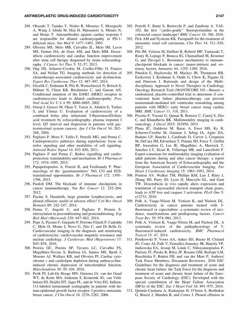
288. Okazaki T, Tanaka Y, Nishio R, Mitsuiye T, MizoguchiA, Wang J, Ishida M, Hiai H, Matsumori A, Minato N,and Honjo T. Autoantibodies against cardiac troponin Iare responsible for dilated cardiomyopathy in PD-1-deficient mice. Nat Med 9: 1477–1483, 2003.
289. Oliveira MS, Melo MB, Carvalho JL, Melo IM, LavorMS, Gomes DA, de Goes AM, and Melo MM. Doxor-ubicin cardiotoxicity and cardiac function improvementafter stem cell therapy diagnosed by strain echocardiog-raphy. J Cancer Sci Ther 5: 52–57, 2013.
290. Ong DS, Scherrer-Crosbie M, Coelho-Filho O, FrancisSA, and Neilan TG. Imaging methods for detection ofchemotherapy-associated cardiotoxicity and dysfunction.Expert Rev Cardiovasc Ther 12: 487–497, 2014.
291. Ozcelik C, Erdmann B, Pilz B, Wettschureck N, Britsch S,Hubner N, Chien KR, Birchmeier C, and Garratt AN.Conditional mutation of the ErbB2 (HER2) receptor incardiomyocytes leads to dilated cardiomyopathy. ProcNatl Acad Sci U S A 99: 8880–8885, 2002.
292. Oztop I, Gencer M, Okan T, Yaren A, Altekin E, TurkerS, and Yilmaz U. Evaluation of cardiotoxicity of acombined bolus plus infusional 5-fluorouracil/folinicacid treatment by echocardiography, plasma troponin Ilevel, QT interval and dispersion in patients with gas-trointestinal system cancers. Jpn J Clin Oncol 34: 262–268, 2004.
293. Pagliaro P, Moro F, Tullio F, Perrelli MG, and Penna C.Cardioprotective pathways during reperfusion: focus onredox signaling and other modalities of cell signaling.Antioxid Redox Signal 14: 833–850, 2011.
294. Pagliaro P and Penna C. Redox signalling and cardio-protection: translatability and mechanism. Br J Pharmacol172: 1974–1995, 2015.
295. Papapetropoulos A, Foresti R, and Ferdinandy P. Phar-macology of the ‘gasotransmitters’ NO, CO and H2S:translational opportunities. Br J Pharmacol 172: 1395–1396, 2015.
296. Pardoll DM. The blockade of immune checkpoints incancer immunotherapy. Nat Rev Cancer 12: 252–264,2012.
297. Paydas S. Dasatinib, large granular lymphocytosis, andpleural effusion: useful or adverse effect? Crit Rev OncolHematol 89: 242–247, 2014.
298. Penna C, Angotti C, and Pagliaro P. Protein S-nitrosylation in preconditioning and postconditioning. ExpBiol Med (Maywood) 239: 647–662, 2014.
299. Pepe A, Pizzino F, Gargiulo P, Perrone-Filardi P, CadedduC, Mele D, Monte I, Novo G, Zito C, and Di Bella G.Cardiovascular imaging in the diagnosis and monitoringof cardiotoxicity: cardiovascular magnetic resonance andnuclear cardiology. J Cardiovasc Med (Hagerstown) 17:S45–S54, 2016.
300. Pereira GC, Pereira SP, Tavares LC, Carvalho FS,Magalhaes-Novais S, Barbosa IA, Santos MS, Bjork J,Moreno AJ, Wallace KB, and Oliveira PJ. Cardiac cyto-chrome c and cardiolipin depletion during anthracycline-induced chronic depression of mitochondrial function.Mitochondrion 30: 95–104, 2016.
301. Perik PJ, Lub-De Hooge MN, Gietema JA, van der GraafWT, de Korte MA, Jonkman S, Kosterink JG, van Veld-huisen DJ, Sleijfer DT, Jager PL, and de Vries EG. Indium-111-labeled trastuzumab scintigraphy in patients with hu-man epidermal growth factor receptor 2-positive metastaticbreast cancer. J Clin Oncol 24: 2276–2282, 2006.
302. Petrelli F, Barni S, Bertocchi P, and Zaniboni A. TAS-102, the first ‘‘cardio-gentle’’ fluoropyrimidine in thecolorectal cancer landscape? BMC Cancer 16: 386, 2016.
303. Pick AM and Nystrom KK. Pazopanib for the treatment ofmetastatic renal cell carcinoma. Clin Ther 34: 511–520,2012.
304. Pitt JM, Vetizou M, Daillere R, Roberti MP, Yamazaki T,Routy B, Lepage P, Boneca IG, Chamaillard M, KroemerG, and Zitvogel L. Resistance mechanisms to immune-checkpoint blockade in cancer: tumor-intrinsic and -ex-trinsic factors. Immunity 44: 1255–1269, 2016.
305. Pituskin E, Haykowsky M, Mackey JR, Thompson RB,Ezekowitz J, Koshman S, Oudit G, Chow K, Pagano JJ,and Paterson I. Rationale and design of the Multi-disciplinary Approach to Novel Therapies in CardiologyOncology Research Trial (MANTICORE 101—Breast): arandomized, placebo-controlled trial to determine if con-ventional heart failure pharmacotherapy can preventtrastuzumab-mediated left ventricular remodeling amongpatients with HER2+ early breast cancer using cardiacMRI. BMC Cancer 11: 318, 2011.
306. Pizzino F, Vizzari G, Qamar R, Bomzer C, Carerj S, ZitoC, and Khandheria BK. Multimodality imaging in cardi-ooncology. J Oncol 2015: 263950, 2015.
307. Plana JC, Galderisi M, Barac A, Ewer MS, Ky B,Scherrer-Crosbie M, Ganame J, Sebag IA, Agler DA,Badano LP, Banchs J, Cardinale D, Carver J, CerqueiraM, DeCara JM, Edvardsen T, Flamm SD, Force T, GriffinBP, Jerusalem G, Liu JE, Magalhaes A, Marwick T,Sanchez LY, Sicari R, Villarraga HR, and Lancellotti P.Expert consensus for multimodality imaging evaluation ofadult patients during and after cancer therapy: a reportfrom the American Society of Echocardiography and theEuropean Association of Cardiovascular Imaging. EurHeart J Cardiovasc Imaging 15: 1063–1093, 2014.
308. Pointon AV, Walker TM, Phillips KM, Luo J, Riley J,Zhang SD, Parry JD, Lyon JJ, Marczylo EL, and GantTW. Doxorubicin in vivo rapidly alters expression andtranslation of myocardial electron transport chain genes,leads to ATP loss and caspase 3 activation. PLoS One 5:e12733, 2010.
309. Polk A, Vaage-Nilsen M, Vistisen K, and Nielsen DL.Cardiotoxicity in cancer patients treated with 5-fluorouracil or capecitabine: a systematic review of inci-dence, manifestations and predisposing factors. CancerTreat Rev 39: 974–984, 2013.
310. Polk A, Vistisen K, Vaage-Nilsen M, and Nielsen DL. Asystematic review of the pathophysiology of 5-fluorouracil-induced cardiotoxicity. BMC PharmacolToxicol 15: 47, 2014.
311. Ponikowski P, Voors AA, Anker SD, Bueno H, ClelandJG, Coats AJ, Falk V, Gonzalez-Juanatey JR, Harjola VP,Jankowska EA, Jessup M, Linde C, Nihoyannopoulos P,Parissis JT, Pieske B, Riley JP, Rosano GM, Ruilope LM,Ruschitzka F, Rutten FH, and van der Meer P; Authors/Task Force Members; Document Reviewers. 2016 ESCGuidelines for the diagnosis and treatment of acute andchronic heart failure: the Task Force for the diagnosis andtreatment of acute and chronic heart failure of the Euro-pean Society of Cardiology (ESC). Developed with thespecial contribution of the Heart Failure Association(HFA) of the ESC. Eur J Heart Fail 18: 891–975, 2016.
312. Quintas-Cardama A, Kantarjian H, O’brien S, BorthakurG, Bruzzi J, Munden R, and Cortes J. Pleural effusion in
ANTINEOPLASTIC DRUG-INDUCED CARDIOTOXICITY 2147
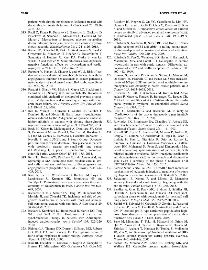
patients with chronic myelogenous leukemia treated withdasatinib after imatinib failure. J Clin Oncol 25: 3908–3914, 2007.
313. Racil Z, Razga F, Drapalova J, Buresova L, Zackova D,Palackova M, Semerad L, Malaskova L, Haluzik M, andMayer J. Mechanism of impaired glucose metabolismduring nilotinib therapy in patients with chronic myelog-enous leukemia. Haematologica 98: e124–e126, 2013.
314. Rainer PP, Doleschal B, Kirk JA, Sivakumaran V, Saad Z,Groschner K, Maechler H, Hoefler G, Bauernhofer T,Samonigg H, Hutterer G, Kass DA, Pieske B, von Le-winski D, and Pichler M. Sunitinib causes dose-dependentnegative functional effects on myocardium and cardio-myocytes. BJU Int 110: 1455–1462, 2012.
315. Ranpura V, Hapani S, Chuang J, and Wu S. Risk of car-diac ischemia and arterial thromboembolic events with theangiogenesis inhibitor bevacizumab in cancer patients: ameta-analysis of randomized controlled trials. Acta Oncol49: 287–297, 2010.
316. Rastogi S, Sharov VG, Mishra S, Gupta RC, Blackburn B,Belardinelli L, Stanley WC, and Sabbah HN. Ranolazinecombined with enalapril or metoprolol prevents progres-sive LV dysfunction and remodeling in dogs with mod-erate heart failure. Am J Physiol Heart Circ Physiol 295:H2149–H2155, 2008.
317. Rea D, Mirault T, Cluzeau T, Gautier JF, Guilhot F,Dombret H, and Messas E. Early onset hypercholester-olemia induced by the 2nd-generation tyrosine kinase in-hibitor nilotinib in patients with chronic phase-chronicmyeloid leukemia. Haematologica 99: 1197–1203, 2014.
318. Reck M, Kaiser R, Mellemgaard A, Douillard JY, OrlovS, Krzakowski M, von Pawel J, Gottfried M, BondarenkoI, Liao M, Gann CN, Barrueco J, Gaschler-Markefski B,Novello S, and LUME-Lung 1 Study Group. Docetaxelplus nintedanib versus docetaxel plus placebo in patientswith previously treated non-small-cell lung cancer(LUME-Lung 1): a phase 3, double-blind, randomisedcontrolled trial. Lancet Oncol 15: 143–155, 2014.
319. Reus TL, Robert AW, Da Costa MB, de Aguiar AM, andStimamiglio MA. Secretome from resident cardiac stro-mal cells stimulates proliferation, cardiomyogenesis andangiogenesis of progenitor cells. Int J Cardiol 221: 396–403, 2016.
320. Riad A, Bien S, Westermann D, Becher PM, Loya K,Landmesser U, Kroemer HK, Schultheiss HP, andTschope C. Pretreatment with statin attenuates the cardi-otoxicity of Doxorubicin in mice. Cancer Res 69: 695–699, 2009.
321. Richards CJ, Je Y, Schutz FA, Heng DY, Dallabrida SM,Moslehi JJ, and Choueiri TK. Incidence and risk of con-gestive heart failure in patients with renal and nonrenalcell carcinoma treated with sunitinib. J Clin Oncol 29:3450–3456, 2011.
322. Rickard J, Kumbhani DJ, Baranowski B, Martin DO, TangWH, and Wilkoff BL. Usefulness of cardiac re-synchronization therapy in patients with Adriamycin-induced cardiomyopathy. Am J Cardiol 105: 522–526,2010.
323. Ridnour LA, Thomas DD, Donzelli S, Espey MG, RobertsDD, Wink DA, and Isenberg JS. The biphasic nature ofnitric oxide responses in tumor biology. Antioxid RedoxSignal 8: 1329–1337, 2006.
324. Rini BI, Escudier B, Tomczak P, Kaprin A, Szczylik C,Hutson TE, Michaelson MD, Gorbunova VA, Gore ME,
Rusakov IG, Negrier S, Ou YC, Castellano D, Lim HY,Uemura H, Tarazi J, Cella D, Chen C, Rosbrook B, KimS, and Motzer RJ. Comparative effectiveness of axitinibversus sorafenib in advanced renal cell carcinoma (axis):a randomised phase 3 trial. Lancet 378: 1931–1939,2011.
325. Rohrbach S, Niemann B, Silber RE, and Holtz J. Neur-egulin receptors erbB2 and erbB4 in failing human myo-cardium—depressed expression and attenuated activation.Basic Res Cardiol 100: 240–249, 2005.
326. Rohrbach S, Yan X, Weinberg EO, Hasan F, Bartunek J,Marchionni MA, and Lorell BH. Neuregulin in cardiachypertrophy in rats with aortic stenosis. Differential ex-pression of erbB2 and erbB4 receptors. Circulation 100:407–412, 1999.
327. Romano S, Fratini S, Procaccini V, Stifano G, Mancini M,Di Mauro M, Ficorella C, and Penco M. Serial measure-ments of NT-proBNP are predictive of not-high-dose an-thracycline cardiotoxicity in breast cancer patients. Br JCancer 105: 1663–1668, 2011.
328. Rosenthal A, Luthi J, Belohlavek M, Kortum KM, Moo-kadam F, Mayo A, Fonseca R, Bergsagel PL, Reeder CB,Mikhael JR, and Stewart AK. Carfilzomib and the cardi-orenal system in myeloma: an endothelial effect? BloodCancer J 6: e384, 2016.
329. Rosti G, Martinelli G, and Baccarani M. In reply to‘Cardiotoxicity of the cancer therapeutic agent imatinibmesylate’. Nat Med 13: 15, 2007.
330. Rowinsky EK, Eisenhauer EA, Chaudhry V, Arbuck SG,and Donehower RC. Clinical toxicities encountered withpaclitaxel (Taxol). Semin Oncol 20: 1–15, 1993.
331. Russell SD, Lyon A, Lenihan DJ, Moreau P, Joshua D,ChngW-J, Palumbo A, Goldschmidt H, Hajek R, Facon T,Ludwig H, Pour L, Niesvizky R, Oriol A, Rosinol L,Suvorov A, Gaidano G, Goranova-Marinova V, Gillen-water HH, Mohamed N, Feng S, and Dimopoulos MA.Serial echocardiographic assessment of patients (pts) withrelapsed multiple myeloma (RMM) receiving carfilzomiband dexamethasone (Kd) vs bortezomib and dexametha-sone (Vd): a substudy of the phase 3 Endeavor Trial(NCT01568866). Blood 126: 4250, 2015.
332. Salesse S and Verfaillie CM. BCR/ABL: from molecularmechanisms of leukemia induction to treatment of chronicmyelogenous leukemia. Oncogene 21: 8547–8559, 2002.
333. Salvatorelli E, Menna P, and Minotti G. Managinganthracycline-induced cardiotoxicity: beginning with theend in mind. Future Cardiol 11: 363–366, 2015.
334. Sandler A, Gray R, Perry MC, Brahmer J, Schiller JH,Dowlati A, Lilenbaum R, and Johnson DH. Paclitaxel-carboplatin alone or with bevacizumab for nonsmall-celllung cancer. N Engl J Med 355: 2542–2550, 2006.
335. Sandri MT, Salvatici M, Cardinale D, Zorzino L, PasseriniR, Lentati P, Leon M, Civelli M, Martinelli G, and CipollaCM. N-terminal pro-B-type natriuretic peptide after high-dose chemotherapy: a marker predictive of cardiac dys-function? Clin Chem 51: 1405–1410, 2005.
336. Sano M, Minamino T, Toko H, Miyauchi H, Orimo M,Qin Y, Akazawa H, Tateno K, Kayama Y, Harada M,Shimizu I, Asahara T, Hamada H, Tomita S, MolkentinJD, Zou Y, and Komuro I. p53-induced inhibition of HIF-1 causes cardiac dysfunction during pressure overload.Nature 446: 444–448, 2007.
337. Santos DL, Moreno AJM, Leino RL, Froberg MK, andWallace KB. Carvedilol protects against doxorubicin-
2148 TOCCHETTI ET AL.
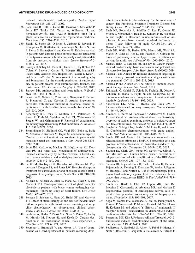
induced mitochondrial cardiomyopathy Toxicol ApplPharmacol 185: 218–227, 2002.
338. Sanz-Ruiz R, Bolli R, Gersh BJ, Janssens S, Menasche P,Perin EC, Taylor DA, Terzic A, Willerson JT, andFernandez-Aviles. The TACTIS initiative: time for aglobal alliance on cardiovascular regenerative medicine.Eur Heart J 37: 2208–2211, 2016.
339. Sasaki K, Strom SS, O’Brien S, Jabbour E, Ravandi F,Konopleva M, Borthakur G, Pemmaraju N, Daver N, JainP, Pierce S, Kantarjian H, and Cortes JE. Relative survivalin patients with chronic-phase chronic myeloid leukaemiain the tyrosine-kinase inhibitor era: analysis of patient datafrom six prospective clinical trials. Lancet Haematol 5:e186–e193, 2015.
340. Sawaya H, Sebag IA, Plana JC, Januzzi JL, Ky B, Tan TC,Cohen V, Banchs J, Carver JR, Wiegers SE, Martin RP,Picard MH, Gerszten RE, Halpern EF, Passeri J, Kuter I,and Scherrer-Crosbie M. Assessment of echocardiographyand biomarkers for the estende prediction of cardiotoxi-city in patients treated with anthracyclines, taxanes, andtrastuzumab. Circ Cardiovasc Imaging 5: 596–603, 2012.
341. Sawyer DB. Anthracyclines and heart failure. N Engl JMed 368: 1154–1156, 2013.
342. Scartozzi M, Galizia E, Chiorrini S, Giampieri R, BerardiR, Pierantoni C, and Cascinu S. Arterial hypertensioncorrelates with clinical outcome in colorectal cancer pa-tients treated with first-line bevacizumab. Ann Oncol 20:227–230, 2009.
343. Schermuly RT, Dony E, Ghofrani HA, Pullamsetti S,Savai R, Roth M, Sydykov A, Lai YJ, Weissmann N,Seeger W, and Grimminger F. Reversal of experimentalpulmonary hypertension by PDGF inhibition. J Clin Invest115: 2811–2821, 2005.
344. Schmidinger M, Zielinski CC, Vogl UM, Bojic A, BojicM, Schukro C, Ruhsam M, Hejna M, and Schmidinger H.Cardiac toxicity of sunitinib and sorafenib in patients withmetastatic renal cell carcinoma. J Clin Oncol 26: 5204–5212, 2008.
345. Scott JM, Khakoo A, Mackey JR, Haykowsky MJ, Dou-glas PS, and Jones LW. Modulation of anthracycline-induced cardiotoxicity by aerobic exercise in breast can-cer: current evidence and underlying mechanisms. Cir-culation 124: 642–650, 2011.
346. Scott JM, Koelwyn GJ, Hornsby WE, Khouri M, Pep-percorn J, Douglas PS, and Jones LW. Exercise therapy astreatment for cardiovascular and oncologic disease after adiagnosis of early-stage cancer. Semin Oncol 40: 218–228,2013.
347. Seicean S, Seicean A, Alan N, Plana JC, Budd GT, andMarwick TH. Cardioprotective effect of b-adrenoceptorblockade in patients with breast cancer undergoing che-motherapy: follow-up study of heart failure. Circ HeartFail 6: 420–426, 2013.
348. Seicean S, Seicean A, Plana JC, Budd GT, and MarwickTH. Effect of statin therapy on the risk for incident heartfailure in patients with breast cancer receiving anthracy-cline chemotherapy: an observational clinical cohortstudy. J Am Coll Cardiol 60: 2384–2390, 2012.
349. Seidman A, Hudis C, Pierri MK, Shak S, Paton V, AshbyM, Murphy M, Stewart SJ, and Keefe D. Cardiac dys-function in the trastuzumab clinical trials experience. JClin Oncol 20: 1215–1221, 2002.
350. Seymour L, Bramwell V, and Moran LA. Use of dexra-zoxane as a cardioprotectant in patients receiving doxo-
rubicin or epirubicin chemotherapy for the treatment ofcancer. The Provincial Systemic Treatment Disease SiteGroup. Cancer Prev Control 3: 145–159, 1999.
351. Shah NP, Rousselot P, Schiffer C, Rea D, Cortes JE,Milone J, Mohamed H, Healey D, Kantarjian H, HochhausA, and Saglio G. Dasatinib in imatinib-resistant or -in-tolerant chronic-phase, chronic myeloid leukemia pa-tients: 7-year follow-up of study CA180-034. Am JHematol 91: 869–874, 2016.
352. Shah NP, Wallis N, Farber HW, Mauro MJ, Wolf RA,Mattei D, Guha M, Rea D, and Peacock A. Clinical fea-tures of pulmonary arterial hypertension in patients re-ceiving dasatinib. Am J Hematol 90: 1060–1064, 2015.
353. Shalkey-Hahn V, Lenihan DJ, and Ky B. Cancer therapy-induced cardiotoxicity: basic mechanisms and potential car-dioprotective therapies. J Am Heart Assoc 3: e000665, 2014.
354. Sharma P and Allison JP. Immune checkpoint targeting incancer therapy: toward combination strategies with cura-tive potential. Cell 161: 205–214, 2015.
355. Sharma P and Allison JP. The future of immune check-point therapy. Science 348: 56–61, 2015.
356. Shimazaki C, Ochiai N, Uchida R, Fuchida SI, Okano A,Ashihara E, Inaba T, Fujita N, and Nakagawa M. In-tramuscular edema as a complication of treatment withimatinib. Leukemia 17: 804–805, 2003.
357. Shoemaker LK, Arora U, Rocha, and Lima CM. 5-fluorouracil-induced coronary vasospasm. Cancer Control11: 46–49, 2004.
358. Simunek T, Sterba M, Popelova O, Adamcova M, HrdinaR, and Gersl V. Anthracycline-induced cardiotoxicity:overview of studies examining the roles of oxidative stressand free cellular iron. Pharmacol Rep 61: 154–171, 2009.
359. Singh CK, Siddigui IA, El-Abd S, Mukhtar H, and AhmadN. Combination chemoprevention with grape antioxi-dants. Mol Nutr Food Res 60: 1406–1415, 2016.
360. Singla DK and Abdelli LS. Embryonic stem cells andreleased factors stimulate c-kit/FLK-1 progenitor cells andpromote neovascularization in doxorubicin-induced car-diomyopathy. Cell Transplant 24: 1043–1052, 2015.
361. Slamon DJ, Clark GM, Wong SG, Levin WJ, Ullrich A,and McGuire WL. Human breast cancer: correlation ofrelapse and survival with amplification of the HER-2/neuoncogene. Science 235: 177–182, 1987.
362. Slamon DJ, Leyland-Jones B, Shak S, Fuchs H, Paton V,Bajamonde A, Fleming T, Eiermann W, Wolter J, PegramM, Baselga J, and Norton L. Use of chemotherapy plus amonoclonal antibody against her2 for metastatic breastcancer that overexpresses HER2. N Engl J Med 344: 783–792, 2001.
363. Smith RR, Barile L, Cho HC, Leppo MK, Hare JM,Messina E, Giacomello A, Abraham MR, and Marban E.Regenerative potential of cardiosphere-derived cells ex-panded from percutaneous endomyocardial biopsy speci-mens. Circulation 115: 896–908, 2007.
364. Soga M, Kamal FA, Watanabe K, Ma M, Palaniyandi S,Prakash P, Veeraveedu P, Mito S, Kunisaki M, TachikawaH, Kodama M, and Aizawa Y. Effects of angiotensin IIreceptor blocker (candesartan) in daunorubicin-inducedcardiomyopathic rats. Int J Cardiol 110: 378–385, 2006.
365. Sorrentino MF, Kim J, Foderaro AE, and Truesdell AG. 5-fluorouracil induced cardiotoxicity: review of the litera-ture. Cardiol J 19: 453–458, 2012.
366. Spallarossa P, Garibaldi S, Altieri P, Fabbi P, Manca V,Nasti S, Rossettin P, Ghigliotti G, Ballestrero A, Patrone F,
ANTINEOPLASTIC DRUG-INDUCED CARDIOTOXICITY 2149
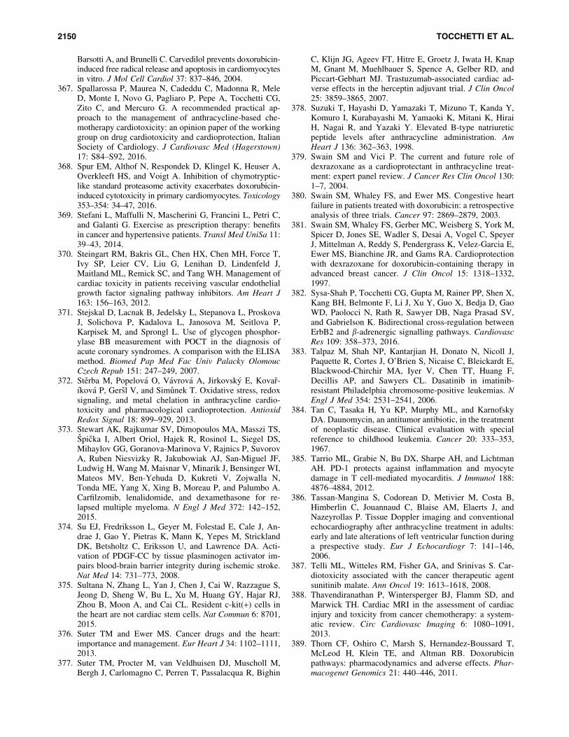
Barsotti A, and Brunelli C. Carvedilol prevents doxorubicin-induced free radical release and apoptosis in cardiomyocytesin vitro. J Mol Cell Cardiol 37: 837–846, 2004.
367. Spallarossa P, Maurea N, Cadeddu C, Madonna R, MeleD, Monte I, Novo G, Pagliaro P, Pepe A, Tocchetti CG,Zito C, and Mercuro G. A recommended practical ap-proach to the management of anthracycline-based che-motherapy cardiotoxicity: an opinion paper of the workinggroup on drug cardiotoxicity and cardioprotection, ItalianSociety of Cardiology. J Cardiovasc Med (Hagerstown)17: S84–S92, 2016.
368. Spur EM, Althof N, Respondek D, Klingel K, Heuser A,Overkleeft HS, and Voigt A. Inhibition of chymotryptic-like standard proteasome activity exacerbates doxorubicin-induced cytotoxicity in primary cardiomyocytes. Toxicology353–354: 34–47, 2016.
369. Stefani L, Maffulli N, Mascherini G, Francini L, Petri C,and Galanti G. Exercise as prescription therapy: benefitsin cancer and hypertensive patients. Transl Med UniSa 11:39–43, 2014.
370. Steingart RM, Bakris GL, Chen HX, Chen MH, Force T,Ivy SP, Leier CV, Liu G, Lenihan D, Lindenfeld J,Maitland ML, Remick SC, and Tang WH. Management ofcardiac toxicity in patients receiving vascular endothelialgrowth factor signaling pathway inhibitors. Am Heart J163: 156–163, 2012.
371. Stejskal D, Lacnak B, Jedelsky L, Stepanova L, ProskovaJ, Solichova P, Kadalova L, Janosova M, Seitlova P,Karpisek M, and Sprongl L. Use of glycogen phosphor-ylase BB measurement with POCT in the diagnosis ofacute coronary syndromes. A comparison with the ELISAmethod. Biomed Pap Med Fac Univ Palacky OlomoucCzech Repub 151: 247–249, 2007.
372. Sterba M, Popelova O, Vavrova A, Jirkovsky E, Kovar-ıkova P, Gersl V, and Simunek T. Oxidative stress, redoxsignaling, and metal chelation in anthracycline cardio-toxicity and pharmacological cardioprotection. AntioxidRedox Signal 18: 899–929, 2013.
373. Stewart AK, Rajkumar SV, Dimopoulos MA, Masszi TS,Spicka I, Albert Oriol, Hajek R, Rosinol L, Siegel DS,Mihaylov GG, Goranova-Marinova V, Rajnics P, SuvorovA, Ruben Niesvizky R, Jakubowiak AJ, San-Miguel JF,Ludwig H, Wang M, Maisnar V, Minarik J, Bensinger WI,Mateos MV, Ben-Yehuda D, Kukreti V, Zojwalla N,Tonda ME, Yang X, Xing B, Moreau P, and Palumbo A.Carfilzomib, lenalidomide, and dexamethasone for re-lapsed multiple myeloma. N Engl J Med 372: 142–152,2015.
374. Su EJ, Fredriksson L, Geyer M, Folestad E, Cale J, An-drae J, Gao Y, Pietras K, Mann K, Yepes M, StricklandDK, Betsholtz C, Eriksson U, and Lawrence DA. Acti-vation of PDGF-CC by tissue plasminogen activator im-pairs blood-brain barrier integrity during ischemic stroke.Nat Med 14: 731–773, 2008.
375. Sultana N, Zhang L, Yan J, Chen J, Cai W, Razzague S,Jeong D, Sheng W, Bu L, Xu M, Huang GY, Hajar RJ,Zhou B, Moon A, and Cai CL. Resident c-kit(+) cells inthe heart are not cardiac stem cells. Nat Commun 6: 8701,2015.
376. Suter TM and Ewer MS. Cancer drugs and the heart:importance and management. Eur Heart J 34: 1102–1111,2013.
377. Suter TM, Procter M, van Veldhuisen DJ, Muscholl M,Bergh J, Carlomagno C, Perren T, Passalacqua R, Bighin
C, Klijn JG, Ageev FT, Hitre E, Groetz J, Iwata H, KnapM, Gnant M, Muehlbauer S, Spence A, Gelber RD, andPiccart-Gebhart MJ. Trastuzumab-associated cardiac ad-verse effects in the herceptin adjuvant trial. J Clin Oncol25: 3859–3865, 2007.
378. Suzuki T, Hayashi D, Yamazaki T, Mizuno T, Kanda Y,Komuro I, Kurabayashi M, Yamaoki K, Mitani K, HiraiH, Nagai R, and Yazaki Y. Elevated B-type natriureticpeptide levels after anthracycline administration. AmHeart J 136: 362–363, 1998.
379. Swain SM and Vici P. The current and future role ofdexrazoxane as a cardioprotectant in anthracycline treat-ment: expert panel review. J Cancer Res Clin Oncol 130:1–7, 2004.
380. Swain SM, Whaley FS, and Ewer MS. Congestive heartfailure in patients treated with doxorubicin: a retrospectiveanalysis of three trials. Cancer 97: 2869–2879, 2003.
381. Swain SM, Whaley FS, Gerber MC, Weisberg S, York M,Spicer D, Jones SE, Wadler S, Desai A, Vogel C, SpeyerJ, Mittelman A, Reddy S, Pendergrass K, Velez-Garcia E,Ewer MS, Bianchine JR, and Gams RA. Cardioprotectionwith dexrazoxane for doxorubicin-containing therapy inadvanced breast cancer. J Clin Oncol 15: 1318–1332,1997.
382. Sysa-Shah P, Tocchetti CG, Gupta M, Rainer PP, Shen X,Kang BH, Belmonte F, Li J, Xu Y, Guo X, Bedja D, GaoWD, Paolocci N, Rath R, Sawyer DB, Naga Prasad SV,and Gabrielson K. Bidirectional cross-regulation betweenErbB2 and b-adrenergic signalling pathways. CardiovascRes 109: 358–373, 2016.
383. Talpaz M, Shah NP, Kantarjian H, Donato N, Nicoll J,Paquette R, Cortes J, O’Brien S, Nicaise C, Bleickardt E,Blackwood-Chirchir MA, Iyer V, Chen TT, Huang F,Decillis AP, and Sawyers CL. Dasatinib in imatinib-resistant Philadelphia chromosome-positive leukemias. NEngl J Med 354: 2531–2541, 2006.
384. Tan C, Tasaka H, Yu KP, Murphy ML, and KarnofskyDA. Daunomycin, an antitumor antibiotic, in the treatmentof neoplastic disease. Clinical evaluation with specialreference to childhood leukemia. Cancer 20: 333–353,1967.
385. Tarrio ML, Grabie N, Bu DX, Sharpe AH, and LichtmanAH. PD-1 protects against inflammation and myocytedamage in T cell-mediated myocarditis. J Immunol 188:4876–4884, 2012.
386. Tassan-Mangina S, Codorean D, Metivier M, Costa B,Himberlin C, Jouannaud C, Blaise AM, Elaerts J, andNazeyrollas P. Tissue Doppler imaging and conventionalechocardiography after anthracycline treatment in adults:early and late alterations of left ventricular function duringa prespective study. Eur J Echocardiogr 7: 141–146,2006.
387. Telli ML, Witteles RM, Fisher GA, and Srinivas S. Car-diotoxicity associated with the cancer therapeutic agentsunitinib malate. Ann Oncol 19: 1613–1618, 2008.
388. Thavendiranathan P, Wintersperger BJ, Flamm SD, andMarwick TH. Cardiac MRI in the assessment of cardiacinjury and toxicity from cancer chemotherapy: a system-atic review. Circ Cardiovasc Imaging 6: 1080–1091,2013.
389. Thorn CF, Oshiro C, Marsh S, Hernandez-Boussard T,McLeod H, Klein TE, and Altman RB. Doxorubicinpathways: pharmacodynamics and adverse effects. Phar-macogenet Genomics 21: 440–446, 2011.
2150 TOCCHETTI ET AL.
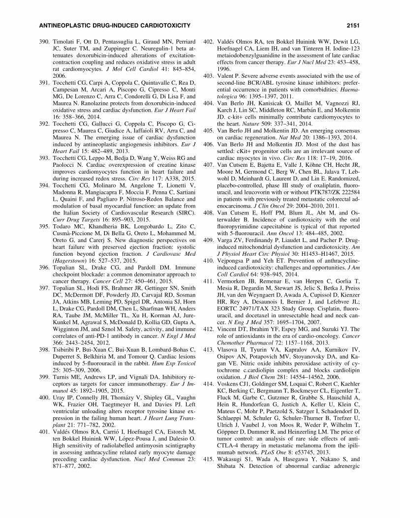
390. Timolati F, Ott D, Pentassuglia L, Giraud MN, PerriardJC, Suter TM, and Zuppinger C. Neuregulin-1 beta at-tenuates doxorubicin-induced alterations of excitation-contraction coupling and reduces oxidative stress in adultrat cardiomyocytes. J Mol Cell Cardiol 41: 845–854,2006.
391. Tocchetti CG, Carpi A, Coppola C, Quintavalle C, Rea D,Campesan M, Arcari A, Piscopo G, Cipresso C, MontiMG, De Lorenzo C, Arra C, Condorelli G, Di Lisa F, andMaurea N. Ranolazine protects from doxorubicin-inducedoxidative stress and cardiac dysfunction. Eur J Heart Fail16: 358–366, 2014.
392. Tocchetti CG, Gallucci G, Coppola C, Piscopo G, Ci-presso C, Maurea C, Giudice A, Iaffaioli RV, Arra C, andMaurea N. The emerging issue of cardiac dysfunctioninduced by antineoplastic angiogenesis inhibitors. Eur JHeart Fail 15: 482–489, 2013.
393. Tocchetti CG, Leppo M, Bedja D, Wang Y, Weiss RG andPaolocci N. Cardiac overexpression of creatine kinaseimproves cardiomyocytes function in heart failure andduring increased redox stress. Circ Res 117: A338, 2015.
394. Tocchetti CG, Molinaro M, Angelone T, Lionetti V,Madonna R, Mangiacapra F, Moccia F, Penna C, SartianiL, Quaini F, and Pagliaro P. Nitroso-Redox Balance andmodulation of basal myocardial function: an update fromthe Italian Society of Cardiovascular Research (SIRC).Curr Drug Targets 16: 895–903, 2015.
395. Todaro MC, Khandheria BK, Longobardo L, Zito C,Cusma-Piccione M, Di Bella G, Oreto L, Mohammed M,Oreto G, and Carerj S. New diagnostic perspectives onheart failure with preserved ejection fraction: systolicfunction beyond ejection fraction. J Cardiovasc Med(Hagerstown) 16: 527–537, 2015.
396. Topalian SL, Drake CG, and Pardoll DM. Immunecheckpoint blockade: a common denominator approach tocancer therapy. Cancer Cell 27: 450–461, 2015.
397. Topalian SL, Hodi FS, Brahmer JR, Gettinger SN, SmithDC, McDermott DF, Powderly JD, Carvajal RD, SosmanJA, Atkins MB, Leming PD, Spigel DR, Antonia SJ, HornL, Drake CG, Pardoll DM, Chen L, Sharfman WH, AndersRA, Taube JM, McMiller TL, Xu H, Korman AJ, Jure-Kunkel M, Agrawal S, McDonald D, Kollia GD, Gupta A,Wigginton JM, and Sznol M. Safety, activity, and immunecorrelates of anti-PD-1 antibody in cancer. N Engl J Med366: 2443–2454, 2012.
398. Tsibiribi P, Bui-Xuan C, Bui-Xuan B, Lombard-Bohas C,Duperret S, Belkhiria M, and Tomour Q. Cardiac lesionsinduced by 5-fluorouracil in the rabbit. Hum Exp Toxicol25: 305–309, 2006.
399. Turnis ME, Andrews LP, and Vignali DA. Inhibitory re-ceptors as targets for cancer immunotherapy. Eur J Im-munol 45: 1892–1905, 2015.
400. Uray IP, Connelly JH, Thomazy V, Shipley GL, VaughnWK, Frazier OH, Taegtmeyer H, and Davies PJ. Leftventricular unloading alters receptor tyrosine kinase ex-pression in the failing human heart. J Heart Lung Trans-plant 21: 771–782, 2002.
401. Valdes Olmos RA, Carrio I, Hoefnagel CA, Estorch M,ten Bokkel Huinink WW, Lopez-Pousa J, and Dalesio O.High sensitivity of radiolabelled antimyosin scintigraphyin assessing anthracycline related early myocyte damagepreceding cardiac dysfunction. Nucl Med Commun 23:871–877, 2002.
402. Valdes Olmos RA, ten Bokkel Huinink WW, Dewit LG,Hoefnagel CA, Liem IH, and van Tinteren H. Iodine-123metaiodobenzylguanidine in the assessment of late cardiaceffects from cancer therapy. Eur J Nucl Med 23: 453–458,1996.
403. Valent P. Severe adverse events associated with the use ofsecond-line BCR/ABL tyrosine kinase inhibitors: prefer-ential occurrence in patients with comorbidities. Haema-tologica 96: 1395–1397, 2011.
404. Van Berlo JH, Kanisicak O, Maillet M, Vagnozzi RJ,Karch J, Lin SC, Middleton RC, Marban E, and MolkentinJD. c-kit+ cells minimally contribute cardiomyocytes tothe heart. Nature 509: 337–341, 2014.
405. Van Berlo JH and Molkentin JD. An emerging consensuson cardiac regeneration. Nat Med 20: 1386–1393, 2014.
406. Van Berlo JH and Molkentin JD. Most of the dust hassettled: cKit+ progenitor cells are an irrelevant source ofcardiac myocytes in vivo. Circ Res 118: 17–19, 2016.
407. Van Cutsem E, Bajetta E, Valle J, Kohne CH, Hecht JR,Moore M, Germond C, Berg W, Chen BL, Jalava T, Leb-wohl D, Meinhardt G, Laurent D, and Lin E. Randomized,placebo-controlled, phase III study of oxaliplatin, fluoro-uracil, and leucovorin with or without PTK787/ZK 222584in patients with previously treated metastatic colorectal ad-enocarcinoma. J Clin Oncol 29: 2004–2010, 2011.
408. Van Cutsem E, Hoff PM, Blum JL, Abt M, and Os-terwalder B. Incidence of cardiotoxicity with the oralfluoropyrimidine capecitabine is typical of that reportedwith 5-fluorouracil. Ann Oncol 13: 484–485, 2002.
409. Varga ZV, Ferdinandy P, Liaudet L, and Pacher P. Drug-induced mitochondrial dysfunction and cardiotoxicity. AmJ Physiol Heart Circ Physiol 30: H1453–H1467, 2015.
410. Vejpongsa P and Yeh ET. Prevention of anthracycline-induced cardiotoxicity: challenges and opportunities. J AmColl Cardiol 64: 938–945, 2014.
411. Vermorken JB, Remenar E, van Herpen C, Gorlia T,Mesia R, Degardin M, Stewart JS, Jelic S, Betka J, PreissJH, van den Weyngaert D, Awada A, Cupissol D, KienzerHR, Rey A, Desaunois I, Bernier J, and Lefebvre JL;EORTC 24971/TAX 323 Study Group. Cisplatin, fluoro-uracil, and docetaxel in unresectable head and neck can-cer. N Eng J Med 357: 1695–1704, 2007.
412. Vincent DT, Ibrahim YF, Espey MG, and Suzuki YJ. Therole of antioxidants in the era of cardio-oncology. CancerChemother Pharmacol 72: 1157–1168, 2013.
413. Vlasova II, Tyurin VA, Kapralov AA, Kurnikov IV,Osipov AN, Potapovich MV, Stoyanovsky DA, and Ka-gan VE. Nitric oxide inhibits peroxidase activity of cy-tochrome c.cardiolipin complex and blocks cardiolipinoxidation. J Biol Chem 281: 14554–14562, 2006.
414. Voskens CJ1, Goldinger SM, Loquai C, Robert C, KaehlerKC, Berking C, Bergmann T, Bockmeyer CL, Eigentler T,Fluck M, Garbe C, Gutzmer R, Grabbe S, Hauschild A,Hein R, Hundorfean G, Justich A, Keller U, Klein C,Mateus C, Mohr P, Paetzold S, Satzger I, Schadendorf D,Schlaeppi M, Schuler G, Schuler-Thurner B, Trefzer U,Ulrich J, Vaubel J, von Moos R, Weder P, Wilhelm T,Goppner D, Dummer R, and Heinzerling LM. The price oftumor control: an analysis of rare side effects of anti-CTLA-4 therapy in metastatic melanoma from the ipili-mumab network. PLoS One 8: e53745, 2013.
415. Wakasugi S1, Wada A, Hasegawa Y, Nakano S, andShibata N. Detection of abnormal cardiac adrenergic
ANTINEOPLASTIC DRUG-INDUCED CARDIOTOXICITY 2151
neuron activity in adriamycin-induced cardiomyopathywith iodine-125-metaiodobenzylguanidine. J Nucl Med33: 208–214, 1992.
416. Walker J, Bhullar N, Fallah-Rad N, Lytwyn M, Golian M,Fang T, Summers AR, Singal PK, Barac I, Kirkpatrick ID,and Jassal DS. Role of three-dimensional echocardiogra-phy in breast cancer: comparison with two-dimensionalechocardiography, multiple-gated acquisition scans, andcardiac magnetic resonance imaging. J Clin Oncol 28:3429–3436, 2010.
417. Walsh K and Shiojima I. Cardiac growth and angiogenesiscoordinated by intertissue interactions. J Clin Invest 117:3176–3179, 2007.
418. Wang J, Okazaki IM, Yoshida T, Chikuma S, Kato Y,Nakaki F, Hiai H, Honjo T, and Okazaki T. PD-1 defi-ciency results in the development of fatal myocarditis inMRL mice. Int Immunol 22: 443–452, 2010.
419. Wang X, Ha T, Liu L, Zou J, Zhang X, Kalbfleisch J, GaoX, Williams D, and Li C. Increased expression of microRNA-146a decreases myocardial ischaemia/reperfusion injury.Cardiovasc Res 97: 432–442, 2013.
420. Weber JS, Kahler KC, and Hauschild A. Management ofimmune-related adverse events and kinetics of responsewith ipilimumab. J Clin Oncol 30: 2691–2697, 2012.
421. Welti J, Loges S, Dimmeler S, and Carmeliet P. Recentmolecular discoveries in angiogenesis and antiangiogenictherapies in cancer. J Clin Invest 123: 3190–3200, 2013.
422. Will Y, Dykens JA, Nadanaciva S, Hirakawa B, JamiesonJ, Marroguin LD, Hynes J, Patyna S, and Jessen BA. Ef-fect of the multitargeted tyrosine kinase inhibitors im-atinib, dasatinib, sunitinib and sorefenib on mitochondrialfunction in isolated rat heart mitochondria and H9c2 cells.Toxicol Sci 106: 153–161, 2008.
423. Willis MS and Patterson C. Into the heart: the emergingrole of the ubiquitin-proteasome system. J Mol CellCardiol 41: 567–579, 2006.
424. Willis MS and Patterson C. Proteotoxicity and cardiacdysfunction—Alzheimer’s disease of the heart? N Engl JMed 368: 455–464, 2013.
425. Wouters KA, Kremer LC, Miller TL, Herman EH, andLipshultz SE. Protecting against anthracycline-inducedmyocardial damage: a review of the most promisingstrategies. Br J Haematol 131: 561–578, 2005.
426. Xi L, Zhu SG, Das A, Chen Q, Durrant D, Hobbs DC,Lesnefsky EJ, and Kukreja RC. Dietary inorganic nitratealleviates doxorubicin cardiotoxicity: mechanisms andimplications. Nitric Oxide 26: 274–284, 2012.
427. Xiao Y, Yin J, Wei J, and Shang Z. Incidence and risk ofcardiotoxicity associated with bortezomib in the treatmentof cancer: a systematic review and meta-analysis. PLoSOne 9: e87671, 2014.
428. Yamagishi S and Takeuchi M. Telmisartan is a promisingcardiometabolic sartan due to its unique PPAR-gamma-inducing property. Med Hypotheses 64: 476–478, 2005.
429. Yeh ET and Bickford CL. Cardiovascular complicationsof cancer therapy: incidence, pathogenesis, diagnosis, andmanagement. J Am Coll Cardiol 53: 2231–2247, 2009.
430. Yue TL, Cheng HY, Lysko PG, McKenna PJ, FeuersteinR, Gu JL, Lysko KA, Davis LL, and Feuerstein G. Car-vedilol, a new vasodilator and beta adrenoceptor antago-nist, is an antioxidant and free radical scavenger. JPharmacol Exp Ther 263: 92–98, 1992.
431. Yun S, Vincelette ND, Mansour I, Hariri D, and MotamedS. Late onset ipilimumab-induced pericarditis and peri-cardial effusion: a rare but life threatening complication.Case Rep Oncol Med 2015: 794842, 2015.
432. Zamorano JL, Lancellotti P, Rodriguez Munoz D, Abo-yans V, Asteggiano R, Galderisi M, Habib G, Lenihan DJ,Lip GY, Lyon AR, Lopez Fernandez T, Mohty D, PiepoliMF, Tamargo J, Torbicki A, and Suter TM; Authors/TaskForce Members; ESC Committee for Practice Guidelines(CPG). 2016 ESC Position Paper on cancer treatments andcardiovascular toxicity developed under the auspices ofthe ESC Committee for Practice Guidelines: The TaskForce for cancer treatments andcardiovascular toxicity ofthe European Society of Cardiology (ESC). Eur Heart J37: 2768–2801, 2016.
433. Zanardelli M, Micheli L, Cinci L, Failli P, Ghelardini C,and Di Cesare Mannelli L. Oxalipla: 2tin neurotoxicityinvolves peroxisome alterations. PPARc agonism as pre-ventive pharmacological approach. PLoS One 9: e102758,2014.
434. Zang J, Wu S, Tang L, Xu X, Bai J, Ding C, Chang Y,Yue L, Kang E, and He J. Incidence and risk of QTcinterval prolongation among cancer patients treated withvandetanib: a systematic review and meta-analysis. PLoSOne 7: e30353Z, 2012.
435. Zhang S, Liu X, Bawa-Khalfe T, Lu LS, Lyu YL, Liu LF,and Yeh ET. Identification of the molecular basis ofdoxorubicin-induced cardiotoxicity. Nat Med 18: 1639–1642, 2012.
436. Zhang Y, Sivakumaran P, Newcomb AE, Hernandez D,Harris N, Khana bdali R, Liu GS, Kelly DJ, Pebay A,Hewitt AW, Boyle A, Harvey R, Morrison WA, ElliotDA, Dusting GJ, and Lim SY. Cardiac repair with a novelpopulation of mesenchymal stem cells resident in thehuman heart. Stem Cells 33: 3100–3113, 2015.
437. Zhu W, Zou Y, Aikawa R, Harada K, Kudoh S, UozumiH, Hayashi D, Gu Y, Yamazaki T, Nagai R, Yazaki Y,and Komuro I. MAPK superfamily plays an important rolein daunomycin-induced apoptosis of cardiac myocytes.Circulation 100: 2100–2107, 1999.
438. Zito C, Longobardo L, Cadeddu C, Monte I, Novo G,Dell’Oglio S, Pepe A, Madonna R, Tocchetti CG, andMele D. Cardiovascular imaging in the diagnosis andmonitoring of cardiotoxicity: role of echocardiography. JCardiovasc Med (Hagerstown) 17: S35–S44, 2016.
Address correspondence to:Dr. Pasquale Pagliaro
Department of Clinical and Biological SciencesUniversity of Turin
Regione Gonzole 1010043 Turin
Italy
E-mail: [email protected]
Date of first submission to ARS Central, October 26, 2016;date of final revised submission, April 6, 2017; date of ac-ceptance, April 10, 2017.
2152 TOCCHETTI ET AL.
Abbreviations Used
b-AR¼ b-adrenergic receptor5-FU¼ 5-fluorouracil
ACE-Is¼ angiotensin-converting enzyme inhibitorsALL¼ acute lymphoblastic leukemia
AMPK¼AMP-activated protein kinaseANTs¼ anthracyclinesAPCs¼ antigen-presenting cellsARBs¼ angiotensin receptor blockersATE¼ arterial thromboembolic events
BDNF¼ brain-derived neurotrophic factorBNP¼ brain natriuretic peptideCAD¼ coronary artery diseaseCML¼ chronic myeloid leukemiaCMR¼ cardiac magnetic resonance
CPCs/CSCs¼ cardiac-derived progenitor or stem cellsCSCs¼ cardiac stem cell cardiotoxicity
CSF1R¼ colony-stimulating factor 1 receptorCTLA-4¼ cytotoxic T lymphocyte-associated
protein 4CTX¼ cardiotoxicity
CV¼ cardiovascularCVD¼ cardiovascular diseaseECG¼ electrocardiogram
EGFR¼ epidermal growth factor receptoreNOS¼ endothelial NOS
ER¼ endoplasmic reticulumErbB2 (HER2)¼ human epidermal growth factor receptor 2
ERK¼ extracellular signal-regulated kinaseFABP¼ fatty acid-binding proteinFLT3¼ FMS-related tyrosine kinase 3
HF¼ heart failureGLS¼ global longitudinal strain
GPBB¼ glycogen phosphorylase BBGPCR¼G protein-coupled receptor
GPx¼ glutathione peroxidaseH2O2¼ hydrogen peroxide
H2S¼ hydrogen sulfideIL-6¼ interleukin 6
iNOS¼ inducible nitric oxide synthaseIRAEs¼ immune-related adverse events
LV¼ left ventricleLVEF¼ left ventricular ejection fraction
MAPK¼mitogen-activated protein kinasemiR-146a¼microRNA-146a
mPTPs¼mitochondrial permeability transitionpores
MUGA¼multiple-gated acquisitionNO¼ nitric oxide
NOSs¼ nitric oxide synthasesNOXs¼NAD(P)H oxidasesNRG1¼ neuregulin 1
ONOO-¼ peroxynitritePAD¼ peripheral arterial diseasePAH¼ pulmonary arterial hypertensionPD-1¼ programmed cell death protein 1
PDE-5¼ phosphodiesterase-5PDGFR¼ platelet-derived growth factor receptorPD-L1¼ programmed death-ligand 1
PRADA¼ prevention of cardiac dysfunction duringadjuvant breast cancer therapy
RAAS¼ renin-angiotensin-aldosterone systemRET¼RE arranged during transfectionRNS¼ reactive nitrogen speciesROS¼ reactive oxygen speciesSNO¼ S-nitrosylationSOD¼ superoxide dismutaseTCR¼T cell receptor
TE¼ thromboembolismTKIs¼ tyrosine kinase inhibitorsTop2¼ topoisomerase 2TrkB¼ tyrosine kinase receptor
VEGF¼ vascular endothelial growth factorVEGFR¼VEGF receptor
ANTINEOPLASTIC DRUG-INDUCED CARDIOTOXICITY 2153


