Embed Size (px)
Citation preview

Antioxidant activity of Phyllanthus niruri L. herbs:in vitro and in vivo models and isolation ofactive compound
Muhammad Da’i, Arifah Sri Wahyuni, Ika Trisharyanti DK, Tanti Azizah, Andi Suhendi, Azis Saifudin
Faculty of Pharmacy, Universitas Muhammadiyah Surakarta, Indonesia..
Correspondence to: Muhammad Da’i, E-mail: [email protected]
Received October 5, 2015. Accepted October 15, 2015
||ABSTRACT
Background: Phyllanthus niruri, Thymus vulgaris, Centella asiatica, and Apium graveolens L. have been frequently used asmedical herbs for various diseases. However, the use of the herbs is not based on any evidence of their contents. Aims andObjective: To examine their potential activity of becoming antioxidant, using in vitro and in vivo models and isolate activecompound. Materials and Methods: The antioxidant activity of the samples was assessed using 1,1-diphenyl-2-picrylhydrazyl (DPPH) as radical compounds. The decrease of DPPH radical was measured using spectrophotometer at 517nm, after an incubation for 30 min. The highest antioxidant activity of the sample was then continued with in vivo modelusing Sprague Dawley (SD) rats. Paracetamol was used as an inducer to increase the malondialdehyde (MDA) levels inplasma, with doses of 2.5 g/kg BW. The rats were divided into four groups: negative control, dose of 50 mg/kg BW, dose of100 mg/kg BW, and dose of 200 mg/kg BW. Isolation method was guided by antioxidant activity by using fractionation andcolumn chromatography. The obtained compound was then characterized by 1H-nuclear magnetic resonance (NMR) andcompared with the earlier study. Result: The IC50 value of these samples were 14.21 ± 0.73; 14.41 ± 1.13; 98.66 ± 9.59;and 237.33 ± 17.26 mg/mL, respectively, and the total phenolic contents were 81.59 ± 2.85; 154.09 ± 11.61; 6.36 ±
3.99; 2.03 ± 0.78 mg/g of sample extract, respectively. The in vivo study showed that P. niruri with dose of 100 mg/kg BWand 200 mg/kg BW were able to reduce MDA levels by 90.44% and 92.64%, respectively. Meanwhile, the proton and carbonNMR spectra showed that the isolate was quercetin. The IC50 value of the isolate, being 5.85 mg/mL, was lower than that ofvitamin E, which was 6.85 mg/mL. Conclusion: The extract shows a potential effect with antioxidant activity, both in in vitroand in vivo models. Quercetin is the active compound, the antioxidant activity of which is higher than that of vitamin E.
KEY WORDS: Antioxidant; Phyllanthus niruri L.; Quercetin
||INTRODUCTION
Free radicals have one or more electrons that are not paired inthe outer orbitals[1] and produced under normal circumstances
of metabolic processes.[2] Free radicals can damage proteins,lipids, carbohydrates, and nucleic acids.[3] Over longer terms,some damages can lead to degenerative diseases.[4] In diabeticpatients, for example, hyperglycemia causes glucose autooxidation, glycation of proteins, and activation of the polyolmetabolic pathway that will accelerate the formation ofreactive oxygen species (ROS).[5] This will result in animbalance between antioxidants in the body and free radicals,which is the beginning of oxidative damage, known as oxidativestress. The risk of the disease can be minimized by the use ofantioxidant compounds that can stabilize radicals.[6,7]
Obviously, the sources of antioxidants are natural andsynthetic antioxidants. The Indonesian people have used avariety of medicinal plants to heal various diseases. A previous
Access this article online
Website: http://www.njppp.comQuick Response Code:
DOI: 10.5455/njppp.2015.5.0510201575
National Journal of Physiology, Pharmacy and Pharmacology Online 2016. © 2016 Muhammad Da’i. This is an Open Access article distributed under the terms of the Creative Commons Attribution 4.0
International License (http://creativecommons.org/licenses/by/4.0/), allowing third parties to copy and redistribute the material in any medium or format and to remix, transform, and build upon
the material for any purpose, even commercially, provided the original work is properly cited and states its license.
2016 | Vol 6 | Issue 1 National Journal of Physiology, Pharmacy and Pharmacology32
Research Article
study on 23 medicinal plants in Indonesia found four plants(Phyllanthus niruri, Thymus vulgaris, Centella asiatica, andApium graveolens) that are potential as antioxidant agents, theactivity of which is better or equal to that of vitamin E.[8] Theseplants contain alkaloids, flavonoids, lignin, tannins, coumarin,polyphenols, saponins, and terpenoids.[9] Active compoundsthat may work as antioxidants are tannins and[10] alkaloids[11]
and phenolic components, such as phenolic diterpene, phenolicacids, and flavonoids.[12] Owing to reducing properties, theantioxidant activity of the phenolic content can stabilize singletoxygen, capture free radicals, electron donors, and chelatingmetals.[13]
Therefore, this study was conducted to explore the potentialof antioxidants from those four medicinal plants that have thebest antioxidant activity based on in vitro and in vivo models,isolate the active compounds, and characterize the structure byproton and carbon nuclear magnetic resonance (NMR). Theantioxidant activity is generally contributed by the phenoliccontent of the extract. The isolate was assessed throughantioxidant activity and compared with vitamin E as anestablished antioxidant agent.
||MATERIALS AND METHODS
The laboratory equipment used for this study was glassware(Pyrex), analytical balance (Ohaus), minispin (Eppendorf),centrifuge (Centrifuge PLC Series), Spectrophotometer uv-vis(Shimadzu 1140), ultrasound (Branson), and NMR (JEOL).
The materials used in this study consisted of P. niruri L.,T. vulgaris, C. asiatica, and A. graveolens (local market), ratsSprague Dawley (SD) strain, paracetamol 2.5 g/kg BW(Pharmaceutical grade, Brataco Chemica), TMP-X (SigmaAldrich), TCA 20% (Merck), and TBA 0.67% (Sigma Aldrich,T5500), 1,1-diphenyl-2-picrylhydrazyl (DPPH) (Merck), ethanol(Merck), gallic acid (Merck), Follin–Ciocalteu (Merck), Na2CO3
(Merck), Vitamin E (Sigma Aldrich), and plate sillica gel GF254.This study was approved by Health Research Ethics Committeeof Faculty of Medicine, Universitas Muhammadiyah Surakarta.
ExtractionThe extraction process was performed by maceration usingethanol 96% as a solvent with a 1:7 ratio. Liquid extract wasevaporated using a rotary evaporator and then heated in thewater bath until a thick extract was formed.
Determination of Antioxidant in In Vitro ModelSeven hundred mL of DPPH solution (400mM) plus 1.5 mL ofsample solution of various concentrations were then left in thedark for 30 min and then read at a wavelength of 517 nm. Theblank used was 1.5 mL sample solution added by ethanol up to5.0 mL, while the control used was 700 mL DPPH solution addedby ethanol up to 5.0 mL. Inhibition concentration (IC50) was theconcentration of extract or fraction that gave antioxidantactivity of 50%.
Determination of total phenolic contentsThe total phenolic content was determined using the Folin–Ciocalteu reagent according to the method of Waterhouse.[14]
The reaction mixture consisted of 100 mL of extract and 100 mLFolin–Ciocalteu reagent. The mixture was kept in darkness for5 min, then combined with 75 mL of sodium carbonate andmade up to 5.0 mL of aquadest. In order to complete thereaction, the mixture was kept in darkness for 45 min at roomtemperature. The absorbance at 742.5 nm was measured usinga UV-Vis spectrophotometer (Shimadzu Corp.). Gallic acid wasused as a standard, and the result was expressed as gallic acidequivalent (GAE)/g extract.
Determination of Antioxidant In VivoModel[15]
The hepatoprotective activity of P. niruri extract was deter-mined with hepatotoxic rat model using paracetamol as theinducer. After 7 days of acclimatization, the rats were classifiedinto four groups of five. The treatments were done for 8 days.Group I served as the normal control and received onlyaquabidest (0.4 mL/kg BW) for 8 days. The group II served asthe toxic control and was administered with paracetamolsuspension (2.5 g/kg BW, po) once in every 72 h. Groups IIIuntil V were administered with extract (50; 100; and 200 mg/kg BW, po) daily and paracetamol suspension (2.5 g/kg BW, po)once in 5 days. About 1 mL of blood was collected from venalateralis of rats on days 0, 5, and 8. The blood samples werecentrifuged at 3,000 rpm at room temperature to separateserum and used to estimate the MDA levels.
Determination of malondialdehyde (MDA)[16] was used toevaluate lipid peroxidation in tissue samples. A volume of theserum (0.10 mL) was transferred to a vial and mixed with 2.45mL of 20% (w/v) TCA solution and 2.45 L of a 0.67% (w/v)solution of TBA, and the final volume was adjusted to 5.0 mlwith distilled water. Each vial was tightly capped and heated ina boiling water bath for 20 min. The vials were then cooled toambient temperature. The absorbance of supernatant wasmeasured at 532 nm (Shimadzu, UV Mini). The calibrationcurve used TMP-X as the standard, and the result was convertedto MDA. The series of standard were 0.16; 0.33; 0.66; 1.32; and2.64 mg/mL of TMP-X.
Isolation and CharacterizationIsolation of active compound of P. niruri was conducted byguiding antioxidant in in vitro models. The extract of P. niruriwas fractionated to obtain n-hexene fraction, ethyl acetatefraction, and residues fraction. All fractions obtained were thenassessed with antioxidant activity. Then, ethyl acetate fraction,which has a potential activity, was fractionated with vacuumcolumn chromatography. Stationary and mobile phases usedwere silica gel (Merck 7734) and a mixture of CHCl3:n-hexene(8:2), and CHCl3:methanol (9:1, 8:2, 7:3, 6:4, and 5:5). Elutionwas performed three times with volume of each mobile phase of350 mL. The volume of the collected fraction was 130 mL.Altogether, 58 fractions were obtained. On the basis of the thin
National Journal of Physiology, Pharmacy and Pharmacology 2016 | Vol 6 | Issue 1 33
Antioxidant activity of Phyllanthus niruri L. herbs Da’i et al.
layer chromatography (TLC) examination, the same profiles offractions were then combined.
After TLC examination, nine subfractions were collected,and, then, the antioxidant activity was determined. The highestantioxidant activity of subfraction, then fractionated by flashchromatography, silica G 60 (Cat Number 7731) as stationaryphase and mixtures of n-hexene:ethyl acetate (4:1; 3:2; 2:3; and1:4), CHCl3:ethyl acetate (4:1; 3:2; 2:3; and 1:4), and CHCl3:methanol (7:3) as the mobile phase. Elution was performedthree times, each using 200 mL. Fraction was collected; theseparation profiles were then evaluated by TLC. The sameprofiles combined resulted in four subfractions, and, then, theantioxidant activity was assessed. Owing to the impuritycontent of subfraction, the purification was performed bypreparative RP-TLC (Silica-C18, as a stationary phase andmixtures of acetonitrile:methanol:water = 1:1:1 as mobilephase). Separated subfraction was then cleaned up using flashchromatography [methanol:CHCl3 = 3:7, as mobile phase, andsilica G 60 (Cat Number 7731), as stationary phase]. The isolatecharacterized by proton NMR (JEOL, 500 MHZ), CDCl3 was usedas a solvent. Antioxidant activity of isolate was assessed andcompared with vitamin E.
||RESULT
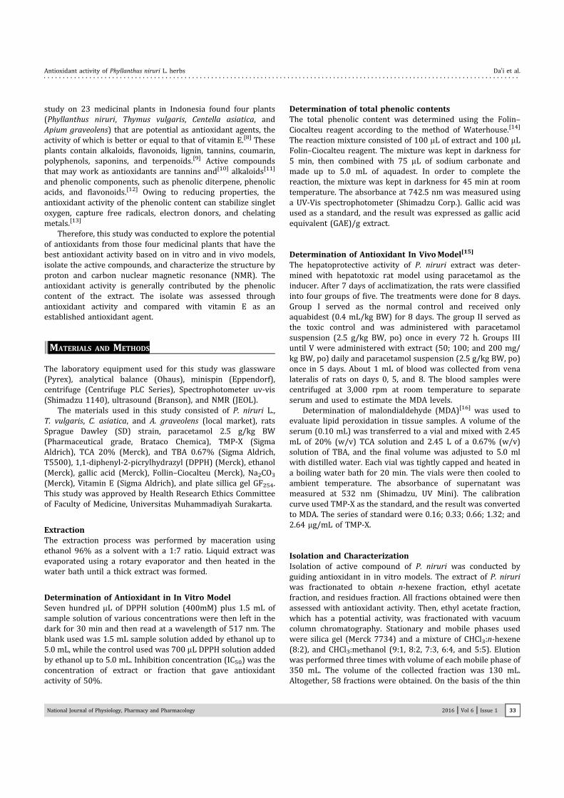
Screening Antioxidant ActivityPotential antioxidant activity was classified based on the IC50value.[17] The result showed that the antioxidant activity ofP. niruri and T. vulgaris showed similar values, around 14 mg/mL, which were close to that of the vitamin E [Table 1]. Theother extracts are not active as an antioxidant.
Antioxidant of In Vivo ModelsThe objective of in vivo models was to optimize the induc-tion time of paracetamol as hepatotoxic inducer, which is
administered per oral. Induction of hepatic may increase MDAlevels in serum. As shown in Table 2, the optimum time tomeasure the MDA levels is 72 h after induction. The increase ofMDA is almost double of the first time, about 62.88 ± 12.38mg/mL.
Paracetamol metabolite, N-acetyl-p-benzoquinone imine(NAPQI) is a reactive electrophilic that induces tissuesimpairment,[18] indicated with the increase of MDA levels inthe serum.[19]
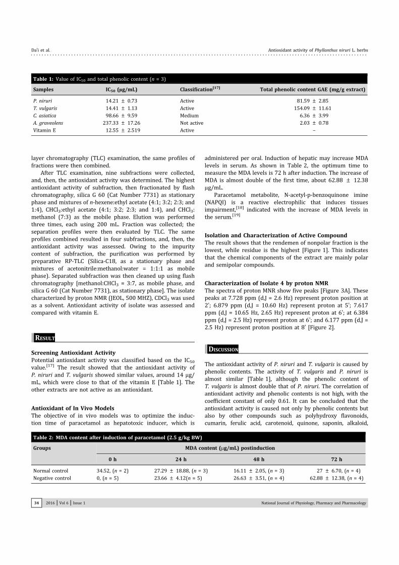
Isolation and Characterization of Active CompoundThe result shows that the rendemen of nonpolar fraction is thelowest, while residue is the highest [Figure 1]. This indicatesthat the chemical components of the extract are mainly polarand semipolar compounds.
Characterization of Isolate 4 by proton NMRThe spectra of proton MNR show five peaks [Figure 3A]. Thesepeaks at 7.728 ppm (d,J = 2.6 Hz) represent proton position at2’; 6.879 ppm (d,J = 10.60 Hz) represent proton at 5’; 7.617ppm (d,J = 10.65 Hz, 2.65 Hz) represent proton at 6’; at 6.384ppm (d,J = 2.5 Hz) represent proton at 6’; and 6.177 ppm (d,J =2.5 Hz) represent proton position at 8’ [Figure 2].
||DISCUSSION
The antioxidant activity of P. niruri and T. vulgaris is caused byphenolic contents. The activity of T. vulgaris and P. niruri isalmost similar [Table 1], although the phenolic content ofT. vulgaris is almost double that of P. niruri. The correlation ofantioxidant activity and phenolic contents is not high, with thecoefficient constant of only 0.61. It can be concluded that theantioxidant activity is caused not only by phenolic contents butalso by other compounds such as polyhydroxy flavonoids,cumarin, ferulic acid, carotenoid, quinone, saponin, alkaloid,
Table 1: Value of IC50 and total phenolic content (n = 3)
Samples IC50 (lg/mL) Classification[17] Total phenolic content GAE (mg/g extract)
P. niruri 14.21 ± 0.73 Active 81.59 ± 2.85
T. vulgaris 14.41 ± 1.13 Active 154.09 ± 11.61
C. asiatica 98.66 ± 9.59 Medium 6.36 ± 3.99
A. graveolens 237.33 ± 17.26 Not active 2.03 ± 0.78
Vitamin E 12.55 ± 2.519 Active –
Table 2: MDA content after induction of paracetamol (2.5 g/kg BW)
Groups MDA content (mg/mL) postinduction
0 h 24 h 48 h 72 h
Normal control 34.52, (n = 2) 27.29 ± 18.88, (n = 3) 16.11 ± 2.05, (n = 3) 27 ± 6.70, (n = 4)
Negative control 0, (n = 5) 23.66 ± 4.12(n = 5) 26.63 ± 3.51, (n = 4) 62.88 ± 12.38, (n = 4)
2016 | Vol 6 | Issue 1 National Journal of Physiology, Pharmacy and Pharmacology34
Da’i et al. Antioxidant activity of Phyllanthus niruri L. herbs
and tanins. The differences of antioxidant activity can be theinfluence of several factors, such as the solvent of extraction,chemical composition of samples, and determination methods.[21]
Evaluation of antioxidant of in vivo models was conducted byreferring to the activity of previous study on antioxidant and
antidiabetic effects of P. niruri.[22] The data [Table 3] shows thatP. niruri presents an antioxidant activity that is characterized bythe decrease of MDA levels in the serum. The decrease of P. niruridose of 100 and 200 mg/kg BWwas almost close. This study alsoshows a similar result[23] that P. niruri presents an antioxidant
Figure 1: Scheme for isolation of antioxidant compound from extract P. niruri.
National Journal of Physiology, Pharmacy and Pharmacology 2016 | Vol 6 | Issue 1 35
Antioxidant activity of Phyllanthus niruri L. herbs Da’i et al.

activity using in vivo model. Chemical composition of P. niruri,such as transiphytol, 4-t-butylpyrocatechol, phyllanthin, andhypophyllantin can protect cytotoxicity hepatocyte culture.[24,25]
Isolation is guided by the antioxidant activity, initiated byredissolving the extract by using n-hexene (as nonpolarsolvent), ethyl acetate (as semipolar solvent), and residue(as polar fraction).On the basis of the evaluation of the
antioxidant, the candidate to be isolated was the ethyl acetatefraction. In order to obtain simple components in the fractions,gradient polarity mobile phase is used in the vacuum columnchromatography. The fraction obtained classified based on theTLC profiles became nine subfractions. The assessment ofantioxidant activity shows that group VI shows the highestactivity [Figure 1]. On the basis of the results in Fig 1, theantioxidant activity of isolated compound are higher thanvitamin E (IC50 = 6.85 mg/mL) which is known as an establishedantioxidant compound. Isolate 4 can be elucidated by using 1H-MR and compared with the reference. The spectra of isolate[Figure 3A] are quite similar to the result of an earlier study(Fig. 3B).[26] Therefore, the isolate is quercetin.
||CONCLUSION
P. niruri L. herb is a promising antioxidant herb that has thebest DPPH radical scavenging activity when compared withT. vulgaris, C. asiatica, and Apium graveolens L.; meanwhile, theextract at dose 200 mg/kg BW could decrease MDA levelsaround 92.64%. Isolation guided by the antioxidant activity ofP. niruri as a promising antioxidant herb showed the fractions
Figure 2: Structure of quercetin.
Figure 3: Proton NMR spectra of isolate from P. niruri (A) and quercetin (B)[26].
Table 3: MDA levels of groups treatments
Groups MDA levels (lg/mL) days Reduction MDA levels (%)
0 5 8
Negative control ND, (n = 5) 36.34 ± 6.59, (n = 5) 81.65 ± 11.09, (n = 5) –
P. niruri, 50 mg/kg BW 10.60 ± 1.50,(n = 3) 70.52, (n = 2) 32.27 ± 14.08*, (n = 3) 60.47
P. niruri, 100 mg/kg BW ND, (n = 5) 1.58, (n = 2) 6.99*, (n = 1) 91.44
P. niruri, 200 mg/kg BW ND, (n = 5) 22.55 ± 2.73, (n = 5) 6.01*, (n = 1) 92.64
ND = not detected.*p o 0.05, compared with negative control group (one-way ANOVA, followed by a t-test)
2016 | Vol 6 | Issue 1 National Journal of Physiology, Pharmacy and Pharmacology36
Da’i et al. Antioxidant activity of Phyllanthus niruri L. herbs
of n-hexane, ethyl acetate, and ethanol, which showed values ofIC50 such as 14.21 ± 0.73, 282.84 ± 11.77, 2.67 ± 0.74, and8.85 ± 1.54 mg/mL, respectively. Quercetin was successfullyisolated from the extract and showed a higher antioxidantactivity than vitamin E.
||REFERENCES
1. Zou Y, Lu Y, Wei D. Antioxidant activity of flavonoid-rich extract ofHypericum perforatum L. in vitro. J Agric Food Chem. 2004;52(16):5032–9.
2. Hariyatmi. Kemampuan Vitamin E sebagai Antioksidan terhadapRadikal Bebas pada Lanjut Usia. MIPA, Jurusan Pendidikan BiologiFKIP UMS. Surakarta. 2004;14(1):52–60.
3. Suirta IWNM, Puspawati, Gumiati NK. Isolasi dan IdentifikasiSenyawa Aktif Larvasida dari Biji Mimba (Azadirachta indika A.Juss) Terhadap Larva Nyamuk Demam Berdarah (Aedes aegypti).J Kimia. 2007;1(1):47–54.
4. Temple NJ. Antioxidants and disease: more questions thananswers. Nutrition Research. 2000;20(3):449–59.
5. Ueno Y, Kizaki M, Nakagiri R, Kamiya T, Sumi H, Oswa T. Dietaryglutathione protects rats from diabetic nephropathy and neuro-pathy. J. Nutr. 2000;132(5):897–900.
6. Amrun MH, Umiyah Evi UU. Uji Aktivitas Antioksidan Ekstrak Air danEkstrak Metanol beberapa Varian Buah Kenitu (Chrysophyllum cainitoL.) dari Daerah Jember Berkala Penelelitian Hayati. 2007;13:45–50.
7. Rosen P, Tritschler HJ, Packer L. Vascular complications in diabetes:mechanisms and the influence of antioxidants In:(Eds.) Handbook ofAntioxidants, 2nd edn. , New York: Marcel Dekker, 2000. pp. 511–24.
8. Da’i M, Wahyuni AS, Kusumowati ITD, Setiawan D, Dhi’fi HJ,Suhendi A, et al. The Corrrelation of Phenolic Content with AntioxidantActivity Five Types Indonesian Herbs International Seminar onTranslational Research in Cancer Chemoprevention2011.
9. Paithankar VV, Raut KS, Charde RM, Vyas JV. Phyllanthus niruri:a magic herb. Res Pharm. 2011;1(4):1–9.
10. Zhang LL, Lin YM. Antioxidant tannins from Syzygium cumini fruitAfr J Biotechnol. 2009;8(10):2301–9.
11. Alfarabi M, Bintang M, Suryani, Safithri M. The Comparative ability ofantioxidant activity of Piper crocatum in inhibiting fatty acid oxidationand free radical scavenging. Hayati J Biosci. 2010;17(4):201–4.
12. Javanmardi J, Stushnoff C, Locke E, Vivanco JM. Antioxidant activityand total phenolic content of Iranian Ocimum accessions. FoodChem. 2003;83(4):547–50.
13. Karadeniz F, Burdurlu HS, Koca N, Soyer Y. Antioxidant activity ofselected fruits and vegetables grown in Turkey. Turk J Agric For.2004;29:297–303.
14. Waterhouse AL. Current Protocols in Food Analytical Chemistry.United States: John Wiley and Sons, 2000. pp. 11–8.
15. Sriningsih. Efek protektif pemberian ekstrak etanol herba meniran(Phyllantus niruri L.) terhadap aktivitas dan kapasitas fagositosismakrofag peritoneum tikus. Artocarpus. 2006;6(2):91–6.
16. Ohkawa H, Ohishi N, Yagi K. Assay for lipid peroxides in animaltissues by thiobarbituric acid reaction. Anal Biochem. 1979;95(2):351–8.
17. Reynertson KA. Phytochemical Analysis of Bioactive Constituentsfrom Edible Myrtaceae Fruit, Dissertation, The City University ofNew York2007. pp. 70–4.
18. Corcoran GB, Racz WJ, Smith CV, Mitchell JR. Effects ofN-acetylcysteine on acetaminophen covalent binding and hepaticnecrosis in mice. J Pharmacol Exp Ther. 1985;232(3):864–72.
19. Josephy PD. Molecular Toxicology. , New York: Oxford UniversityPress, 2007. pp. 97–104.
20. Widhihastuti E. Pengukuran Aktivitas Antioksidan dengan MetodeDPPH serta Korelasinya dengan Kadar Fenolik pada Lima JenisHerba Bahan Obat Alam Indonesia. Skripsi. Fakultas FarmasiUniversitas Muhammadiyah Surakarta. 2011.
21. Widyastuti N. Pengukuran Aktivitas Antioksidan dengan MetodeCUPRAC, DPPH, dan FRAP serta Korelasinya dengan Fenol danFlavonoid pada Enam Tanaman. Skripsi. Fakultas MIPA InstitutPertanian Bogor2010.
22. Da’i M, Wahyuni AS, Kusumowati ITD, Suhendi A, Azizah T.Pemanfaatan Antioksidan Alami Pada Upaya Pencegahan Kompli-kasi Akibat Stres Oksidatif Pada Tikus Yang Diinduksi DiabetesMelitus, Laporan Penelitian LPPM UMS2012.
23. Shivanandappa T, Harish R. Antioxidant activity and hepatopro-tective potential of Phyllanthus niruri. Food Chem. 2006;95(2):180–5.
24. Shamasundar KV, Singh B, Thakur RS, Husain A, Kiso Y, Hikino H.Antihepatotoxic principles of Phyllanthus niruri herbs. J Ethno-pharmacol. 1985;14(1):41–4.
25. Makoshi MS, Adanyeguh IM, Nwatu LI. Hepatoprotective effect ofPhyllanthus niruri aqueous extract in acetaminophen sub-acutecexposure rabbits. J Vet Med Anim Health. 2013;5(1):8–15.
26. Foo LY, Lu Y, Molan AL, Woodfield DR, McNabb WC. The phenolsand prodelphinidins of white clover flowers. Phytochemistry.2000;54(5):539–48.
How to cite this article: Da’i M, Wahyuni AS, DK IT, Azizah T,Suhendi A, Saifudin A. Antioxidant activity of Phyllanthus niruri L.herbs: in vitro and in vivo models and isolation of active compound.Natl J Physiol Pharm Pharmacol 2016;6:32-37.
Source of Support: Nil, Conflict of Interest: None declared.
National Journal of Physiology, Pharmacy and Pharmacology 2016 | Vol 6 | Issue 1 37
Antioxidant activity of Phyllanthus niruri L. herbs Da’i et al.


![[PHAR06] Anti Helicobacter pylori from extracts of ten species of Phyllanthus sp. with special emphasis on chloroform extracts of Phyllanthus pulcher](https://img.dokumen.tips/doc/110x75/63529537c02ce502c103a4cf/phar06-anti-helicobacter-pylori-from-extracts-of-ten-species-of-phyllanthus-sp.jpg)