Embed Size (px)
Citation preview
AR
Da
b
c
d
a
ARRAA
KGcRAM
1
tglctBkcttlN
ctaa
Uf
0d
Carbohydrate Polymers 84 (2011) 1282–1288
Contents lists available at ScienceDirect
Carbohydrate Polymers
journa l homepage: www.e lsev ier .com/ locate /carbpol
ntioxidant activity of gallate-chitooligosaccharides in mouse macrophageAW264.7 cells
ai-Hung Ngoa, Zhong-Ji Qianb, Thanh-Sang Voa, BoMi Ryua, Dai-Nghiep Ngoc, Se-Kwon Kima,d,∗
Department of Chemistry, Pukyong National University, Busan 608-737, Republic of KoreaMarine Life Research and Education Center, Chosun University, Gwangju 501-759, Republic of KoreaDepartment of Biochemistry, Faculty of Biology, University of Science, VNU-HCM, Ho Chi Minh City, Viet NamMarine Bioprocess Research Center, Pukyong National University, Busan 608-737, Republic of Korea
r t i c l e i n f o
rticle history:eceived 27 October 2010eceived in revised form 11 January 2011ccepted 14 January 2011vailable online 21 January 2011
a b s t r a c t
In this research, a novel derivative of chitooligosaccharides (COS) was synthesized by covalently linkinggallic acid and COS (gallate-COS) via carbodiimide to improve cellular antioxidant activity. The directintracellular radical scavenging effect of gallate-COS by 2′,7′-dichlorofluorescin diacetate (DCFH-DA)method, and the inhibition of oxidation of cellular macromolecules, such as DNA, protein and lipid in
eywords:allic acid covalently-linkedhitooligosaccharides (gallate-COS)eactive oxygen species (ROS)ntioxidant
mouse macrophages (RAW264.7 cells) were determined. Furthermore, with the treatment of gallate-COS, the expression levels of antioxidant enzymes such as superoxide dismutase (SOD) and glutathione(GSH) were significantly increased according to reverse transcription polymerase chain reaction (RT-PCR)and Western blot analysis. In addition, gallate-COS decreased reactive oxygen species induced activationof the nuclear transcription factor (NF-�B). Collectively, these results suggest that gallate-COS can be usedas a potential natural compound-based antioxidant in the functional food and pharmaceutical industries.
ouse macrophages (RAW264.7 cells)
. Introduction
Antioxidants may have a positive effect on human health sincehey can protect human body against deterioration by reactive oxy-en species (ROS), which attack biomolecules such as proteins,ipids and DNA, lead to many health disorders including aging,ancer, diabetes, neurodegenerative, cardiovascular and inflamma-ory diseases. Recent studies have shown that nuclear factor-kappa
(NF-�B) is a redox-associated transcription factor and plays aey role in regulating the immune responses, cell survival andell proliferation via its target genes. Cells possess enzymatic sys-em such as superoxide dismutase (SOD) and glutathione (GSH)o scavenge radicals and protect oxidative damage to major cel-ular biomolecules (Butterfield et al., 2006; Dhalla, Temsah, &etticadan, 2000; Seven, Guzel, Aslan, & Hamuryudan, 2008).
Many studies have been carried out to find novel antioxidative
ompounds from marine resources, such as chitin, chitosan andheir derivatives. Chitosan, a partially deacetylated polymer of N-cetyl glucosamine, is prepared by alkaline deacetylation of chitinnd chitooligosaccharides (COS) are partially hydrolyzed products∗ Corresponding author at: Marine Bioprocess Research Center, Pukyong Nationalniversity, Busan 608-737, Republic of Korea. Tel.: +82 51 629 7094;
ax: +82 51 629 7099.E-mail addresses: [email protected], [email protected] (S.-K. Kim).
144-8617/$ – see front matter © 2011 Elsevier Ltd. All rights reserved.oi:10.1016/j.carbpol.2011.01.022
© 2011 Elsevier Ltd. All rights reserved.
of chitosan. COS has important biological properties in medicinaland pharmaceutical applications such as antioxidative (Park, Je, &Kim, 2003), anti-bacterial (Jeon & Kim, 2000), anticoagulant (Park,Je, Jung, & Kim, 2004), immuno-stimulant (Jeon & Kim, 2001), adi-pogenesis inhibitory (Cho et al., 2008) and anticancer (Shen, Chen,Chan, Jeng, & Wang, 2009) activities.
In recent years, phenolic compounds are of great interest in com-bining with COS to improve their biological properties. Therefore,the aim of this research is to develop a novel COS derivative bycovalently linking gallic acid (3,4,5-trihydroxy benzoic acid) andCOS to improve the antioxidant activity of COS due to high antiox-idative property of gallic acid (Cho, Kim, Ahn, & Je, 2011; Pasanphan,Buettner, & Chirachanchai, 2010; Pasanphan & Chirachanchai,2008).
2. Materials and methods
2.1. Materials
COS (molecular weight 3.0–5.0 kDa) was kindly provided byKitto Life Co. (Seoul, Republic of Korea). Gallic acid was purchased
from the Acros Organics (New Jersey, USA). All the chemicalsrequired for synthesis were obtained from the Sigma Chemi-cal Co. (St. Louis, MO, USA). Mouse macrophages (RAW264.7)cell line was obtained from American Type Culture Collection(Manassas, VA, USA). Dulbecco’s Modified Eagle Medium (DMEM),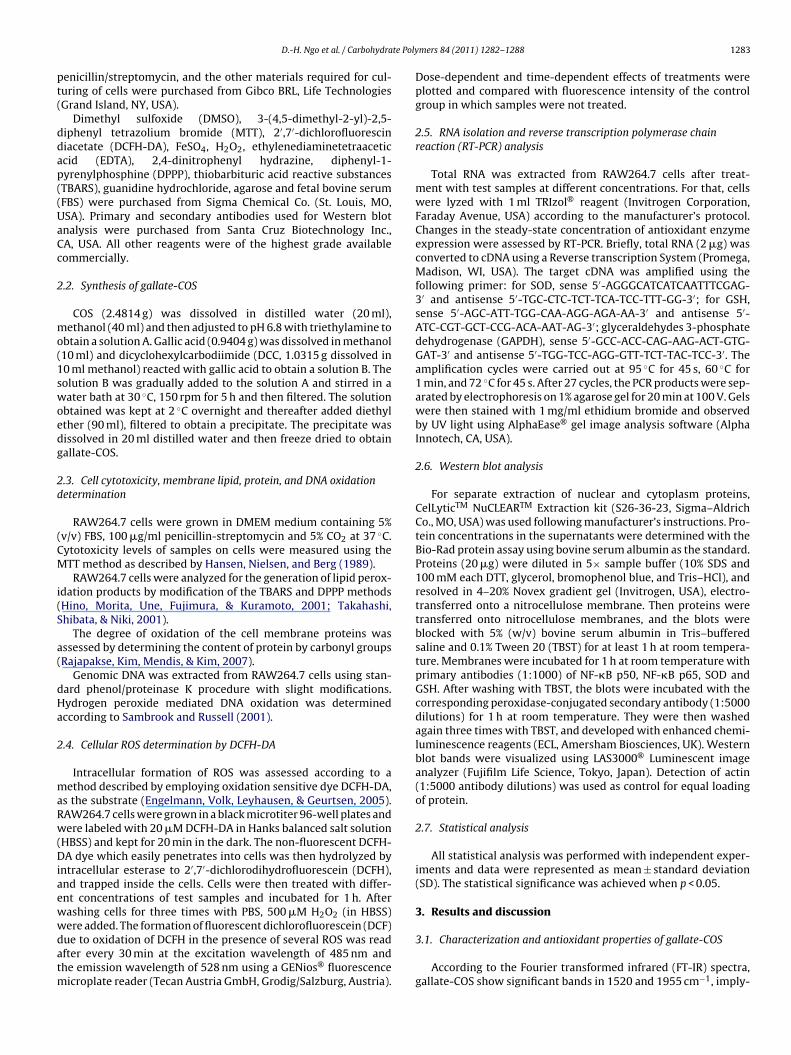
te Poly
pt(
ddap((UaCc
2
mo(1swoedg
2d
(CM
i(S
a(
dHa
2
maRw(Diaewwdatm
D.-H. Ngo et al. / Carbohydra
enicillin/streptomycin, and the other materials required for cul-uring of cells were purchased from Gibco BRL, Life TechnologiesGrand Island, NY, USA).
Dimethyl sulfoxide (DMSO), 3-(4,5-dimethyl-2-yl)-2,5-iphenyl tetrazolium bromide (MTT), 2′,7′-dichlorofluoresciniacetate (DCFH-DA), FeSO4, H2O2, ethylenediaminetetraaceticcid (EDTA), 2,4-dinitrophenyl hydrazine, diphenyl-1-yrenylphosphine (DPPP), thiobarbituric acid reactive substancesTBARS), guanidine hydrochloride, agarose and fetal bovine serumFBS) were purchased from Sigma Chemical Co. (St. Louis, MO,SA). Primary and secondary antibodies used for Western blotnalysis were purchased from Santa Cruz Biotechnology Inc.,A, USA. All other reagents were of the highest grade availableommercially.
.2. Synthesis of gallate-COS
COS (2.4814 g) was dissolved in distilled water (20 ml),ethanol (40 ml) and then adjusted to pH 6.8 with triethylamine to
btain a solution A. Gallic acid (0.9404 g) was dissolved in methanol10 ml) and dicyclohexylcarbodiimide (DCC, 1.0315 g dissolved in0 ml methanol) reacted with gallic acid to obtain a solution B. Theolution B was gradually added to the solution A and stirred in aater bath at 30 ◦C, 150 rpm for 5 h and then filtered. The solution
btained was kept at 2 ◦C overnight and thereafter added diethylther (90 ml), filtered to obtain a precipitate. The precipitate wasissolved in 20 ml distilled water and then freeze dried to obtainallate-COS.
.3. Cell cytotoxicity, membrane lipid, protein, and DNA oxidationetermination
RAW264.7 cells were grown in DMEM medium containing 5%v/v) FBS, 100 �g/ml penicillin-streptomycin and 5% CO2 at 37 ◦C.ytotoxicity levels of samples on cells were measured using theTT method as described by Hansen, Nielsen, and Berg (1989).RAW264.7 cells were analyzed for the generation of lipid perox-
dation products by modification of the TBARS and DPPP methodsHino, Morita, Une, Fujimura, & Kuramoto, 2001; Takahashi,hibata, & Niki, 2001).
The degree of oxidation of the cell membrane proteins wasssessed by determining the content of protein by carbonyl groupsRajapakse, Kim, Mendis, & Kim, 2007).
Genomic DNA was extracted from RAW264.7 cells using stan-ard phenol/proteinase K procedure with slight modifications.ydrogen peroxide mediated DNA oxidation was determinedccording to Sambrook and Russell (2001).
.4. Cellular ROS determination by DCFH-DA
Intracellular formation of ROS was assessed according to aethod described by employing oxidation sensitive dye DCFH-DA,
s the substrate (Engelmann, Volk, Leyhausen, & Geurtsen, 2005).AW264.7 cells were grown in a black microtiter 96-well plates andere labeled with 20 �M DCFH-DA in Hanks balanced salt solution
HBSS) and kept for 20 min in the dark. The non-fluorescent DCFH-A dye which easily penetrates into cells was then hydrolyzed by
ntracellular esterase to 2′,7′-dichlorodihydrofluorescein (DCFH),nd trapped inside the cells. Cells were then treated with differ-nt concentrations of test samples and incubated for 1 h. Afterashing cells for three times with PBS, 500 �M H2O2 (in HBSS)
ere added. The formation of fluorescent dichlorofluorescein (DCF)ue to oxidation of DCFH in the presence of several ROS was readfter every 30 min at the excitation wavelength of 485 nm andhe emission wavelength of 528 nm using a GENios® fluorescenceicroplate reader (Tecan Austria GmbH, Grodig/Salzburg, Austria).
mers 84 (2011) 1282–1288 1283
Dose-dependent and time-dependent effects of treatments wereplotted and compared with fluorescence intensity of the controlgroup in which samples were not treated.
2.5. RNA isolation and reverse transcription polymerase chainreaction (RT-PCR) analysis
Total RNA was extracted from RAW264.7 cells after treat-ment with test samples at different concentrations. For that, cellswere lyzed with 1 ml TRIzol® reagent (Invitrogen Corporation,Faraday Avenue, USA) according to the manufacturer’s protocol.Changes in the steady-state concentration of antioxidant enzymeexpression were assessed by RT-PCR. Briefly, total RNA (2 �g) wasconverted to cDNA using a Reverse transcription System (Promega,Madison, WI, USA). The target cDNA was amplified using thefollowing primer: for SOD, sense 5′-AGGGCATCATCAATTTCGAG-3′ and antisense 5′-TGC-CTC-TCT-TCA-TCC-TTT-GG-3′; for GSH,sense 5′-AGC-ATT-TGG-CAA-AGG-AGA-AA-3′ and antisense 5′-ATC-CGT-GCT-CCG-ACA-AAT-AG-3′; glyceraldehydes 3-phosphatedehydrogenase (GAPDH), sense 5′-GCC-ACC-CAG-AAG-ACT-GTG-GAT-3′ and antisense 5′-TGG-TCC-AGG-GTT-TCT-TAC-TCC-3′. Theamplification cycles were carried out at 95 ◦C for 45 s, 60 ◦C for1 min, and 72 ◦C for 45 s. After 27 cycles, the PCR products were sep-arated by electrophoresis on 1% agarose gel for 20 min at 100 V. Gelswere then stained with 1 mg/ml ethidium bromide and observedby UV light using AlphaEase® gel image analysis software (AlphaInnotech, CA, USA).
2.6. Western blot analysis
For separate extraction of nuclear and cytoplasm proteins,CelLyticTM NuCLEARTM Extraction kit (S26-36-23, Sigma–AldrichCo., MO, USA) was used following manufacturer’s instructions. Pro-tein concentrations in the supernatants were determined with theBio-Rad protein assay using bovine serum albumin as the standard.Proteins (20 �g) were diluted in 5× sample buffer (10% SDS and100 mM each DTT, glycerol, bromophenol blue, and Tris–HCl), andresolved in 4–20% Novex gradient gel (Invitrogen, USA), electro-transferred onto a nitrocellulose membrane. Then proteins weretransferred onto nitrocellulose membranes, and the blots wereblocked with 5% (w/v) bovine serum albumin in Tris–bufferedsaline and 0.1% Tween 20 (TBST) for at least 1 h at room tempera-ture. Membranes were incubated for 1 h at room temperature withprimary antibodies (1:1000) of NF-�B p50, NF-�B p65, SOD andGSH. After washing with TBST, the blots were incubated with thecorresponding peroxidase-conjugated secondary antibody (1:5000dilutions) for 1 h at room temperature. They were then washedagain three times with TBST, and developed with enhanced chemi-luminescence reagents (ECL, Amersham Biosciences, UK). Westernblot bands were visualized using LAS3000® Luminescent imageanalyzer (Fujifilm Life Science, Tokyo, Japan). Detection of actin(1:5000 antibody dilutions) was used as control for equal loadingof protein.
2.7. Statistical analysis
All statistical analysis was performed with independent exper-iments and data were represented as mean ± standard deviation(SD). The statistical significance was achieved when p < 0.05.
3. Results and discussion
3.1. Characterization and antioxidant properties of gallate-COS
According to the Fourier transformed infrared (FT-IR) spectra,gallate-COS show significant bands in 1520 and 1955 cm−1, imply-
1284 D.-H. Ngo et al. / Carbohydrate Polymers 84 (2011) 1282–1288
FD
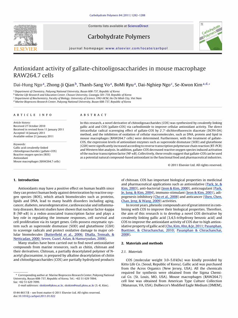
i(c1
b
ig. 1. 13C NMR (100 MHz, D2O) spectrum of gallate-COS (A) and 1H NMR (400 MHz,2O) spectrum of gallate-COS (B).
ng that the amide and ester linkages between gallic acid and COS
data not shown). This shows that the gallyl group of gallic acid wasovalently linked with COS via amide and ester linkages. Moreover,3carbon (13C) NMR spectra, gallate-COS shows the aromatic car-on of the gallyl group at 109.54, 128.01, 135.84, and 144.45 ppmFig. 2. Cell viability assessed by the MTT assay. The results shown are representativeof separate experiments performed in triplicate. Error bars represent the standarderror (SD).
(C C) and 174.8 ppm (C O) (Fig. 1(A)). From the proton nuclearmagnetic resonance (1H NMR) spectra, gallate-COS show a newpeak at 6.98 ppm belonging to the phenyl protons as comparedwith COS (Fig. 1(B)). These results confirmed the successful linkgallic acid and COS.
In this research, RAW264.7 cells were treated with varyingconcentrations of COS and gallate-COS in order to determine thecytotoxic effect of COS and gallate-COS by the MTT assay. Theresults showed that both COS and gallate-COS exert no cytotoxiceffects on RAW264.7 cells at all tested concentrations (Fig. 2).Therefore, those concentrations of COS and gallate-COS were usedfor further experiments.
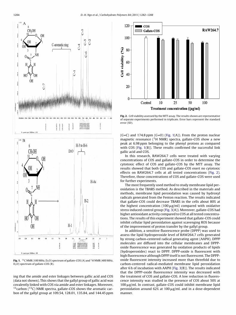
The most frequently used method to study membrane lipid per-oxidation is the TBARS method. As described in the materials andmethods, membrane lipid peroxidation was caused by hydroxylradicals generated from the Fenton reaction. The results indicatedthat gallate-COS could decrease TBARS in the cells about 80% atthe highest concentration (100 �g/ml) compared with oxidativestress induced control group (Fig. 3(A)). Moreover, gallate-COS hadhigher antioxidant activity compared to COS at all tested concentra-tions. The results of this experiment showed that gallate-COS couldinhibit cellular lipid peroxidation against scavenging ROS becauseof the improvement of proton transfer by the gallyl group.
In addition, a sensitive fluorescence probe (DPPP) was used toassess the lipid hydroperoxide level of RAW264.7 cells generatedby strong carbon-centered radical generating agent (AAPH). DPPPmolecules are diffused into the cellular membranes and DPPP-oxide fluorescence was generated by oxidation products of lipids(hydroperoxides) react to DPPP. DPPP-oxide is fluorescent withhigh fluorescence although DPPP itself is not fluorescent. The DPPP-oxide fluorescent intensity increased more than threefold due tocarbon-centered radical-mediated membrane lipid peroxidationafter 6 h of incubation with AAPH (Fig. 3(B)). The results indicatedthat the DPPP-oxide fluorescence intensity was decreased withthe treatment of COS and gallate-COS. A low reduction in fluores-
cence intensity was studied in the presence of COS about 50% at100 �g/ml. In contrast, gallate-COS could inhibit membrane lipidperoxidation around 62% at 100 �g/ml. and in a dose-dependentmanner.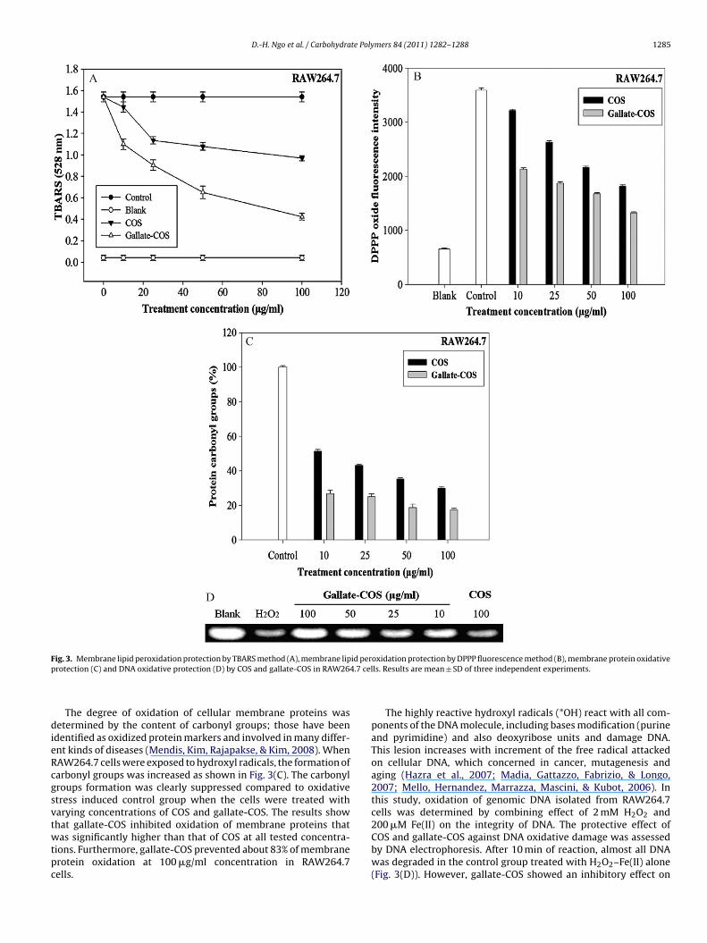
D.-H. Ngo et al. / Carbohydrate Polymers 84 (2011) 1282–1288 1285
F id perp 4.7 cel
dieRcgsvtwtpc
ig. 3. Membrane lipid peroxidation protection by TBARS method (A), membrane liprotection (C) and DNA oxidative protection (D) by COS and gallate-COS in RAW26
The degree of oxidation of cellular membrane proteins wasetermined by the content of carbonyl groups; those have been
dentified as oxidized protein markers and involved in many differ-nt kinds of diseases (Mendis, Kim, Rajapakse, & Kim, 2008). WhenAW264.7 cells were exposed to hydroxyl radicals, the formation ofarbonyl groups was increased as shown in Fig. 3(C). The carbonylroups formation was clearly suppressed compared to oxidativetress induced control group when the cells were treated witharying concentrations of COS and gallate-COS. The results show
hat gallate-COS inhibited oxidation of membrane proteins thatas significantly higher than that of COS at all tested concentra-ions. Furthermore, gallate-COS prevented about 83% of membranerotein oxidation at 100 �g/ml concentration in RAW264.7ells.
oxidation protection by DPPP fluorescence method (B), membrane protein oxidativels. Results are mean ± SD of three independent experiments.
The highly reactive hydroxyl radicals (*OH) react with all com-ponents of the DNA molecule, including bases modification (purineand pyrimidine) and also deoxyribose units and damage DNA.This lesion increases with increment of the free radical attackedon cellular DNA, which concerned in cancer, mutagenesis andaging (Hazra et al., 2007; Madia, Gattazzo, Fabrizio, & Longo,2007; Mello, Hernandez, Marrazza, Mascini, & Kubot, 2006). Inthis study, oxidation of genomic DNA isolated from RAW264.7cells was determined by combining effect of 2 mM H2O2 and
200 �M Fe(II) on the integrity of DNA. The protective effect ofCOS and gallate-COS against DNA oxidative damage was assessedby DNA electrophoresis. After 10 min of reaction, almost all DNAwas degraded in the control group treated with H2O2–Fe(II) alone(Fig. 3(D)). However, gallate-COS showed an inhibitory effect on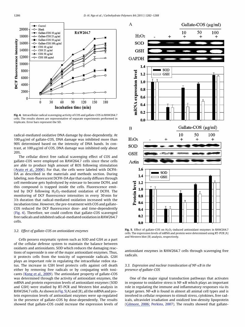
1286 D.-H. Ngo et al. / Carbohydrate Polymers 84 (2011) 1282–1288
Fct
r19t2
ga(Dlcttm3iC(fc
3
ootiptecwmaRtis
ig. 4. Intracellular radical scavenging activity of COS and gallate-COS in RAW264.7ells. The results shown are representative of separate experiments performed inriplicate. Error bars represent the SD.
adical-mediated oxidative DNA damage by dose-dependently. At00 �g/ml of gallate-COS, DNA damage was inhibited more than0% determined based on the intensity of DNA bands. In con-rast, at 100 �g/ml of COS, DNA damage was inhibited only about0%.
The cellular direct free radical scavenging effect of COS andallate-COS were employed on RAW264.7 cells since these cellsre able to produce high amount of ROS following stimulationArato et al., 2006). For that, the cells were labeled with DCFH-A as described in the materials and methods section. During
abeling, non-fluorescent DCFH-DA dye that easily diffuses throughell membrane gets hydrolyzed by esterase to become DCFH, andhis compound is trapped inside the cells. Fluorescence emit-ed by DCF following H2O2-mediated oxidation of DCFH. The
onitoring of DCF fluorescence intensities in every 30 min forh duration that radical-mediated oxidation increased with the
ncubation time. However, the pre-treatment with COS and gallate-OS reduced the DCF fluorescence dose- and time-dependentlyFig. 4). Therefore, we could confirm that gallate-COS scavengedree radicals and inhibited radical-mediated oxidation in RAW264.7ells.
.2. Effect of gallate-COS on antioxidant enzymes
Cells possess enzymatic system such as SOD and GSH as a partf the cellular defense system to maintain the balance betweenxidants and antioxidants. SOD which reduces the damaging reac-ions of superoxide is one of the major antioxidant enzymes. Thus,t protects cells from the toxicity of superoxide radicals. GSHlays an important role in regulating the intracellular redox sta-us. The increase in GSH level protects cells against cell deathither by removing free radicals or by conjugating with toxi-ants (Kong et al., 2009). The antioxidant property of gallate-COSas determined through the activity of antioxidant enzymes, theRNA and protein expression levels of antioxidant enzymes (SOD
nd GSH) were studied by RT-PCR and Western blot analysis inAW264.7 cells. As shown in Fig. 5(A) and (B), all the mRNA and pro-ein expression levels of antioxidant enzymes were up-regulatedn the presence of gallate-COS by dose-dependently. The resultshowed that gallate-COS could increase the expression levels of
Fig. 5. Effect of gallate-COS on H2O2-induced antioxidant enzymes in RAW264.7cells. The expression levels of mRNA and protein were determined using RT-PCR (A)and Western blot (B) analysis, respectively.
antioxidant enzymes in RAW264.7 cells through scavenging freeradicals.
3.3. Expression and nuclear translocation of NF-�B in thepresence of gallate-COS
One of the major signal transduction pathways that activatesin response to oxidative stress is NF-�B which plays an importantrole in regulating the immune and inflammatory responses via its
target genes. NF-�B is found in almost all animal cell types and isinvolved in cellular responses to stimuli stress, cytokines, free rad-icals, ultraviolet irradiation and oxidized low-density lipoprotein(Gilmore, 2006; Perkins, 2007). The results showed that gallate-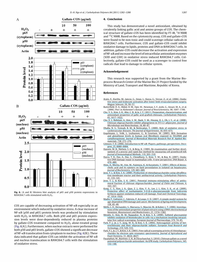
D.-H. Ngo et al. / Carbohydrate Poly
FR
CeNwsb(bodao
Park, P. J., Je, J. Y., & Kim, S. K. (2003). Free radical scavenging activity of chitooligosac-
ig. 6. (A and B) Western blot analysis of p65 and p50 protein expressions inAW264.7 cells stimulated with H2O2.
OS are capable of decreasing activation of NF-�B especially in annvironment which induced by oxidative stress. A clear increase ofF-�B (p50 and p65) protein levels was produced by stimulationith H2O2 in RAW264.7 cells. Both p50 and p65 protein expres-
ion levels were dose-dependently reduced in plasma proteinsy gallate-COS treatment compared to H2O2 alone-treated groupFig. 6(A)). Furthermore, when nuclear extracts were performed foroth p50 and p65 levels, gallate-COS showed a significant decrease
f NF-�B translocation from cytoplasm to nucleus (Fig. 6(B)). Theseata indicated that gallate-COS can inhibit the activation of NF-�Bnd nucleus translocation in RAW264.7 cells with the stimulationf oxidative stress.mers 84 (2011) 1282–1288 1287
4. Conclusion
This study has demonstrated a novel antioxidant, obtained bycovalently linking gallic acid and amino groups of COS. The chem-ical structure of gallate-COS has been identified by FT-IR, 1H NMRand 13C NMR. Based on the cytotoxicity assay, COS and gallate-COSwere found to be non-toxic and could scavenge cellular radicals inRAW264.7 cells. Furthermore, COS and gallate-COS could inhibitoxidative damage to lipids, proteins and DNA in RAW264.7 cells. Inaddition, gallate-COS could decrease the activation and expressionof NF-�B and increase the level of intracellular antioxidant enzymes(SOD and GSH) in oxidative stress induced RAW264.7 cells. Col-lectively, gallate-COS could be used as a scavenger to control freeradicals that lead to damage to cellular system.
Acknowledgement
This research was supported by a grant from the Marine Bio-process Research Center of the Marine Bio 21 Project funded by theMinistry of Land, Transport and Maritime, Republic of Korea.
References
Arato, E., Kurthy, M., Jancso, G., Sinay, L., Kasza, G., Verzar, Z., et al. (2006). Oxida-tive stress and leukocyte activation after lower limb revascularization surgery.Magyar Sebeszet, 59, 50–57.
Butterfield, D. A., Abdul, H. M., Opii, W., Newman, S. F., Joshi, G., Ansari, M. A., et al.(2006). Pin1 in Alzheimer’s disease. Journal of Neurochemistry, 98, 1697–1706.
Cho, Y. S., Kim, S. K., Ahn, C. B., & Je, J. Y. (2011). Preparation, characterization, andantioxidant properties of gallic acid-grafted-chitosans. Carbohydrate Polymers,83, 1617–1622.
Cho, E. J., Rahman, A., Kim, S. W., Baek, Y. M., Hwang, H. J., Oh, J. Y., et al. (2008).Chitosan oligosaccharides inhibit adipogenesis in 3T3-L1 adipocytes. Journal ofMicrobiology and Biotechnology, 18, 80–87.
Dhalla, N. S., Temsah, R. M., & Netticadan, T. (2000). Role of oxidative stress incardiovascular diseases. The Journal of Hypertension, 18, 655–673.
Engelmann, J., Volk, J., Leyhausen, G., & Geurtsen, W. (2005). ROS formationand glutathione levels in human oral fibroblasts exposed to TEGDMA andcamphorquinone. Journal of Biomedical Materials Research Part B: Applied Bio-materials, 75B, 272–276.
Gilmore, T. D. (2006). Introduction to NF-�B: Players, pathways, perspectives. Onco-gene, 25, 6680–6684.
Hansen, M. B., Nielsen, S. E., & Berg, K. (1989). Re-examination and further devel-opment of a precise and rapid dye method for measuring cell growth/cell kill.Journal of Immunological Methods, 119, 203–210.
Hazra, T. K., Das, A., Das, S., Choudhury, S., Kow, Y. W., & Roy, R. (2007). Oxida-tive DNA damage repair in mammalian cells: A new perspective. DNA Repair, 6,470–480.
Hino, A., Morita, M., Une, M., Fujimura, K., & Kuramoto, T. (2001). Effects of deoxy-cholic acid and its epimers on lipid peroxidation in isolated rat hepatocytes.Journal of Biochemistry, 129, 683–689.
Jeon, Y. J., & Kim, S. K. (2000). Production of chitooligosaccharides using ultrafiltra-tion membrane reactor and their antibacterial activity. Carbohydrate Polymers,41, 133–141.
Jeon, Y. J., & Kim, S. K. (2001). Potential immuno-stimulating effect of antitu-moral fraction of chitosan oligosaccharides. Journal of Chitin and Chitosan, 6,163–167.
Kong, C. S., Kim, J. A., Qian, Z. J., Kim, Y. A., Lee, J. I., Kim, S. K., et al. (2009).Protective effect of isorhamnetin 3-O-�-d-glucopyranoside from Salicorniaherbacea against oxidation-induced cell damage. Food and Chemical Toxicology,47, 1914–1920.
Madia, F., Gattazzo, C., Fabrizio, P., & Longo, V. D. (2007). A simple model system forage-dependent DNA damage and cancer. Mechanisms of Ageing and Development,128, 45–49.
Mello, L. D., Hernandez, S., Marrazza, S., Mascini, M., & Kubot, L. T. (2006). Investiga-tions of the antioxidant properties of plant extracts using a DNA-electrochemicalbiosensor. Biosensensors and Bioelectronics, 21, 1374–1382.
Mendis, E., Kim, M. M., Rajapakse, N., & Kim, S. K. (2008). Sulfated glucosamineinhibits oxidation of biomolecules in cells via a mechanism involving intracel-lular free radical scavenging. European Journal of Pharmacology, 579, 74–85.
Park, P. J., Je, J. Y., Jung, W. K., & Kim, S. K. (2004). Anticoagulant activity of het-erochitosans and their oligosaccharide sulfates. European Food Research andTechnology, 219, 529–533.
charides by electron spin resonance spectrometry. Journal of Agricultural andFood Chemistry, 51, 4624–4627.
Pasanphan, W., Buettner, G. R., & Chirachanchai, S. (2010). Chitosan gallate as a novelpotential polysaccharide antioxidant: An EPR study. Carbohydrate Polymers, 345,132–140.
1 te Pol
P
P
R
S
Shen, K. T., Chen, M. H., Chan, H. Y., Jeng, J. H., & Wang, Y. J. (2009). Inhibitory effects
288 D.-H. Ngo et al. / Carbohydra
asanphan, W., & Chirachanchai, S. (2008). Conjugation of gallic acid onto chitosan:An approach for green and water-based antioxidant. Carbohydrate Polymers, 72,169–177.
erkins, N. D. (2007). Integrating cell-signalling pathways with NF-�B and IKK func-
tion. Nature Reviews Molecular Cell Biology, 8, 49–62.ajapakse, N., Kim, M. M., Mendis, E., & Kim, S. K. (2007). Inhibition of freeradical-mediated oxidation of cellular biomolecules by carboxylated chi-tooligosaccharides. Bioorganic and Medicinal Chemistry, 15, 997–1003.
ambrook, J., & Russell, D. (2001). Molecular cloning: A laboratory manual. New York:Cold Spring Harbor Laboratory Press., pp. 84–87.
ymers 84 (2011) 1282–1288
Seven, A., Guzel, S., Aslan, M., & Hamuryudan, V. (2008). Lipid, protein, DNA oxi-dation and antioxidant status in rheumatoid arthritis. Clinical Biochemistry, 41,538–543.
of chitooligosaccharides on tumor growth and metastasis. Food and ChemicalToxicology, 47, 1864–1871.
Takahashi, M., Shibata, M., & Niki, E. (2001). Cytotoxic effect of formaldehyde withfree radicals via increment of cellular reactive oxygen species. Free Radical Biol-ogy and Medicine, 31, 164–174.

![Tocotrienol-Rich Fraction, [6]-Gingerol and Epigallocatechin Gallate Inhibit Proliferation and Induce Apoptosis of Glioma Cancer Cells](https://img.dokumen.tips/doc/110x75/63385d684959065769077f57/tocotrienol-rich-fraction-6-gingerol-and-epigallocatechin-gallate-inhibit-proliferation.jpg)


