Embed Size (px)
Citation preview
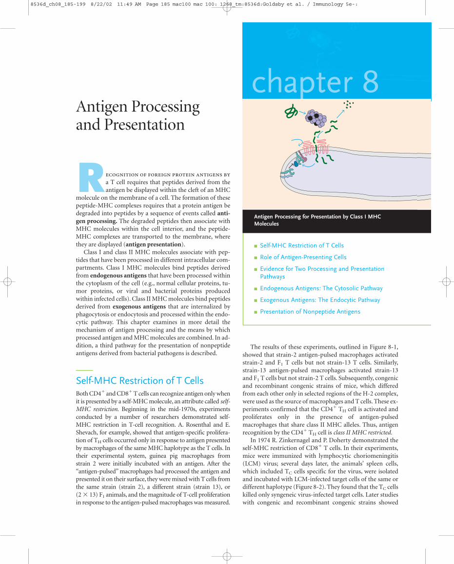
chapter 8
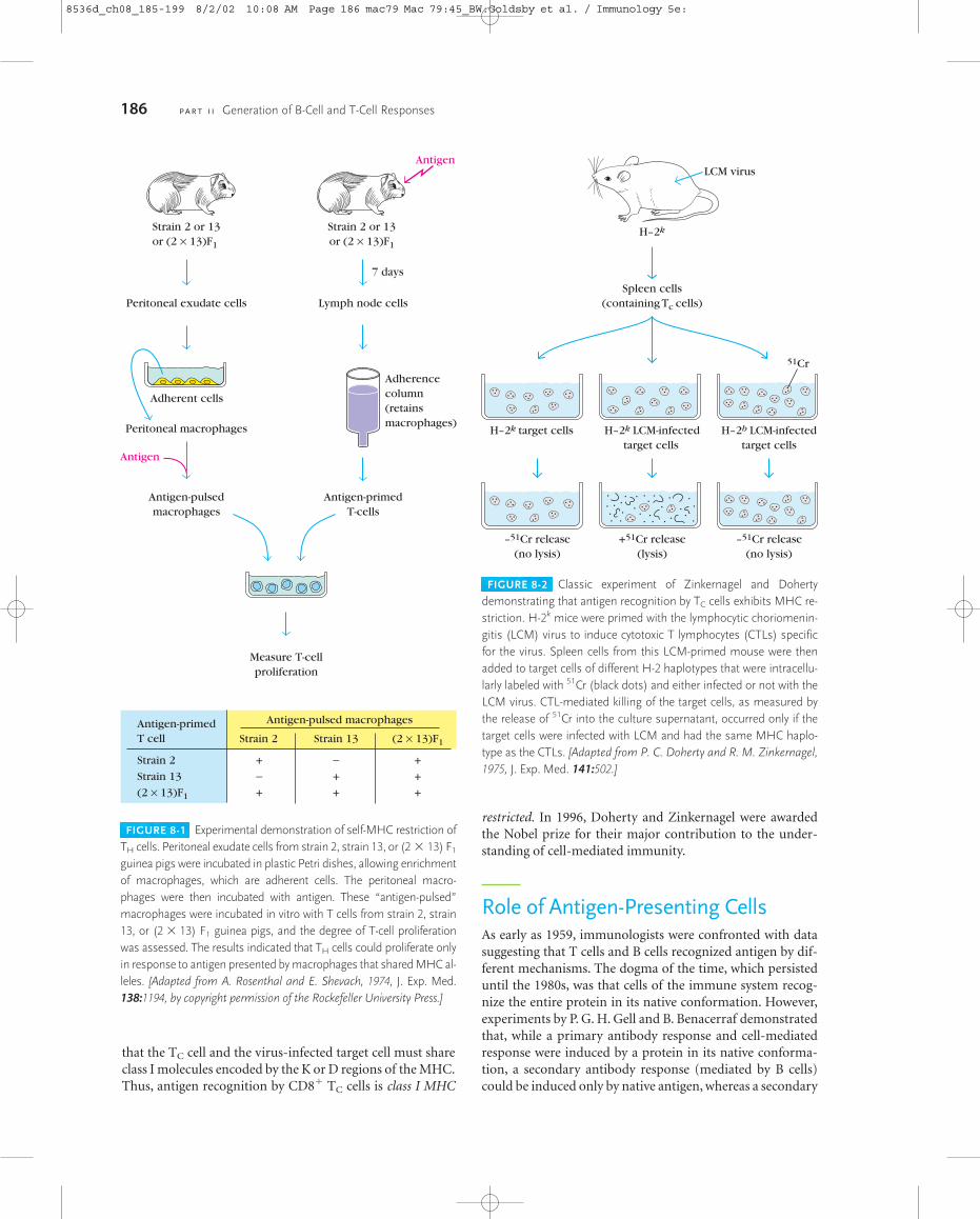
The results of these experiments, outlined in Figure 8-1,showed that strain-2 antigen-pulsed macrophages activatedstrain-2 and F1 T cells but not strain-13 T cells. Similarly,strain-13 antigen-pulsed macrophages activated strain-13and F1 T cells but not strain-2 T cells. Subsequently, congenicand recombinant congenic strains of mice, which differedfrom each other only in selected regions of the H-2 complex,were used as the source of macrophages and T cells. These ex-periments confirmed that the CD4� TH cell is activated andproliferates only in the presence of antigen-pulsedmacrophages that share class II MHC alleles. Thus, antigenrecognition by the CD4� TH cell is class II MHC restricted.
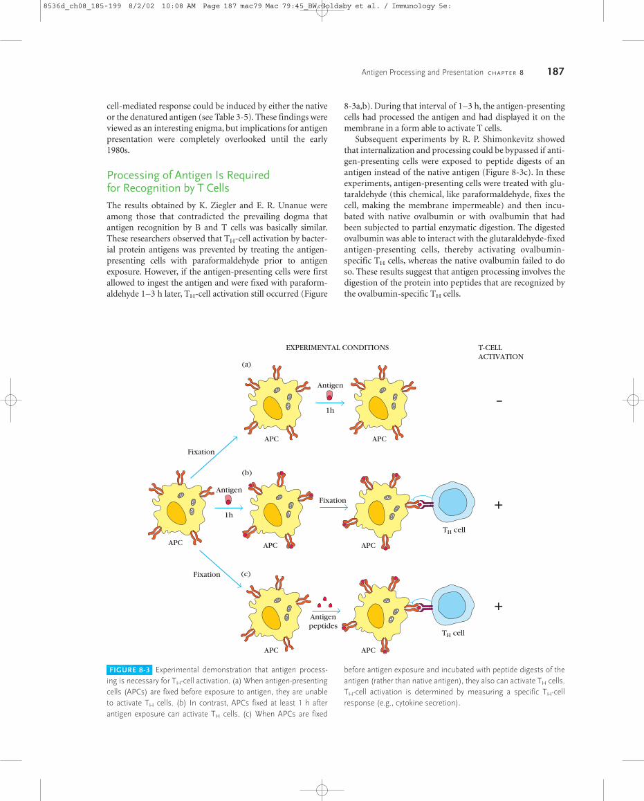
In 1974 R. Zinkernagel and P. Doherty demonstrated theself-MHC restriction of CD8� T cells. In their experiments,mice were immunized with lymphocytic choriomeningitis(LCM) virus; several days later, the animals’ spleen cells,which included TC cells specific for the virus, were isolatedand incubated with LCM-infected target cells of the same ordifferent haplotype (Figure 8-2). They found that the TC cellskilled only syngeneic virus-infected target cells. Later studieswith congenic and recombinant congenic strains showed
� Self-MHC Restriction of T Cells
� Role of Antigen-Presenting Cells
� Evidence for Two Processing and PresentationPathways
� Endogenous Antigens: The Cytosolic Pathway
� Exogenous Antigens: The Endocytic Pathway
� Presentation of Nonpeptide Antigens
Antigen Processing and Presentation
R
a T cell requires that peptides derived from theantigen be displayed within the cleft of an MHC
molecule on the membrane of a cell. The formation of thesepeptide-MHC complexes requires that a protein antigen bedegraded into peptides by a sequence of events called anti-gen processing. The degraded peptides then associate withMHC molecules within the cell interior, and the peptide-MHC complexes are transported to the membrane, wherethey are displayed (antigen presentation).
Class I and class II MHC molecules associate with pep-tides that have been processed in different intracellular com-partments. Class I MHC molecules bind peptides derivedfrom endogenous antigens that have been processed withinthe cytoplasm of the cell (e.g., normal cellular proteins, tu-mor proteins, or viral and bacterial proteins producedwithin infected cells). Class II MHC molecules bind peptidesderived from exogenous antigens that are internalized byphagocytosis or endocytosis and processed within the endo-cytic pathway. This chapter examines in more detail themechanism of antigen processing and the means by whichprocessed antigen and MHC molecules are combined. In ad-dition, a third pathway for the presentation of nonpeptideantigens derived from bacterial pathogens is described.
Self-MHC Restriction of T CellsBoth CD4� and CD8� T cells can recognize antigen only whenit is presented by a self-MHC molecule, an attribute called self-MHC restriction. Beginning in the mid-1970s, experimentsconducted by a number of researchers demonstrated self-MHC restriction in T-cell recognition. A. Rosenthal and E.Shevach, for example, showed that antigen-specific prolifera-tion of TH cells occurred only in response to antigen presentedby macrophages of the same MHC haplotype as the T cells. Intheir experimental system, guinea pig macrophages fromstrain 2 were initially incubated with an antigen. After the“antigen-pulsed” macrophages had processed the antigen andpresented it on their surface, they were mixed with T cells fromthe same strain (strain 2), a different strain (strain 13), or (2 � 13) F1 animals, and the magnitude of T-cell proliferationin response to the antigen-pulsed macrophages was measured.
Antigen Processing for Presentation by Class I MHCMolecules
8536d_ch08_185-199 8/22/02 11:49 AM Page 185 mac100 mac 100: 1268_tm:8536d:Goldsby et al. / Immunology 5e-:
restricted. In 1996, Doherty and Zinkernagel were awardedthe Nobel prize for their major contribution to the under-standing of cell-mediated immunity.
Role of Antigen-Presenting CellsAs early as 1959, immunologists were confronted with datasuggesting that T cells and B cells recognized antigen by dif-ferent mechanisms. The dogma of the time, which persisteduntil the 1980s, was that cells of the immune system recog-nize the entire protein in its native conformation. However,experiments by P. G. H. Gell and B. Benacerraf demonstratedthat, while a primary antibody response and cell-mediatedresponse were induced by a protein in its native conforma-tion, a secondary antibody response (mediated by B cells)could be induced only by native antigen, whereas a secondary
186 P A R T I I Generation of B-Cell and T-Cell Responses
Antigen-pulsed macrophagesAntigen-primedT cell Strain 2 Strain 13 (2 × 13)F1
Strain 2
Strain 13
(2 × 13)F1
+
+
−−+
+
+
+
+
Strain 2 or 13or (2 × 13)F1
Strain 2 or 13or (2 × 13)F1
Antigen
Peritoneal exudate cells
Peritoneal macrophages
Adherent cells
Antigen
Antigen-pulsedmacrophages
Measure T-cellproliferation
Lymph node cells
Antigen-primedT-cells
Adherencecolumn(retainsmacrophages)
7 days
FIGURE 8-1 Experimental demonstration of self-MHC restriction ofTH cells. Peritoneal exudate cells from strain 2, strain 13, or (2 � 13) F1
guinea pigs were incubated in plastic Petri dishes, allowing enrichmentof macrophages, which are adherent cells. The peritoneal macro-phages were then incubated with antigen. These “antigen-pulsed”macrophages were incubated in vitro with T cells from strain 2, strain13, or (2 � 13) F1 guinea pigs, and the degree of T-cell proliferationwas assessed. The results indicated that TH cells could proliferate onlyin response to antigen presented by macrophages that shared MHC al-leles. [Adapted from A. Rosenthal and E. Shevach, 1974, J. Exp. Med.138:1194, by copyright permission of the Rockefeller University Press.]
that the TC cell and the virus-infected target cell must shareclass I molecules encoded by the K or D regions of the MHC.Thus, antigen recognition by CD8� TC cells is class I MHC
Spleen cells(containing Tc cells)
H–2k target cells H–2k LCM-infectedtarget cells
H–2b LCM-infectedtarget cells
–51Cr release(no lysis)
–51Cr release(no lysis)
+51Cr release(lysis)
H–2k
LCM virus
51Cr
FIGURE 8-2 Classic experiment of Zinkernagel and Dohertydemonstrating that antigen recognition by TC cells exhibits MHC re-striction. H-2k mice were primed with the lymphocytic choriomenin-gitis (LCM) virus to induce cytotoxic T lymphocytes (CTLs) specificfor the virus. Spleen cells from this LCM-primed mouse were thenadded to target cells of different H-2 haplotypes that were intracellu-larly labeled with 51Cr (black dots) and either infected or not with theLCM virus. CTL-mediated killing of the target cells, as measured bythe release of 51Cr into the culture supernatant, occurred only if thetarget cells were infected with LCM and had the same MHC haplo-type as the CTLs. [Adapted from P. C. Doherty and R. M. Zinkernagel,1975, J. Exp. Med. 141:502.]
8536d_ch08_185-199 8/2/02 10:08 AM Page 186 mac79 Mac 79:45_BW:Goldsby et al. / Immunology 5e:
cell-mediated response could be induced by either the nativeor the denatured antigen (see Table 3-5). These findings wereviewed as an interesting enigma, but implications for antigenpresentation were completely overlooked until the early1980s.
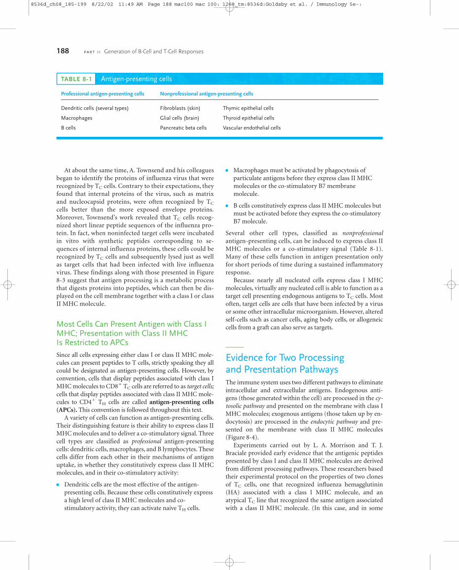
Processing of Antigen Is Required for Recognition by T CellsThe results obtained by K. Ziegler and E. R. Unanue wereamong those that contradicted the prevailing dogma thatantigen recognition by B and T cells was basically similar.These researchers observed that TH-cell activation by bacter-ial protein antigens was prevented by treating the antigen-presenting cells with paraformaldehyde prior to antigenexposure. However, if the antigen-presenting cells were firstallowed to ingest the antigen and were fixed with paraform-aldehyde 1–3 h later, TH-cell activation still occurred (Figure
8-3a,b). During that interval of 1–3 h, the antigen-presentingcells had processed the antigen and had displayed it on themembrane in a form able to activate T cells.
Subsequent experiments by R. P. Shimonkevitz showedthat internalization and processing could be bypassed if anti-gen-presenting cells were exposed to peptide digests of anantigen instead of the native antigen (Figure 8-3c). In theseexperiments, antigen-presenting cells were treated with glu-taraldehyde (this chemical, like paraformaldehyde, fixes thecell, making the membrane impermeable) and then incu-bated with native ovalbumin or with ovalbumin that hadbeen subjected to partial enzymatic digestion. The digestedovalbumin was able to interact with the glutaraldehyde-fixedantigen-presenting cells, thereby activating ovalbumin-specific TH cells, whereas the native ovalbumin failed to doso. These results suggest that antigen processing involves thedigestion of the protein into peptides that are recognized bythe ovalbumin-specific TH cells.
Antigen Processing and Presentation C H A P T E R 8 187
FIGURE 8-3 Experimental demonstration that antigen process-ing is necessary for TH-cell activation. (a) When antigen-presentingcells (APCs) are fixed before exposure to antigen, they are unableto activate TH cells. (b) In contrast, APCs fixed at least 1 h afterantigen exposure can activate TH cells. (c) When APCs are fixed
before antigen exposure and incubated with peptide digests of theantigen (rather than native antigen), they also can activate TH cells.TH-cell activation is determined by measuring a specific TH-cellresponse (e.g., cytokine secretion).
T-CELLACTIVATION
EXPERIMENTAL CONDITIONS
+Antigenpeptides
Fixation
APC
Fixation
–
APCAPC
Antigen
1h
Antigen
1h
APC
APC
TH cell
APC
+Fixation
APC
TH cell
(a)
(b)
(c)
8536d_ch08_185-199 8/2/02 10:08 AM Page 187 mac79 Mac 79:45_BW:Goldsby et al. / Immunology 5e:
At about the same time, A. Townsend and his colleaguesbegan to identify the proteins of influenza virus that wererecognized by TC cells. Contrary to their expectations, theyfound that internal proteins of the virus, such as matrixand nucleocapsid proteins, were often recognized by TC
cells better than the more exposed envelope proteins.Moreover, Townsend’s work revealed that TC cells recog-nized short linear peptide sequences of the influenza pro-tein. In fact, when noninfected target cells were incubatedin vitro with synthetic peptides corresponding to se-quences of internal influenza proteins, these cells could berecognized by TC cells and subsequently lysed just as wellas target cells that had been infected with live influenzavirus. These findings along with those presented in Figure8-3 suggest that antigen processing is a metabolic processthat digests proteins into peptides, which can then be dis-played on the cell membrane together with a class I or classII MHC molecule.
Most Cells Can Present Antigen with Class IMHC; Presentation with Class II MHC Is Restricted to APCsSince all cells expressing either class I or class II MHC mole-cules can present peptides to T cells, strictly speaking they allcould be designated as antigen-presenting cells. However, byconvention, cells that display peptides associated with class IMHC molecules to CD8� TC cells are referred to as target cells;cells that display peptides associated with class II MHC mole-cules to CD4� TH cells are called antigen-presenting cells(APCs). This convention is followed throughout this text.
A variety of cells can function as antigen-presenting cells.Their distinguishing feature is their ability to express class IIMHC molecules and to deliver a co-stimulatory signal. Threecell types are classified as professional antigen-presentingcells: dendritic cells, macrophages, and B lymphocytes. Thesecells differ from each other in their mechanisms of antigenuptake, in whether they constitutively express class II MHCmolecules, and in their co-stimulatory activity:
� Dendritic cells are the most effective of the antigen-presenting cells. Because these cells constitutively expressa high level of class II MHC molecules and co-stimulatory activity, they can activate naive TH cells.
� Macrophages must be activated by phagocytosis ofparticulate antigens before they express class II MHCmolecules or the co-stimulatory B7 membrane molecule.
� B cells constitutively express class II MHC molecules butmust be activated before they express the co-stimulatoryB7 molecule.
Several other cell types, classified as nonprofessionalantigen-presenting cells, can be induced to express class IIMHC molecules or a co-stimulatory signal (Table 8-1).Many of these cells function in antigen presentation onlyfor short periods of time during a sustained inflammatoryresponse.
Because nearly all nucleated cells express class I MHCmolecules, virtually any nucleated cell is able to function as atarget cell presenting endogenous antigens to TC cells. Mostoften, target cells are cells that have been infected by a virusor some other intracellular microorganism. However, alteredself-cells such as cancer cells, aging body cells, or allogeneiccells from a graft can also serve as targets.
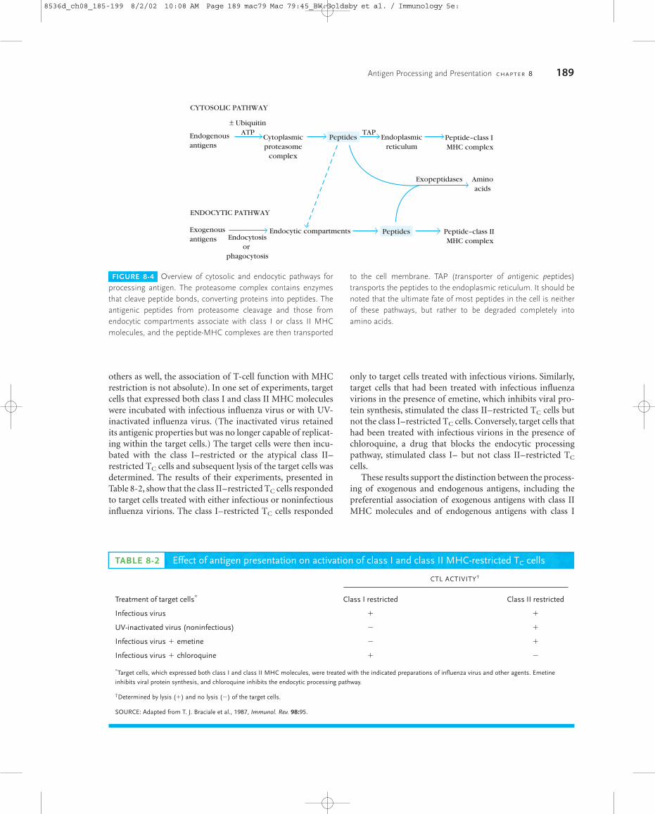
Evidence for Two Processing and Presentation PathwaysThe immune system uses two different pathways to eliminateintracellular and extracellular antigens. Endogenous anti-gens (those generated within the cell) are processed in the cy-tosolic pathway and presented on the membrane with class IMHC molecules; exogenous antigens (those taken up by en-docytosis) are processed in the endocytic pathway and pre-sented on the membrane with class II MHC molecules(Figure 8-4).
Experiments carried out by L. A. Morrison and T. J.Braciale provided early evidence that the antigenic peptidespresented by class I and class II MHC molecules are derivedfrom different processing pathways. These researchers basedtheir experimental protocol on the properties of two clonesof TC cells, one that recognized influenza hemagglutinin(HA) associated with a class I MHC molecule, and an atypical TC line that recognized the same antigen associatedwith a class II MHC molecule. (In this case, and in some
188 P A R T I I Generation of B-Cell and T-Cell Responses
TABLE 8-1 Antigen-presenting cells
Professional antigen-presenting cells Nonprofessional antigen-presenting cells
Dendritic cells (several types) Fibroblasts (skin) Thymic epithelial cells
Macrophages Glial cells (brain) Thyroid epithelial cells
B cells Pancreatic beta cells Vascular endothelial cells
8536d_ch08_185-199 8/22/02 11:49 AM Page 188 mac100 mac 100: 1268_tm:8536d:Goldsby et al. / Immunology 5e-:
others as well, the association of T-cell function with MHCrestriction is not absolute). In one set of experiments, targetcells that expressed both class I and class II MHC moleculeswere incubated with infectious influenza virus or with UV-inactivated influenza virus. (The inactivated virus retainedits antigenic properties but was no longer capable of replicat-ing within the target cells.) The target cells were then incu-bated with the class I–restricted or the atypical class II–restricted TC cells and subsequent lysis of the target cells wasdetermined. The results of their experiments, presented inTable 8-2, show that the class II–restricted TC cells respondedto target cells treated with either infectious or noninfectiousinfluenza virions. The class I–restricted TC cells responded
only to target cells treated with infectious virions. Similarly,target cells that had been treated with infectious influenzavirions in the presence of emetine, which inhibits viral pro-tein synthesis, stimulated the class II–restricted TC cells butnot the class I–restricted TC cells. Conversely, target cells thathad been treated with infectious virions in the presence ofchloroquine, a drug that blocks the endocytic processingpathway, stimulated class I– but not class II–restricted TC
cells.These results support the distinction between the process-
ing of exogenous and endogenous antigens, including thepreferential association of exogenous antigens with class IIMHC molecules and of endogenous antigens with class I
Antigen Processing and Presentation C H A P T E R 8 189
FIGURE 8-4 Overview of cytosolic and endocytic pathways forprocessing antigen. The proteasome complex contains enzymesthat cleave peptide bonds, converting proteins into peptides. Theantigenic peptides from proteasome cleavage and those fromendocytic compartments associate with class I or class II MHCmolecules, and the peptide-MHC complexes are then transported
to the cell membrane. TAP (transporter of antigenic peptides)transports the peptides to the endoplasmic reticulum. It should benoted that the ultimate fate of most peptides in the cell is neitherof these pathways, but rather to be degraded completely intoamino acids.
CYTOSOLIC PATHWAY
ENDOCYTIC PATHWAY
Endogenousantigens
± UbiquitinATP
Exogenousantigens
Cytoplasmicproteasome
complex
Peptides
Peptides
TAPEndoplasmic
reticulumPeptide–class I MHC complex
Peptide–class II MHC complex
Exopeptidases Aminoacids
Endocytosisor
phagocytosis
Endocytic compartments
TABLE 8-2 Effect of antigen presentation on activation of class I and class II MHC-restricted TC cells
CTL ACTIVITY†
Treatment of target cells* Class I restricted Class II restricted
Infectious virus � �
UV-inactivated virus (noninfectious) � �
Infectious virus � emetine � �
Infectious virus � chloroquine � �
*Target cells, which expressed both class I and class II MHC molecules, were treated with the indicated preparations of influenza virus and other agents. Emetine
inhibits viral protein synthesis, and chloroquine inhibits the endocytic processing pathway.
†Determined by lysis (�) and no lysis (�) of the target cells.
SOURCE: Adapted from T. J. Braciale et al., 1987, Immunol. Rev. 98:95.
8536d_ch08_185-199 8/2/02 10:08 AM Page 189 mac79 Mac 79:45_BW:Goldsby et al. / Immunology 5e:
MHC molecules. Association of viral antigen with class IMHC molecules required replication of the influenza virusand viral protein synthesis within the target cells; associationwith class II did not. These findings suggested that the pep-tides presented by class I and class II MHC molecules aretrafficked through separate intracellular compartments; classI MHC molecules interact with peptides derived from cy-tosolic degradation of endogenously synthesized proteins,class II molecules with peptides derived from endocyticdegradation of exogenous antigens. The next two sectionsexamine these two pathways in detail.
Endogenous Antigens: The Cytosolic PathwayIn eukaryotic cells, protein levels are carefully regulated.Every protein is subject to continuous turnover and is de-graded at a rate that is generally expressed in terms of its half-life. Some proteins (e.g., transcription factors, cyclins, andkey metabolic enzymes) have very short half-lives; dena-tured, misfolded, or otherwise abnormal proteins also are de-graded rapidly. The pathway by which endogenous antigensare degraded for presentation with class I MHC moleculesutilizes the same pathways involved in the normal turnoverof intracellular proteins.
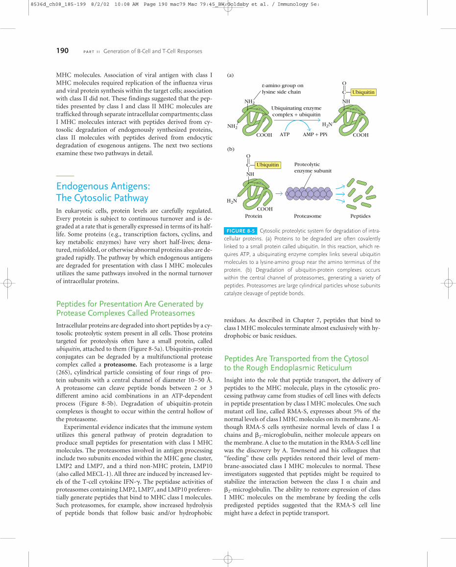
Peptides for Presentation Are Generated byProtease Complexes Called ProteasomesIntracellular proteins are degraded into short peptides by a cy-tosolic proteolytic system present in all cells. Those proteinstargeted for proteolysis often have a small protein, called ubiquitin, attached to them (Figure 8-5a). Ubiquitin-proteinconjugates can be degraded by a multifunctional proteasecomplex called a proteasome. Each proteasome is a large(26S), cylindrical particle consisting of four rings of pro-tein subunits with a central channel of diameter 10–50 Å.A proteasome can cleave peptide bonds between 2 or 3 different amino acid combinations in an ATP-dependentprocess (Figure 8-5b). Degradation of ubiquitin-proteincomplexes is thought to occur within the central hollow ofthe proteasome.
Experimental evidence indicates that the immune systemutilizes this general pathway of protein degradation to produce small peptides for presentation with class I MHCmolecules. The proteasomes involved in antigen processinginclude two subunits encoded within the MHC gene cluster,LMP2 and LMP7, and a third non-MHC protein, LMP10(also called MECL-1). All three are induced by increased lev-els of the T-cell cytokine IFN-�. The peptidase activities ofproteasomes containing LMP2, LMP7, and LMP10 preferen-tially generate peptides that bind to MHC class I molecules.Such proteasomes, for example, show increased hydrolysis of peptide bonds that follow basic and/or hydrophobic
residues. As described in Chapter 7, peptides that bind toclass I MHC molecules terminate almost exclusively with hy-drophobic or basic residues.
Peptides Are Transported from the Cytosol to the Rough Endoplasmic ReticulumInsight into the role that peptide transport, the delivery ofpeptides to the MHC molecule, plays in the cytosolic pro-cessing pathway came from studies of cell lines with defectsin peptide presentation by class I MHC molecules. One suchmutant cell line, called RMA-S, expresses about 5% of thenormal levels of class I MHC molecules on its membrane. Al-though RMA-S cells synthesize normal levels of class I �chains and �2-microglobulin, neither molecule appears onthe membrane. A clue to the mutation in the RMA-S cell linewas the discovery by A. Townsend and his colleagues that“feeding” these cells peptides restored their level of mem-brane-associated class I MHC molecules to normal. Theseinvestigators suggested that peptides might be required tostabilize the interaction between the class I � chain and �2-microglobulin. The ability to restore expression of class I MHC molecules on the membrane by feeding the cellspredigested peptides suggested that the RMA-S cell linemight have a defect in peptide transport.
190 P A R T I I Generation of B-Cell and T-Cell Responses
COOH
H2N
NH
C
O
Ubiquitin
(b)
COOH
NH2
(a)
ε-amino group on lysine side chain
COOH
H2N
NH
C
O
Ubiquitin
NH2
Ubiquinating enzymecomplex + ubiquitin
AMP + PPiATP
Protein Proteasome Peptides
Proteolyticenzyme subunit
FIGURE 8-5 Cytosolic proteolytic system for degradation of intra-cellular proteins. (a) Proteins to be degraded are often covalentlylinked to a small protein called ubiquitin. In this reaction, which re-quires ATP, a ubiquinating enzyme complex links several ubiquitinmolecules to a lysine-amino group near the amino terminus of theprotein. (b) Degradation of ubiquitin-protein complexes occurswithin the central channel of proteasomes, generating a variety ofpeptides. Proteasomes are large cylindrical particles whose subunitscatalyze cleavage of peptide bonds.
8536d_ch08_185-199 8/2/02 10:08 AM Page 190 mac79 Mac 79:45_BW:Goldsby et al. / Immunology 5e:
Antigen Processing and Presentation C H A P T E R 8 191
(a)
Amino acids
Peptides
Calreticulin Tapasin
Class I α chain
Calnexin
(b) Cytosol
TAP
Protein
RERlumen
RERlumen
TAP1 TAP2
CytosolATP ATP
RER membrane
ATPADP + Pi
Class I MHC
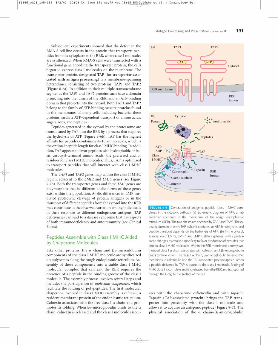
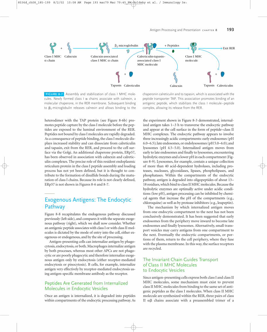
FIGURE 8-6 Generation of antigenic peptide–class I MHC com-plexes in the cytosolic pathway. (a) Schematic diagram of TAP, a het-erodimer anchored in the membrane of the rough endoplasmicreticulum (RER). The two chains are encoded by TAP1 and TAP2. The cy-tosolic domain in each TAP subunit contains an ATP-binding site, andpeptide transport depends on the hydrolysis of ATP. (b) In the cytosol,association of LMP2, LMP7, and LMP10 (black spheres) with a protea-some changes its catalytic specificity to favor production of peptides thatbind to class I MHC molecules. Within the RER membrane, a newly syn-thesized class I � chain associates with calnexin until �2-microglobulinbinds to the � chain. The class I � chain/�2-microglobulin heterodimerthen binds to calreticulin and the TAP-associated protein tapasin. Whena peptide delivered by TAP is bound to the class I molecule, folding ofMHC class I is complete and it is released from the RER and transportedthrough the Golgi to the surface of the cell.
Subsequent experiments showed that the defect in theRMA-S cell line occurs in the protein that transports pep-tides from the cytoplasm to the RER, where class I moleculesare synthesized. When RMA-S cells were transfected with afunctional gene encoding the transporter protein, the cellsbegan to express class I molecules on the membrane. Thetransporter protein, designated TAP (for transporter asso-ciated with antigen processing) is a membrane-spanningheterodimer consisting of two proteins: TAP1 and TAP2(Figure 8-6a). In addition to their multiple transmembranesegments, the TAP1 and TAP2 proteins each have a domainprojecting into the lumen of the RER, and an ATP-bindingdomain that projects into the cytosol. Both TAP1 and TAP2belong to the family of ATP-binding cassette proteins foundin the membranes of many cells, including bacteria; theseproteins mediate ATP-dependent transport of amino acids,sugars, ions, and peptides.
Peptides generated in the cytosol by the proteasome aretranslocated by TAP into the RER by a process that requiresthe hydrolysis of ATP (Figure 8-6b). TAP has the highestaffinity for peptides containing 8–10 amino acids, which isthe optimal peptide length for class I MHC binding. In addi-tion, TAP appears to favor peptides with hydrophobic or ba-sic carboxyl-terminal amino acids, the preferred anchorresidues for class I MHC molecules. Thus, TAP is optimizedto transport peptides that will interact with class I MHCmolecules.
The TAP1 and TAP2 genes map within the class II MHCregion, adjacent to the LMP2 and LMP7 genes (see Figure 7-15). Both the transporter genes and these LMP genes arepolymorphic; that is, different allelic forms of these genesexist within the population. Allelic differences in LMP-me-diated proteolytic cleavage of protein antigens or in thetransport of different peptides from the cytosol into the RERmay contribute to the observed variation among individualsin their response to different endogenous antigens. TAP deficiencies can lead to a disease syndrome that has aspectsof both immunodeficiency and autoimmunity (see Clinical Focus).
Peptides Assemble with Class I MHC Aidedby Chaperone MoleculesLike other proteins, the � chain and �2-microglobulincomponents of the class I MHC molecule are synthesizedon polysomes along the rough endoplasmic reticulum. As-sembly of these components into a stable class I MHCmolecular complex that can exit the RER requires thepresence of a peptide in the binding groove of the class Imolecule. The assembly process involves several steps andincludes the participation of molecular chaperones, whichfacilitate the folding of polypeptides. The first molecularchaperone involved in class I MHC assembly is calnexin, aresident membrane protein of the endoplasmic reticulum.Calnexin associates with the free class I � chain and pro-motes its folding. When �2-microglobulin binds to the �chain, calnexin is released and the class I molecule associ-
ates with the chaperone calreticulin and with tapasin.Tapasin (TAP-associated protein) brings the TAP trans-porter into proximity with the class I molecule and allows it to acquire an antigenic peptide (Figure 8-7). Thephysical association of the � chain–�2-microglobulin
8536d_ch08_185-199 8/2/02 10:08 AM Page 191 mac79 Mac 79:45_BW:Goldsby et al. / Immunology 5e:
192 P A R T I I Generation of B-Cell and T-Cell Responses
of the upper respiratory tract, and in thesecond decade begins to have chronic in-fection of the lungs. It is thought that apost-nasal-drip syndrome common inyounger patients promotes the bacteriallung infections in later life. Noteworthy isthe absence of any severe viral infection,which is common in immunodeficien-cies with T-cell involvement (see Chapter19). Bronchiectasis (dilation of thebronchial tubes) often occurs and recur-ring infections can lead to lung damagethat may be fatal. The most characteristicmark of the deficiency is the occurrenceof necrotizing skin lesions on the extrem-ities and the midface. These lesions ul-cerate and may cause disfigurement (seefigure). The skin lesions are probably dueto activated NK cells and �� T cells; NK
cells were isolated from biopsied skinfrom several patients, supporting thispossibility. Normally, the activity of NKcells is limited through the action ofkiller-cell-inhibitory receptors (KIRs),which deliver a negative signal to the NKcell following interaction with class Imolecules (see Chapter 14). The defi-ciency of class I molecules in TAP-relatedBLS patients explains the excessive activ-ity of the NK cells. Activation of NK cellsfurther explains the absence of severevirus infections, which are limited by NKand �� cells.
The best treatment for the character-istic lung infections appears to be antibi-otics and intravenous immunoglobulin.Attempts to limit the skin disease by im-munosuppressive regimens, such assteroid treatment or cytotoxic agents,can lead to exacerbation of lesions and istherefore contraindicated. Mutations inthe promoter region of TAP that precludeexpression of the gene were found forseveral patients, suggesting the possibil-ity of gene therapy, but the cellular distri-bution of class I is so widespread that itis not clear what cells would need to becorrected to alleviate all symptoms.
A relatively rare con-dition known as bare lymphocyte syn-drome, or BLS, has been recognized formore than 22 years. The lymphocytes inBLS patients express MHC molecules atbelow-normal levels and, in some cases,not at all. In type 1 BLS, a deficiency inMHC class I molecules exists; in type 2BLS, expression of class II molecules isimpaired. The pathogenesis of one typeof BLS underscores the importance ofthe class I family of MHC molecules intheir dual roles of preventing autoim-munity as well as defending againstpathogens.
Defects in promoter sequences thatpreclude MHC gene transcription werefound for some type 2 BLS cases, but inmany instances the nature of the under-lying defect is not known. A recent studyhas identified a group of patients withtype 1 BLS due to defects in TAP1 orTAP2 genes. Manifestations of the TAPdeficiency were consistent in this patientgroup and define a unique disease. Asdescribed earlier in this chapter, TAP pro-teins are necessary for the loading ofpeptides onto class I molecules, a stepthat is essential for expression of class IMHC molecules on the cell surface. Lym-phocytes in individuals with TAP defi-ciency express levels of class I moleculessignificantly lower than normal controls.Other cellular abnormalities include in-creased numbers of NK and �� T cells,and decreased levels of CD8� �� T cells.As we shall see, the disease manifesta-tions are reasonably well explained bythese deviations in the levels of certaincells involved in immune function.
In early life the TAP-deficient individ-ual suffers frequent bacterial infections
C L I N I C A L F O C U S
Deficiency in TransportersAssociated with AntigenPresentation (TAP) Leads to aDiverse Disease Spectrum
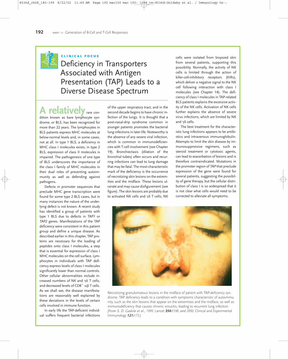
Necrotizing granulomatous lesions in the midface of patient with TAP-deficiency syn-drome. TAP deficiency leads to a condition with symptoms characteristic of autoimmu-nity, such as the skin lesions that appear on the extremities and the midface, as well asimmunodeficiency that causes chronic sinusitis, leading to recurrent lung infection.[From S. D. Gadola et al., 1999, Lancet 354:1598, and 2000, Clinical and ExperimentalImmunology 121:173.]
8536d_ch08_185-199 8/22/02 11:49 AM Page 192 mac100 mac 100: 1268_tm:8536d:Goldsby et al. / Immunology 5e-:
Antigen Processing and Presentation C H A P T E R 8 193
heterodimer with the TAP protein (see Figure 8-6b) pro-motes peptide capture by the class I molecule before the pep-tides are exposed to the luminal environment of the RER.Peptides not bound by class I molecules are rapidly degraded.As a consequence of peptide binding, the class I molecule dis-plays increased stability and can dissociate from calreticulinand tapasin, exit from the RER, and proceed to the cell sur-face via the Golgi. An additional chaperone protein, ERp57,has been observed in association with calnexin and calretic-ulin complexes. The precise role of this resident endoplasmicreticulum protein in the class I peptide assembly and loadingprocess has not yet been defined, but it is thought to con-tribute to the formation of disulfide bonds during the matu-ration of class I chains. Because its role is not clearly defined,ERp57 is not shown in Figures 8-6 and 8-7.
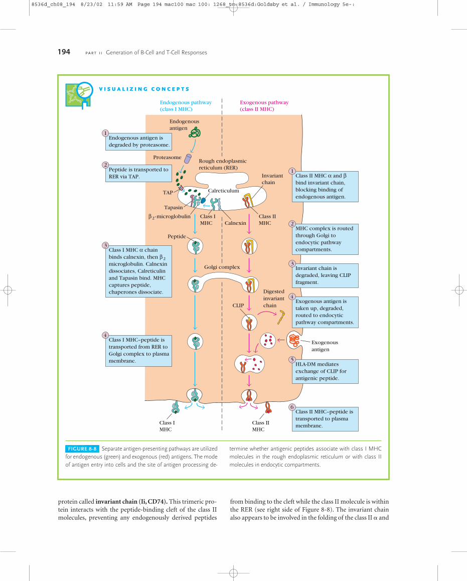
Exogenous Antigens: The EndocyticPathwayFigure 8-8 recapitulates the endogenous pathway discussedpreviously (left side), and compares it with the separate exoge-nous pathway (right), which we shall now consider. Whetheran antigenic peptide associates with class I or with class II mol-ecules is dictated by the mode of entry into the cell, either ex-ogenous or endogenous, and by the site of processing.
Antigen-presenting cells can internalize antigen by phago-cytosis, endocytosis, or both. Macrophages internalize antigenby both processes, whereas most other APCs are not phago-cytic or are poorly phagocytic and therefore internalize exoge-nous antigen only by endocytosis (either receptor-mediatedendocytosis or pinocytosis). B cells, for example, internalizeantigen very effectively by receptor-mediated endocytosis us-ing antigen-specific membrane antibody as the receptor.
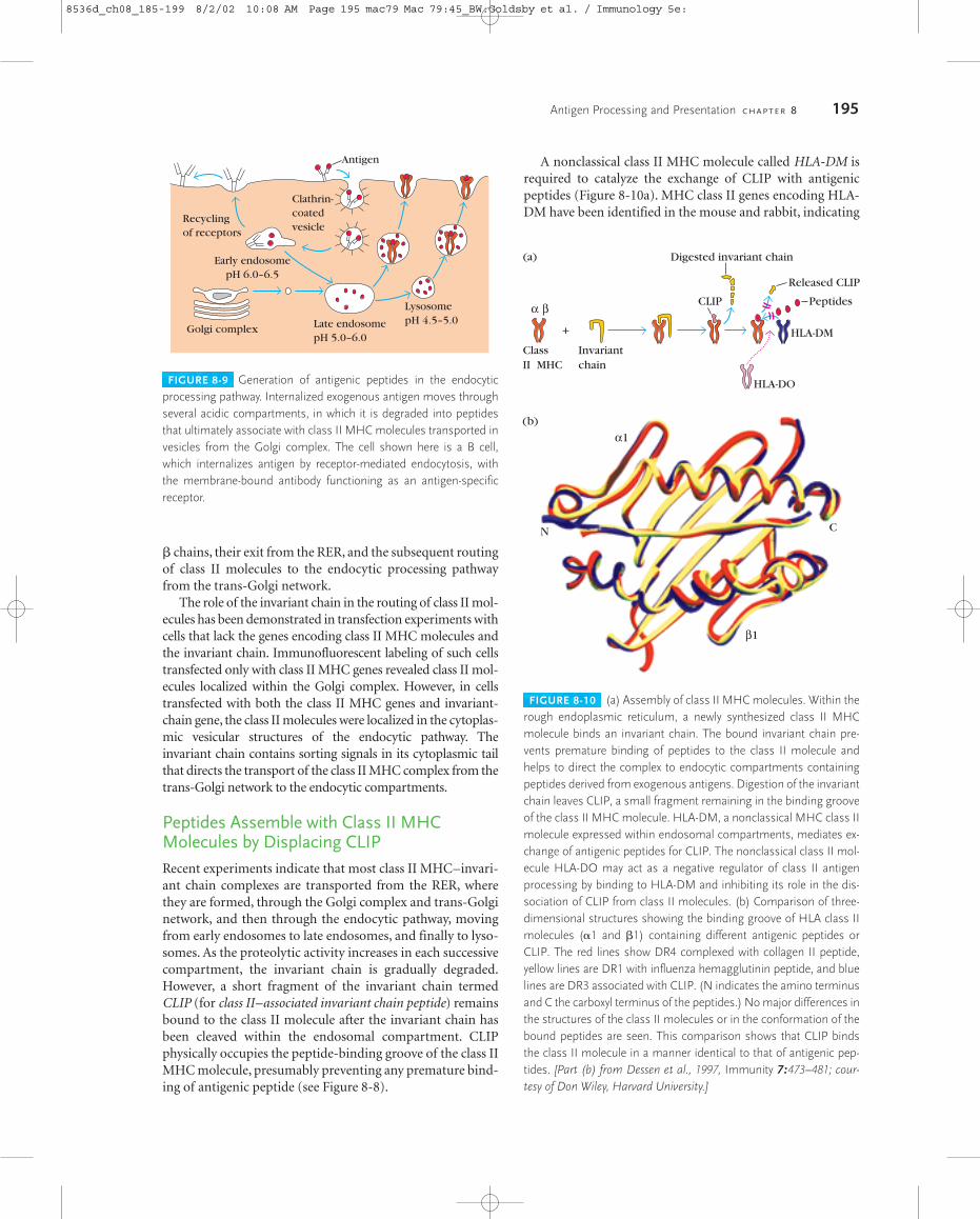
Peptides Are Generated from InternalizedMolecules in Endocytic VesiclesOnce an antigen is internalized, it is degraded into peptideswithin compartments of the endocytic processing pathway. As
the experiment shown in Figure 8-3 demonstrated, internal-ized antigen takes 1–3 h to transverse the endocytic pathwayand appear at the cell surface in the form of peptide–class IIMHC complexes. The endocytic pathway appears to involvethree increasingly acidic compartments: early endosomes (pH6.0–6.5); late endosomes, or endolysosomes (pH 5.0–6.0); andlysosomes (pH 4.5–5.0). Internalized antigen moves fromearly to late endosomes and finally to lysosomes, encounteringhydrolytic enzymes and a lower pH in each compartment (Fig-ure 8-9). Lysosomes, for example, contain a unique collectionof more than 40 acid-dependent hydrolases, including pro-teases, nucleases, glycosidases, lipases, phospholipases, andphosphatases. Within the compartments of the endocyticpathway, antigen is degraded into oligopeptides of about 13–18 residues,which bind to class II MHC molecules.Because thehydrolytic enzymes are optimally active under acidic condi-tions (low pH), antigen processing can be inhibited by chemi-cal agents that increase the pH of the compartments (e.g.,chloroquine) as well as by protease inhibitors (e.g., leupeptin).
The mechanism by which internalized antigen movesfrom one endocytic compartment to the next has not beenconclusively demonstrated. It has been suggested that earlyendosomes from the periphery move inward to become lateendosomes and finally lysosomes. Alternatively, small trans-port vesicles may carry antigens from one compartment tothe next. Eventually the endocytic compartments, or por-tions of them, return to the cell periphery, where they fusewith the plasma membrane. In this way, the surface receptorsare recycled.
The Invariant Chain Guides Transport of Class II MHC Molecules to Endocytic VesiclesSince antigen-presenting cells express both class I and class IIMHC molecules, some mechanism must exist to preventclass II MHC molecules from binding to the same set of anti-genic peptides as the class I molecules. When class II MHCmolecule are synthesized within the RER, three pairs of classII �� chains associate with a preassembled trimer of a
FIGURE 8-7 Assembly and stabilization of class I MHC mole-cules. Newly formed class I � chains associate with calnexin, amolecular chaperone, in the RER membrane. Subsequent bindingto �2-microglobulin releases calnexin and allows binding to the
chaperonin calreticulin and to tapasin, which is associated with thepeptide transporter TAP. This association promotes binding of anantigenic peptide, which stabilizes the class I molecule–peptidecomplex, allowing its release from the RER.
++ Peptides
Exit RER
Calnexin
Calnexin
Class I MHCα chain
Class I MHCmolecule
Calreticulin-tapasin–associated class I MHC molecule
Calnexin-associatedclass I MHC α chain
β2 microglobulin
+ +
CalreticulinTapasinCalreticulinTapasin
8536d_ch08_185-199 8/2/02 10:08 AM Page 193 mac79 Mac 79:45_BW:Goldsby et al. / Immunology 5e:
194 P A R T I I Generation of B-Cell and T-Cell Responses
V I S U A L I Z I N G C O N C E P T S
FIGURE 8-8 Separate antigen-presenting pathways are utilizedfor endogenous (green) and exogenous (red) antigens. The modeof antigen entry into cells and the site of antigen processing de-
termine whether antigenic peptides associate with class I MHCmolecules in the rough endoplasmic reticulum or with class IImolecules in endocytic compartments.
Endogenous pathway(class I MHC)
Exogenous pathway(class II MHC)
Peptide
TAP
Invariantchain
Class IIMHC
Class IMHC
Class IMHC
Class IIMHC
Rough endoplasmicreticulum (RER)
Proteasome
Calreticulum
Tapasin
β2-microglobulin
Golgi complex
Digestedinvariantchain
Exogenousantigen
CLIP
Calnexin
Endogenousantigen
Class II MHC α and βbind invariant chain,blocking binding ofendogenous antigen.
1
Endogenous antigen isdegraded by proteasome.
1
Peptide is transported toRER via TAP.
2
MHC complex is routedthrough Golgi toendocytic pathwaycompartments.
2
Class I MHC α chainbinds calnexin, then β2microglobulin. Calnexindissociates, Calreticulinand Tapasin bind. MHCcaptures peptide,chaperones dissociate.
3
Invariant chain isdegraded, leaving CLIPfragment.
3
Exogenous antigen istaken up, degraded,routed to endocyticpathway compartments.
Class I MHC–peptide istransported from RER toGolgi complex to plasmamembrane.
4
4
HLA-DM mediatesexchange of CLIP forantigenic peptide.
5
Class II MHC–peptide istransported to plasmamembrane.
6
protein called invariant chain (Ii, CD74). This trimeric pro-tein interacts with the peptide-binding cleft of the class IImolecules, preventing any endogenously derived peptides
from binding to the cleft while the class II molecule is withinthe RER (see right side of Figure 8-8). The invariant chainalso appears to be involved in the folding of the class II � and
8536d_ch08_194 8/23/02 11:59 AM Page 194 mac100 mac 100: 1268_tm:8536d:Goldsby et al. / Immunology 5e-:
� chains, their exit from the RER, and the subsequent routingof class II molecules to the endocytic processing pathwayfrom the trans-Golgi network.
The role of the invariant chain in the routing of class II mol-ecules has been demonstrated in transfection experiments withcells that lack the genes encoding class II MHC molecules andthe invariant chain. Immunofluorescent labeling of such cellstransfected only with class II MHC genes revealed class II mol-ecules localized within the Golgi complex. However, in cellstransfected with both the class II MHC genes and invariant-chain gene, the class II molecules were localized in the cytoplas-mic vesicular structures of the endocytic pathway. Theinvariant chain contains sorting signals in its cytoplasmic tailthat directs the transport of the class II MHC complex from thetrans-Golgi network to the endocytic compartments.
Peptides Assemble with Class II MHCMolecules by Displacing CLIPRecent experiments indicate that most class II MHC–invari-ant chain complexes are transported from the RER, wherethey are formed, through the Golgi complex and trans-Golginetwork, and then through the endocytic pathway, movingfrom early endosomes to late endosomes, and finally to lyso-somes. As the proteolytic activity increases in each successivecompartment, the invariant chain is gradually degraded.However, a short fragment of the invariant chain termedCLIP (for class II–associated invariant chain peptide) remainsbound to the class II molecule after the invariant chain hasbeen cleaved within the endosomal compartment. CLIPphysically occupies the peptide-binding groove of the class IIMHC molecule, presumably preventing any premature bind-ing of antigenic peptide (see Figure 8-8).
A nonclassical class II MHC molecule called HLA-DM isrequired to catalyze the exchange of CLIP with antigenicpeptides (Figure 8-10a). MHC class II genes encoding HLA-DM have been identified in the mouse and rabbit, indicating
Antigen Processing and Presentation C H A P T E R 8 195
Recyclingof receptors
Early endosomepH 6.0–6.5
Clathrin- coatedvesicle
Antigen
Late endosomepH 5.0–6.0
LysosomepH 4.5–5.0
Golgi complex
FIGURE 8-9 Generation of antigenic peptides in the endocyticprocessing pathway. Internalized exogenous antigen moves throughseveral acidic compartments, in which it is degraded into peptidesthat ultimately associate with class II MHC molecules transported invesicles from the Golgi complex. The cell shown here is a B cell,which internalizes antigen by receptor-mediated endocytosis, withthe membrane-bound antibody functioning as an antigen-specific receptor.
(a)
+
Released CLIP
CLIP
Digested invariant chain
Peptides
HLA-DM
HLA-DO
ClassII MHC
Invariantchain
α β
N C
β1
α1
(b)
FIGURE 8-10 (a) Assembly of class II MHC molecules. Within therough endoplasmic reticulum, a newly synthesized class II MHCmolecule binds an invariant chain. The bound invariant chain pre-vents premature binding of peptides to the class II molecule andhelps to direct the complex to endocytic compartments containingpeptides derived from exogenous antigens. Digestion of the invariantchain leaves CLIP, a small fragment remaining in the binding grooveof the class II MHC molecule. HLA-DM, a nonclassical MHC class IImolecule expressed within endosomal compartments, mediates ex-change of antigenic peptides for CLIP. The nonclassical class II mol-ecule HLA-DO may act as a negative regulator of class II antigenprocessing by binding to HLA-DM and inhibiting its role in the dis-sociation of CLIP from class II molecules. (b) Comparison of three-dimensional structures showing the binding groove of HLA class IImolecules (�1 and �1) containing different antigenic peptides orCLIP. The red lines show DR4 complexed with collagen II peptide,yellow lines are DR1 with influenza hemagglutinin peptide, and bluelines are DR3 associated with CLIP. (N indicates the amino terminusand C the carboxyl terminus of the peptides.) No major differences inthe structures of the class II molecules or in the conformation of thebound peptides are seen. This comparison shows that CLIP bindsthe class II molecule in a manner identical to that of antigenic pep-tides. [Part (b) from Dessen et al., 1997, Immunity 7:473–481; cour-tesy of Don Wiley, Harvard University.]
8536d_ch08_185-199 8/2/02 10:08 AM Page 195 mac79 Mac 79:45_BW:Goldsby et al. / Immunology 5e:
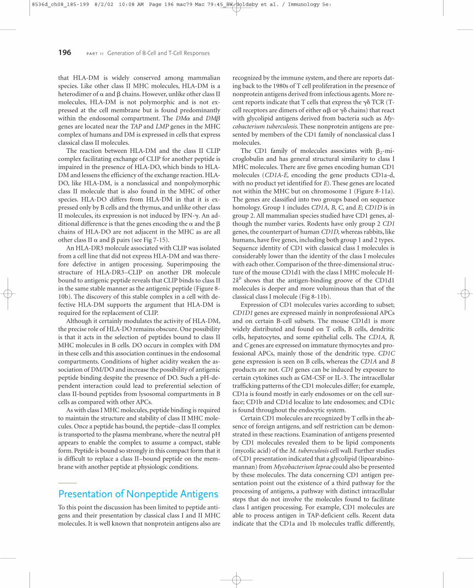
that HLA-DM is widely conserved among mammalianspecies. Like other class II MHC molecules, HLA-DM is aheterodimer of � and � chains. However, unlike other class IImolecules, HLA-DM is not polymorphic and is not ex-pressed at the cell membrane but is found predominantlywithin the endosomal compartment. The DM� and DM�genes are located near the TAP and LMP genes in the MHCcomplex of humans and DM is expressed in cells that expressclassical class II molecules.
The reaction between HLA-DM and the class II CLIPcomplex facilitating exchange of CLIP for another peptide isimpaired in the presence of HLA-DO, which binds to HLA-DM and lessens the efficiency of the exchange reaction. HLA-DO, like HLA-DM, is a nonclassical and nonpolymorphicclass II molecule that is also found in the MHC of otherspecies. HLA-DO differs from HLA-DM in that it is ex-pressed only by B cells and the thymus, and unlike other classII molecules, its expression is not induced by IFN-�. An ad-ditional difference is that the genes encoding the � and the �chains of HLA-DO are not adjacent in the MHC as are allother class II � and � pairs (see Fig 7-15).
An HLA-DR3 molecule associated with CLIP was isolatedfrom a cell line that did not express HLA-DM and was there-fore defective in antigen processing. Superimposing thestructure of HLA-DR3–CLIP on another DR moleculebound to antigenic peptide reveals that CLIP binds to class IIin the same stable manner as the antigenic peptide (Figure 8-10b). The discovery of this stable complex in a cell with de-fective HLA-DM supports the argument that HLA-DM isrequired for the replacement of CLIP.
Although it certainly modulates the activity of HLA-DM,the precise role of HLA-DO remains obscure. One possibilityis that it acts in the selection of peptides bound to class IIMHC molecules in B cells. DO occurs in complex with DMin these cells and this association continues in the endosomalcompartments. Conditions of higher acidity weaken the as-sociation of DM/DO and increase the possibility of antigenicpeptide binding despite the presence of DO. Such a pH-de-pendent interaction could lead to preferential selection ofclass II-bound peptides from lysosomal compartments in Bcells as compared with other APCs.
As with class I MHC molecules, peptide binding is requiredto maintain the structure and stability of class II MHC mole-cules. Once a peptide has bound, the peptide–class II complexis transported to the plasma membrane, where the neutral pHappears to enable the complex to assume a compact, stableform. Peptide is bound so strongly in this compact form that itis difficult to replace a class II–bound peptide on the mem-brane with another peptide at physiologic conditions.
Presentation of Nonpeptide AntigensTo this point the discussion has been limited to peptide anti-gens and their presentation by classical class I and II MHCmolecules. It is well known that nonprotein antigens also are
recognized by the immune system, and there are reports dat-ing back to the 1980s of T cell proliferation in the presence ofnonprotein antigens derived from infectious agents. More re-cent reports indicate that T cells that express the �� TCR (T-cell receptors are dimers of either �� or �� chains) that reactwith glycolipid antigens derived from bacteria such as My-cobacterium tuberculosis. These nonprotein antigens are pre-sented by members of the CD1 family of nonclassical class Imolecules.
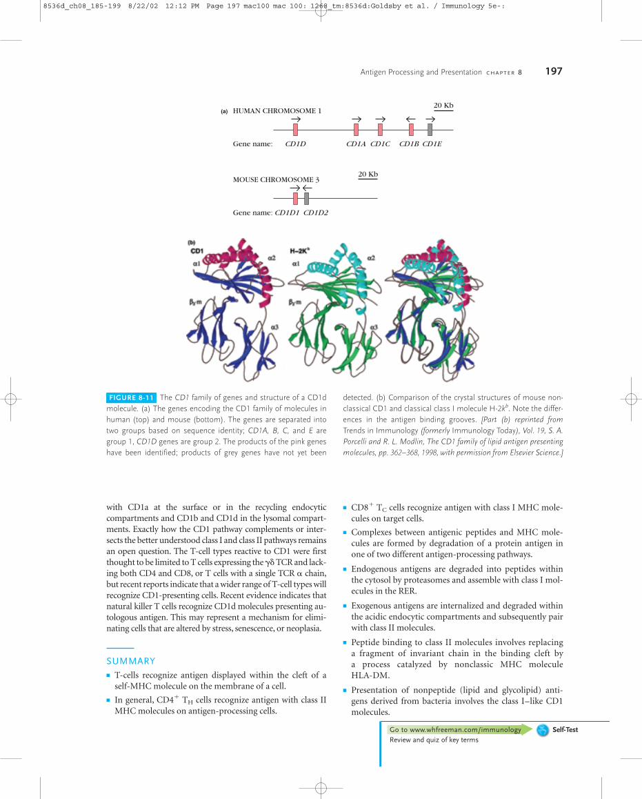
The CD1 family of molecules associates with �2-mi-croglobulin and has general structural similarity to class IMHC molecules. There are five genes encoding human CD1molecules (CD1A-E, encoding the gene products CD1a-d,with no product yet identified for E). These genes are locatednot within the MHC but on chromosome 1 (Figure 8-11a).The genes are classified into two groups based on sequencehomology. Group 1 includes CD1A, B, C, and E; CD1D is ingroup 2. All mammalian species studied have CD1 genes, al-though the number varies. Rodents have only group 2 CD1genes, the counterpart of human CD1D, whereas rabbits, likehumans, have five genes, including both group 1 and 2 types.Sequence identity of CD1 with classical class I molecules isconsiderably lower than the identity of the class I moleculeswith each other. Comparison of the three-dimensional struc-ture of the mouse CD1d1 with the class I MHC molecule H-2kb shows that the antigen-binding groove of the CD1d1molecules is deeper and more voluminous than that of theclassical class I molecule (Fig 8-11b).
Expression of CD1 molecules varies according to subset;CD1D1 genes are expressed mainly in nonprofessional APCsand on certain B-cell subsets. The mouse CD1d1 is morewidely distributed and found on T cells, B cells, dendriticcells, hepatocytes, and some epithelial cells. The CD1A, B,and C genes are expressed on immature thymocytes and pro-fessional APCs, mainly those of the dendritic type. CD1Cgene expression is seen on B cells, whereas the CD1A and Bproducts are not. CD1 genes can be induced by exposure tocertain cytokines such as GM-CSF or IL-3. The intracellulartrafficking patterns of the CD1 molecules differ; for example,CD1a is found mostly in early endosomes or on the cell sur-face; CD1b and CD1d localize to late endosomes; and CD1cis found throughout the endocytic system.
Certain CD1 molecules are recognized by T cells in the ab-sence of foreign antigens, and self restriction can be demon-strated in these reactions. Examination of antigens presentedby CD1 molecules revealed them to be lipid components(mycolic acid) of the M. tuberculosis cell wall. Further studiesof CD1 presentation indicated that a glycolipid (lipoarabino-mannan) from Mycobacterium leprae could also be presentedby these molecules. The data concerning CD1 antigen pre-sentation point out the existence of a third pathway for theprocessing of antigens, a pathway with distinct intracellularsteps that do not involve the molecules found to facilitateclass I antigen processing. For example, CD1 molecules areable to process antigen in TAP-deficient cells. Recent data indicate that the CD1a and 1b molecules traffic differently,
196 P A R T I I Generation of B-Cell and T-Cell Responses
8536d_ch08_185-199 8/2/02 10:08 AM Page 196 mac79 Mac 79:45_BW:Goldsby et al. / Immunology 5e:
with CD1a at the surface or in the recycling endocytic compartments and CD1b and CD1d in the lysomal compart-ments. Exactly how the CD1 pathway complements or inter-sects the better understood class I and class II pathways remainsan open question. The T-cell types reactive to CD1 were firstthought to be limited to T cells expressing the �� TCR and lack-ing both CD4 and CD8, or T cells with a single TCR � chain,but recent reports indicate that a wider range of T-cell types willrecognize CD1-presenting cells. Recent evidence indicates thatnatural killer T cells recognize CD1d molecules presenting au-tologous antigen. This may represent a mechanism for elimi-nating cells that are altered by stress, senescence, or neoplasia.
SUMMARY� T-cells recognize antigen displayed within the cleft of a
self-MHC molecule on the membrane of a cell.
� In general, CD4� TH cells recognize antigen with class IIMHC molecules on antigen-processing cells.
� CD8� TC cells recognize antigen with class I MHC mole-cules on target cells.
� Complexes between antigenic peptides and MHC mole-cules are formed by degradation of a protein antigen inone of two different antigen-processing pathways.
� Endogenous antigens are degraded into peptides withinthe cytosol by proteasomes and assemble with class I mol-ecules in the RER.
� Exogenous antigens are internalized and degraded withinthe acidic endocytic compartments and subsequently pairwith class II molecules.
� Peptide binding to class II molecules involves replacing a fragment of invariant chain in the binding cleft by a process catalyzed by nonclassic MHC molecule HLA-DM.
� Presentation of nonpeptide (lipid and glycolipid) anti-gens derived from bacteria involves the class I–like CD1molecules.
Antigen Processing and Presentation C H A P T E R 8 197
FIGURE 8-11 The CD1 family of genes and structure of a CD1dmolecule. (a) The genes encoding the CD1 family of molecules inhuman (top) and mouse (bottom). The genes are separated intotwo groups based on sequence identity; CD1A, B, C, and E aregroup 1, CD1D genes are group 2. The products of the pink geneshave been identified; products of grey genes have not yet been
detected. (b) Comparison of the crystal structures of mouse non-classical CD1 and classical class I molecule H-2kb. Note the differ-ences in the antigen binding grooves. [Part (b) reprinted fromTrends in Immunology (formerly Immunology Today), Vol. 19, S. A.Porcelli and R. L. Modlin, The CD1 family of lipid antigen presentingmolecules, pp. 362–368, 1998, with permission from Elsevier Science.]
HUMAN CHROMOSOME 120 Kb
Gene name: CD1D CD1ECD1BCD1CCD1A
MOUSE CHROMOSOME 320 Kb
Gene name: CD1D1 CD1D2
(a)
Go to www.whfreeman.com/immunology Self-TestReview and quiz of key terms
8536d_ch08_185-199 8/22/02 12:12 PM Page 197 mac100 mac 100: 1268_tm:8536d:Goldsby et al. / Immunology 5e-:
ReferencesAlfonso, C., and L. Karlsson. 2000. Nonclassical class II mole-
cules. Ann. Rev. Immunol. 18:113.
Brodsky, F. M., et al. 1999. Human pathogen subversion of anti-gen presentation. Immunol. Reviews. 168:199.
Busch, R., et al. 2000. Accessory molecules for MHC class II pep-tide loading. Curr. Opinion in Immunol. 12:99.
Doherty, P. C., and R. M. Zinkernagel. 1975. H-2 compatibility isrequired for T-cell mediated lysis of target cells infected withlymphocytic choriomeningitis virus. J. Exp. Med. 141:502.
Gadola, S. D., et al. 2000. TAP deficiency syndrome. Clin. Exp.Immunol. 121:173.
Ghosh P., M. Amaya, E. Mellins, and D. C. Wiley. 1995. Thestructure of an intermediate in class II MHC maturation: CLIPbound to HLA-DR3. Nature 378:457.
Jayawardena-Wolf, J., and A. Bendelac. 2001. CD1 and lipid anti-gens: intracellular pathways for antigen presentation. Curr.Opinions in Immunol. 13:109.
Matsuda J. L., and M. Kroneberg. 2001. Presentation of self andmicrobial lipids by CD1 molecules. Curr. Opinion in Immunol.13:19.
Ortmann, B., et al. 1997. A critical role for tapasin in the assem-bly and function of multimeric MHC class I–TAP complexes.Science 277:1306.
Pamer, E., and P. Cresswell. 1998. Mechanisms of MHC class I–restricted antigen processing. Annu. Rev. Immunol. 16:323.
Porcelli, S. A., and R. L. Modlin. 1999. The CD1 System: Antigen-presenting molecules for T-cell recognition of lipids and gly-colipids. Ann. Rev. Immunol. 17:297.
Roche, P. A. 1999. Intracellular protein traffic in lymphocytes:“How do I get there from here?” Immunity 11:391.
Van Ham, M., et al. 2000. What to do with HLA-DO? Immuno-genetics 51:765.
Yewdell, J. W. 2001. Not such a dismal science: The economics ofprotein synthesis, folding, degradation, and antigen process-ing. Trends in Cell Biol. 11: 294
Study QuestionsCLINICAL FOCUS QUESTION Patients with TAP deficiency havepartial immunodeficiency as well as autoimmune manifesta-tions. How do the profiles for patients’ immune cells explain thepartial immunodeficiency? Why is it difficult to design a genetherapy treatment for this disease, despite the fact that a singlegene defect is implicated?
1. Explain the difference between the terms antigen-presentingcell and target cell, as they are commonly used in immunology.
2. Define the following terms:
a. Self-MHC restrictionb. Antigen processingc. Endogenous antigend. Exogenous antigen
3. L. A. Morrison and T. J. Braciale conducted an experiment todetermine whether antigens presented by class I or II MHCmolecules are processed in different pathways. Their resultsare summarized in Table 8-2.
a. Explain why the class I–restricted TC cells did not re-spond to target cells infected with UV-inactivated in-fluenza virus.
b. Explain why chloroquine inhibited the response of theclass II–restricted TC cells to live virus.
c. Explain why emetine inhibited the response of class I–restricted but not class II–restricted TC cells to live virus.
4. For each of the following cell components or processes, indi-cate whether it is involved in the processing and presentationof exogenous antigens (EX), endogenous antigens (EN), orboth (B). Briefly explain the function of each item.
a. ______Class I MHC moleculesb. ______Class II MHC moleculesc. ______Invariant (Ii) chainsd. ______Lysosomal hydrolasese. ______TAP1 and TAP2 proteinsf. ______Transport of vesicles from the RER to the Golgi
complexg. ______Proteasomesh. ______Phagocytosis or endocytosisi. ______Calnexinj. ______CLIPk. ______Tapasin
5. Antigen-presenting cells have been shown to presentlysozyme peptide 46–61 together with the class II IAk mole-cule. When CD4�TH cells are incubated with APCs and nativelysozyme or the synthetic lysozyme peptide 46–61, TH-cellactivation occurs.
a. If chloroquine is added to the incubation mixture, presen-tation of the native protein is inhibited, but the peptidecontinues to induce TH-cell activation. Explain why thisoccurs.
b. If chloroquine addition is delayed for 3 h, presentation ofthe native protein is not inhibited. Explain why this occurs.
6. Cells that can present antigen to TH cells have been classifiedinto two groups—professional and nonprofessional APCs.
a. Name the three types of professional APCs. For each typeindicate whether it expresses class II MHC molecules and aco-stimulatory signal constitutively or must be activatedbefore doing so.
b. Give three examples of nonprofessional APCs. When arethese cells most likely to function in antigen presentation?
7. Predict whether TH-cell proliferation or CTL-mediated cytol-ysis of target cells will occur with the following mixtures ofcells. The CD4� TH cells are from lysozyme-primed mice, andthe CD8� CTLs are from influenza-infected mice. Use R toindicate a response and NR to indicate no response.
a. ______H-2k TH cells � lysozyme-pulsed H-2k
macrophagesb. ______H-2k TH cells � lysozyme-pulsed H-2b/k
macrophages
198 P A R T I I Generation of B-Cell and T-Cell Responses
8536d_ch08_185-199 8/2/02 10:08 AM Page 198 mac79 Mac 79:45_BW:Goldsby et al. / Immunology 5e:
c. ______H-2k TH cells � lysozyme-primed H-2d
macrophagesd. ______H-2k CTLs � influenza-infected H-2k
macrophagese. ______H-2k CTLs � influenza-infected H-2d
macrophagesf. ______H-2d CTLs � influenza-infected H-2d/k
macrophages
8. HLA-DM and HLA-DO are termed nonclassical MHC class IImolecules. How do they differ from the classical MHC classII? How do they differ from each other?
9. Molecules of the CD1 family were recently shown to presentnonpeptide antigens.
a. What is a major source of nonpeptide antigens?b. Why are CD1 molecules not classified as members of
the MHC family even though they associate with �2-microglobulin?
c. What evidence suggests that the CD1 pathway is differentfrom that utilized by classical class I MHC molecules?
Antigen Processing and Presentation C H A P T E R 8 199
8536d_ch08_185-199 8/2/02 10:08 AM Page 199 mac79 Mac 79:45_BW:Goldsby et al. / Immunology 5e:


