Embed Size (px)
Citation preview

Pure Appl. Chem., Vol. 75, Nos. 2–3, pp. 157–166, 2003.© 2003 IUPAC
157
Antiangiogenic heparin-derived heparan sulfatemimics*
Benito Casu‡ and Annamaria Naggi
G. Ronzoni Institute for Chemical and Biochemical Research, Milan, Italy
Abstract: Heparan sulfate (HS) is a glycosaminoglycan (GAG) widely distributed as a pro-teoglycan on the cell surface and in the extracellular matrix of animal tissues. Among otherimportant physiological functions, its polysaccharide chains mediate cell proliferation bybinding growth factors [fibroblast growth factor (FGF), vascular endothelial growth factor(VEGF)], which are released in active form through the action of the enzyme heparanaseoverexpressed by tumor cells. HS is constituted of alternating disaccharide sequences of var-iously sulfated uronic acid (D-glucuronic, GlcA, or L-iduronic, IdoA) and glucosamine (N-acetylated, GlcNAc, or N-sulfated, GlcNSO3). HS mimics can be obtained by chemical mod-ification of heparin, a more highly sulfated GAG clinically used as an anticoagulant andantithrombotic drug. With the aim of obtaining antagonists of FGFs as potential inhibitors oftumor neo-vascularization (angiogenesis), arrays of short FGF-binding sequences have beenobtained by generating sulfation gaps within the prevalent (IdoA2SO3–GlcNSO36SO3)nsequences of heparin, by controlled base-catalyzed removal of 2-O-sulfate groups ofIdoA2SO3 residues. The C(2)–C(3) bond of all nonsulfated uronic acid residues have thenbeen split with periodate, to generate flexible joints along the polysaccharide chain. The novelheparin derivative (poly-PST.sU), chiefly made up of the repeating tetrasaccharide units–GlcNSO36SO3–IdoA2SO3–GlcNSO36SO3–sU– (where sU is a glycol-split and reduceduronic acid residue) binds FGF2 as strongly as heparin. However, it is a poor inducer of for-mation of FGF2 dimers and of complexes with FGF receptors, required for triggering mito-genic signals. NMR and molecular modeling studies indicate that formation of these higher-order complexes is prevented by the unfavorable conformation induced by glycol-splitresidues. In a parallel study, partially N-acetylated heparins have been obtained that efficientlyinhibit heparanase upon glycol-splitting. It is noteworthy that glycol-splitting involves inacti-vation of the active site for antithrombin, with consequent loss of anticoagulant activity. Incontrast, poly-PST.sU and some of its analogs show potent antiangiogenic activity in in vivomodels in which heparin is either proangiogenic or inactive.
INTRODUCTION
Heparan sulfate (HS) is a carbohydrate polymer widely distributed on cell surfaces and extracellularmatrix as a proteoglycan (HSPG) [1–3]. Among its multiple functions, HS controls cell adhesion andproliferation through binding to a variety of proteins, among which growth factors of the FGF family[3–5]. Not unexpectedly, HSPGs and their fragments modulate the onset and development of cancer [6].The enzyme heparanase, overexpressed by tumor cells [7], releases FGFs, which are stored by the HSchains of HSPG in inactive form [8]. As schematized in Fig. 1, fragments of the original polysaccharide
*Pure Appl. Chem. 75, 141–419 (2003). An issue of reviews and research papers based on lectures presented at the 23rd IUPACInternational Symposium on the Chemistry of Natural Products, Florence, Italy, 28 July–2 August 2002 on the theme of naturalproducts.‡Corresponding author

chains induce dimerization of the growth factor and its activation through binding to FGF cellular recep-tors (FGFRs), a prerequisite for triggering mitogenic signals (Fig. 1).
Like heparin, which is clinically used as an anticoagulant and antithrombotic drug, HS is a GAGconstituted of repeating disaccharide sequences of a uronic acid (D-glucuronic, GlcA, and L-iduronic,IdoA) and D-glucosamine (either N-acetylated, GlcNAc, or N-sulfated, GlcN), with variable degrees ofO-sulfation on both residues [1]. Though at different locations (heparin is mainly confined in mast cellsof connective tissues), heparin and HS are biosynthesized in a similar way, through sequential action ofenzymes (N-deacetylase/N-sulfatase, C5-epimerases, O-sulfotransferases) on the common precursorN-acetylheparosan, constituted of repeating GlcA–GlcNAc disaccharide sequences. Whereas thebiosynthesis of heparin proceeds to reach high degrees of N- and O-sulfation [their structure beingprevalently accounted for by NS domains chiefly constituted by sequences of the trisulfated disaccha-ride (TD) IdoA2SO3–GlcNSO36SO3], HS has a much more hybrid structure, with prevalent GlcA- andGlcNAc-containing regions (NA- and NA/NS domains), which are only minor constituents of heparin.Both GAGs contain the minor but important pentasaccharide sequence of the antithrombin-bindingdomain (AT-bd), essential for the expression of significant anticoagulant properties [1] (Fig. 2).
B. CASU AND A. NAGGI
© 2003 IUPAC, Pure and Applied Chemistry 75, 157–166
158
Fig. 1 Simplified representation of an HSPG molecule and its involvement in cellular signaling. HSPGs consist ofHS chains linked to a peptide core. Among other proteins, HS stores growth factors such as FGFs in inactive form.The HS chains may be cleaved (arrows) by the enzyme heparanase (a). The released FGFs are activated by HSfragments, which favor their dimerization and formation of a ternary complex with cellular FGFRs (b).
Fig. 2 Biosynthesis and major structural domains of heparin/HS (adapted from [1]).
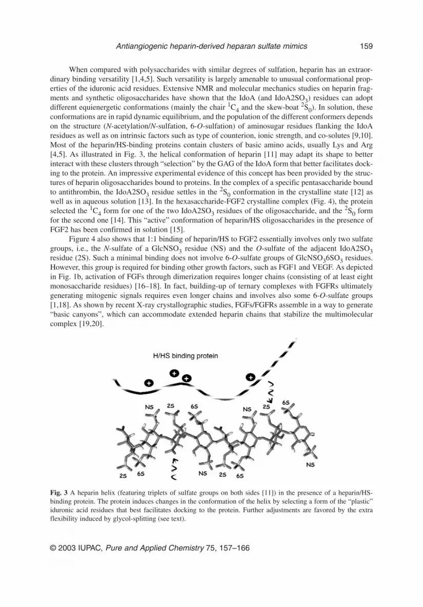
When compared with polysaccharides with similar degrees of sulfation, heparin has an extraor-dinary binding versatility [1,4,5]. Such versatility is largely amenable to unusual conformational prop-erties of the iduronic acid residues. Extensive NMR and molecular mechanics studies on heparin frag-ments and synthetic oligosaccharides have shown that the IdoA (and IdoA2SO3) residues can adoptdifferent equienergetic conformations (mainly the chair 1C4 and the skew-boat 2S0). In solution, theseconformations are in rapid dynamic equilibrium, and the population of the different conformers dependson the structure (N-acetylation/N-sulfation, 6-O-sulfation) of aminosugar residues flanking the IdoAresidues as well as on intrinsic factors such as type of counterion, ionic strength, and co-solutes [9,10].Most of the heparin/HS-binding proteins contain clusters of basic amino acids, usually Lys and Arg[4,5]. As illustrated in Fig. 3, the helical conformation of heparin [11] may adapt its shape to betterinteract with these clusters through “selection” by the GAG of the IdoA form that better facilitates dock-ing to the protein. An impressive experimental evidence of this concept has been provided by the struc-tures of heparin oligosaccharides bound to proteins. In the complex of a specific pentasaccharide boundto antithrombin, the IdoA2SO3 residue settles in the 2S0 conformation in the crystalline state [12] aswell as in aqueous solution [13]. In the hexasaccharide-FGF2 crystalline complex (Fig. 4), the proteinselected the 1C4 form for one of the two IdoA2SO3 residues of the oligosaccharide, and the 2S0 formfor the second one [14]. This “active” conformation of heparin/HS oligosaccharides in the presence ofFGF2 has been confirmed in solution [15].
Figure 4 also shows that 1:1 binding of heparin/HS to FGF2 essentially involves only two sulfategroups, i.e., the N-sulfate of a GlcNSO3 residue (NS) and the O-sulfate of the adjacent IdoA2SO3residue (2S). Such a minimal binding does not involve 6-O-sulfate groups of GlcNSO36SO3 residues.However, this group is required for binding other growth factors, such as FGF1 and VEGF. As depictedin Fig. 1b, activation of FGFs through dimerization requires longer chains (consisting of at least eightmonosaccharide residues) [16–18]. In fact, building-up of ternary complexes with FGFRs ultimatelygenerating mitogenic signals requires even longer chains and involves also some 6-O-sulfate groups[1,18]. As shown by recent X-ray crystallographic studies, FGFs/FGFRs assemble in a way to generate“basic canyons”, which can accommodate extended heparin chains that stabilize the multimolecularcomplex [19,20].
© 2003 IUPAC, Pure and Applied Chemistry 75, 157–166
Antiangiogenic heparin-derived heparan sulfate mimics 159
Fig. 3 A heparin helix (featuring triplets of sulfate groups on both sides [11]) in the presence of a heparin/HS-binding protein. The protein induces changes in the conformation of the helix by selecting a form of the “plastic”iduronic acid residues that best facilitates docking to the protein. Further adjustments are favored by the extraflexibility induced by glycol-splitting (see text).

FGFs (especially FGF2) and VEGFs are potent inducers of neo-vascularization [6] and are poten-tial targets to develop antitumor drugs. FGFs and VEGFs are heparin-binding proteins [4], and becausethe NS domains prevalent in heparin are also putative binding domains for these growth factors, heparinrepresents the starting material of choice to generate angiogenesis inhibitors. Heparin itself is beingconsidered as a potential anticancer drug. In fact, heparin has shown antimetastatic activity in animalmodels [21]. Moreover, heparin and low-molecular-weight heparin administered as an antithromboticdrug significantly prolong the life of cancer patients [22]. However, the use of unmodified heparininvolves hemorrhagic risks. Heparin has also intriguing effects on cells. In fact, it can stimulate orinhibit growth factors in different cellular systems, apparently depending on different types and con-centrations of HSPGs [4,23].
Reported strategies to inhibit FGFs with HS mimics are based on the use of heparin fragments orsynthetic oligosaccharides able to complex the growth factors, but shorter than necessary to induce theirdimerization and activation [24], and (for FGF2) on removal from heparin of the 6-O-sulfate groups notrequired for 1:1 binding, but necessary for activation [25].
TARGETING FIBROBLAST GROWTH FACTORS
Our design of FGF2 inhibitors was based on interruption of the regular TD repeating sequences alongthe NS region of heparin by selective desulfation, in order to generate arrays of sequences that containthe minimal binding site to the growth factor, but are too short to induce its dimerization and activation.The sulfation gaps were obtained by alkali-induced 2-O-desulfation of IdoA2SO3 residues throughintermediate epoxides [26–28]. In addition, the C2–C3 bonds of the nonsulfated uronic acid residueswere cleaved with periodate, and the resulting polydialdehydes were stabilized by reduction with boro-hydride. The glycol-split residues were expected to act as flexible joints along the polysaccharide chains[29], thus facilitating 1:1 binding to FGF2 while disrupting the relatively rigid conformation requiredto stabilize FGF-FGFR assemblies.
The prevalent structure of heparin (1), intermediates 2, 3, and of the final product 4 of the desul-fation via epoxide under conditions to obtain 2-O-desulfation and glycol-splitting of about 50 % ofIdoA2SO3 residues in the NS regions [30], as confirmed/determined by mono- and two-dimensionalNMR spectroscopy, are reported in Fig. 5. The spectra of the final product are remarkably clean, with
B. CASU AND A. NAGGI
© 2003 IUPAC, Pure and Applied Chemistry 75, 157–166
160
Fig. 4 Conformation of a heparin hexasaccharide in a co-crystal with FGF2 (adapted from [14]), showing that thetwo IdoA2SO3 residues are in different conformations. Primary binding involves essentially the two sulfate groupsNS and 2S of the hexasaccharide.

only weak signals reminiscent of the original, minor NA regions unaffected by the reactions. The struc-ture of 4 was further confirmed by Smith degradation, which cleaved the modified heparin at the levelof glycol-split residues and generated the trisaccharide GlcNSO36SO3–IdoA2SO3–GlcNSO36SO3–R(where R is the remnant of a glycol-split uronic acid residue) as a major fragment [31].
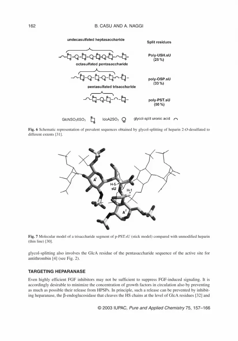
The 2-O-desulfation–glycol-splitting reaction was performed under controlled conditions in orderto obtain longer NS sequences separated by glycol-split residues. The prevalent structure of productsobtained by splitting a total of about 25, 33, and 50 % uronic acid residues is schematized in Fig. 6,showing that graded modification of heparin yields oligosaccharide sequences of different lengths sep-arated by glycol-split residues.
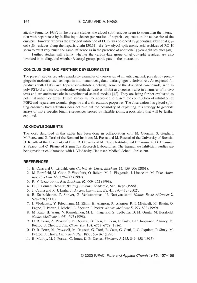
NMR and molecular mechanics studies on the 50 %-split product (prevalently poly-pentasulfatedpentasaccharide, p-PST.sU) confirmed the assumption that glycol-split residues drastically modify theconformation of the heparin chains. As illustrated in Fig. 7 for one of the major conformers of 4 com-patible with measured nuclear Overhauser effects (NOEs), the chain of the glycol-split product deviatesfrom the linearity of the heparin helix that can be accommodated in the basic canyon of FGF/FGRFassemblies. It was then expected that, whereas still able to form 1:1 complexes, product 4 (and possi-bly also some of the heparin derivatives with somewhat longer NS sequences framed between glycol-split residues) is unable to induce formation of higher-order complexes and would accordingly act as anangiogenesis inihibitor.
Indeed, 4 was found to retain the FGF2-binding ability of the original heparin, as shown by itscapacity to protect FGF2 from trypsin cleavage and to prevent the formation of HSPS/FGF2/FGFR1ternary complexes. However, when compared to heparin it showed a reduced capacity to induce FGF2dimerization, and to favor the interaction of [125I]FGF2 with FGFR1 in HSPG-deficient, FGFR1-trans-fected CHO cells. Accordingly, it was more effective than heparin in inhibiting the mitogenic activityexerted by FGF2 in cultured endothelial cells. Finally, it inhibited angiogenesis in a chick embryochoroallantoic membrane (CAM) assay in which heparin is inactive. [30]. Such an antiangiogenic activ-ity is retained by corresponding low-molecular-weight derivatives [31]. Although somewhat less active,derivatives prevalently corresponding to poly-OSP.sU (see Fig. 6) also bound FGF2 and inhibited bothFGF2-stimulated growth of cultured endothelial cells and capillaries growth in the CAM model [31]. Itis worth noting that the presently described derivatization, and especially glycol-splitting, leads to lossof the anticoagulant properties of heparin, which are undesirable owing to hemorrhagic risks. In fact,
© 2003 IUPAC, Pure and Applied Chemistry 75, 157–166
Antiangiogenic heparin-derived heparan sulfate mimics 161
Fig. 5 Prevalent tetrasaccharide units of heparin (1), its epoxi derivative 2, its galacto derivative 3, and the glycol-split derivative 4 [30].
glycol-splitting also involves the GlcA residue of the pentasaccharide sequence of the active site forantithrombin [4] (see Fig. 2).
TARGETING HEPARANASE
Even highly efficient FGF inhibitors may not be sufficient to suppress FGF-induced signaling. It isaccordingly desirable to minimize the concentration of growth factors in circulation also by preventingas much as possible their release from HPSPs. In principle, such a release can be prevented by inhibit-ing heparanase, the β-endoglucosidase that cleaves the HS chains at the level of GlcA residues [32] and
B. CASU AND A. NAGGI
© 2003 IUPAC, Pure and Applied Chemistry 75, 157–166
162
Fig. 6 Schematic representation of prevalent sequences obtained by glycol-splitting of heparin 2-O-desulfated todifferent extents [31].
Fig. 7 Molecular model of a trisaccharide segment of p-PST.sU (stick model) compared with unmodified heparin(thin line) [30].

releases FGFs in active form [8]. By preserving the integrity of HSPSs in the extracellular matrix, inhi-bition of heparanase is expected also to prevent invasion of cancer cells through the connective tissue[7].
X-ray structures of heparanase-substrate complexes are not yet available. However, similaritiesbetween the structure of the enzyme and members of several groups of glycosyl hydrolase families havepermitted identification of the active-site acid residues and mapping of clusters of basic aminoacids asputative ligand binding domains [33]. HS sequences binding to these clusters most likely belong to theNS (heparin-like) domain of the GAG, and may thus be mimicked by heparin.
Indeed, heparin, as well as N-acetyl heparin (i.e., heparin whose N-sulfate groups are completelyreplaced by N-acetyl groups) efficiently inhibits heparanase [34,35]. Although different preparations ofheparin inhibited the enzyme to significantly different extents [36], suggesting subtle specificities,activities observed for heparin derivatives did not unravel structural features essential for heparanaseinhibition. Total substitution of heparin N-sulfate groups with N-acetyl groups [35] nor completeremoval of 2-O-sulfate groups [37] were reported to significantly impair the heparanase-inhibitionproperties of heparin. On the other hand, also extensively sulfated, nonglycosaminoglycan polysaccha-rides of widely different structures are good heparanase inhibitors [38,39]. Extensive screening of sul-fated carbohydrate polymers and their fragments indicated that the enzyme can be strongly inhibited bysome persulfated oligosaccharides as small as hexa- and pentasaccharide, such as maltohexaose sulfateand phosphomannopentaose sulfate [38].
Interestingly, reduced oxyheparin (RO-heparin, obtained by glycol-splitting of all the nonsulfateduronic acid residues preexisting in heparin) turned out to be at least as effective an inhibitor of theenzyme as the parent GAG [37]. This observation prompted revisitation of heparins and heparin deriv-atives as heparanase inhibitors. Whereas removal of 2-O-sulfate groups was confirmed to have littleeffect on the inhibition activity as previously reported [37], the activity increased with increasing con-tent of 6-O-sulfate groups [40]. Using a more sensitive test than in previous reports, fully N-acetylatedheparin was found to be one order of magnitude less active as heparanase inhibitor than heparin. Inmore detail, while the inhibition activity is barely affected by replacement of about 40 % of the origi-nal NSO3 groups by N-acetyl groups, it sharply decreases for higher N-acetyl contents, indicating thatat least one NSO3 group per tetrasaccharide unit is needed for binding to (and inhibition of) the enzyme.However, the most dramatic influence of chemical modification on the heparanase-inhibiting activitywas observed upon glycol-splitting. As illustrated in Fig. 8, glycol-splitting enhances the activity ofN-acetylated heparins irrespective of their N-acetylation level, the most notable activity increase beingobserved at about 70 % N-acetylation [40]. As earlier observed for lipoprotein lipase [41] and system-
© 2003 IUPAC, Pure and Applied Chemistry 75, 157–166
Antiangiogenic heparin-derived heparan sulfate mimics 163
Fig. 8 Heparanase inhibition by heparin at different degrees of N-acetylation (�) and of the same products afterglycol-splitting of nonsulfated uronic acid residues (○) [40].
atically found for FGF2 in the present studies, the glycol-split residues seem to strengthen the interac-tion with heparanase by facilitating a deeper penetration of heparin sequences in the active site of theenzyme. However, whereas the strongest inhibition of FGF2 was observed by generating additional gly-col-split residues along the heparin chain [30,31], the few glycol-split uronic acid residues of RO–Hseem to exert very much the same influence as in the presence of additional glycol-split residues [40].
Further studies will clarify whether the carboxylate group of glycol-split residues are alsoinvolved in binding, and whether N-acetyl groups participate in the interaction.
CONCLUSIONS AND FURTHER DEVELOPMENTS
The present studies provide remarkable examples of conversion of an anticoagulant, prevalently proan-giogenic molecule such as heparin into nonanticoagulant, antiangiogenic derivatives. As expected forproducts with FGF2- and heparanase-inhibiting activity, some of the described compounds, such aspoly-PST.sU and its low-molecular-weight derivatives inhibit angiogenesis also in a number of in vivotests and are antimetastatic in experimental animal models [42]. They are being further evaluated aspotential antitumor drugs. Future studies will be addressed to dissect the contribution of inhibition ofFGF2 and heparanase to antiangiogenic and antimetastatic properties. The observation that glycol-split-ting enhances both activities does not rule out the possibility of exploiting this strategy to generatearrays of more specific binding sequences spaced by flexible joints, a possibility that will be furtherexplored.
ACKNOWLEDGMENTS
The work described in this paper has been done in collaboration with M. Guerrini, S. Guglieri, M. Perez, and G. Torri of the Ronzoni Institute; M. Presta and M. Rusnati of the University of Brescia;D. Ribatti of the University of Bari; R. Giavazzi of M. Negri Institute; and P. Carminati, G. Giannini,S. Penco, and C. Pisano of Sigma-Tau Research Laboratories. The heparanase-inhibition studies arebeing made in collaboration with I. Vlodavsky, Hadassah Medical School, Jerusalem.
REFERENCES
1. B. Casu and U. Lindahl. Adv. Carbohydr. Chem. Biochem. 57, 159–206 (2001). 2. M. Bernfield, M. Götte, P. Woo Park, O. Reizes, M. L. Fitzgerald, J. Lincecum, M. Zako. Annu.
Rev. Biochem. 68, 729–777 (1999).3. R. V. Iozzo. Annu. Rev. Biochem. 67, 609–652 (1998).4. H. E. Conrad. Heparin Binding Proteins, Academic, San Diego (1998).5. J. Capila and R. J. Linhardt. Angew. Chem., Int. Ed. 41, 390–412 (2002).6. R. Sasisekharan, Z. Shriver, G. Venkataraman, U. Narayanasami. Nature Reviews/Cancer 2,
521–528 (2002).7. I. Vlodavsky, Y. Friedmann, M. Elkin, H. Aingorn, R. Atzmon, R.-I. Michaeli, M. Bitain, O.
Pappo, T. Peretz, I. Michal, L. Spector, I. Pecker. Nature Medicine 5, 793–802 (1999).8. M. Kato, H. Wang, V. Kainulainen, M. L. Fitzgerald, S. Ledbetter, D. M. Ornitz, M. Bernfield.
Nature Medicine 4, 691–697 (1998).9. D. R. Ferro, A. Provasoli, M. Ragazzi, G. Torri, B. Casu, G. Gatti, J.-C. Jacquinet, P. Sinaÿ, M.
Petitou, J. Choay. J. Am. Chem. Soc. 108, 6773–6778 (1986).10. D. R. Ferro, M. Provasoli, M. Ragazzi, G. Torri, B. Casu, G. Gatti, J.-C. Jaquinet, P. Sinaÿ, M.
Petitou, J. Choay. Carbohydr. Res. 185, 157–167 (1990).11. B. Mulloy, M. J. Forster, C. Jones, D. B. Davies. Biochem. J. 293, 849–858 (1993).
B. CASU AND A. NAGGI
© 2003 IUPAC, Pure and Applied Chemistry 75, 157–166
164
12. L. Jin, J. Abrahams, R. Skinner, M. Petitou, R. N. Pike, R. W. Carrell. Proc. Nat. Acad. Sci. USA94, 14683–14688 (1997).
13. M. Hricovini, M. Guerrini, A. Bisio, G. Torri, B. Casu. Biochem. J. 359, 265–272 (2001).14. S. Faham, R. E. Hileman, J. R. Fromm, R. J. Linhardt, D. C. Rees. Science 271, 1116–1120
(1996).15. H. Hricovini, M. Guerrini, A. Bisio, G. Torri, A. Naggi, B. Casu. Seminars Thromb. Hemost. 28,
325–334 (2002).16. S. Guimond, M. Maccarana, B. B. Olvin, U. Lindahl, A. C. Rapraeger. J. Biol. Chem. 268,
23906–23914 (1993). 17. D. A. Pye, R. D. Vives, J. E. Turnbull, P. Hyde, J. T. Gallagher. J. Biol. Chem. 36, 22935–22942
(1998).18. L. Pellegrini. Curr. Opin. Struct. Biol. 11, 629–634 (2001).19. A. N. Plotnikov, J. Schlessinger, S. H. Hubbard, M. Mohammadi. Cell 413–424 (2000).20. D. J. Stauber, A. D. Di Gabriele, W. A. Hendrikson. Proc. Natl. Acad. Sci. USA 97, 49–54 (2000).21. I. Vlodavsky, R. Ishai-Michaeli, M. Mohsen, R. Bar-Shavit, R. Catane, H.-P. Ekre, C. M. Svahn.
In Heparin and Related Polysaccharides, D. A. Lane, I. Björk, U. Lindahl (Eds.), pp. 317–327,Plenum, New York (1992).
22. S. M. Smolenburg and C. J. F. Van Noorden. Pharmacol. Rev. 65, 93–105 (2001).23. M. Fannon, K. E. Forsten, M. A. Nugent. Biochemistry 39, 1434–1445 (2000).24. D. M. Ornitz, A. B. Herr, M. Nilsson, J. Westman, C.-M. Scahn, G. Waksman. Science 268,
432–436 (1995).25. L. Lundin, H. Larsson, J. Krueger, S. Kanda, U. Lindahl, M. Salmivirta, L. Claesson-Welsh. J.
Biol. Chem. 275, 24653–24660 (2000).26. R. N. Rej and A. S. Perlin. Carbohydr. Res. 200, 437–447 (1990).27. S. Piani, B. Casu, E. G. Marchi, G. Torri, F. Ungarelli. J. Carbohydr. Chem. 12, 507–521 (1993).28. K. Holme, W. Liang, F. Lapierre, P. N. Shaklee, L. Lam. In Nonanticoagulant Actions of
Glycosaminoglycans, J. Harenberg and B. Casu (Eds.), pp. 139–162, Plenum, New York (1996).29. B. Casu. In New Trends in Haemostasis, J. Harenberg, D. L. Heene, G. Stehle, G. Schettler (Eds.),
pp. 2–11, Springer Verlag, Heidelberg (1990).30. B. Casu, M. Guerrini, A. Naggi, M. Perez, G. Torri, D. Ribatti, P. Carminati, G. Giannini, S.
Penco, C. Pisano, M. Belleri, M. Rusnati, M. Presta. Biochemistry 41, 10519–10528 (2002).31. B. Casu, M. Guerrini, S. Guglieri, A. Naggi, M. Perez, G. Torri, D. Ribatti, P. Carminati, G.
Giannini, S. Penco, C. Pisano, M. Belleri, M. Rusnati, M. Presta. J. Med. Chem. Submitted forpublication.
32. D. S. Pikas, J. P. Li, I. Vlodavsky, U. Lindahl. J. Biol. Chem. 273, 18770–18777 (1998). 33. M. D. Hulett, J. R. Hornby, S. J. Ohms, J. Zuegg, C. Freeman, J. E. Gready, C. R. Parish.
Biochemistry 39, 15659–15667 (2000).34. T. Irimura, M. Nakajima, G. L. Nicholson. Biochemistry 25, 5322–5328 (1986).35. R. Ishai-Michaeli, C. M. Svahn, M. Weber, T. Chajeck-Shaul, G. Korner, H.-P. Ekre, I. Vlodavsky.
Biochemistry 31, 2080–2088 (1992).36. D. R. Coombe, C. R. Parish, I. A. Ramshaw, J. M. Snowden. Int. J. Cancer 39, 82–88 (1987).37. F. Lapierre, K. Holme, L. Lam, R. J. Tressler, N. Storm, J. Wee, R. J. Stack, J. Castelot, D. J.
Tyrrell. Glycobiology 6, 355–366 (1996).38. C. R. Parish, C. Freeman, K. J. Brown, D. J. Francis, W. B. Cowden. Cancer Res. 59, 3433–3441
(1999).39. H.-Q. Miao, M. Elkin, E. Aingorn, R. Isshai-Michaeli, C. A. Stein, I. Vlodavsky. Int. J. Cancer
83, 424–431 (1999).40. A. Naggi, M. Perez, G. Torri, S. Penco, C. Pisano, G. Giannini, I. Vlodavsky, B. Casu.
Glycobiology. In preparation.
© 2003 IUPAC, Pure and Applied Chemistry 75, 157–166
Antiangiogenic heparin-derived heparan sulfate mimics 165

41. B. Casu, G. Diamantini, G. Fedeli, M. Mantovani, P. Oreste, R. Pescador, R. Porta, G. Torrri, G.Zoppetti. Arzneim.-Forsch. (Drug Res.) 36, 637–642 (1986).
42. C. Pisano, M. L. Cervoni, I. Chiarucci, R. Foderà, V. Giordano, K. Lombardo, M. Marcellini, T.Riccioni, M. A. Stasi, L. Vesci, S. Penco, M. Poli, R. Giavazzi, P. Carminati. Commun. NCI-EORTC-AARC Symposium (2002).
B. CASU AND A. NAGGI
© 2003 IUPAC, Pure and Applied Chemistry 75, 157–166
166


