Embed Size (px)
Citation preview
BookID 148921_ChapID 10_Proof# 1 - 18 / 09 / 2009
Chapter 10
Analysis of miRNA Modifications
Bin Yu and Xuemei Chen
Abstract
After transcription, a large number of cellular RNAs employ modifications to increase their diversity and functional potential. Modifications can occur on the base, ribose, or both, and are important steps in the maturation of many RNAs. Our lab recently showed that plant microRNAs (miRNAs) possess a 2¢-O-methyl group on the ribose of the 3¢ terminal nucleotide, and that this methyl group is added after miRNA/miRNA* formation. One function of this modification is to protect miRNAs from 3¢ terminal uridylation by an unknown enzymatic activity. It is possible that uridylation of miRNAs triggers their degradation. Here we describe a protocol to purify a specific miRNA in order to determine its molecular mass so that the presence of a modification can be inferred, an in vivo method to detect 3¢ terminal modification of miRNAs, and an (a-32P) dATP incorporation assay to study 3¢ terminal uridylation of miRNAs.
Key words: miRNA, Methylation, Uridylation, b elimination
1. Introduction
MicroRNAs (miRNAs) are short noncoding RNAs that recognize partially or completely complementary sequences inside target mRNAs and guide cleavage or translational inhibition of target mRNAs (1). This ability has made miRNAs important regulators of gene expression in both animals and plants (1). miRNAs are generated from long stem-loop precursor transcripts known as pri-miRNAs (1). In animals, an RNAase III enzyme Drosha processes pri-miRNAs into pre-miRNAs, which are processed by another RNAase III enzyme Dicer to generate transient 20–24 nucleotide (nt) miRNA/miRNA* duplexes (2–5). In plants, an RNAse III enzyme DICER LIKE1 (DCL1) processes priamiRNAs to preamiRNAs and pre-miRNAs to miRNA/miRNA* duplexes (6, 7) with the aid of HYL1 and SERRATE (8–11). miRNA/miRNA* duplexes show typical features of RNAase III products, 5¢ P, 3¢OH and a 2 nt overhang on each strand (4, 12).
137
B.C. Meyers and P.J. Green (eds.), Plant MicroRNAs, Methods in Molecular Biology, vol. 592DOI 10.1007/978-1-60327-005-2_10, © Humana Press, a part of Springer Science + Business Media, LLC 2010

138 Yu and Chen
BookID 148921_ChapID 10_Proof# 1 - 18 / 09 / 2009 BookID 148921_ChapID 10_Proof# 1 - 18 / 09 / 2009
Recently, our lab showed that Arabidopsis miRNA/miRNA* duplexes have an additional feature, 2¢-O-methylation on the 3¢ terminal ribose (13, 14), and an enzyme named HUA ENHANCER1 (HEN1; 15) catalyzes the methylation reaction (13). We revealed the presence of a methyl group on miR173 via mass spectrometry analysis of miR173 purified from Arabidopsis total RNAs. We also demonstrated the presence of methylation at the 3¢ termini of miRNAs by treating miRNAs with sodium peri-odate followed by b elimination (13). Complete or partial loss-of-function mutations in HEN1, such as hen1-1 or hen1-2, result in reduced accumulation and size heterogeneity of miRNAs and pleiotropic developmental defects (6, 15, 16). With the cloning of particular miRNAs and with an (a-32P) dATP incorporation assay, we showed that the size heterogeneity of miRNAs in hen1 mutants comes from 3¢ terminal uridylation, suggesting that unmethylated miRNAs are modified by an unknown polymerase activity in plants (17).
In this chapter, we describe a procedure to purify a specific miRNA for mass spectrometry analysis and a protocol to perform b elimination to detect modifications on the 3¢ terminal ribose of miRNAs. We also describe an (a-32P) dATP incorporation assay to detect 3¢ terminal uridylation.
2. Materials
1. Tri-reagent (Molecular Research Center, Inc. Cat# TR 118).2. Diethyl pyrocarbonate (DEPC)-treated water. Add 1 ml DEPC
to 1 l deionized water, stir overnight, and autoclave the next day.
3. Chloroform (VWR, cat# EM-CX1055-14).4. Isopropanol (VWR, cat# EM-PX1838-1).5. 100% ethanol and 70% ethanol. Mix 70-ml 100% ethanol with
30-ml DEPC-treated water.
1. Biotinylated probe. 5¢ biotin-aagtgatttctctctgcaagcgaa 3¢ (see Note 1).
2. 20× SSC.3. RNasin Plus RNase Inhibitor (Promega, cat # N2615).
1. Streptavidin magnetic particles (Roche, cat# 11641778001).2. 0.5× SSC.3. Magnetic stand (Promega).
2.1. Purifying miR173 with a Complementary Oligonucleotide Probe Coupled to Biotin
2.1.1. Extraction of RNAs
2.1.2. Annealing of Probe
2.1.3. Preparation of Streptavidin Magnetic Particles
BookID 148921_ChapID 10_Proof# 1 - 18 / 09 / 2009
Analysis of miRNA Modifications 139
BookID 148921_ChapID 10_Proof# 1 - 18 / 09 / 2009
1. Exonuclease I (GE Healthcare, cat# E70073Z).
1. 5× TBE and 0.5× TBE.2. 2× RNA loading buffer. Mix 8 ml formamide, 2 ml 5× TBE,
10 mg bromophenol blue and 10 mg xylene cyanol.3. 15% polyacrylamide gel containing 42% urea. Dissolve 42 g
urea in 4 ml 5× TBE and 15 ml 40% acrylamide (acrylamide:bis-acrylamide, 29:1) and add water to 40 ml. Add 320 ml 10% APS and 24 ml TEMED (see Note 2).
4. Zeta-probe GT membrane (BioRad, cat# 162-093).5. Ultrahyb-Oligo hybridization buffer (Ambion, cat# AM8663).6. T4 polynucleotide kinase (NEB, cat# M0201S).7. (g-32P) ATP (PerkinElmer).8. DNA probe 5¢ gtgatttctctctgcaagcgaa 3¢ and synthesized
miR173 5¢ UUCGCUUGCAGAGAGAAAUCAC 3¢ (a, t, g and c are deoxyribonucleotides; A, U, G and C are ribon- ucleotides).
9. 2× SSC with 0.5% SDS.
1. 0.06 M borax/boric acid buffer (pH 8.6; see Note 3).2. 0.055 M borax/boric acid/NaOH (pH 9.5).3. 200 mM sodium periodate (see Note 4).4. Glycerol.5. Glycogen (Fermentas, cat# R0551).6. 3 M sodium acetate (pH 5.2, DEPC-treated).7. G25 column (GE Healthcare).
1. 50% polyethylene glycol 8000 (DEPC-treated).2. 5 M NaCl (DEPC-treated).
2.1.4. Capturing of Annealed Biotinylated-Oligonucleotide/miRNA Hybrids and Elution of miRNA
3. Quantification of Purified miRNA by RNA Filter Hybridization
3.1. Monitoring 3 ¢ Terminal Methylation by b Elimination
3.1.1. Extraction of RNAs (See Subheading 2.1.1)
3.1.2. Periodate Treatment and b Elimination
3.2. Monitoring 3 ¢ Uridylation by an [a-32P] dATP Incorporation Assay
3.2.1. Enrichment of Small RNAs
140 Yu and Chen
BookID 148921_ChapID 10_Proof# 1 - 18 / 09 / 2009 BookID 148921_ChapID 10_Proof# 1 - 18 / 09 / 2009
1. Decade™ markers (Ambion, cat# AM7778).2. RNA elution buffer containing 20 mM Tris-HCL (pH 7.5),
0.5 M sodium Acetate, 10 mM EDTA, and 1% SDS.3. Glass wool.4. Chloroform/Phenol (1:1).
1. Alkaline Phosphatase, Calf Intestinal (CIP) (NEB, cat# M0290L).
2. RNA ligase (GE Healthcare, cat# E2050Y).3. 3¢ adaptor. 5¢ P-UUUctgtaggcaccatcaat-iT 3¢ (P is phosphate,
a, t, g and c are deoxyribonucleotides; U is ribonucleotide, iT is inverted deoxythymidine).
1. microP2 primer. 5¢ attgatggtgcctacagttt 3¢.2. miR167P1. 5¢ tgaagctgccagcatga 3¢ (see Note 5).3. M-MuLV reverse transcriptase (NEB, cat# M0253S).4. 10 mM dNTP.5. Gotaq DNA polymerase (Promega, cat# M3005).
1. 6× DNA loading buffer. 40% sucrose, 1 mg/ml bromophenol blue, and 1 mg/ml xylene cyanol.
2. 12% native polyacrylamide gel.3. TrackIt 10 bp DNA ladder (Invitrogen, cat# 10488-019).4. 0.3 M Sodium acetate (DEPC-treated).
1. (a-32p) dATP (PerkinElmer).
4. Methods
Outline of the methods described below:1. Purification of miR173.2. Detection of RNA 3¢ terminal methylation by b elimination.3. Detection of 3¢ uridylation by an (a-32P) dATP incorporation
assay.
We describe an affinity procedure to purify miR173 from total RNAs (see Fig. 1). Briefly, a complementary oligonucleotide probe coupled to biotin will be annealed with miR173 in a high salt solution and the hybrids will then be captured by magnetic streptavidin particles. After washes, miR173 will be eluted with water.
3.2.2. Isolation of 18–30 nt Small RNAs by Electrophoresis
3.2.3. Ligation to 3 ¢ Adaptor and Purification of Small RNAs After 3 ¢ Adaptor Ligation
3.2.4. Reverse Transcription and Amplification of miR167
3.2.5. Purification of DNA by Electrophoresis
3.2.6. [a-32P] dATP Incorporation Assay
4.1. Purification of miR173 with a Complementary Oligonucleotide Probe Coupled to Biotin

BookID 148921_ChapID 10_Proof# 1 - 18 / 09 / 2009
Analysis of miRNA Modifications 141
BookID 148921_ChapID 10_Proof# 1 - 18 / 09 / 2009
1. Grind Arabidopsis tissue in liquid nitrogen to fine powder with a mortar and pestle.
2. Transfer the powder to a centrifuge tube, add tri-reagent (10 ml per 1 g of fresh tissue), mix vigorously by vortexing and incubate at room temperature (RT) for 5 min.
3. Add chloroform (1/5 volume), mix vigorously, and incubate at RT for 15 min.
4. Centrifuge at 12,000 g for 15 min at 4°C.5. Transfer the aqueous phase to a fresh centrifuge tube, add iso-
propanol (1/2 volume), mix, and incubate for 10 min at RT.6. Centrifuge at 12,000 g for 10 min at 4°C.7. Remove supernatant, wash with 70% ethanol (1 ml per 1 ml
tri-reagent used), and air-dry pellet for 5 min (see Note 6).8. Dissolve RNA in water by mixing through a pipette tip and
incubating for 10–15 min at 60°C.
4.1.1. Extraction of Total RNAs
Fig. 1. A schematic illustration of the purification of miR173 from Arabidopsis total RNAs. The purification is achieved in three steps. The first step is the annealing of a biotiny-lated antisense miR173 probe to miR173 in total RNAs. The second step is the magnetic capturing of the duplex. The third step is the elution of miR173 after washes. Small box indicates biotin; SMP, streptavidin magnetic particle.
142 Yu and Chen
BookID 148921_ChapID 10_Proof# 1 - 18 / 09 / 2009 BookID 148921_ChapID 10_Proof# 1 - 18 / 09 / 2009
1. Transfer 500 ml of total RNA (1–2 mg/ml) to an RNAse free tube and incubate for 15 min at 65°C (see Note 7).
2. Add 3 ml biotinylated oligonucleotide probe, 5 ml RNase inhibitor and 13 ml 20× SSC to RNA, and incubate at 50°C for 5–12 h.
1. Transfer 50 ml of streptavidin magnetic particles (SMPs) to an RNAase-free tube. Capture the particles by placing the tube in the magnetic stand until the SMPs have collected on one side of the tube (approximately 30 s).
2. Carefully remove the supernatant. Do not centrifuge the particles.
3. Wash the SMPs by adding 250 ml of 0.5× SSC followed by the capture of the SMPs using the magnetic stand and carefully removing the supernatant. Repeat these steps two more times.
1. Transfer the annealing reaction to the tube containing the washed SMPs.
2. Incubate at RT for 20 min. Gently mix by inverting the tube every 1–2 min.
3. Capture the SMPs using the magnetic stand and carefully remove the supernatant without disturbing the SMP pellet (see Note 8).
4. Wash the particles four times with 0.5× SSC (200 ml per wash). After the final wash, remove as much of the supernatant as possible without disturbing the SMPs.
5. Elute the miRNA from the SMPs by adding 50 ml of H2O followed by incubation at 65°C for 5 min.
6. Add 2 ml of exonuclease I and incubate for 1 h to degrade any DNA oligonucleotide that is co-eluted with the miRNA.
The amount of purified miR173 can be estimated by northern blotting and comparing its signal intensity to that of a series of standards of known concentrations.1. Prepare solutions of the synthesized miR173 standard in four
different concentrations by adding 0.5 ng, 1 ng, 2.5 ng and 5 ng miR173 in 5 ml H2O. Add 5 ml of RNA loading buffer to 5 ml of purified miR173 and the four standards, incubate at 65°C for 5 min, and leave on ice.
2. Resolve RNAs on a 15% polyacrylamide gel containing 42% urea.
3. Transfer the RNAs to Zeta-probe GT membrane using a semi-dry transfer apparatus (see Note 9).
4. Fix RNA to the membrane by ultraviolet cross-linking for 1 min followed by baking at 80°C for 1 h.
4.1.2. Annealing of Probe
4.1.3. Preparation of Streptavidin Magnetic Particles
4.1.4. Capturing of Annealed Oligonucleotide-miRNA Hybrids and Elution of the miRNA
4.1.5. Quantification of the Purified miRNA by RNA Filter Hybridization

BookID 148921_ChapID 10_Proof# 1 - 18 / 09 / 2009
Analysis of miRNA Modifications 143
BookID 148921_ChapID 10_Proof# 1 - 18 / 09 / 2009
5. Prehybridize in Ultrahyb-Oligo hybridization buffer for 1.5 h at 42°C.
6. Prepare the 5¢ end labeled probe by incubating a mixture of 34.5 ml H2O, 5 ml 10× T4 polynucleotide kinase (PNK) buffer (700 mM Tris–HCl, 100 mM MgCl2 and 50 mM Dithiothre-itol, pH 76.6), 5 ml PNK, 0.5 ml 100 mM DNA oligonucle-otide, and 5 ml (g-32P) ATP (6,000 Ci/mMol) at 37°C for 1 h.
7. Pass the labeling reaction through a G-25 column to eliminate the free ATP.
8. Add the probe to the prehybridization reaction and incubate for 18 h in a hybridization oven.
9. Wash the membrane three times with 2× SSC/0.5% SDS at 42°C.
10. Visualize and quantify the radioactive signals with a PhosphoImager.
The presence of a methyl group on the 3¢ terminal ribose of miR173 was detected by filter hybridization of total RNAs that have been treated with sodium periodate followed by b elimination (13). As shown in Fig. 2a, periodate cleaves the vicinal hydroxyl groups of the last nucleoside of miR173 to produce a dialdehyde when free hydroxyl groups are present in both 2¢ and 3¢ positions on the ribose of the last nucleotide (18). The b elimination reaction then removes the last nucleotide to generate an RNA that is 1 nt shorter and that has a phosphate group at the 3¢ terminus (see Fig. 2a). Thus, after the chemical treatments, miR173 with two free hydroxyl groups at the 3¢ terminus will migrate approximately 2 nt faster than it will without treatment, which can be detected by RNA filter hybridization (see Fig. 2b, hen1-1). If methylation occurs on the 3¢ terminal ribose of miR173, the methyl group will block the chemi-cal reactions. Therefore, the chemical treatment will not change the mobility of methylated miR173 (see Fig. 2b, Ler).
1. Dissolve ~100 mg of RNA in 88 ml borax/boric acid buffer and add 12.5 ml of sodium periodate.
2. Incubate in the dark at RT for 1 h. 3. Add 10 ml of glycerol and incubate for another 30 min to
stop the reaction. 4. Add 1 ml glycogen, 10 ml sodium acetate, and 300 ml ethanol
to precipitate RNA. 5. Dissolve precipitated RNA in 100 ml of borax/boric acid/
NaOH and incubate for 90 min at 45°C. 6. Pass the reaction through a G25 column to remove salts
(optional). 7. Precipitate RNA with ethanol.
4.2. Detection of 3 ¢ Terminal Methylation by b Elimination
4.2.1. Preparation of RNAs from Ler and hen1-1 (See subheading 3.1.1)
4.2.2. Periodate Treatment and b Elimination

144 Yu and Chen
BookID 148921_ChapID 10_Proof# 1 - 18 / 09 / 2009 BookID 148921_ChapID 10_Proof# 1 - 18 / 09 / 2009
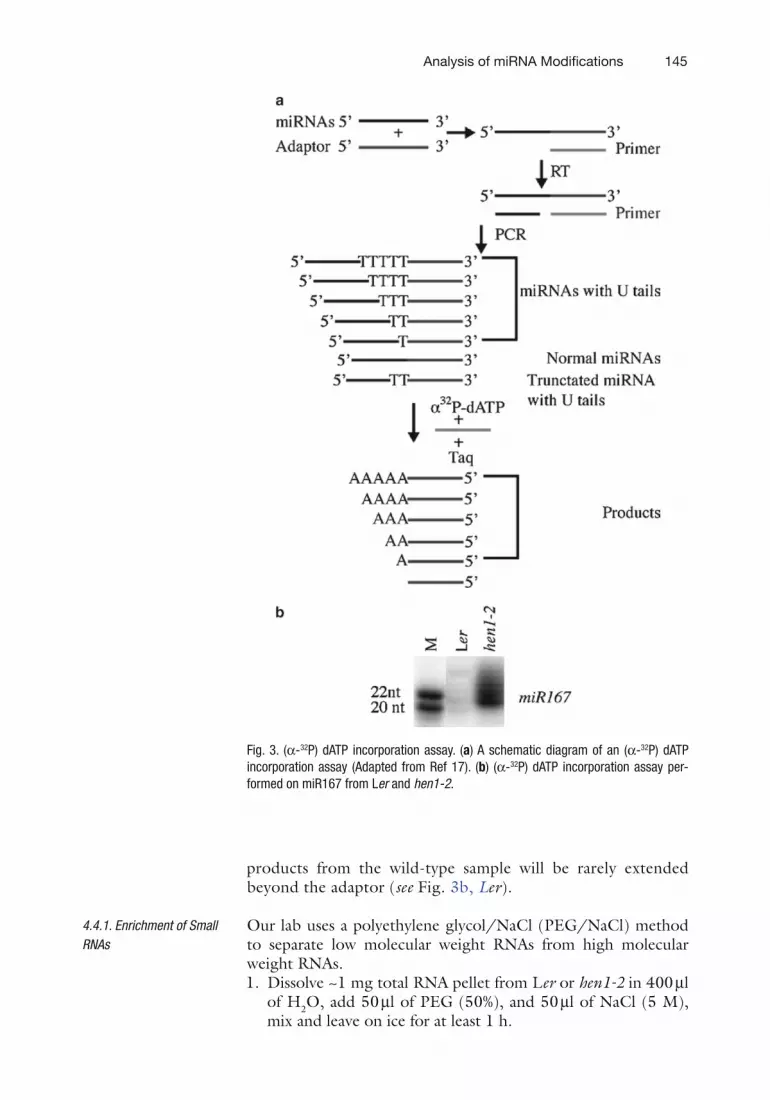
We employ an (a-32P) dATP incorporation assay to study the 3¢ uridylation of miRNAs (17). After small RNAs are isolated, they are ligated to a 3¢ adaptor, and reverse transcribed with a primer complementary to the 3¢ adaptor (see Fig. 3a). After this, miR167 is selectively amplified with an miRNA-specific primer that corresponds to the 5¢ portion of miR167 and the 3¢ adaptor primer. miR167 with U-tails in hen1-2 will generate a pool of PCR products with various numbers of T residues adja-cent to the 3¢ adaptor, whereas miR167 from the wild type will produce products in which no Ts are adjacent to the 3¢ adaptor (see Fig. 3a). Taq DNA polymerase will be used to extend the RT-PCR products with a primer complementary to the 3¢ adap-tor, in the presence of only (a-32P) dATP (see Fig. 3a). In this primer extension, the templates from U-tailed miRNAs will generate a ladder of products with varying numbers of A resi-dues in the hen1-2 sample (see Fig. 3b, hen1-2), whereas the
4.3. Probing miR173 by RNA Filter Hybridization (See Subheading 3.1.5)
4.4. Detection of 3 ¢ Uridylation with an [a-32P] dATP Incorporation Assay
Fig. 2. Detection of miRNA methylation by b elimination. (a) Diagram of periodate treat-ment followed by b elimination. The last two nucleotides of miR173 are shown. The vici-nal hydroxyl groups of the 3¢ terminal ribose react with periodate such that the last nucleoside is converted into a dialdehyde, which is subsequently removed by b elimina-tion. The resulting miR173 is one nucleotide shorter and carries a 3¢ P. (b) The methyla-tion status of miR173 in Ler (wild type) and hen1-1. Total RNAs of Ler or hen1-1 were treated with sodium periodate followed by b elimination, resolved by gel electrophore-sis, and hybridized to an antisense miR173 probe, and the hybridization signals were visualized using a PhosphoImager.
BookID 148921_ChapID 10_Proof# 1 - 18 / 09 / 2009
Analysis of miRNA Modifications 145
BookID 148921_ChapID 10_Proof# 1 - 18 / 09 / 2009
products from the wild-type sample will be rarely extended beyond the adaptor (see Fig. 3b, Ler).
Our lab uses a polyethylene glycol/NaCl (PEG/NaCl) method to separate low molecular weight RNAs from high molecular weight RNAs.1. Dissolve ~1 mg total RNA pellet from Ler or hen1-2 in 400 ml
of H2O, add 50 ml of PEG (50%), and 50 ml of NaCl (5 M), mix and leave on ice for at least 1 h.
4.4.1. Enrichment of Small RNAs
Fig. 3. (a-32P) dATP incorporation assay. (a) A schematic diagram of an (a-32P) dATP incorporation assay (Adapted from Ref 17). (b) (a-32P) dATP incorporation assay per-formed on miR167 from Ler and hen1-2.

146 Yu and Chen
BookID 148921_ChapID 10_Proof# 1 - 18 / 09 / 2009 BookID 148921_ChapID 10_Proof# 1 - 18 / 09 / 2009
2. Centrifuge at 13,000 g for 10 min. Transfer the supernatant to a new tube.
3. Add 1 ml of glycogen, 50 ml of sodium acetate, and 3 volumes of 100% ethanol. Incubate at −20°C for at least 2 h.
4. Centrifuge at maximum speed for 20 min at 4°C. Wash the pellet with 70% ethanol.
5. Air-dry the pellet for 5 min and dissolve in DEPC-treated water.
1. Resolve small RNAs and 32P-labelled RNA size markers on a 15% polyacrylamide gel containing 42% urea.
2. Excise 20–30 nt small RNAs (sizes were estimated based on RNA decade markers) from the gel.
3. Elute small RNAs by incubating the gel slice in RNA elution buffer at 65°C for 4 h. Pass the solution through glass wool, extract with equal volumes of chloroform/phenol twice, and precipitate RNAs with three volumes of 100% ethanol.
4. Air-dry the pellet for 5 min and dissolve in 25 ml of DEPC-treated water.
1. Dephosphorylate small RNAs by adding 3 ml of 10× NEB Buffer 3 (500 mM tris–HCl, 1,000 mM NaCl, 10 mM MgCl2, and 10 mM Dithiothreitol, pH 7.9) and 2 ml of CIP. Incubate at 37°C for 1 h.
2. Add 70 ml of water, extract with 100 ml of chloroform/phenol and precipitate with ethanol.
3. Dissolve RNAs in 10 ml of water and add 3 ml 10× ligation buf-fer (500 mM Tris–HCl, 100 mM MgCl2, 10 mM ATP and 100 mM Dithiothreitol, pH7.8), 3 ml BSA, 13 ml adaptor and 1 ml T4 RNA ligase. Incubate for 16 h at 8°C.
4. Purify small RNAs ligated to the 3¢ adaptor by electrophoresis (see Subheading 3.3.2)
1. Mix 13.5 ml of small RNAs ligated to the adaptor and 2 ml of microP2 primer, incubate at 65°C for 5 min, and leave on ice.
2. Add 2 ml 10× RT buffer (500 mM tris–HCl, 750 mM KCl, 30 mM MgCl2 and 100 mM Dithiothreitol, pH 8.0), 1 ml dNTP (10 mM), 0.5 ml RNase inhibitor, and 1 ml MuLV reverse transcriptase. Incubate at 42°C for 1 h.
3. Perform PCR in the solution containing 38.5 ml H2O, 4 ml RT products, 5 ml 10× PCR buffer (2,000 mM Tris–HCl, 500 mM KCl and15-mM MgCl2, pH 8.4), 1 ml dNTP (10 mM), 1 ml miR167P1, 1 ml microP2, and 0.5 ml Taq DNA polymerase.
4.4.2. Isolation of 18–30 nt Small RNAs by Electrophoresis
4.4.3. Ligation to 3¢ Adaptor and Purification of Small RNAs Ligated to the 3¢ Adaptor
4.4.4. Reverse Transcription and PCR Amplification (RT-PCR)

BookID 148921_ChapID 10_Proof# 1 - 18 / 09 / 2009
Analysis of miRNA Modifications 147
BookID 148921_ChapID 10_Proof# 1 - 18 / 09 / 2009
1. Resolve PCR products and DNA size markers on a 12% native polyacrylamide gel and visualize DNA by ethidium bromide staining.
2. Excise the DNA band from the gel and cut the gel slices into many small pieces.
3. Add 500 ml of 300 mM sodium acetate (pH 5.2), and shake at 37°C for 1 h.
4. Pass the solution through glass wool, extract with equal vol-umes of chloroform/phenol twice and precipitate with two volumes of 100% ethanol.
5. Dissolve the DNA pellet in 50 ml of water.
1. Mix 12.2 ml H2O, 1 ml DNA (see Subheading 3.3.5), 1.5 ml 10× PCR buffer (2,000 mM Tris–HCl, 500 mM KCl and 15 mM MgCl2, pH 8.4) 0.2 ml (a-32P) dATP, 0.4 ml microP2 (10 mM), and 0.2 ml Taq DNA polymerase.
2. Perform one cycle PCR (94°C for 90 s, 55°C for 30 s, and 72°C for 10 s).
3. Add 15 ml of 2× loading buffer and resolve 5 ml of the reaction in a 15% polyacrylamide gel containing 42% urea.
4. Visualize the radioactive signals with a PhosphoImager.
5. Notes
1. The molecular weight of the biotinylated probe should have a large difference from that of the miRNA to be isolated. This is to prevent the biotinylated probe, which will be inevitably eluted in the purification process together with the miRNA, from interfering with the mass spectrometry analysis of the miRNA.
2. It is convenient to make a 1 l stock without the addition of APS and TEMED. The stock can be stored at 4°C in the dark.
3. To make borax/boric acid buffer (0.06 M, pH 8.6), make 0.06 M borax and 0.06 M boric acid. Use borax to adjust the pH of the boric acid to 8.6.
4. Sodium periodate needs to be kept in the dark, as it is sensitive to light.
5. As this experiment is to study the 3¢ terminus of the miRNA, the miRNA-specific primer should correspond to the 5¢ por-tion of the miRNA.
6. Do not completely dry the RNA pellet, as this will greatly decrease its solubility.
4.4.5. Purification of DNA by Electrophoresis
4.4.6. (a-32P) dATP Incorporation Assay

148 Yu and Chen
BookID 148921_ChapID 10_Proof# 1 - 18 / 09 / 2009
7. To obtain enough miRNA for mass spectrometry analysis, the starting amount of total RNAs should be scaled up based on the amount described here.
8. Save the supernatant from step 3 until you are certain that satisfactory binding and elution of the miRNA have occurred.
9. The current for the transfer is 2 mA per cm2 membrane, but this needs to be experimentally determined for other transfer apparatus.
References
1. Bartel DP (2004) MicroRNAs: genomics, biogenesis, mechanism, and function. Cell 116:281–297
2. Grishok A, Pasquinelli AE, Conte D, Li N, Parrish S, Ha I, Baillie DL, Fire A, Ruvkun G, Mello CC (2001) Genes and mechanisms related to RNA interference regulate expres-sion of the small temporal RNAs that control C. elegans developmental timing. Cell 106:23–34
3. Hutvágner G, McLachlan J, Pasquinelli AE, Balint É, Tuschl T, Zamore PD (2001) A cellular function for the RNA-interference enzyme Dicer in the maturation of the let-7 small temporal RNA. Science 293:834–838
4. Lee Y, Ahn C, Han J, Choi H, Kim J, Yim J, Lee J, Provost P, Radmark O, Kim S, Kim VN (2003) The nuclear RNase III Drosha initiates microRNA processing. Nature 425: 415–419
5. Ketting RF, Fischer SE, Bernstein E, Sijen T, Hannon GJ, Plasterk RH (2001) Dicer func-tions in RNA interference and in synthesis of small RNA involved in developmental timing in C. elegans. Genes Dev 15:2654–2659
6. Park W, Li J, Song R, Messing J, Chen X (2002) CARPEL FACTORY, a Dicer homolog, and HEN1, a novel protein, act in microRNA metabolism in Arabidopsis thaliana. Curr Biol 12:1484–1495
7. Reinhart BJ, Weinstein EG, Rhoades MW, Bartel B, Bartel DP (2002) MicroRNAs in plants. Genes Dev 16:1616–1626
8. Lobbes D, Rallapalli G, Schmidt DD, Martin C, Clarke J (2006) SERRATE: a new player on the plant microRNA scene. EMBO Rep 7:1052–1058
9. Yang L, Liu Z, Lu F, Dong A, Huang H (2006) SERRATE is a novel nuclear regulator in primary microRNA processing in Arabidopsis. Plant J 47:841–850
10. Fang Y, Spector DL (2007) Identification of nuclear dicing bodies containing proteins for microRNA biogenesis in living Arabidopsis plants. Curr Biol 17:818–823
11. Song L, Han MH, Lesicka J, Fedoroff N (2007) Arabidopsis primary microRNA pro-cessing proteins HYL1 and DCL1 define a nuclear body distinct from the Cajal body. Proc Natl Acad Sci U S A 104:5437–5442
12. Basyuk E, Suavet F, Doglio A, Bordonne R, Bertrand E (2003) Human let-7 stem-loop pre-cursors harbor features of RNase III cleavage products. Nucleic Acids Res 31:6593–6597
13. Yu B, Yang Z, Li J, Minakhina S, Yang M, Padgett RW, Steward R, Chen X (2005) Methylation as a crucial step in plant microRNA biogenesis. Science 307:932–935
14. Yang Z, Ebright YW, Yu B, Chen X (2006) HEN1 recognizes 21–24 nt small RNA duplexes and deposits a methyl group onto the 2¢ OH of the 3¢ terminal nucleotide. Nucleic Acids Res 34:667–675
15. Chen X, Liu J, Cheng Y, Jia D (2002) HEN1 functions pleiotropically in Arabidopsis devel-opment and acts in C function in the flower. Development 129:1085–1094
16. Boutet S, Vazquez F, Liu J, Beclin C, Fagard M, Gratias A, Morel JB, Crete P, Chen X, Vaucheret H (2003) Arabidopsis HEN1: a genetic link between endogenous miRNA controlling development and siRNA control-ling transgene silencing and virus resistance. Curr Biol 13:843–848
17. Li J, Yang Z, Yu B, Liu J, Chen X (2005) Methylation protects miRNAs and siRNAs from a 3’-end uridylation activity in Arabidopsis. Curr Biol 15:1501–1507
18. Alefelder S, Patel BK, Eckstein F (1998) Incorporation of terminal phosphorothioates into oligonucleotides. Nucleic Acids Res 26: 4983–4988


