Embed Size (px)
Citation preview
Citation: Prišlin, M.; Vlahovic, D.;
Kostešic, P.; Ljolje, I.; Brnic, D.; Turk,
N.; Lojkic, I.; Kunic, V.; Karadjole, T.;
Krešic, N. An Outstanding Role of
Adipose Tissue in Canine Stem Cell
Therapy. Animals 2022, 12, 1088.
https://doi.org/10.3390/
ani12091088
Academic Editors: Eleonora Iacono
and Barbara Merlo
Received: 18 March 2022
Accepted: 20 April 2022
Published: 22 April 2022
Publisher’s Note: MDPI stays neutral
with regard to jurisdictional claims in
published maps and institutional affil-
iations.
Copyright: © 2022 by the authors.
Licensee MDPI, Basel, Switzerland.
This article is an open access article
distributed under the terms and
conditions of the Creative Commons
Attribution (CC BY) license (https://
creativecommons.org/licenses/by/
4.0/).
animals
Review
An Outstanding Role of Adipose Tissue in Canine StemCell TherapyMarina Prišlin 1 , Dunja Vlahovic 2, Petar Kostešic 2, Ivana Ljolje 3, Dragan Brnic 1 , Nenad Turk 2,Ivana Lojkic 1 , Valentina Kunic 1, Tugomir Karadjole 2 and Nina Krešic 1,*
1 Croatian Veterinary Institute, Savska Cesta 143, 10000 Zagreb, Croatia; [email protected] (M.P.);[email protected] (D.B.); [email protected] (I.L.); [email protected] (V.K.)
2 Faculty of Veterinary Medicine, University of Zagreb, Heinzelova 55, 10000 Zagreb, Croatia;[email protected] (D.V.); [email protected] (P.K.); [email protected] (N.T.); [email protected] (T.K.)
3 Veterinary Clinic for Small Animals Buba, Dore Pfanove 11, 10000 Zagreb, Croatia; [email protected]* Correspondence: [email protected]
Simple Summary: Throughout history, the role of adipose tissue has changed for humans, andregarding canines: the role has changed from connective tissue to restoration of physiologicalfunctions. The adipose tissue cells have extraordinary mechanisms of healing tissue function, and themost outstanding component of adipose tissue discovered are mesenchymal stem cells. It has beenalmost fifteen years since their discovery in canine adipose tissue. Since then, numerous studies haveinvestigated the possibilities of adipose-derived mesenchymal stem cells in treating various caninediseases. This review summarised the progress of confirming the therapeutic role of adipose tissuecomponents, focusing on stem cells as the most researched and with the highest potential in enablinga better quality of life for canines.
Abstract: Adipose tissue, previously known as connective tissue with a role in energy storage, iscurrently changing the course of treatments in veterinary medicine. Recent studies have revealedone particularly impressive function among all the newly discovered functions of adipose tissue.The interactive cells hosted by adipose tissue, the stromal vascular fraction (SVF), and their role intreating numerous diseases have provided a prospective course of research with positive outcomes inregenerative veterinary medicine (RVM). This review describes the main features of adipose tissue,emphasizing an eclectic combination of cells within the SVF and its thus far researched therapeuticpossibilities in canine RVM. An afterwards focus is on a highly researched component of the SVF,adipose-derived mesenchymal stem cells (ASCs), which were shown to have an extraordinary impactrelying on several proposed mechanisms of action on mitigating pathologies in canines. Furthermore,ASC therapy showed the most significant results in the orthopaedics field and in neurology, derma-tology, ophthalmology, gastroenterology, and hepatology, which elevates the possibilities of ASCtherapy to a whole new level. Therefore, this review article aims to raise awareness of the importanceof research on cellular components, within abundant and easily accessible adipose tissue, in thedirection of regenerative therapy in canines, considering the positive outcomes so far. Although thefocus is on the positive aspects of cellular therapy in canines, the researchers should not forget theimportance of identifying the potential negative aspects within published and upcoming research.Safe and standardized treatment represents a fundamental prerequisite for positively impacting thelives of canine patients.
Keywords: canine; adipose-derived mesenchymal stem cells; stem cell therapy; regenerative veteri-nary medicine; stromal vascular fraction; adipose tissue
1. Introduction
Adipose tissue (AT) was once considered to be the only connective tissue involved inenergy storage. Currently, recognition of AT function is beyond simple fat storage, and is
Animals 2022, 12, 1088. https://doi.org/10.3390/ani12091088 https://www.mdpi.com/journal/animals
Animals 2022, 12, 1088 2 of 19
well known as a metabolic and endocrine organ [1–3]. Consequently, this review aims toraise awareness on the importance of research on cellular components, within abundantand easily accessible adipose tissue, in the direction of regenerative therapy in canines,considering positive outcomes so far and failures of current treatment options.
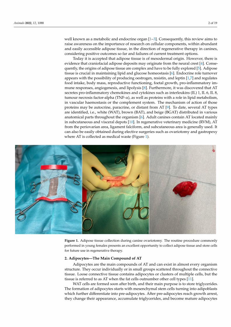
Today it is accepted that adipose tissue is of mesodermal origin. However, there isevidence that craniofacial adipose deposits may originate from the neural crest [4]. Conse-quently, the origins of adipose tissue are complex and have to be fully explored [5]. Adiposetissue is crucial in maintaining lipid and glucose homeostasis [6]. Endocrine role turnoverappears with the possibility of producing oestrogen, resistin, and leptin [1,7] and regulatesfood intake, body mass, reproductive functioning, foetal growth, pro-inflammatory im-mune responses, angiogenesis, and lipolysis [8]. Furthermore, it was discovered that ATsecretes pro-inflammatory chemokines and cytokines such as interleukins (IL) 1, IL 6, IL 8,tumour necrosis factor-alpha (TNF-α), as well as proteins with a role in lipid metabolism,in vascular haemostasis or the complement system. The mechanism of action of thoseproteins may be autocrine, paracrine, or distant from AT [9]. To date, several AT typesare identified, i.e., white (WAT), brown (BAT), and beige (BGAT) distributed in variousanatomical parts throughout the organism [6]. Adult canines contain AT located mainlyin subcutaneous and visceral depots [10]. In regenerative veterinary medicine (RVM), ATfrom the periovarian area, ligament falciform, and subcutaneous area is generally used. Itcan also be easily obtained during elective surgeries such as ovariotomy and gastropexywhere AT is collected as medical waste (Figure 1).
Animals 2022, 12, x FOR PEER REVIEW 2 of 20
1. Introduction Adipose tissue (AT) was once considered to be the only connective tissue involved
in energy storage. Currently, recognition of AT function is beyond simple fat storage, and is well known as a metabolic and endocrine organ [1–3]. Consequently, this review aims to raise awareness on the importance of research on cellular components, within abundant and easily accessible adipose tissue, in the direction of regenerative therapy in canines, considering positive outcomes so far and failures of current treatment options.
Today it is accepted that adipose tissue is of mesodermal origin. However, there is evidence that craniofacial adipose deposits may originate from the neural crest [4]. Con-sequently, the origins of adipose tissue are complex and have to be fully explored [5]. Adipose tissue is crucial in maintaining lipid and glucose homeostasis [6]. Endocrine role turnover appears with the possibility of producing oestrogen, resistin, and leptin [1,7] and regulates food intake, body mass, reproductive functioning, foetal growth, pro-inflamma-tory immune responses, angiogenesis, and lipolysis [8]. Furthermore, it was discovered that AT secretes pro-inflammatory chemokines and cytokines such as interleukins (IL) 1, IL 6, IL 8, tumour necrosis factor-alpha (TNF-α), as well as proteins with a role in lipid metabolism, in vascular haemostasis or the complement system. The mechanism of action of those proteins may be autocrine, paracrine, or distant from AT [9]. To date, several AT types are identified, i.e., white (WAT), brown (BAT), and beige (BGAT) distributed in var-ious anatomical parts throughout the organism [6]. Adult canines contain AT located mainly in subcutaneous and visceral depots [10]. In regenerative veterinary medicine (RVM), AT from the periovarian area, ligament falciform, and subcutaneous area is gen-erally used. It can also be easily obtained during elective surgeries such as ovariotomy and gastropexy where AT is collected as medical waste (Figure 1).
Figure 1. Adipose tissue collection during canine ovariotomy. The routine procedure commonly performed in young females presents an excellent opportunity to collect adipose tissue and store cells for future use in regenerative therapy.
2. Adipocytes—The Main Compound of AT Adipocytes are the main compounds of AT and can exist in almost every organism
structure. They occur individually or in small groups scattered throughout the connective tissue. Loose connective tissue contains adipocytes or clusters of multiple cells, but the tissue is referred to as AT when the fat cells outnumber other cell types [11].
Figure 1. Adipose tissue collection during canine ovariotomy. The routine procedure commonlyperformed in young females presents an excellent opportunity to collect adipose tissue and store cellsfor future use in regenerative therapy.
2. Adipocytes—The Main Compound of AT
Adipocytes are the main compounds of AT and can exist in almost every organismstructure. They occur individually or in small groups scattered throughout the connectivetissue. Loose connective tissue contains adipocytes or clusters of multiple cells, but thetissue is referred to as AT when the fat cells outnumber other cell types [11].
WAT cells are formed soon after birth, and their main purpose is to store triglycerides.The formation of adipocytes starts with mesenchymal stem cells turning into adipoblastswhich further differentiate into pre-adipocytes. After pre-adipocytes reach growth arrest,they change their appearance, accumulate triglycerides, and become mature adipocytes
Animals 2022, 12, 1088 3 of 19
with lost ability of division [12]. BAT cells develop before birth and specialize in defendingmammals against hypothermia [13]. The morphogenetic protein (BMP)-7 is responsible forthe differentiation process of brown pre-adipocytes into BAT [14].
BAT is equipped with the metabolic machinery comprising the numerous mitochon-dria and the appropriate enzymes that allow fatty acids to oxidize at enhanced rates thanthat of WAT. In addition, the mitochondria of brown adipose tissue cells primarily generateheat rather than adenosine triphosphate (ATP) and can sustain body heat during prolongedperiods of cold [15].
The last discovered and current highly researched type of adipocytes are beige orbrite.These have the characteristics of WAT and BAT cells [16]. The synthesis of BGAT is a highlyinvestigated topic in diabetes and metabolism research [17,18].
As mentioned, AT was once viewed as a passive triglyceride depot, but AT is nowknown as a complex tissue giving residence to various interacting cells, also known as thestromal vascular fraction (SVF) [1,13].
3. Stromal Vascular Fraction—Interacting Cells Hosted by AT
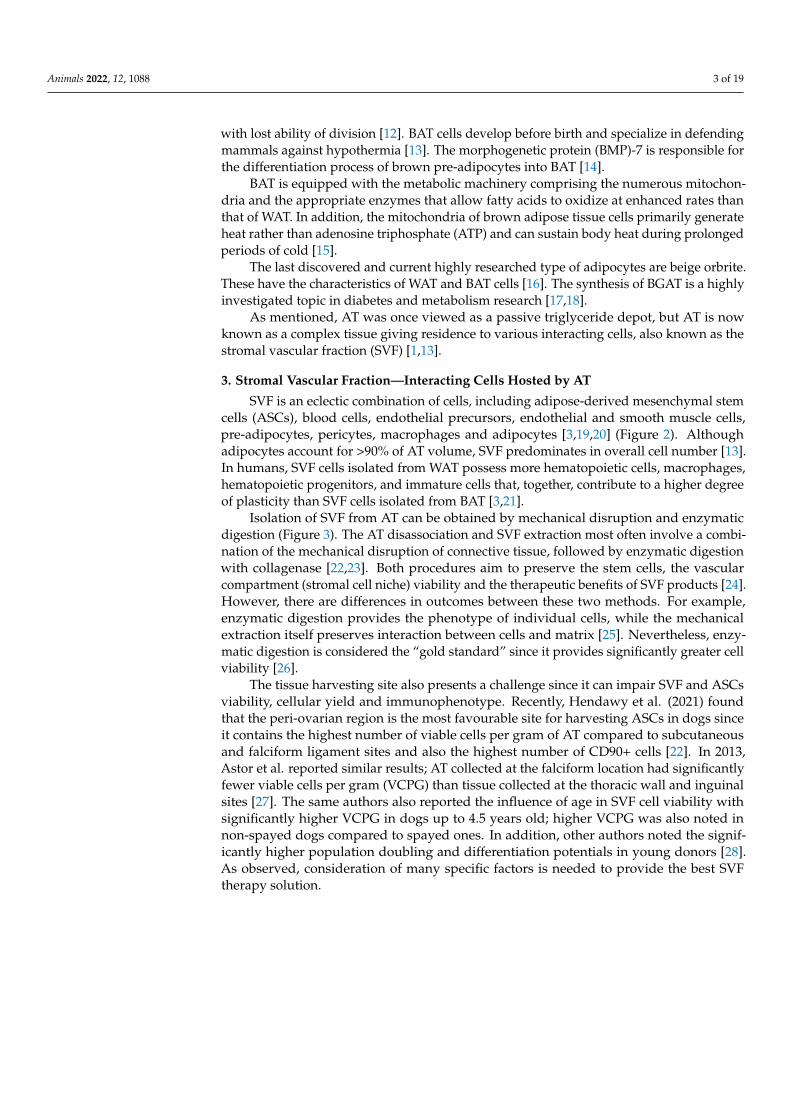
SVF is an eclectic combination of cells, including adipose-derived mesenchymal stemcells (ASCs), blood cells, endothelial precursors, endothelial and smooth muscle cells,pre-adipocytes, pericytes, macrophages and adipocytes [3,19,20] (Figure 2). Althoughadipocytes account for >90% of AT volume, SVF predominates in overall cell number [13].In humans, SVF cells isolated from WAT possess more hematopoietic cells, macrophages,hematopoietic progenitors, and immature cells that, together, contribute to a higher degreeof plasticity than SVF cells isolated from BAT [3,21].
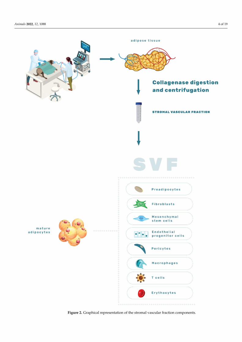
Isolation of SVF from AT can be obtained by mechanical disruption and enzymaticdigestion (Figure 3). The AT disassociation and SVF extraction most often involve a combi-nation of the mechanical disruption of connective tissue, followed by enzymatic digestionwith collagenase [22,23]. Both procedures aim to preserve the stem cells, the vascularcompartment (stromal cell niche) viability and the therapeutic benefits of SVF products [24].However, there are differences in outcomes between these two methods. For example,enzymatic digestion provides the phenotype of individual cells, while the mechanicalextraction itself preserves interaction between cells and matrix [25]. Nevertheless, enzy-matic digestion is considered the “gold standard” since it provides significantly greater cellviability [26].
The tissue harvesting site also presents a challenge since it can impair SVF and ASCsviability, cellular yield and immunophenotype. Recently, Hendawy et al. (2021) foundthat the peri-ovarian region is the most favourable site for harvesting ASCs in dogs sinceit contains the highest number of viable cells per gram of AT compared to subcutaneousand falciform ligament sites and also the highest number of CD90+ cells [22]. In 2013,Astor et al. reported similar results; AT collected at the falciform location had significantlyfewer viable cells per gram (VCPG) than tissue collected at the thoracic wall and inguinalsites [27]. The same authors also reported the influence of age in SVF cell viability withsignificantly higher VCPG in dogs up to 4.5 years old; higher VCPG was also noted innon-spayed dogs compared to spayed ones. In addition, other authors noted the signif-icantly higher population doubling and differentiation potentials in young donors [28].As observed, consideration of many specific factors is needed to provide the best SVFtherapy solution.
Animals 2022, 12, 1088 4 of 19Animals 2022, 12, x FOR PEER REVIEW 4 of 20
Figure 2. Graphical representation of the stromal vascular fraction components.
Figure 2. Graphical representation of the stromal vascular fraction components.
Animals 2022, 12, 1088 5 of 19Animals 2022, 12, x FOR PEER REVIEW 5 of 20
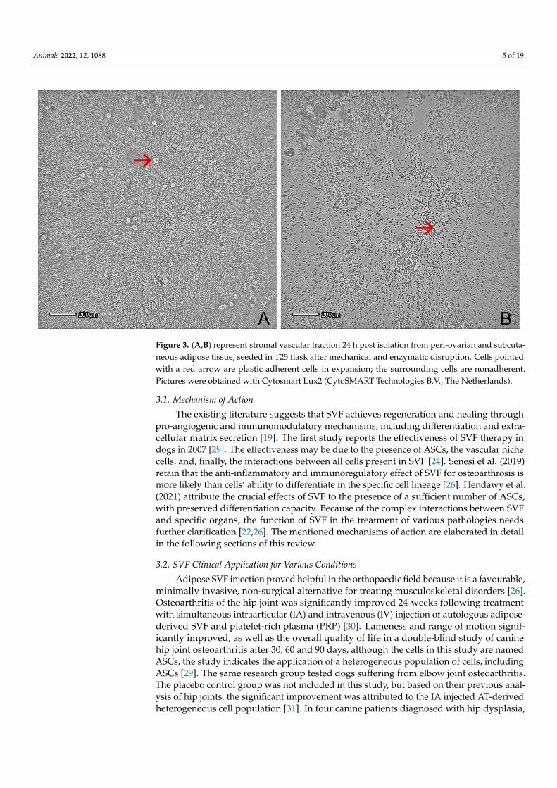
Figure 3. (A,B) represent stromal vascular fraction 24 h post isolation from peri-ovarian and subcu-taneous adipose tissue, seeded in T25 flask after mechanical and enzymatic disruption. Cells pointed with a red arrow are plastic adherent cells in expansion; the surrounding cells are nonadherent. Pictures were obtained with Cytosmart Lux2 (CytoSMART Technologies B.V., The Netherlands).
3.1. Mechanism of Action The existing literature suggests that SVF achieves regeneration and healing through
pro-angiogenic and immunomodulatory mechanisms, including differentiation and ex-tracellular matrix secretion [19]. The first study reports the effectiveness of SVF therapy in dogs in 2007 [29]. The effectiveness may be due to the presence of ASCs, the vascular niche cells, and, finally, the interactions between all cells present in SVF [24]. Senesi et al. (2019) retain that the anti-inflammatory and immunoregulatory effect of SVF for osteoar-throsis is more likely than cells’ ability to differentiate in the specific cell lineage. Hen-dawy et al. (2021) attribute the crucial effects of SVF to the presence of a sufficient number of ASCs, with preserved differentiation capacity. Because of the complex interactions be-tween SVF and specific organs, the function of SVF in the treatment of various pathologies needs further clarification [22,26]. The mentioned mechanisms of action are elaborated in detail in the following sections of this review.
3.2. SVF Clinical Application for Various Conditions Adipose SVF injection proved helpful in the orthopaedic field because it is a favour-
able, minimally invasive, non-surgical alternative for treating musculoskeletal disorders [26]. Osteoarthritis of the hip joint was significantly improved 24-weeks following treat-ment with simultaneous intraarticular (IA) and intravenous (IV) injection of autologous adipose-derived SVF and platelet-rich plasma (PRP) [30]. Lameness and range of motion significantly improved, as well as the overall quality of life in a double-blind study of canine hip joint osteoarthritis after 30, 60 and 90 days; although the cells in this study are named ASCs, the study indicates the application of a heterogeneous population of cells, including ASCs [29]. The same research group tested dogs suffering from elbow joint os-teoarthritis. The placebo control group was not included in this study, but based on their previous analysis of hip joints, the significant improvement was attributed to the IA in-jected AT-derived heterogeneous cell population [31]. In four canine patients diagnosed
Figure 3. (A,B) represent stromal vascular fraction 24 h post isolation from peri-ovarian and subcuta-neous adipose tissue, seeded in T25 flask after mechanical and enzymatic disruption. Cells pointedwith a red arrow are plastic adherent cells in expansion; the surrounding cells are nonadherent.Pictures were obtained with Cytosmart Lux2 (CytoSMART Technologies B.V., The Netherlands).
3.1. Mechanism of Action
The existing literature suggests that SVF achieves regeneration and healing throughpro-angiogenic and immunomodulatory mechanisms, including differentiation and extra-cellular matrix secretion [19]. The first study reports the effectiveness of SVF therapy indogs in 2007 [29]. The effectiveness may be due to the presence of ASCs, the vascular nichecells, and, finally, the interactions between all cells present in SVF [24]. Senesi et al. (2019)retain that the anti-inflammatory and immunoregulatory effect of SVF for osteoarthrosis ismore likely than cells’ ability to differentiate in the specific cell lineage [26]. Hendawy et al.(2021) attribute the crucial effects of SVF to the presence of a sufficient number of ASCs,with preserved differentiation capacity. Because of the complex interactions between SVFand specific organs, the function of SVF in the treatment of various pathologies needsfurther clarification [22,26]. The mentioned mechanisms of action are elaborated in detailin the following sections of this review.
3.2. SVF Clinical Application for Various Conditions
Adipose SVF injection proved helpful in the orthopaedic field because it is a favourable,minimally invasive, non-surgical alternative for treating musculoskeletal disorders [26].Osteoarthritis of the hip joint was significantly improved 24-weeks following treatmentwith simultaneous intraarticular (IA) and intravenous (IV) injection of autologous adipose-derived SVF and platelet-rich plasma (PRP) [30]. Lameness and range of motion signif-icantly improved, as well as the overall quality of life in a double-blind study of caninehip joint osteoarthritis after 30, 60 and 90 days; although the cells in this study are namedASCs, the study indicates the application of a heterogeneous population of cells, includingASCs [29]. The same research group tested dogs suffering from elbow joint osteoarthritis.The placebo control group was not included in this study, but based on their previous anal-ysis of hip joints, the significant improvement was attributed to the IA injected AT-derivedheterogeneous cell population [31]. In four canine patients diagnosed with hip dysplasia,
Animals 2022, 12, 1088 6 of 19
autologous SVF acupoint injection showed marked improvement, compared with baselineresults after the first week of treatment [32].
The use of allogenic SVF in degenerative joint disease of the spine in dogs revealedan increased serum level of the vascular endothelial growth factor of affected animals inthe second week of treatment. In the eighth week, the levels were decreased [33]. Thesame study published that decreased pain and reduced lameness were noticed a few daysfollowing therapy, overall concluding the improvement of joint regeneration capacity. Thelack of research in the veterinary field indicates a significant need for further investigationof SVF benefits.
4. Adipose-Derived Mesenchymal Stem Cells—An Outstanding Component ofthe SVF
It is well known that stem cells provide tissues and organs with a fresh cellularcompartment that can replace cells that have expired naturally and provide physiologicalbalance in the organism. In addition, the expiration of cells due to natural processes ordamage enables regeneration of the tissues [34]. The significant discovery of a stem cellsystem within AT occurred twenty years ago [35,36]. This finding raised considerableinterest in the veterinary scientific community. The results were first documented in 2008when scientists successfully isolated and fully described ASCs in canines [37] which laidthe foundation for RVM.
The ASCs, a subpopulation within SVF, are non-hemopoietic stem cells originatingfrom the mesoderm [38]. What makes them intriguing for cell research and therapy, amongMSC properties such as self-renewal, in vitro proliferation, non-specialization, and abilityto differentiate in another type of cell, is their easy accessibility. To address AT isolated cellsas ASCs, the International Society for Cellular Therapy (ISCT) and The International Feder-ation for Adipose Therapeutics (IFATS) have provided guidelines and recommendationsfor the minimal essential characterization of human ASCs. The established criteria were:capacity to proliferate as adherent cells in cultures, the ability of minimal three lineagesin vitro differentiation (osteogenic, chondrogenic and adipogenic) (Figure 4), phenotypicalpositivity for CD90, CD73, CD105 and negativity for CD14, CD34, CD45, CD11b, CD19 orCD79α [24,39]. Although scientists apply those rules for canine ASCs research, the exactcriteria are still not wholly established for this species. Though, numerous studies arecontributing to ASCs characterization. In this context, the investigation of these changesin surface marker expression (CD73, CD90, CD29, CD44, CD271, CD45 and CD14) hasbeen performed through six passages, providing a timeframe the ASCs cultivated in vitropossess optimal surface marker expression for use in therapy [23].
From the moment of their discovery, ASCs features were exploited in vitro to generatesufficient cell numbers to reach therapeutic doses depending on the disease for whichthe ASCs are being tested; meanwhile, their properties, gene expression and surfacemarker expression can be heavily influenced by such manipulation. Inevitably, prolongedcultivation in vitro carries side effects in terms of affection of the characteristic ASCsmembrane markers responsible for their positive impact [23]. Therefore, basic research onASCs properties in vitro is needed to further reveal their molecular signatures.
4.1. Mechanism of Action
As already well documented for MSC in general, the healing properties are probablya result of the secretion of many factors influencing the immune system, with anti-apoptotic,anti-inflammatory, chemotactic and pro-angiogenic functions [10,40–42].
Animals 2022, 12, 1088 7 of 19Animals 2022, 12, x FOR PEER REVIEW 7 of 20
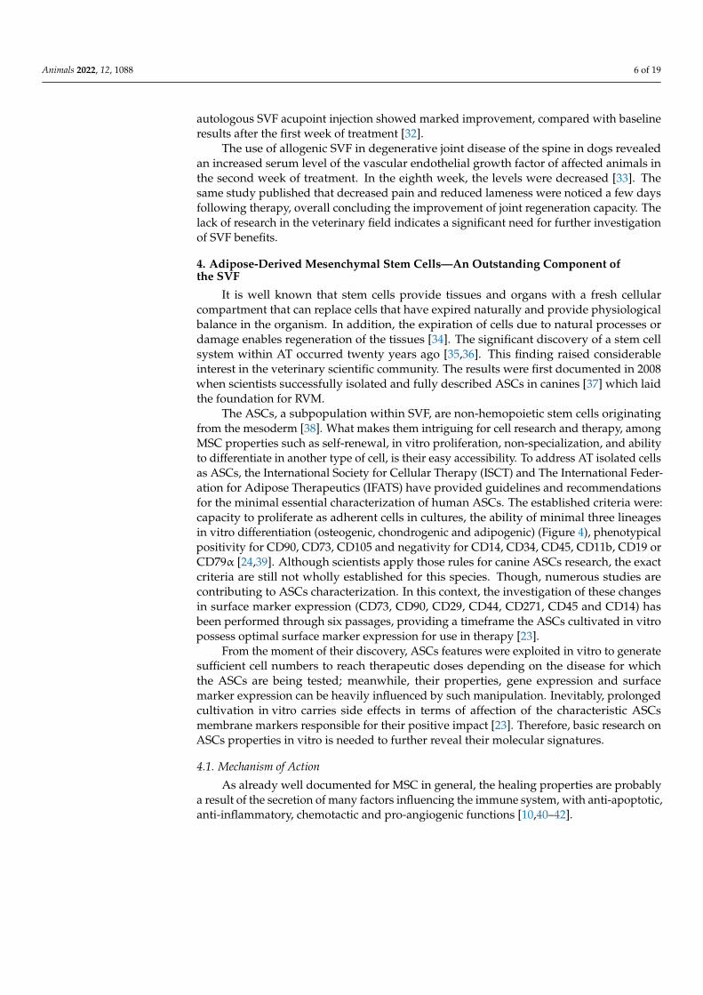
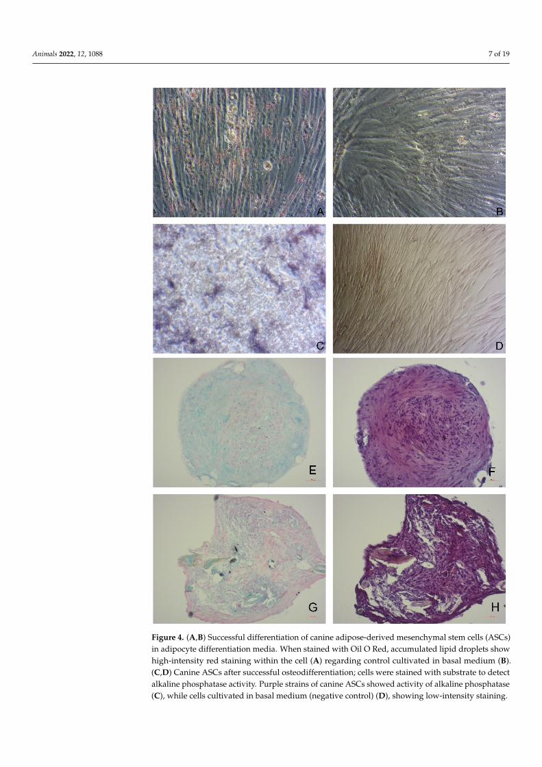
Figure 4. (A,B) Successful differentiation of canine adipose-derived mesenchymal stem cells (ASCs) in adipocyte differentiation media. When stained with Oil O Red, accumulated lipid droplets show high-intensity red staining within the cell (A) regarding control cultivated in basal medium (B). (C,D) Canine ASCs after successful osteodifferentiation; cells were stained with substrate to detect alkaline phosphatase activity. Purple strains of canine ASCs showed activity of alkaline phospha-tase (C), while cells cultivated in basal medium (negative control) (D), showing low-intensity stain-ing. 4E-H Images of histological sections of paraffin-embedded spheroids (20×, Zeiss, Germany) of canine ASCs three-dimensional culture after successful chondrodifferentiation. ASCs spheroids
Figure 4. (A,B) Successful differentiation of canine adipose-derived mesenchymal stem cells (ASCs)in adipocyte differentiation media. When stained with Oil O Red, accumulated lipid droplets showhigh-intensity red staining within the cell (A) regarding control cultivated in basal medium (B).(C,D) Canine ASCs after successful osteodifferentiation; cells were stained with substrate to detectalkaline phosphatase activity. Purple strains of canine ASCs showed activity of alkaline phosphatase(C), while cells cultivated in basal medium (negative control) (D), showing low-intensity staining.
Animals 2022, 12, 1088 8 of 19
4E-H Images of histological sections of paraffin-embedded spheroids (20×, Zeiss, Germany) of canineASCs three-dimensional culture after successful chondrodifferentiation. ASCs spheroids were stainedwith Alcian blue to detect the presence of aggrecan (E,G) and with H&E (F,H). Microscopic images(20×) (A–H) were taken with Zeiss Axiovert, Carl Zeiss AG, Jena, Germany.
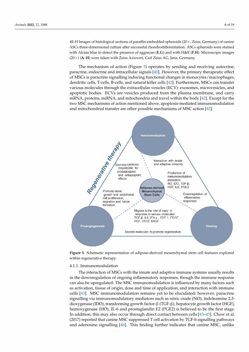
The mechanism of action (Figure 5) operates by sending and receiving autocrine,paracrine, endocrine and intracellular signals [40]. However, the primary therapeutic effectof MSCs is paracrine signalling inducing functional changes in monocytes/macrophages,dendritic cells, T-cells, B-cells, and natural killer cells [42]. Furthermore, MSCs can transfervarious molecules through the extracellular vesicles (ECV): exosomes, microvesicles, andapoptotic bodies. ECVs are vesicles produced from the plasma membrane, and carrymRNA, proteins, miRNA, and mitochondria and travel within the body [42]. Except for thetwo MSC mechanisms of action mentioned above, apoptosis-mediated immunomodulationand mitochondrial transfer are other possible mechanisms of MSC action [42].
Animals 2022, 12, x FOR PEER REVIEW 8 of 20
were stained with Alcian blue to detect the presence of aggrecan (E,G) and with H&E (F,H). Micro-scopic images (20×) (A–H) were taken with Zeiss Axiovert, Carl Zeiss AG, Jena, Germany.
4.1. Mechanism of Action As already well documented for MSC in general, the healing properties are probably
a result of the secretion of many factors influencing the immune system, with anti-apop-totic, anti-inflammatory, chemotactic and pro-angiogenic functions [10,40–42].
The mechanism of action (Figure 5) operates by sending and receiving autocrine, paracrine, endocrine and intracellular signals [40]. However, the primary therapeutic ef-fect of MSCs is paracrine signalling inducing functional changes in monocytes/macro-phages, dendritic cells, T-cells, B-cells, and natural killer cells [42]. Furthermore, MSCs can transfer various molecules through the extracellular vesicles (ECV): exosomes, microvesi-cles, and apoptotic bodies. ECVs are vesicles produced from the plasma membrane, and carry mRNA, proteins, miRNA, and mitochondria and travel within the body [42]. Except for the two MSC mechanisms of action mentioned above, apoptosis-mediated immuno-modulation and mitochondrial transfer are other possible mechanisms of MSC action [42].
Figure 5. Schematic representation of adipose-derived mesenchymal stem cell features explored within regenerative therapy.
4.1.1. Immunomodulation The interaction of MSCs with the innate and adaptive immune systems usually re-
sults in the downregulation of ongoing inflammatory responses, though the immune re-sponse can also be upregulated. The MSC immunomodulation is influenced by many fac-tors such as activation, tissue of origin, dose and time of application, and interaction with immune cells [43]. MSC immunomodulation remains yet to be elucidated; however, para-crine signalling via immunomodulatory mediators such as nitric oxide (NO), indoleamine 2,3-dioxygenase (IDO), transforming growth factor-β (TGF-β), hepatocyte growth factor (HGF), hemoxygenase (HO), IL-6 and prostaglandin E2 (PGE2) is believed to be the first stage. In addition, this may also occur through direct contact between cells [43–45]. Chow et al. (2017) reported that canine MSC suppressed T cell activation by TGF-b signalling
Figure 5. Schematic representation of adipose-derived mesenchymal stem cell features exploredwithin regenerative therapy.
4.1.1. Immunomodulation
The interaction of MSCs with the innate and adaptive immune systems usually resultsin the downregulation of ongoing inflammatory responses, though the immune responsecan also be upregulated. The MSC immunomodulation is influenced by many factors suchas activation, tissue of origin, dose and time of application, and interaction with immunecells [43]. MSC immunomodulation remains yet to be elucidated; however, paracrinesignalling via immunomodulatory mediators such as nitric oxide (NO), indoleamine 2,3-dioxygenase (IDO), transforming growth factor-β (TGF-β), hepatocyte growth factor (HGF),hemoxygenase (HO), IL-6 and prostaglandin E2 (PGE2) is believed to be the first stage.In addition, this may also occur through direct contact between cells [43–45]. Chow et al.(2017) reported that canine MSC suppressed T cell activation by TGF-b signalling pathwaysand adenosine signalling [46]. This finding further indicates that canine MSC, unlike
Animals 2022, 12, 1088 9 of 19
human and rodent MSC, relies primarily on cyclooxygenase and TGF-b pathways for Tcell suppression rather than on NO or IDO-mediated pathways. Besides suppressing Tcells, MSCs suppress B cell activation and proliferation, dendritic cells maturation, inhibitNK cell proliferation and cytotoxicity, and promote regulatory T cell generation via solublefactors or cell-cell contact [44]. T cell necrosis by canine MSC is an additional mechanismof immune modulation [46]. Canine ASCs can suppress lipopolysaccharide mediatedactivation/maturation of canine dendritic cells (DC). The impact in vivo of such squelchedDC activation would undoubtedly result in an attenuated ability to appropriately prime Tcell responses. This effect would be exacerbated if the ASCs were first activated with IFNg,suggesting that the suppressive effect would be optimal in an inflammatory environmenttypical of autoimmune or pro-inflammatory conditions [47].
Another “immune-privileged” MSC property is their low immunogenicity attributedto low expression of MHC I, absence of co-stimulating CD80, CD86 and CD40, MHC IIdeficiency and whole paracrine spectrum of biomolecules and growth factors throughwhich they establish their action [48–50]. Each of the mentioned pathways reflects thepossibilities these cells offer to treat various disorders and organ systems. However, allaforementioned mechanisms also imply the differences between species and offer space fornew acknowledgements.
4.1.2. Homing
The MSCs have a remarkable ability to locate damaged tissues [3,42]. In response tochemotactic signals, MSCs reach the circulation and migrate to the site of injury, wherethey secrete molecules to promote regeneration. However, it is unclear which chemotacticsignals guide MSCs to appropriate microenvironments [51]. The homing of MSCs iscurrently inefficient, and after they are systemically administered a small percentageof cells reach the target tissue [52]. The process of migration from the bloodstream totissue involves steps for lymphocyte migration: (1) tethering and rolling, (2) activation,(3) firm adhesion, (4) transmigration or diapedesis, resulting in migration into tissue dueto chemotaxis as described by Sackstein [53]. The migration of MSCs occur in response tovarious chemokines and growth factors, including TNF-α (tumour necrosis factor-α), IL-6,IL-8 [54]. Unlike comprehensive knowledge on blood cell homing, MSC homing remainspoorly understood as tethering or rolling and transmigration.
The therapy research of MSCs bears one of the most significant aims, i.e., improvingtheir homing efficiency. MSC homing can be categorized into (1) targeted administration—administration of ASCs at or near the target tissue, (2) magnetic guidance—cells labelledwith magnetic particles are directed to the organ of interest using an external magnetic field,(3) genetic modification—permanent overexpression of homing factors via viral transduc-tion, (4) cell surface engineering—temporarily chemical engineering by enzymes or ligands,(5) in vitro priming—altering culture conditions to affect gene expression, (6) and modifica-tion of the target tissue by direct injection of homing factors, genetic modification of targettissue, scaffold implantation, or using radiotherapeutic and ultrasound techniques[52].
4.1.3. Pro-Angiogenic and Anti-Apoptotic Mechanism of Action
MSCs secrete various cytokines responsible for pro-angiogenic and anti-apoptoticeffects and in doing so, MSCs enable tissue regeneration and revascularization. Solubleangiogenic factors secreted by MSCs include fibroblast growth factors, hepatocyte growthfactor and the vascular endothelial growth factor.
The lack of canine-specific antibodies has hampered identification of growth factors inthe secretome of canine MSCs. Likely, secretomes of other species are similar to secretomessecreted by canine MSCs thus there is an idea on the secretome composition of canine MSCsbased on this information [55]. Canine MSCs promote nerve growth and endothelial cellproliferation, migration and tubule formation by secretion of neurotrophic and angiogenicfactors. Delfi et al., 2016 demonstrated MSC paracrine activity on nerves and blood vesselsin the vicinity of the wound site. It was shown that MSC transplants promote increased
Animals 2022, 12, 1088 10 of 19
neuronal function in dogs with central nervous system damage [55]. The following studyby the same authors, revealed that the conditioned medium from human and canine MSCscultures exhibited neurogenic and angiogenic effects and increased SH-SY5Y neuronalproliferation, βIII tubulin immunoreactivity, neurite outgrowth, and EA.hy926 endothelialcell proliferation, migration and the formation of endothelial tubule-like structures, toa significantly greater extent than control medium, indicating marked trophic activity [55].
Regarding anti-apoptotic action, it was shown that ASCs protect against radiation-induced dermatitis by exerting an anti-apoptotic effect through inhibition of cathepsin F(CTSF) expression. In addition, ASCs markedly attenuated radiation-induced apoptosis,downregulated CTSF and downstream pro-apoptotic proteins (Bid, BAX, and caspase 9),and upregulated anti-apoptotic proteins (Bcl-2 and Bcl-XL) [56].
4.2. ASCs Clinical Application for Various Conditions
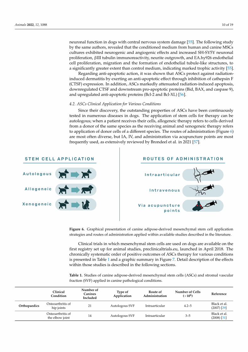
Since their discovery, the outstanding properties of ASCs have been continuouslytested in numerous diseases in dogs. The application of stem cells for therapy can beautologous; when a patient receives their cells, allogeneic therapy refers to cells derivedfrom a donor of the same species as the receiving animal and xenogeneic therapy refersto application of donor cells of a different species. The routes of administration (Figure 6)are most often diverse, but IA, IV, and administration via acupuncture points are mostfrequently used, as extensively reviewed by Brondeel et al. in 2021 [57].
Animals 2022, 12, x FOR PEER REVIEW 10 of 20
endothelial cell proliferation, migration and tubule formation by secretion of neurotrophic and angiogenic factors. Delfi et al., 2016 demonstrated MSC paracrine activity on nerves and blood vessels in the vicinity of the wound site. It was shown that MSC transplants promote increased neuronal function in dogs with central nervous system damage [55]. The following study by the same authors, revealed that the conditioned medium from human and canine MSCs cultures exhibited neurogenic and angiogenic effects and in-creased SH-SY5Y neuronal proliferation, βIII tubulin immunoreactivity, neurite out-growth, and EA.hy926 endothelial cell proliferation, migration and the formation of en-dothelial tubule-like structures, to a significantly greater extent than control medium, in-dicating marked trophic activity [55].
Regarding anti-apoptotic action, it was shown that ASCs protect against radiation-induced dermatitis by exerting an anti-apoptotic effect through inhibition of cathepsin F (CTSF) expression. In addition, ASCs markedly attenuated radiation-induced apoptosis, downregulated CTSF and downstream pro-apoptotic proteins (Bid, BAX, and caspase 9), and upregulated anti-apoptotic proteins (Bcl-2 and Bcl-XL) [56].
4.2. ASCs Clinical Application for Various Conditions Since their discovery, the outstanding properties of ASCs have been continuously
tested in numerous diseases in dogs. The application of stem cells for therapy can be au-tologous; when a patient receives their cells, allogeneic therapy refers to cells derived from a donor of the same species as the receiving animal and xenogeneic therapy refers to ap-plication of donor cells of a different species. The routes of administration (Figure 6) are most often diverse, but IA, IV, and administration via acupuncture points are most fre-quently used, as extensively reviewed by Brondeel et al. in 2021 [57].
Figure 6. Graphical presentation of canine adipose-derived mesenchymal stem cell application strat-egies and routes of administration applied within available studies described in the literature.
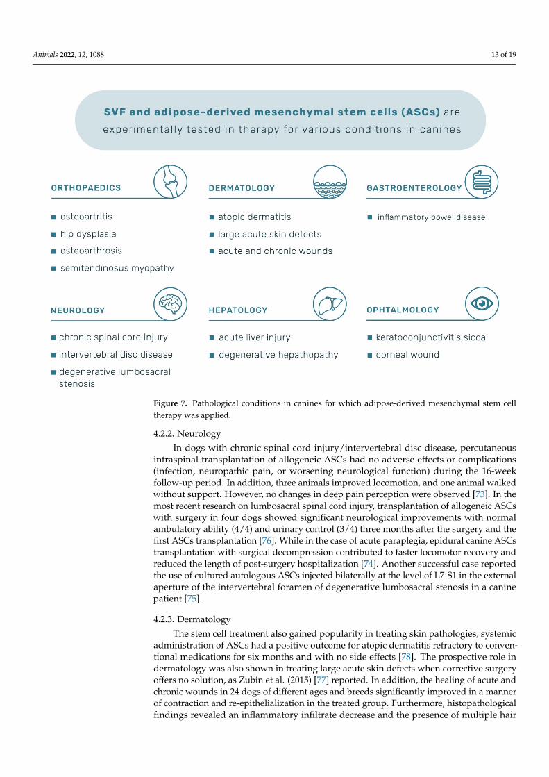
Clinical trials in which mesenchymal stem cells are used on dogs are available on the first registry set up for animal studies, preclinicaltrials.eu, launched in April 2018. The chronically systematic order of positive outcomes of ASCs therapy for various conditions is presented in Table 1 and a graphic summary in Figure 7. Detail description of the effects within those studies is described in the following sections.
Figure 6. Graphical presentation of canine adipose-derived mesenchymal stem cell applicationstrategies and routes of administration applied within available studies described in the literature.
Clinical trials in which mesenchymal stem cells are used on dogs are available on thefirst registry set up for animal studies, preclinicaltrials.eu, launched in April 2018. Thechronically systematic order of positive outcomes of ASCs therapy for various conditionsis presented in Table 1 and a graphic summary in Figure 7. Detail description of the effectswithin those studies is described in the following sections.
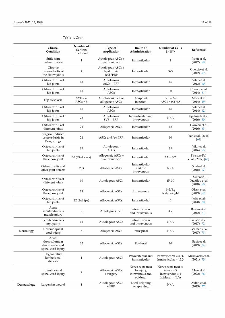
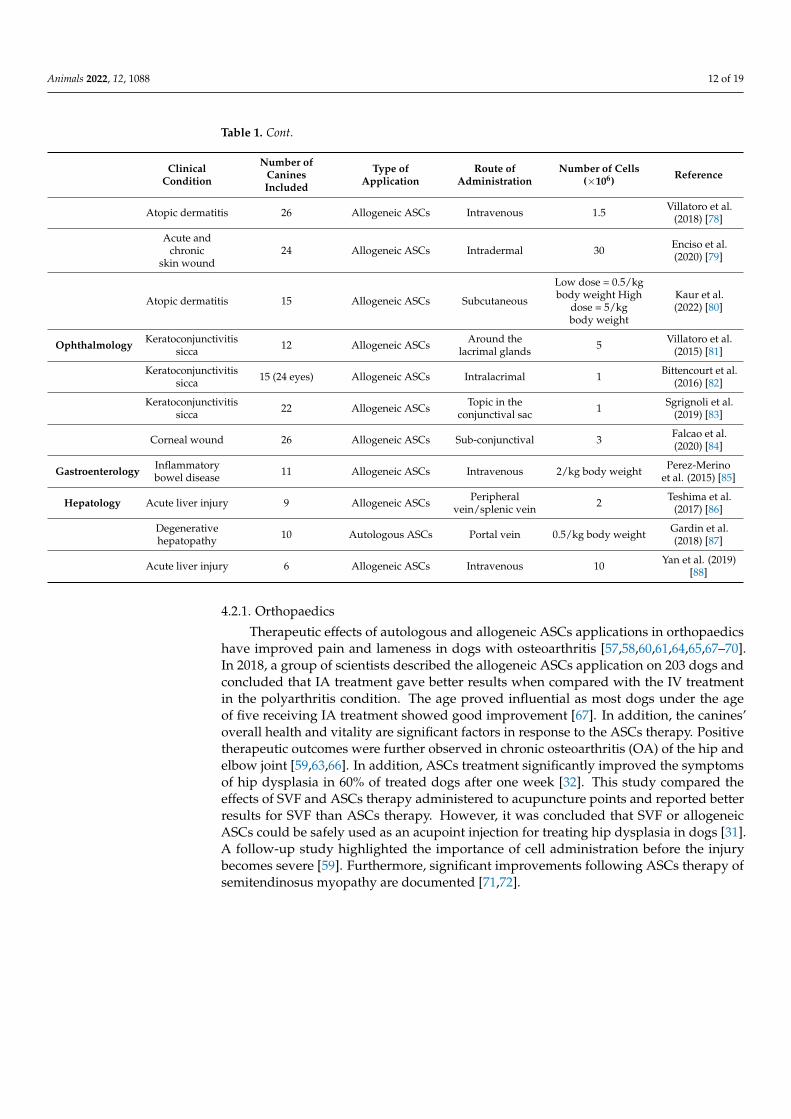
Table 1. Studies of canine adipose-derived mesenchymal stem cells (ASCs) and stromal vascularfraction (SVF) applied in canine pathological conditions.
ClinicalCondition
Number ofCaninesIncluded
Type ofApplication
Route ofAdministration
Number of Cells(×106) Reference
Orthopaedics Osteoarthritis ofhip joints 21 Autologous SVF Intraarticular 4.2–5 Black et al.
(2007) [29]
Osteoarthritis ofthe elbow joint 14 Autologous SVF Intraarticular 3–5 Black et al.
(2008) [31]
Animals 2022, 12, 1088 11 of 19
Table 1. Cont.
ClinicalCondition
Number ofCaninesIncluded
Type ofApplication
Route ofAdministration
Number of Cells(×106) Reference
Stifle jointosteoarthrosis 1 Autologous ASCs +
hyaluronic acid intraarticular 1 Yoon et al.(2012) [58]
Chronicosteoarthritis ofthe elbow joints
4Autologous ASCs +
hyaluronicacid/PRP
Intraarticular 3–5 Guercio et al.(2012) [59]
Osteoarthritis ofhip joints 13 Autologous
ASCs + PRP Intraarticular 15 Vilar et al.(2013) [60]
Osteoarthritis ofhip joints 18 Autologous
ASCs Intraarticular 30 Cuervo et al.(2014) [61]
Hip dysplasia SVF = 4ASCs = 5
Autologous SVF orallogeneic ASCs
Acupointinjection
SVF = 2–5ASCs = 0.2–0.8
Marx et al.(2014) [49]
Osteoarthritis ofhip joints 15 Autologous
ASCs Intraarticular 15 Vilar et al.(2014) [62]
Osteoarthritis ofhip joints 22 Autologous
SVF + PRPIntraarticular and
intravenous N/A Upchurch et al.(2016) [30]
Osteoarthritis ofdifferent joints 74 Allogeneic ASCs Intraarticular 12 Harman et al.
(2016) [63]
Surgical-inducedosteoarthritis in
Beagle dogs24 ASCs and/or PRP Intraarticular 10 Yun et al. (2016)
[64]
Osteoarthritis ofhip joints 15 Autologous
ASCs Intraarticular 15 Vilar et al.(2016) [65]
Osteoarthritis ofthe elbow joint 30 (39 elbows) Allogeneic ASCs +
hyaluronic acid Intraarticular 12 ± 3.2 Kriston-Palet al. (2017) [66]
Osteoarthritis andother joint defects 203 Allogeneic ASCs
Intraarticularand/or
intravenousN/A Shah et al.
(2018) [67]
Osteoarthritis ofdifferent joints 10 Autologous ASCs Intraarticular 15–30
SrzenticDražilov et al.
(2018) [68]
Osteoarthritis ofthe elbow joint 13 Allogeneic ASCs Intravenous 1–2/kg
body weightOlsen et al.(2019) [69]
Osteoarthritis ofhip joints 12 (24 hips) Allogeneic ASCs Intraarticular 5 Wits et al.
(2020) [70]
Acutesemitendinosusmuscle injury
2 Autologous SVF Intramuscularand intravenous 4.7 Brown et al.
(2012) [71]
Semitendinosusmyopathy 11 Autologous ASCs Intramuscular
and intravenous N/A Gibson et al.(2017) [72]
Neurology Chronic spinalcord injury 6 Allogeneic ASCs Intraspinal N/A Escalhao et al.
(2017) [73]
Acutethoracolumbar
disc disease andspinal cord injury
22 Allogeneic ASCs Epidural 10 Bach et al.(2019) [74]
Degenerativelumbosacral
stenosis1 Autologous ASCs Paravertebral and
intraarticularParavertebral = 30.6Intraarticular = 15.3
Mrkovacki et al.(2021) [75]
Lumbosacralspinal cord injury 4 Allogeneic ASCs
+ surgery
Nerve roots nextto injury,
intravenous andepidural
Nerve roots next toinjury = 5
Intravenous = 4Epidural = N/A
Chen et al.(2022) [76]
Dermatology Large skin wound 1 Autologous ASCs+ PRP
Local drippingor spraying N/A Zubin et al.
(2015) [77]
Animals 2022, 12, 1088 12 of 19
Table 1. Cont.
ClinicalCondition
Number ofCaninesIncluded
Type ofApplication
Route ofAdministration
Number of Cells(×106) Reference
Atopic dermatitis 26 Allogeneic ASCs Intravenous 1.5 Villatoro et al.(2018) [78]
Acute andchronic
skin wound24 Allogeneic ASCs Intradermal 30 Enciso et al.
(2020) [79]
Atopic dermatitis 15 Allogeneic ASCs Subcutaneous
Low dose = 0.5/kgbody weight High
dose = 5/kgbody weight
Kaur et al.(2022) [80]
Ophthalmology Keratoconjunctivitissicca 12 Allogeneic ASCs Around the
lacrimal glands 5 Villatoro et al.(2015) [81]
Keratoconjunctivitissicca 15 (24 eyes) Allogeneic ASCs Intralacrimal 1 Bittencourt et al.
(2016) [82]
Keratoconjunctivitissicca 22 Allogeneic ASCs Topic in the
conjunctival sac 1 Sgrignoli et al.(2019) [83]
Corneal wound 26 Allogeneic ASCs Sub-conjunctival 3 Falcao et al.(2020) [84]
Gastroenterology Inflammatorybowel disease 11 Allogeneic ASCs Intravenous 2/kg body weight Perez-Merino
et al. (2015) [85]
Hepatology Acute liver injury 9 Allogeneic ASCs Peripheralvein/splenic vein 2 Teshima et al.
(2017) [86]
Degenerativehepatopathy 10 Autologous ASCs Portal vein 0.5/kg body weight Gardin et al.
(2018) [87]
Acute liver injury 6 Allogeneic ASCs Intravenous 10 Yan et al. (2019)[88]
4.2.1. Orthopaedics
Therapeutic effects of autologous and allogeneic ASCs applications in orthopaedicshave improved pain and lameness in dogs with osteoarthritis [57,58,60,61,64,65,67–70].In 2018, a group of scientists described the allogeneic ASCs application on 203 dogs andconcluded that IA treatment gave better results when compared with the IV treatmentin the polyarthritis condition. The age proved influential as most dogs under the ageof five receiving IA treatment showed good improvement [67]. In addition, the canines’overall health and vitality are significant factors in response to the ASCs therapy. Positivetherapeutic outcomes were further observed in chronic osteoarthritis (OA) of the hip andelbow joint [59,63,66]. In addition, ASCs treatment significantly improved the symptomsof hip dysplasia in 60% of treated dogs after one week [32]. This study compared theeffects of SVF and ASCs therapy administered to acupuncture points and reported betterresults for SVF than ASCs therapy. However, it was concluded that SVF or allogeneicASCs could be safely used as an acupoint injection for treating hip dysplasia in dogs [31].A follow-up study highlighted the importance of cell administration before the injurybecomes severe [59]. Furthermore, significant improvements following ASCs therapy ofsemitendinosus myopathy are documented [71,72].
Animals 2022, 12, 1088 13 of 19Animals 2022, 12, x FOR PEER REVIEW 13 of 20
Figure 7. Pathological conditions in canines for which adipose-derived mesenchymal stem cell ther-apy was applied.
4.2.1. Orthopaedics Therapeutic effects of autologous and allogeneic ASCs applications in orthopaedics
have improved pain and lameness in dogs with osteoarthritis [57,58,60,61,64,65,67–70]. In 2018, a group of scientists described the allogeneic ASCs application on 203 dogs and con-cluded that IA treatment gave better results when compared with the IV treatment in the polyarthritis condition. The age proved influential as most dogs under the age of five re-ceiving IA treatment showed good improvement [67]. In addition, the canines’ overall health and vitality are significant factors in response to the ASCs therapy. Positive thera-peutic outcomes were further observed in chronic osteoarthritis (OA) of the hip and elbow joint [59,63,66]. In addition, ASCs treatment significantly improved the symptoms of hip dysplasia in 60% of treated dogs after one week [32]. This study compared the effects of SVF and ASCs therapy administered to acupuncture points and reported better results for SVF than ASCs therapy. However, it was concluded that SVF or allogeneic ASCs could be safely used as an acupoint injection for treating hip dysplasia in dogs [31]. A follow-up study highlighted the importance of cell administration before the injury becomes severe [59]. Furthermore, significant improvements following ASCs therapy of semitendinosus myopathy are documented [71,72].
4.2.2. Neurology In dogs with chronic spinal cord injury/intervertebral disc disease, percutaneous in-
traspinal transplantation of allogeneic ASCs had no adverse effects or complications
Figure 7. Pathological conditions in canines for which adipose-derived mesenchymal stem celltherapy was applied.
4.2.2. Neurology
In dogs with chronic spinal cord injury/intervertebral disc disease, percutaneousintraspinal transplantation of allogeneic ASCs had no adverse effects or complications(infection, neuropathic pain, or worsening neurological function) during the 16-weekfollow-up period. In addition, three animals improved locomotion, and one animal walkedwithout support. However, no changes in deep pain perception were observed [73]. In themost recent research on lumbosacral spinal cord injury, transplantation of allogeneic ASCswith surgery in four dogs showed significant neurological improvements with normalambulatory ability (4/4) and urinary control (3/4) three months after the surgery and thefirst ASCs transplantation [76]. While in the case of acute paraplegia, epidural canine ASCstransplantation with surgical decompression contributed to faster locomotor recovery andreduced the length of post-surgery hospitalization [74]. Another successful case reportedthe use of cultured autologous ASCs injected bilaterally at the level of L7-S1 in the externalaperture of the intervertebral foramen of degenerative lumbosacral stenosis in a caninepatient [75].
4.2.3. Dermatology
The stem cell treatment also gained popularity in treating skin pathologies; systemicadministration of ASCs had a positive outcome for atopic dermatitis refractory to conven-tional medications for six months and with no side effects [78]. The prospective role indermatology was also shown in treating large acute skin defects when corrective surgeryoffers no solution, as Zubin et al. (2015) [77] reported. In addition, the healing of acute andchronic wounds in 24 dogs of different ages and breeds significantly improved in a mannerof contraction and re-epithelialization in the treated group. Furthermore, histopathologicalfindings revealed an inflammatory infiltrate decrease and the presence of multiple hair
Animals 2022, 12, 1088 14 of 19
follicles on day seven after treatment with ASCs [79]. Most recently, Kaur et al. (2022) per-formed the first double-blinded, placebo-controlled evaluation of the efficacy of allogeneiccanine ASCs to treat canine atopic dermatitis. No severe side effects were observed in anypatient in this study. Furthermore, the high dose ASCs treatment proved to be efficaciousin alleviating the clinical signs of atopic dermatitis until 30 days after the last subcutaneousadministration of MSCs[80].
4.2.4. Ophthalmology
Reviewing ophthalmological benefits, an immune-mediated condition common inhumans and canines, keratoconjunctivitis sicca (KCS), was studied. Results in canines withKCS revealed that a single infusion of ACSs into lacrimal glands of 15 dogs resulted in noside effects during 12-months follow up.
Furthermore, a significant clinical improvement was observed in all patients, singleadministration was effective, and daily use of corticosteroids was not required [82]. In2019, topical application into the conjunctival sac resulted in decreased expression of pro-inflammatory markers, which implies ASCs as an adjuvant therapy in treating KCS indogs and humans. [83]. Another successful study of ASCs for KCS was reported withsignificant outcomes in canines where allogeneic ASCs were applied [81]. Falcao et al., in2020, evaluated the use of sub-conjunctival applied ASCs in dogs diagnosed with deepcorneal ulcers. Allogeneic ASCs therapy in 22 out of 26 dogs presented complete ulcerwound healing within 14 days, totalling 84.6%, indicating that this therapy is a simplesolution to substitute surgery with satisfying results [84].
4.2.5. Gastroenterology
The ASCs therapy was also tested for currently uncurable inflammatory bowel disease(IBD); administration of a single IV ASCs infusion showed no acute reaction or side effectsduring the follow-up of 11 dogs. Furthermore, 9 out of 11 dogs were in clinical remission.As the primary goal of treatment is to reduce symptoms, achieve and maintain remission,and prevent complications, ASCs were well tolerated and appeared to produce clinicalbenefits in dogs with severe IBD [85].
4.2.6. Hepatology
Liver diseases share clinical and pathological features in humans and canines, thus,dogs may be a representative model for humans. Autologous ASCs transplantation in dogswith liver diseases significantly ameliorated liver function; decreased liver biomarkersand observed effects seem to be related to stem cells’ immunomodulatory mechanism ofaction [87,88]. The effects of allogenic ASCs on acute liver injury by carbon tetrachloride indogs were investigated by Teshima et al. (2017). It was observed that serum liver enzymesdecreased significantly. In the liver, the mRNA expression levels of pro-inflammatorycytokines, such as IL-1, IL-6, IL-8, and IFNγ decreased significantly, but anti-inflammatorycytokines such as IL-4 and IL-10, HGF and VEGFA, were significantly increased after thefirst ASCs injection. The authors suggest that allogenic ASCs ameliorate acute hepaticinjury in dogs [86].
5. The Importance of the Regulatory Considerations and Safety Aspects Using ofAnimal Cell-Based Products in Regenerative Therapy
As demonstrated by these positive examples, the use of ASCs therapy for numerousconditions holds excellent promise and encourages more research to provide safe, effective,and quality treatment. The European Medicines Agency (EMA), 2015, published the firstdraft problem statement agreed by Ad Hoc Expert Group on Veterinary Novel Therapies(ADVENT), which raised questions concerning the sterility of animal-cell-based products.The conclusion, published in 2019, was that sterility assurance of the finished stem-cellproduct is critical in light of the fact that the product may be administered prior to finalsterility result being obtained [89]. The Novel Therapies and Technologies Working Party
Animals 2022, 12, 1088 15 of 19
(NTWP) of the EMA Committee for Medicinal Products for Veterinary Use is currentlypreparing scientific guidance on the requirements for authorization of novel therapy veteri-nary medicines, which involves guidelines on veterinary cell-based therapy products takinginto consideration the mechanism of action, potency and clinical effects [90]. In order to im-plement safe new treatments for animals, the FDA published guidelines for the applicationand handling of animal-cell-based products, and all cell-based products require premarketreview and FDA approval to be legally marketed. Precautionary steps in therapeutic useinclude the control of transmitting infectious agents, tumorigenicity or unintended tissueformation, immunogenicity, long-term safety, cell survival and biodistribution [91].
6. Conclusions
To conclude, while AT was once considered an energy depot, today it is well knownthat, among others, AT hosts components with extraordinary potential in relieving pain andtreating numerous diseases. The canine SVF and ASCs treatments provide many benefits,starting with the degenerative orthopaedic pathologies, and also regenerative possibilitieswithin other organs such as skin, bowel, and eyes. In addition, although this review focusedon positive aspects of therapy in canines, the possible side effects it can carry should notbe overlooked, such as the transmission of infectious agents, tumorigenicity, immuno-genicity, donor selection, long-term safety, cell survival, biodistribution or ectopic tissueformation [91]. The (NTWP) of the EMA Committee for Medicinal Products for VeterinaryUse is currently preparing scientific guidance on the requirements for authorization ofnovel therapy veterinary medicines, which involves guidelines on veterinary cell-basedtherapy products taking into consideration the mechanism of action, potency and clinicaleffects [90].
Although therapy will be available in the near future, there remains much laboratoryand clinical work to undertake to better understand the complexity behind the healingmechanisms of canine ASCs. In this context, the development of regenerative veterinarymedicine is essential not only for pets and their health but also for humans since caninesrepresent an important model for human conditions. Nevertheless, it is evident that thecourse of research in this field is expanding, which welcomes further high quality basic,translational, and clinical research in stem cell regenerative therapy. Furthermore, in orderto positively impact the lives of canine patients, adequate research for safe and standardizedtreatment is a fundamental prerequisite.
Author Contributions: Conceptualization: M.P. and N.K.; writing—original draft preparation: M.P.and N.K.; writing—review and editing: M.P., D.V., P.K., I.L. (Ivana Ljolje), T.K., D.B., V.K., I.L. (IvanaLojkic), N.T. and N.K.; visualization M.P. and N.K.; supervision: N.K. and N.T.; project administration:N.K.; funding: N.K. All authors have read and agreed to the published version of the manuscript.
Funding: This review article was conducted within the Installation Research Project (UIP-2019-04-2178) “Revealing the adipose-derived mesenchymal stem cell transcriptome and secretome”—SECRET funded by Croatian Science Foundation.
Institutional Review Board Statement: Not applicable.
Informed Consent Statement: Not applicable.
Data Availability Statement: Not applicable.
Acknowledgments: Graphical illustrations were produced by graphic designer Mirna Mrcelja.
Conflicts of Interest: The authors declare no conflict of interest.
References1. Kershaw, E.E.; Flier, J.S. Adipose tissue as an endocrine organ. J. Clin. Endocrinol. Metab. 2004, 89, 2548–2556. [CrossRef] [PubMed]2. Esteve Ràfols, M. Adipose tissue: Cell heterogeneity and functional diversity. Endocrinol. y Nutr. (English Ed.) 2014, 61, 100–112.
[CrossRef] [PubMed]3. Ramakrishnan, V.M.; Boyd, N.L. The Adipose Stromal Vascular Fraction as a Complex Cellular Source for Tissue Engineering
Applications. Tissue Eng.—Part B Rev. 2018, 24, 289–299. [CrossRef] [PubMed]
Animals 2022, 12, 1088 16 of 19
4. Billon, N.; Iannarelli, P.; Monteiro, M.C.; Glaviuex-Pardanaud, C.; Richardson, W.D.; Kessaris, N.; Dani, C.; Dupin, E. Thegeneration of adipocytes by the neural crest. Development 2007, 134, 2283–2292. [CrossRef]
5. Trevor, L.V.; Riches-Suman, K.; Mahajan, A.L.; Thornton, M.J. Adipose tissue: A source of stem cells with potential for regenerativetherapies for wound healing. J. Clin. Med. 2020, 9, 2161. [CrossRef]
6. Chait, A.; den Hartigh, L.J. Adipose Tissue Distribution, Inflammation and Its Metabolic Consequences, Including Diabetes andCardiovascular Disease. Front. Cardiovasc. Med. 2020, 7, 22. [CrossRef]
7. Simpson, E.R. Sources of estrogen and their importance. J. Steroid Biochem. Mol. Biol. 2003, 86, 225–230. [CrossRef]8. Obradovic, M.; Sudar-Milovanovic, E.; Soskic, S.; Essack, M.; Arya, S.; Stewart, A.J.; Gojobori, T.; Isenovic, E.R. Leptin and
Obesity: Role and Clinical Implication. Front. Endocrinol. (Lausanne) 2021, 12, 585887. [CrossRef]9. Trayhurn, P.; Beattie, J.H. Physiological role of adipose tissue: White adipose tissue as an endocrine and secretory organ. Proc.
Nutr. Soc. 2001, 60, 329–339. [CrossRef]10. Bahamondes, F.; Flores, E.; Cattaneo, G.; Bruna, F.; Conget, P. Omental adipose tissue is a more suitable source of canine
Mesenchymal stem cells. BMC Vet. Res. 2017, 13, 166. [CrossRef]11. Eurell, J.A.; Van Sickle, D.C. Connective and Supportive Tissues. In Dellmann’s Textbook of Veterinary Histology; Eurell, J.A.,
Freappier, B.L., Eds.; Blackwell Publishing: Ames, IA, USA, 2006; pp. 1–405. ISBN 9780781741484.12. Otto, T.C.; Lane, M.D. Adipose development: From stem cell to adipocyte. Crit. Rev. Biochem. Mol. Biol. 2005, 40, 229–242.
[CrossRef] [PubMed]13. Cohen, P.; Spiegelman, B.M. Cell biology of fat storage. Mol. Biol. Cell 2016, 27, 2523–2527. [CrossRef] [PubMed]14. Tseng, Y.H.; Kokkotou, E.; Schulz, T.J.; Huang, T.L.; Winnay, J.N.; Taniguchi, C.M.; Tran, T.T.; Suzuki, R.; Espinoza, D.O.;
Yamamoto, Y.; et al. New role of bone morphogenetic protein 7 in brown adipogenesis and energy expenditure. Nature 2008, 454,1000–1004. [CrossRef]
15. Lee, J.H.; Park, A.; Oh, K.J.; Lee, S.C.; Kim, W.K.; Bae, K.H. The role of adipose tissue mitochondria: Regulation of mitochondrialfunction for the treatment of metabolic diseases. Int. J. Mol. Sci. 2019, 20, 4924. [CrossRef] [PubMed]
16. Wu, J.; Boström, P.; Sparks, L.M.; Ye, L.; Choi, J.H.; Giang, A.H.; Khandekar, M.; Virtanen, K.A.; Nuutila, P.; Schaart, G.; et al.Beige adipocytes are a distinct type of thermogenic fat cell in mouse and human. Cell 2012, 150, 366–376. [CrossRef]
17. Bargut, T.C.L.; Souza-Mello, V.; Aguila, M.B.; Mandarim-De-Lacerda, C.A. Browning of white adipose tissue: Lessons fromexperimental models. Horm. Mol. Biol. Clin. Investig. 2017, 31, 1–13. [CrossRef]
18. Kaisanlahti, A.; Glumoff, T. Browning of white fat: Agents and implications for beige adipose tissue to type 2 diabetes. J. Physiol.Biochem. 2019, 75, 1–10. [CrossRef]
19. Guo, J.; Nguyen, A.; Banyard, D.A.; Fadavi, D.; Toranto, J.D.; Wirth, G.A.; Paydar, K.Z.; Evans, G.R.D.; Widgerow, A.D. Stromalvascular fraction: A regenerative reality? Part 2: Mechanisms of regenerative action. J. Plast. Reconstr. Aesthetic Surg. 2016, 69,180–188. [CrossRef]
20. Bora, P.; Majumdar, A.S. Adipose tissue-derived stromal vascular fraction in regenerative medicine: A brief review on biologyand translation. Stem Cell Res. Ther. 2017, 8, 145. [CrossRef]
21. Prunet-Marcassus, B.; Cousin, B.; Caton, D.; André, M.; Pénicaud, L.; Casteilla, L. From heterogeneity to plasticity in adiposetissues: Site-specific differences. Exp. Cell Res. 2006, 312, 727–736. [CrossRef]
22. Hendawy, H.; Uemura, A.; Ma, D.; Namiki, R.; Samir, H.; Ahmed, M.F.; Elfadadny, A.; El-husseiny, H.M.; Chieh-jen, C.; Tanaka, R.Tissue harvesting site effect on the canine adipose stromal vascular fraction quantity and quality. Animals 2021, 11, 460. [CrossRef][PubMed]
23. Krešic, N.; Prišlin, M.; Vlahovic, D.; Kostešic, P.; Ljolje, I.; Brnic, D.; Turk, N.; Musulin, A.; Habrun, B. The expression pattern ofsurface markers in canine adipose-derived mesenchymal stem cells. Int. J. Mol. Sci. 2021, 22, 7476. [CrossRef] [PubMed]
24. Andia, I.; Maffulli, N.; Burgos-Alonso, N. Stromal vascular fraction technologies and clinical applications. Expert Opin. Biol. Ther.2019, 19, 1289–1305. [CrossRef] [PubMed]
25. van Dongen, J.A.; Stevens, H.P.; Parvizi, M.; van der Lei, B.; Harmsen, M.C. The fractionation of adipose tissue procedure toobtain stromal vascular fractions for regenerative purposes. Wound Repair Regen. 2016, 24, 994–1003. [CrossRef] [PubMed]
26. Senesi, L.; De Francesco, F.; Farinelli, L.; Manzotti, S.; Gagliardi, G.; Papalia, G.F.; Riccio, M.; Gigante, A. Mechanical andenzymatic procedures to isolate the stromal vascular fraction from adipose tissue: Preliminary results. Front. Cell Dev. Biol. 2019,7, 88. [CrossRef]
27. Astor, D.E.; Hoelzler, M.G.; Harman, R.; Bastian, R.P. Patient factors influencing the concentration of stromal vascular fraction(SVF) for adipose-derived stromal cell (ASC) therapy in dogs. Can. J. Vet. Res. 2013, 77, 177–182.
28. Lee, J.; Lee, K.S.; Kim, C.L.; Byeon, J.S.; Gu, N.Y.; Cho, I.S.; Cha, S.H. Effect of donor age on the proliferation and multipotency ofcanine adipose-derived mesenchymal stem cells. J. Vet. Sci. 2017, 18, 141–148. [CrossRef]
29. Black, L.L.; Gaynor, J.; Gahring, D.; Adams, C.; Aron, D.; Harman, S.; Gingerich, D.A.; Harman, R. Effect of adipose-derivedmesenchymal stem and regenerative cells on lameness in dogs with chronic osteoarthritis of the coxofemoral joints: A randomized,double-blinded, multicenter, controlled trial. Vet. Ther. 2007, 8, 272–284.
30. Upchurch, D.A.; Renberg, W.C.; Roush, J.K.; Milliken, G.A.; Weiss, M.L. Effects of administration of adipose-derived stromalvascular fraction and platelet-rich plasma to dogs with osteoarthritis of the hip joints. Am. J. Vet. Res. 2016, 77, 940–951. [CrossRef]
Animals 2022, 12, 1088 17 of 19
31. Black, L.L.; Gaynor, J.; Adams, C.; Dhupa, S.; Sams, A.E.; Taylor, R.; Harman, S.; Gingerich, D.A.; Harman, R. Effect of intraarticularinjection of autologous adipose-derived mesenchymal stem and regenerative cells on clinical signs of chronic osteoarthritis of theelbow joint in dogs. Vet. Ther. 2008, 9, 192–200.
32. Marx, C.; Silveira, M.D.; Selbach, I.; Da Silva, A.S.; Braga, L.M.G.D.M.; Camassola, M.; Nardi, N.B. Acupoint injection ofautologous stromal vascular fraction and allogeneic adipose-derived stem cells to treat hip dysplasia in dogs. Stem Cells Int. 2014,2014, 6. [CrossRef] [PubMed]
33. Kemilew, J.; Sobczynska-Rak, A.; Zylinska, B.; Szponder, T.; Nowicka, B.; Urban, B. The use of allogenic stromal vascular fraction(SVF) cells in degenerative joint disease of the spine in dogs. In Vivo (Brooklyn) 2019, 33, 1109–1117. [CrossRef] [PubMed]
34. Caplan, A.I.; Dennis, J.E. Mesenchymal stem cells as trophic mediators. J. Cell. Biochem. 2006, 98, 1076–1084. [CrossRef]35. Zuk, P.A.; Zhu, M.; Mizuno, H.; Huang, J.; Futrell, J.W.; Katz, A.J.; Benhaim, P.; Lorenz, H.P.; Hedrick, M.H. Multilineage cells
from human adipose tissue: Implications for cell-based therapies. Tissue Eng. 2001, 7, 211–228. [CrossRef] [PubMed]36. Zuk, P.A.; Min, Z.; Ashjian, P.; De Ugarte, D.A.; Huang, J.I.; Mizuno, H.; Alfonso, Z.C.; Fraser, J.K.; Benhaim, P.; Hedrick, M.H.
Human Adipose Tissue Is a Source of Multipotent Stem Cells. Mol. Biol. Cell 2002, 13, 4279–4295. [CrossRef]37. Neupane, M.; Chang, C.-C.; Kiupel, M.; Yuzbasiyan-Gurkan, V. Isolation and Characterization of Canine Adipose–Derived
Mesenchymal Stem Cells. Tissue Eng. Part A 2008, 14, 1007–1015. [CrossRef]38. Marcoccia, R.; Nesci, S.; Merlo, B.; Ballotta, G.; Algieri, C.; Pagliarani, A.; Iacono, E. Biological characteristics and metabolic
profile of canine mesenchymal stem cells isolated from adipose tissue and umbilical cord matrix. PLoS ONE 2021, 16, e0247567.[CrossRef]
39. Dominici, M.; Le Blanc, K.; Mueller, I.; Slaper-Cortenbach, I.; Marini, F.C.; Krause, D.S.; Deans, R.J.; Keating, A.; Prockop, D.J.;Horwitz, E.M. Minimal criteria for defining multipotent mesenchymal stromal cells. The International Society for CellularTherapy position statement. Cytotherapy 2006, 8, 315–317. [CrossRef]
40. Kulus, M.; Kulus, J.; Jankowski, M.; Borowiec, B.; Jeseta, M.; Bukowska, D.; Brüssow, K.P.; Kempisty, B.; Antosik, P. The use ofmesenchymal stem cells in veterinary medicine. Med. J. Cell Biol. 2018, 6, 101–107. [CrossRef]
41. Gugjoo, M.B.; Amarpal, A.; Sharma, G.T. Mesenchymal stem cell basic research and applications in dog medicine. J. Cell. Physiol.2019, 234, 16779–16811. [CrossRef]
42. Voga, M.; Adamic, N.; Vengust, M.; Majdic, G. Stem Cells in Veterinary Medicine—Current State and Treatment Options. Front.Vet. Sci. 2020, 7, 278. [CrossRef] [PubMed]
43. Dias, I.E.; Pinto, P.O.; Barros, L.C.; Viegas, C.A.; Dias, I.R.; Carvalho, P.P. Mesenchymal stem cells therapy in companion animals:Useful for immune-mediated diseases? BMC Vet. Res. 2019, 15, 358. [CrossRef] [PubMed]
44. Gao, F.; Chiu, S.M.; Motan, D.A.L.; Zhang, Z.; Chen, L.; Ji, H.L.; Tse, H.F.; Fu, Q.L.; Lian, Q. Mesenchymal stem cells andimmunomodulation: Current status and future prospects. Cell Death Dis. 2016, 7, e2062. [CrossRef] [PubMed]
45. Ayala-Cuellar, A.P.; Kang, J.H.; Jeung, E.B.; Choi, K.C. Roles of mesenchymal stem cells in tissue regeneration and immunomodu-lation. Biomol. Ther. 2019, 27, 25–33. [CrossRef]
46. Chow, L.; Johnson, V.; Coy, J.; Regan, D.; Dow, S. Mechanisms of Immune Suppression Utilized by Canine Adipose and BoneMarrow-Derived Mesenchymal Stem Cells. Stem Cells Dev. 2017, 26, 374–389. [CrossRef]
47. Wheat, W.H.; Chow, L.; Kurihara, J.N.; Regan, D.P.; Coy, J.W.; Webb, T.L.; Dow, S.W. Suppression of Canine Dendritic CellActivation/Maturation and Inflammatory Cytokine Release by Mesenchymal Stem Cells Occurs Through Multiple DistinctBiochemical Pathways. Stem Cells Dev. 2017, 26, 249–262. [CrossRef]
48. Herrero, C.; Pérez-Simón, J.A. Immunomodulatory effect of mesenchymal stem cells. Brazilian J. Med. Biol. Res. 2010, 43, 425–430.[CrossRef]
49. Marx, C.; Silveira, M.D.; Nardi, N.B. Adipose-Derived Stem Cells in Veterinary Medicine: Characterization and TherapeuticApplications. Stem Cells Dev. 2015, 24, 803–813. [CrossRef]
50. Krešic, N.; Šimic, I.; Lojkic, I.; Bedekovic, T. Mezenhimske maticne stanice u veterinarskoj medicini. Vet. Stanica 2019, 50, 149–154.51. Ponte, A.L.; Marais, E.; Gallay, N.; Langonné, A.; Delorme, B.; Hérault, O.; Charbord, P.; Domenech, J. The In Vitro Migration
Capacity of Human Bone Marrow Mesenchymal Stem Cells: Comparison of Chemokine and Growth Factor ChemotacticActivities. Stem Cells 2007, 25, 1737–1745. [CrossRef]
52. Ullah, M.; Liu, D.D.; Thakor, A.S. Mesenchymal Stromal Cell Homing: Mechanisms and Strategies for Improvement. iScience2019, 15, 421–438. [CrossRef] [PubMed]
53. Sackstein, R. The lymphocyte homing receptors: Gatekeepers of the multistep paradigm. Curr. Opin. Hematol. 2005, 12, 444–450.[CrossRef] [PubMed]
54. Kwon, Y.W.; Heo, S.C.; Jeong, G.O.; Yoon, J.W.; Mo, W.M.; Lee, M.J.; Jang, I.H.; Kwon, S.M.; Lee, J.S.; Kim, J.H. Tumor necrosisfactor-α-activated mesenchymal stem cells promote endothelial progenitor cell homing and angiogenesis. Biochim. Biophys.Acta—Mol. Basis Dis. 2013, 1832, 2136–2144. [CrossRef] [PubMed]
55. Al Delfi, I.R.; Sheard, J.J.; Wood, C.R.; Vernallis, A.; Innes, J.F.; Myint, P.; Johnson, W.E.B. Canine mesenchymal stem cells areneurotrophic and angiogenic: An in vitro assessment of their paracrine activity. Vet. J. 2016, 217, 10–17. [CrossRef] [PubMed]
56. Yao, C.; Zhou, Y.; Wang, H.; Deng, F.; Chen, Y.; Zhu, X.; Kong, Y.; Pan, L.; Xue, L.; Zhou, X.; et al. Adipose-derived stem cellsalleviate radiation-induced dermatitis by suppressing apoptosis and downregulating cathepsin F expression. Stem Cell Res. Ther.2021, 12, 447. [CrossRef]
Animals 2022, 12, 1088 18 of 19
57. Brondeel, C.; Pauwelyn, G.; de Bakker, E.; Saunders, J.; Samoy, Y.; Spaas, J.H. Review: Mesenchymal Stem Cell Therapy in CanineOsteoarthritis Research: “Experientia Docet” (Experience Will Teach Us). Front. Vet. Sci. 2021, 8, 668881. [CrossRef]
58. Yoon, H.-Y.; Lee, J.; Jeong, S. Long-term follow-up after implantation of autologous adipose tissue derived mesenchymal stemcells to treat a dog with stifle joint osteoarthrosis. J. Vet. Clin. 2012, 29, 82–86.
59. Guercio, A.; Di Marco, P.; Casella, S.; Cannella, V.; Russotto, L.; Purpari, G.; Di Bella, S.; Piccione, G. Production of caninemesenchymal stem cells from adipose tissue and their application in dogs with chronic osteoarthritis of the humeroradial joints.Cell Biol. Int. 2012, 36, 189–194. [CrossRef]
60. Vilar, J.M.; Morales, M.; Santana, A.; Spinella, G.; Rubio, M.; Cuervo, B.; Cugat, R.; Carrillo, J.M. Controlled, blinded forceplatform analysis of the effect of intraarticular injection of autologous adipose-derived mesenchymal stem cells associated toPRGF-Endoret in osteoarthritic dogs. BMC Vet. Res. 2013, 9, 131. [CrossRef]
61. Cuervo, B.; Rubio, M.; Sopena, J.; Dominguez, J.M.; Vilar, J.; Morales, M.; Cugat, R.; Carrillo, J.M. Hip osteoarthritis in dogs:A randomized study using mesenchymal stem cells from adipose tissue and plasma rich in growth factors. Int. J. Mol. Sci. 2014,15, 13437–13460. [CrossRef]
62. Vilar, J.M.; Batista, M.; Morales, M.; Santana, A.; Cuervo, B.; Rubio, M.; Cugat, R.; Sopena, J.; Carrillo, J.M. Assessment of theeffect of intraarticular injection of autologous adipose-derived mesenchymal stem cells in osteoarthritic dogs using a doubleblinded force platform analysis. BMC Vet. Res. 2014, 10, 143. [CrossRef] [PubMed]
63. Harman, R.; Carlson, K.; Gaynor, J.; Gustafson, S.; Dhupa, S.; Clement, K.; Hoelzler, M.; McCarthy, T.; Schwartz, P.; Adams, C.A prospective, randomized, masked, and placebo-controlled efficacy study of intraarticular allogeneic adipose stem cells for thetreatment of osteoarthritis in dogs. Front. Vet. Sci. 2016, 3, 81. [CrossRef] [PubMed]
64. Yun, S.; Ku, S.K.; Kwon, Y.S. Adipose-derived mesenchymal stem cells and platelet-rich plasma synergistically ameliorate thesurgical-induced osteoarthritis in Beagle dogs. J. Orthop. Surg. Res. 2016, 11, 9. [CrossRef] [PubMed]
65. Vilar, J.M.; Cuervo, B.; Rubio, M.; Sopena, J.; Domínguez, J.M.; Santana, A.; Carrillo, J.M. Effect of intraarticular inoculation ofmesenchymal stem cells in dogs with hip osteoarthritis by means of objective force platform gait analysis: Concordance withnumeric subjective scoring scales. BMC Vet. Res. 2016, 12, 223. [CrossRef]
66. Kriston-Pál, É.; Czibula, Á.; Gyuris, Z.; Balka, G.; Seregi, A.; Sükösd, F.; Süth, M.; Kiss-Tóth, E.; Haracska, L.; Uher, F.; et al.Characterization and therapeutic application of canine adipose. Can. J. Vet. Res. 2017, 81, 73–78.
67. Shah, K.; Drury, T.; Roic, I.; Hansen, P.; Malin, M.; Boyd, R.; Sumer, H.; Ferguson, R. Outcome of allogeneic adult stem cell therapyin dogs suffering from osteoarthritis and other joint defects. Stem Cells Int. 2018, 2018, 7. [CrossRef]
68. Srzentic Dražilov, S.; Mrkovacki, J.; Spasovski, V.; Fazlagic, A.; Pavlovic, S.; Nikcevic, G. The use of canine mesenchymal stemcells for the autologous treatment of osteoarthritis. Acta Vet. Hung. 2018, 66, 376–389. [CrossRef]
69. Olsen, A.; Johnson, V.; Webb, T.; Santangelo, K.S.; Dow, S.; Duerr, F.M. Evaluation of Intravenously Delivered AllogeneicMesenchymal Stem Cells for Treatment of Elbow Osteoarthritis in Dogs: A Pilot Study. Vet. Comp. Orthop. Traumatol. 2019, 32,173–181. [CrossRef]
70. Wits, M.I.; Tobin, G.C.; Silveira, M.D.; Baja, K.G.; Braga, L.M.M.; Sesterheim, P.; Camassola, M.; Nardi, N.B. Combining caninemesenchymal stromal cells and hyaluronic acid for cartilage repair. Genet. Mol. Biol. 2020, 43, 1–8. [CrossRef]
71. Brown, G.S.; Harman, J.R.; Black, L.L. Adipose-derived stem cell therapy for severe muscle tears in working German shepherds:Two case reports. Stem Cell Discov. 2012, 02, 41–44. [CrossRef]
72. Gibson, M.A.; Brown, S.G.; Brown, N.O. Semitendinosus myopathy and treatment with adipose-derived stem cells in workingGerman shepherd police dogs. Can. Vet. J. 2017, 58, 241–246. [PubMed]
73. Escalhão, C.C.M.I.; Ramos, I.P.; Hochman-Mendez, C.; Brunswick, T.H.K.; Souza, S.A.L.; Gutfilen, B.; Dos Santos Goldenberg,R.C.; Coelho-Sampaio, T. Safety of allogeneic canine adipose tissue-derived mesenchymal stem cell intraspinal transplantation indogs with chronic spinal cord injury. Stem Cells Int. 2017, 2017, 11. [CrossRef] [PubMed]
74. Bach, F.S.; Rebelatto, C.L.K.; Fracaro, L.; Senegaglia, A.C.; Fragoso, F.Y.I.; Daga, D.R.; Brofman, P.R.S.; Pimpão, C.T.; EngraciaFilho, J.R.; Montiani-Ferreira, F.; et al. Comparison of the Efficacy of Surgical Decompression Alone and Combined With CanineAdipose Tissue-Derived Stem Cell Transplantation in Dogs With Acute Thoracolumbar Disk Disease and Spinal Cord Injury.Front. Vet. Sci. 2019, 6, 383. [CrossRef] [PubMed]
75. Mrkovacki, J.; Srzentic Dražilov, S.; Spasovski, V.; Fazlagic, A.; Pavlovic, S.; Nikcevic, G. Case Report: Successful Therapy ofSpontaneously Occurring Canine Degenerative Lumbosacral Stenosis Using Autologous Adipose Tissue-Derived MesenchymalStem Cells. Front. Vet. Sci. 2021, 8, 1055. [CrossRef]
76. Chen, C.C.; Yang, S.F.; Wang, I.K.; Hsieh, S.Y.; Yu, J.X.; Wu, T.L.; Huang, W.J.; Su, M.H.; Yang, H.L.; Chang, P.C.; et al. TheLong-Term Efficacy Study of Multiple Allogeneic Canine Adipose Tissue-Derived Mesenchymal Stem Cells TransplantationsCombined With Surgery in Four Dogs With Lumbosacral Spinal Cord Injury. Cell Transplant. 2022, 31, 09636897221081487.[CrossRef]
77. Zubin, E.; Conti, V.; Leonardi, F.; Zanichelli, S.; Ramoni, R.; Grolli, S. Regenerative therapy for the management of a large skinwound in a dog. Clin. Case Reports 2015, 3, 598–603. [CrossRef]
78. Villatoro, A.J.; Hermida-Prieto, M.; Fernández, V.; Fariñas, F.; Alcoholado, C.; Isabel Rodríguez-García, M.; Mariñas-Pardo, L.;Becerra, J. Allogeneic adipose-derived mesenchymal stem cell therapy in dogs with refractory atopic dermatitis: Clinical efficacyand safety. Vet. Rec. 2018, 183, 654. [CrossRef]
Animals 2022, 12, 1088 19 of 19
79. Enciso, N.; Avedillo, L.; Fermín, M.L.; Fragío, C.; Tejero, C. Cutaneous wound healing: Canine allogeneic ASC therapy. Stem CellRes. Ther. 2020, 11, 261. [CrossRef]
80. Kaur, G.; Ramirez, A.; Xie, C.; Clark, D.; Dong, C.; Maki, C.; Ramos, T.; Izadyar, F.; Najera, S.O.L.; Harb, J.; et al. A double-blindedplacebo-controlled evaluation of adipose-derived mesenchymal stem cells in treatment of canine atopic dermatitis. Vet. Res.Commun. 2022, 46, 251–260, 10. [CrossRef]
81. Villatoro, A.J.; Fernández, V.; Claros, S.; Rico-Llanos, G.A.; Becerra, J.; Andrades, J.A. Use of adipose-derived mesenchymal stemcells in keratoconjunctivitis sicca in a canine model. Biomed Res. Int. 2015, 2015, 527926. [CrossRef]
82. Bittencourt, M.K.W.; Barros, M.A.; Martins, J.F.P.; Vasconcellos, J.P.C.; Morais, B.P.; Pompeia, C.; Bittencourt, M.D.;Evangelho, K.D.S.; Kerkis, I.; Wenceslau, C.V. Allogeneic Mesenchymal Stem Cell Transplantation in Dogs with Kerato-conjunctivitis Sicca. Cell Med. 2016, 8, 63–77. [CrossRef] [PubMed]
83. Sgrignoli, M.R.; Silva, D.A.; Nascimento, F.F.; Sgrignoli, D.A.M.; Nai, G.A.; da Silva, M.G.; de Barros, M.A.; Bittencourt,M.K.W.; de Morais, B.P.; Dinallo, H.R.; et al. Reduction in the inflammatory markers CD4, IL-1, IL-6 and TNFα in dogs withkeratoconjunctivitis sicca treated topically with mesenchymal stem cells. Stem Cell Res. 2019, 39, 101525. [CrossRef] [PubMed]
84. Falcão, M.S.A.; Brunel, H.D.S.S.; Peixer, M.A.S.; Dallago, B.S.L.; Costa, F.F.; Queiroz, L.M.; Campbell, P.; Malard, P.F. Effect ofallogeneic mesenchymal stem cells (MSCs) on corneal wound healing in dogs. J. Tradit. Complement. Med. 2020, 10, 440–445.[CrossRef] [PubMed]
85. Pérez-Merino, E.M.; Usón-Casaús, J.M.; Zaragoza-Bayle, C.; Duque-Carrasco, J.; Mariñas-Pardo, L.; Hermida-Prieto, M.; Barrera-Chacón, R.; Gualtieri, M. Safety and efficacy of allogeneic adipose tissue-derived mesenchymal stem cells for treatment of dogswith inflammatory bowel disease: Clinical and laboratory outcomes. Vet. J. 2015, 206, 385–390. [CrossRef]
86. Teshima, T.; Matsumoto, H.; Michishita, M.; Matsuoka, A.; Shiba, M.; Nagashima, T.; Koyama, H. Allogenic adipose tissue-derivedmesenchymal stem cells ameliorate acute hepatic injury in dogs. Stem Cells Int. 2017, 2017, 12. [CrossRef] [PubMed]
87. Gardin, C.; Ferroni, L.; Bellin, G.; Rubini, G.; Barosio, S.; Zavan, B. Therapeutic potential of autologous adipose-derived stem cellsfor the treatment of liver disease. Int. J. Mol. Sci. 2018, 19, 4064. [CrossRef]
88. Yan, Y.; Fang, J.; Wen, X.; Teng, X.; Li, B.; Zhou, Z.; Peng, S.; Arisha, A.H.; Liu, W.; Hua, J. Therapeutic applications ofadipose-derived mesenchymal stem cells on acute liver injury in canines. Res. Vet. Sci. 2019, 126, 233–239. [CrossRef]
89. Anonymus. European Medicines Agency: Questions and Answers on Allogeneic Stem Cell-Based Products for Veterinary Use.2019. Available online: https://www.ema.europa.eu/en/documents/scientific-guideline/questions-answers-allogeneic-stem-cell-based-products-veterinary-use-specific-questions-sterility_en.pdf (accessed on 31 January 2022).
90. Anonymous. European Medicines Agency: Concept Paper on the Development and Data Requirements of Potency Tests forCell-Based Therapy Products and the Relation to Clinical Efficacy. 2022. Available online: https://www.ema.europa.eu/en/documents/scientific-guideline/concept-paper-development-data-requirements-potency-tests-cell-based-therapy-products-relation_en.pdf (accessed on 3 February 2022).
91. Lynne Boxer. Food and Drug Administration: Guidance for Industry Cell-Based Products for Animal Use. 2015. Availableonline: https://www.fda.gov/media/88925/download?source=govdelivery&utm_medium=email&utm_source=govdelivery(accessed on 21 December 2021).


