Embed Size (px)
Citation preview

Research Article
An immunohistochemical study of the clearance of apoptoticcellular fragmentsM. P. G. Leers a,*, V. Björklundb, B. Björklundb, H. Jörnvall c and M. Nap a
a Department of Pathology, Atrium Medical Center Heerlen, PO Box 4446, 6401 CX Heerlen, (The Netherlands),Fax + 31 45 5766502, e-mail: [email protected] Cancer Council, Stockholm (Sweden)c Department of Medical Biochemistry and Biophysics, Karolinska Institutet, S-171 77 Stockholm (Sweden)
Received 1 July 2002; accepted 1 July 2002
Abstract. We investigated the distribution and fate ofapoptotic bodies during human development and in theadult, using an antibody (M30) that recognizes a neo-epi-tope formed early in the apoptotic cascade by caspasecleavage of cytokeratin 18. In the fetus, we found exten-sive accumulation of M30-positive, non-phagocytosedfragments in the red pulp of the spleen, subcutaneous andsubmucosal vessels, the interstitium of the lung, and theglomerular mesangium of the kidneys. In the liver, M30-immunoreactive fragments were found inside macro-phages in the sinusoids. The number of these fragmentsand the intensity of the immunostaining increased withthe gestational age of the fetus. In the adult, M30-positivefragments were barely detectable in normal tissues. How-ever, many pathological situations, including bothchronic degenerative processes and metastatic cancer,
CMLS, Cell. Mol. Life Sci. 59 (2002) 001–081420-682X/02/010001-08 $ 1.50 + 0.20/0© Birkhäuser Verlag, Basel, 2002 CMLS Cellular and Molecular Life Sciences
CMLS 2163/M
were associated with accumulation of M30-positive frag-ments in the red pulp of the spleen. In the liver and kid-ney, no fragments could be detected. Remarkably, 13 ofthe 16 patients with metastasized cancer showed pro-nounced accumulation of M30-positive fragments con-taining hematoxylin-reactive material in the red pulp ofthe spleen. In the non-cancerous cases, such DNA-con-taining fragments were only seen in 9 of 94 cases. The re-sults show that when apoptotic activity is high, as duringdevelopment in the fetus or during metastasis and otherpathological processes in the adult, the phagocytic clear-ance of apoptotic bodies can be overloaded. These apop-totic fragments then accumulate in the spleen. The visualdetection of apoptotic fragments is concluded to reflectincreased cell turnover.
Key words. Apoptosis; immunohistochemistry; caspase-cleaved keratin; human development; macrophage;spleen; liver.
Normal development of multicellular eukaryotic organ-isms requires an ordered process of cell proliferation andapoptosis. Apoptosis is an important mechanism notonly for maintaining a balance between tissue growthand breakdown but also for the development and differ-entiation of organs. It plays a major morphogenetic rolein the formation of limbs and interdigital clefts [1, 2], the
* Corresponding author.
palate [3, 4], and the heart [5], and in the involution ofphylogenetic vestiges [6]. Apoptosis first affects scat-tered individual cells and, once initiated, proceedsrapidly. The characteristic morphological feature ofapoptosis is cell shrinkage, chromatin condensation, py-cnosis of the nucleus, and nuclear disintegration. The nu-cleus may disintegrate into fragments (karyorrhexis)[7]. The cell frequently develops cytoplasmic processesthat contain condensed nuclear fragments [8]. Whenthese fragments separate from the cells, apoptotic bodies

are formed which are phagocytosed by monocytes,macrophages, epithelial cells, vascular endothelium, ortumor cells [9].The average proportion of apoptotic cells that can be seenin a hematoxylin-eosin(H&E)-stained section of normaltissue is generally low, due to the rapid degradationprocess. We have previously characterized an antibody(M30), recognising a cytokeratin 18 (CK18) neo-epitopethat is expressed during the first steps of caspase cleavagein apoptosis [10]. Since this antibody works on formalin-fixed, paraffin-embedded tissues, archival paraffinblocks can be used to monitor apoptotic progress.In the present study, an immunohistochemical M30 assaywas used to investigate the fate of apoptotic epithelialcells during human development and in adulthood. Thestudy focuses on the liver and spleen. The expression ofM30-positive activity was compared with staining forCK18, and with the TUNEL assay. A monoclonal anti-body directed against the CD68 epitope was used as amacrophage marker.
Materials and methods
Tissue collection and preparationTissue samples were collected from 21 fetuses (fromspontaneous abortions and still births) ranging in gesta-tional age from 5 to 35 weeks. The samples were fixedin neutral buffered formalin for at least 24 h and sub-jected to paraffin embedding for routine histological ex-amination with H&E. Similarly, from 110 consecutiveautopsies of adult persons with full reports available,liver, spleen, and kidney samples were processed forparaffin embedding. In addition to routine H&E stain-ing, in a selection of cases, 3-mm thin sections werestained according to Feulgen: hydrolysis in 1 N HCl for30 min at 60°C, rinsing in 1 N HCl at room temperature,staining with fresh Shiff reagent (Sigma, St. Louis, Mo.)for 45 min, and washing in tap water for 15 min. Slideswere then counterstained with 1% Light Green (Sigma)for 2 min. Finally, the specimens were dehydrated inalcohol and mounted in Entellan (Merck, Darmstadt,Germany).
ImmunohistochemistryThe following panel of antibodies was used: CD68 (clonePG-M1; DAKO, Glostrup, Denmark; dilution 1:100, mi-crowave pretreatment) [11, 12], CK18 (clone M3; IDL,Sweden; dilution 1:10, pepsin digestion pretreatment)[13], and M30 (Roche Molecular Biochemicals, Ger-many; dilution 1:150, microwave pretreatment). M30,recognizing the epitope of interest, is an IgG2b mousemonoclonal antibody, raised by immunization of Balb/cmice with CK18 fragments from cell culture supernatantfrom the WiDr CCL218 colon cancer cell line. The neo-
2 M. P. G. Leers et al. Clearance of apoptotic cellular fragments
epitope recognized by this antibody is formed by caspasecleavage at the sequence DALD-S, situated at a liberatedC terminus of a CK18 fragment [10].Sections of 3 mm were cut from the paraffin-embeddedblocks, mounted on APES- (3-aminopropyltriethoxysi-lane; Sigma) coated slides, and air-dried overnight at37°C. For immunostaining, the sections were deparaf-finized in xylene and rehydrated in a descendingethanol series. Endogenous peroxidase activity wasblocked by immersion for 10 min in 3% hydrogen per-oxide in methanol, after which the slides were rinsed inphosphate-buffered saline (PBS; pH 7.2–7.4). Samplesfor tests with the anti-cytokeratin antibody M3 neededpretreatment with 0.1% pepsin/0.1 N HCl (Sigma),whereas samples for treatment with the other antibodieswere not enzymatically pretreated, but placed in a 0.1 Mcitrate solution (pH 6.0) at 90°C in a microwave ovenfor 10 min and then cooled for 15 min in citrate solutionat room temperature (RT). After preincubation with 1%bovine serum albumin (Sigma)/PBS for 10 min, the pri-mary antibody was applied at the appropriate dilutionfor 1 h at RT. After washing in PBS, the secondary anti-body (biotin-labeled goat anti-mouse Ig (1:400 diluted;DAKO) was applied for 45 min at room temperature.After washing in PBS, the slides were incubated withstreptavidin conjugated with horseradish peroxidase(1:600; DAKO). After washing in PBS, peroxidase ac-tivity was detected with aminoethylcarbazole (AEC)staining solution (Zymed, South San Francisco, Calif.).Finally, sections were counterstained with Harris’ hema-toxylin and embedded in Kaiser’s glycerin gelatin(Merck).
Terminal dUTP nick end labeling of fragmented DNATissue sections were deparaffinized and rehydrated as de-scribed above. After blocking the endogenous peroxidaseactivity in 3% hydrogen peroxide/methanol, the sectionswere treated with proteinase K (20 mg/ml; Sigma) at37°C for 15 min, washed in PBS buffer, and incubatedwith TdT (0.3 units/ml; Boehringer Mannheim, Ger-many) and FITC-labeled 11-deoxyuridine triphosphate(dUTP-FITC; 20 mM; Boehringer Mannheim) in TdTbuffer (30 mM TRIZMA base, pH 7.2, 140 mM sodiumcacodylate, 1 mM cobalt chloride). After 1 h at 37°C, theslides were rinsed in 4 ¥ SSC (0.6 M NaCl, 0.06 Msodium citrate) for 5 min at RT, rinsed in PBS, incubatedwith the alkaline phosphatase converter antibody (alka-line phosphatase-labeled antibodies directed againstFITC; Boerhinger Mannheim) for 30 min at 37°C, andrinsed in PBS. The slides were developed for 15 min at37°C, using a specified substrate [14, 15], containingnaphthol-AS-MX-phosphate/Fast Red TR (Sigma; redcolor). After rinsing in distilled water, the slides werecounterstained in Harris’ hematoxylin and embedded inKaiser’s glycerin gelatin.

Scoring of M30 immunostainingA two-person consensus score of the immunostainingwas established and restricted to the localization of theepitope. Scoring of the M30-positive structures was per-formed by a semiquantitative estimation of the cells andfragments positive for this antibody. Analyses of the pro-portion of stained structures within each section yieldedthree groups: no fragments or reactive cells (score 0), de-tectable fragments or immunopositive cells (score 1), andfragments in all microscopic fields (score 2). All micro-scopic fields were examined by ¥ 40 enlargement. Whenfragments contained hematoxylin-reactive material(DNA) this was noted. Differences in staining intensitywere not evaluated.
Results
The fetusA low number of epithelial cells were positive for M30.However, in a few cases, an increase in M30-positive ep-ithelial cells could be found in the mucosa of the gas-trointestinal tract (fig. 1), and in the bronchial epitheliumof the lung. Whereas epithelial linings of many organswere not often M30 positive, the supportive stroma of in-testines, esophagus, kidneys, heart, lung, and stomachshowed the presence of M30-positive fragments, either asfree particles or in the cytoplasm of macrophages. Thesefragments varied in size from the limits of visibility(when examined by light microscopy) to almost 10 mm,the size of an erythrocyte, and were irregularly shaped. Inalmost all cases, the M30-positive fragments outsidemacrophages could be detected in the lumen of blood orlymph vessels, at constant short distances from the mu-cosal surface. The fragments did not react with antibod-ies against CK18 and were not visible in routine H&E-stained sections.CD68-positive macrophages, also immunoreactive withM30, were often present outside vascular structures in thestroma of several organs, including intestines, lung, kid-neys, spleen, and liver (fig. 2). Very similar M30-positive
macrophages were also found in the dermis, especially inthe peripheral regions of the upper and lower extremities.In the spleen, accumulation of M30-positive small cyto-plasmic fragments was seen in the vascular spaces of thered pulp. Only incidental macrophages with M30-im-munoreactive material in their cytoplasm were seen,whereas in the sinusoids of the liver, the opposite situa-tion occured (table 1, Figs. 2, 3). Despite the fact that thetissue had sometimes undergone autolytic changes, themajority of apoptotic fragments remained immunoreac-tive for M30.There was a wide distribution of TUNEL-positive cells inthe fetus at different stages of development. The labelingwas not only confined to the nucleus. In the spleen, muchbut weak cytoplasmic nick-end reactivity was detected.Most positive cells were found in the stroma of several or-gans and in the periphery of the limbs. Double-immunos-taining experiments showed that the areas with TUNEL-positive cells coincided with those containing M30-posi-tive cells. In epithelia, the number of TUNEL-positivecells was also low. In the intestines, cells at the tip of thevilli were stained, although these cells did not have the
CMLS, Cell. Mol. Life Sci. Vol. 59, 2002 Research Article 3
Figure 1. Immunostaining for M30 in the small intestines of a fe-tus of 28 (A) and 18 weeks (B). As shown, the numbers of apoptoticcells vary at different fetal ages. The immunostained cells showstrong cytoplasmic staining; some in A show signs of cytoplasm de-tachment from the environment: the nucleus of those cells becomesexpelled toward the lumen of the gut.
Table 1. M30 immunostaining of fetal liver and spleen (n = 21).
IHC score
0 1 2
Liver hepatocytes 20 (93%) 1 (7%) 0fragments in sinusoids 18 (85%) 3 (15%) 0macrophages 0 8 (39%) 13 (61%)
Spleen fragments in red pulp 0 9 (43%) 12 (57%)macrophages 0 0 21 (100%)
score 0, no fragments or reactive cells; score 1, detectable fragments or immunopositive cells; score 2, fragments in all microscopic fields.
F

morphological criteria of apoptosis. When examined bydouble immunostaining, there were macrophages whichwere positive with both M30 and the TUNEL assay. In theliver of older fetuses, TUNEL-positive Kupffer cellscould be detected in the sinusoids (fig. 2C). In theTUNEL experiments, separating TUNEL-positive cellsand fragments from background staining in the spleenwas difficult. Autolytic and necrotic tissues showed astrong homogeneous background staining.
AdultsThe results of 110 autopsy cases could be broadly classi-fied into five categories (table 2). In adult tissues, non-phagocytosed fragments were rarely detected in the inter-stitium of the tissues, and only a scattering of M30-posi-tive fragments were normally detected in the red pulp of
the spleen. Only in classes 2, 3, and 4 (table 2) was an in-crease in M30-positive apoptotic epithelial fragmentsfound. In these patients, M30-positive fragments couldalso be detected in the blood and lymph vessels in the dis-eased organs. Patients with chronic inflammatory disease(class 2) showed extensive accumulation of apoptoticfragments in the red pulp of the spleen, whereas patientswith respiratory failure or cardiac decompensationshowed less or no fragments. The fragments showed noimmunoreactivity with antibodies directed against CK18.When the number of M30-positive fragments was veryhigh in the spleen, a scattering of fragments could befound in the liver. In one case, M30-positive fragmentswere observed in bile thrombi present in bile canaliculi ofthe liver. This was from a patient with metastasized ade-nocarcinoma to the liver with many apoptotic tumor cells.
4 M. P. G. Leers et al. Clearance of apoptotic cellular fragments
Figure 2. Immunohistochemistry with M30 (A) and CD68 (B), and TUNEL assay (C) in a liver of a 12-week fetus. (A) The M30 im-munostaining shows numerous immunopositive macrophages in the hepatic sinusoids. The number of free M30-positive fragments is low.(B) Staining for CD68 shows that cells in the hepatic sinusoids are macrophages. (C) The TUNEL assay shows that those cells with faintcytoplasmic staining may be from ingested apoptotic nucleosomal particles. A few apoptotic hepatocytes with TUNEL-positive nuclei arealso present.
Figure 3. Expression of the M30 epitope in the spleen of a fetus of 12 (A) and 28 weeks (B). In the splenic sinusoid of the red pulp thereare many M30-positive fragments and macrophages are also present loaded with M30-positive particles. As shown at higher magnification(B), the spleen in the older fetus becomes replete with epithelial apoptotic fragments.
F
F
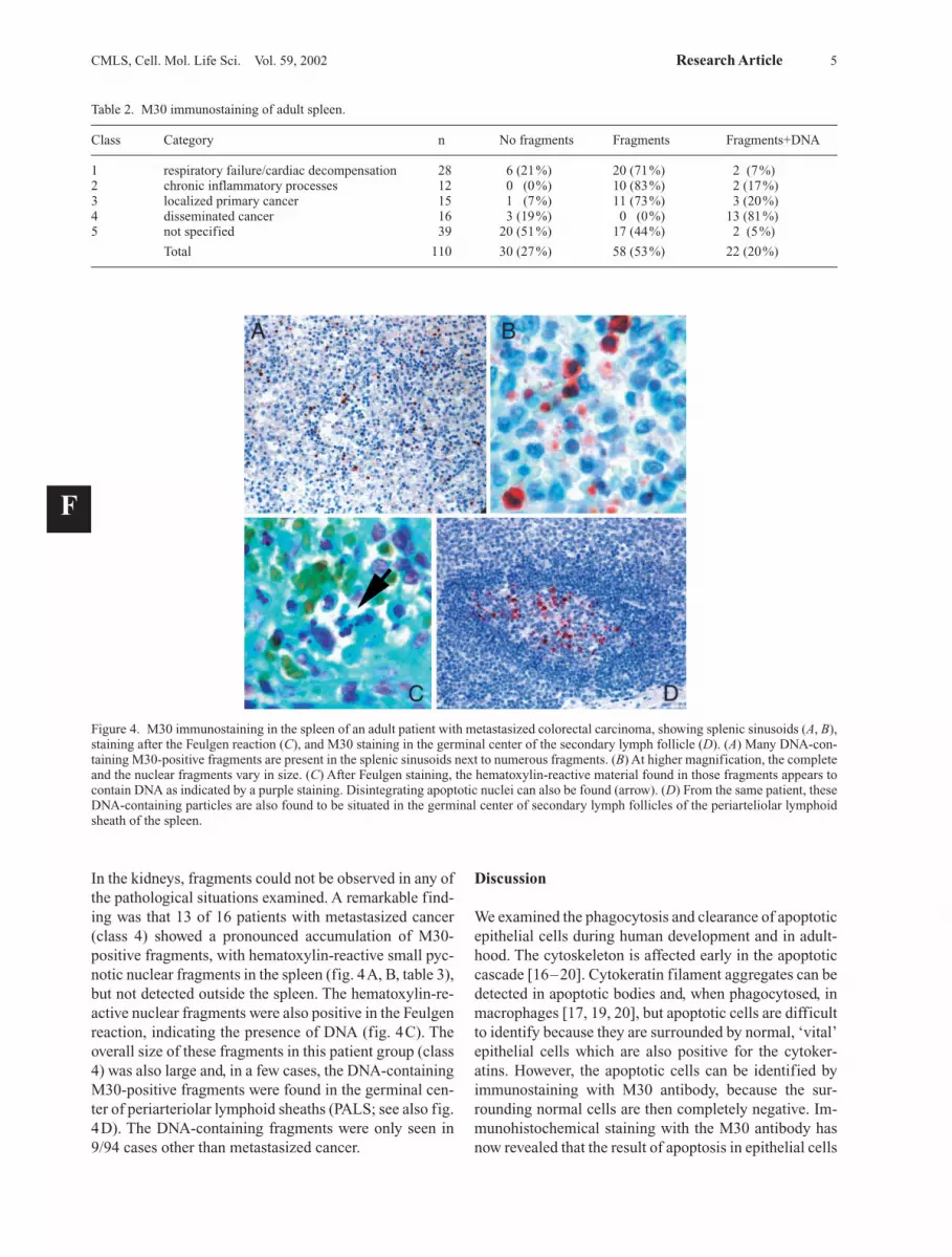
In the kidneys, fragments could not be observed in any ofthe pathological situations examined. A remarkable find-ing was that 13 of 16 patients with metastasized cancer(class 4) showed a pronounced accumulation of M30-positive fragments, with hematoxylin-reactive small pyc-notic nuclear fragments in the spleen (fig. 4A, B, table 3),but not detected outside the spleen. The hematoxylin-re-active nuclear fragments were also positive in the Feulgenreaction, indicating the presence of DNA (fig. 4C). Theoverall size of these fragments in this patient group (class4) was also large and, in a few cases, the DNA-containingM30-positive fragments were found in the germinal cen-ter of periarteriolar lymphoid sheaths (PALS; see also fig.4D). The DNA-containing fragments were only seen in9/94 cases other than metastasized cancer.
Discussion
We examined the phagocytosis and clearance of apoptoticepithelial cells during human development and in adult-hood. The cytoskeleton is affected early in the apoptoticcascade [16–20]. Cytokeratin filament aggregates can bedetected in apoptotic bodies and, when phagocytosed, inmacrophages [17, 19, 20], but apoptotic cells are difficultto identify because they are surrounded by normal, ‘vital’epithelial cells which are also positive for the cytoker-atins. However, the apoptotic cells can be identified byimmunostaining with M30 antibody, because the sur-rounding normal cells are then completely negative. Im-munohistochemical staining with the M30 antibody hasnow revealed that the result of apoptosis in epithelial cells
CMLS, Cell. Mol. Life Sci. Vol. 59, 2002 Research Article 5
Table 2. M30 immunostaining of adult spleen.
Class Category n No fragments Fragments Fragments+DNA
1 respiratory failure/cardiac decompensation 28 6 (21%) 20 (71%) 2 (7%)2 chronic inflammatory processes 12 0 (0%) 10 (83%) 2 (17%)3 localized primary cancer 15 1 (7%) 11 (73%) 3 (20%)4 disseminated cancer 16 3 (19%) 0 (0%) 13 (81%)5 not specified 39 20 (51%) 17 (44%) 2 (5%)
Total 110 30 (27%) 58 (53%) 22 (20%)
Figure 4. M30 immunostaining in the spleen of an adult patient with metastasized colorectal carcinoma, showing splenic sinusoids (A, B),staining after the Feulgen reaction (C), and M30 staining in the germinal center of the secondary lymph follicle (D). (A) Many DNA-con-taining M30-positive fragments are present in the splenic sinusoids next to numerous fragments. (B) At higher magnification, the completeand the nuclear fragments vary in size. (C) After Feulgen staining, the hematoxylin-reactive material found in those fragments appears tocontain DNA as indicated by a purple staining. Disintegrating apoptotic nuclei can also be found (arrow). (D) From the same patient, theseDNA-containing particles are also found to be situated in the germinal center of secondary lymph follicles of the periarteliolar lymphoidsheath of the spleen.
F

can be detected as scattered individual fragments. Duringadulthood, in normal situations, apoptotic fragments werenot found by light microscopy, but in situations with in-creased apoptotic activity, M30-positive fragments andmacrophages could be found. From the findings pre-sented in this study, we can evaluate several aspects of theapoptotic cytokeratin fragments.
Lack of splenic apoptotic fragmentswith anti-cytokeratin antibodiesAfter induction of apoptosis in epithelial cell cultures, apositive reaction in the cytoskeleton can be observed withmost cytokeratin antibodies [19, 20]. We observed bothCK18 and M30 reactivity under similar conditions [10].However, in M30-positive cell fragments in vessel spacesand in the spleen, no cytokeratin immunoreactivity wasobserved with the commercially available antibodies. Thefinding that the M30-positive fragments do not react withordinary cytokeratin antibodies can be explained by thedifferent locations of the epitope on the CK18 filamentprotein: the M30 neo-epitope is created during earlyapoptotic cleavage and is situated near the C terminus ofthe CK18 protein [10]. We demonstrated earlier that inlate apoptotic cells, the cleavage process results in aCK18 fragment of approximately 20 kDa, in which theM30 epitope is retained but the M3 epitope and other cy-tokeratin epitopes are not [10].
The splenic M30-positive material may indicate accu-mulation of apoptotic cellular debris in the spleenEnlargment of the spleen in sepsis is generally believed tobe caused by the accumulation of cells and cellular de-bris. Normal breakdown of erythrocytes takes place in thespleen, followed by transportation to the liver for furtherprocessing and final excretion into the bile. In a balancedsituation, the apoptotic bodies are removed by macro-phages. Sometimes, we observe a weakly positive cyto-plasmic staining for M30 in macrophages. In this situa-tion, the spleen only shows a few scattered M30-positivefragments (class 1 of the autopsy cases). However, whenthe apoptotic activity is higher and the load of fragmentsis high in chronic inflammation such as ulcerative colitisor in patients with metastasized malignant epithelial tu-
mors, the affected tissues show an increased expressionof M30-positive apoptotic cells. In those cases, a huge ac-cumulation of M30-positive fragments was also found in the red pulp of the spleen. Clearance by the spleen isobviously effective, because in the majority of the adultautopsy analyses, very few fragments could be detectedin the liver and none in the kidney. However, an overloadof the phagocytic clearance capacity has previously been observed when massive apoptosis in the liver wasinduced by administration of anti-Fas antibodies to mice [21].Whereas an accumulation of macrophages was seen inthe adult spleen under pathological conditions, the num-ber of M30-positive macrophages in the spleen is lowduring fetal development. However, in fetal liver, a largenumber of M30-positive macrophages were detected. Inaddition, very few free M30-positive fragments were de-tected in the fetal hepatic sinusoids. This observationmight be understood if one considers the macrophages astransport vehicles from the spleen to the liver in fetalgrowth, development, and recycling of cell constituents.
Epithelial apoptotic fragments in the spleen may playa role in the development of immunological tolerancefor or rejection of neo-epitopesThe accumulation of M30-positive fragments in thespleen during development in the fetus may possibly actas a normal procedure to induce tolerance for this andother neo-epitopes. Impaired completion of tolerance in-duction, or the formation of other epitopes during apop-tosis in the adult may initiate autoimmune reactions. Theobservation of M30-positive DNA-containing apoptoticbodies in the germinal center of the PALS system of thespleen in five of our patients might be interpreted in thiscontext. In support of this conclusion, observations onapoptotic keratinocytes are of interest. Two populationsof surface structures have been shown to exist on suchkeratinocytes: small buds containing fragmented endo-plasmic reticulum and ribosomes, and large buds (apop-totic bodies) containing nucleosomal DNA and nuclearribonucleoproteins [22]. In those studies, the membrane-buds, surrounding the keratinocytes undergoing apopto-sis, were presumed to contain concentrates of neo-epi-topes or autoantigens and to have an important functionin antigen presentation to the immune system. In sys-temic lupus erythematosus, there is evidence that defectsin the apoptotic process are linked to the pathogenesis ofthe disease, and in SLE patients, proteins cleaved by theICE family of proteases during apoptosis could result inthe formation of autoantibody products [23–25]. Fur-thermore, a subpopulation of macrophages involved inthe phagocytic clearance of apoptotic cells in rat colontumors expresses cell surface molecules associated with antigen presentation and stimulation of naive spleno-cytes [26].
6 M. P. G. Leers et al. Clearance of apoptotic cellular fragments
Table 3. M30 immunostaining of DNA-containing fragments
Class 4 Other Totalclasses
Fragments + DNA 13 9 22(No) fragments - DNA 3 85 88
Total 16 94 110
Sensitivity = (13/16) ¥100 = 81%Specificity = (88/97) ¥100 = 91%

Role of DNA-containing apoptotic fragmentsin late metastasesIn the category of patients with metastatic tumors, a pref-erential occurrence of DNA-containing M30-positivefragments in the spleen was found. In all other categories,the occurrence of the DNA-containing fragments wasmuch lower and, in most cases, absent. Because thosefragments were also larger, the most reasonable interpre-tation is that these fragments constitute apoptotic bodies,containing nuclear fragments, which have escaped localphagocytic clearance. Recent experiments [27] haveshown that DNA remnants within apoptotic bodies can betransferred to another cell where they are integrated intothe recipient DNA, as demonstrated by cocultivation ofapoptotic bodies of Epstein-Barr virus (EBV)-carryingcells with EBV-negative human fibroblasts, macro-phages, and bovine aortic endothelial cells. This resultedin expression of EBV-encoded genes in the recipient cellsat high frequency and the transferred DNA was stableover time. Recently, oncogenes present in apoptotic bod-ies were also shown to be horizontally transferred to eu-karyotic cells resulting in aneuploidy and accumulationof genetic changes necessary for tumor formation [28].The accumulation of DNA-containing M30-positivefragments in the spleen of patients with metastatic malig-nancies, found in the present study, may similarly lead totransfer and integration of tumor DNA remnants fromthose apoptotic bodies into other normal cells.In conclusion, our study visualized the existence of rem-nants of the apoptotic process by a simple immunochemi-cal assay. When the apoptotic load is too high, phagocyticclearance of apoptotic bodies can apparently become inef-ficient and fragments may remain intact before they aretransported to the spleen where they are filtered out of theblood. Detection of M30-positive fragments in the blood-stream may reflect the activity of disease or therapy.
1 Saunders J. and Fallon J. (1966) Cell death in morphogensis. In:Major Problems in Developmental Biology, pp. 289–314,Locke M. (ed.), Academic Press, New York
2 Prindull G. (1995) Apoptosis in the embryo and tumorigenesis.Eur. J. Cancer 1: 116–123
3 Hinrichsen K. (1985) The early development of morphologyand patterns of the face in the human embryo. In: Advances inAnatomy of Embryologic Biology, pp. 98–101, Beck F. H. W.,Kriz W., Ortmann R., Pauly J. E. and Schieber T. M. (eds),Springer, Berlin
4 Goldmann A., Baker M., Peddington R. and Herold R. (1983)Inhibition of programmed cell death in mouse embryonicpalate in vitro by cortisone and phenytoin: receptor involve-ment and requirement of protein synthesis. Proc. Soc. Exp.Biol. Med. 174: 239–243
5 Pexieder T. (1975) Cell death in the morphogenesis and terato-genesis of the heart. Adv. Anat. Embryol Cell. Biol. 51: 5–99
6 Saunders J. (1966) Death in the embryonic system. Science154: 604–612
7 Kerr J. (1971) Shrinkage necrosis: a distinct mode of cellulardeath. J. Pathol. 105: 13–20
8 Kerr J. and Harmon B. (1994) Definition and incidence ofapoptosis: an historical perspective. In: Apoptosis: The Mole-cular Basis of Cell Death, pp � –�, Tomei L. and Cope F. (eds),Cold Spring Harbor Laboratory Press. Cold Spring Harbor, N. Y.
9 Bosman F., Visser B. and Oeveren J. van (1996) Apoptosis:pathophysiology of programmed cell death. Pathol. Res. Pract.192: 676–683
10 Leers M. P. G., Kolgen W., Bjorklund V., Bergman T., TribbickG., Persson B. et al. (1999) Immunocytochemical detection andmapping of a cytokeratin 18 neo-epitope exposed during earlyapoptosis. J. Pathol. 187: 567–572
11 Holness C. and Simmons D. (1993) Molecular cloning ofCD68, a human macrophage marker related to lysosomal gly-coproteins. Blood 81: 1607–1613
12 Falini B., Flenghi L., Pileri S., Gambacorta M., Bigerna B.,Durkop H. et al. (1993) PG-M1: a new monoclonal antibody di-rected against a fixative-resistant epitope on the macrophage-restricted form of the CD68 molecule. Am. J. Pathol. 142:1359–1372
13 Bonfrer J. M., Groeneveld E. M., Korse C. M., Dalen A. van,Oomen L. C. and Ivanyi D. (1994) Monoclonal antibody M3used in tissue polypeptide-specific antigen assay for the quan-tification of tissue polypeptide antigen recognizes keratin 18.Tumour Biol. 15: 210–222
14 Speel E. J., Jansen M. P., Ramaekers F. C. S. and Hopman A. H.(1994) A novel triple-color detection procedure for brightfieldmicroscopy, combining in situ hybridization with immunocyto-chemistry. J. Histochem. Cytochem. 42: 1299–1307
15 Speel E. J., Herbergs J., Ramaekers F. C. S. and Hopman A. H.(1994) Combined immunocytochemistry and fluorescence insitu hybridization for simultaneous tricolor detection of cellcycle, genomic, and phenotypic parameters of tumor cells. J.Histochem. Cytochem. 42: 961–966
16 Engeland M. van, Nieland L. J., Ramaekers F. C. S., Schutte B.and Reutelingsperger C. P. (1998) Annexin V-affinity assay: areview on an apoptosis detection system based on phos-phatidylserine exposure. Cytometry 31: 1–9
17 Caulin C., Salvesen G. S. and Oshima R. G. (1997) Caspasecleavage of keratin 18 and reorganization of intermediate fila-ments during epithelial cell apoptosis. J. Cell Biol. 138: 1379–1394
18 Ku N. O., Liao J. and Omary M. B. (1997) Apoptosis generatesstable fragments of human type I keratins. J. Biol. Chem. 2:33197–33203
19 Tinnemans M. M., Lenders M. H., Velde G. P. ten, RamaekersF. C. S. and Schutte B. (1995) Alterations in cytoskeletal andnuclear matrix-associated proteins during apoptosis. Eur. J.Cell Biol. 68: 35–46
20 Engeland M. van, Kuijpers H. J., Ramaekers F. C. S., Reute-lingsperger C. P. and Schutte B. (1997) Plasma membrane al-terations and cytoskeletal changes in apoptosis. Exp. Cell Res.235: 421–430
21 Ogasawara J., Watanabe-Fukunaga R., Adachi M., MatsuzawaA., Kasugai T., Kitamura Y. et al. (1993) Lethal effect of theanti-Fas antibody in mice. Nature 364: 806–809
22 Casciola-Rosen L., Anhalt G. and Rosen A. (1994) Autoan-tigens targeted in systemic lupus erythematosus are clustered in two populations of surface structures on apoptotic ker-atinocytes. J. Exp. Med. 179: 1317–1330
23 Casciola-Rosen L., Anhalt G. and Rosen A. (1995) DNA-depen-dent protein kinase is one of a subset of autoantigens specificallycleaved early during apoptosis. J. Exp. Med. 182: 1625–1634
24 Nicholson D. W., Ali A., Thornberry N. A., Vaillancourt J. P.,Ding C. K., Gallant M. et al. (1995) Identification and inhibi-tion of the ICE/CED-3 protease necessary for mammalianapoptosis. Nature 376: 37–43
25 Tewari M., Quan L.T., O’Rourke K., Desnoyers S., Zeng Z.,Beidler D. R. et al. (1995) Yama/CPP32 beta, a mammalian ho-
CMLS, Cell. Mol. Life Sci. Vol. 59, 2002 Research Article 7

molog of CED-3, is a CrmA-inhibitable protease that cleavesthe death substrate poly(ADP-ribose) polymerase. Cell 81:801–809
26 Henry F., Bretaudeau L., Barbieux I., Meflah K. and GregoireM. (1998) Induction of antigen presentation by macrophagesafter phagocytosis of tumour apoptotic cells. Res. Immunol.149: 673–679
27 Holmgren L., Szeles A., Rajnavolgyi E., Folkman J., Klein G.,Ernberg I. et al. (1999) Horizontal transfer of DNA by the up-take of apoptotic bodies. Blood 93: 3956–3963
28 Bergsmedh A., Szeles A., Henriksson M., Bratt A., Folkman M.J., Spetz A. L. et al. (2001) Horizontal transfer of oncogenes byuptake of apoptotic bodies. Proc. Natl. Acad. Sci. USA 98:6407–6411
8 M. P. G. Leers et al. Clearance of apoptotic cellular fragments


