Embed Size (px)
Citation preview

10070 Biochemistry 1990, 29, 10070-10080
a-Macroglobulin from Limulus polyphemus Exhibits Proteinase Inhibitory Activity and Participates in a Hemolytic System)
Jan J. Enghild,t Ida B. Th~gersen , t Guy Salvesen,: Georg H. Fey , l Nancy L. Figler,§ Steven L. Gonias,§ and Salvatore V. Pizzo*J
Duke University Medical Center, Durham, North Carolina 2771 0, Departments of Pathology and Biochemistry, Health Sciences Center, University of Virginia, Chariottesville, Virginia 22908, and Department of Immunology, Research Institute of Scripps
Clinic, La Jolla, California 92037 Received March 5, 1990; Revised Manuscript Receiced June 11, I990
ABSTRACT: Significant primary sequence homology between the a-macroglobulin family of proteinase inhibitors and the complement components C3, C4, and C5 implies that these proteins arose from a common ancestor. Hemolymph from the ancient invertebrate Limulus polyphemus contains both complement-like and proteinase inhibitory activity. In this report, we present evidence that L. polyphemus a-macroglobulin not only possesses proteinase inhibitory activity, but it also participates in the lytic system of the horseshoe crab. The protein is a disulfide-linked dimer of subunits of molecular mass 185 kDa. Upon reaction with proteinase or methylamine, L. polyphemus a-macroglobulin underwent a major conformational change and no proteinase-associated multimerization was detected. L. polyphemus a-macroglobulin is the only detectable inhibitor of a number of proteinases in L. polyphemus hemolymph. Proteinase inhibition follows the general “trapping” mechanism shared by most a-macroglobulins; however, no covalent linking of proteinases to the inhibitor was detected despite the presence of a functional thiolester. Moreover, the inhibitor demonstrated thiolester-mediated binding to sheep erythrocytes, a property also observed with complement components such as C3. Depletion of functional protein by treatment of hemolymph with methylamine destroyed the proteinase inhibitory capacity and the lytic activity of the hemolymph. Both activities were restored by adding purified protein to depleted hemolymph. Studies with purified L. polyphemus a-macroglobulin demonstrated that the thiolester incorporates glycerol as well as methylamine, a property shared by human C3. The data support the hypothesis that L. polyphemus a-macroglobulin is both a proteinase inhibitor and part of a lytic system, providing a link between the two distinct sides of the a-macroglobulin family. Because both properties are contained in one molecule, we propose the name “limac” to describe this G m u l u s a-macroglobulin complement-like protein.
a-Macroglobulins have been conserved throughout evolution (Starkey & Barrett, 1982) and have been isolated from in- vertebrates, reptiles, birds, and many mammalian species [see Sottrup-Jensen (1 987, 1989) for reviews]. The family com- prises proteinase inhibitors, such as human q-macroglobulin (a2M)’ and the complement factors C3, C4, and C5. The evolutionary relationship between these proteins became ob- vious after their protein sequences were determined. These proteins are synthesized as 185-kDa precursors and contain several stretches of sequence similarity (Sottrup-Jensen et al., 1985). This relationship is further strengthened by the presence of an internal 8-cysteinyl-y-glutamyl thiolester in C3, C4, and a2M (Swenson & Howard, 1979; Tack et al., 1980; Sottrup-Jensen et al., 1980; Janatova et al., 1980). The re- activity of the thiolester to small nucleophiles differs when human C3 and aZM are compared. Thus, human C3 incor- porates [3H]glycerol much more efficiently than does human a2M (Dodds & Law, 1988), while [3H]methylamine is in-
corporated into the thiolesters of both C3 or a z M (Swenson & Howard, 1979; Sottrup-Jensen et al., 1980; Law, 1983). There is no evidence that the complement members of the a-macroglobulin family inhibit proteinases, but all other known members of the family have been characterized on the basis of proteinase inhibition.
Most proteinases are inhibited by the a-macroglobulin proteinase inhibitors irrespective of catalytic mechanism or substrate specificity (Barrett, 1982; Travis & Salvesen, 1983; Roberts, 1986). The inhibitory mechanism has been analyzed in greatest detail by using human a2M, but it is shared by other inhibitory a-macroglobulins (Sottrup-Jensen, 1987). The mechanism, best described by the “trap hypothesis” of Barrett and Starkey (1973), involves a conformational change of the inhibitor encaging the attacking proteinase. This compart- mentalizes and restrains the proteinase, hindering physical access to macromolecular substrates. The conformational
‘This work was supported by the National Heart and Blood Institute Grant HL-24066 and National Institute of Allergy and Infectious Dis- eases Grants AI-22166 and AI-19651. SLG is supported by a Research Career Development Award from the National Institutes of Health (HL-02272) and by the Pew Scholars Program.
* Address correspondence to this author a t Pathology Department, P.O. Box 3712, Duke University Medical Center, Durham, N C 27710.
*Duke University Medical Center.
8 University of Virginia. Research Institute of Scripps Clinic.
’ Abbreviations: CY,^,, a,-inhibitor3; a2M, a,-macroglobulin; C3, C4, and C5, complement factors 3,4, and 5 ; HNE, human neutrophil elastase (E.C. 3.4.21.37); DTT, dithiothreitol; DTNB, 5’,5-dithiobis(2-nitro- benzoic acid); SDS-PAGE, sodium dodecyl sulfate-polyacrylamide gel electrophoresis; limac, Limulus a-macroglobulin complement-like pro- tein; DCI, 3,4-dichloroisocoumarin; E-64, N-[ [ [N-[(~-3-trans-carboxy- oxiran-2-yl)carbonyl]-~-leucyl]amino] butyl]guanidine; GVBZ+, 1.8 mM sodium barbital, 3.1 mM barbituric acid, 145 mM NaC1, 0.15 mM CaCI,, 1 mM MgCI,, 0.1% gelatin, pH 7.4; PTH, phenylthiohydantoin; HPLC, high-performance liquid chromatography; Tris, tris(hydroxy- methy1)aminomethane; EDTA, (ethylenedinitri1o)tetraacetic acid.
0006-2960/90/0429-10070$02.50/0 0 1990 American Chemical Society
L. polyphemus a-Macroglobulin
change greatly decreases the stability of the internal thiolester. It becomes a nascent binding site that will interact with ad- jacent nucleophiles. The activated thiolester will often react with the e-amino group of lysine residues on the surface of the proteinase, forming covalent dysyl-y-glutamyl cross-links (Howard, 1981; Barrett, 1982; Roberts, 1986). Complete suppression of this covalent interaction does not alter the inhibitory ability of human a2M (Salvesen, et al., 1981). The monomeric rat a,inhibitor, (al13) (Enghild et al., 1989a) and the dimeric human pregnancy zone protein (PZP) (Christensen et al., 1989), however, are completely dependent on this ability to form covalent cross-links, although inhibition results from steric shielding as in the other a-macroglobulins.
The complement system is comprised of a group of proteins that play a significant role in host resistance to infection. TWO primary pathways of complement activation are known, the classical and the alternative. The third component of com- plement, C3, is the central protein of both pathways. After activation, C3b, generated after cleavage by either pathway, can bind covalently to cell surface carbohydrates or immune aggregates (Muller-Eberhard et al., 1966; Miller & Nus- senzweig, 1975). The other thiolester-containing complement component, C4, serves as an opsonin and as a covalent anchor for the complex proteinases of the classical pathway of com- plement [see Law and Reid (1988) for a review].
Limulus polyphemus is one of the oldest surviving multi- cellular organisms, dating back more than 500 million years (Rudloe, 1979). The hemolymph of this animal contains both complement-like lytic activity (Day et al., 1970) and an a- macroglobulin-like proteinase inhibitor (Quigley & Armstrong, 1983). In the present study we demonstrate that the proteinase inhibitory and lytic activity of L. polyphemus hemolymph were eliminated by reagents that destroy thiolesters. These data suggest that a single thiolester-containing protein is responsible for proteinase inhibitory activity and is part of the lytic system of the hemolymph. To test this hypothesis, the protein was purified, characterized, and identified as the L. polyphemus a-macroglobulin (Quigley & Armstrong, 1983). It is dem- onstrated that L . polyphemus a-macroglobulin reacts with proteinases without forming a covalent link between the pro- teinase and the a-macroglobulin. This protein incorporates both glycerol and methylamine into its thiolester when the a-macroglobulin is treated with proteinase. Furthermore, following proteinase activation, L. polyphemus a-macro- globulin binds to sheep erythrocytes. This protein may rep- resent the link that has been postulated to exist between the a-macroglobulin proteinase inhibitors and the complement components C3, C4, and C5.
MATERIALS A N D METHODS Chemicals and Reagents. Pig pancreatic elastase, ther-
molysin, dithiothreitol (DTT), iodoacetamide, sodium 5,5- dimethylbarbital, sodium 5,5-diethylbarbituric acid, guanidine hydrochloride, l,lO-phenanthroline, and CNBr-activated Se- pharose CL-4B were from Sigma. Hide powder azure was from Calbiochem. TPCK-treated bovine trypsin, bovine chymotrypsin, the general serine proteinase inhibitor 3,4-di- chloroisocoumarin (DCI), and the general cysteine proteinase inhibitor N- [ [ [N- [ (~-3-trans-carboxyoxiran-2-yl)carbonyl]- ~-leucyl] amino] butyllguanidine (E-64) were from Boehringer Mannheim. Iodoacetic acid was from Eastman Kodak. Iodo[14C]acetamide (55 mCi/mmol), iod~[~H]acetic acid (75 mCi/mmol), and [I4C]methylamine hydrochloride (55.4 mCi/mmol) were from Amersham. [3H]Glycerol (40 Ci/ mmol) and Protosol were from New England Nuclear. MONO Q, Superose 6 columns, and Sephacryl S-300 H R
Biochemistry, Vol. 29, No. 43, 1990 10071
were from Pharmacia. Rabbit anti-limac sera were obtained from Accurate Chemical and Scientific Corp. Sheep eryth- rocytes and cobra venom factor were generously provided by Dr. Wendell Rosse, Duke University Medical Center. Human neutrophil elastase (HNE) was a kind gift of Drs. Wieslaw Watorek and James Travis, University of Georgia.
Protein Purifi:cation. L. polyphemus a-macroglobulin (limac) was purified essentially as described by Quigley and Armstrong (1985). Briefly, L. polyphemus specimens were randomly selected from the tanks of Duke Marine Laboratory, Beaufort, NC. The animals were chilled to 4 OC in the cold room before they were bled by cardiac puncture into sterile tubes. Fractions of 20 mL were collected and the cells were immediately removed by centrifugation at 5000 rpm for 5 min to prevent clotting. The plasma was centrifuged at IOOOOOg (Sorvall Ultracentrifuge OTD 50B) for 4 h to sediment he- mocyanin and the clear supernatant (high-speed supernatant) was stored a t -20 or 0 OC. Fifty milliliters of plasma was dialyzed (1 5 kDa cutoff) against 12 L of 50 mM Tris-HC1 and 50 mM NaCl, pH 7.4, overnight at 4 OC. Following removal of the precipitate, the supernatant was applied to a MONO Q column in 50 mM Tris-HC1, pH 7.4. The column was eluted with a linear gradient from 0.0 to 0.5 M NaCl (1 .O mM mL-I) by using a Pharmacia FPLC system. Fractions containing proteinase inhibitory activity were pooled and concentrated by ultrafiltration using a Spectrapor 100-kDa cutoff membrane. The final purification step was gel filtration on Sephacryl S-300 HR in 50 mM Tris-HC1 and 150 mM NaC1, pH 7.4. Human a2M was purified as described by Kurecki et al. (1979). Rat a113 was purified as described by Enghild et al. (1989a). The extinction coefficient was de- termined by amino acid analysis using norleucine to correct for loss during transfer and hydrolysis (Kupke & Dorrier, 1978).
Labeling of Thiolester Proteins in the Hemolymph. The pH of 10 mL of hemolymph was adjusted to 8.3 with 2 M Tris-HC1, pH 8.3. The sample was made 25 mM in iodo- acetamide and 50 mCi ['4C]methylamine was added. The reaction was allowed to continue overnight before 300 mM methylamine was added. The reaction mixture was dialyzed and radioactive proteins were purified as described above.
Labeling of Limac with 10do[~H]acetic Acid, lodo[14C]- acetamide, and [I4C]Methylamine Hydrochloride. Double- labeled limac was prepared by reacting 10 mg of limac in 100 mM Tris-HC1 and 150 mM NaC1, pH 8.3, with 100 pCi [ I4C]methylamine hydrochloride and 25 pCi i~do[~H]ace t ic acid at 25 "C. After 4 h, the sample was made 300 mM in methylamine hydrochloride and 25 mM in iodoacetic acid and left overnight at 25 OC. Single-labeled limac was prepared as described above but substituting methylamine hydrochloride for [ I4C]methylamine hydrochloride and adding 25 pCi i~do[~H]acetic acid or 25 pCi i~do['~C]acetamide. The next day, the sample was dialyzed against 100 mM Tris-HC1 and 50 mM NaCl, pH 8.3, before the disulfides were reduced with 10 mM DTT. The resulting sulfhydryl groups were modified with 0.5% 4-vinylpyridine in the presence of 6 M guanidine hydrochloride as described by Friedman et al. (1970). The sample was dialyzed against 50 mM NH4HC03 before frag- mentation.
Proteolytic Digestion. Batches of 3.5 mg of alkylated limac were digested with Staphylococcus aureus V8 proteinase, clostripain, bovine chymotrypsin, or H N E in 50 mM NH4- HCO, by using an enzymembstrate ratio of 1:50 (w/w). The clostripain digestion was carried out in the presence of 5 mM DTT and the stock enzyme solution was made 0.5 mM in DCl,

10072
0.1 mM in E-64, and 10 mM in 1,lO-phenanthroline to inhibit any undesired proteolytic activity, before it was added to the limac solution. The digestions were continued for 4 h a t 37 OC and the reactions were terminated with 50 pM DCI ( S . aureus V8 proteinase, bovine chymotrypsin, human neutrophil elastase). The clostripain digest was separated immediately on HPLC.
Peptide Purification. The radioactive peptides were purified by reverse-phase HPLC by using various columns supplied by Applied Biosystems (Aquapore RP-300, 2.1 X 220 mm and 4.6 X 22 mm), Vydac (Vydac C18, 4.6 X 250 mm), and Waters (Nova-pak clg, 3.9 X 150 mm). An LKB 2152 controller and two LKB 21 50 pumps were used to form gradients from (A) 0.1% trifluoroacetic acid (TFA) to (B) 90% acetonitrile (or propan-2-01), 9.9% H,O, and 0.1% TFA. The peptides were detected at 220 or 214 nm in an LKB 2140 diode array de- tector as described previously (Enghild et al., 1989b). Aliquots from each peak were counted for 3H or 14C radioactivity in a Beckman LS 5000 TD liquid scintillation system to localize the radioactive peptides.
Amino Acid Compositions and Sequence Analysis of Pep- tides. Peptides, 1-2 nmol, were hydrolyzed in gas phase for 24 h at 110 "C (Meltzer et al., 1987) and analyzed in a Beckman 6300 amino acid analyzer using sodium citrate buffers provided by the manufacturer. Automated Edman degradation was carried out in an Applied Biosystems 477A sequencer with on-line PTH analysis using an Applied Bio- systems 120A HPLC. The instruments were operated as recommended in the manuals and user bulletins distributed by the manufacturer. Fifty-microliter aliquots of the PTH derivatives were analyzed in the 120A HPLC and the re- maining sample was delivered directly into scintillation vials with a Pharmacia Frac 100 fraction collector. After adding 3.5 mL of scintillation cocktail, the level of 'H or I4C radio- activity in each fraction was measured in a Beckman LS 5000 T D liquid scintillation system.
Limac-Proteinase Interaction. The ability of limac to prevent bovine chymotrypsin, bovine trypsin, pig pancreatic elastase, and thermolysin from digesting the high molecular weight substrate hide powder azure (Salvesen & Nagase, 1989) was investigated. The activity of limac was determined by thiol titration according to Ellman (1959). The batch used for the following experiments was 88% active. Increasing amounts of limac were reacted with 0.04 nmol of active- site-standardized proteinase (Salvesen & Nagase, 1989) in 0.6 mL of 50 mM Tris-HC1, 100 mM NaCI, and 1 mM EDTA, pH 8.0 (2 mM CaCl, substituted for EDTA when thermolysin was used). Reactants were incubated for 30 min at 25 "C before 0.4 mL of blue hide powder suspension, 12.5 mg/mL in 0.6 M sucrose and 0.05% Triton X-100, was added. The tube was incubated with sufficient agitation to keep the par- ticulate substrate in suspension. The reaction was stopped by adding 0.3 mL of 3 M glycine hydrochloride, pH 3.0. The blue powder was pelleted by centrifugation and the absorbance of the supernatant was determined at 595 nm. The ability of limac to covalently bind the proteinases was investigated by reacting '251-labeled bovine chymotrypsin, bovine trypsin, pig pancreatic elastase, and thermolysin with limac at a molar ratio of 0.5: 1 proteinase/limac. The reactions were continued for 15 min before the mixture was made 50 pM in DCI (bovine chymotrypsin, bovine trypsin, and pig pancreatic elastase) and 1 mM in l,lO-phenanthroline (thermolysin). The sample was separated on a Superose 12 gel filtration column to remove unbound proteinase. The limac-proteinase complex was then analyzed by pore limit gel electrophoresis, SDS-PAGE with
Biochemistry, Vol. 29, No. 43, 1990 Enghild et al.
and without reduction, and autoradiography. Electron Microscopy. Thin carbon films were floated on
100-pL aliquots of a solution of limac or limac reacted with trypsin. The protein concentration was 10 pg/mL. Adsorbed protein was treated with 2% glutaraldehyde for IO min and washed with deionized water. Treatment with glutaraldehyde, after the limac was adsorbed to the carbon film, enhanced image quality without altering detectable protein structure, consistent with previous studies of human a 2 M (Gonias et al., 1988; Gonias & Figler, 1989). Films were stained with 2.0% uranyl formate and transferred to 300-mesh nickel hexagon grids. Air-dried grids were analyzed by conventional trans- mission electron microscopy using a Zeiss 902 electron mi- croscope at 80 kV. The image was of elastically scattered electrons. The photomicrographs are shown at a magnification of 112000. Measurements were made by using a Laboratory Computer Systems Micro-Plan I1 image analyzer. The re- ported values represent the mean f l standard deviation ( n = 25).
Assay for Hemolytic Activity. The hemolytic assay used was adapted from the procedure of Day et al. (1970). He- molymph- or limac-depleted L . polyphemus hemolymph (high-speed supernatant) was diluted 6-fold with 1.8 mM sodium barbital, 3.1 mM barbituric acid, 145 mM NaC1, 0.15 m M CaCI,, and 1 mM MgC1, containing 0.1% gelatin (GVB2+). To a row of test tubes containing 300 pL of sus- pended erythrocytes (1 X lo8 cells/mL), increasing amount of the diluted hemolymph was added. The final volume was brought to 800 pL with GVB2+. The mixture was incubated in a shaking waterbath at 30 "C for 4 h. Two milliliters of cold saline was added before the cells were sedimented by centrifugation. The absorbance of the solution was measured in a Shimadzu UV16OU spectrophotometer at 412 nm against a blank containing GVB2+ and saline. Spontaneous lysis was determined as the amount of lysis without hemolymph and was usually less than 2%. Complete lysis was determined by adding water instead of saline.
Limac-depleted hemolymph was prepared by adjusting the pH to 8.3 with 2 M Tris-HC1, pH 8.3, and adding 10 mM methylamine. The reaction was continued for 24 h at 25 O C
before it was used for hemolytic or proteinase inhibitory assays. Control samples treated the same, except that methylamine was omitted, did not show any loss of activity.
Reaction of Limac and Human a2M with Proteinase in the Presence of [3H]Glycerol. Limac and human a 2 M were compared for their ability to incorporate [3H]glycerol when incubated with HNE. H N E reacts readily with limac and a2M but does not contain Lys residues (Salvesen et al., 1987). Binding of Lys residues to the activated thiolester was thereby prevented and competition between €-amino groups of the proteinase and glycerol avoided. Samples of 20 pg of limac in 25 mM HEPES and 150 mM NaC1, pH 7.4, containing 10 mM glycerol (400 mCi/mmol) were incubated with and without 1.55 pg of H N E at 37 O C . The reactions were ter- minated after 5 min by adding DCI to a final concentration of 250 pM. Similar experiments were performed where 20 pg of human a,M were incubated with and without 1.55 pg of HNE. The four samples were electrophoresed on a 5-1 5% polyacrylamide-SDS gel without reduction, stained with Coomassie blue, and destained in 10% aqueous acetic acid. The bands were excised and transferred to vials containing 4 mL of scintillation cocktail and 200 pL of Protosol. After solubilization of the gel pieces, the vials were counted for 3H radioactivity in a Beckman LS 5000 TD liquid scintillation system.

L. polyphemus a-Macroglobulin Biochemistry, Vol. 29, No. 43, 1990 10073
Binding of Limac to Erythrocytes. Twenty-five microliters of suspended sheep erythrocytes (4 X IO9 cell/mL) in GVB2+ was transferred to a row of microcentrifuge tubes. '251-LabeIed limac (1.4 pg) or human a2M (2.8 pg) and GVB2+ were added to a final volume of 40 pL. The samples were incubated with increasing amounts of pig pancreatic elastase for 5 min at 25 OC before the reaction was stopped by making the reaction mixture 100 pM in DCI. Unbound 1251-labeled limac was removed as described by Pangburn et al. (1983) by using human C3. Samples were removed and layered on 300 pL of 20% sucrose in a microfuge tube. The cells were sedimented by centrifugation for 30 s in a microcentrifuge. The super- natant was aspirated and discarded. The cells were then washed with GVB2+ two times before the bottom of the tube was removed and the percent of maximum bound radioactivity was determined.
Protein Radioiodination. Proteins were labeled with 1251 by the solid-state lactoperoxidase method of David and Reis- feld (1974). No loss of activity of the 1251-labeled limac or proteinase was detected.
Polyacrylamide Gel Electrophoresis (PAGE). Sodium dodecyl sulfate (SDS)-PAGE was performed on samples treated for 15 min at 25 "C in 1% SDS on 5 1 5 % linear gradient gels ( I O X 10 X 0.1 cm) with the glycine/2-amino- 2-methyl- 1,3-propanedioI/HCl system described by Bury ( I 98 I ) . Nondenaturing pore limit gel electrophoresis was performed in 4-20% gradient gels (10 X 10 X 0.1 cm) in a Tris/EDTA/boric acid buffer system according to Manwell ( 1 977) with continuous circulation of lower and upper reservoir buffers.
Other Techniques. The binding of limac and limac-ther- molysin complexes to mouse peritoneal macrophages was performed as described previously (Enghild et al., 1989b). Circular dichroic spectroscopy (CD) of limac and its trypsin derivative was performed as described previously (Gonias et al., 1982). Amino acid sequence analysis of protein material electroblotted to poly(viny1idene difluoride) (PVDF) mem- branes was performed according to Matsudaira ( 1 987). The apparent size of limac and limac-trypsin was determined by employing a calibrated Superose 6 gel filtration column. The globular protein standards employed to calibrate the column were the following: thyroglobulin, 669 kDa; ferritin, 440 kDa; catalase, 232 kDa; lactate dehydrogenase, 140 kDa; bovine serum albumin, 67 kDa. Zone electrophoresis was carried out in a 2% agarose gel by using a Beckman Paragon 6558 elec- trophoretic system and the barbital buffers supplied by the manufacturer. DTNB titrations of proteins were performed as previously described (Ellman, 1959).
RESULTS Inhibitors of Proteinases in L. polyphemus Hemolymph.
The ability of L. polyphemus hemolymph to inhibit the pro- teolytic activity of bovine chymotrypsin, bovine trypsin, pig pancreatic elastase and thermolysin was compared before and after the hemolymph was treated with methylamine. The inhibitory capacity of the hemolymph against all the protei- nases was completely suppressed after methylamine treatment. Since all the inhibitory capacity in the hemolymph is sensitive to methylamine, a thiolester-containing proteinase inhibitor is probably responsible for the proteinase inhibitory activity in the hemolymph. I f other non-methylamine-sensitive pro- teinase inhibitors were present in the hemolymph, the con- centration would be less than 50 nM on the basis of the sen- sitivity of the proteinase assay. This is less than 1% of the a-macroglobulin concentration ( 5 pM). We conclude that the major protcinasc inhibitor in the L. polyphemus hemolymph
Table I: Limac-Proteinase Stoichiometry proteinase:limac molar binding ratios"
bovinc Pig chymo- pancreatic trypsin bovine trypsin elastase thermolysin 0.96 0.84 0.99 0.9 1
" Proteinases were titrated with increasing amounts of limac at room temperature for 30 min before the residual activity against blue hide powder was determined. All the titration plots were linear and showed >95% inhibition at the binding ratios noted above.
o b c d e f g h
w 669- - 440- - 0 0 & 4
&-- 232- 140- -
FIGURE 1 : Comparison of a-macroglobulins by nondenaturing pore-limit gel electrophoresis. Electrophoresis was performed on a 4-20% gradient gel as described under Materials and Methods. The gel lanes are as follows: The protein standards thyroglobulin, ferritin, catalase, and lactate dehydrogenase (lane a); human a2M in the native conformation (lane b); partially reduced human a2M (lane c ) showing tetramers, dimers, and monomers and the monomeric rat a,l, (lanc d) arc compared with different limac species. Partially reduced limac (lanc c), native limac (lane 9, native limac after reaction with trypsin 1:O.S mol/mol (lane f), and limac treated with methylamine (lanc h).
is inactivated by methylamine and is probably an a-macro- globulin homologue.
Purijkation of Limac. The proteinase inhibitor was purified by a combination of dialysis, anion-exchange chromatography, and gel filtration. The fractions were assayed for inhibitory activity against pig pancreatic elastase, bovine trypsin, and thermolysin by using hide powder azure as a substrate. Only one peak of inhibitory activity was found after both purification steps, suggesting that the major inhibitor in L. polyphemus hemolymph is the a-macroglobulin homologue (limac) pre- viously identified by Quigley and Armstrong ( 1 983). Limac is the only detectable proteinase inhibitor of thermolysin in hemolymph and it forms an equimolar complex with the proteinase (Table I ) . On the basis of blue hide powder ti- tration, we calculated a final limac concentration of 2 mg/mL. The concentration of limac appeared to vary seasonally and from animal to animal. I n the summer (April and August), the concentration was 2 mg/mL but in animals collected in the winter (December and January) the concentration ranged from 0.5 to 1 mg/mL. The protein was purified in a yield of 50% and was homogeneous when analyzed by amino terminal sequence analysis, nondenaturing gel electrophoresis (Figure 1, lane h) and Superose 6 gel filtration. The following 25 amino acids were identified by amino terminal sequence analysis: KSG FILTAPKSLTPGKSN ILNLH LF. No sig- nificant similarity to the available protein sequences was de- tected (National Biomedical Research Foundation Protein Identification Resources, release number 20). A faint band just below the major band was visible when purified material

10074 Biochemistry, Vol. 29, No. 43, 1990
a b c d e f g
Enghild et al.
a b c d e f g h
97- - 66- 0
43- - 31- - 21- - 14- -
FIGURE 2: Comparison of a-macroglobulins by SDS-PAGE. Elec- trophoresis was performed on a 5-1 5% gradient gel as described under Materials and Methods. Molecular weight standards are shown in lane a, and the tctrameric human a2M, the dimeric limac, and the monomeric rat cul l3 are analyzed with (lanes b-d) and without re- duction (lancs e-g).
was analyzed by SDS-PAGE (Figure 2, lane f). Amino terminal sequence analysis of the two bands after electro- blotting to poly(viny1idene difluoride) (PVDF) membranes resulted in an identical sequence. This result suggests that the two bands are the same protein, but with heterogeneous posttranslational modifications or carboxyl terminal trimming. Zone electrophoresis showed that limac migrates between the P, and PZ fractions. The extinction coefficient was E( 196, I cm, 280 nm) = 11.7.
Evidence That Limac Is the Only Thiolester-Containing Protein in Hemolymph. The hemolymph was incubated with [ 14C] methylamine as described under Materials and Methods. Methylamine will react with the P-cysteinyl-y-glutamyl thiolester of a-macroglobulins and a residue of y-glutamyl methylamine is formed (Swensen & Howard, 1979; Tack et al., 1980; Sottrup-Jensen et al., 1980; Janatova et al., 1980). To identify thiolester-containing proteins the hemolymph was dialyzed and separated by anion exchange chromatography and gel filtration. All fractions were analyzed for I4C ra- dioactivity. Only one radioactive protein was recovered and identified by amino terminal sequence analysis as limac.
Chain Stoichiometry and Conformational Change. It has been suggested that limac is a trimer of 185-kDa subunits (Quigley & Armstrong, 1985) and experiments were con- ducted to test this hypothesis. The apparent molecular weight of the native protein before and after methylamine or pro- teinase treatment was examined by gel filtration on a cali- brated Superose 6 gel filtration column and by pore-limit gel electrophoresis (Figure 1). The molecular mass for the de- natured subunit was determined by SDS-PAGE (Figure 2). The native protein demonstrated an apparent molecular mass of 550 kDa by gel filtration, based on the globular protein standards, but after proteinase or methylamine treatment the apparent molecular mass decreased to 360 kDa. SDS-PAGE yielded a subunit molecular mass of 185 kDa for the reduced protein (Figure 2). The change in migration between limac and proteinase- or methylamine-treated limac was also de- tected by pore-limit gel electrophoresis (Figure l) . Analysis of nonreduced samples of limac by SDS-PAGE resulted in a single band, indicating that the protein is composed of subunits with similar size or subunits linked by disulfides. Limited reduction followed by SDS-PAGE resulted in one molecular mass shift from 400 kDa to 185 kDa with no in- termediates (Figure 3). These data suggest that the protein is composed of two disulfide-linked subunits arranged as a
97- - 66- - 43- - 31- - 21- - 14- -
FIGURE 3: Limited reduction of limac examined by SDS-PAGE. Electrophoresis was performed on a 5-1 5% gradient gel as described under Materials and Methods. Limac, 5 pg, was incubated with 0, 5.50, 100,500, and 1000 pM DTT (lanes b-h) for I5 min at 37 “C. The reaction was stopped by adding IO mM iodoacetamide. Lane a shows molecular weight standards.
I
Wovelength (nm)
FIGURE 4: Circular dichroic spectroscopy of limac. The lower curve shows the spectrum of native unreacted limac and the upper curve of the protein after reaction with trypsin. The experimental procedures employed for these studies are described in detail elsewhere (Gonias et al., 1982).
dimer. The native protein could, of course, be composed of two of these dimers. To address this question, quantitative amino terminal sequence analysis were performed. When 100 pmol of limac was analyzed by automated Edman degradation, the recovery of PTH-Lys in the first cycle was 148 pmol. Since the initial yield of PTH-Lys in our hands usually is 8096, the expected initial yield was 160 pmol. The 148-pmol yield obtained, therefore, is consistent with the hypothesis that the protein is a dimer composed of two identical disulfide-linked subunits.
Circular dichroism (CD) was performed to analyze changes in secondary structure of limac before and after proteinase activation (Figure 4). The spectra demonstrate a negative ellipticity at wavelengths between 205 and 235 nm with a minimum at 21 5-21 6 nm for the native protein and 21 8 nm for the complex. Reaction with trypsin significantly decreased the magnitude of the mean residue rotation. These results are very similar to those previously observed with human, bovine, chicken, and frog a-macroglobulins (Gonias et al., 1982; Strickland et al., 1984; Feldman & Pizzo, 1984). Changes in secondary structure measured by CD do not necessarily correlate with changes in tertiary structure such as are detected in pore-limit gels of the protein (Feldman & Pizzo, 1984; Barrett & Starkey, 1973). We conclude that limac is a dimer that undergoes a very large change in both secondary and tertiary structure when it reacts with proteinase or methyl- amine. These data suggest that the nonreacted limac must
L. polyphemus a-Macroglobulin Biochemistry, Vol. 29, No. 43, 1990 10075
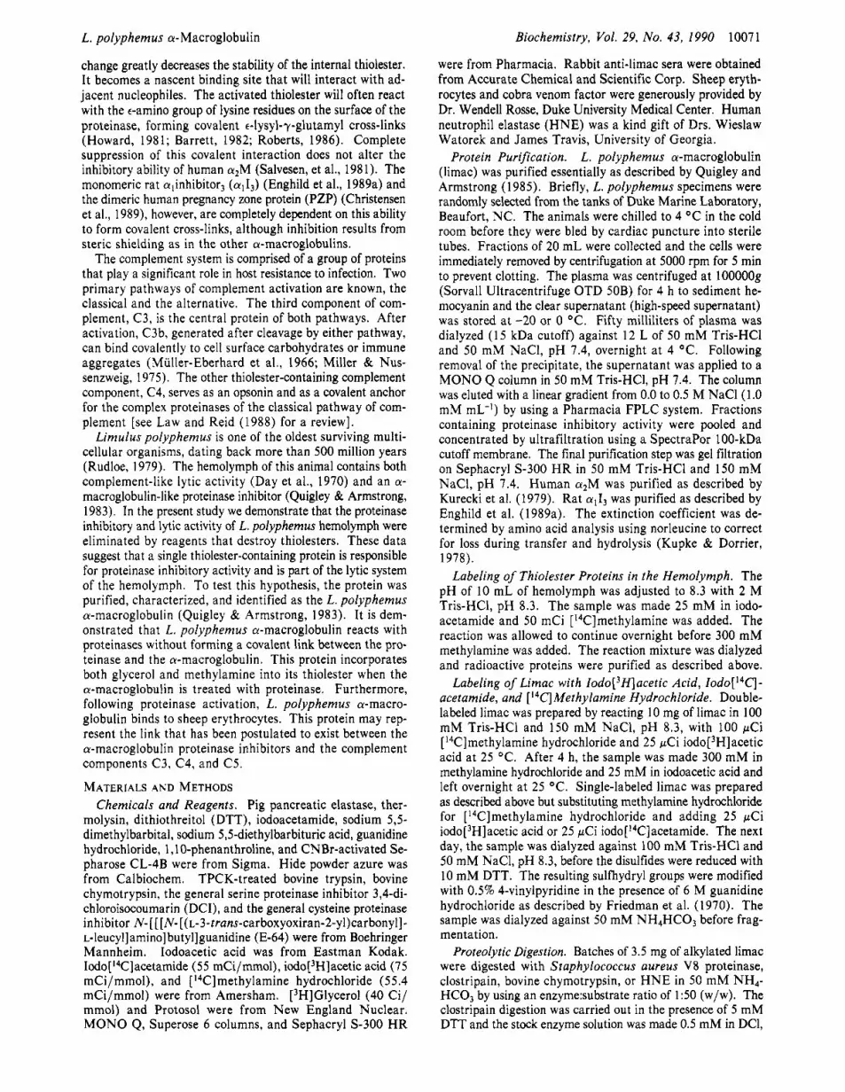
FIGURE 5: Elcctron micrographs of limac (A) and purified limac-trypsin complex (B). Protein was adsorbed to carbon films and negatively stained with uranyl acetate. Photomicrographs are shown at a magnification of 1 12000. The upper panels show representative fields. The arrows in panel A show molecules with the apparent globular structures discussed in the text. The composite of individual molecules at the bottom of the figure includes four images of native limac (left) and four images of limac-trypsin complex (right). The high-magnification images of limac-trypsin were selected to show "doughnut" and "number 8" structures described in the text. In the representative fields and in the high-magnification composite, the bars represent 20 nm.
be very asymmetric, with a larger Stokes radius than would be expected for a globular protein of this size. The confor- mational change induced by trypsin was further analyzed by electron microscopy.
Electron Microscopy. Limac underwent a major con for- mational change after reaction with trypsin, as determined by electron microscopy. Images of the native protein showed a highly elongated, irregularly shaped structure that oriented somewhat variably on the carbon film (Figure 5 ) . Some projections showed three distinct stain-excluding apparent globular structures oriented in a chain (see arrows in panel A). This structural organization does not imply the existence of three separate subunits (polypeptide chains) or support a specific organization of subunits. A similar triglobular structure has been reported for fibrinogen, which consists of six separated polypeptide chains (Hall & Slayter, 1959).
The average maximum length of native limac (measured from apex to apex of the stain-excluding area) was 26.7 f 3.7 nm (n = 25). This value is an estimate that does not account for the irregular folding of the protein. After reaction with trypsin, limac adopted a "compacted structure". The molecular diameter (maximum length) was decreased to 14.5 f 1.4 nm (n = 25). Some molecules showed one or two stain-including regions centrally, yielding images suggestive of a doughnut
or the number "8". These electron microscopy studies suggest that the limac conformational change involves a "rodlike" to globular structure transition.
This large change in shape was also observed by pore-limit gel electrophoresis as a characteristic rapid migration after reaction with proteinase or methylamine (Figure 1). We conclude that limac undergoes a conformational change much greater than that of human a2M. Moreover, the conforma- tional change did not result in exposure of determinants rec- ognized by the mammalian a2M receptor (data not shown).
It has previously been demonstrated that the proteinase retains activity against low molecular weight substrates following binding to limac. This indicates that the inhibitory mechanism is similar to that of other a-macroglobulin proteinase inhibitors (Quigley & Armstrong, 1983). In the present study 1251-labeled proteinase was reacted with limac to probe the covalent linking reaction. Unbound enzyme was removed by gel filtration, following which the sample was analyzed by pore-limit gel electropho- resis, SDS-PAGE, and autoradiography. The pore-limit gels revealed that the radioactive proteinase was associated only with the more rapidly migrating limac band (Figure 6). 1251-Labeled bovine chymotrypsin, bovine trypsin, pig pan- creatic elastase, and thermolysin were reacted with limac and
Limac-Proteinase Interaction.

Enghild et al. 10076 Biochemistry, Vol. 29, No. 43, 1990
o b c d e f g o b c d e f g
232- 140- -
FIGURE 6: Nondenaturing pore-limit gel electrophoresis of limac- proteinase complexes. Electrophoresis was performed on a 4-2096 gradient gel as described under Materials and Methods. Limac was incubated with 1251-labeled trypsin 1:0, l : i /m, l : i /16, I:'/*, 1 : 1 / 2 mol/mol (lanes b-g). Left panel: Coomassie blue stained gel. As is evident from the autoradiography of the same gel (right panel), proteinase is associated only with the faster migrating band. The standard proteins thyroglobulin, ferritin, catalase, and lactate de- hydrogenase are shown in lane a.
o b c d e f 9
- - - 190- - - 97- - 66- m
43- m
31 - - 21 -- 14-o
d e f g
~ --..____-. - FIGURE 7: SDS-PAGE of limac-proteinase complexes. Electro- phoresis was performed on a 5-1 5% gradient gel as described under Materials and Methods. Molecular weight standards are shown in lane a. Unreduced limac (lane b) and reduced limac (lane c) before reaction with proteinase limac after incubation with the '251-labeled proteinases bovine chymotrypsin (lane c), bovine trypsin (lane d), pig pancreatic elastase (lane 9, and thermolysin (lane g), analyzed under reducing conditions. Left panel: Coomassie blue stained gel. As is evident from the autoradiograph of the same gels (right panel), proteinase shows little or no covalent association with limac.
the complex was separated by SDS-PAGE under both re- ducing (Figure 7) and nonreducing conditions (data not shown). Less than 1 % of the radioactivity was associated with bands originating from limac (Fig. 7), although all the pro- teinases tested were inhibited, forming equimolar complexes (Table I). We conclude that covalent proteinase cross-linking is not part of the inhibitory mechanism of limac.
Protein Sequence Analysis of the Thiolester. Limac was reacted with [ i4C]methylamine and [3H]iodoacetamide. After reduction and alkylation, the protein was fragmented with S. aureus V8 proteinase, chymotrypsin, human neutrophil elas- tase, or clostripain. The radioactive peptide(s) was (were) purified on HPLC and submitted to automated Edman deg- radation. The results are presented in Figure 8. During Edman degradation of the putative thiolester sequence, -[3H ]Cy~-Gly-Glu-[~~C]Xxx-, radioactivity was released as indicated. This is consistent with the reactivity of an a-ma- croglobulin thiolester. The amino-reactive residue, here labeled Xxx, is encoded as Gln in rat a l M and other thiolester-con- taining a-macroglobulins (Figure 8). The limac thiolester
CHVYO CLOSIRTPAN ve C , I V Y o
MI OSAROVVS I TGOLMOPA I KNLOHLVRL P T G C G E Z N M I K F V P N I F V L O V L T A T O S I T O HIE
L h U C m
OM1 T V l A V V 0 1 OWEK GMT[TVIAV V L D TEOWEK A M T H T V I A V V L D TEOWOK GMT T V l A V Y V L I T L A A S Y OKTEOWST Y L I T L T A S V D TEOWSK V L I T L T A S I V DRT OWSK S Wq V E T G N H W N I S I 4 I I H W N I
9) Wman C3
FIGURE 8: Thiolester sequence of limac compared to those of other members of the a-macroglobulin family. The 57 amino acids around the thiolester in limac are compared with the thiolester region of other a-macroglobulins. The limac sequence was generated by protein sequence analysis of peptides derived from digestion with S. aureus V8 proteinase (V8), bovine chymotrypsin (chymo), human neutrophil elastase (HNE), or clostripain. The rat a l M sequence was established by cDNA sequencing (the complete amino acid sequence derived from cDNA sequencing will be the subject of a full-length manuscript by G. Eggertsen, G. Hudson, B. R. Shields, D. Reed, K. Lonberg-Holm, B. F. Tack, and G. H. Fey). The other a-macroglobulin sequences were from ( I ) Sottrup-Jensen et al. ( 1 984) and Kan et al. ( 1 985), (2) Sand et al. (1989, (3) Bjork and Jornvall (l986), (4) Gehring et al. (1987), (5) Braciak et al. (1988) and Aiello et al. (1988), (6) Nagase and Brew (1987), (7) Spycher et al. (1987), (8) Hall et al. (l989), (9) de Bruijn and Fey (1989, ( IO) Wetsel et al. (l984), ( I 1 ) Kusano et al. ( 1986), ( 1 2) Thomas and Tack ( 1 983), ( 1 3) Belt et al. ( 1984), ( 14) Nonaka et al. ( 1985), ( I 5) Nonaka et al. ( 1984), ( 16) Wetsel et al. ( 1 988), and ( 1 7) Wetsel et al. ( 1987). Amino acids that are identical with those in limac are boxed.
sequence showed regions of identity with both the a-macro- globulin proteinase inhibitors and the complement factors. In Figure 8, 57 amino acids around the putative thiolester of 19 a-macroglobulins were aligned. The overall identity between human a2M and limac was 51%. The identity between human C3 and limac was 30%, slightly more similar than human C3 and human a2M (24%). The sequence LXXXPXG- CGEQNMXXXXPXXXXXXYL was highly conserved among all the a-macroglobulins and may represent a thiolester consensus sequence. We conclude that the limac contains a putative thiolester showing extensive sequence identity with this region in C3, C4, and C5, and the a-macroglobulin in- hibitors.
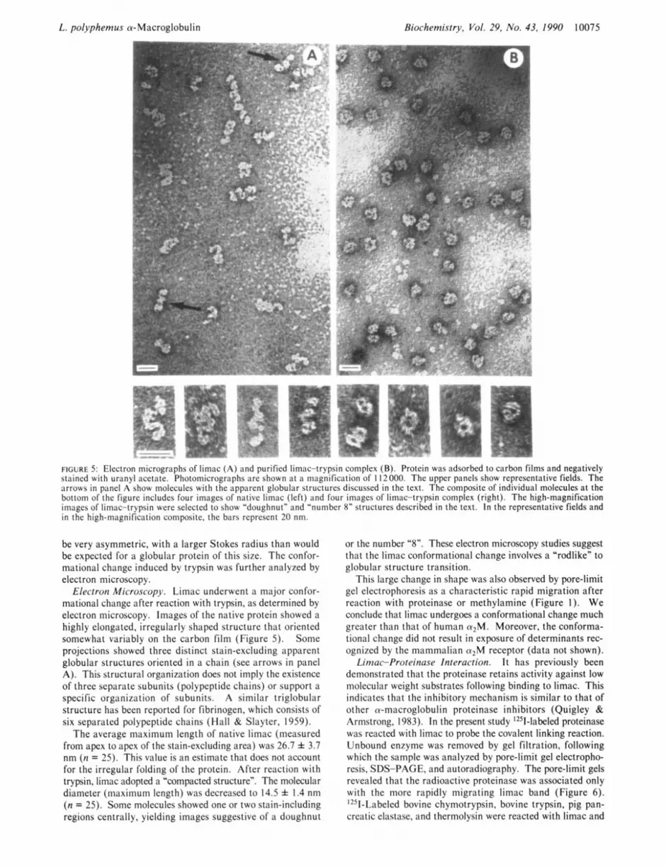
Hemolytic Activity. L. polyphemus hemolymph possesses the ability to lyse erythrocytes, indicating the presence of a primitive lytic system (Day et al., 1970). To investigate whether limac participates in this system, a hemolytic assay was employed. The hemolytic activity present in the hemo- lymph was labile under certain conditions. The activity was greatly diminished by freezing, but if stored on ice in poly- propylene tubes the activity was stable for 2 weeks. Fur- thermore, the activity was completely abolished if the hemo- lymph was passed through or incubated with Sepharose CL-4B or Sephadex (3-25. The activity was destroyed within 5-10 min as judged by batch adsorption followed by hemolytic assays. If hemolymph was transferred to a dialysis membrane (Spectrapore) and kept in a humid environment, but without submersing the membrane in dialysis buffer, the activity was also destroyed. The reason for this is unknown but two ex- planations seem feasible. Either components in the hemolymph necessary for the lytic activity adsorbed to the membrane or the column material, or the lytic system is activated and thereby depleted. This prohibited us from using affinity-pu- rified limac antibodies coupled to Sepharose CL-4B to deplete the hemolymph. We found that the most gentle way to pre- pare limac-depleted hemolymph was by incubating with 10 mM methylamine for 24 h at 25 "C. This material was de-
L. polyphemus a-Macroglobulin
60
50
40
VI In r .- -
30 2 e
20
i
0 20 40 60 B O 100
L. polyphemus hemolymph [pll FIGURE 9: Hemolytic activity of limac. Sheep erythrocytes treated with the hemolymph of L. polyphemus (B) demonstrating the ability to lyse the erythrocytes. Sheep erythrocytes treated with hemolymph in the presence of 50 mM EDTA (VI. Similar results were obtained if the hemolymph was boiled for 5 min or incubated with Sepharose. Treatment of the hemolymph with 10 mM methylamine for 24 h demonstrating significant depletion of the lytic activity (A). The activity was significantly restored by adding limac back to the depleted hemolymph (+). Addition of human cu2M to depleted hemolymph showed no restoration or activity.
pleted with respect to lytic (Figure 9) and proteinase inhibitory activity. However, as determined by DTNB titration, me- thylamine reacts with the thiolester of limac at least 10 times more slowly than observed with human a2M. This observation accounts for the residual hemolytic activity of 10 mM me- thylamine-treated hemolymph (Figure 9). Adding back pu- rified limac restored a significant amount of the lytic activity, while human crzM added as a control had no effect (Figure 9). The lytic activity of the hemolymph was completely abolished when the assay was performed in the presence of 50 mM EDTA. Boiling the hemolymph for 5 min had the same effect. These observations support the hypothesis that limac participates in the lytic system in L . polyphemus he- molymph.
Incorporation of [3H]Glycerol into the Thiolester of Limac. Limac and human a 2 M were activated with H N E in the presence of [3H] glycerol. Under the reaction conditions em- ployed, limac incorporated approximately 0.1 mol of [3H]- glycerol/mol of limac subunit while human a2M incorporated approximately 0.01 mol of [3H]glycerol/mol of a2M subunit.
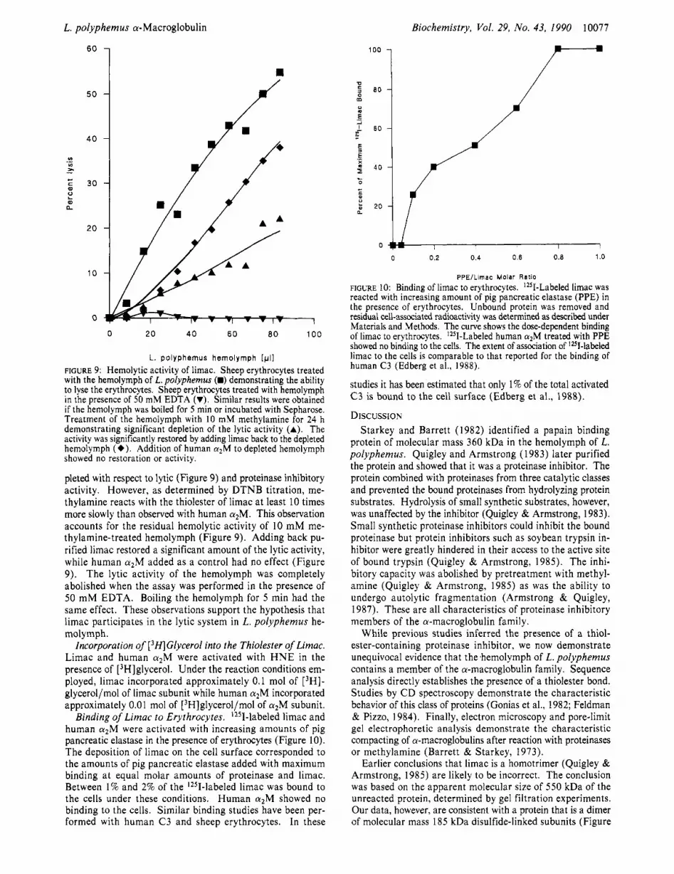
Binding of Limac to Erythrocytes. '251-labeled limac and human a z M were activated with increasing amounts of pig pancreatic elastase in the presence of erythrocytes (Figure 10). The deposition of limac on the cell surface corresponded to the amounts of pig pancreatic elastase added with maximum binding at equal molar amounts of proteinase and limac. Between 1 % and 2% of the '251-labeled limac was bound to the cells under these conditions. Human a 2 M showed no binding to the cells. Similar binding studies have been per- formed with human C3 and sheep erythrocytes. In these
Biochemistry, Vol. 29, No. 43, 1990 10077
100
0 s 80 m u .- 2
5
-I I 60 N - E .- X
40 F 0
c 0
e
- kl 20
0 0 0.2 0.4 0.6 0.8 1 .o
PPEIL imac Molar Rat io
FIGURE 10: Binding of limac to erythrocytes. IZ5I-Labeled limac was reacted with increasing amount of pig pancreatic elastase (PPE) in the presence of erythrocytes. Unbound protein was removed and residual cell-associated radioactivity was determined as described under Materials and Methods. The curve shows the dose-dependent binding of limac to erythrocytes. 'Z51-Labeled human cuzM treated with PPE showed no binding to the cells. The extent of association of lzI-labeled limac to the cells is comparable to that reported for the binding of human C3 (Edberg et ai., 1988).
studies it has been estimated that only 1% of the total activated C3 is bound to the cell surface (Edberg et al., 1988).
DISCUSSION Starkey and Barrett (1982) identified a papain binding
protein of molecular mass 360 kDa in the hemolymph of L. polyphemus. Quigley and Armstrong (1983) later purified the protein and showed that it was a proteinase inhibitor. The protein combined with proteinases from three catalytic classes and prevented the bound proteinases from hydrolyzing protein substrates. Hydrolysis of small synthetic substrates, however, was unaffected by the inhibitor (Quigley & Armstrong, 1983). Small synthetic proteinase inhibitors could inhibit the bound proteinase but protein inhibitors such as soybean trypsin in- hibitor were greatly hindered in their access to the active site of bound trypsin (Quigley & Armstrong, 1985). The inhi- bitory capacity was abolished by pretreatment with methyl- amine (Quigley & Armstrong, 1985) as was the ability to undergo autolytic fragmentation (Armstrong & Quigley, 1987). These are all characteristics of proteinase inhibitory members of the a-macroglobulin family.
While previous studies inferred the presence of a thiol- ester-containing proteinase inhibitor, we now demonstrate unequivocal evidence that the ,hemolymph of L. polyphemus contains a member of the a-macroglobulin family. Sequence analysis directly establishes the presence of a thiolester bond. Studies by CD spectroscopy demonstrate the characteristic behavior of this class of proteins (Gonias et al., 1982; Feldman & Pizzo, 1984). Finally, electron microscopy and pore-limit gel electrophoretic analysis demonstrate the characteristic compacting of a-macroglobulins after reaction with proteinases or methylamine (Barrett & Starkey, 1973).
Earlier conclusions that limac is a homotrimer (Quigley & Armstrong, 1985) are likely to be incorrect. The conclusion was based on the apparent molecular size of 550 kDa of the unreacted protein, determined by gel filtration experiments. Our data, however, are consistent with a protein that is a dimer of molecular mass 185 kDa disulfide-linked subunits (Figure

10078
3). Since limac has an asymmetric conformation (Figure 5 ) , it probably has a larger Stokes radius than would be expected for a protein of this size. Such asymmetric proteins typically behave in an anomalous manner when studied by gel filtration and pore-limit gel electrophoresis. After methylamine or proteinase treatment, limac undergoes a conformational change (Figure 4 and 5 ) that results in a more globular form (Figure 5 ) . The apparent molecular mass of this molecule is 360 kDa, which is similar to that of other a-macroglobulin dimers. When limac proteinase complexes were analyzed by pore-limit gel electrophoresis, the proteinase was only associated with the rapidly migrating band (Figure 6). No multimerization of the proteinase-complexed material was detected. This is in contrast to the dimeric human a-macroglobulin homologue pregnancy zone protein (PZP), which forms tetramers when reacted with proteinase (Christensen et al., 1987).
Most a-macroglobulin proteinase inhibitors contain a pu- tative internal thiolester, which is often detected by its ability to covalently incorporate small nucleophiles such as methyl- amine (Swenson & Howard, 1979; Tack et al., 1980; Sott- rup-Jensen et al., 1980; Janatova et al., 1980). Limac in- corporated radiolabeled iodoacetic acid and methylamine during methylamine treatment and the location of incorporated radioactivity was consistent with the reactivity of a functional thiolester as determined by amino acid sequence analysis (Figure 8). The sequence was similar to the thiolester region of other a-macroglobulins. The 57 amino acid stretch was more similar to the proteinase inhibitors than to the comple- ment components. It might be difficult from these sequence data to reach any firm conclusions regarding degree of rela- tionship. Overall, limac was more similar to the a-macro- globulin inhibitors. On the other hand, the similarity between human C3 and limac was slightly greater than the identity between human C3 and human a2M, suggesting that limac is more related to the complement components than is human
During proteinase-induced conformational change, a-ma- croglobulin thiolesters become extremely reactive with a broad range of nucleophiles. Often, the proteinase itself becomes covalently bound to the a-macroglobulin inhibitor via lysine residues on the surface (Sottrup-Jensen, 1987). The extent of covalent linking varies from 20 to 100% (Sottrup-Jensen, 1987). Since suppression of the covalent interaction does not alter the ability of human a2M to inhibit proteinases (Salvesen et al., 1981), the function of this thiolester remains unclear. Moreover, chicken ovostatin contains no thiolester, but it is still able to inhibit a number of proteinases (Nagase et al., 1983). In contrast, the proteinase inhibitory activity of the monomeric a-macroglobulin rat a , I, and the dimeric human pregnancy zone protein (PZP) depends completely on covalent linking for inhibition (Enghild et al., 1989a; Christensen et al., 1989). We have speculated that the thiolester is a pre- requisite for monomeric a-macroglobulin (Enghild et al., 1989a) but redundant in tetramers such as human aZM and chicken ovostatin. It is also presumably redundant for in- hibition by limac since less than 1% of the proteinases tested were covalently bound (Figure 7). This led us to consider an alternative function for the limac thiolester.
The ability of L. polyphemus hemolymph to lyse erythro- cytes in a complement-like manner was first demonstrated by Day et al. (1970). This claim was later disputed by Hall et al. (1972) on the basis of experiments with lobster serum (clotted hemolymph). We found that the hemolytic activity in the L. polyphemus hemolymph was depleted during clotting. Consequently, conclusions about lytic activity cannot be drawn
Biochemistry, Vol. 29, No. 43, 1990
a2M.
Enghild et al.
from experiments on clotted hemolymph. Day et al. (1970) initiated the lytic system with cobra venom factor. We did not find this necessary. As with the human alternative pathway of complement (Platts-Mills & Ishizaha, 1974), the L. polyphemus lytic system is activated spontaneously by foreign erythrocytes (Figure 9). The lytic activity was de- stroyed if hemolymph came in contact with column materials such as Sepharose CL-4B or dialysis membranes (Figure 9). Significantly, the alternative pathway of the human comple- ment system is also activated by Sepharose, resulting in de- pletion of several lytic factors (Goldstein et al., 1976). Perhaps a similar mechanism is responsible for the depletion of the lytic activity in hemolymph.
In L. polyphemus hemolymph, ['4C]methylamine was in- corporated into only one protein, limac, suggesting that it is the only thiolester-containing protein in the hemolymph. Since the lytic activity was destroyed when the hemolymph was treated with methylamine, we reason that limac participates in the lytic system. Immunodepletion, desalting, and dialysis could not be employed to prepare limac-depleted hemolymph; however, the lytic activity could be depleted by incubation with methylamine. When purified limac was added to methyl- amine-depleted hemolymph the lytic activity was restored (Figure 9).
We also studied the ability of limac thiolesters to react with [3H]glycerol. When treated with HNE, limac subunits in- corporated IO-fold more ['H]glycerol than did human a2M subunits. [3H]Glycerol is characteristically incorporated into the thiolester of human C3 but not a 2 M (Law, 1983; Dodds & Law, 1988; Law & Reid, 1988). The rate of reaction of proteinase-treated human C3 with [3H]glycerol is about 19- fold greater than observed with human aZM (Dodds & Law, 1988). Depending on reaction conditions, the human C3 thiolester can incorporate from 0.034 to 0.162 mol of [3H]- glycerol/mol of C3 (Law, 1983). In our studies, limac in- corporated approximately 0.1 mol of [3H]glycerol/mol of limac subunit, while human a 2 M incorporated no more than 0.01 mol of [3H]glycerol/mol of aZM subunit. These data, together with the failure of limac to form covalent cross-links with proteinases, support the hypothesis that the proteinase inhibitor limac must also function as part of a complement-like lytic system in L. polyphemus hemolymph.
The key step in distinction of self and nonself by the mam- malian complement system is the thiolester-mediated binding of C3 to foreign particles. Significantly, limac also bound to erythrocyte in a dose-dependent manner when activated with proteinase (Figure IO). The binding of limac to sheep erythrocytes is quantitatively similar in extent to the of binding observed in similar experiments with human C3 (Edberg et al., 1988).
We can only speculate as to whether the lytic system of L. polyphemus is evolutionarily related to the mammalian com- plement system; however, it does share several distinct features with the mammalian alternative pathway of complement. The lytic activity (1) is abolished by EDTA, ( 2 ) is activated by foreign cell surfaces or Sepharose, (3) is not antibody de- pendent, and (4) depends on a thiolester-containing protein. Our data suggest that at some stage during protein evolution, an a-macroglobulin with complement-like lytic activity and proteinase inhibitory activity was present. These distinct activities are now apparently separated in most animals but captured in a single protein in the ancient horseshoe crab.
ACKNOWLEDGMENTS We thank Dr. Joseph Bonaventura and Mr. Gerald Godette
of Duke Marine Laboratory for providing L. polyphemus and

L. polyphemus a-Macroglobulin
facilities, Mr. Frederic Brucato for assisting with bleeding of the animals, Dr. Wayne Beyer for performing CD measure- ments, Mr. David Rubenstein and Dr. Wendell Rosse for reading the manuscript, and Ms. Andrea Tillotson for prep- aration of the manuscript.
Registry No. Proteinase, 9001 -92-7; methylamine, 74-89-5; glycerol, 56-8 1-5.
Biochemistry, Vol. 29, No. 43, 1990 10079
Kan, C.-C., Solomon, E., Belt, K. T., Chain, A. C., Hiorns, L. R., & Fey, G. (1985) Proc. Natl. Acad. Sci. U.S.A. 82,
Kupke, D. W., & Dorrier, T. E. (1978) Methods Enzymol.
Kurecki, T., Kress, L., & Laskowski, M. (1979) Anal. Bio- chem. 99, 4 15-420.
Kusano, M., Choi, N.-H., Tomita, M., Yamamoto, K.-i., Migita, S . , Sekiya, T., & Nishimura, S . (1986) Immunol. Invest. 15, 365-378.
Law, S.-K. A. (1983) Ann. N.Y. Acad. Sci. 421 , 246-258. Law, S.-K. A., & Reid, K. B. M. (1988) in Complement
(Male, D., Ed.) IRL Press Limited, Oxford. Lonberg-Holm, K., Reed, D. L., Roberts, R. C., Hebert, R.
R., Hillman, M. C., & Kutney, R. M. (1987) J . Biol. Chem.
2282-2286.
48, 155-162.
262, 438-445. Manwell, C. (1977) Biochem. J . 165, 487-495. Matsudaira, P. (1987) J . Biol. Chem. 262, 10035-10038. Meltzer, N. M., Tour, G. I., & Stein, S . (1987) Anal. Bio-
Miller, G. W., & Mussenzweig, V. (1975) Proc. Natl. Acad.
Muller-Eberhard, H. J., Dalmasso, A. P., & Calcott, M. A.
Nagase, H., & Brew, K. (1987) FEBS Lett. 222, 83-88. Nagase, H., Harris, E. D., Jr., Woessner, J. F., Jr., & Brew,
K. (1983) J . Biol. Chem. 258, 7481-7489. Nonaka, M., Takahashi, M., Natsuume-Sakai, S . , Nonaka,
M., Tanaka, S . , Shimizu, A,, & Honjo, T. (1984) Proc. Natl. Acad. Sci. U.S.A. 81, 6822-6826.
Nonaka, M., Nakayama, K., Yeul, Y. D., & Takahashi, M. (1985) J . Biol. Chem. 260, 10936-10943.
Pangburn, M. K., Schreiber, R. D., & Muller-Eberhard, H. J. (1983) J . Immunol. 131, 1930-1935.
Platts-Mills, T. A. E., & Ishizaka, K. (1974) J . Immunol. 113,
Quigley, J. P., & Armstrong, P. B. (1983) J . Biol. Chem. 258,
Quigley, J. P., & Armstrong, P. B. (1985) J . Biol. Chem. 260,
Roberts, R. C. (1986) Rev. Hematol. 2, 129-224. Rudloe, A. (1979) in Biomedical Applications of the Horse
Shoe Crab (Cohen, E., Ed.) pp 27-35, Alan R. Liss, New York.
Salvesen, G., & Nagase, H. (1989) in Proteolytic Enzymes; A Practical Approach (Beynon, R., & Bond, J., Eds.) IRL Press Ltd., Oxford.
Salvesen, G., Sayers, C. A., & Barrett, A. J. (1981) Biochem.
Salvesen, G., Farley, J., Przybyla, A., Reilly, C., & Travis, J . (1987) Biochemistry 26, 2289-2293.
Sand, O., Folkersen, J., Westergaard, J. G., & Sottrup-Jensen, L. (1985) J . Biol. Chem. 260, 15723-15735.
Sottrup-Jensen, L. (1987) in The Plasma Proteins (Putnam, F. W., Ed.) Vol. V, pp 191-291, Academic Press, New York.
Sottrup-Jensen, L. (1989) J . Biol. Chem. 264, 11539-1 1542. Sottrup-Jensen, L., Petersen, T. E., & Magnusson, S . (1980)
FEBS Lett. 121, 275-279. Sottrup-Jensen, L., Stepanik, T. M., Kristensen, T., Weirz-
bicki, D. M., Jones, C. M., Lanblad, P. B., Magnusson, S . , & Petersen, T. E. (1984) J . Biol. Chem. 259, 8318-8327.
Sottrup-Jensen, L., Stepanik, T. M., Kristensen, T., Lanblad, P. B., Jones, C . M., Wierzbicki, D. M., Magnusson, S . , Domdey, H., Wetsel, R. A., Lundwall, A., Tack, B. F., &
chem. 160, 356-361.
Sci. U.S.A. 72, 418-422.
(1966) J . Exp. Med. 123, 33-54.
348-358.
7903-7906.
127 15-127 19.
J . 195, 453-461.
REFERENCES
Aiello, L. P., Shia, M. A., Robinson, G. S . , Pilch, P. F., & Farmer, S . R. (1988) J . Biol. Chem. 263, 4013-4022.
Armstrong, P. B., & Quigley, J. P. (1987) Biochem. J . 248,
Barrett, A. J . (1982) Methods Enzymol. 80, 737-754. Barrett, A. J., & Starkey, P. M. (1973) Biochem. J . 133,
Belt, K. T., Carroll, M. C., & Porter, R. R. (1984) Cell 36,
Bjork, I . , & Fish, W. W. (1982) Biochem. J . 207, 347-356. Bjork, I., & Jornvall, H. (1986) FEBS Lett. 205, 87-91. Braciak, T. A., Northemann, W., Hudson, G. O., Shiels, B.
R., Gehring, M. R., & Fey, G. H. (1988) J . Biol. Chem.
703-707.
709-724.
907-9 1 4.
263, 3999-401 2. Bury, A. F. (1981) J . Chromatogr. 213, 491-500. Christensen, U., Simonsen, M., Harrit, M., & Sottrup-Jensen,
David, D. S., & Reisfeld, R. A. (1974) Biochemistry 13,
Day, N. K. B., Gewurz, H., Johannsen, R., Finstak, J., &
de Bruijn, M. H. L., & Fey, G. H. (1985) Proc. Natl. Acad.
Dodds, A. W., & Law, A. (1988) Complement 5 , 89-97. Edberg, J. C., Tosie, L., Wright, E. L., Sutherland, W. M.,
& Taylor, R. P. (1988) J . Immunol. 141, 4258-4265. Ellman, G. L. (1959) Arch. Biochem. Biophys. 82, 70-77. Enghild, J. J., Salvesen, G., Thagersen, I. B., & Pizzo, S . V.
Enghild, J. J., Thagersen, I. B., Roche, P. A,, & Pizzo, S . V.
Feldman, S. R., & Pizzo, S. V. (1984) Arch. Biochem. Bio-
Freidman, M., Krull, L. H., & Cavins, J. F. (1970) J . Biol. Chem. 245, 3868-3871.
Gehring, M. R., Shiels, B. R., Northemann, W., de Bruijn, M. H. L., Kan, C-C., Chain, A. C., Noonan, D. J., & Fey, G. H. (1987) J . Biol. Chem. 262, 446-454.
Goldstein, I. M., Kaplan, H. B., Radin, A., & Frosch, M. (1976) J . Immunol. 117, 1282-1287.
Gonias, S . L., & Figler, N. L. (1989) J . Biol. Chem. 264,
Gonias, S . L., Reynolds, J. A., & Pizzo, S . V. (1982) Biochim. Biophys. Acta 705, 306-3 14.
Gonias, S . L., Allietta, M. M, Pizzo, S . V., Castellino, F. S . , & Tillack, T. W. (1988) J. Biol. Chem. 263, 10903-10906.
Hall, C . , & Slayter, H. (1959) J . Biophys. Biochem. Cytol.
Hall, J . L, Rowlands, D. T., Jr., & Nilsson, U. R. (1972) J .
Hall, M., Soderhall, K., & Sottrup-Jensen, L. (1989) FEBS
Howard, J. B. (1981) Proc. Natl. Acad. Sci. U.S.A. 870,
Janatova, J., Lorentz, P. E., Schechter, A. N., Prahal, J. W.,
L. ( 1 989) Biochemistry 28, 9324-9336.
1 0 1 4-1 02 1.
Good, R. A. (1970) J . Exp . Med. 132,941-950.
Sci. U.S.A. 82, 708-712.
(1989a) J . Biol. Chem. 264, 11428-11435.
(1 989b) Biochemistry 28, 1406-1 41 2.
phys. 235, 267-275.
9565-9570.
5, 11-16.
Immunol. 109, 816-823.
Lett. 254, 11 1-1 14.
2235-2239.
& Tack, B. F. (1980) Biochemistry 19, 4471-4478.

10080 Biochemistry 1990, 29, 10080-1 0089
Fey, G. H. (1985) Proc. Natl. Acad. Sci. U.S.A. 82, 9-13. Spycher, S . E., Arya, S . , Isenman, D. E., & Painter, R. H.
(1987) J . Biol. Chem. 262, 14606-14611. Starkey, P. M., & Barrett, A. J. (1982) Biochem. J. 205,
Strickland, D. K . , Steiner, J. P., Feldman, S . R., & Pizzo, S . V. (1984) Biochemistry 23, 6679-6685.
Swenson, R. P., & Howard, J . B. (1979) Proc. Nat l . Acad. Sci. U.S.A. 76, 43 13-43 16.
Tack, B. F., Harrison, R. A., Janatova, J., Thomas, M. L., & Prahl, J . W. (1980) Proc. Nat l . Acad. Sei. U.S.A. 77,
91-95.
5 764-5 768.
Thomas, M. L., & Tack, B. F. (1983) Biochemistry 22,
Travis, J., & Salvesen, G. S . (1983) Annu. Rev. Biochem. 52,
Wetsel, R. A., Lundwall, A., Davidson, F., Gibson, T., Tack, B. F., & Fey, G. H. (1984) J . Biol. Chem. 259,
Wetsel, R. A., Ogata, R. T., & Tack, B. F. (1 987) Biochem- istry 26, 737-743.
Wetsel, R. A., Lemons, R. S . , Le Beau, M. M., Barnum, S . R., Noack, D., & Tack, B. F . (1988) Biochemistry 27,
942-947.
655-709.
13857-13862.
1474-1482.
Synthesis and Characterization of Extended and Deleted Recombinant Analogues of Parathyroid Hormone-( 1-84): Correlation of Peptide Structure with Function?
Shafaat A. Rabbani,l Stephanie M. Kaiser,* Janet E. Henderson,* Suzanne M. Bernier,s Andrew J. Modand,% Denis R. Roy, Diana M. Zahab, Wing L. Sung, David Goltzman, and Geoffrey N. Hendy*vil
Calcium Research Laboratory, Departments of Medicine and Physiology, McCill University and Royal Victoria Hospital, Montreal, Quebec, Canada H3A 1 A l , and Institute for Biological Sciences, National Research Council of Canada, Ottawa,
Ontario, Canada K I A OR6 Received May 14, 1990; Revised Manuscript Received July 13, 1990
ABSTRACT: Recombinant analogues of human parathyroid hormone [hPTH-( 1-84)] were expressed in Escherichia cofi harboring plasmids containing synthetic genes under the control of the lac promoter. The level of expression of the gene encoding the truncated analogue, hPTH-(3-84), was greater than that of the gene encoding full-length hPTH-( 1-84) but less than that of the gene encoding proparathyroid hormone (hProPTH). This may be due in part to the relative efficiency of translation of the m R N A as suggested by secondary structure analysis and in part because of enhanced stability of the extended peptide. For- mylmethionyl derivatives of hProPTH and of hPTH-( 3-84) and underivatized hPTH-(3-84) were purified by HPLC, and their identity was confirmed by NH2-terminal sequencing and amino acid analysis. The bioactivity of these recombinant peptides was then tested in skeletal and renal adenylate cyclase assays in vitro and in assays examining effects on plasma and urine calcium and phosphate levels and on urine cyclic A M P levels in vivo. The NH2-terminally extended analogue fMet-hProPTH displayed 10% of the in vitro activity of hPTH-( 1-84) and was a partial agonist in vivo. The peptides hPTH-(3-84) and fMet-hPTH-(3-84) were inert in vitro and were very weak in vitro antagonists when compared to the NH2-terminal analogue bovine [Nle8J8Tyr34] PTH-( 3-34)-NH2. In vivo, hPTH-(3-84) and the bPTH-(3-34) analogue, when assayed at a 1O:l molar ratio relative to bPTH-(1-84), were each inert, and neither demonstrated antagonist activity at these concentrations. The results demonstrate a variation in expression levels of synthetic genes encoding deleted, full-length, and extended forms of hPTH-(1-84) and show that either NH2-terminal or COOH- terminal extension of the active region of the P T H molecule may produce conformational changes which apparently alter receptor binding and reduce both agonist and antagonist activity.
E r a t h y r o i d hormone (PTH)' (Potts et al., 1982; Goltzman & Hendy, 1990) interacts with renal and skeletal receptors to regulate extracellular calcium and phosphate concentrations at least in part through stimulation of the adenylate cyclase system. Although the major glandular, and secreted, bioactive form of the hormone is an 84 amino acid straight-chain pep-
tide, PTH-(1-84), the active core of the molecule is believed to reside within the NH2-terminal sequence 1-27 (Tregear et al., 1973), and to date, studies correlating PTH structure with function have employed mainly analogues of the synthetic NH,-terminal peptide, PTH-( 1-34). The latter peptide in- teracts with specific, high-affinity receptors in renal and os- seous tissue and appears to carry out most of the functions of
+This work was supported by Grants MA-9315 and MT-5775 from the Medical Research Council of Canada. This is NRCC Publication No. 31885.
* T o whom correspondence should be addressed at the Calcium Re- search Laboratory, Royal Victoria Hospital, Room H4.67,687 Pine Ave. W., Montreal, Quebec, Canada H3A 1Al.
$Recipient of a Fellowship Award from the MRC of Canada. *Recipient of a Scholarship Award from the Fonds pour la Formation
de Chercheurs et I'Aide 5 la Recherche du Quebec. Recipient of a Scholarship Award from the MRC of Canada.
the intact molecule. Nevertheless, specific binding sites for the midregion and carboxyl regions of PTH-( 1-84) have been described (McKee & Murray, 1985; Demay et al., 1985), and recent studies have implicated these domains in hormonal effects on cell replication and differentiation (Schluter et al.,
Abbreviations: PTH, parathyroid hormone; h, human; b, bovine; fMet, formylmethionyl; HPLC, high-performance liquid chromatogra- phy; IPTG, isopropyl thiogalactoside.
0006-2960/90/0429- 10080$02.50/0 0 1990 American Chemical Society


![[IMAGE][IMAGE]-Macroglobulin Bait Region Variants](https://img.dokumen.tips/doc/110x75/635d8d00fd007d475b0336aa/imageimage-macroglobulin-bait-region-variants.jpg)

