Embed Size (px)
Citation preview
Original Paper
Indoor and BuiltEnvironment Indoor Built Environ 2004;13:63–74 Accepted: May 7, 2003
Airborne Fungi andActinomycetesConcentrations in the Air ofEskisehir City (Turkey)
Ahmet Asana Semra Ilhanb Burhan Sena
Ismuhan Potoglu Erkarab Cansu Filikb Ahmet Cabukb
Rasime Demirelb Mevlut Turec Suzan Sarica Oktena
Suleyman Tokurb
aTrakya University, Faculty of Arts and Sciences, Department of Biology, TR-22030 Edirne, TurkeybOsmangazi University, Faculty of Arts and Sciences, Department of Biology, TR-26480Meselik, TurkeycTrakya University, Medical Faculty, Department of Biostatics, TR-22030 Erdine, Turkey
Key Words
Fungi E Actinomycetes E Urban air E Biomass E
Fungal and Actinomycetes distribution E Bioaerosols
E Airspora
AbstractThe present study investigated the isolation and iden-
tification of airborne fungi from three different urban
stations located in Eskisehir (Turkey). Air samples were
taken by exposing a Petri dish with Rose-Bengal
streptomycin agar medium for 15min and after incuba-
tion the number of growing colonies was counted. The
sampling procedure for fungi was performed 35 times
at the research stations weekly between March and
November 2001. A total of 2518 fungal and 465
actinomycetes colonies were counted on 420 Petri
plates over a nine-month period. In total, some 20
mould species belonging to 12 genera were isolated.
Alternaria alternata, Cladosporium cladosporioides and
Scopulariopsis brevicaulis were the most abundant
species in the study area (13.66, 5.80 and 5.50% of the
total, respectively). Relationships between fungal spore
numbers, aerosol air pollutants (that is the particulate
matter in the air) and sulphur dioxide together with the
meteorological conditions were examined using statis-
tical analysis. Number of fungi and actinomycetes were
tested by multivariate analysis (MANOVA) according to
the areas and months. Fungal numbers were non-
significant according to the areas and months (p>0.05),
but the number of actinomycetes recorded was sig-
nificant (p<0.01).
Introduction
Living particles such as fungal propagules are ubiqui-
tous in the atmosphere. Fungal spores have even been
detected in the air of Signy Island in the maritime
Antarctic [1]. Fungal spores and other airborne structures
are found both indoors and out. Madelin [2], explained
that fungal aerosols are important in the spread of plant,
animal and human disease. Airborne fungi may cause
allergy in humans [3,4], so they are important from this
� 2004 Sage PublicationsDOI: 10.1177/1420326X04033843Accessible online at:www.sagepublications.com
Prof. Ahmet AsanTrakya University, Faculty of Arts and Sciences, Department of BiologyTR-22030 Edirne, TurkeyTel. þ90 284 2356405, Fax þ90 284 2354010, E-Mail [email protected]
point of view. Some species of fungi such as Cladosporium
cladosporioides, C. herbarum, Penicillium brevicompactum,
P. chrysogenum,Aspergillus candidus,A. niger,A. versicolor,
etc. can provoke extreme allergic reactions in humans [5].
According to Singh [6], mould growth may contribute
to the sick-building syndrome as well as to allergy and
other environmental health problems. Furthermore,
airborne fungi cause spoilage of foods and are responsible
for many adverse health effects; the mycotoxins which they
produce may affect human and animals, and fungal
propagules can serve as an infective agent of plant disease.
In addition, these bioaerosols may cause eye and sinus
irritation, sore throat, headache, fatigue and dizziness [7].
The concentration of airborne micro-organisms are linked
to the level of airborne dust and various human activities.
In addition, the concentration of fungal spores in the air is
linked to both geographical region and seasonal variations,
meaning that it is dependent on wind, humidity, tempera-
ture, rainfall, altitude, vegetation and some specific
reservoirs of contamination [8–10]. Also, the air velocity
above any surface contaminated with moulds, the texture
of that surface, and vibration or other movement of the
contaminated material, will affect the number of fungal
spores released to the atmosphere [11]. Overall, the
prevalence of airborne fungi is highly variable and
determined by many factors.
Nocard (see [12]) recognised the pathogenic potential
of actinomycetes for the first time in 1888 and since then,
several aerobic actinomycetes have been a major source
of materials for the commercial drug industry, some of
which have proved to be useful for the production of new
antimicrobial agents [12]. Reponen et al. [13] noted that
airborne actinomycetes spores are important contaminants
in occupational and residential environments and the
spores of several actinomycetes species have been related
to the occurrence of allergic alveolitis and other health
effects.
There have been very few studies on airborne fungi in
Turkish cities. The studies that have been carried out have
monitored fungi in only a handful of cities including
Istanbul [14], Edirne [15–17], Bursa [18], Ankara [19]
and Izmir [20]. Harmanci et al. [21] determined that
Cladosporium and Aspergillus spores caused allergic reac-
tions in adult patients with asthma and/or rhinitis in
Eskisehir; they also explained that Cladosporium was the
commonest cause. The first study on airborne fungi in
Eskisehir was done by Atik and Tamer in 1994 [22]. These
workers also studied airborne bacteria in this city [23].
However, the city is developing and expanding year by
year and the population and industrial activities are
increasing. As a consequence the earlier work is now out
of date with the airborne fungal and actinomycetes
concentrations effectively unknown. So, the objective
of the present study was the determination of fungal
and actinomycetes concentrations in the different study
areas of the present city of Eskisehir. In particular, so far
as we can ascertain, studies of airborne actinomycetes have
not been made in Turkey although studies have been
carried out in other parts of the world (for example
[13,24]).
The rationale behind the work was that since actino-
mycetes and many fungi are potential allergens the study
may provide the basis for future work on health effects.
Because of the paucity of information of this type from
Turkey, the study has also increased our knowledge of
the distribution of fungal and actinomycetes flora in our
country. Mould allergy is common in Eskisehir city. The
official number of patients who applied to the Osmangazi
University Hospital at Eskisehir for mould allergy tests
are: 948 in 1999, 1185 in 2000, and 1053 in 2001 (Source:
Osmangazi University Hospital records) (Allergens from
several fungi were included in the mould tests: Penicillium
notatum, Cladosporium herbarum, Aspergillus fumigatus
and Alternaria alternata). As an aside, we did have the
expectation that we would record for the first time fungal
species new to Turkey and, perhaps, the world, but this
was not to be.
Materials and Methods
Sampling Sites
The salient features of the research stations, the number
of samples and other relevant information is given in
Table 1.
The concentration of airborne fungal spores and
actinomycetes from three urban areas in Eskisehir City
(1. Osmangazi University Meselik Campus; 2. Anadolu
University Yunusemre Campus; 3. Anadolu University Iki
Eylul Campus) (Figure 1 and Table 1) have been
measured. The first station has rich flora; there are 240
plant species and 41 varieties. Asteraceae and Fabaceae
families are very common. There are 363 plant species at
the second station where the Asteraceae, Fabaceae and
Lamiaceae families are common. The third station is in a
residential area, but some plants species belonging to the
Poaceae, Asteraceae and Fabaceae families are common
[25,26]. The first and second stations are in the north of the
city, but the third station is in the south. Distances
64 Indoor Built Environ 2004;13:63–74 Asan et al.
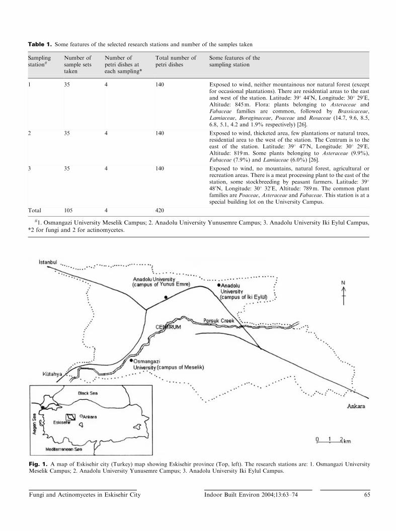
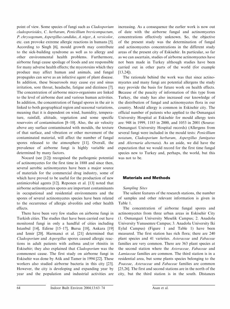
Fig. 1. A map of Eskisehir city (Turkey) map showing Eskisehir province (Top, left). The research stations are: 1. Osmangazi UniversityMeselik Campus; 2. Anadolu University Yunusemre Campus; 3. Anadolu University Iki Eylul Campus.
Table 1. Some features of the selected research stations and number of the samples taken
Samplingstation#
Number ofsample setstaken
Number ofpetri dishes ateach sampling*
Total number ofpetri dishes
Some features of thesampling station
1 35 4 140 Exposed to wind, neither mountainous nor natural forest (exceptfor occasional plantations). There are residential areas to the eastand west of the station. Latitude: 39� 440N, Longitude: 30� 290E,Altitude: 845m. Flora: plants belonging to Asteraceae andFabaceae families are common, followed by Brassicaceae,Lamiaceae, Boraginaceae, Poaceae and Rosaceae (14.7, 9.6, 8.5,6.8, 5.1, 4.2 and 1.9% respectively) [26].
2 35 4 140 Exposed to wind, thicketed area, few plantations or natural trees,residential area to the west of the station. The Centrum is to theeast of the station. Latitude: 39� 470N, Longitude: 30� 290E,Altitude: 819m. Some plants belonging to Asteraceae (9.9%),Fabaceae (7.9%) and Lamiaceae (6.0%) [26].
3 35 4 140 Exposed to wind, no mountains, natural forest, agricultural orrecreation areas. There is a meat processing plant to the east of thestation, some stockbreeding by peasant farmers. Latitude: 39�
480N, Longitude: 30� 320E, Altitude: 789m. The common plantfamilies are Poaceae, Asteraceae and Fabaceae. This station is at aspecial building lot on the University Campus.
Total 105 4 420
#1. Osmangazi University Meselik Campus; 2. Anadolu University Yunusemre Campus; 3. Anadolu University Iki Eylul Campus,
*2 for fungi and 2 for actinomycetes.
Fungi and Actinomycetes in Eskisehir City Indoor Built Environ 2004;13:63–74 65
between the research stations and a meteorological station
were measured by GARMIN GPS 12 CX device (Global
Positioning System; Made in Taiwan, under USA patent).
This device was also used to measure the altitude of the
research stations.
Sampling and Isolation Methods
The samples were taken in the morning (10.00–12.00) at
1.50m above the ground level. The sampling time was
always at the same time of the day. Concentration of
airborne fungi and actinomycetes were conducted over a
period of nine months at different sites (from March to
November 2001). Sampling was performed at one-week
intervals; but we did not sample on rainy days; thus,
counts of airborne microfungi and actinomycetes were
conducted only on days when the weather was stable and
dry. At the end sampling had been carried out for 35 weeks
(of 39) over the nine-month period.
The method used for the isolation of fungi and
actinomycetes was either the Petri plate gravitational
method or the Settle plate method [27,28]. Rose Bengal
streptomycin agar medium was used for isolation of the
fungi while Czapek’s solution agar (CZ) (Merck,
Germany) with cycloheximide (50 mg �mL�1) was used
for actinomycetes. The agar medium for the fungi was
made from: Dextrose 10 g, peptone 5 g, KH2PO4 1 g,
MgSO4 � 7H2O 0.5 g, Agar agar 15 g, sterile pure water
1000mL and used according to the following procedure:
1 g powdered streptomycin (Deva Inc., Turkey) was
dissolved in 33mL sterile pure water; 2mL of this solution
was added to 1000mL of the medium; and then 10mL of
a solution of 0.5 g powdered Rose Bengal stain (Sigma
Chemical Co., USA) dissolved in 150mL sterile pure
water was added. Two Petri dishes were used for each
sampling. After incubation at 27� 1�C, the concentrations
of airborne fungi and actinomycetes were calculated as
CFU (Colony forming units) (CFU/plate/15min).
Each colony of fungi was inoculated onto malt extract
agar (MEA) (Merck, Germany), CZ and potato dextrose
agar (PDA) (Difco, USA) media for identification and
incubated at room temperature (27� 1�C) for a period of
seven days after which colony diameters were measured.
Petri plates were first examined under the dissecting
microscope (a stereomicroscope) and then under a high
resolution light microscope to determine the colonial
features and the morphological structures of the fungi. The
determination of the morphological structures was carried
out on material mounted in a modified mounting medium,
Lacto-Cotton Blue, as proposed by Sime et al. [29].
Identification
Many different culture media were used for the iden-
tification of species. Fungal species were identified based
on micro- and macro-morphology, reverse and surface
colouration of colonies grown on CZ, MEA and PDA
media. Fungi were identified to genus level using Barnett
and Hunter’s work [30]. Cultures were identified to species
level (except actinomycetes) according to various mycolog-
ical references as below: Pitt [31] was followed for the
identification of Penicillium species. These species were
grown on three different media all prepared according to
the recipes of Pitt [31]. So, Czapek Yeast Extract agar
(CYA), MEA, and 25% glycerol nitrate agar (G25N) were
used for cultivation of Penicillium species and prepared
according to Pitt [31]. Each Penicillium culture was
inoculated in triplicate onto each medium and incubated
at three different temperatures (5, 25 and 37�C) for a
period of seven days in the dark. The works of Raper and
Fennell [32] and Klich [33] were used for identification of
Aspergillus species. So, Czapek Yeast Agar with 20%
sucrose (CY20S) and Czapek Yeast Agar (CYA25,
CYA37) were used to cultivate some Aspergillus species
and prepared according to Klich [33]. We also prepared
Czapek concentrate [31,33] from NaNO3 (30 g), KCl (5 g),
MgSO4�7H2O (5 g), FeSO4 � 7H2O (0.1 g), ZnSO4 � 7H2O
(0.1 g), CuSO4�5H2O (0.05 g) dissolved in 100mL distilled
water for addition to CYA, G25N, CYA25 and CYA37
media. An aliquot portion of 15mL of media was poured
into the Petri dishes as a standard procedure. The volume
of medium is important since depth of medium or head
space differences can lead to morphological changes
[33,34]. The Cladosporium and Alternaria sp. conidia and
other structures present on the microscope slides were
identified according to the descriptions of Ellis [35] and
Ellis and Ellis [36]. Fusarium sp. was identified following
the concepts of Nelson et al. [37]. Scopulariopsis brevicaulis
and Trichotecium roseum were identified according to
Hasenekoglu [38]; Ellis [35] was used for identification of
Ulocladium tuberculatum, Wardomyces ovalis and Torula
sp. Actinomycetes species could not be identified at the
genus or species level and only numerical results are
presented. Each colony of actinomycetes was inoculated
onto Tryptone Yeast Extract Agar (TYEA) prepared
from: tryptone (0.5 g), yeast extract (0.3 g), agar agar
(1.5 g) and sterilised and distilled pure water (100mL)
and incubated for a period of seven days at 27� 1�C.
Macroscopic and microscopic investigation of actinomy-
cetes was carried out according to Schall [39,40] and
Waksman [41].
66 Indoor Built Environ 2004;13:63–74 Asan et al.
All isolates identified in our study were deposited
in the Culture Collection at the Osmangazi University,
Department of Biology at the Eskisehir city (Turkey).
Citation of the author names presented in this paper
are standardised according to the Authors of Fungal
Names [42] (New online version of this revised book
can be obtained from: http://www.indexfungorum.org/
AuthorsOfFungalNames.htm). The list of accepted species
and synonyms in the family Trichocomaceae [43] was fol-
lowed for acceptable names of Penicillium and Aspergillus
species.
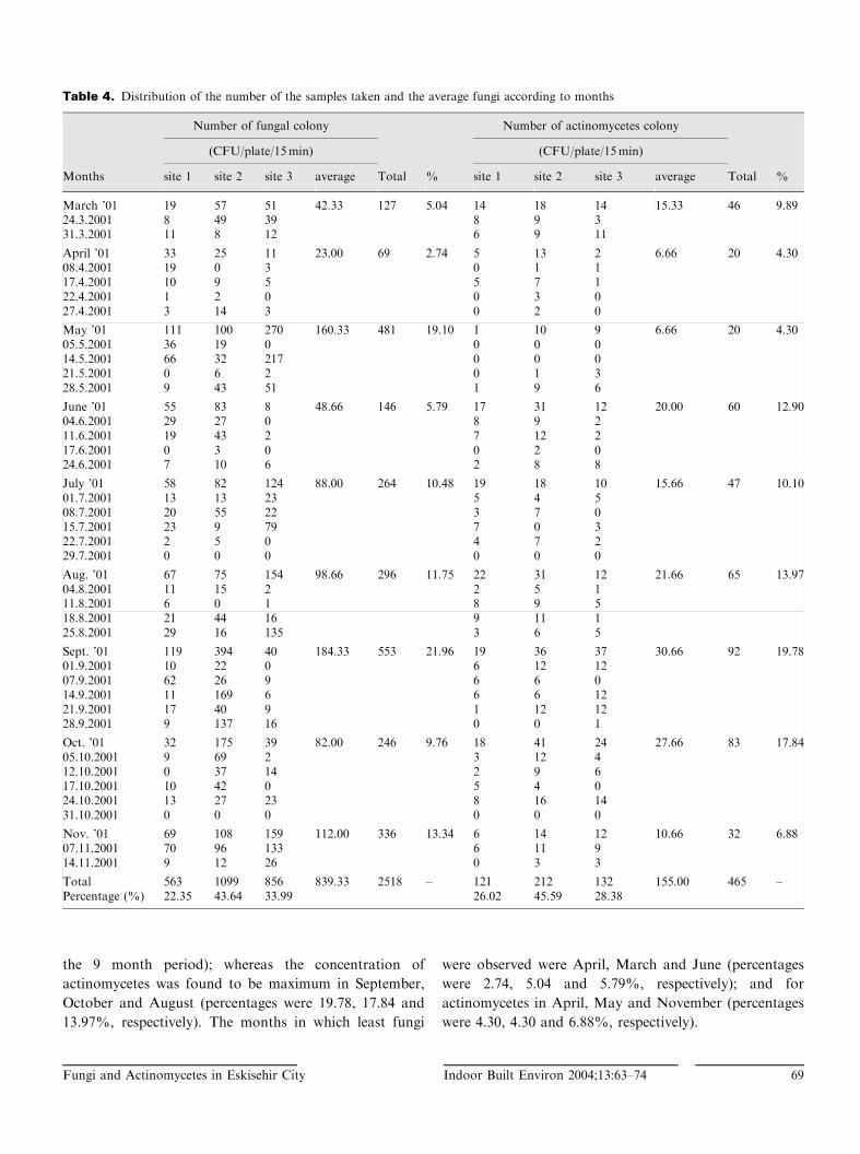
Climatological and Partial Air Pollutants Data
Weather data (monthly average temperature, monthly
total rainfall, monthly average relative humidity, monthly
average wind velocity and average monthly sunny times
(Table 3) had been recorded and was obtained from the
Directorate of Eskisehir Meteorological Office which is
positioned 9.2 km from the first station, 3.9 km from the
second station, and 0.5 km from the third station. Some air
pollutants including sulphur dioxide (SO2) (mg �m�3) and
respirablesuspendedparticulatematter (PM)(mg �m�3)were
also recorded. SO2 and PM concentrations in the urban air
were provided from continuous monitoring sites operated
by the Head Office of the H|fz|s|hha Institute (Public
Health Institute) in Eskisehir monthly (Table 2). Table 2
gives the monthly and annual mean of SO2 and PM con-
centrations in Eskisehir from March to November 2001.
Statistical Analysis
Statistical analysis (multivariate analysis (MANOVA))
of the data including fungal and actinomycetes colony
numbers and meteorological factors was performed.
Results
A total of 2518 fungal and 465 actinomycetes colonies
were isolated from 420 Petri dishes, quantified to deter-
mine the frequency of occurrence and then identified
(excepting the actinomycetes) (Tables 4 and 5). Some 20
fungal species could be identified, among them the species
Alternaria alternata that was generally found as the
predominant fungus (13.66%) at all sites, followed by
Cladosporium cladosporioides (5.80%) and Scopulariopsis
brevicaulis (5.50%) (Table 5). A. alternata, Aspergillus
wentii, C. cladosporioides, Penicillium chrysogenum,
S. brevicaulis and Ulocladium tuberculatum were found at
all sampling sites.
During the nine months of 2001 which were monitored,
the maximum concentration of airborne actinomycetes
was found in September (19.78%). The number of fungal
colonies on culture media plates was between 69 to a
maximum count of 553. The corresponding numbers
for actinomycetes were 20 and 92. In total 77.49% of the
fungal species could be identified in this study. The
remainder (22.51%) could not be identified using the
literature sources available because some of the genera
have similar morphological aspects, or no distinctive prop-
erties or there had been bacterial contamination (Table 5).
Statistical analysis of the data showed that the numbers
of fungi did not vary significantly by months and by areas
NOVEMBER
OCTOBER
SEPTEMBER
AUGUST
JULY
JUNE
MAY
APRIL
MARCH
Cou
nt o
f Act
inom
ycet
es (
cfu/
plat
e/15
min
)
10
8
6
4
2
0
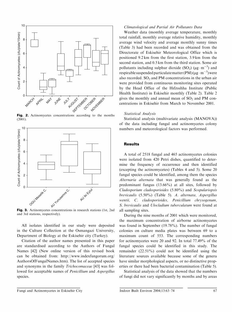
Fig. 2. Actinomycetes concentrations according to the months(2001).
AU Iki E
ylul
AU Yun
usem
re C
.
OGU Mes
elik C
.
Cou
nt o
f Act
inom
ycet
es (
cfu/
plat
e/15
min
) 10
8
6
4
2
0
Fig. 3. Actinomycetes concentrations in research stations (1st, 2ndand 3rd stations, respectively).
Fungi and Actinomycetes in Eskisehir City Indoor Built Environ 2004;13:63–74 67
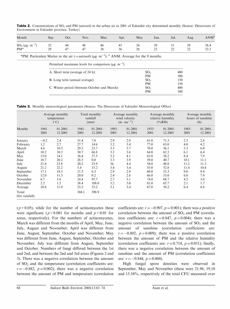
( p>0.05), while for the number of actinomycetes these
were significant ( p<0.001 for months and p<0.01 for
areas, respectively). For the numbers of actinomycetes,
March was different from the months of April, May, June,
July, August and November; April was different from
June, August, September, October and November; May
was different from June, August, September, October and
November; July was different from August, September
and October. Numbers of fungi differed between the 1st
and 2nd, and between the 2nd and 3rd areas (Figures 2 and
3). There was a negative correlation between the amount
of SO2 and the temperature (correlation coefficients are:
r¼�0.882, p¼ 0.002); there was a negative correlation
between the amount of PM and temperature (correlation
coefficients are: r¼�0.907, p¼ 0.001); there was a positive
correlation between the amount of SO2 and PM (correla-
tion coefficients are: r¼ 0.847, p¼ 0.004); there was a
negative correlation between the amount of SO2 and the
amount of sunshine (correlation coefficients are:
r¼�0.802, p¼ 0.009); there was a positive correlation
between the amount of PM and the relative humidity
(correlation coefficients are: r¼ 0.714, p¼ 0.031); finally,
there was a negative correlation between the amount of
sunshine and the amount of PM (correlation coefficients
are: r¼�0.844, p¼ 0.004).
High fungal spore densities were observed in
September, May and November (these were 21.96, 19.10
and 13.34%, respectively of the total CFU measured over
Table 3. Monthly meteorological parameters (Source: The Directorate of Eskisehir Meteorological Office)
Average monthlytemperature
(�C)
Total monthlyrainfall(mm)
Average monthlywind velocity(m � sec�1)
Average monthlyrelative humidity
(%RH)
Average monthlyhours of sunshine
(h)
Months 1941–2001
01.2001–12.2001
1941–2001
01.2001–12.2001
1985–2001
01.2001–12.2001
1933–2001
01.2001–12.2001
1985–2001
01.2001–12.2001
January �0.2 2.4 31.4 7.0 2.9 2.9 81.0 71.3 2.5 2.6February 1.2 2.7 27.7 14.6 3.2 3.4 77.0 65.0 4.0 4.2March 4.8 10.3 29.5 23.7 3.5 3.7 70.0 56.1 5.3 6.0April 10.2 10.3 30.7 66.8 3.3 3.6 64.0 62.3 6.1 6.4May 15.0 14.1 36.4 37.5 3.2 4.1 63.0 58.2 8.4 7.9June 18.7 20.2 28.5 0.0 3.3 3.9 59.0 40.7 10.1 11.1July 21.4 23.8 20.2 23.9 36. 4.4 54.0 48.8 11.2 11.3August 21.2 22.2 5.4 15.2 3.4 3.4 55.0 52.8 11.4 10.4September 17.1 18.3 11.5 6.3 2.9 2.9 60.0 53.3 9.0 9.4October 12.0 11.3 20.0 0.2 2.4 2.8 66.0 55.0 6.0 7.9November 6.7 5.6 24.4 95.7 2.7 3.1 74.0 68.7 4.2 3.0December 2.2 1.5 38.4 108.0 3.2 3.0 81.0 65.7 2.1 1.7Average 10.8 11.9 25.3 33.2 3.1 3.4 67.0 58.2 6.4 6.8
Total(for rainfall)
304.1 398.9
Table 2. Concentrations of SO2 and PM (aerosol) in the urban air in 2001 of Eskisehir city determined monthly (Source: Directorate ofEnvironment in Eskisehir province, Turkey)
Month Sep. Oct. Nov. Mar. Apr. May Jun. Jul. Aug. ANM#
SO2 (mg �m�3) 32 44 48 46 43 34 39 31 29 38.4
PM* 29 47 47 38 36 28 22 22 22 32.3
*PM: Particulate Matter in the air (¼ aerosol) (mg �m�3); # ANM: Average for the 9 months.
Permitted maximum levels for comparison (mg �m�3)
A. Short term (average of 24 h): SO2 400PM 300
B. Long term (annual average): SO2 150PM 150
C. Winter period (between October and March): SO2 400PM 300
68 Indoor Built Environ 2004;13:63–74 Asan et al.
the 9 month period); whereas the concentration of
actinomycetes was found to be maximum in September,
October and August (percentages were 19.78, 17.84 and
13.97%, respectively). The months in which least fungi
were observed were April, March and June (percentages
were 2.74, 5.04 and 5.79%, respectively); and for
actinomycetes in April, May and November (percentages
were 4.30, 4.30 and 6.88%, respectively).
Table 4. Distribution of the number of the samples taken and the average fungi according to months
Number of fungal colony Number of actinomycetes colony
(CFU/plate/15min) (CFU/plate/15min)
Months site 1 site 2 site 3 average Total % site 1 site 2 site 3 average Total %
March ’01 19 57 51 42.33 127 5.04 14 18 14 15.33 46 9.8924.3.2001 8 49 39 8 9 331.3.2001 11 8 12 6 9 11
April ’01 33 25 11 23.00 69 2.74 5 13 2 6.66 20 4.3008.4.2001 19 0 3 0 1 117.4.2001 10 9 5 5 7 122.4.2001 1 2 0 0 3 027.4.2001 3 14 3 0 2 0
May ’01 111 100 270 160.33 481 19.10 1 10 9 6.66 20 4.3005.5.2001 36 19 0 0 0 014.5.2001 66 32 217 0 0 021.5.2001 0 6 2 0 1 328.5.2001 9 43 51 1 9 6
June ’01 55 83 8 48.66 146 5.79 17 31 12 20.00 60 12.9004.6.2001 29 27 0 8 9 211.6.2001 19 43 2 7 12 217.6.2001 0 3 0 0 2 024.6.2001 7 10 6 2 8 8
July ’01 58 82 124 88.00 264 10.48 19 18 10 15.66 47 10.1001.7.2001 13 13 23 5 4 508.7.2001 20 55 22 3 7 015.7.2001 23 9 79 7 0 322.7.2001 2 5 0 4 7 229.7.2001 0 0 0 0 0 0
Aug. ’01 67 75 154 98.66 296 11.75 22 31 12 21.66 65 13.9704.8.2001 11 15 2 2 5 111.8.2001 6 0 1 8 9 518.8.2001 21 44 16 9 11 125.8.2001 29 16 135 3 6 5
Sept. ’01 119 394 40 184.33 553 21.96 19 36 37 30.66 92 19.7801.9.2001 10 22 0 6 12 1207.9.2001 62 26 9 6 6 014.9.2001 11 169 6 6 6 1221.9.2001 17 40 9 1 12 1228.9.2001 9 137 16 0 0 1
Oct. ’01 32 175 39 82.00 246 9.76 18 41 24 27.66 83 17.8405.10.2001 9 69 2 3 12 412.10.2001 0 37 14 2 9 617.10.2001 10 42 0 5 4 024.10.2001 13 27 23 8 16 1431.10.2001 0 0 0 0 0 0
Nov. ’01 69 108 159 112.00 336 13.34 6 14 12 10.66 32 6.8807.11.2001 70 96 133 6 11 914.11.2001 9 12 26 0 3 3
Total 563 1099 856 839.33 2518 – 121 212 132 155.00 465 –Percentage (%) 22.35 43.64 33.99 26.02 45.59 28.38
Fungi and Actinomycetes in Eskisehir City Indoor Built Environ 2004;13:63–74 69
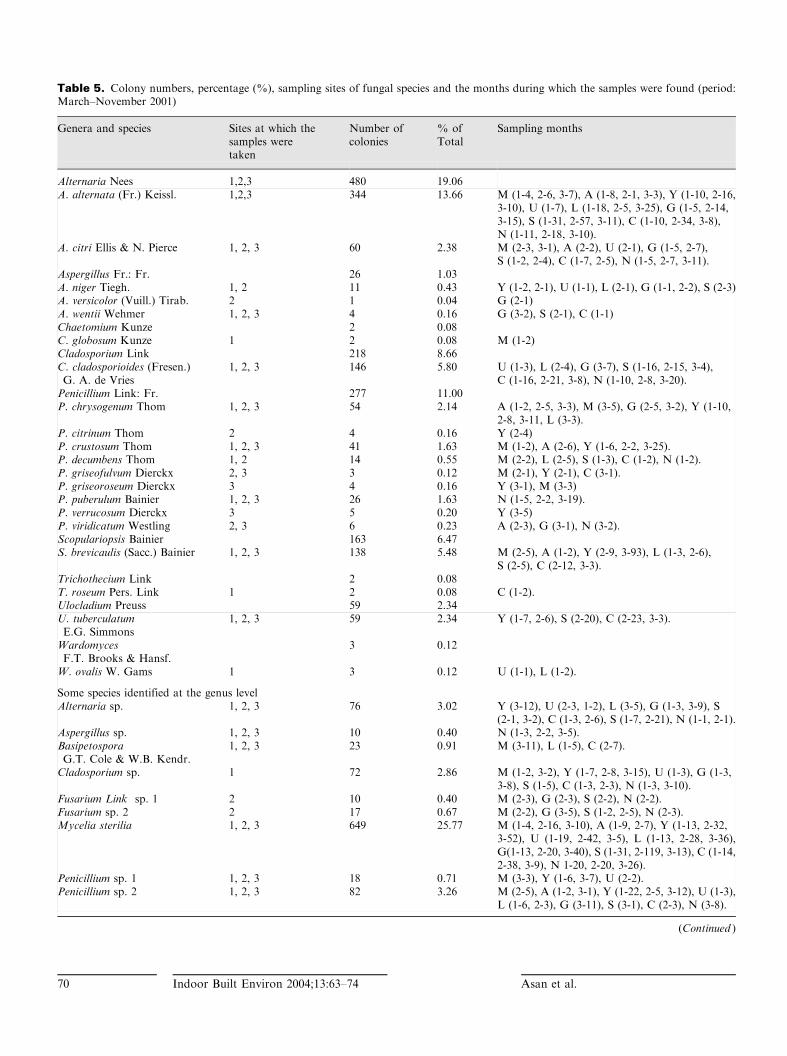
Table 5. Colony numbers, percentage (%), sampling sites of fungal species and the months during which the samples were found (period:March–November 2001)
Genera and species Sites at which thesamples weretaken
Number ofcolonies
% ofTotal
Sampling months
Alternaria Nees 1,2,3 480 19.06A. alternata (Fr.) Keissl. 1,2,3 344 13.66 M (1-4, 2-6, 3-7), A (1-8, 2-1, 3-3), Y (1-10, 2-16,
3-10), U (1-7), L (1-18, 2-5, 3-25), G (1-5, 2-14,3-15), S (1-31, 2-57, 3-11), C (1-10, 2-34, 3-8),N (1-11, 2-18, 3-10).
A. citri Ellis & N. Pierce 1, 2, 3 60 2.38 M (2-3, 3-1), A (2-2), U (2-1), G (1-5, 2-7),S (1-2, 2-4), C (1-7, 2-5), N (1-5, 2-7, 3-11).
Aspergillus Fr.: Fr. 26 1.03A. niger Tiegh. 1, 2 11 0.43 Y (1-2, 2-1), U (1-1), L (2-1), G (1-1, 2-2), S (2-3)A. versicolor (Vuill.) Tirab. 2 1 0.04 G (2-1)A. wentii Wehmer 1, 2, 3 4 0.16 G (3-2), S (2-1), C (1-1)Chaetomium Kunze 2 0.08C. globosum Kunze 1 2 0.08 M (1-2)Cladosporium Link 218 8.66C. cladosporioides (Fresen.)G. A. de Vries
1, 2, 3 146 5.80 U (1-3), L (2-4), G (3-7), S (1-16, 2-15, 3-4),C (1-16, 2-21, 3-8), N (1-10, 2-8, 3-20).
Penicillium Link: Fr. 277 11.00P. chrysogenum Thom 1, 2, 3 54 2.14 A (1-2, 2-5, 3-3), M (3-5), G (2-5, 3-2), Y (1-10,
2-8, 3-11, L (3-3).P. citrinum Thom 2 4 0.16 Y (2-4)P. crustosum Thom 1, 2, 3 41 1.63 M (1-2), A (2-6), Y (1-6, 2-2, 3-25).P. decumbens Thom 1, 2 14 0.55 M (2-2), L (2-5), S (1-3), C (1-2), N (1-2).P. griseofulvum Dierckx 2, 3 3 0.12 M (2-1), Y (2-1), C (3-1).P. griseoroseum Dierckx 3 4 0.16 Y (3-1), M (3-3)P. puberulum Bainier 1, 2, 3 26 1.63 N (1-5, 2-2, 3-19).P. verrucosum Dierckx 3 5 0.20 Y (3-5)P. viridicatum Westling 2, 3 6 0.23 A (2-3), G (3-1), N (3-2).Scopulariopsis Bainier 163 6.47S. brevicaulis (Sacc.) Bainier 1, 2, 3 138 5.48 M (2-5), A (1-2), Y (2-9, 3-93), L (1-3, 2-6),
S (2-5), C (2-12, 3-3).Trichothecium Link 2 0.08T. roseum Pers. Link 1 2 0.08 C (1-2).Ulocladium Preuss 59 2.34U. tuberculatumE.G. Simmons
1, 2, 3 59 2.34 Y (1-7, 2-6), S (2-20), C (2-23, 3-3).
WardomycesF.T. Brooks & Hansf.
3 0.12
W. ovalis W. Gams 1 3 0.12 U (1-1), L (1-2).
Some species identified at the genus levelAlternaria sp. 1, 2, 3 76 3.02 Y (3-12), U (2-3, 1-2), L (3-5), G (1-3, 3-9), S
(2-1, 3-2), C (1-3, 2-6), S (1-7, 2-21), N (1-1, 2-1).Aspergillus sp. 1, 2, 3 10 0.40 N (1-3, 2-2, 3-5).BasipetosporaG.T. Cole & W.B. Kendr.
1, 2, 3 23 0.91 M (3-11), L (1-5), C (2-7).
Cladosporium sp. 1 72 2.86 M (1-2, 3-2), Y (1-7, 2-8, 3-15), U (1-3), G (1-3,3-8), S (1-5), C (1-3, 2-3), N (1-3, 3-10).
Fusarium Link sp. 1 2 10 0.40 M (2-3), G (2-3), S (2-2), N (2-2).Fusarium sp. 2 2 17 0.67 M (2-2), G (3-5), S (1-2, 2-5), N (2-3).Mycelia sterilia 1, 2, 3 649 25.77 M (1-4, 2-16, 3-10), A (1-9, 2-7), Y (1-13, 2-32,
3-52), U (1-19, 2-42, 3-5), L (1-13, 2-28, 3-36),G(1-13, 2-20, 3-40), S (1-31, 2-119, 3-13), C (1-14,2-38, 3-9), N 1-20, 2-20, 3-26).
Penicillium sp. 1 1, 2, 3 18 0.71 M (3-3), Y (1-6, 3-7), U (2-2).Penicillium sp. 2 1, 2, 3 82 3.26 M (2-5), A (1-2, 3-1), Y (1-22, 2-5, 3-12), U (1-3),
L (1-6, 2-3), G (3-11), S (3-1), C (2-3), N (3-8).
(Continued )
70 Indoor Built Environ 2004;13:63–74 Asan et al.
Discussion
The present study will contribute to our knowledge of
the levels and types of airborne fungi and actinomycetes in
the urban air of Eskisehir. Although similar studies [22, 23]
have been undertaken in this city, one of these [23] focused
on airborne bacteria and the other [22] looked at airborne
fungi, some of which were determined only to genus level.
New studies are needed because of the rapid development
in urbanisation, with concomitant increasing air pollution
and industrialisation. One of the studies noted above [22]
was published nine years ago and it contains no data
related to concentration and distribution of airborne
fungal species. In the present study, concentrations were
measured and several of the fungal species were identified.
Unfortunately, the nature of mycoflora is such that it was
difficult to specify all the airborne fungi found.
It is well known that microfungi can live under extreme
conditions in almost all regions and all climates. They
usually live in soil and can be dispersed into the atmos-
phere under the influence of various factors. In recent
years, aerobiologists have shown a great interest in
airborne fungi due to both their ubiquitous presence in
the air and the increase in allergies attributed to them
[9,44]. Monitoring airborne fungi in both outdoor and
indoor environments provides valuable data for evaluating
type and distribution. Aspergillus and Penicillium spores
are the most widespread aeroallergens in the world.
According to qualitative and quantitative reports, the
first is the dominant species in tropical regions while the
latter is dominant over the rest of the world [45].
Everybody may be exposed to moulds, but fungal density
in the air and the time for which people are exposed are
both important for assessing any adverse effect of moulds.
In this study Alternaria was the most frequent and
the predominant genus detected, followed by Penicillium,
Cladosporium and Scopulariopsis genera (Table 5).
According to Gambale et al. [46], the genus Alternaria
with powerful allergenic properties has been isolated with
16% frequency in sampled collections. Downs et al. [47]
noted that Alternaria is known to be allergenic and is
one of the most common fungi world-wide and they have
suggested that Alternaria allergens contribute to severe
asthma. Myszkowska et al. [48] noted that some 4–7% of
the European population shows sensitivity to Alternaria
and Cladosporium spores. C. cladosporioides, determined
in our study, has been reported to be the agent
causing phaeohyphomycosis along with other species of
Cladosporium [49]. Also we identified Aspergillus niger
which is well-known to cause many health problems
such as extrinsic alveolitis, allergic bronchopulmonary
aspergillosis, keratitis, endophthalmitis, primary cuta-
neous aspergillosis and necrosing otitis in humans [50].
Although the prevalence of fungal sensitivity in asthma
is not completely understood, A. alternata species
(which were found as an appreciable fraction of the
whole in our study) are a common cause of asthma [51].
Although spore numbers of Alternaria sp. lower than those
of Cladosporium sp. have been found in some studies
[15,52], the spore volume of Alternaria sp. is greater than
Cladosporium sp., so together they are comparable in
biomass (Dr. Julie M. Corden, Derby-UK, personal
communication). Alternaria, Penicillium, Aspergillus and
Fusarium genera were found to be the dominant fungi in
some studies such as the one by Savino and Caretta [53].
Cladosporium sp. is the most common fungus living as a
saprophyte, mainly on dying and/or dead herbaceous
plants and other organic matter. It produces chains of dry
Table 5. Continued
Genera andspecies
Sites at which thesamples weretaken
Number ofcolonies
% ofTotal
Sampling months
Penicillium sp. 3 1, 2, 3 20 0.79 M (3-3), A (3-2), G (2-8), Y (1-2), S (2-2), N (2-3).Scopulariopsis sp. 2, 3 25 0.99 A (3-2), Y (2-1), S (2-15), C (2-4, 3-3).Torula Pers. sp. 1, 2, 3 22 0.87 A (1-3), M (1-4), G (2-2, 3-1), S (3-4), C (1-5),
N (1-3).Unidentified 1, 2, 3 567 22.51 M (1-3, 2-15, 3-9), A (1-7, 2-2, 3-3), Y (1-10,
2-23, 3-42), U (1-8, 2-23,3-3), L (1-8, 2-30, 3-41),G (1-17, 2-13, 3-53), S (1-12, 2-109, 3-7),C (1-14, 2-23, 3-6), N (1-12, 2-30, 3-44).
Total 2518
Letters indicate: M: March, A: April, Y: May, U: June, L: July, G: August, S: September, C: October, N: November. The first
(and second and third where appropriate) number in parenthesis in the last column is the station number with related month.
Fungi and Actinomycetes in Eskisehir City Indoor Built Environ 2004;13:63–74 71
conidia that easily become airborne (Dr. Hugues Beguin,
Brussels – Belgium; personal communication).
Although there are different methods for sampling
fungi from air, we used the Petri Plate Gravitational
Settling Method for the isolation of airborne fungi and
actinomycetes because of its practicality and low cost. This
method is useful for the enumeration of fungal spores, but
gives only a rough approximation of the kinds and
numbers of airborne fungi [54]. According to Chen et al.
[55], there is no official and universally accepted bioaero-
sols sampling method. There are many types of sampler
(Rotorod, Tauber, Burjerd 7 day, personal Burkard, air-o-
cell, Andersen, SAS, etc.) for sampling aeroallergens from
air; but although each has its advantages and disadvan-
tages they all have some limitations [56]. In addition,
although many types of culture media are used for
sampling micro-organisms from air we used Rose-Bengal
streptomycin agar medium. According to Madan et al.
[57], this medium is the most suitable for sampling fungi
from air. Also according to Morring et al. [58], Rose-
Bengal streptomycin agar can be used for aeromycological
sampling. Streptomycin antibiotic was used to control
reproduction of bacteria and Rose-Bengal stain was used
to limit the growth of fast-growing moulds (e.g., Rhizopus
and Trichoderma spp.). Also, Wu et al. [59] reported that
dichloron (present in dichloron glycerol-18 agar¼DG18)
restricts the growth of fast-growing genera but we did not
use it. Wu et al. [60] compared some media for sampling
fungi from a hospital environment. They proposed DG18
medium for sampling but this medium has a low water
activity and is best used for xerophilic moulds and
osmophilic yeasts. Also Levetin and Horner [56] noted
that MEA is generally suggested for mesophilic fungi.
Sampling time was between 10.00 and 12.00 in our study
after Levetin and Horner [56] who pointed out that the
spores of many asexual fungi peaked in air in early to mid-
afternoon but were low in early morning.
Alternaria and Penicillium genera were found more
abundantly than other fungi at all the research stations.
A. alternata was present throughout the year but was at a
maximum in September, October and November. A. citri
occurred throughout seven months: it was not seen in
December and July but was especially abundant in
November. Corden and Millington [61] determined that
the Alternaria sp. spores peak daily count usually occurred
in August but occasionally in late July or early September.
Annual variations in the composition of the vegetation
around the research station may cause the differences.
Mitakakis and Guest [62] noted the seasonality of spore
levels of Cladosporium and Alternaria sp. which peaked in
spring and summer. Bandyopadhyay et al. [63] found a
positive correlation between heavy rainfall and Fusarium
sp. concentration. Other researchers [8,63] including Di
Giorgio et al. [10] reported that various meteorological
factors affected the type and concentrations of airborne
fungi. Among these wind velocity, relative humidity and
temperature were particularly important. Pasanen et al.
[64] reported that the minimum air velocity at which
Cladosporium sp. released spores was 1.0m � s�1, however
Aspergillus fumigatus and Penicillium sp. released great
numbers of spores at 0.5m � s�1. So, air velocity is
significant for dispersion of fungal spores into the air.
Reponen et al. [65] reported that the hygroscopicity of
fungal airborne spores significantly affected their aero-
dynamic diameter.
Aspergillus niger, A. versicolor, A. wentii and Penicillium
chrysogenum identified in our study are widespread in
Turkey and have been determined in other studies [66].
Some species of Cladosporium, Alternaria, Penicillium,
Fusarium and Aspergillus genera (determined in our study)
can cause allergy [67–69] and these genera are known to be
an important part of the airborne microfungi. Although
Aspergillus fumigatus is the most common agent of
invasive aspergillosis, A. niger, found in our study, has
also been implicated [70]. The airborne fungal species
identified in our study may be allergenic to people residing
in Eskisehir and the population may be exposed to dust
containing mycotoxins derived from fungi. Therefore,
fungal spore monitoring in Eskisehir city may be useful
from the allergological point of view. Future investigations
are needed to examine further the effects of mould
exposures on the related health problems.
We showed above that our study revealed the existence
of a rich population of airborne fungi and actinomycetes
in the urban air. However, it must be borne in mind that
the culture methods used reveal only a portion of the
airborne micro-organisms because some fungi are not able
to grow at all in culture media and some of them may lose
viability [56]. Therefore, the air of Eskisehir city may be
even richer in fungi and actinomycetes than our results
have shown.
72 Indoor Built Environ 2004;13:63–74 Asan et al.
References
1 Marshall WA: Seasonality in Antarctic air-borne fungal spores. Appl Environ Microbiol1997;63:2240–2245.
2 Madelin TM: Fungal aerosols – a review. J AerSci 1994;25:1405–1412.
3 Simeray J, Mandin D, Chaumont JP : Anaeromycological study of sawmills: Effects oftype of installation and timber on mycofloraand inhalation hazards for workers. IntBiodeter Biodegradation 1997;40:11–17.
4 Burge HA, Rogers CA: Outdoor allergens.Environ Health Perspect 2000;108:653–659.
5 Simeray J, Mandin D, Chaumont JP:Variations in the distribution of fungal sporesin the atmosphere of bakehouses. Impact onthe study of allergies. Grana 1995;34:269–274.
6 Singh J: Occupational exposure to moulds inbuildings. Indoor Built Environ2001;10:172–178.
7 Buttner MP, Cruz-Perez P, Stetzenbach LD:Enhanced detection of surface-associated bac-teria in indoor environments by quantitativePCR. Appl Environ Microbiol 2001;67:2564–2570.
8 Agarwal MK, Shivpuri DN: Studies on theallergenic fungal spores of the Delhi, India,metropolitan area – Botanical aspects (aero-mycology). J Allergy 1969;44:193–203.
9 Pasanen AL: Airborne mesophilic fungalspores in various residental environments.Atmos Environ 1992;26A:2861–2868.
10 DiGiorgio C, Krempff A,GuiraudH, Binder P,Tiret C, Dumenil G: Atmospheric pollutionby airborne microorganisms in the city ofMarseilles. Atmos Environ 1996;30:155–160.
11 Gorny RL, Reponen T, Grinshpun SA,Willeke K: Source strength of fungal sporeaerosolization from moldy building material.Atmos Environ 2001;35:4853–4862.
12 McNeil, MM. and Brown JM: The medicallyimportant aerobic actinomycetes: epidemiol-ogy and microbiology. Clin Microbiol Rev1994;7:357–417. (includes reference to thehistoric work of Nocard ME: Note sur lamaladie des bouefs de la Gouadelopue, connuesous le nom de Farcin. Ann Inst Pasteur1888;2:293–302.)
13 Reponen TA, Gazenko SV, Grinshpun SA,Willeke K, Cole EC: Characteristics of air-borne actinomycete spores. Appl EnvironMicrobiol 1998;64:3807–3812.
14 Colakoglu G: Fungal spore concentrations inthe atmosphere at the Anatolia Quarter ofIstanbul, Turkey. J Basic Microbiol1996;36:155–162.
15 Sen B, Asan A: Airborne fungi in vegetablegrowing areas of Edirne, Turkey. Aerobiologia2001;17:69–75.
16 Asan A, Sen B, Sarica S: Airborne fungi inurban air of Edirne city (Turkey). Biologia2002;57:59–68.
17 Sarica S, Asan A, Tatman-Otkun M, Ture M:Monitoring indoor airborne fungi and bacteriain the different areas of Trakya UniversityHospital (Edirne-Turkey). Indoor BuiltEnviron 2002;11:285–292.
18 Simsekli Y, Gucin F, Asan A: Isolation andidentification of indoor airborne fungal con-taminants of food production facilities and
warehouses in Bursa, Turkey. Aerobiologia1999;15:225–231.
19 Sakiyan N, Inceoglu O: Atmospheric concen-tration of Cladosporium and Alternaria sporesin Ankara and the effect of meteorologicalfactors. Turk J Bot 2003;27:77–81.
20 Ayata C, Coskun S, Okyay T: y|l|nda aylaragore Izmir ilinin ces� itli semtlerinde havan|nfungal floras| ve bunun allerjik hastal|klaryonunden onemi. Turk Mikrobiyol CemDerg. 1991;21:219–226. (Fungal flora of theair in several parts of Izmir according to monthsin 1989 and its importance regarding in theallergic diseases) (Turkish, with Englishabstract).
21 Harmanci E, Metintas M, Erginel S:Respiratory allergy to moulds among adultsin Eskisehir Anatolia), Turkey. AllergImmunol (Paris) 2000;32:49–51.
22 Atik S, Tamer AU: Eskisehir (merkez ilce)’demikrofungal hava kirliligi. Ege Univ Fen FakDerg. Seri B. Ek. 1994;16/1:227–238.
23 Atik S, Tamer AU: Eskisehir (merkez ilce)’debakteriyal hava kirliligi. II. Ulusal Ekolojive Cevre Kongresi Bildirileri. 1995;734–748.11–13 Eylul 1995, Ankara-Turkey.
24 Diab A, Omar SA, Hertani H: Airborneactinomycetes in the atmosphere of Kuwait.Zent Bakteriol Parasitenk Infekt Hyg1977;132:273–282.
25 Davis PH (ed): Flora of Turkey and theEast Aegaean Island. 1965–1988;Vols. 1–10,Edinburgh, Edinburgh University Press.
26 Ocak A, Ture C: The flora of Mes� elik campusof the Osmangazi University (Eskis� ehir-Turkey). Ot Sist Bot Derg. (The Herb J SystBot) 2001;8:19–46.
27 Ismail MA, Chebon SK, Nakamya R:Preliminary surveys of outdoor and indooraeromycobiota in Uganda. Mycopathol 1999;148:41–51.
28 Martinez Ordaz VA, Rincon-Castaneda CB,Esquivel Lopez G, Lazo-Saenz JG, LlorenzMeraz MT, Velasco Rodriguez VM: Fungalspores in the environment of the asthmaticpatient in a semi-desert area of Mexico. RevAlerg Mex 2002;49:2–7.
29 Sime AD, Abbott LL, Abbott SP: Mountingmedium for use in Indoor Air Quality spore-trap analyses. Mycologia 2002;94:1087–1088.
30 Barnett HL, Hunter BB: Illustrated Genera ofImperfect Fungi, ed 4. The American Phyto-pathological Society. St. Paul, Minnesota,USA, APS Press, 1998, pp 218.
31 Pitt JI: The genus Penicillium and its tele-omorphic states Eupenicillium andTalaromyces. London, Academic Press Inc.,1979, 634 pp.
32 Raper KB, Fennell DI: The genus Aspergillus.Baltimore, USA, The Williams & WilkinsComp., 1965, 686 pp.
33 Klich MA: Identification of commonAspergillus species. 2002; First ed. Utrecht,The Netherlands, Centraalbureau voor Schim-melcultures, 122 pp.
34 Okuda T, Klich MA, Seifert KA, Ando K:Media and incubation effects on morphologi-cal characteristics of Penicillium andAspergillus. Samson RA, Pitt JI (eds):
Integration of modern taxonomic methodsfor Penicillium and Aspergillus classification.510 pp. Singapore, Harwood AcademicPublishers, 2000;83–99.
35 Ellis MB: Dematiaceous Hyphomycetes.London and Reading. Commonwealth Myco-logical Institute. Kew, Surrey, UK, The EasternPress Ltd., 1971, 608 pp.
36 Ellis MB, Ellis JP: Microfungi on Land Plants.An Identification Handbook. Enlarged Ed.UK, The Richmond Publishing Co. Ltd.,1997, 868 pp.
37 Nelson PE, Toussoun TA, Marasas WFO:Fusarium Species – An Illustrated Manual forIdentification. USA, The Pennsylvania StateUniversity Press, University Park and London,1983, 193 pp.
38 Hasenekoglu I: Toprak mikrofunguslar|.1991;Vol. 1–7. Ataturk Unv. Yay. No: 689,Erzurum-Turkey. (Soil Microfungi) (Turkish).
39 Schall PK: Actinomyces. in Peter HA Sneath(ed): Bergey’s Manual of SystematicBacteriology, Vol. 2, Williams & Wilkins,1986; pp. 1383–1417. Baltimore, USA.
40 Schall PK: Actinomyces, arcanobacterium,and rothia. in Balows A, Truper HG,Dworkin M, Harder W, and Schleifer KH(eds): The Procaryotes, 1991;Vol. 1. ed 2,Springer-Verlag, pp. 850–905.
41 Waksman SA. Actinomyces. New York, TheRonald Press Company, 1967. (Translated intoTurkish in 1989, by Mehmet ONER).
42 Kirk PM, Ansell AE: Authors of fungal names.Index of fungi supplement. Kew, Surrey (UK),International Mycological Institute. AnInstitute of CAB International, 1992, 95 pp.
43 Pitt JI, Samson RA, Frisvad JC: List ofaccepted species and synonyms in the familyTrichocomaceae. 2000, pp 9–49, in SamsonRA, Pitt JI (eds). Integration of ModernTaxonomic methods for Penicillium and Asper-gillus Classification. Singapore, HarwoodAcademic Publishers, 510 pp.
44 Larsen L, Gravesen S: Seasonal variation ofoutdoor airborne viable microfungi inCopenhagen, Denmark, Grana 1991;30:467–471.
45 Rosas I, Calderon C, Escamilla B, Ulioa M:Seasonal distribution of Aspergillus in the airof an urban area: Mexico City, Grana1992;31:315–319.
46 Gambale, W, Paula, RC, Buck, N, Gambale V:Airborne fungi of presidente prudente SP.Brasil Rev Microbiol 1985;16:9–14.
47 Downs SH, Mitakakis TZ, Marks GB, CarNG, Belousova EG, Leuppi JD, Xuan W,Downie SR, Tobias A, Peat JK: Clinicalimportance of Alternaria exposure in children.Amer J Resp Critical Care Med2001;164:455–459.
48 Myszkowska D, Ste�palska D, Krystyna K,Pore�bski G: The relationship between airbornepollen and fungal spore concentrations andseasonal pollen allergy symptoms in Cracow in1997–1999. Aerobiologia 2002;18:153–161.
49 Kantarcioglu AS, Yucel A, De Hoog GS: Casereport. Isolation of Cladosporium cladospor-ioides from cerebrospinal fluid. Mycoses2002;45:500–503.
Fungi and Actinomycetes in Eskisehir City Indoor Built Environ 2004;13:63–74 73
50 Severo LC, Geyer GR, Porto NS, Wagner MB,Londero AT: Pulmonary Aspergillus nigerintracavitary colonization. Report of 23 casesand a review of the literature. Rev IberoamMicol 1997;14:104–110.
51 Sanchez H, Bush RK: A review of Alternariaalternata sensitivity. Rev Iberocoam Micol2001;18:56–59.
52 Huang CY, Lee CC, Li FC, Ma YP, Su HJJ:The seasonal distribution of bioaerosols inmunicipal landfill sites: a 3-yr study. AtmosEnviron 2002;36:4385–4395.
53 Savino E, Caretta G: Airborne fungi in anItalian rice mill. Aerobiologia 1992;8:267–274.
54 Pelczar MJ, Chan ECS, Krieg NR:Microbiology: Concepts and applications. 966pp. International ed. New York, McGraw-Hill, Inc., 1993, p. 796.
55 Chen CC, Yu TS, Chang JY, Chang CW, ShihTS, Hwang JS: A computer simulation study onbioaerosol colony counting error due to mask-ing effect. Ann Occup Hyg 1998;42:501–510.
56 Levetin E, Horner WE: Fungal aerobiology:Exposure and measurement. in Breitenbach M,Crameri R, Lahrer SB (eds): Fungal Allergyand Pathogenicity. Chem Immunol, KargerBasel, 2002;81:10–27.
57 Madan P, Lamba LC, Aneja KR: The most-suitable medium for trapping fungi from air.Sci Cult 1982;48:77–78.
58 Morring, KL, Sorenson WG, Attfield MD:Sampling for airborne fungi: A statisticalcomparison of media. Am Ind Hyg Assoc J1983;44:662–664.
59 Wu PC, Su HJJ, Lin CY: Characteristics ofindoor and outdoor airborne fungi at suburbanand urban homes in two seasons. Sci TotalEnviron 2000a;253:111–118.
60 Wu PC, Su HJJ, Ho HM: A comparison ofsampling media for environmental viable fungicollected in a hospital environment. EnvironRes 2000b;82:253–257.
61 Corden JM, Millington WM: The long-termtrends and seasonal variation of the aeroaller-gen Alternaria in Derby, UK. Aerobiologia2001;17:127–136.
62 Mitakakis TZ, Guest Di: A fungal sporecalendar for the atmosphere of Melbourne,Australia, for the year 1993. Aerobiologia2001;17:171–176.
63 Bandyopadhyay R, Mughogho LK,Satyanarayana MV: Occurrence of airbornespores of fungi causing grain mould over asorghum crop. Mycol Res 1991;95:1315–1320.
64 Pasanen AL, Pasanen P, Jantunen MJ,Kalliokoski P: Significance of air humidityand air velocity for fungal spore release in theair. Atmos Environ 1991;25A:459–462.
65 ReponenT,WillekeK,UleviciusV,ReponenA,Grinshpun SA: Effect of relative humidity onthe aerodynamic diameter and respiratorydeposition of fungal spores. Atmos Environ1996;30:3967–3974.
66 Asan A: Check list of Aspergillus andPenicillium species reported from Turkey.Turk J Bot 2000;24:151–167.
67 Chih-Shan L, Yu-Mei K: Characteristics ofairborne microfungi in subtropical homes. SciTotal Environ 1994;155:267–271.
68 Chih-Shan L, Li-Yuan H, Chen-Cheng C,Kue-Hsiung H: Fungus allergens insideand outside the residences of atopic andcontrol children. Arch Environ Health1995;50:38–43.
69 Adhikari A, Sen MM, Gupta-Bhattacharya S,Chanda S: Incidence of allergenically signifi-cant fungal aerosol in a rural bakery of WestBengal, India. Mycopathol 2000;149:35–45.
70 Abarca ML: Taxonomy and identification ofthe species involved in nosocomial aspergillo-sis. Rev Iberoam Micol 2000;17:S79–S84.
74 Indoor Built Environ 2004;13:63–74 Asan et al.


