Embed Size (px)
Citation preview

R E S E A R C H A R T I C L E
Activebacteriaandarchaeacells¢xingbicarbonate inthedarkalongthewater columnofa strati¢ed eutrophic lagoonMarc Lliros1, Laura Alonso-Saez2, Frederic Gich1, Anna Plasencia1, Olga Auguet1, Emilio O. Casamayor2
& Carles M. Borrego1
1Group of Molecular Microbial Ecology, Institute of Aquatic Ecology, University of Girona, Girona, Spain; and 2Group of Biogeodynamics & Biodiversity
Interactions, Department of Continental Ecology, Centre d’Estudis Avancats de Blanes-CSIC, Blanes, Spain
Correspondence: Carles M. Borrego, Group
of Molecular Microbial Ecology, Institute of
Aquatic Ecology, University of Girona,
E-17071 Girona, Spain. Tel.: 134 972 418
177; fax: 134 972 418 150; e-mail:
Present addresses: Marc Lliros, Laboratory
of Freshwater Ecology, Research Unit in
Organismic Biology, Department of Biology,
University of Namur, B-5000 Namur, Belgium.
Laura Alonso-Saez, Centro Oceanografico de
Gijon-IEO, E-33212, Gijon, Spain.
Received 24 January 2011; revised 13 April
2011; accepted 14 April 2011.
Final version published online 20 May 2011.
DOI:10.1111/j.1574-6941.2011.01117.x
Editor: Riks Laanbroek
Keywords
dark carbon fixation; sulfidic redoxclines;
anaplerotic reactions; chemoautotrophy;
heterotrophic carbon fixation; karstic lakes.
Abstract
We studied the carbon dioxide fixation activity in a stratified hypereutrophic
karstic lagoon using a combination of fingerprinting techniques targeting bacterial
and archaeal 16S rRNA genes, functional gene cloning [the acetyl-CoA carboxylase
(accC)], and isotopic labelling (14C-bicarbonate) coupled to single-cell analyses
[microautoradiography combined with catalyzed reported deposition-FISH
(MAR-CARD-FISH)]. The microbial planktonic community was dominated by
bacteria with maximal abundances of archaea just below the oxic/anoxic transition
zone (7% of total cells). In situ incubations with radiolabelled bicarbonate showed
maximal photoassimilation activity in the oxic epilimnion, whereas dark CO2
fixation was consistently observed throughout the water column, with a maximum
at the oxic/anoxic interface (8.6 mg C m�3 h�1). The contributions of light and dark
carbon fixation activities in the whole water column were 69% and 31% of the total
C incorporated, respectively. MAR-CARD-FISH incubations corroborated these
results and revealed that the highest fraction of bacterial and archaeal cells actively
uptaking bicarbonate in the light was found at the surface. The bacterial
community was mainly composed of green sulfur bacteria (Chlorobi) and
members of the Betaproteobacteria and the Bacteroidetes. The archaeal assemblage
was composed of phylotypes of the Miscellaneous Crenarchaeotic Group and a few
methanogens. Clone libraries of the accC gene showed an absolute dominance of
bacterial carboxylases. Our results suggest that the dark carbon fixation activity
measured was mainly related to CO2 incorporation by heterotrophs rather than to
the activity of true chemoautotrophs.
Introduction
Inorganic carbon photoassimilation has traditionally been
considered the dominant fixation process in planktonic
environments. However, high dark CO2 fixation rates mea-
sured in several stratified lakes with sharp oxic/anoxic
interfaces have called this assumption into question (Culver
& Brunskill, 1969; Jorgensen et al., 1979; Cloern et al., 1983;
Pedros-Alio & Guerrero, 1991; Shively et al., 1998; Camacho
et al., 2001; Hadas et al., 2001; Casamayor et al., 2008;
Casamayor, 2010). In some of these habitats, the values of
dark fixation rates integrated for the entire water column
were equivalent to those calculated for light-driven
processes, indicating that dark assimilation could not be
easily disregarded. Besides, the complexity of the microbial
assemblages thriving in environments with oxic/anoxic
interfaces and the availability of different energy sources
and electron donors favor the presence of a large diversity of
microorganisms involved in chemoautotrophic activities
(Casamayor, 2010).
In the last decade, several studies have provided detailed
analyses of dark CO2 assimilation by distinct microbial
guilds in stratified lakes (Camacho et al., 2001; Garcıa-
Cantizano et al., 2005; Casamayor et al., 2008; Casamayor,
2010), but none have focused on the potential contribution
of planktonic archaea in the process. This is of special
FEMS Microbiol Ecol 77 (2011) 370–384c� 2011 Federation of European Microbiological SocietiesPublished by Blackwell Publishing Ltd. All rights reserved
MIC
ROBI
OLO
GY
EC
OLO
GY
Dow
nloaded from https://academ
ic.oup.com/fem
sec/article/77/2/370/500140 by guest on 30 July 2022
interest considering the general distribution and abundance
of archaea in aquatic ecosystems (e.g. DeLong, 1992, 1998;
DeLong & Pace, 2001; Robertson et al., 2005; Schleper et al.,
2005; Chaban et al., 2006; Auguet et al., 2010) and the
particular high richness of archaeal taxa in anoxic waters of
stratified lakes (Lliros et al., 2008, 2010). To date, archaeal
carbon fixation has mainly been studied in pure cultures of
extremophiles, most of which have been in relation to sulfur
metabolism (Kletzin et al., 2004; Berg et al., 2010) and
methanogens (Jones et al., 1987; Simpson & Whitman,
1993). Very recently, the incorporation of inorganic carbon
by ammonia-oxidizing archaea (AOA) has been confirmed
both in the laboratory (Konneke et al., 2005; Martens-
Habbena et al., 2009) and in situ (Wuchter et al., 2003;
Herndl et al., 2005; Ingalls et al., 2006; Varela et al., 2008).
Besides, isotopic analyses of membrane lipids from marine
AOA have provided additional evidence of their ability to
incorporate inorganic carbon into their cell structures
(Schouten et al., 2000, 2008; Sinninghe Damste et al.,
2002). Moreover, the discovery of a new biosynthetic route
for the assimilation of CO2 by autotrophic crenarchaeota – the
3-hydroxypropionate/4-hydroxybutyrate cycle – has provided
a metabolic framework to explain how these carboxylation
reactions take place (Berg et al., 2007, 2010; Walker et al.,
2010). In this regard, the identification of acetyl-CoA/
propionyl CoA carboxylase (accC) as a key enzyme in
this pathway allowed the design of new molecular markers
to trace the potential autotrophic activity of mesophilic
crenarchaeota in environmental samples (Auguet et al.,
2008; Yakimov et al., 2009). Thus, planktonic archaea may
play a relevant role linking the sulfur, nitrogen, and carbon
cycling in aquatic environments. The lack of cultivated
representatives for most of the ubiquitous archaeal lineages
in aquatic and terrestrial environments (Konneke et al.,
2005) hinders the proper interpretation of activity measure-
ments under an ecophysiological and biogeochemical
framework.
In the present work, we described the structure of the
microbial planktonic community in an ammonium- and
sulfide-rich stratified karstic lagoon, and attempted to
ascertain whether or not planktonic archaea were able to
incorporate inorganic carbon in the dark and how they
contributed to the overall dark fixation processes along the
water column. An experimental approach combining 16S
rRNA gene fingerprinting [denaturing gradient gel electro-
phoresis (DGGE)], isotopic labelling (14C) coupled to
single-cell analyses [microautoradiography combined with
catalyzed reported deposition-FISH (MAR-CARD-FISH)],
and cloning the acetyl-CoA carboxylase gene was applied to
samples collected at depths selected along the vertical
physico-chemical gradient to represent the fully oxic
epilimnion, the oxic/anoxic interface, and the sulfide-rich,
anoxic hypolimnion.
Materials and methods
Study site and sample collection
Sampling was carried out in the Coromina lagoon, (42180N,
21450E) located in the karstic lacustrine system of Banyoles
in the Northeast of Spain, on May 2007. The lagoon is
round-shaped and protected against wind due to its sharp
shore slope (4 501) and surrounding vegetation. Similar to
other water bodies of the area, the lagoon develops a thermal
stratification during late spring-summer and it has been
classified as holomictic (Borrego & Garcia-Gil, 1994). The
lagoon receives high external inputs of organic matter from
adjacent crops and a cattle farm that heavily affect its trophic
status (the maximal reported values of ammonia and total
phosphorous at the hypolimnion are of 60 and 3.5 mg L�1,
respectively; Borrego & Garcia-Gil, 1994). Blooms of photo-
synthetic microbial populations composed both by green
algae (e.g. Chlamydomonas sp. and Chlorella sp.) and by
anoxygenic phototrophic sulfur bacteria (both purple- and
green-colored species) have consistently been reported
(Borrego & Garcia-Gil, 1994; Gich et al., 2001).
Depth profiles for water temperature, conductivity, pH,
redox potential (EH), and oxygen concentration were deter-
mined in situ using a multiparametric probe YSI-556MPS
(Yellow Spring Instruments, OH). Water samples for biolo-
gical and chemical analyses were selected along the vertical
physico-chemical gradient to represent the fully oxic
epilimnion, the oxic/anoxic interface, and the sulfide-rich,
anoxic hypolimnion. Samples were then collected from
different depths in 1-L sterile glass bottles using a weighted
double cone device designed to minimize disruption of the
vertical water stratification (Jorgensen et al., 1979). The
double cone is connected to a battery-powered pump by a
10-m calibrated plastic hose. This system allows the cone to
be placed at the selected depth and water to be pumped to
the surface, where it is collected. On the boat, water samples
were kept on ice and protected from light in a portable
icebox until further analysis within 24 h. For sulfide analysis,
10 mL of water was collected in sterile screw-capped glass
tubes and fixed in situ by adding zinc acetate (0.1 M final
concentration) under alkaline conditions (NaOH, 0.1 M
final concentration).
Chemical and pigment analyses
The sulfide concentration was measured using the leuco-
methylene blue method (Brock et al., 1971). Dissolved
organic carbon (DOC) and humic substance concentrations
were estimated from optical absorbance at 250 and 365 nm
of 0.2-mm-filtrated water samples as described previously
(Jezberova et al., 2010). The ratio A250 nm/A365 nm was used
as a proxy for DOC quality because this ratio is related to the
FEMS Microbiol Ecol 77 (2011) 370–384 c� 2011 Federation of European Microbiological SocietiesPublished by Blackwell Publishing Ltd. All rights reserved
371Dark C fixation by planktonic bacteria and archaea
Dow
nloaded from https://academ
ic.oup.com/fem
sec/article/77/2/370/500140 by guest on 30 July 2022

proportion of small molecules in the DOC pool (De Haan,
1972).
For pigment analyses, 500 mL of water samples collected
at selected depths were passed through 0.45 mm pore-size
membrane filters (47 mm filter diameter) previously covered
with a thin layer of 2.5% MgCO3 (Guerrero et al., 1985).
Cells retained on the filters were scrapped using a sterile
spatula and transferred to light-preserved, screw-capped
glass tubes containing the extraction solvent (acetone :
methanol 7 : 2 v/v, HPLC grade, Scharlau, Germany).
Pigments were extracted after a mild sonication in the dark
at 4 1C for 30 s (B.Braun-Labsonic 2000, B.Braun, Germany)
and stored at � 30 1C for 24 h. HPLC analyses were
performed as described previously (Borrego & Garcia-Gil,
1995).
Biological uptake of inorganic carbon
The bulk uptake of inorganic carbon was measured in 70-
mL plastic flasks fully filled to minimize aeration in hypo-
limnetic samples. Samples were spiked with radiolabelled
bicarbonate (NaH14CO3; specific activity 4 mCi mL�1; DHI,
Denmark) at a final concentration of 0.15–2.0mCi mL�1.
The incubation lasted 4 h (noon period) under in situ light
conditions. The incubation set included two clear (light)
and two dark incubation flasks and one additional formal-
dehyde-killed flask as a control. After incubation, water
samples were immediately fixed with formaldehyde (final
concentration 3.7%) to stop microbial activity (Camacho &
Vicente, 1998), and cells were collected on white 0.22-mm
pore-size nitrocellulose filters (25-mm filter diameter) at a
low vacuum pressure. Filters were exposed overnight to HCl
fumes to release precipitated bicarbonate. Scintillation cock-
tail (4 mL; Optiphase Hisafe 2) was added and radioactivity
was measured in a Beckton-Dickinson LS6000 scintillation
counter (Beckman). Alkalinity and pH were determined for
each sample to estimate the total inorganic carbon content
(Margalef, 1982). Photosynthetic carbon incorporation was
calculated by subtracting the disintegrations per minute
(d.p.m.) measured in the ‘dark’ flasks from that measured
in the ‘clear’ flasks, whereas chemosynthetic incorporation
was calculated by subtracting d.p.m. measured in the killed
control from d.p.m. measured in ‘dark’ flasks (Pedros-Alio
et al., 1993). Almost identical results were obtained from
duplicate subsamples and the results are presented as mean
values.
CARD-FISH
The abundance of different prokaryotic groups was analyzed
by CARD-FISH (Pernthaler et al., 2002) using horseradish
peroxidase-labelled oligonucleotide probes targeting members
of the Domain Bacteria and Archaea (Table 1). Because biases
of the archaeal probe ARC915 unspecifically hybridizing with
some members of the Bacteroidetes – formerly known as
Cytophaga–Flavobacteria–Bacteroides cluster – have been
reported by several authors (Battin et al., 2001; Klammer
et al., 2002; Amann & Fuchs, 2008), the abundance of
members of the Bacteroidetes was measured in parallel
samples using the specific probe CF319a (Manz et al., 1996)
Table 1. Oligonucleotides sequences for the PCR primers and probes used in this study
Primer/probe name Target Sequence (50–30) Source
16S rRNA gene primers
341f Bacteria CCTACgggAggCAgCAg Muyzer & Ramsing (1995)
907r Bacteria CCgTCAATTCCTTTgAgTTT Muyzer & Ramsing (1995)
Arch21F Archaea TTCCggTTgATCCYgCCggA DeLong (1992)
Arch958R Archaea YCCggCgTTgAMTCCAATT DeLong (1992)
ARC344f Archaea ACggggYgCAgCAggCgCgA Casamayor et al. (2001)
ARC337f Crenarchaeota� ATgggCACTgAgACAAgg Lliros et al. (2008)
ARC915r Archaea gTgCTCCCCCgCCAATTCCT Raskin et al. (1994)
accC gene primers
ACAC254f accC gCTgATgCTATACATCCWggWTAYgg Auguet et al. (2008)
ACAC720r accC gCTggAgATggAgCYTCYTCWATA Auguet et al. (2008)
AccAF573 accC gTTYgTYACDggDCCYgAYG Yakimov et al. (2009)
AccAR279 accC TgATRTRRTCCATRCAHTCRTA Yakimov et al. (2009)
Probes
EUB338-Iw Most Bacteria gCTgCCTCCCgTAggAgT Amann et al. (1990)
EUB338-IIw Planctomycetales gCAgCCACCCgTAggTgT Daims et al. (1999)
EUB338-IIIw Verrucomicrobia gCAgCCACCCgTAggTgT Daims et al. (1999)
ARC915 Archaea gTgCTCCCCCgCCAATTCCT Raskin et al. (1994)
CF319a Bacteroidetes TggTCCgTgTCTCAgTAC Manz et al. (1996)
�In nested PCR, this primer is biased towards lacustrine Crenarchaeota, but a few euryarchaeotal phylotypes can also be unspecifically amplified (Lliros
et al., 2008).wEUB338 mix = UB338-I, -II and -III.
FEMS Microbiol Ecol 77 (2011) 370–384c� 2011 Federation of European Microbiological SocietiesPublished by Blackwell Publishing Ltd. All rights reserved
372 M. Lliros et al.
Dow
nloaded from https://academ
ic.oup.com/fem
sec/article/77/2/370/500140 by guest on 30 July 2022

to detect double hybridizations and correct false positives in
archaeal counts. Hybridizations were performed overnight
at 35 1C and by adding 55% formamide to hybridization
buffers. Filter sections were counter-stained with 40-
6-diamidino-2-phenylindole (DAPI) (1mg mL�1). Between
250 and 600 DAPI-stained cells were counted in 10 ran-
domly selected microscopic fields using an Axioskop epi-
fluorescence microscope (Zeiss, Germany).
MAR-CARD-FISH
MAR-CARD-FISH was carried out as described previously
(Alonso & Pernthaler, 2005). Briefly, between 0.1 and 2.0 mL
water subsamples from NaH14CO3 uptake incubations were
passed through 0.22 mm pore-size, 25 mm diameter white
polycarbonate filters (Millipore, Germany) at a low vacuum
pressure. Filters were then hybridized following the CARD-
FISH protocol described above and glued on glass slides
using epoxy adhesive (UHU plus, UHU GmbH, Germany).
For autoradiography, slides were embedded in 46 1C tem-
pered photographic emulsion (KODAK NTB-2) containing
0.1% low-gelling-point agarose in a darkroom. The slides
were then placed on black boxes containing a drying agent
and incubated at 4 1C until development. The optimal
exposure time was determined for each sample and resulted
in 14 and 24 days (data not shown). Exposed slides were
developed and fixed following the manufacturer’s specifica-
tions, i.e. a 3-min immersion in photographic commercial
developer (KODAK D19; 1 : 1 dilution with Milli-Q water),
30-s rinsing in Milli-Q water, 3 min of emulsion fixation
(KODAK Tmax; 1 : 4 dilution with Milli-Q water), and two
final 30-s rinsing steps with distilled and tap water. After-
wards, the slides were dried in a desiccator overnight,
counter-stained with DAPI (1 mg mL�1 final concentration),
and examined under an Axioskop epifluorescence micro-
scope (Zeiss, Germany). Active cells were distinguished by
the presence of silver grains surrounding the cell. Cells
showing positive hybridization by the bacterial and archaeal
probes and surrounded by silver grains are named from now
on as EUB1M1 and ARC1M1, respectively.
DNA extraction, PCR, and DGGE fingerprinting
DNA extraction was carried out from water samples as
described previously (Lliros et al., 2008). Extraction of
DNA from the sediment was carried out on 10 g of fresh
sample using the PowerMaxTM Soil DNA Extraction Kit
(MoBio Laboratories Inc.) according to the manufacturer’s
instructions. Amplifications of bacterial and archaeal 16S
rRNA gene fragments were carried out using a universal
primer combinations for Bacteria and Archaea as described
by Casamayor et al. (2000) (Table 1). To increase the
resolution for the archaeal community, nested-PCR reac-
tions using the primer combination ARC337F-ARC915R
(Lliros et al., 2008) were run on amplicons obtained with the
universal primer pair Arch21F-Arch958R (DeLong, 1992).
Nested products of the expected size (�578 bp) were
obtained from all samples analyzed. DGGE for Bacteria and
Archaea were run as described previously (Casamayor et al.,
2000; Lliros et al., 2008). A DGGE ladder composed by a
mixture of known SSU rRNA gene fragments was loaded in
all gels to allow intergel comparison of band migration.
After the run, gels were stained for 30 min with 1� SYBR
Gold nucleic acid stain (Molecular Probes Inc.) in 1�TAE
buffer, rinsed, and visualized under UV radiation using a
GelPrinter system (TDI, Spain). Individual DNA bands were
excised using a sterile scalpel and rehydrated overnight in
50 mL of Tris-HCl 10 mM buffer (pH = 7.4). DNA was eluted
after incubation at 65 1C for 3 h and reamplified using
the corresponding primer pairs (without GC clamp)
and sizing down the number of PCR cycles up to 20.
Reamplified products were further purified and sequenced
on both strands by an external company (Macrogen Inc.,
Seoul, Korea).
Phylogenetic analysis
Bacterial and archaeal 16S rRNA gene sequences obtained
from excised DGGE bands were analyzed for the presence of
chimeras using BELLEROPHON (Huber et al., 2004). Consensus
sequences were obtained after the alignment of forward and
reverse strands using BIOEDIT (Hall, 1999) and then aligned in
MOTHUR (http://www.mothur.org; Schloss et al., 2009) using
the SILVA bacterial and archaeal reference alignments pro-
vided by the MOTHUR project. Neighbor-joining (NJ) (Saitou
& Nei, 1987) distance matrices were calculated by MOTHUR
using the Jukes–Cantor (JC) correction and used to assign
sequences to operational taxonomic units (OTUs) defined at
the 97% cutoff using the furthest-neighbor algorithm.
Representative sequences for each OTU were identified
using the implemented tool in MOTHUR.
Bacterial and archaeal phylogenetic trees were con-
structed after importing MOTHUR alignments into the ARB
software package (Ludwig et al., 2004) loaded with the
SILVA 16S rRNA gene-ARB-compatible database (SSURef-
102, February 2010) and checked manually for errors.
Bacterial and archaeal backbone trees were built with
reference sequences of at least 900 bp in length using the
NJ algorithm and JC-corrected distances. The aligned
bacterial and archaeal sequences were then added to the
corresponding trees using the parsimony ‘quick add marked
tool’, thereby maintaining the overall tree topology.
Bootstrap support (1000 replicates) was calculated in PHYLIP
(Felsenstein, 2007) using JC evolutionary distances and the
NJ method. The archaeal cluster names and grouping used
in this work were based on widely used cluster definitions
for Euryarchaeota and Crenarchaeota (Takai et al., 2001;
FEMS Microbiol Ecol 77 (2011) 370–384 c� 2011 Federation of European Microbiological SocietiesPublished by Blackwell Publishing Ltd. All rights reserved
373Dark C fixation by planktonic bacteria and archaea
Dow
nloaded from https://academ
ic.oup.com/fem
sec/article/77/2/370/500140 by guest on 30 July 2022

Inagaki et al., 2003) and recently reviewed by Teske &
Sorensen (2008).
Clone libraries of acetyl-CoA carboxylase (accC)genes
Genes for the biotin carboxylase subunit (accC) of the
acetyl-CoA carboxylase enzyme (ACCase) were amplified
using two primer combinations: ACAC-254F/-720R
(Auguet et al., 2008) and AccA-F573/-R279 (Yakimov et al.,
2009) (Table 1). Although originally designed to selectively
target archaeal accC genes, the application of the primer pair
ACAC-254F/-720R to environmental samples resulted in
unspecific amplification of bacterial accC gene and genes
coding for related enzymes (propionyl and pyruvate carbox-
ylases and carbamoyl-phosphate synthases) (Auguet et al.,
2008; Alonso-Saez et al., 2010). In turn, the primer pair
AccA-F573/-R279 seems to be highly specific for auto-
trophic AOA (Yakimov et al., 2009) and was used to increase
the resolution on the detection of these microorganisms if
present.
DNA extracts from the oxic epilimnion (a mixture of
equal volumes of extracts from 0 and 1 m depths), the oxic/
anoxic interphase (2 m depth), the anoxic hypolimnion (4 m
depth), and a sediment sample were used as templates for
PCR reactions targeting the accC gene as described pre-
viously (Auguet et al., 2008; Yakimov et al., 2009). PCR
products (50 mL) were purified using the QIAGEN PCR
purification kit and cloned using the TOPO-TA cloning kit
as described previously (Eiler & Bertilsson, 2004). For each
sample, 50 clones were picked and checked for the presence
of the correct insert, purified, and sequenced on both
strands by an external company (Macrogen Inc.).
The resulting sequences were checked for chimeras using
BELLEROPHON (Huber et al., 2004) and aligned and translated
to the predicted protein sequences using BIOEDIT (Hall,
1999). Functional gene annotation of the final set of
sequences (155) was carried out using BLAST2GO software
(http://www.blast2go.org; Conesa et al., 2005).
Nucleotide sequence accession numbers
Archaeal and bacterial 16S rRNA gene sequences were
deposited in GenBank under accession numbers
HQ890472–HQ890486 and HQ890487–HQ890502, respec-
tively. Carboxylase clone sequences were deposited under
accession numbers HQ901203–HQ901357.
Results
Physico-chemical profiles
The lagoon was thermally stratified showing three water
compartments defined by the physico-chemical vertical
gradient: an oxic epilimnion ranging from the surface to
2 m depth, an oxic/anoxic transition zone (metalimnion)
from 2 to 2.25 m depth, and an anoxic hypolimnion from
this latter depth to the bottom (Fig. 1a). Oxygen concentra-
tions decreased from c. 17 mg L�1 at the surface to extinction
at 2.25 m depth, with a noticeable oxygen peak at 1 m depth
(17.2 mg L�1). Sulfide increased with depth, reaching values
of 381 mM at the sediment surface (4.75 m), with the
maximum values (c. 450 mM) at 4 m. The slight increase in
conductivity with depth was due to the accumulation of
reduced compounds that created a mild biogenic meromixis
(Borrego & Garcia-Gil, 1994). A250 nm indicated a high
concentration of dissolved humic material (A250 nm of 0.49
and 0.23 at 1.25 m and 4 m depth, respectively). The A250 nm/
A365 nm ratio at 1.25 and 4 m depth (11.3 and 6.2, respec-
tively) indicated a DOC pool rich in labile molecules
(De Haan, 1972).
Vertical distribution of photosynthetic microbialpopulations
Different planktonic populations of oxygenic and anoxy-
genic photosynthetic microorganisms were identified along
the water column, showing a distinct distribution along the
vertical gradient according to their physiological require-
ments (Fig. 1b). High chlorophyll a (Chl a) concentrations
were measured in the oxic epilimnion, reaching maximal
values (175mg L�1) at the upper layer of the oxic/anoxic
transition (2 m depth). Microscopical observations revealed
a conspicuous abundance of Cryptomonas spp. (mainly
Cryptomonas phaseolus). This species has consistently been
found in other stratified, sulfide-rich lakes forming dense
populations at the oxic/anoxic interface (Gasol et al., 1993;
Camacho & Vicente, 1998; Garcıa-Cantizano et al., 2005).
The comparison of the vertical profiles of Chl a and
dissolved oxygen concentrations indicated a high photosyn-
thetic activity in epilimnetic waters. Anoxic conditions and
the accumulation of sulfide below 2.25 m depth favored the
growth of anoxygenic green and purple sulfur bacterial
populations. In this regard, the presence of green-colored
three-dimensional nets composed by cells with ternary
fission resembling those of Chlorobium clathratiforme
(formerly Pelodictyon clathratiforme) were easily distin-
guished on the microscope. This branching species of green
sulfur bacteria (GSB) contains bacteriochlorophyll (BChl) c
and d (Overmann, 2006) and it has repeatedly been found as
the dominant species of GSB in the lagoon (Borrego &
Garcia-Gil, 1994; Gich et al., 2001). In this study,
Chl. clathratiforme constituted a dense population as indicated
by the high concentrations of their signature pigments
(970mg L�1 of BChl c1d, Fig. 1b) measured at the metalim-
nion and the intensity of bands assigned to phylum Chlorobi
in bacterial 16S rRNA gene fingerprints (bands B13 and B10 in
FEMS Microbiol Ecol 77 (2011) 370–384c� 2011 Federation of European Microbiological SocietiesPublished by Blackwell Publishing Ltd. All rights reserved
374 M. Lliros et al.
Dow
nloaded from https://academ
ic.oup.com/fem
sec/article/77/2/370/500140 by guest on 30 July 2022

Supporting Information, Fig. S1). In turn, purple sulfur
bacteria were rarely observed on the microscope, agreeing
both with the low BChl a concentrations measured (27mg L�1,
Fig. 1b) and with previous studies (Gich et al., 2001).
Abundance of planktonic prokaryotes
The total abundances (DAPI-stained cells) ranged from
2.1� 0.3� 106 to 4.0� 0.7� 106 cells mL�1, but no consis-
tent differences were observed along the vertical profile (Fig.
1c). CARD-FISH counts indicated the dominance of bacter-
ia over archaea (averaged relative contributions of 60.7%
and 2.3% of DAPI cell counts, respectively) and a different
vertical distribution pattern for both groups. While Bacteria
had a homogeneous distribution along the water column
with a slight decrease in concentration with depth, archaeal
cells were practically absent from the oxygenic epilimnion
and showed maximal abundances just below the oxic/anoxic
transition (7% of the total DAPI counts at 2.5 m depth, Fig.
1c). To correct potential biases on archaeal counts due to
unspecific hybridization of probe ARC915 to cells of Bacter-
oidetes (Battin et al., 2001; Klammer et al., 2002; Amann &
Fuchs, 2008), additional hybridization tests using probes
ARC915 and CF319a were carried out in parallel samples.
No correlations were observed for the estimated abundance
of Archaea and Bacteroidetes under the hybridization condi-
tions used (55% formamide). Further tests using double
hybridizations did not detect cross-interference by both
probes ARC915 and CF319a. Accordingly, ARC915 counts
were considered valid archaeal cell estimations. However, a
high unspecific hybridization of probe ARC915 with
CF319a-positive cells was observed after reducing hybridiza-
tion stringency (formamide concentration o 55%, data not
shown), indicating a strong dependence of probe ARC915
specificity on formamide concentration.
Phylogenetic identification of the dominantmicrobial populations
A total of 16 sequences of bacterial 16S rRNA gene were
retrieved from DGGE fingerprints (Fig. S1). These se-
quences were grouped into 13 OTUs (defined at 97% cutoff,
Temperature (°C)5 10 15 20
Depth (m
)
0
1
2
3
4
5
Conductivity (µS cm–1)
400 600 800 1000
Oxygen (mg L–1)
0 5 10 15 20
H2S (µM)
0 100 200 300 400 500
0 500 1000
Chl a, BChl a (µg L–1)
50 100 150 200
BChl c+dBChl aChl a
Probe+ cells (%)10 20 60 80
Dep
th (
m)
0
1
2
3
4
5
DAPI (cells mL–1)
105 106 107
EUB+ARC+
BChl c+d (µg L–1)
(a) (b) (c)
Fig. 1. Depth profiles of main physico-chemical variables (a), photosynthetic pigments (b), and total cells (DAPI staining) and the relative abundances of
bacterial and archaeal cells targeted by specific CARD-FISH probes (c) in Coromina lagoon on the day of sampling.
FEMS Microbiol Ecol 77 (2011) 370–384 c� 2011 Federation of European Microbiological SocietiesPublished by Blackwell Publishing Ltd. All rights reserved
375Dark C fixation by planktonic bacteria and archaea
Dow
nloaded from https://academ
ic.oup.com/fem
sec/article/77/2/370/500140 by guest on 30 July 2022

Table S1) that affiliated to Alphaproteobacteria (OTU-5),
Betaproteobacteria (OTU-3, -4, -8, -9, and -13), Bacteroidetes
(OTU-2 and -12), Chlorobi (OTU-7 and -10), high G1C
Gram-positive bacteria (i.e. Actinobacteria; OTU-11), and
low G1C Gram-positive bacteria (i.e. Firmicutes; OTU-6).
The remaining three sequences were recovered from the oxic
epilimnion and grouped into a single OTU affiliated with
plastids from diatoms (OTU-1 in Fig. 2 and bands b1, b4,
and b5 in Fig. S1).
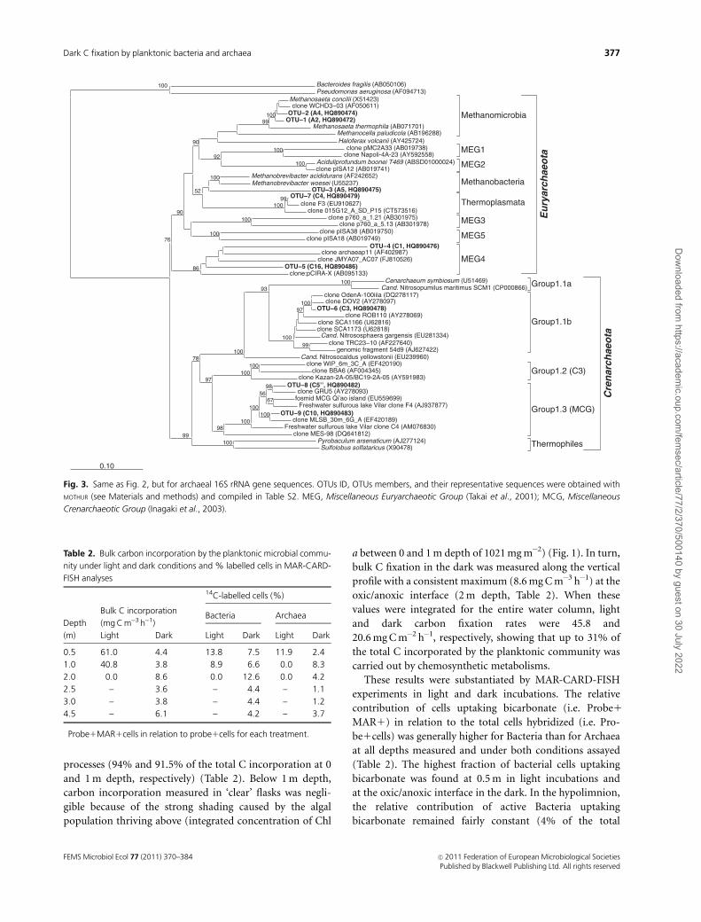
Fingerprinting of archaeal 16S rRNA gene fragments
using the general primer pair ARC344f-ARC915r only
retrieved four sequences from water samples collected at
the anoxic hypolimnion. All these sequences were assigned
to OTUs affiliated to methanogenic euryarchaeota from the
Methanosaeta cluster (OTU-1 and OTU-2, Fig. 3 and Table
S2) and Methanobacteriales (A5, OTU-3). Considering the
low archaeal richness detected using this general primer pair,
a second primer combination (ARC337f-ARC915r) was
applied to detect archaeal phylotypes potentially overlooked
by the primer pair ARC344F-ARC915R. When used in
nested reactions, the primer pair ARC337f-ARC915r is
biased towards lacustrine crenarchaeota and it considerably
improves the recovery of crenarchaeota phylotypes as de-
scribed previously for the neighboring Lake Vilar (Lliros
et al., 2008). Using this primer pair, 11 additional sequences
were recovered (Fig. S1). These sequences grouped into six
archaeal OTUs (97% cutoff, Table S2) affiliated to crenarch-
aeal lineages such as the Soil Group 1.1b (OTU-6) and the
Miscellaneous Crenarchaeotic Group (MCG, OTU-8 and -9)
and to the euryarchaeal Miscellaneous Euryarchaeotic Group
(MEG, OTU-4 and -5) and Thermoplasmatales (OTU-7)
(Fig. 3 and Table S2).
Carbon dioxide incorporation by the planktonicprokaryotic community
Samples from each water compartment (oxic epilimnion,
oxic/anoxic interface, and anoxic hypolimnion) were col-
lected to study the vertical distribution of bulk inorganic
carbon incorporation under light and dark incubation
conditions. Incubations carried out in the epilimnion
showed the maximal contribution of photosynthetic
Algal plastids
Firmicutes
Betaproteobacteria
Actinobacteria
Chlorobi
Bacteroidetes
Alphaproteobacteria
0.10
Fig. 2. NJ phylogenetic tree generated by the ARB software package showing the affiliation of the bacterial 16S rRNA gene sequences retrieved from the
lagoon (in bold) and grouped into OTUs (only the representative sequence for each OTU is shown; see Table S1 for details). Bootstrap support values
450% (1000 replicates) are shown. The scale bar indicates 10% estimated sequence divergence. The OTU number corresponds to the identification
number shown in Table S1. Sequences named following the code used in DGGE fingerprints (Fig. S1).
FEMS Microbiol Ecol 77 (2011) 370–384c� 2011 Federation of European Microbiological SocietiesPublished by Blackwell Publishing Ltd. All rights reserved
376 M. Lliros et al.
Dow
nloaded from https://academ
ic.oup.com/fem
sec/article/77/2/370/500140 by guest on 30 July 2022
processes (94% and 91.5% of the total C incorporation at 0
and 1 m depth, respectively) (Table 2). Below 1 m depth,
carbon incorporation measured in ‘clear’ flasks was negli-
gible because of the strong shading caused by the algal
population thriving above (integrated concentration of Chl
a between 0 and 1 m depth of 1021 mg m�2) (Fig. 1). In turn,
bulk C fixation in the dark was measured along the vertical
profile with a consistent maximum (8.6 mg C m�3 h�1) at the
oxic/anoxic interface (2 m depth, Table 2). When these
values were integrated for the entire water column, light
and dark carbon fixation rates were 45.8 and
20.6 mg C m�2 h�1, respectively, showing that up to 31% of
the total C incorporated by the planktonic community was
carried out by chemosynthetic metabolisms.
These results were substantiated by MAR-CARD-FISH
experiments in light and dark incubations. The relative
contribution of cells uptaking bicarbonate (i.e. Probe1
MAR1) in relation to the total cells hybridized (i.e. Pro-
be1cells) was generally higher for Bacteria than for Archaea
at all depths measured and under both conditions assayed
(Table 2). The highest fraction of bacterial cells uptaking
bicarbonate was found at 0.5 m in light incubations and
at the oxic/anoxic interface in the dark. In the hypolimnion,
the relative contribution of active Bacteria uptaking
bicarbonate remained fairly constant (4% of the total
Euryarch
aeota
MEG1
Methanomicrobia
MEG2
MEG3
MEG5
Thermoplasmata
MEG4
Methanobacteria
Crenarch
aeota
Group1.1b
Group1.3 (MCG)
Group1.1a
Thermophiles
Group1.2 (C3)
0.10
Fig. 3. Same as Fig. 2, but for archaeal 16S rRNA gene sequences. OTUs ID, OTUs members, and their representative sequences were obtained with
MOTHUR (see Materials and methods) and compiled in Table S2. MEG, Miscellaneous Euryarchaeotic Group (Takai et al., 2001); MCG, Miscellaneous
Crenarchaeotic Group (Inagaki et al., 2003).
Table 2. Bulk carbon incorporation by the planktonic microbial commu-
nity under light and dark conditions and % labelled cells in MAR-CARD-
FISH analyses
Depth
(m)
Bulk C incorporation
(mg C m�3 h�1)
14C-labelled cells (%)�
Bacteria Archaea
Light Dark Light Dark Light Dark
0.5 61.0 4.4 13.8 7.5 11.9 2.4
1.0 40.8 3.8 8.9 6.6 0.0 8.3
2.0 0.0 8.6 0.0 12.6 0.0 4.2
2.5 – 3.6 – 4.4 – 1.1
3.0 – 3.8 – 4.4 – 1.2
4.5 – 6.1 – 4.2 – 3.7
�Probe1MAR1cells in relation to probe1cells for each treatment.
FEMS Microbiol Ecol 77 (2011) 370–384 c� 2011 Federation of European Microbiological SocietiesPublished by Blackwell Publishing Ltd. All rights reserved
377Dark C fixation by planktonic bacteria and archaea
Dow
nloaded from https://academ
ic.oup.com/fem
sec/article/77/2/370/500140 by guest on 30 July 2022

EUB1hybridized cells). For Archaea, the highest percen-
tages of cells uptaking bicarbonate were observed again at
0.5 m in light incubations (�12% of the total ARC9151
hybridized cells), and both at the epilimnion (with a clear
maximum located at 1 m depth) and at the bottom of the
lagoon, in the dark.
Cloning of the biotin acetyl-CoA carboxylasegene
Amplification of accC genes from water samples only yielded
positive results with the primer set described by Auguet et al.
(2008), whereas the AccA-F573/-R279 primer combination
yielded no amplicons (data not shown) even after careful
DNA purification and optimization of the original PCR
conditions (Yakimov et al., 2009). Clone libraries were
constructed for the oxic epilimnion (0–1 m depth), the
metalimnion (2–2.25 m depth), the hypolimnion (4 m
depth), and the sediment of the lagoon. Overall, 133 biotin
acetyl-CoA carboxylase sequences were obtained (Table 3).
Annotation and sequence comparison against databases
showed only bacterial sequences, which affiliated to a broad
range of species, the Betaproteobacteria (63%) and the
Bacteroidetes (8%) being the most frequent taxa in the top-
hit BLAST species distribution (Table S3). Particularly, 46 and
30 accC clones showed high sequence identity (4 95%)
with gene sequences from Polynucleobacter necessarius ssp.
asymbioticus QLW-P1DMWA-1 and Ralstonia pickettii 12J,
respectively. The clone library constructed from the sedi-
ment sample recovered 17 sequences (Table 3), with iden-
tities ranging from 74% to 83.4% to pyruvate carboxylases
from Dehalococcoides ethenogenes (15 sequences, Table S3),
Candidatus ‘Methanoregula boonei’ (one sequence), and
Croceibacter atlanticus (one sequence). Five sequences
retrieved from different libraries (Table 3) showed a high
identity to carbamoyl-phosphate synthases from the
Betaproteobacteria (Polaromonas sp. JS666 and Leptothrix
chlolodnii SP-6) and the Chloroflexi (Oscillochloris trichoides)
(Table S3).
Discussion
The assimilation of inorganic carbon in the dark has
traditionally been considered negligible in photic zones of
both marine and freshwater environments due to the over-
whelming dominance of photosynthetic processes. How-
ever, in environments characterized by the presence of oxic/
anoxic interfaces, chemoautotrophic assimilation of CO2 by
planktonic microorganisms has been shown to be as im-
portant as photosynthesis (Cloern et al., 1983; Camacho
et al., 2001; Garcıa-Cantizano et al., 2005). The relevance of
dark carbon fixation within the C cycle in such environ-
ments progressively gained significance as new data accu-
mulated in the literature (Camacho et al., 2001; Hadas et al.,
2001; Garcıa-Cantizano et al., 2005; Casamayor et al., 2008;
Grote et al., 2008; Glaubitz et al., 2009, 2010; Casamayor,
2010; Jost et al., 2010). Most of these studies also identified
the different bacterial taxa potentially involved in these
processes such as the purple sulfur bacteria (Garcıa-Canti-
zano et al., 2005; Casamayor et al., 2008), the Epsilonproteo-
bacteria [e.g. Sulfurimonas (Grote et al., 2007, 2008)], and
the aerobic sulfur-oxidizing bacteria [e.g. Thiobacilli (Casa-
mayor, 2010)]. However, the capability of lacustrine archaea
other than ammonia oxidizers to incorporate CO2 in the
dark has not been assessed in aquatic environments with
well-defined oxic/anoxic interfaces. To date, archaeal che-
moautotrophy has been found in (hyper)thermophiles
(Berg et al., 2010), methanogens (Jones et al., 1987; Simpson
& Whitman, 1993), and ammonia-oxidizing crenarchaeota
(Konneke et al., 2005; Hallam et al., 2006; Martens-Habbena
et al., 2009; Walker et al., 2010), although the high diversity
of archaea in the most diverse habitats (Schleper et al., 2005)
provides the possibility of a wider occurrence of autotrophy
over the entire archaeal domain.
Coromina is a small lagoon of the Banyoles Karstic
System characterized by the high concentrations of ammo-
nium and sulfide in the anoxic water compartment and the
prevalence of photosynthetic primary producers that are
either eukaryotic (green and brown algae) or prokaryotic
Table 3. Distribution of gene sequences in the four accC clone libraries constructed from water and sediment samples of the lagoon (see text for
details)
Clone library
Clones with
correct insert (%)�Unspecific
sequences (%)
Valid
sequences
(%)
Acetyl-CoA
carboxylasew
(%)
Pyruvate
carboxylasew
(%)
Carbamoyl-
phosphate
synthasew (%)
Epilimnion (0–1 m) 44 (88) 7 (16) 37 (84) 36 (97) 0 1 (3)
Metalimnion (2–2.5 m) 46 (92) 7 (15) 39 (85) 36 (92) 0 3 (8)
Hypolimnion (4 m) 48 (96) 10 (21) 38 (79) 38 (100) 0 0
Sediment 49 (98) 8 (16) 41 (84) 23 (56) 17 (42) 1 (2)
Total 187 (93.5) 32 (17) 155 (83) 133 (86) 17 (11) 5 (3)
�A total of 50 clones were picked for each library (200 screened clones in total).wSequence description was obtained by automatic annotation of nucleotide sequences using BLAST2GO software.
The relative contribution (in %) of each sequence cluster with respect to the number of correct sequences for that group is also indicated.
FEMS Microbiol Ecol 77 (2011) 370–384c� 2011 Federation of European Microbiological SocietiesPublished by Blackwell Publishing Ltd. All rights reserved
378 M. Lliros et al.
Dow
nloaded from https://academ
ic.oup.com/fem
sec/article/77/2/370/500140 by guest on 30 July 2022

(green anoxygenic photosynthetic bacteria) (Borrego &
Garcia-Gil, 1994; Gich et al., 2001). In fact, the data
presented here agree with this picture because bicarbonate
photoassimilation accounted for up to 69% of the total
carbon incorporated by the planktonic community in the
entire water column. However, when integrated values of
light and dark C fixation rates were calculated on a daily
basis assuming: (1) an average value of 10 h of solar
radiation and (2) that both processes operate at constant
rates, the bulk dark C fixation (494 mg C m�2 day�1) exceeds
photosynthetic assimilation (459 mg C m�2 day�1). These
data support the significance of dark C fixation processes
mediated by planktonic microorganisms in stratified fresh-
water systems and agree with those calculated in other
karstic lakes with oxic/anoxic interfaces (Table 4).
These results were corroborated by MAR-CARD-FISH
experiments. Archaeal abundance (up to 7% of the total
DAPI counts) was within the range reported for other
freshwater environments (Casamayor & Borrego, 2009).
The maximum archaeal counts were found at the upper
layers of the hypolimnion, although both the bulk dark C
incorporation rates and the abundance of active archaeal
cells in MAR-CARD-FISH incubations showed their max-
imal values at the oxic/anoxic interface and above (Table 2).
Of particular interest was the relatively high fraction of
archaeal cells (�12%) actively uptaking bicarbonate under
light conditions at the surface. Stimulation of CO2 fixation
in the light has been reported in proteorhodopsin-contain-
ing marine Flavobacteria (Gomez-Consarnau et al., 2007;
Gonzalez et al., 2008), but it has never been reported for
archaea. Current data are not sufficient to properly explain
this observation and further work is needed to resolve the
extent of such stimulation of CO2 uptake by light in archaea,
if any. Besides, we cannot rule out a mixotrophic or even a
heterotrophic metabolism on labelled organic compounds
excreted by phototrophs, especially considering that several
authors have reported the ability of some marine archaeal
groups to incorporate organic matter (Ouverney & Fuhr-
man, 2000; Perez et al., 2003; Kirchman et al., 2007).
At the oxic/anoxic interface and in the hypolimnion, all
the archaeal phylotypes recovered affiliated to crenarchaeota
of the Miscellaneous Crenarchaeotic Group (MCG, OTU-8,
and -9) (Fig. 3) and to methanogenic euryarchaeotal
lineages [either to acetoclastic Methanosaeta (OTU-1 and -
2) or to hydrogenotrophic representatives close to Methano-
brevibacter woesei (OTU-3)]. A high richness of MCG
phylotypes is a distinctive feature of anoxic water layers in
stratified, sulfide-rich lakes (Lliros et al., 2008, 2010),
although the use of a PCR primer combination biased
towards lacustrine crenarchaeota may overestimate their
actual relevance in the ecosystem (for details on primer
design and specificity, see Lliros et al., 2008). In clear
contrast to the wealth of information available for ammo-
nia-oxidizing crenarchaeota, very little is known about the
metabolic capabilities and ecological role of the MCG
archaea. This phylogenetically diverse group seems to be
especially abundant and active in anoxic, nutrient-rich
habitats such as deep subsurface sediments (Parkes et al.,
2005; Biddle et al., 2006) and they have been classified as
heterotrophic anaerobes (Teske & Sorensen, 2008). This
putative metabolism agrees with the physico-chemical con-
ditions prevalent in the lagoon.
The co-occurrence of methanogenic archaea with sulfate-
reducing bacteria is not unusual and has been reported for
other freshwater environments (Lovley & Klug, 1983; Sinke
et al., 1992; Holmer & Storkholm, 2001), although metha-
nogenesis is usually low in comparison with sulfate respira-
tion when sulfate is not limiting (Holmer & Storkholm,
2001; Kallistova et al., 2006). Although no data on methane
concentration are available for the studied lagoon, the
vertical profile of sulfide concentration (Fig. 1) and the high
amount of sulfate present in underground waters of the
Banyoles karstic system [�10 mM, (Guerrero et al., 1985)]
suggest an active sulfate reduction both in the planktonic
compartment and in the sediment. On the other hand, no
evidences on the presence of archaea potentially involved in
anaerobic oxidation of methane, either free-living or asso-
ciated with sulfate-reducing bacteria [ANME groups (Hin-
richs & Boetius, 2002)], were found in the molecular
fingerprints, although the detection limit of the DGGE may
explain that low abundant (o 1%) microbial populations
remained undetected (Casamayor et al., 2000). The detec-
tion of archaeal cells actively uptaking bicarbonate in the
anoxic waters (Table 2) may be related either by the activity
of autotrophic methanogens (Simpson & Whitman, 1993)
or by the heterotrophic incorporation of CO2 by archaea as
reported for bacteria.
The bacterial planktonic community was mainly com-
posed by phylotypes affiliated to Betaproteobacteria, Bacter-
oidetes, and Chlorobi (Fig. 2). Minor components of the
community were ascribed to Alphaproteobacteria, high and
low G1C Gram-positive bacteria, and algal plastids. With
the exception of those phylotypes affiliated to either algae or
GSB recovered from the oxic epilimnion and the
Table 4. Values of dark carbon fixation rates integrated for the entire
water column and their relative contribution to total assimilated C in
different lakes with an oxygen/sulfide interface
System
Dark C fixation
(mg C m�2 h�1)
Contribu-
tion (%) Source
Coromina 20.6 31 This study
Vilar 18.9 32 Casamayor et al. (2008)
Ciso 14.5 52 Garcıa-Cantizano et al.
(2005)
Big Soda Lake 17 30 Cloern et al. (1983)
Kinneret 13 16 Hadas et al. (2001)
FEMS Microbiol Ecol 77 (2011) 370–384 c� 2011 Federation of European Microbiological SocietiesPublished by Blackwell Publishing Ltd. All rights reserved
379Dark C fixation by planktonic bacteria and archaea
Dow
nloaded from https://academ
ic.oup.com/fem
sec/article/77/2/370/500140 by guest on 30 July 2022

metalimnion, respectively, the remaining OTUs showed
close similarities to heterotrophic bacteria capable of growth
on complex organic matter (Table S1). Besides, the observa-
tion that all analyzed accC clones were most closely related
to carboxylases from Betaproteobacteria and the Bacteroi-
detes points to a large relevance of heterotrophs at the meta-
and hipolimnion. Particularly interesting is the large num-
ber of accC clones showing high sequence identity (4 95%)
to accC genes from both P. necessarius ssp. asymbioticus
QLW-P1DMWA-1 (PnecC) and R. pickettii 12J (Table S3).
The latter is a usual inhabitant of polluted soils and aquatic
environments having the capacity to degrade a wide range of
complex organic substances (e.g. tricloroethylene and aro-
matic hydrocarbons) (Ryan et al., 2007). In turn, PnecC is
an obligately free-living Polynucleobacter strain that is ubi-
quitous in freshwater habitats characterized by high con-
centrations of humic substances (Hahn et al., 2009;
Jezberova et al., 2010). The measured values for proxies of
humic substances suggest a high content of these organic
compounds in the lagoon, which may derive from the high
external inputs of organic matter into the system (Borrego &
Garcia-Gil, 1994). The capacity of P. necessarius to grow on
photodegradation products of humic substances (Buck
et al., 2009; Hahn et al., 2009; Watanabe et al., 2009) might
then confer some competitive advantage over other bacterial
species as suggested previously to explain their ubiquity in
freshwater lakes (Jezberova et al., 2010).
Overall, the results from MAR-CARD-FISH incubations,
16S rRNA gene fingerprinting, and accC clone libraries
suggest that the dark CO2 assimilation activity measured
was mainly carried out by heterotrophs rather than by true
chemoautotrophs. Heterotrophic CO2 assimilation is a well-
known process in chemoorganotrophic microorganisms
either to equilibrate their C metabolism via anaplerotic
reactions or to fuel carboxylation reactions for the synthesis
of different cellular constituents, for example fatty acids,
nucleotides, and amino acids (Dijkhuizen & Harder, 1984,
1985; Roslev et al., 2004). The occurrence of these peripheral
metabolic pathways was demonstrated in environmental
samples and pure cultures of heterotrophic bacteria (Roslev
et al., 2004) and it has been proposed to explain the high
rates of dark carbon fixation both in oxic/anoxic interfaces
(Casamayor et al., 2008) and in laboratory-controlled batch
enrichments of Arctic seawater and dominated by hetero-
trophs (Alonso-Saez et al., 2010). Although no evidences of
heterotrophic CO2 fixation by archaea have been reported so
far, it would be reasonable to assume that heterotrophic
archaea also need to compensate their carbon metabolism
using different anaplerotic pathways. In this regard, the
potential capacity of mesophilic archaea other than ammo-
nia-oxidizers to fix inorganic carbon is an interesting issue
that should be addressed properly. Considering the high
microbial diversity and activity found in oxic/anoxic inter-
faces of stratified lakes, these microenvironments constitute
optimal systems for the investigation of such processes.
Acknowledgements
We are indebted to Dr J.M. Gasol for allowing us to use
laboratory facilities at the Institut de Ciencies del Mar
(ICM-CSIC) for MAR-CARD-FISH analyses. We also thank
two anonymous reviewers for their helpful comments and
suggestions. This study was funded through the projects
VIARC (Ref. REN 2003-08333-GLO) and CRENYC
(CGL2006-12058) to C.M.B. and E.O.C. from the Spanish
Ministerio de Ciencia e Innovacion (MCINN). M.L. and
A.P. are recipients of PhD student fellowships from the
Spanish government and the Generalitat de Catalunya,
respectively.
References
Alonso C & Pernthaler J (2005) Incorporation of glucose under
anoxic conditions by bacterioplankton from coastal North Sea
surface waters. Appl Environ Microb 71: 1709–1716.
Alonso-Saez L, Galand PE, Casamayor EO, Pedros-Alio C &
Bertilsson S (2010) High bicarbonate assimilation in the dark
by Arctic bacteria. ISME J 4: 1581–1590.
Amann R & Fuchs BM (2008) Single-cell identification in
microbial communities by improved fluorescence in situ
hybridization techniques. Nat Rev Microbiol 6: 339–348.
Amann RI, Binder BJ, Olson RJ, Chisholm SW, Devereux R &
Stahl DA (1990) Combination of 16S rRNA-targeted
oligonucleotide probes with flow cytometry for analyzing
mixed microbial populations. Appl Environ Microb 56:
1919–1925.
Auguet JC, Borrego CM, Baneras L & Casamayor EO (2008)
Fingerprinting the genetic diversity of the biotin carboxylase
gene (accC) in aquatic ecosystems as a potential marker for
studies of carbon dioxide assimilation in the dark. Environ
Microbiol 10: 2527–2536.
Auguet JC, Barberan A & Casamayor EO (2010) Global ecological
patterns in uncultured Archaea. ISME J 4: 182–190.
Battin TJ, Wille A, Sattler B & Psenner R (2001) Phylogenetic and
functional heterogeneity of sediment biofilms along
environmental gradients in a glacial stream. Appl Environ
Microb 67: 799–807.
Berg IA, Kockelkorn D, Buckel W & Fuchs G (2007) A 3-
hydroxypropionate/4-hydroxybutyrate autotrophic carbon
dioxide assimilation pathway in Archaea. Science 318:
1782–1786.
Berg IA, Ramos-Vera WH, Petri A, Huber H & Fuchs G (2010)
Study of the distribution of autotrophic CO2 fixation cycles in
Crenarchaeota. Microbiology 156: 256–269.
Biddle JF, Lipp JS, Lever MA et al. (2006) Heterotrophic Archaea
dominate sedimentary subsurface ecosystems off Peru. P Natl
Acad Sci USA 103: 3846–3851.
FEMS Microbiol Ecol 77 (2011) 370–384c� 2011 Federation of European Microbiological SocietiesPublished by Blackwell Publishing Ltd. All rights reserved
380 M. Lliros et al.
Dow
nloaded from https://academ
ic.oup.com/fem
sec/article/77/2/370/500140 by guest on 30 July 2022

Borrego C & Garcia-Gil J (1994) Caracterizacion limnologica de
la Coromina, una laguna hipereutrofica del sistema lacustre de
Banyoles. Limnetica 10: 43–51.
Borrego CM & Garcia-Gil LJ (1995) Rearrangement of light
harvesting bacteriochlorophyll homologs as a response of
Green Sulfur Bacteria to low light intensities. Photosynth Res
45: 21–30.
Brock TD, Brock ML, Bott TL & Edwards MR (1971) Microbial
life at 90 1C: the sulfur bacteria of Boulder Spring. J Bacteriol
107: 303–314.
Buck U, Grossart HP, Amann R & Pernthaler J (2009) Substrate
incorporation patterns of bacterioplankton populations in
stratified and mixed waters of a humic lake. Environ Microbiol
11: 1854–1865.
Camacho A & Vicente E (1998) Carbon photoassimilation by
sharply stratified phototrophic communities at the chemocline
of Lake Arcas (Spain). FEMS Microbiol Ecol 25: 11–22.
Camacho A, Erez J, Chicote A, Florin M, Squires MM, Lehmann C
& Bachofen R (2001) Microbial microstratification, inorganic
carbon photoassimilation and dark carbon fixation at the
chemocline of the meromictic Lake Cadagno (Switzerland) and
its relevance to the food web. Aquat Sci 63: 91.
Casamayor EO (2010) Vertical distribution of planktonic
autotrophic thiobacilli and dark CO2 fixation rates in lakes
with oxygen-sulfide interfaces. Aquat Microb Ecol 59: 217–228.
Casamayor EO & Borrego CM (2009) Archaea. Encyclopedia of
Inland Waters, Vol. 3 (Likens GE, ed), pp. 167–181. Elsevier,
Oxford.
Casamayor EO, Schafer H, Baneras L, Pedros-Alio C & Muyzer G
(2000) Identification of and spatio-temporal differences
between microbial assemblages from two neighboring
sulfurous lakes: comparison by microscopy and denaturing
gradient gel electrophoresis. Appl Environ Microb 66: 499–508.
Casamayor EO, Muyzer G & Pedros-Alio C (2001) Composition
and temporal dynamics of planktonic archaeal assemblages
from anaerobic sulfurous environments studied by 16S rDNA
denaturing gradient gel electrophoresis and sequencing. Aquat
Microb Ecol 25: 237–246.
Casamayor EO, Garcıa-Cantizano J & Pedros-Alio C (2008)
Carbon dioxide fixation in the dark by photosynthetic bacteria
in sulfide-rich stratified lakes with oxic–anoxic interfaces.
Limnology and Oceanography. Limnol Oceanogr 53:
1193–1203.
Chaban B, Ng SY & Jarrell KF (2006) Archaeal habitats–from the
extreme to the ordinary. Can J Microbiol 52: 73–116.
Cloern JE, Cole BE & Oremland RS (1983) Autotrophic processes
in meromictic Soda Lake, Nevada. Limnol Oceanogr 28:
1049–1061.
Conesa A, Gotz S, Garcia-Gomez JM, Terol J, Talon M & Robles
M (2005) Blast2GO: a universal tool for annotation,
visualization and analysis in functional genomics research.
Bioinformatics 21: 3674–3676.
Culver DA & Brunskill GJ (1969) Fayetteville Green Lake, New
York. V. Studies of primary production and zooplankton in a
meromictic marl lake. Limnol Oceanogr 14: 862–873.
Daims H, Bruhl A, Amann R, Schleifer K & Wagner M (1999) The
domain-specific probe eub338 is insufficient for the detection
of all Bacteria: development and evaluation of a more
comprehensive probe set. Syst Appl Microbiol 22: 434–444.
De Haan H (1972) Molecular-size distribution of soluble humic
compounds from different natural waters. Freshwater Biol 2:
235–241.
DeLong EF (1992) Archaea in coastal marine environments.
P Natl Acad Sci USA 89: 5685–5689.
DeLong EF (1998) Everything in moderation: archaea as ‘non-
extremophiles’. Curr Opin Genet Dev 8: 649–654.
DeLong EF & Pace NR (2001) Environmental diversity of bacteria
and archaea. Syst Biol 50: 470–478.
Dijkhuizen L & Harder W (1984) Current views on the regulation
of autotrophic carbon dioxide fixation via the Calvin cycle in
bacteria. Anton Leeuw Int J G 50: 473–487.
Dijkhuizen L & Harder W (1985) Microbial metabolism of
carbon dioxide. Comprehensive Biotechnology, Vol. 1 (Dalton
H, ed), pp. 409–423. Pergamon Press, Oxford.
Eiler A & Bertilsson S (2004) Composition of freshwater bacterial
communities associated with cyanobacterial blooms in four
Swedish lakes. Environ Microbiol 6: 1228–1243.
Felsenstein J (2007) PHYLIP (Phylogeny Inference Package).
Department of Genetics, University of Washington, Seattle,
WA.
Garcıa-Cantizano J, Casamayor EO, Gasol JM, Guerrero R &
Pedros-Alio C (2005) Partitioning of CO2 incorporation
among planktonic microbial guilds and estimation of in situ
specific growth rates. Microb Ecol 50: 230–241.
Gasol JM, Garcıa-Cantizano J, Massana R, Guerrero R & Pedros-
Alio C (1993) Physiological ecology of a metalimnetic
Cryptomonas population: relationships to light, sulfide, and
nutrients. J Plankton Res 15: 255–275.
Gich FB, Borrego CM, Martınez-Planells A, Steensgaard DB,
Garcia-Gil LJ & Holzwarth AR (2001) Variability of the
photosynthetic antenna of a Pelodictyon clathratiforme
population from a freshwater holomictic pond. FEMS
Microbiol Ecol 37: 11–19.
Glaubitz S, Lueders T, Abraham WR, Jost G, Jurgens K & Labrenz
M (2009) 13C-isotope analyses reveal that
chemolithoautotrophic Gamma- and Epsilonproteobacteria
feed a microbial food web in a pelagic redoxcline of the central
Baltic Sea. Environ Microbiol 11: 326–337.
Glaubitz S, Labrenz M, Jost G & Jurgens K (2010) Diversity of
active chemolithoautotrophic prokaryotes in the sulfidic zone
of a Black Sea pelagic redoxcline as determined by rRNA-based
stable isotope probing. FEMS Microbiol Ecol 74: 32–41.
Gomez-Consarnau L, Gonzalez JM, Coll-Llado M et al. (2007)
Light stimulates growth of proteorhodopsin-containing
marine Flavobacteria. Nature 445: 210–213.
Gonzalez JM, Fernandez-Gomez B, Fernandez-Guerra A et al.
(2008) Genome analysis of the proteorhodopsin-containing
marine bacterium Polaribacter sp. MED152 (Flavobacteria).
P Natl Acad Sci USA 105: 8724–8729.
FEMS Microbiol Ecol 77 (2011) 370–384 c� 2011 Federation of European Microbiological SocietiesPublished by Blackwell Publishing Ltd. All rights reserved
381Dark C fixation by planktonic bacteria and archaea
Dow
nloaded from https://academ
ic.oup.com/fem
sec/article/77/2/370/500140 by guest on 30 July 2022

Grote J, Labrenz M, Pfeiffer B, Jost G & Jurgens K (2007)
Quantitative distributions of Epsilonproteobacteria and a
Sulfurimonas subgroup in pelagic redoxclines of the central
Baltic Sea. Appl Environ Microb 73: 7155–7161.
Grote J, Jost G, Labrenz M, Herndl GJ & Jurgens K (2008)
Epsilonproteobacteria represent the major portion of
chemoautotrophic bacteria in sulfidic waters of pelagic
redoxclines of the Baltic and Black Seas. Appl Environ Microb
74: 7546–7551.
Guerrero R, Montesinos E, Pedros-Alio C et al. (1985)
Phototrophic sulfur bacteria in two Spanish lakes: vertical
distribution and limiting factors. Limnol Oceanogr 30:
919–931.
Hadas O, Pinkas R & Erez J (2001) High chemoautotrophic
primary production in Lake Kinneret, Israel: A neglected link
in the carbon cycle of the lake. Limnol Oceanogr 46:
1968–1976.
Hahn MW, Lang E, Brandt U, Wu QL & Scheuerl T (2009)
Emended description of the genus Polynucleobacter and the
species Polynucleobacter necessarius and proposal of two
subspecies, P. necessarius subsp. necessarius subsp. nov. and
P. necessarius subsp. asymbioticus subsp. nov. Int J Syst Evol
Micr 59: 2002–2009.
Hall TA (1999) BioEdit: a user-friendly biological sequence
alignment editor and analysis program for Windows 95/98/
NT. Nucl Acids Symp Ser 41: 95–98.
Hallam SJ, Mincer TJ, Schleper C, Preston CM, Roberts K,
Richardson PM & DeLong EF (2006) Pathways of carbon
assimilation and ammonia oxidation suggested by
environmental genomic analyses of marine Crenarchaeota.
PLoS Biology 4: e95.
Herndl GJ, Reinthaler T, Teira E, van Aken H, Veth C, Pernthaler
A & Pernthaler J (2005) Contribution of Archaea to total
prokaryotic production in the deep Atlantic Ocean. Appl
Environ Microb 71: 2303–2309.
Hinrichs KU & Boetius A (2002) The anaerobic oxidation of
methane: new insights in microbial ecology and
biogeochemistry. Ocean Margin Systems (Wefer G, Billet D,
Hebbeln D, Jørgensen BB, Schluter M & Van Weering TC, eds),
pp. 457–477. Springer-Verlag, Heidelberg.
Holmer M & Storkholm P (2001) Sulphate reduction and
sulphur cycling in lake sediments: a review. Freshwater Biol 46:
431–451.
Huber T, Faulkner G & Hugenholtz P (2004) Bellerophon: a
program to detect chimeric sequences in multiple sequence
alignments. Bioinformatics 20: 2317–2319.
Inagaki F, Suzuki M, Takai K et al. (2003) Microbial communities
associated with geological horizons in coastal subseafloor
sediments from the Sea of Okhotsk. Appl Environ Microb 69:
7224–7235.
Ingalls AE, Shah SR, Hansman RL, Aluwihare LI, Santos GM,
Druffel ER & Pearson A (2006) Quantifying archaeal
community autotrophy in the mesopelagic ocean using
natural radiocarbon. P Natl Acad Sci USA 103: 6442–6447.
Jezberova J, Jezbera J, Brandt U, Lindstrom ES, Langenheder S &
Hahn MW (2010) Ubiquity of Polynucleobacter necessarius ssp.
asymbioticus in lentic freshwater habitats of a heterogeneous
2000 km area. Environ Microbiol 12: 658–669.
Jones WJ, Nagle DP Jr. & Whitman WB (1987) Methanogens and
the diversity of archaebacteria. Microbiol Rev 51: 135–177.
Jorgensen BB, Kuenen JG & Cohen Y (1979) Microbial
transformation of sulfur compounds in a stratified lake (Solar
Lake, Sinai). Limnol Oceanogr 24: 799–822.
Jost G, Martens-Habbena W, Pollehne F, Schnetger B & Labrenz
M (2010) Anaerobic sulfur oxidation in the absence of nitrate
dominates microbial chemoautotrophy beneath the pelagic
chemocline of the eastern Gotland Basin, Baltic Sea. FEMS
Microbiol Ecol 71: 226–236.
Kallistova A, Kevbrina MV, Pimenov NV, Rusanov II, Rogozin D,
Wehrli B & Nozhevnikova AN (2006) Sulfate reduction and
methanogenesis in the Shira and Shunet meromictic lakes
(Khakass Republic, Russia). Mikrobiologiia 75: 828–835.
Kirchman DL, Elifantz H, Dittel AI, Malmstrom RR & Cottrell
MT (2007) Standing stocks and activity of Archaea and Bacteria
in the western Arctic Ocean. Limnol Oceanogr 52: 495–507.
Klammer S, Posch T, Sonntag B, Griebler C, Mindl B & Psenner R
(2002) Dynamics of bacterial abundance, biomass, activity,
and community composition in the oligotrophic Traunsee and
the Traun river (Austria). Water Air Soil Poll 2: 137–163.
Kletzin A, Urich T, Muller F, Bandeiras TM & Gomes CM (2004)
Dissimilatory oxidation and reduction of elemental sulfur in
thermophilic archaea. J Bioenerg Biomembr 36: 77–91.
Konneke M, Bernhard AE, de la Torre JR, Walker CB, Waterbury
JB & Stahl DA (2005) Isolation of an autotrophic ammonia-
oxidizing marine archaeon. Nature 437: 543–546.
Lliros M, Casamayor EO & Borrego C (2008) High archaeal
richness in the water column of a freshwater sulfurous karstic
lake along an interannual study. FEMS Microbiol Ecol 66:
331–342.
Lliros M, Gich F, Plasencia A et al. (2010) Vertical distribution of
ammonia-oxidizing crenarchaeota and methanogens in the
epipelagic waters of Lake Kivu. Appl Environ Microb 76:
6853–6863.
Lovley DR & Klug MJ (1983) Sulfate reducers can outcompete
methanogens at freshwater sulfate concentrations. Appl
Environ Microb 45: 187–192.
Ludwig W, Strunk O, Westram R et al. (2004) ARB: a software
environment for sequence data. Nucleic Acids Res 32:
1363–1371.
Manz W, Amann R, Ludwig W, Vancanneyt M & Schleifer KH
(1996) Application of a suite of 16S rRNA-specific
oligonucleotide probes designed to investigate bacteria of the
phylum cytophaga–flavobacter–bacteroides in the natural
environment. Microbiology 142: 1097–1106.
Margalef R (1982) Ecologıa. Omega, Barcelona.
Martens-Habbena W, Berube PM, Urakawa H, de la Torre JR &
Stahl DA (2009) Ammonia oxidation kinetics determine niche
separation of nitrifying Archaea and Bacteria. Nature 461:
976–979.
FEMS Microbiol Ecol 77 (2011) 370–384c� 2011 Federation of European Microbiological SocietiesPublished by Blackwell Publishing Ltd. All rights reserved
382 M. Lliros et al.
Dow
nloaded from https://academ
ic.oup.com/fem
sec/article/77/2/370/500140 by guest on 30 July 2022

Muyzer G & Ramsing NB (1995) Molecular methods to study the
organization of microbial communities. Water Sci Technol 32:
1–9.
Ouverney CC & Fuhrman JA (2000) Marine planktonic archaea
take up amino acids. Appl Environ Microb 66: 4829–4833.
Overmann J (2006) The family Chlorobiaceae. The Prokaryotes: A
Handbook on the Biology of Bacteria, Vol. 7 (Dworkin M,
Falkow S, Rosenberg E, Schleifer KH & Stackebrandt E, eds),
pp. 359–378. Springer, Heidelberg.
Parkes RJ, Webster G, Cragg BA et al. (2005) Deep sub-seafloor
prokaryotes stimulated at interfaces over geological time.
Nature 436: 390–394.
Pedros-Alio C & Guerrero R (1991) Abundance and activity of
bacterioplankton in warm lakes. Verh Int Verein Limnol 24:
1212–1219.
Pedros-Alio C, Garcıa-Cantizano J & Calderon Paz JI (1993)
Bacterial production in anarobic water columns. Current
Methods in Aquatic Microbial Ecology (Kempf PF,
Sherr BF, Sherr EB & Cole JJ, eds), pp. 519–530. Lewis, Boca
Raton, FL.
Perez MT, Pausz C & Herndl GJ (2003) Major shift in
bacterioplankton utilization of enantiomeric aminoacids
between surface waters and the ocean’s interior. Limnol
Oceanogr 48: 755–763.
Pernthaler A, Pernthaler J & Amann R (2002) Fluorescence in situ
hybridization and catalyzed reported deposition for the
identification of marine bacteria. Appl Environ Microb 68:
3094–4101.
Raskin L, Stromley JM, Rittmann BE & Stahl DA (1994) Group-
specific 16S rRNA hybridization probes to describe natural
communities of methanogens. Appl Environ Microb 60:
1232–1240.
Robertson CE, Harris JK, Spear JR & Pace NR (2005)
Phylogenetic diversity and ecology of environmental Archaea.
Curr Opin Microbiol 8: 638–642.
Roslev P, Larsen MB, Jorgensen D & Hesselsoe M (2004) Use of
heterotrophic CO2 assimilation as a measure of metabolic
activity in planktonic and sessile bacteria. J Microbiol Meth 59:
381–393.
Ryan MP, Pembroke JT & Adley CC (2007) Ralstonia pickettii in
environmental biotechnology: potential and applications.
J Appl Microbiol 103: 754–764.
Saitou N & Nei M (1987) The neighbor-joining method: a new
method for reconstructing phylogenetic trees. Mol Biol Evol 4:
406–425.
Schleper C, Jurgens G & Jonuscheit M (2005) Genomic studies of
uncultivated archaea. Nat Rev Microbiol 3: 479–488.
Schloss PD, Westcott SL, Ryabin T et al. (2009) Introducing
mothur: open-source, platform-independent, community-
supported software for describing and comparing microbial
communities. Appl Environ Microb 75: 7537–7541.
Schouten S, Hopmans EC, Pancost RD & Sinninghe Damste J
(2000) Widespread occurrence of structurally diverse
tetraether membrane lipids: evidence for the ubiquitous
presence of low-temperature relatives of hyperthermophiles.
P Natl Acad Sci USA 97: 14421–14426.
Schouten S, Hopmans EC, Baas M et al. (2008) Intact membrane
lipids of ‘Candidatus Nitrosopumilus maritimus,’ a cultivated
representative of the cosmopolitan mesophilic group I
Crenarchaeota. Appl Environ Microb 74: 2433–2440.
Shively JM, van Keulen G & Meijer WG (1998) Something from
almost nothing: carbon dioxide fixation in chemoautotrophs.
Annu Rev Microbiol 52: 191–230.
Simpson PG & Whitman WB (1993) Anabolic pathways in
methanogens. Methanogenesis: Ecology, Physiology,
Biochemistry and Genetics (Ferry JG, ed), pp. 445–472.
Chapman & Hall Inc., New York.
Sinke AJC, Cornelese AA, Cappenberg TE & Zehnder AJB (1992)
Seasonal variation in sulfate reduction and methanogenesis in
peaty sediments of eutrophic Lake Loosdrecht, The
Netherlands. Biogeochemistry 16: 43–61.
Sinninghe Damste JS, Rijpstra WI, Hopmans EC, Prahl FG,
Wakeham SG & Schouten S (2002) Distribution of membrane
lipids of planktonic Crenarchaeota in the Arabian Sea. Appl
Environ Microb 68: 2997–3002.
Takai K, Moser DP, DeFlaun M, Onstott TC & Fredrickson JK
(2001) Archaeal diversity in waters from deep South African
gold mines. Appl Environ Microb 67: 5750–5760.
Teske A & Sorensen KB (2008) Uncultured archaea in deep
marine subsurface sediments: have we caught them all? ISME J
2: 3–18.
Varela MM, van Aken HM, Sintes E & Herndl GJ (2008)
Latitudinal trends of Crenarchaeota and Bacteria in the meso-
and bathypelagic water masses of the Eastern North Atlantic.
Environ Microbiol 10: 110–124.
Walker CB, de la Torre JR, Klotz MG et al. (2010) Nitrosopumilus
maritimus genome reveals unique mechanisms for
nitrification and autotrophy in globally distributed marine
crenarchaea. P Natl Acad Sci USA 107: 8818–8823.
Watanabe K, Komatsu N, Ishii Y & Negishi M (2009) Effective
isolation of bacterioplankton genus Polynucleobacter from
freshwater environments grown on photochemically degraded
dissolved organic matter. FEMS Microbiol Ecol 67: 57–68.
Wuchter C, Schouten S, Boschker HT & Sinninghe Damste JS
(2003) Bicarbonate uptake by marine Crenarchaeota. FEMS
Microbiol Lett 219: 203–207.
Yakimov MM, La Cono V & Denaro R (2009) A first insight into
the occurrence and expression of functional amoA and accA
genes of autotrophic and ammonia-oxidizing bathypelagic
Crenarchaeota of Tyrrhenian Sea. Deep-Sea Res II 56: 748–754.
Supporting Information
Additional Supporting Information may be found in the
online version of this article:
Fig. S1. DGGE fingerprints for 16S rRNA bacterial (left) and
archaeal (middle and right gels) 16S rRNA gene fragments
recovered in the water column of the lagoon.
FEMS Microbiol Ecol 77 (2011) 370–384 c� 2011 Federation of European Microbiological SocietiesPublished by Blackwell Publishing Ltd. All rights reserved
383Dark C fixation by planktonic bacteria and archaea
Dow
nloaded from https://academ
ic.oup.com/fem
sec/article/77/2/370/500140 by guest on 30 July 2022

Table S1. Identification number for the assigned bacterial
OTUs, phylogenetic affiliation, representative sequence for
each OTU, OTU members, and % similarity of the OTU
representative sequence with the first BLAST and the closest
cultured representative found in the BLAST search result list.
Table S2. Identification number for the assigned archaeal
OTUs, phylogenetic affiliation, representative sequence for
each OTU, OTU members, and % similarity of the repre-
sentative sequence with the first BLAST and the closest
cultured representative found in BLAST search result list.
Table S3. Top-hit BLAST species, phylogenetic affiliation,
sequence description, enzyme codes, average identity values,
and number of clones for each BLAST hit obtained in accC
clone libraries constructed from different depths of the
water column and the sediment (see text for details).
Please note: Wiley-Blackwell is not responsible for the
content or functionality of any supporting materials supplied
by the authors. Any queries (other than missing material)
should be directed to the corresponding author for the article.
FEMS Microbiol Ecol 77 (2011) 370–384c� 2011 Federation of European Microbiological SocietiesPublished by Blackwell Publishing Ltd. All rights reserved
384 M. Lliros et al.
Dow
nloaded from https://academ
ic.oup.com/fem
sec/article/77/2/370/500140 by guest on 30 July 2022


