Embed Size (px)
Citation preview
BioMed CentralBMC Neuroscience
ss
Open AcceResearch articleAbnormal resting-state cortical coupling in chronic tinnitusWinfried Schlee*1, Thomas Hartmann1, Berthold Langguth2 and Nathan Weisz1Address: 1University of Konstanz, Konstanz, Department of Psychology, Germany and 2University of Regensburg, Regensburg, Department of Psychiatry, Bezirksklinikum Regensburg, Germany
Email: Winfried Schlee* - [email protected]; Thomas Hartmann - [email protected]; Berthold Langguth - [email protected]; Nathan Weisz - [email protected]
* Corresponding author
AbstractBackground: Subjective tinnitus is characterized by an auditory phantom perception in theabsence of any physical sound source. Consequently, in a quiet environment, tinnitus patients differfrom control participants because they constantly perceive a sound whereas controls do not. Wehypothesized that this difference is expressed by differential activation of distributed corticalnetworks.
Results: The analysis was based on a sample of 41 participants: 21 patients with chronic tinnitusand 20 healthy control participants. To investigate the architecture of these networks, we usedphase locking analysis in the 1–90 Hz frequency range of a minute of resting-state MEG recording.We found: 1) For tinnitus patients: A significant decrease of inter-areal coupling in the alpha (9–12Hz) band and an increase of inter-areal coupling in the 48–54 Hz gamma frequency range relativeto the control group. 2) For both groups: an inverse relationship (r = -.71) of the alpha and gammanetwork coupling. 3) A discrimination of 83% between the patient and the control group based onthe alpha and gamma networks. 4) An effect of manifestation on the distribution of the gammanetwork: In patients with a tinnitus history of less than 4 years, the left temporal cortex waspredominant in the gamma network whereas in patients with tinnitus duration of more than 4years, the gamma network was more widely distributed including more frontal and parietal regions.
Conclusion: In the here presented data set we found strong support for an alteration of long-range coupling in tinnitus. Long-range coupling in the alpha frequency band was decreased fortinnitus patients while long-range gamma coupling was increased. These changes discriminate wellbetween tinnitus and control participants. We propose a tinnitus model that integrates this findingin the current knowledge about tinnitus. Furthermore we discuss the impact of this finding totinnitus therapies using Transcranial Magnetic Stimulation (TMS).
BackgroundPatients that suffer from chronic tinnitus complain of anongoing perception of a phantom sound in the absence ofany physical source for it. About 5–15% of the population
in western societies experience a phantom tinnitus soundand 1–3% of the population suffer from severe tinnitusthat affects their daily life and is accompanied in 50% ofthe cases by depression, in 40% of the cases by insomnia
Published: 19 February 2009
BMC Neuroscience 2009, 10:11 doi:10.1186/1471-2202-10-11
Received: 1 February 2009Accepted: 19 February 2009
This article is available from: http://www.biomedcentral.com/1471-2202/10/11
© 2009 Schlee et al; licensee BioMed Central Ltd. This is an Open Access article distributed under the terms of the Creative Commons Attribution License (http://creativecommons.org/licenses/by/2.0), which permits unrestricted use, distribution, and reproduction in any medium, provided the original work is properly cited.
Page 1 of 11(page number not for citation purposes)
BMC Neuroscience 2009, 10:11 http://www.biomedcentral.com/1471-2202/10/11
and about 20% of the patients complain of an importantdecrease in their quality of life [1,2]. Unfortunately, theunderlying mechanisms responsible for the tinnitus per-ception is currently not known. Tinnitus therapies typi-cally concentrate on coping with the tinnitus but there isno therapy that reliably reduces the perception of tinnitus.
Tinnitus is often accompanied by damage to the periph-eral hearing system and a series of plastic changes in thecentral auditory system are observed in parallel to that. Itis thought that a deafferentation of the hearing systemtriggers a series of reorganization processes at all levels ofthe auditory system [3]. Indeed, abnormal neuronal activ-ity in tinnitus has been demonstrated for the auditorynerve fibers, the dorsal cochlear nucleus, the inferior col-liculus, the primary and the secondary auditory cortex(see a review of this in [3]). Furthermore, it has beenfound that a dissection of the auditory nerve in tinnituspatients does not lead to relief in tinnitus and most of thepatients still experience tinnitus after surgery [4,5]. Thus,there is an agreement that the tinnitus phantom sound isgenerated in the central nervous system – most likely as aresult of the reorganization that is going on in the audi-tory system after hearing loss.
However, there are also studies that demonstrated tinni-tus-related cortical abnormalities outside the auditory sys-tem. Using methods as different as Positron EmissionTomography (PET), Voxel Based Morphometry (VBM)and Magnetoencephalography (MEG) differences in corti-cal activity have been shown for the frontal cortex, theparietal lobe, mesial posterior regions and the subcollosalregion including the nucleus accumbens [6-8]. As hypoth-esized earlier by Jastreboff [9] it might be that tinnitus isgenerated within the auditory system while non-auditoryregions are involved in encoding the conscious perceptwell as the emotional evaluation of it. This idea also fitswith a recently established model of the global neuronalworkspace by Deheane and colleagues [10]. This groupsuggests the existence of workspace neurons that arelocated mainly in the parietal lobe, the frontal, the cingu-late cortex and the sensory systems. In order to form aconscious percept of a stimulus, two conditions arerequired: First, neuronal activity of the sensory cortex ofthe respective modality. Second, an entry into the globalneuronal workspace and thus long-range couplingbetween the widely distributed workspace neurons.According to this model, coupling within this fronto-pari-etal-cingulate network is needed for conscious perception(i.e. awareness of the stimulus). Activity of the sensoryareas without this coupling would remain preconscious.
Different brain regions need to communicate with eachother in order to integrate information, perform their spe-cific function and distribute information to other brain
areas. It has been suggested that this communication isperformed by neuronal synchronization between thosebrain areas and the functional importance of this inter-areal coupling has been shown in several studies [11-18].In this literature, the terms 'coherence', 'synchrony', and'coupling' are used with slightly different connotations.To avoid misunderstandings we want to use the term 'cou-pling' throughout this manuscript to describe the func-tional interaction between distant Neuronal CellAssemblies.
The importance of long-range functional coupling hasbeen shown recently by many authors in different fields ofneuroscience. For instance, Supp et al. [17] demonstrateddifferent patterns of long-range coupling in the gammaband between visually presented familiar and unfamiliarobjects and Miltner et al. [15] found enhanced gammaband coupling during associative learning. Melloni etal.[14] used different masks to manipulate whether a teststimuli was visible or invisible to the participants. Theyfound significant differences of gamma phase lockingbetween the 'visible' and the 'invisible' condition. Hum-mel and Gerloff [13] showed an increase of alpha bandcoupling between occipital and left central areas correlateswith behavioral performance in a visuotactile integrationtask. Uhlhaas and colleagues reviewed abnormal neuro-nal coupling in a large variety of brain disorders, namelyschizophrenia, epilepsy, autism, Alzheimer's disease andParkinson's disease [18]. In a behavioral experiment,Gross et al. [12] showed that changes in the inter-regionalcoupling vary with changes of the behavioral taskdemands.
To the best of our knowledge there is currently no studyon long-range functional coupling in chronic tinnitus. Inprevious studies we investigated abnormal power changesin the spontaneous activity of tinnitus patients and foundan increase in delta-power (<4 Hz) and a decrease inalpha-power (8–12 Hz). These changes were most promi-nent in the temporal region, however abnormalities werealso found in the left frontal and right parietal cortex. Thisalready suggested a frequency-specific long-range corticalnetwork, however no measure of functional coupling wasapplied. In another study using Magnetoencephalographyto describe power changes in the temporal cortex weshowed an increase of gamma band activity in chronic tin-nitus patients [19]. However, these changes were only cal-culated for time windows around slow-wave peaks and wedid not investigate long-range coupling. Theoretically,power changes of Neuronal Cell Assemblies and couplingbetween them can be completely independent [20,21]and thus we were not able to deduce knowledge aboutinter-regional coupling from these studies.
Page 2 of 11(page number not for citation purposes)
BMC Neuroscience 2009, 10:11 http://www.biomedcentral.com/1471-2202/10/11
With the present study we aimed to investigate inter-arealfunctional coupling of spontaneous activity in tinnituspatients and to compare them with normal controls.Functional coupling was measured in a broad frequencyrange from 1 to 90 Hz by means of phase locking analysis.We used a phase locking method described by Lachaux etal. [22], which measures the phase difference between tworecorded signals to quantify whether this phase differenceis constant over time. A perfect coupling of the two signalsresults in a constant phase difference and is operational-ized with a phase locking value of one. Lower values indi-cate weaker phase locking and the value of zero reflects nophase coupling at all.
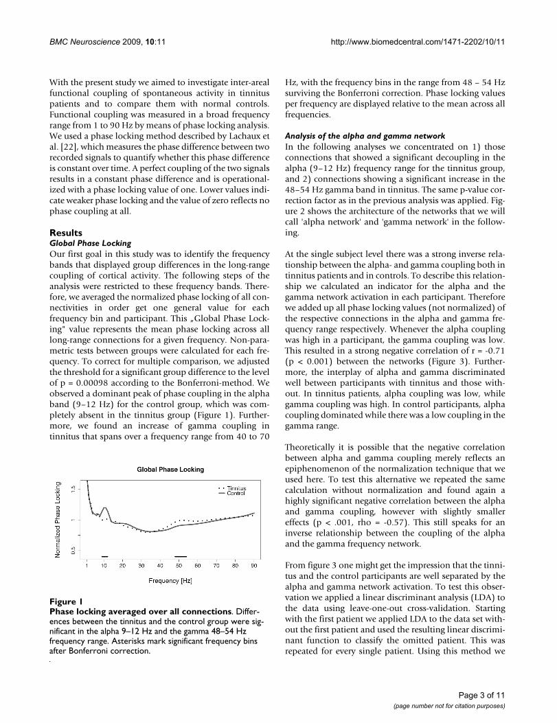
ResultsGlobal Phase LockingOur first goal in this study was to identify the frequencybands that displayed group differences in the long-rangecoupling of cortical activity. The following steps of theanalysis were restricted to these frequency bands. There-fore, we averaged the normalized phase locking of all con-nectivities in order get one general value for eachfrequency bin and participant. This „Global Phase Lock-ing" value represents the mean phase locking across alllong-range connections for a given frequency. Non-para-metric tests between groups were calculated for each fre-quency. To correct for multiple comparison, we adjustedthe threshold for a significant group difference to the levelof p = 0.00098 according to the Bonferroni-method. Weobserved a dominant peak of phase coupling in the alphaband (9–12 Hz) for the control group, which was com-pletely absent in the tinnitus group (Figure 1). Further-more, we found an increase of gamma coupling intinnitus that spans over a frequency range from 40 to 70
Hz, with the frequency bins in the range from 48 – 54 Hzsurviving the Bonferroni correction. Phase locking valuesper frequency are displayed relative to the mean across allfrequencies.
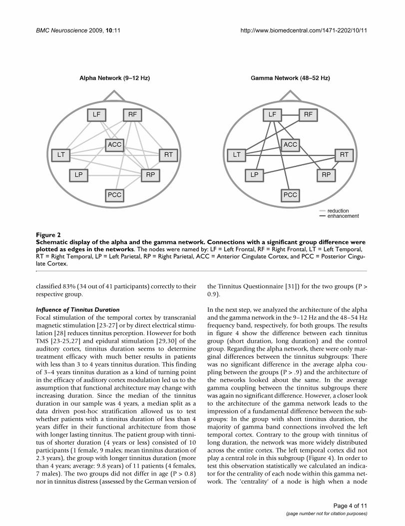
Analysis of the alpha and gamma networkIn the following analyses we concentrated on 1) thoseconnections that showed a significant decoupling in thealpha (9–12 Hz) frequency range for the tinnitus group,and 2) connections showing a significant increase in the48–54 Hz gamma band in tinnitus. The same p-value cor-rection factor as in the previous analysis was applied. Fig-ure 2 shows the architecture of the networks that we willcall 'alpha network' and 'gamma network' in the follow-ing.
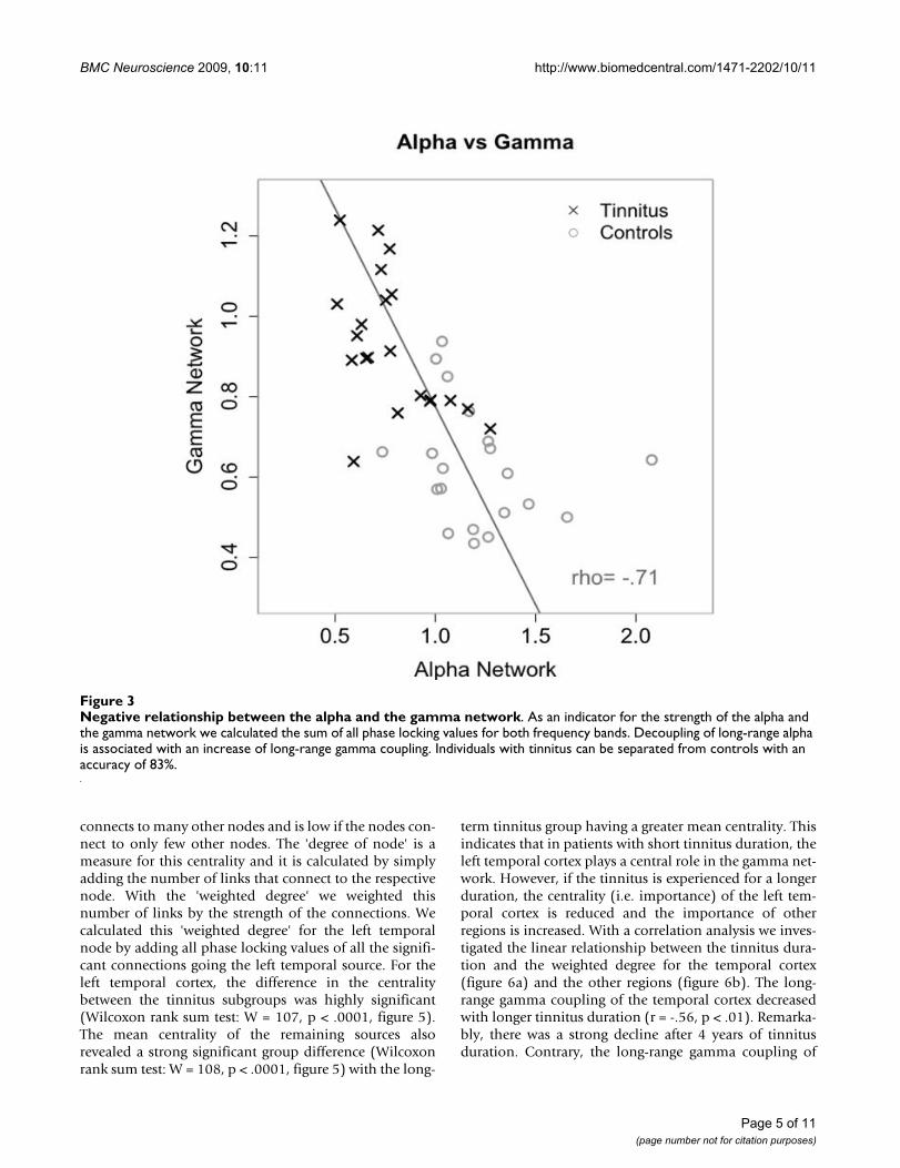
At the single subject level there was a strong inverse rela-tionship between the alpha- and gamma coupling both intinnitus patients and in controls. To describe this relation-ship we calculated an indicator for the alpha and thegamma network activation in each participant. Thereforewe added up all phase locking values (not normalized) ofthe respective connections in the alpha and gamma fre-quency range respectively. Whenever the alpha couplingwas high in a participant, the gamma coupling was low.This resulted in a strong negative correlation of r = -0.71(p < 0.001) between the networks (Figure 3). Further-more, the interplay of alpha and gamma discriminatedwell between participants with tinnitus and those with-out. In tinnitus patients, alpha coupling was low, whilegamma coupling was high. In control participants, alphacoupling dominated while there was a low coupling in thegamma range.
Theoretically it is possible that the negative correlationbetween alpha and gamma coupling merely reflects anepiphenomenon of the normalization technique that weused here. To test this alternative we repeated the samecalculation without normalization and found again ahighly significant negative correlation between the alphaand gamma coupling, however with slightly smallereffects (p < .001, rho = -0.57). This still speaks for aninverse relationship between the coupling of the alphaand the gamma frequency network.
From figure 3 one might get the impression that the tinni-tus and the control participants are well separated by thealpha and gamma network activation. To test this obser-vation we applied a linear discriminant analysis (LDA) tothe data using leave-one-out cross-validation. Startingwith the first patient we applied LDA to the data set with-out the first patient and used the resulting linear discrimi-nant function to classify the omitted patient. This wasrepeated for every single patient. Using this method we
Phase locking averaged over all connectionsFigure 1Phase locking averaged over all connections. Differ-ences between the tinnitus and the control group were sig-nificant in the alpha 9–12 Hz and the gamma 48–54 Hz frequency range. Asterisks mark significant frequency bins after Bonferroni correction.
Page 3 of 11(page number not for citation purposes)
BMC Neuroscience 2009, 10:11 http://www.biomedcentral.com/1471-2202/10/11
classified 83% (34 out of 41 participants) correctly to theirrespective group.
Influence of Tinnitus DurationFocal stimulation of the temporal cortex by transcranialmagnetic stimulation [23-27] or by direct electrical stimu-lation [28] reduces tinnitus perception. However for bothTMS [23-25,27] and epidural stimulation [29,30] of theauditory cortex, tinnitus duration seems to determinetreatment efficacy with much better results in patientswith less than 3 to 4 years tinnitus duration. This findingof 3–4 years tinnitus duration as a kind of turning pointin the efficacy of auditory cortex modulation led us to theassumption that functional architecture may change withincreasing duration. Since the median of the tinnitusduration in our sample was 4 years, a median split as adata driven post-hoc stratification allowed us to testwhether patients with a tinnitus duration of less than 4years differ in their functional architecture from thosewith longer lasting tinnitus. The patient group with tinni-tus of shorter duration (4 years or less) consisted of 10participants (1 female, 9 males; mean tinnitus duration of2.3 years), the group with longer tinnitus duration (morethan 4 years; average: 9.8 years) of 11 patients (4 females,7 males). The two groups did not differ in age (P > 0.8)nor in tinnitus distress (assessed by the German version of
the Tinnitus Questionnaire [31]) for the two groups (P >0.9).
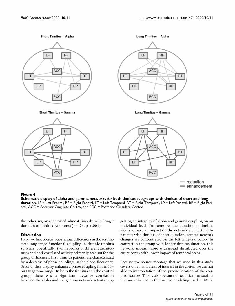
In the next step, we analyzed the architecture of the alphaand the gamma network in the 9–12 Hz and the 48–54 Hzfrequency band, respectively, for both groups. The resultsin figure 4 show the difference between each tinnitusgroup (short duration, long duration) and the controlgroup. Regarding the alpha network, there were only mar-ginal differences between the tinnitus subgroups: Therewas no significant difference in the average alpha cou-pling between the groups (P > .9) and the architecture ofthe networks looked about the same. In the averagegamma coupling between the tinnitus subgroups therewas again no significant difference. However, a closer lookto the architecture of the gamma network leads to theimpression of a fundamental difference between the sub-groups: In the group with short tinnitus duration, themajority of gamma band connections involved the lefttemporal cortex. Contrary to the group with tinnitus oflong duration, the network was more widely distributedacross the entire cortex. The left temporal cortex did notplay a central role in this subgroup (Figure 4). In order totest this observation statistically we calculated an indica-tor for the centrality of each node within this gamma net-work. The 'centrality' of a node is high when a node
Schematic display of the alpha and the gamma networkFigure 2Schematic display of the alpha and the gamma network. Connections with a significant group difference were plotted as edges in the networks. The nodes were named by: LF = Left Frontal, RF = Right Frontal, LT = Left Temporal, RT = Right Temporal, LP = Left Parietal, RP = Right Parietal, ACC = Anterior Cingulate Cortex, and PCC = Posterior Cingu-late Cortex.
Page 4 of 11(page number not for citation purposes)
BMC Neuroscience 2009, 10:11 http://www.biomedcentral.com/1471-2202/10/11
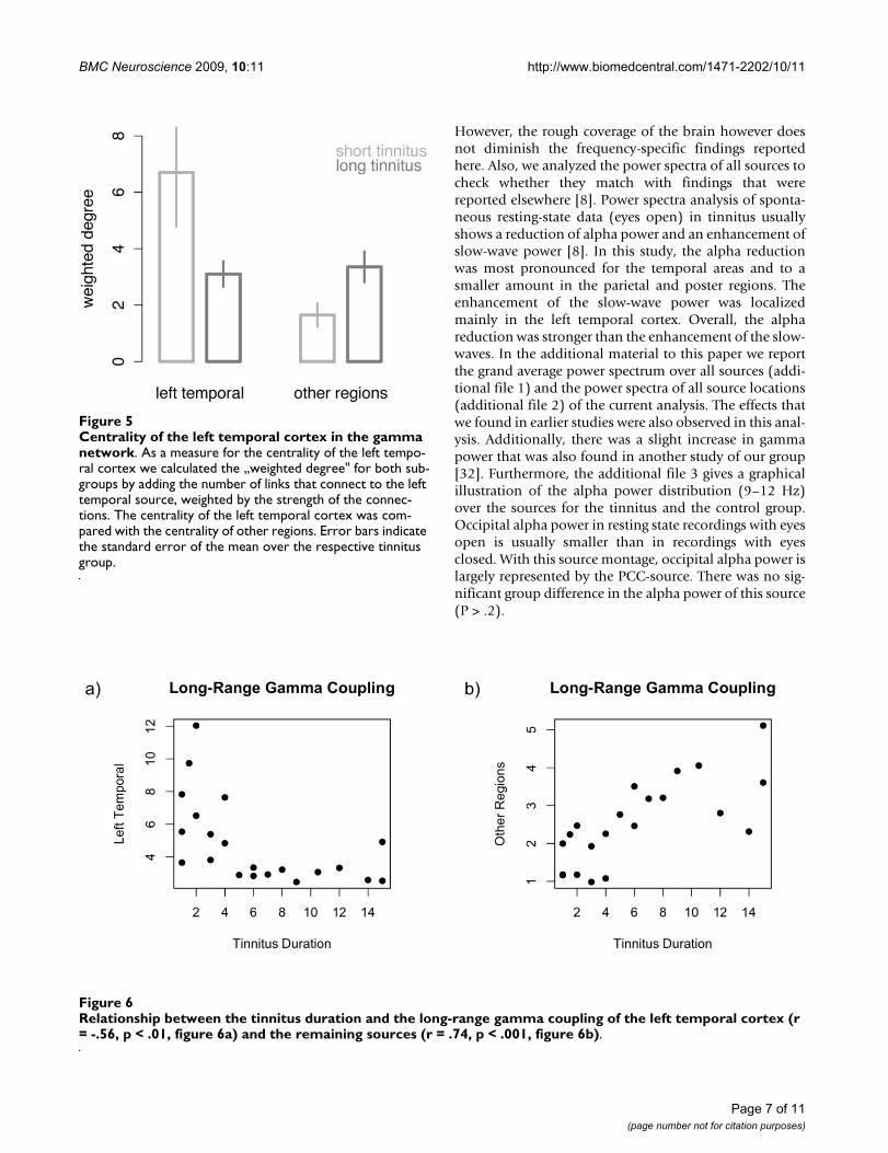
connects to many other nodes and is low if the nodes con-nect to only few other nodes. The 'degree of node' is ameasure for this centrality and it is calculated by simplyadding the number of links that connect to the respectivenode. With the 'weighted degree' we weighted thisnumber of links by the strength of the connections. Wecalculated this 'weighted degree' for the left temporalnode by adding all phase locking values of all the signifi-cant connections going the left temporal source. For theleft temporal cortex, the difference in the centralitybetween the tinnitus subgroups was highly significant(Wilcoxon rank sum test: W = 107, p < .0001, figure 5).The mean centrality of the remaining sources alsorevealed a strong significant group difference (Wilcoxonrank sum test: W = 108, p < .0001, figure 5) with the long-
term tinnitus group having a greater mean centrality. Thisindicates that in patients with short tinnitus duration, theleft temporal cortex plays a central role in the gamma net-work. However, if the tinnitus is experienced for a longerduration, the centrality (i.e. importance) of the left tem-poral cortex is reduced and the importance of otherregions is increased. With a correlation analysis we inves-tigated the linear relationship between the tinnitus dura-tion and the weighted degree for the temporal cortex(figure 6a) and the other regions (figure 6b). The long-range gamma coupling of the temporal cortex decreasedwith longer tinnitus duration (r = -.56, p < .01). Remarka-bly, there was a strong decline after 4 years of tinnitusduration. Contrary, the long-range gamma coupling of
Negative relationship between the alpha and the gamma networkFigure 3Negative relationship between the alpha and the gamma network. As an indicator for the strength of the alpha and the gamma network we calculated the sum of all phase locking values for both frequency bands. Decoupling of long-range alpha is associated with an increase of long-range gamma coupling. Individuals with tinnitus can be separated from controls with an accuracy of 83%.
Page 5 of 11(page number not for citation purposes)
BMC Neuroscience 2009, 10:11 http://www.biomedcentral.com/1471-2202/10/11
the other regions increased almost linearly with longerduration of tinnitus symptoms (r = .74, p < .001).
DiscussionHere, we first present substantial differences in the resting-state long-range functional coupling in chronic tinnitussufferers. Specifically, two networks of different architec-tures and anti-correlated activity primarily account for thegroup differences. First, tinnitus patients are characterizedby a decrease of phase couplings in the alpha frequency.Second, they display enhanced phase coupling in the 48–54 Hz gamma range. In both the tinnitus and the controlgroup, there was a significant negative correlationbetween the alpha and the gamma network activity, sug-
gesting an interplay of alpha and gamma coupling on anindividual level. Furthermore, the duration of tinnitusseems to have an impact on the network architecture. Inpatients with tinnitus of short duration, gamma networkchanges are concentrated on the left temporal cortex. Incontrast in the group with longer tinnitus duration, thisnetwork appears more widespread distributed over theentire cortex with lower impact of temporal areas.
Because the source montage that we used in this studycovers only main areas of interest in the cortex, we are notable to interpretation of the precise location of the cou-pled sources. This is also because of technical constraintsthat are inherent to the inverse modeling used in MEG.
Schematic display of alpha and gamma networks for both tinnitus subgroups with tinnitus of short and long durationFigure 4Schematic display of alpha and gamma networks for both tinnitus subgroups with tinnitus of short and long duration. LF = Left Frontal, RF = Right Frontal, LT = Left Temporal, RT = Right Temporal, LP = Left Parietal, RP = Right Pari-etal, ACC = Anterior Cingulate Cortex, and PCC = Posterior Cingulate Cortex.
Page 6 of 11(page number not for citation purposes)
BMC Neuroscience 2009, 10:11 http://www.biomedcentral.com/1471-2202/10/11
However, the rough coverage of the brain however doesnot diminish the frequency-specific findings reportedhere. Also, we analyzed the power spectra of all sources tocheck whether they match with findings that werereported elsewhere [8]. Power spectra analysis of sponta-neous resting-state data (eyes open) in tinnitus usuallyshows a reduction of alpha power and an enhancement ofslow-wave power [8]. In this study, the alpha reductionwas most pronounced for the temporal areas and to asmaller amount in the parietal and poster regions. Theenhancement of the slow-wave power was localizedmainly in the left temporal cortex. Overall, the alphareduction was stronger than the enhancement of the slow-waves. In the additional material to this paper we reportthe grand average power spectrum over all sources (addi-tional file 1) and the power spectra of all source locations(additional file 2) of the current analysis. The effects thatwe found in earlier studies were also observed in this anal-ysis. Additionally, there was a slight increase in gammapower that was also found in another study of our group[32]. Furthermore, the additional file 3 gives a graphicalillustration of the alpha power distribution (9–12 Hz)over the sources for the tinnitus and the control group.Occipital alpha power in resting state recordings with eyesopen is usually smaller than in recordings with eyesclosed. With this source montage, occipital alpha power islargely represented by the PCC-source. There was no sig-nificant group difference in the alpha power of this source(P > .2).
Centrality of the left temporal cortex in the gamma networkFigure 5Centrality of the left temporal cortex in the gamma network. As a measure for the centrality of the left tempo-ral cortex we calculated the „weighted degree" for both sub-groups by adding the number of links that connect to the left temporal source, weighted by the strength of the connec-tions. The centrality of the left temporal cortex was com-pared with the centrality of other regions. Error bars indicate the standard error of the mean over the respective tinnitus group.
Relationship between the tinnitus duration and the long-range gamma coupling of the left temporal cortex (r = -.56, p < .01, fig-ure 6a) and the remaining sources (r = .74, p < .001, figure 6b)Figure 6Relationship between the tinnitus duration and the long-range gamma coupling of the left temporal cortex (r = -.56, p < .01, figure 6a) and the remaining sources (r = .74, p < .001, figure 6b).
Page 7 of 11(page number not for citation purposes)
BMC Neuroscience 2009, 10:11 http://www.biomedcentral.com/1471-2202/10/11
In this study, we found evidence for abnormal functional-ity in long-range cortical networks between tinnitus andcontrol participants in the resting state, which are specificto the alpha and gamma frequency band. A general inter-action between alpha and gamma power in the brain hasalready been postulated earlier [33]. It is assumed thatalpha directly or indirectly reflects an intrinsic mechanismthat prevents the build-up of gamma coupling within neu-ral cell assemblies during deprivation from input. Func-tionally, such a mechanism appears to be necessary, asstrongly interconnected excitatory oscillators would havea natural tendency to synchronize their activity. A defi-ciency of this mechanism is putatively an important pre-requisite for the emergence of phantom perceptions. Ourfinding suggests that this relationship between alpha andgamma frequency is not limited to local power changes,but also might apply to inter-areal phase coupling.
We found that the inter-regional coupling of the alphaand gamma frequency bands discriminate well betweenthe tinnitus and the control participants. Participants witha tinnitus perception are characterized by a decrease oflong-range alpha coupling and an increase of long-rangegamma coupling. Even though the discrimination of 83%is not sensitive enough to use it as an objective diagnostictool for tinnitus, this is a strong argument that long-rangecouplings play an important part in the neuronal mecha-nisms associated with the tinnitus perception.
Here we propose a tinnitus model that integrates this find-ing with current knowledge on the tinnitus. On a firstlevel the tinnitus is generated within the central auditorysystem and is most likely a result of reorganization proc-esses triggered by damage to the hearing system. This issupported by numerous studies that show functional reor-ganization of the auditory system in tinnitus patients[3,8,34-36]. On a second level, abnormal coupling withhigher-order brain regions outside the auditory systemunderlies its conscious perception [9,10]. We assert thatboth levels are necessary for an ongoing perception of thetinnitus phantom sound.
Even though the alpha and gamma coupling discrimi-nated well between tinnitus and control participants anassociation between the long-range coupling and the sub-jective degree of tinnitus distress was lacking. In an earlierstudy – also with resting-state recordings in the MEG – wefound moderate correlations of the subjective tinnitus rat-ing with alpha power decrease in temporal regions [8]. Itis likely that we investigated two different neuronal mech-anisms: One mechanism that is involved in the generalperception of tinnitus and the other mechanism that isassociated with tinnitus distress. The former studyreported an association between temporal power changesand tinnitus distress. The current study on long-range cou-
pling discriminated well between tinnitus perception andno tinnitus perception. Both mechanisms do not neces-sarily have to be associated.
With respect to tinnitus duration we found that longer-lasting tinnitus (> 4 years) accompanies marked changesin the pattern of the gamma network compared to shorter-lasting tinnitus. The most obvious difference betweenlong and short-lasting tinnitus is a decrease in importanceof the left temporal part of the network, i.e. there are fewerconnections formed within this brain region. On theother side, functional connections between non-auditoryareas are increased in tinnitus of longer duration. Basedon the data that we present here we cannot decidewhether this is only a change of functional coupling orwhether structural changes also occur. A study using diffu-sion tensor imaging could help to clarify this question.
Notwithstanding whether the change in the networkarchitecture is structural or functional, the results offer anexplanation for a so far unresolved riddle in treatingchronic tinnitus with Transcranial Magnetic Stimulation:It has been shown in a series of clinical studies that theefficacy of TMS treatment strongly depends on the dura-tion of tinnitus [24,25,27,37]. In these studies shorter tin-nitus duration predicts better treatment outcome fortherapeutical application of Transcranial Magnetic Stimu-lation applied while the treatment efficacy declines withlonger duration of tinnitus. A tinnitus duration of 3 to 4years seems to represent the turning point and patientsbelow this point benefit only little from the treatment.The intriguing detail about this is that TMS is traditionallyapplied to the left temporal cortex. In the light of our find-ings these negative treatment effects make sense: as thegamma network shows a major hub in the left temporalcortex of patients with short tinnitus duration, stimula-tion of this region exhibits a potentially great impact onthis network. However, since the gamma network is morewidespread in patients with a long history of tinnitus, theimpact of the stimulation to the left hemisphere is largelyreduced. This idea of an alteration of the tinnitus-relatedneural network over time was hypothesized earlier [25]and the data that we presented here are the first experi-mental support for this idea.
ConclusionHere we demonstrate for the first time alterations in thelong-range network during spontaneous activity in tinni-tus patients. The results can be described by an overalldecrease of coupling in the alpha frequency band togetherwith an increase of gamma coupling. This pattern of phasecoupling discriminates with a high percentage (83%) thetinnitus patients from the healthy controls.
Page 8 of 11(page number not for citation purposes)
BMC Neuroscience 2009, 10:11 http://www.biomedcentral.com/1471-2202/10/11
Here we suggest a tinnitus model that incorporates 1)altered activity of the central auditory system that mostlikely generates the tinnitus sound and 2) the couplingacross distant brain regions that is needed for a consciousperception of the tinnitus sound.
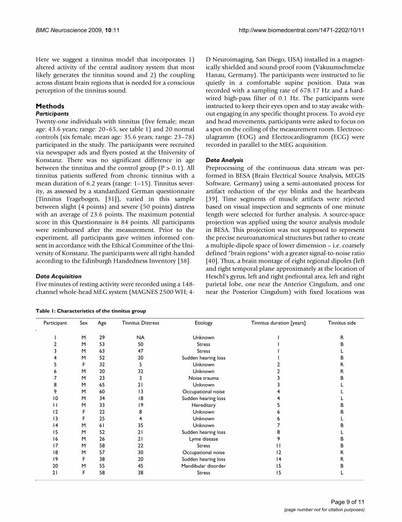
MethodsParticipantsTwenty-one individuals with tinnitus (five female; meanage: 43.6 years; range: 20–65, see table 1) and 20 normalcontrols (six female; mean age: 35.6 years; range: 23–78)participated in the study. The participants were recruitedvia newspaper ads and flyers posted at the University ofKonstanz. There was no significant difference in agebetween the tinnitus and the control group (P > 0.1). Alltinnitus patients suffered from chronic tinnitus with amean duration of 6.2 years (range: 1–15). Tinnitus sever-ity, as assessed by a standardized German questionnaire(Tinnitus Fragebogen, [31]), varied in this samplebetween slight (4 points) and severe (50 points) distresswith an average of 23.6 points. The maximum potentialscore in this Questionnaire is 84 points. All participantswere reimbursed after the measurement. Prior to theexperiment, all participants gave written informed con-sent in accordance with the Ethical Committee of the Uni-versity of Konstanz. The participants were all right-handedaccording to the Edinburgh Handedness Inventory [38].
Data AcquisitionFive minutes of resting activity were recorded using a 148-channel whole-head MEG system (MAGNES 2500 WH; 4-
D Neuroimaging, San Diego, USA) installed in a magnet-ically shielded and sound-proof room (VakuumschmelzeHanau, Germany). The participants were instructed to liequietly in a comfortable supine position. Data wasrecorded with a sampling rate of 678.17 Hz and a hard-wired high-pass filter of 0.1 Hz. The participants wereinstructed to keep their eyes open and to stay awake with-out engaging in any specific thought process. To avoid eyeand head movements, participants were asked to focus ona spot on the ceiling of the measurement room. Electrooc-ulagramm (EOG) and Electrocardiogramm (ECG) wererecorded in parallel to the MEG acquisition.
Data AnalysisPreprocessing of the continuous data stream was per-formed in BESA (Brain Electrical Source Analysis, MEGISSoftware, Germany) using a semi-automated process forartifact reduction of the eye blinks and the heartbeats[39]. Time segments of muscle artifacts were rejectedbased on visual inspection and segments of one minutelength were selected for further analysis. A source-spaceprojection was applied using the source analysis modulein BESA. This projection was not supposed to representthe precise neuroanatomical structures but rather to createa multiple-dipole space of lower dimension – i.e. coarselydefined "brain regions" with a greater signal-to-noise ratio[40]. Thus, a brain montage of eight regional dipoles (leftand right temporal plane approximately at the location ofHeschl's gyrus, left and right prefrontal area, left and rightparietal lobe, one near the Anterior Cingulum, and onenear the Posterior Cingulum) with fixed locations was
Table 1: Characteristics of the tinnitus group
Participant Sex Age Tinnitus Distress Etiology Tinnitus duration [years] Tinnitus side
1 M 29 NA Unknown 1 R2 M 53 50 Stress 1 B3 M 63 47 Stress 1 L4 M 52 20 Sudden hearing loss 1 B5 F 32 5 Unknown 2 R6 M 20 32 Unknown 2 R7 M 23 3 Noise trauma 3 B8 M 65 21 Unknown 3 L9 M 60 13 Occupational noise 4 L10 M 34 18 Sudden hearing loss 4 L11 M 33 19 Hereditary 5 B12 F 22 8 Unknown 6 B13 F 25 4 Unknown 6 L14 M 61 35 Unknown 7 B15 M 52 21 Sudden hearing loss 8 L16 M 26 21 Lyme disease 9 B17 M 58 22 Stress 11 B18 M 57 30 Occupational noise 12 R19 F 38 20 Sudden hearing loss 14 R20 M 55 45 Mandibular disorder 15 B21 F 58 38 Stress 15 L
Page 9 of 11(page number not for citation purposes)
BMC Neuroscience 2009, 10:11 http://www.biomedcentral.com/1471-2202/10/11
used for each participant and adjusted to the individualhead size. The dipole locations were defined prior to theanalysis in order to cover roughly the activity of major cor-tical regions (see supporting information for exact loca-tions).
The data were down-sampled to 450 Hz and one artifact-free minute was selected for subsequent analyses. A Mor-let wavelet (m-factor = 7) was used for estimation theinstantaneous phases in the frequency range of 1 to 90 Hz(1–12 Hz in steps of one Hz, 14–90 Hz in steps of two Hz)using Matlab 7.4 (The MathWorks, Natick, MA). Thephase information over one minute of data was used tocalculate the phase locking value (PLV) that has been sug-gested to be an operationalization for functional coupling[22]. Therefore, the phase difference between two signalsis calculated and tested for stability through all timepoints. The phase locking value (from zero to one)increases the more the distribution within a unit circledeviates from uniformity, with a PLV of one indicatingperfect phase coupling between the two signals. To makethe data comparable across participants in terms of differ-ential coupling values across the frequency bands, we nor-malized the phase locking values with respect tofrequency. That was done by calculation a mean PLV overall frequencies and dividing all phase locking values bythis mean. Thus, the mean „normalized phase locking"over all frequencies equals one for each participant.
Statistical analyses were all conducted in R http://www.r-project.org. Group differences were assessed using thenon-parametric Wilcoxon's rank sum test with a Bonfer-roni correction for multiple comparisons of the fifty-onefrequency bins. Correlations between the alpha andgamma PLVs were determined using a Spearman's rankcorrelation.
Authors' contributionsWS carried out the study, performed the statistical analysisand drafted the manuscript. TH participated in data acqui-sition, supplied analysis tools and helped in drafting themanuscript. BL helped in drafting the paper and the dis-cussion of the results. NW participated in the data collec-tion, data analysis and writing the manuscript.
Additional material
AcknowledgementsWe would like to thank Ursula Lommen, Barabara Awiszus and Christiane Wolf for assistance in collecting the data. This study was supported by the Deutsche Forschungsgemeinschaft (DFG) and the Tinnitus Research Inniti-ative (TRI).
References1. Meyerhoff WL, Cooper JC: Tinnitus. In Otolaryngology 3rd edition.
Philadelphia: Saunders; 1991:1169-75. 2. Phoon WH, Lee HS, Chia SE: Tinnitus in noise-exposed workers.
Occup Med (Lond) 1993, 43:35-38.3. Eggermont JJ, Roberts LE: The neuroscience of tinnitus. Trends
Neurosci 2004, 27:676-682.4. Dandy WE: The surgical treatment of intracranial aneurysms
of the internal carotid artery. Ann Surg 1941, 114:336-340.5. Silverstein H: Transmeatal labyrinthectomy with and without
cochleovestibular neurectomy. Laryngoscope 1976,86:1777-1791.
6. Mirz F, Brahe Pedersen C, Ishizu K, Johannsen P: Positron emissiontomography of cortical centers of tinnitus. Hear Res 1999,134:133-144.
7. Muhlau M, Rauschecker JP, Oestreicher E, Gaser C, Rottinger M,Wohlschlager AM, Simon F, Etgen T, Conrad B, Sander D: Struc-tural brain changes in tinnitus. Cereb Cortex 2006, 16:1283-1288.
8. Weisz N, Moratti S, Meinzer M, Dohrmann K, Elbert T: Tinnitusperception and distress is related to abnormal spontaneousbrain activity as measured by magnetoencephalography.PLoS Med 2005, 2:e153.
9. Jastreboff PJ: Phantom auditory perception (tinnitus): mecha-nisms of generation and perception. Neurosci Res 1990,8:221-254.
10. Dehaene S, Changeux J-P: Neural Mechanisms for access to con-sciousness. In The Cognitive Neurosciences III Edited by: Gazzaniga M.MIT Press; 2004.
11. Fries P, Reynolds JH, Rorie AE, Desimone R: Modulation of oscil-latory neuronal synchronization by selective visual atten-tion. Science 2001, 291:1560-1563.
12. Gross J, Schmitz F, Schnitzler I, Kessler K, Shapiro K, Hommel B,Schnitzler A: Modulation of long-range neural synchronyreflects temporal limitations of visual attention in humans.Proc Natl Acad Sci USA 2004, 101:13050-13055.
13. Hummel F, Gerloff C: Larger interregional synchrony is associ-ated with greater behavioral success in a complex sensoryintegration task in humans. Cereb Cortex 2005, 15:670-678.
14. Melloni L, Molina C, Pena M, Torres D, Singer W, Rodriguez E: Syn-chronization of neural activity across cortical areas corre-lates with conscious perception. J Neurosci 2007, 27:2858-2865.
15. Miltner WH, Braun C, Arnold M, Witte H, Taub E: Coherence ofgamma-band EEG activity as a basis for associative learning.Nature 1999, 397:434-436.
Additional file 1Supplemental figure 1. Grand average of the normalized power spectrum over all sources.Click here for file[http://www.biomedcentral.com/content/supplementary/1471-2202-10-11-S1.pdf]
Additional file 2Supplemental figure 2. Normalized power spectra for all source loca-tions.Click here for file[http://www.biomedcentral.com/content/supplementary/1471-2202-10-11-S2.pdf]
Additional file 3Supplemental figure 3. Graphical illustration of the normalized alpha power (9–12 Hz) over the 8 sources. Then diameter of the circle denotes the strength of the alpha power. The average over the tinnitus group is shown on the left side, the control group on the right side of the figure.Click here for file[http://www.biomedcentral.com/content/supplementary/1471-2202-10-11-S3.pdf]
Page 10 of 11(page number not for citation purposes)
BMC Neuroscience 2009, 10:11 http://www.biomedcentral.com/1471-2202/10/11
Publish with BioMed Central and every scientist can read your work free of charge
"BioMed Central will be the most significant development for disseminating the results of biomedical research in our lifetime."
Sir Paul Nurse, Cancer Research UK
Your research papers will be:
available free of charge to the entire biomedical community
peer reviewed and published immediately upon acceptance
cited in PubMed and archived on PubMed Central
yours — you keep the copyright
Submit your manuscript here:http://www.biomedcentral.com/info/publishing_adv.asp
BioMedcentral
16. Singer W: Neuronal synchrony: a versatile code for the defini-tion of relations? Neuron 2000, 24:49-65. 111–25
17. Supp GG, Schlögl A, Trujillo-Barreto N, Müller MM, Gruber T:Directed cortical information flow during human object rec-ognition: analyzing induced EEG gamma-band responses inbrain's source space. PLoS ONE 2007, 2:e684.
18. Uhlhaas PJ, Singer W: Neural synchrony in brain disorders: rel-evance for cognitive dysfunctions and pathophysiology. Neu-ron 2006, 52:155-168.
19. Weisz N, Müller S, Schlee W, Dohrmann K, Hartmann T, Elbert T:The neural code of auditory phantom perception. J Neurosci2007, 27:1479-1484.
20. Pitkovsky R, Kurths J: Synchronization in a population of glo-bally coupled chaotic oscillators. Europhys Lett 1996,34(3):165-170.
21. Rosenblum MG, Pikovsky AS, Kurths J: Phase synchronization ofchaotic oscillators. Phys Rev Lett 1996, 76:1804-1807.
22. Lachaux JP, Rodriguez E, Martinerie J, Varela FJ: Measuring phasesynchrony in brain signals. Hum Brain Mapp 1999, 8:194-208.
23. De Ridder D, Verstraeten E, Kelen K Van der, De Mulder G, SunaertS, Verlooy J, Heyning P Van de, Moller A: Transcranial magneticstimulation for tinnitus: influence of tinnitus duration onstimulation parameter choice and maximal tinnitus suppres-sion. Otol Neurotol 2005, 26:616-619.
24. Khedr EM, Rothwell JC, Ahmed MA, El-Atar A: Effect of dailyrepetitive transcranial magnetic stimulation for treatmentof tinnitus: comparison of different stimulus frequencies. JNeurol Neurosurg Psychiatry 2008, 79:212-215.
25. Kleinjung T, Steffens T, Sand P, Murthum T: Which tinnituspatients benefit from transcranial magnetic stimulation?Otolaryngol Head Neck Surg 2007, 137:589-595.
26. Langguth B, Kleinjung T, Frank E, Landgrebe M, Sand P, Dvorakova J,Frick U, Eichhammer P, Hajak G: High-frequency priming stimu-lation does not enhance the effect of low-frequency rTMS inthe treatment of tinnitus. Exp Brain Res 2008, 184:587-591.
27. Plewnia C, Reimold M, Najib A, Reischl G, Plontke SK, Gerloff C:Moderate therapeutic efficacy of positron emission tomog-raphy-navigated repetitive transcranial magnetic stimula-tion for chronic tinnitus: a randomised, controlled pilotstudy. J Neurol Neurosurg Psychiatry 2007, 78:152-156.
28. De Ridder D, De Mulder G, Menovsky T, Sunaert S, Kovacs S: Elec-trical stimulation of auditory and somatosensory cortices fortreatment of tinnitus and pain. Prog Brain Res 2007,166:377-388.
29. De Ridder D, De Mulder G, Verstraeten E, Kelen K Van der, SunaertS, Smits M, Kovacs S, Verlooy J, Heyning P Van de, Moller AR: Pri-mary and secondary auditory cortex stimulation for intrac-table tinnitus. ORL J Otorhinolaryngol Relat Spec 2006, 68:48-54.discussion 54–5
30. Seidman MD, Ridder DD, Elisevich K, Bowyer SM, Darrat I, Dria J,Stach B, Jiang Q, Tepley N, Ewing J, Seidman M, Zhang J: Direct elec-trical stimulation of Heschl's gyrus for tinnitus treatment.Laryngoscope 2008, 118:491-500.
31. Goebel G, Hiller W: Tinnitus Fragebogen (TF): Ein Instrumentzur Erfassung von Belastung und Schweregrad bei Tinnitus.Göttingen: Hogrefe; 1998.
32. Mueller S: Analyse des neuromagnetischen Spektrums beiTinnitus. In Diploma Thesis University of Konstanz, Department ofPsychology; 2007.
33. Weisz N, Dohrmann K, Elbert T: The relevance of spontaneousactivity for the coding of the tinnitus sensation. Prog Brain Res2007, 166:61-70.
34. Dietrich V, Nieschalk M, Stoll W, Rajan R, Pantev C: Cortical reor-ganization in patients with high frequency cochlear hearingloss. Hear Res 2001, 158:95-101.
35. Mühlnickel W, Elbert T, Taub E, Flor H: Reorganization of audi-tory cortex in tinnitus. Proc Natl Acad Sci USA 1998,95:10340-10343.
36. Wienbruch C, Paul I, Weisz N, Elbert T, Roberts LE: Frequencyorganization of the 40-Hz auditory steady-state response innormal hearing and in tinnitus. Neuroimage 2006, 33:180-194.
37. De Ridder D, De Mulder G, Walsh V, Muggleton N, Sunaert S, MollerA: Magnetic and electrical stimulation of the auditory cortexfor intractable tinnitus. Case report. J Neurosurg 2004,100:560-564.
38. Oldfield RC: The assessment and analysis of handedness: theEdinburgh inventory. Neuropsychologia 1971, 9:97-113.
39. Ille N, Berg P, Scherg M: Artifact correction of the ongoing EEGusing spatial filters based on artifact and brain signal topog-raphies. J Clin Neurophysiol 2002, 19:113-124.
40. Scherg M, Ille N, Bornfleth H, Berg P: Advanced tools for digitalEEG review: virtual source montages, whole-head mapping,correlation, and phase analysis. J Clin Neurophysiol 2002,19:91-112.
Page 11 of 11(page number not for citation purposes)


