Embed Size (px)
Citation preview
Am. J. Hum. Genet. 77:617–626, 2005
617
A Variant in XPNPEP2 Is Associated with Angioedema Inducedby Angiotensin I–Converting Enzyme InhibitorsQing Ling Duan,1,2,* Borzoo Nikpoor,1,* Marie-Pierre Dube,3 Giuseppe Molinaro,4Inge A. Meijer,1 Patrick Dion,1 Daniel Rochefort,1 Judith Saint-Onge,1 Leah Flury,5Nancy J. Brown,6 James V. Gainer,6 Jean L. Rouleau,3 Angelo Agostoni,7 Massimo Cugno,7Pierre Simon,8 Pierre Clavel,9 Jacky Potier,11 Bassem Wehbe,12 Seddik Benarbia,13
Julien Marc-Aurele,14 Jacques Chanard,10 Tatiana Foroud,5 Albert Adam,4and Guy A. Rouleau1
1Centre de recherche du CHUM, Hopital Notre-Dame, 2Department of Human Genetics, McGill University, and 3Department of Medicine,Montreal Heart Institute, and 4Faculte de Pharmacie, Universite de Montreal, Montreal; 5Department of Medical and Molecular Genetics,Indiana University, Indianapolis, IN; 6Department of Pharmacology, Vanderbilt University, Nashville, TN; 7Department of Internal Medicine,University of Milan, Milan; 8Service de Nephrologie, CH La Beauchee, Saint-Brieuc, France; 9Service de Nephrologie-Hemodialyse, ARPDD,and 10Service de Nephrologie, CH Universitaire-Reims, Reims, France; 11Service d’Hemodialyse, CH Louis Pasteur, Cherbourg, France;12Service d’Hemodialyse, CH Intercommunal de Cornouaille, and 13Service de Nephrologie-Hemodialyse, CH Laennec, Quimper, France; and14Hotel-Dieu de Saint-Jerome, Saint-Jerome, Canada
Angiotensin I–converting enzyme inhibitors (ACEi), which are used to treat common cardiovascular diseases, areassociated with a potentially life-threatening adverse reaction known as angioedema (AE-ACEi). We have previouslydocumented a significant association between AE-ACEi and low plasma aminopeptidase P (APP) activity. Witheight large pedigrees, we hereby demonstrate that this quantitative trait is partially regulated by genetic factors.We tested APP activity using a variance-component QTL analysis of a 10-cM genomewide microsatellite scanenriched with seven markers over two candidate regions. We found significant linkage (LOD p 3.75) to a locusthat includes the XPNPEP2 candidate gene encoding membrane-bound APP. Mutation screening of this QTLidentified a large coding deletion segregating in one pedigree and an upstream single-nucleotide polymorphism (C–2399A SNP), which segregates in the remaining seven pedigrees. Measured genotype analysis strongly suggests thatthe linkage signal for APP activity at this locus is accounted for predominantly by the SNP association. In a separatecase-control study (20 cases and 60 controls), we found significant association of this SNP to ACEi-induced AE( ). In conclusion, our findings provide supporting evidence that the C-2399A variant in XPNPEP2 isP p .0364associated with reduced APP activity and a higher incidence of AE-ACEi.
Introduction
Angiotensin I–converting enzyme inhibitors (ACEi) area class of drugs used by 140 million patients worldwidefor the treatment of cardiovascular diseases such as hy-pertension, congestive heart failure, and diabetes (Ungerand Gohlke 1994; Brown and Vaughan 1998). Angioe-dema (AE) associated with ACEi therapy (AE-ACEi) isa potentially fatal adverse event that affects 0.1%–0.7%of white patients (Israili and Hall 1992; Vleeming et al.1998) and is four to five times more prevalent among
Received March 28, 2005; accepted for publication August 1, 2005;electronically published September 1, 2005.
Address for correspondence and reprints: Dr. Guy A. Rouleau, Cen-tre de recherche du CHUM, Hopital Notre Dame, Laboratoire d’etudedes maladies du cerveau, Pavillon deSeve, local Y-3633, 1560, rueSherbrooke Est, Montreal (Quebec), Canada, H2L 4M1. E-mail: [email protected].
* These authors contributed equally to this work.� 2005 by The American Society of Human Genetics. All rights reserved.
0002-9297/2005/7704-0010$15.00
African Americans (Brown et al. 1996; Coats 2002).This racial difference suggests that genetic factors mod-ulate AE risk, but environmental factors are also im-portant, because smokers taking ACEi have an increasedsusceptibility to AE (Coats 2002; Kostis et al. 2004).The incidence of AE-ACEi is likely underestimated be-cause the clinical symptoms can develop years after start-ing ACEi therapy, thus obscuring the adverse event’srelationship with the drug and often leading to misdi-agnosis (Agostoni et al. 2004).
A majority of AE-ACEi cases do not respond to an-tihistamines or corticosteroids, indicating that thesecases are not allergic reactions (Agostoni and Cicardi2001). Currently, there is no effective treatment for AE-ACEi and no method for identifying individuals withincreased susceptibility to this adverse reaction. Betterunderstanding of the pathogenetic mechanism under-lying this type of AE may also have significant impli-cations for AE associated with other vasopeptidase in-hibitors (i.e., Omapatrilat), which block the activities

618 Am. J. Hum. Genet. 77:617–626, 2005
of both ACE and neutral endopeptidase (NEP). The AErisk associated with these drugs is even higher than withACEi (Coats 2002) and has curtailed the regulatory ap-proval for usage of these vasopeptidase inhibitors totreat cardiovascular diseases.
Previous reports have suggested that bradykinin (BK),a potent vasodilatory and proinflammatory nonapep-tide, plays a central role in the pathophysiology of AE-ACEi (Israili and Hall 1992; Nussberger et al. 1998).BK is rapidly degraded in the plasma of healthy indi-viduals by angiotensin I–converting enzyme (ACE) andaminopeptidase P (APP) (Bhoola et al. 1992). KininaseI enzymes normally transform a minute fraction (3.5%)of BK into its active metabolite, des-arginine9-brady-kinin (des-Arg9-BK) (Blais et al. 2000). This carboxy-truncated metabolite is, in turn, broken down by APPand ACE (Cyr et al. 2001). In the presence of ACEinhibition, however, kininase I activity is increased(transforms 28% of BK into des-Arg9-BK), and APP actsas the major metabolizing enzyme of both BK and des-Arg9-BK (Blais et al. 2000).
An increase of BK has been measured in the plasmaof patients during episodes of AE-ACEi, but, unlike he-reditary forms of AE (MIM 106100), there is no in-crease in cleavage of the BK precursor, high-molecular-weight kininogen (HK) (Nussberger et al. 1998; Ago-stoni et al. 1999; Cugno et al. 2003). This suggests thatimpaired BK metabolism, rather than increased BK pro-duction, plays an important role in AE-ACEi. We pre-viously reported significantly lower plasma APP activ-ities in patients with a history of AE-ACEi, which isstrongly correlated with a significant decrease in des-Arg9-BK degradation in vitro (Blais et al. 1999a; Adamet al. 2002; Molinaro et al. 2002). Blais et al. demon-strated that half of AE-ACEi cases have an enzyme de-fect involved in des-Arg9-BK metabolism (Blais et al.1999b). Accumulated levels of des-Arg9-BK have beenshown to cause proinflammatory effects in vivo (Blaiset al. 1997; Blais et al. 1999a). One study reported thatACEi patients who received subcutaneous injections ofthe APP inhibitor Apstatin developed local inflamma-tions (Kim et al. 2000). Thus, previous data indicatethat reduced plasma APP activity may predict increasedrisk for AE associated with ACEi therapy.
The aim of this study is to determine whether plasmaAPP activity is regulated by genetic factors and to iden-tify the QTL or QTLs that confer susceptibility to AE-ACEi. There are two known APP enzymes in humans:membrane-bound (mAPP) and cytosolic (cAPP). Thegene encoding the former is XPNPEP2 (MIM 300145),localized to chromosome Xq26.1 (Sprinkle et al. 1998),and the latter is the product of XPNPEPL (MIM602443) on chromosome 10q25.1 (Sprinkle et al.2000). Although these represent good candidate genes
for the interindividual variability in plasma APP activity,other genetic loci may serve as important regulators.Plasma APP activity has been shown to form a contin-uous distribution in the general population (Cyr et al.2001), suggesting that plasma APP activity is a complexquantitative trait likely influenced by multiple geneticloci and nongenetic factors (e.g., smoking and hormonereplacement therapy [Gallagher et al. 1999]). Identifi-cation of the genetic factors underlying reduced plasmaAPP activity would provide a better understanding ofthe pathogenesis of AE-ACEi and could facilitate thedevelopment of a clinical assay to detect those individ-uals with greater AE risk.
Subjects and Methods
Blood and Plasma Samples
The ethics committees from Centre Hospitalier del’Universite de Montreal, Institut de Cardiologie deMontreal, and McGill University, all in Montreal, re-viewed and approved all protocols involving human sub-jects. Informed consent was obtained from all partic-ipants. DNA extraction from blood was performedfollowing a standardized protocol (Gentra Systems).
Participants
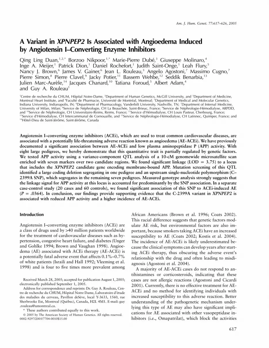
Cohort 1 (fig. 1a), composed of five white pedigrees(88 DNA samples), was collected for a genomewidemicrosatellite scan, followed by a variance-componentlinkage analysis for APP activity. Cohort 2 (fig. 1b), con-sisting of three white pedigrees (35 DNA samples), wassubsequently selected for genotyping of 11 microsatellitemarkers (table 1), which cover four genomic regions thatprovided LOD scores 11 in the linkage analysis of cohort1 and the locus including the XPNPEPL candidate gene.Plasma samples were also collected from all 123 partic-ipants for characterization of APP activity. The kindredswere selected from Canada (Montreal) and France onthe basis of the presence of one individual in each familywith a history of AE and/or another acute adverse effectrelated to ACEi therapy, anaphylactoid reactions (AR-ACEi) (Bright et al. 1999). The clinical diagnosis wasnot considered for linkage analysis because of lack ofpower. Instead, plasma APP activity measurements wereused as the quantitative trait in a variance-componentlinkage analysis.
DNA from 20 independent AE-ACEi cases (table A1[online only]) was extracted for mutation screening ex-periments. All subjects were whites with histories of AE-ACEi and origins in Canada ( ), Belgium ( ),n p 7 n p 3or the United States ( ). Plasma samples weren p 10available for all AE subjects except two from the UnitedStates. The onset of clinical symptoms ranged from a

Duan et al.: XPNPEP2 Associated with Angioedema-ACEi 619
Figure 1 Cohorts included in variance-component linkage analysis. One member of each kindred, depicted in black, developed AE and/or AR associated with ACEi therapy. All available members were quantified for plasma APP activity, shown as numbers that represent unitsof arginine released per minute per milliliter of plasma. Genotypes of the C-2399A SNP upstream of XPNPEP2 are shown as C/C, C/A, or A/A for females, and C or A for males. a, DNA samples from cohort 1 were included in a genomewide microsatellite scan, which was analyzedfor linkage using a variance-component method (SOLAR program). b, Eleven genome-scanned markers were genotyped in cohort 2 to furtherevaluate linkage. A large deletion (DEL) in XPNPEP2 segregates in pedigree VI.
few hours to 8 years after starting ACEi therapy. Inaddition, we collected three unrelated white controls tomatch each AE patient for country of origin and gender( ). Controls from Belgium and Canada had non p 60history of ACEi therapy, whereas the medical historieswere unknown for US controls. Controls were notmatched for age because we did not find age effects in
our samples, which is in agreement with previous find-ings (Cyr et al. 2001).
Quantification of Plasma APP Activity
APP activity was measured in plasma with modifi-cation of an assay described elsewhere (Simmons and

620 Am. J. Hum. Genet. 77:617–626, 2005
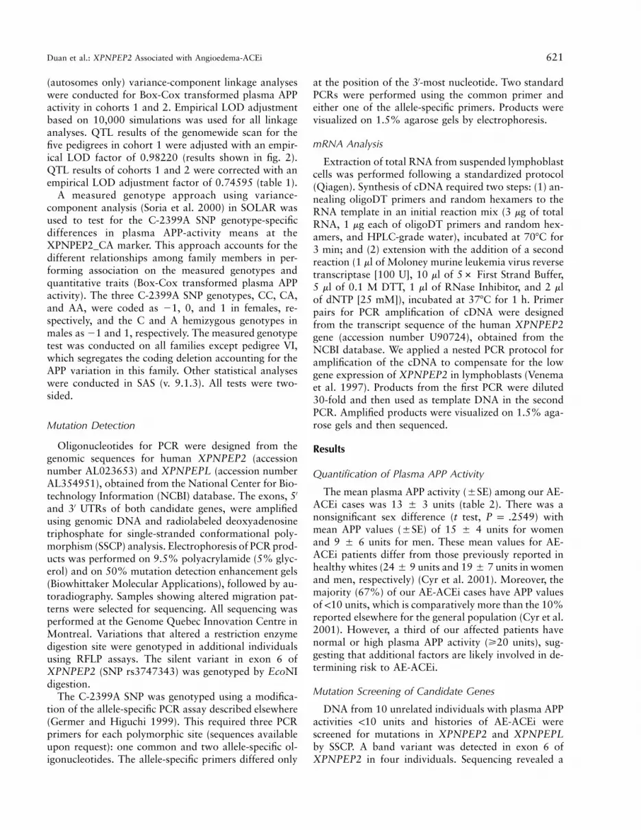
Figure 2 Results of the variance-component QTL analyses inthe five pedigrees of cohort 1. All results were corrected empiricallyfor a � 0.05. a, String diagram of autosomal multipoint results, LODs11, are shown next to a LOD scale. b, Two-point linkage analysisresults including chromosome X.
Table 1
Variance-Component Two-Point QTLs for Markers Genotyped in Cohorts 1 and 2
MARKER
POSITION
(cM)CHROMOSOME
BAND
LOD
Cohorts 1 and 2(8 Pedigrees) Cohort 1 Only
D6S1017 63.3 6p21.1 .97 2.62D6S1280 73.1 6p12.3 .18 1.09D6S1056 102.8 6q16.1 1.32 1.68D8S277 8.0 8p23.1 .22 .69D8S373 164.5 8q24.3 1.72 1.76D9S925 32.2 9p22.2 .16 1.71D9S1122 75.9 9q21.2 .13 .76Markers flanking candidates:
D10S1220 70.2 10q11.23 .24 .74DXS8057 136.4 Xq25 2.10 1.82DXS1047 143.2 Xq25 2.04 .28XPNPEP2_CA 146.0 Xq25 3.75 2.15
NOTE.—Eleven microsatellite markers that were already part of the analysis in cohort 1 weregenotyped in cohort 2 to cover the four loci that provided LOD scores 11 in cohort 1 and the regionproximal to the XPNPEPL candidate gene (D10S1220).
Orawski 1992; Blais et al. 1999a). Our assay used thesynthetic substrate arginine-proline-proline. Plasma APPactivity is expressed as nanomoles of arginine releasedper minute per milliliter of plasma sample (1 unit p 1nmol/min/ml).
Genome Scan and Microsatellite Genotyping
A 10-cM genomewide microsatellite scan (markerpanel SS4 on the ABI-3700 DNA analyzer at the Ge-nome Quebec Innovation Centre, Montreal), enrichedfor seven markers flanking the two APP candidate geneson chromosomes 10 and X (D10S534, D10S1741,D10S562, DXS1212, DXS8057, XPNPEP2_CA, andDXS1047), was performed on cohort 1 (total of 397microsatellite markers). Primers for the XPNPEP2_CArepeat marker located upstream of XPNPEP2 (∼4 kbwere designed for this study) were sense 5′-GCTCTTTC-CCCCTGCTGTGT-3′ and antisense 5′-GGTGCTGTT-GGGTGCCTCATC-3′.
Genotyping of the 11 genome-scanned markers in co-hort 2 was conducted in our laboratory. Genomic DNAwas amplified by radiolabeled (a-35S-dATP) PCR. Theproducts were separated by electrophoresis on 6% de-naturing polyacrylamide gels. The alleles were sized bycomparison to the M13mp18 sequence ladder, and eachindividual was assigned a genotype. Marker-allele sizesand frequencies were obtained from the Fondation JeanDausset CEPH database.
Statistical Analyses
The SOLAR (Sequential Oligogenic Linkage AnalysisRoutines, v. 2.1.4) (Almasy and Blangero 1998) programwas used to estimate heritability of plasma APP activityand to perform a variance-component linkage analysis.
Age and sex were considered as covariates, but were notsignificant (P 1 .05). APP activity was transformed fornormality to avoid convergence failure of SOLAR byusing the Box-Cox transformation (Box and Cox 1964),such that T(APP) p {[(APP � 1) # 0.40 � 1]/0.40} #. The two-point (all chromosomes) and multipoint4
Duan et al.: XPNPEP2 Associated with Angioedema-ACEi 621
(autosomes only) variance-component linkage analyseswere conducted for Box-Cox transformed plasma APPactivity in cohorts 1 and 2. Empirical LOD adjustmentbased on 10,000 simulations was used for all linkageanalyses. QTL results of the genomewide scan for thefive pedigrees in cohort 1 were adjusted with an empir-ical LOD factor of 0.98220 (results shown in fig. 2).QTL results of cohorts 1 and 2 were corrected with anempirical LOD adjustment factor of 0.74595 (table 1).
A measured genotype approach using variance-component analysis (Soria et al. 2000) in SOLAR wasused to test for the C-2399A SNP genotype-specificdifferences in plasma APP-activity means at theXPNPEP2_CA marker. This approach accounts for thedifferent relationships among family members in per-forming association on the measured genotypes andquantitative traits (Box-Cox transformed plasma APPactivity). The three C-2399A SNP genotypes, CC, CA,and AA, were coded as �1, 0, and 1 in females, re-spectively, and the C and A hemizygous genotypes inmales as �1 and 1, respectively. The measured genotypetest was conducted on all families except pedigree VI,which segregates the coding deletion accounting for theAPP variation in this family. Other statistical analyseswere conducted in SAS (v. 9.1.3). All tests were two-sided.
Mutation Detection
Oligonucleotides for PCR were designed from thegenomic sequences for human XPNPEP2 (accessionnumber AL023653) and XPNPEPL (accession numberAL354951), obtained from the National Center for Bio-technology Information (NCBI) database. The exons, 5′
and 3′ UTRs of both candidate genes, were amplifiedusing genomic DNA and radiolabeled deoxyadenosinetriphosphate for single-stranded conformational poly-morphism (SSCP) analysis. Electrophoresis of PCR prod-ucts was performed on 9.5% polyacrylamide (5% glyc-erol) and on 50% mutation detection enhancement gels(Biowhittaker Molecular Applications), followed by au-toradiography. Samples showing altered migration pat-terns were selected for sequencing. All sequencing wasperformed at the Genome Quebec Innovation Centre inMontreal. Variations that altered a restriction enzymedigestion site were genotyped in additional individualsusing RFLP assays. The silent variant in exon 6 ofXPNPEP2 (SNP rs3747343) was genotyped by EcoNIdigestion.
The C-2399A SNP was genotyped using a modifica-tion of the allele-specific PCR assay described elsewhere(Germer and Higuchi 1999). This required three PCRprimers for each polymorphic site (sequences availableupon request): one common and two allele-specific ol-igonucleotides. The allele-specific primers differed only
at the position of the 3′-most nucleotide. Two standardPCRs were performed using the common primer andeither one of the allele-specific primers. Products werevisualized on 1.5% agarose gels by electrophoresis.
mRNA Analysis
Extraction of total RNA from suspended lymphoblastcells was performed following a standardized protocol(Qiagen). Synthesis of cDNA required two steps: (1) an-nealing oligoDT primers and random hexamers to theRNA template in an initial reaction mix (3 mg of totalRNA, 1 mg each of oligoDT primers and random hex-amers, and HPLC-grade water), incubated at 70�C for3 min; and (2) extension with the addition of a secondreaction (1 ml of Moloney murine leukemia virus reversetranscriptase [100 U], 10 ml of 5# First Strand Buffer,5 ml of 0.1 M DTT, 1 ml of RNase Inhibitor, and 2 mlof dNTP [25 mM]), incubated at 37�C for 1 h. Primerpairs for PCR amplification of cDNA were designedfrom the transcript sequence of the human XPNPEP2gene (accession number U90724), obtained from theNCBI database. We applied a nested PCR protocol foramplification of the cDNA to compensate for the lowgene expression of XPNPEP2 in lymphoblasts (Venemaet al. 1997). Products from the first PCR were diluted30-fold and then used as template DNA in the secondPCR. Amplified products were visualized on 1.5% aga-rose gels and then sequenced.
Results
Quantification of Plasma APP Activity
The mean plasma APP activity (�SE) among our AE-ACEi cases was 13 � 3 units (table 2). There was anonsignificant sex difference (t test, ) withP p .2549mean APP values (�SE) of 15 � 4 units for womenand 9 � 6 units for men. These mean values for AE-ACEi patients differ from those previously reported inhealthy whites (24 � 9 units and 19 � 7 units in womenand men, respectively) (Cyr et al. 2001). Moreover, themajority (67%) of our AE-ACEi cases have APP valuesof !10 units, which is comparatively more than the 10%reported elsewhere for the general population (Cyr et al.2001). However, a third of our affected patients havenormal or high plasma APP activity (�20 units), sug-gesting that additional factors are likely involved in de-termining risk to AE-ACEi.
Mutation Screening of Candidate Genes
DNA from 10 unrelated individuals with plasma APPactivities !10 units and histories of AE-ACEi werescreened for mutations in XPNPEP2 and XPNPEPLby SSCP. A band variant was detected in exon 6 ofXPNPEP2 in four individuals. Sequencing revealed a

622 Am. J. Hum. Genet. 77:617–626, 2005
Table 2
C-2399A SNP Genotypes in ACEi-Associated AE Cases and Matched Controls
C-2399A SNP GENOTYPE
AE-ACEi CASES
( )n p 20CONTROLS
( )n p 60
No. of Males No. of FemalesMean APP � SE
(units) No. of Males No. of Females
CC or C 4 8 18 � 4 18 31CA � 4 10 � 5 � 8AA or A 3 1 2 � 2 3 0
All 13 � 3
NOTE.—Mean APP activity is represented as units of arginine released per minute per milliliter of plasma sample.
TrC substitution (SNP rs3747343), which does not re-sult in an amino acid change. Genotyping by RFLP anal-ysis in the family members of these cases and in 20healthy individuals showed no correlation between thisvariant and plasma APP activity. No other sequence var-iants in the candidate genes were detected in thesesubjects.
Given the absence of a causative mutation, we con-cluded that, if a functional variant conferring risk to AE-ACEi exists within these candidate loci, it might be lo-cated in the regulatory regions affecting transcription,splicing, or message stability. Alternatively, APP activitymight be regulated by other genetic loci. For example,variants in genes coding for transcription factors mightmodulate expression of the APP coding gene(s). Ge-nomewide studies in yeast have shown that 75% of tran-scripts are regulated by trans-acting elements (Yvert etal. 2003). Previous studies have also shown that ex-pression differences in humans can be explained by ge-netic variations located elsewhere in the genome (e.g.,Wilm’s Tumor [Discenza and Pelletier 2004]). Thus, weconducted a genomewide scan and variance-componentlinkage analyses, in addition to further mutation screen-ing experiments involving additional AE-ACEi cases, toidentify other potential genetic loci regulating APPactivity.
Variance-Component Linkage Analysis
Cohort 1 (fig. 1a), included in the genome scan andcharacterized for plasma APP activities, was tested forvariance-component QTL analysis using SOLAR. Themean of the transformed trait in the five pedigrees was16.8 units, with SD of 6.3 units, skewness of �0.17units, and kurtosis of �0.14 units. Heritability of APPactivity was estimated at 37.5% � 26.5% (P p
), indicating that the observed phenotypic vari-.0336ability within these families partly results from genotypicdifferences. The genome scan performed on cohort 1resulted in the identification of three loci with LODscores 11 in multipoint linkage analysis (fig. 2a) on chro-mosomes 6, 8, and 9, and one region on chromosome
X that included the XPNPEP2 candidate gene with atwo-point LOD score of 2.15 (fig. 2b).
We proceeded to genotype cohort 2 (fig. 1b) for 11genome-scanned microsatellite markers covering thefour regions above and the XPNPEPL candidate gene(table 1). Heritability of plasma APP activity for all eightpedigrees combined was estimated at 33.6% � 25.1%SD, . Analyses of the combined genotype dataP p .0452from cohorts 1 and 2 produced a maximum two-pointLOD score of 3.75 for the marker XPNPEP2-CA (table1). Review of the linkage data strongly suggested thatthe XPNPEP2 locus is a major genetic factor controllingAPP activity in our families, despite no mutation havingbeen found in the initial mutation screening experiments.This locus, however, is not the only regulator of APPactivity, as multipoint linkage analysis in cohort 1 (fig.2a) provided strong linkage to chromosome 6 (LOD p3.47), but the linkage signal was greatly reduced in com-bined analyses of both cohorts (LOD p 1.43). Our link-age analysis also provided positive linkage to loci onchromosomes 8 and 9. Further investigation of these lociis necessary to either confirm or reject linkage.
Mutation Screening of the XPNPEP2 Locus
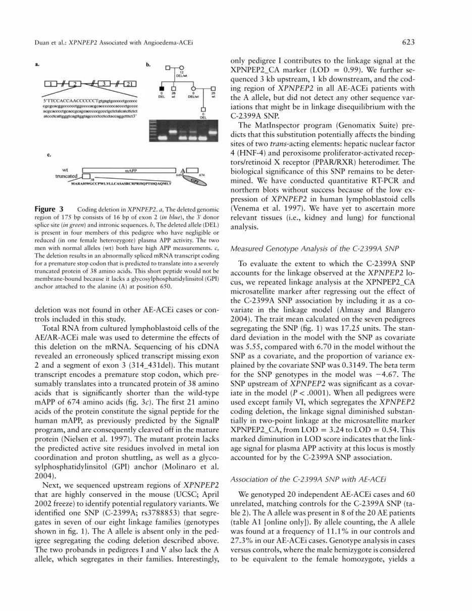
Given evidence of strong linkage to the XPNPEP2locus, we sequenced the coding regions of this candidategene in all available AE-ACEi cases with APP activities!10 units. A coding deletion in XPNPEP2 was detectedin the proband of pedigree VI (fig. 1b) (not included inthe initial mutation screening experiments) who firstsuffered an anaphylactoid reaction (AR) associatedwith ACEi during hemodialysis and subsequently suf-fered an AE-ACEi episode. The 175-bp genomic deletion(g.2953-3127del) includes 16 bp at the 3′ end of exon2, the donor splice site and a fragment of the down-stream intron (fig. 3a). The deleted allele is also presentin three relatives of this AE/AR-ACEi patient, who allhave negligible or reduced plasma APP activities (fig. 3b).The two men with relatively high plasma APP activitiesin this family each have a normal copy of this gene. This
Duan et al.: XPNPEP2 Associated with Angioedema-ACEi 623
Figure 3 Coding deletion in XPNPEP2. a, The deleted genomicregion of 175 bp consists of 16 bp of exon 2 (in blue), the 3′ donorsplice site (in green) and intronic sequences. b, The deleted allele (DEL)is present in four members of this pedigree who have negligible orreduced (in one female heterozygote) plasma APP activity. The twomen with normal alleles (wt) both have high APP measurements. c,The deletion results in an abnormally spliced mRNA transcript codingfor a premature stop codon that is predicted to translate into a severelytruncated protein of 38 amino acids. This short peptide would not bemembrane-bound because it lacks a glycosylphosphatidylinsitol (GPI)anchor attached to the alanine (A) at position 650.
deletion was not found in other AE-ACEi cases or con-trols included in this study.
Total RNA from cultured lymphoblastoid cells of theAE/AR-ACEi male was used to determine the effects ofthis deletion on the mRNA. Sequencing of his cDNArevealed an erroneously spliced transcript missing exon2 and a segment of exon 3 (314_431del). This mutanttranscript encodes a premature stop codon, which pre-sumably translates into a truncated protein of 38 aminoacids that is significantly shorter than the wild-typemAPP of 674 amino acids (fig. 3c). The first 21 aminoacids of the protein constitute the signal peptide for thehuman mAPP, as previously predicted by the SignalPprogram, and are consequently cleaved off in the matureprotein (Nielsen et al. 1997). The mutant protein lacksthe predicted active site residues involved in metal ioncoordination and proton shuttling, as well as a glyco-sylphosphatidylinsitol (GPI) anchor (Molinaro et al.2004).
Next, we sequenced upstream regions of XPNPEP2that are highly conserved in the mouse (UCSC; April2002 freeze) to identify potential regulatory variants. Weidentified one SNP (C-2399A; rs3788853) that segre-gates in seven of our eight linkage families (genotypesshown in fig. 1). The A allele is absent only in the ped-igree segregating the coding deletion described above.The two probands in pedigrees I and V also lack the Aallele, which segregates in their families. Interestingly,
only pedigree I contributes to the linkage signal at theXPNPEP2_CA marker (LOD p 0.99). We further se-quenced 3 kb upstream, 1 kb downstream, and the cod-ing region of XPNPEP2 in all AE-ACEi patients withthe A allele, but did not detect any other sequence var-iations that might be in linkage disequilibrium with theC-2399A SNP.
The MatInspector program (Genomatix Suite) pre-dicts that this substitution potentially affects the bindingsites of two trans-acting elements: hepatic nuclear factor4 (HNF-4) and peroxisome proliferator-activated recep-tors/retinoid X receptor (PPAR/RXR) heterodimer. Thebiological significance of this SNP remains to be deter-mined. We have conducted quantitative RT-PCR andnorthern blots without success because of the low ex-pression of XPNPEP2 in human lymphoblastoid cells(Venema et al. 1997). We have yet to ascertain morerelevant tissues (i.e., kidney and lung) for functionalanalysis.
Measured Genotype Analysis of the C-2399A SNP
To evaluate the extent to which the C-2399A SNPaccounts for the linkage observed at the XPNPEP2 lo-cus, we repeated linkage analysis at the XPNPEP2_CAmicrosatellite marker after regressing out the effect ofthe C-2399A SNP association by including it as a co-variate in the linkage model (Almasy and Blangero2004). The trait mean calculated on the seven pedigreessegregating the SNP (fig. 1) was 17.25 units. The stan-dard deviation in the model with the SNP as covariatewas 5.55, compared with 6.70 in the model without theSNP as a covariate, and the proportion of variance ex-plained by the covariate SNP was 0.3149. The beta termfor the SNP genotypes in the model was �4.67. TheSNP upstream of XPNPEP2 was significant as a covar-iate in the model (P ! .0001). When all pedigrees wereused except family VI, which segregates the XPNPEP2coding deletion, the linkage signal diminished substan-tially in two-point linkage at the microsatellite markerXPNPEP2_CA, from LOD p 3.24 to LOD p 0.54. Thismarked diminution in LOD score indicates that the link-age signal for plasma APP activity at this locus is mostlyaccounted for by the C-2399A SNP association.
Association of the C-2399A SNP with AE-ACEi
We genotyped 20 independent AE-ACEi cases and 60unrelated, matching controls for the C-2399A SNP (ta-ble 2). The A allele was present in 8 of the 20 AE patients(table A1 [online only]). By allele counting, the A allelewas found at a frequency of 11.1% in our controls and27.3% in our AE-ACEi cases. Genotype analysis in casesversus controls, where the male hemizygote is consideredto be equivalent to the female homozygote, yields a

624 Am. J. Hum. Genet. 77:617–626, 2005
(Armitage trend test) when comparing AEP p .0364cases to population controls.
Discussion
This report provides evidence that variable plasma APPactivity is partially regulated by genetic factors. We es-timated that 34% of the phenotypic variation in ourlinkage families results from genotypic differences. Link-age analysis significantly identified a QTL (LOD p 3.75)near XPNPEP2, a candidate gene encoding mAPP. De-spite the absence of mutations detected in the initialgene-screening experiments, further investigation of thislocus identified two sequence variations. The genomicdeletion segregating in pedigree VI results in an erro-neously spliced transcript that is predicted to translateinto a severely truncated protein. The C-2399A SNPgenotype significantly accounts for the linkage signal atthis QTL and is significantly associated with AE-ACEi( ). No other variants that may be in linkageP p .0364disequilibrium with the A allele were found at this locusin additional sequencing experiments, further suggestingthat this SNP may be responsible for the observed re-duction in plasma APP activity.
The above variants were found in 9 of 20 AE-ACEicases (table A1 [online only]). This suggests that the lowplasma APP activity observed in additional AE-ACEipatients may result from other genetic loci or environ-mental factors. Our multipoint variance-componentlinkage analysis of cohort 1 provided evidence of sig-nificant linkage to a locus on chromosome 6 (LOD p3.47) and positive linkage to loci on chromosomes 8and 9. Multipoint linkage analysis including cohort 2,however, reduced linkage to the chromosome 6 locus(LOD p 1.43), as well as the LOD scores for the othertwo autosomal loci. Genotyping of additional markersat these loci in more families are necessary to eitherconfirm or reject linkage.
Given that a third of our AE patients have normalplasma APP activity (�20 units), this quantitative traitclearly explains a fraction of the AE risk associated withACEi therapy. Individuals who are not genetically pre-disposed to have low APP activity may develop AE-ACEi due to nonspecific inhibition of APP by certainkinds of ACEi drugs (Hooper et al. 1992) or other un-identified causes. Other potential risk factors includeelevated levels of the sensory neuropeptide substance P,which correlate with reduced amounts of dipeptidylpeptidase IV activity (Ferreira et al. 2000; Lefebvre etal. 2002). In addition, clinical trials for new vasopep-tidase inhibitors (that inhibit NEP and ACE) result inhigher AE incidence than with ACE inhibition alone,suggesting that NEP activity may play a role in AE risk.Furthermore, other, nongenetic factors (i.e., smoking
and estrogen replacement) may contribute to AE-ACEirisk.
Our study confirms previously published data show-ing that reduced plasma APP activity is a relatively com-mon phenotype in the general population (Lefebvre etal. 2002). Other than predisposition to AE-ACEi, noclinical phenotype has been associated with low APPactivity to date. This suggests that its physiological rolemay be nonessential or that there is functional redun-dancy. Furthermore, certain ACEi patients with lowAPP activity do not develop AE during therapy. Thissuggests that factors (i.e., environmental triggers) actingin pathways other than des-Arg9-BK degradation maycontribute to AE risk.
Finally, low APP activity may determine risk for otherACEi-associated adverse effects. We previously reportedan association between reduced plasma APP activity andanaphylactoid reactions during hemodialysis induced byACEi therapy (Blais et al. 1999a). Further genetic in-vestigations are necessary to determine if variants in theXPNPEP2 locus, such as those described here, play arole in AR-ACEi risk. Other adverse reactions associ-ated with ACEi therapy include chronic cough, whichaffects ∼10%–15% of ACEi patients. It would be in-teresting to further characterize plasma APP activity inthese patients and screen XPNPEP2 as a candidate sus-ceptibility gene for these side effects of ACEi.
In conclusion, this report demonstrates that geneticvariants at the XPNPEP2 locus are partially responsiblefor reduced plasma APP activity. Furthermore, we showa significant association between the C-2399A poly-morphism and AE-ACEi. Thus, we have successfullymapped a QTL that may, in part, predict susceptibilityto a potentially fatal adverse reaction associated withone of the most commonly used drugs worldwide. Ourstudy represents a potential application of pharmaco-genomics in health care, a field destined to transformmedical care, but in which there have been few successesto date. Our findings, in addition to further studies, mayfacilitate the rational design of safer drugs for treatingcardiovascular diseases, as well as clinical assays forpredicting individuals who are genetically more liableto develop AE associated with ACEi or other vasopep-tidase inhibitors.
Acknowledgments
We thank all the participants in this study; Dr. Andre Tou-louse, for molecular expertise; Dominique Verlaan, for carefulreview of this manuscript; Nicole Gervais and Kateri Brisebois,for technical assistance; and the logistical assistance of HospalFrance. This work was funded by the National Institutes ofHealth grant 1-RO1-HL079184. G.A.R. and G.M. are sup-ported by the Canadian Institutes of Health Research. A.A.receives funding from Fonds de Recherche en Sante du Quebec.

Duan et al.: XPNPEP2 Associated with Angioedema-ACEi 625
Web Resources
Accession numbers and URLs for data presented herein areas follows:
Fondation Jean Dausset CEPH, http://www.cephb.fr/MatInspector (Genomatix Suite), http://www.genomatix.de/National Center for Biotechnology Information (NCBI), http:
//www.ncbi.nlm.nih.gov/Online Mendelian Inheritance in Man (OMIM), http://www
.ncbi.nlm.nih.gov/Omim/University of Santa Cruz (UCSC) Genome Browser, http://
genome.ucsc.edu/
References
Adam A, Cugno M, Molinaro G, Perez M, Lepage Y, AgostoniA (2002) Aminopeptidase P in individuals with a history ofangio-oedema on ACE inhibitors. Lancet 359:2088–2089
Agostoni A, Aygoren-Pursun E, Binkley KE, Blanch A, BorkK, Bouillet L, Bucher C, et al (2004) Hereditary and acquiredangioedema: problems and progress: proceedings of thethird C1 esterase inhibitor deficiency workshop and beyond.J Allergy Clin Immunol 114:S51–131
Agostoni A, Cicardi M (2001) Drug-induced angioedema with-out urticaria. Drug Saf 24:599–606
Agostoni A, Cicardi M, Cugno M, Zingale LC, Gioffre D,Nussberger J (1999) Angioedema due to angiotensin-con-verting enzyme inhibitors. Immunopharmacology 44:21–25
Almasy L, Blangero J (1998) Multipoint quantitative-trait link-age analysis in general pedigrees. Am J Hum Genet 62:1198–1211
Almasy L, Blangero J (2004) Exploring positional candidategenes: linkage conditional on measured genotype. Behav Ge-net 34:173–177
Bhoola KD, Figueroa CD, Worthy K (1992) Bioregulation ofkinins: kallikreins, kininogens, and kininases. PharmacolRev 44:1–80
Blais C Jr, Couture R, Drapeau G, Colman RW, Adam A (1997)Involvement of endogenous kinins in the pathogenesis ofpeptidoglycan-induced arthritis in the Lewis rat. ArthritisRheum 40:1327–1333
Blais C Jr, Marc-Aurele J, Simmons WH, Loute G, ThibaultP, Skidgel RA, Adam A (1999a) Des-Arg9-bradykinin me-tabolism in patients who presented hypersensitivity reactionsduring hemodialysis: role of serum ACE and aminopeptidaseP. Peptides 20:421–430
Blais C Jr, Marceau F, Rouleau JL, Adam A (2000) The kal-likrein-kininogen-kinin system: lessons from the quantifi-cation of endogenous kinins. Peptides 21:1903–1940
Blais C Jr, Rouleau JL, Brown NJ, Lepage Y, Spence D, MunozC, Friborg J, Geadah D, Gervais N, Adam A (1999b) Serummetabolism of bradykinin and des-Arg9-bradykinin in pa-tients with angiotensin-converting enzyme inhibitor-associ-ated angioedema. Immunopharmacology 43:293–302
Box GEP, Cox DR (1964) An analysis of transformations. JR Stat Soc Ser B (Methodological) 26:211–252
Bright RA, Torrence ME, Daley WR, McClellan WM (1999)Preliminary survey of the occurrence of anaphylactoid re-
actions during haemodialysis. Nephrol Dial Transplant 14:799–800
Brown NJ, Ray WA, Snowden M, Griffin MR (1996) BlackAmericans have an increased rate of angiotensin convertingenzyme inhibitor-associated angioedema. Clin PharmacolTher 60:8–13
Brown NJ, Vaughan DE (1998) Angiotensin-converting en-zyme inhibitors. Circulation 97:1411–1420
Coats AJ (2002) Omapatrilat—the story of Overture and Oc-tave. Int J Cardiol 86:1–4
Cugno M, Nussberger J, Cicardi M, Agostoni A (2003) Bra-dykinin and the pathophysiology of angioedema. Int Im-munopharmacol 3:311–317
Cyr M, Lepage Y, Blais C Jr, Gervais N, Cugno M, RouleauJL, Adam A (2001) Bradykinin and des-Arg(9)-bradykininmetabolic pathways and kinetics of activation of humanplasma. Am J Physiol Heart Circ Physiol 281:H275–283
Discenza MT, Pelletier J (2004) Insights into the physiologicalrole of WT1 from studies of genetically modified mice. Phys-iol Genomics 16:287–300
Ferreira PK, Campos MM, Calixto JB (2000) The role of sen-sorial neuropeptides in the edematogenic responses mediatedby B(1) agonist des-Arg(9)-BK in rats pre-treated with LPS.Regul Pept 89:29–35
Gallagher PE, Li P, Lenhart JR, Chappell MC, Brosnihan KB(1999) Estrogen regulation of angiotensin-converting en-zyme mRNA. Hypertension 33:323–328
Germer S, Higuchi R (1999) Single-tube genotyping withoutoligonucleotide probes. Genome Res 9:72–78
Hooper NM, Hryszko J, Oppong SY, Turner AJ (1992) In-hibition by converting enzyme inhibitors of pig kidney ami-nopeptidase P. Hypertension 19:281–285
Israili ZH, Hall WD (1992) Cough and angioneurotic edemaassociated with angiotensin-converting enzyme inhibitortherapy. A review of the literature and pathophysiology. AnnIntern Med 117:234–242
Kim KS, Kumar S, Simmons WH, Brown NJ (2000) Inhibitionof aminopeptidase P potentiates wheal response to bradyk-inin in angiotensin-converting enzyme inhibitor-treated hu-mans. J Pharmacol Exp Ther 292:295–298
Kostis JB, Packer M, Black HR, Schmieder R, Henry D, LevyE (2004) Omapatrilat and enalapril in patients with hyper-tension: the Omapatrilat Cardiovascular Treatment vs. En-alapril (OCTAVE) trial. Am J Hypertens 17:103–111
Lefebvre J, Murphey LJ, Hartert TV, Jiao Shan R, SimmonsWH, Brown NJ (2002) Dipeptidyl peptidase IV activity inpatients with ACE-inhibitor-associated angioedema. Hyper-tension 39:460–464
Molinaro G, Boileau G, Adam A (2004) Aminopeptidase Pand vasoactive peptides. In: Hooper NM, Lendeckel (eds)Aminopeptidases in biology and disease. Kluwer Academic/Plenum, New York
Molinaro G, Cugno M, Perez M, Lepage Y, Gervais N, Agos-toni A, Adam A (2002) Angiotensin-converting enzyme in-hibitor-associated angioedema is characterized by a slowerdegradation of des-arginine(9)-bradykinin. J Pharmacol ExpTher 303:232–237
Nielsen H, Engelbrecht J, Brunak S, von Heijne G (1997) Iden-tification of prokaryotic and eukaryotic signal peptides andprediction of their cleavage sites. Protein Eng 10:1–6

626 Am. J. Hum. Genet. 77:617–626, 2005
Nussberger J, Cugno M, Amstutz C, Cicardi M, Pellacani A,Agostoni A (1998) Plasma bradykinin in angio-oedema.Lancet 351:1693–1697
Simmons WH, Orawski AT (1992) Membrane-bound ami-nopeptidase P from bovine lung. Its purification, properties,and degradation of bradykinin. J Biol Chem 267:4897–4903
Soria JM, Almasy L, Souto JC, Tirado I, Borell M, Mateo J,Slifer S, Stone W, Blangero J, Fontcuberta J (2000) Linkageanalysis demonstrates that the prothrombin G20210A mu-tation jointly influences plasma prothrombin levels and riskof thrombosis. Blood 95:2780–2785
Sprinkle TJ, Caldwell C, Ryan JW (2000) Cloning, chromo-somal sublocalization of the human soluble aminopeptidaseP gene (XPNPEP1) to 10q25.3 and conservation of the pu-tative proton shuttle and metal ligand binding sites withXPNPEP2. Arch Biochem Biophys 378:51–56
Sprinkle TJ, Stone AA, Venema RC, Denslow ND, Caldwell
C, Ryan JW (1998) Assignment of the membrane-boundhuman aminopeptidase P gene (XPNPEP2) to chromosomeXq25. Genomics 50:114–116
Unger T, Gohlke P (1994) Converting enzyme inhibitors incardiovascular therapy: current status and future potential.Cardiovasc Res 28:146–158
Venema RC, Ju H, Zou R, Venema VJ, Ryan JW (1997) Clon-ing and tissue distribution of human membrane-bound ami-nopeptidase P. Biochim Biophys Acta 1354:45–48
Vleeming W, van Amsterdam JG, Stricker BH, de Wildt DJ(1998) ACE inhibitor-induced angioedema: incidence, pre-vention and management. Drug Saf 18:171–188
Yvert G, Brem RB, Whittle J, Akey JM, Foss E, Smith EN,Mackelprang R, Kruglyak L (2003) Trans-acting regulatoryvariation in Saccharomyces cerevisiae and the role of tran-scription factors. Nat Genet 35:57–64


