Embed Size (px)
Citation preview
Available online http://arthritis-research.com/content/10/4/R93
Open AccessVol 10 No 4Research articleTH-17 cells in rheumatoid arthritisShiva Shahrara1, QiQuan Huang1, Arthur M Mandelin II1 and Richard M Pope1,2
1Department of Medicine, Feinberg School of Medicine, Northwestern University 240 E. Huron, Suite M220, Chicago, IL 60611, USA2Jesse Brown VA Chicago Healthcare System, 820 S. Damon, Chicago, IL 60612, USA
Corresponding author: Shiva Shahrara, [email protected]
Received: 15 May 2008 Revisions requested: 11 Jun 2008 Revisions received: 8 Aug 2008 Accepted: 18 Aug 2008 Published: 18 Aug 2008
Arthritis Research & Therapy 2008, 10:R93 (doi:10.1186/ar2477)This article is online at: http://arthritis-research.com/content/10/4/R93© 2008 Shahrara et al.; licensee BioMed Central Ltd. This is an open access article distributed under the terms of the Creative Commons Attribution License (http://creativecommons.org/licenses/by/2.0), which permits unrestricted use, distribution, and reproduction in any medium, provided the original work is properly cited.
Abstract
Introduction The aim of this study was to quantify the numberof T-helper (TH)-17 cells present in rheumatoid arthritis (RA)synovial fluid (SF) and to determine the level of interleukin (IL)-17 cytokine in RA, osteoarthritis (OA) and normal synovialtissue, as well as to examine SF macrophages for the presenceof IL-23, IL-27 and interferon (IFN)-γ.
Methods Peripheral blood (PB) mononuclear cells from normaland RA donors and mononuclear cells from RA SF wereexamined either without stimulation or after pretreatment with IL-23 followed by stimulation with phorbol myristate acetate (PMA)plus ionomycin (P/I). The abundance of TH-17 cells in RA SFwas determined by flow cytometry. IL-17 levels were quantifiedin synovial tissue from RA, OA and normal individuals by ELISAand IL-23 was identified in SFs by ELISA. RA SF and control invitro differentiated macrophages were either untreated ortreated with the toll-like receptor (TLR) 2 ligand peptidoglycan,and then IL-23, IL-27 and IFN-γ mRNA levels were quantified byreal-time polymerase chain reaction (RT-PCR).
Results Treatment with P/I alone or combined with IL-23significantly increased the number of TH-17 cells in normal, RA
PB and RA SF. With or without P/I plus IL-23, the percentage ofTH-17 cells was higher in RA SF compared with normal and RAPB. IL-17 levels were comparable in OA and normal synovialtissues, and these values were significantly increased in RAsynovial tissue. Although IL-17 was readily detected in RA SFs,IL-23 was rarely identified in RA SF. However, IL-23 mRNA wassignificantly increased in RA SF macrophages compared withcontrol macrophages, with or without TLR2 ligation. IL-27mRNA was also significantly higher in RA SF compared withcontrol macrophages, but there was no difference in IL-27 levelsbetween RA and control macrophages after TLR2 ligation. IFN-γ mRNA was also detectable in RA SF macrophages but notcontrol macrophages and the increase of IFN-γ mRNA followingTLR2 ligation was greater in RA SF macrophages comparedwith control macrophages.
Conclusion These observations support a role for TH-17 cellsin RA. Our observations do not strongly support a role for IL-23in the generation of TH-17 cells in the RA joint, however, theysuggest strategies that enhance IL-27 or IFN-γ might modulatethe presence of TH-17 cells in RA.
IntroductionInterleukin (IL)-17 may play a critical role in the pathogenesisof rheumatoid arthritis (RA). IL-17 is capable of promotinginflammation by inducing a variety of pro-inflammatory media-tors, including cytokines, chemokines and other mediators ofbone and cartilage destruction in synovial fibroblasts, mono-cytes, macrophages and chondrocytes [1]. RA synovialexplants spontaneously produce IL-17 [2] and increased lev-els of IL-17 are found in RA synovial fluid (SF) compared withosteoarthritis (OA) SF [3]. By immunohistochemistry, IL-17
has previously been identified in T lymphocytes in RA synovialtissue (ST), especially CD4+CD45RO+ T cells [2,3]. Onesubset of T cells, T helper (TH)1 cells, produce IFNγ andanother subset, TH2 cells, produce IL-4 [4]; but as IFNγ andIL-4 are found at very low levels in the RA joint [5], the sourceof IL-17 in the RA joint was unclear until the recent discoveryof a third subset of T cells capable of producing IL-17, the TH-17 cells. However, the abundance of TH-17 cells in the RAjoint has not yet been fully characterised. Nonetheless, thepotential importance of IL-17 in RA is supported by the obser-vation that IL-17 is critical for the development of, and is an
Page 1 of 7(page number not for citation purposes)
DMARDs = disease-modifying anti-rheumatic drugs; IFN = interferon; IL = interleukin; OA = osteoarthritis; PB = peripheral blood; PGN = peptidog-lycan; P/I = PMA (phorbol myristate acetate) plus ionomyocin; RA = rheumatoid arthritis; RT-PCR = real-time polymerase chain reaction; SF = synovial fluid; ST = synovial tissue; TGF = transforming growth factor; TH = T helper; TLR = toll-like receptor; TNF = tumor necrosis factor.
Arthritis Research & Therapy Vol 10 No 4 Shahrara et al.
effective therapeutic target in, a variety of animal models of RA[6].
The mechanisms contributing to the development of TH-17cells in humans have only been recently clarified. Severalgroups have demonstrated that IL-1β, IL-6, IL-23 and trans-forming growth factor (TGF)-β promote human TH-17 cell dif-ferentiation from naive peripheral blood (PB) CD4+ cells,resulting in the expression of IL-17 (also called IL-17A), IL-17F, IL-21, IL-22 and IL-6 [7,8]. Although it was initially sug-gested that the differentiation of human TH-17 cells is inde-pendent of TGF-β, recently published data demonstrate thatthe absence of TGF-β mediates a shift in T cell gene expres-sion from a TH-17 profile to a TH1-like profile [7,8]. Othershave shown that TGF-β and IL-21 uniquely promote the polar-isation of TH-17 cells from human naive CD4+ T cells, and fur-ther that IL-1β together with IL-6 or IL-23 are only capable ofinducing TH-17 cells from human memory CD4+ T cells [9].
Cell-cell contact of human CD4+ T cells with monocytes thathave been activated by lipopolysaccarides (LPS) or peptidog-lycan (PGN) promotes the development of TH-17 cells [10].Consistent with a potential role in RA, IL-23 plays a major rolein the pathogenesis of experimental arthritis, because IL-23-/-mice are resistant to the development of collagen-inducedarthritis [11]. Conversely, IL-27 has recently been shown tosuppress the development of TH-17 cells [12] and to sup-press experimental arthritis [13]. Although IL-1 and IL-6 arehighly expressed in the RA joint and IL-23 has been identifiedby immunohistochemistry in RA ST [14], the role of IL-23 isunclear, due to marked differences in the levels of IL-23reported in previous studies [15-17]. Also, the expression ofthe cytokines IL-27 and IFN-γ, which might suppress TH-17polarisation, have not been examined in RA SF.
In the present study, we document the presence of TH-17cells in RA SF, and demonstrate that the abundance of TH-17cells is significantly increased compared with RA or normalPB. Further, we show that RA ST express higher levels of IL-17 compared with OA and normal ST. We also demonstratethat IL-23 increases the abundance of TH-17 cells followingshort-term activation of mononuclear cells taken from RA SF,but not from normal PB or RA PB. Although IL-23 was rarelydetected in RA SF, IL-23 mRNA was increased in RA SF mac-rophages compared with control macrophages, in theabsence or presence of the toll-like receptor (TLR) 2 agonist,PGN. IL-27 was also increased in RA SF macrophages,although it was only modestly induced by PGN. While IFN-γmRNA levels were undetectable in control macrophages, lowlevels were detected in RA SF macrophages and induction ofIFN-γ mRNA by PGN was greater in RA SF macrophagescompared with control macrophages. These observationssupport a role of TH-17 cells in the pathogenesis of RA,although the mechanisms responsible for the generation ofthese cells in the RA joint require further clarification.
Materials and methodsPatientsSFs were obtained from patients with RA, diagnosed accord-ing to the 1987 revised criteria of the American College ofRheumatology [18]. RA SF was obtained from 12 women andtwo men (mean age ± SE = 52.8 ± 6.3 years). Two patientswere only taking prednisone (generic) (<10 mg/day) at thetime of joint aspiration, seven patients were taking methotrex-ate (generic) plus an anti-tumor necrosis factor (TNF) 3 on ananti-TNF alone, one patient was only taking methotrexate, andone patient was taking azathioprine (generic) plus abatacept(Orencia, Bristol-Myers Squibb) Dosages vary widely bypatient and were not tracked for the purposes of this study. Ofthe patients whose SFs were examined for cytokines (meanage = 52.6 ± 2.7 years), 15 were taking an anti-TNF, eitheralone (n = 1), with a non-biological disease-modifying anti-rheumatic drug (DMARD) (n = 10; methotrexate, leflunomide(generic) or azulfidine) or with low-dose prednisone (n = 4,<10 mg/day). Eight patients were taking low-dose pred-nisone, either alone (n = 4) or with methotrexate (n = 4). Fourpatients were taking no medication at the time of arthrocenthe-sis. RA PB was obtained from nine women (mean age 56.3 ±7.5 years), of whom there were three taking methotrexatealone, two taking methotrexate plus an anti-TNF, one takingmethotrexate and leflunomide, two taking methotrexate andabatacept and one patient taking rituximab (Rituxan,Genentech).
The studies were approved by the Northwestern UniversityInstitutional Ethics Review Board and all donors gave informedwritten consent.
Cell isolation and cultureMacrophages were differentiated in vitro for seven days frommonocytes, which were purified by elutriation from the PBmononuclear cells of healthy donors as previously described[19]. Heparinised SFs were centrifuged at 800 g at room tem-perature for 10 minutes to obtain cell-free SF. RA PB and RASF mononuclear cells were isolated by Histopaque gradientcentrifugation (Sigma-aldrich, St. Louis, MO, USA) RA SFmacrophages were isolated by adherence for one hour, aspreviously described [19]. The control and RA SF macro-phages were either untreated or treated with PGN (1 μg/ml)for four hours.
Flow cytometric analysis of TH-17 cellsMononuclear cells were either left untreated or incubated for18 hours with IL-23 (20 ng/ml). Subsequently, the cells wereincubated with PMA (50 ng/ml) and ionomyocin (1 μg/ml) plusbrefeldin A (10 μg/ml) for four hours. Cells were blocked with50% human serum and 0.5% bovine serum albumin in phos-phate buffered saline and incubated with allophycocyanin-conjugated monoclonal anti-CD3 (eBioscience, San Diego,CA) for 30 minutes, fixed with 2% formaldehyde for 10 min-utes, and then permeabilised with 0.1% NP40 for 10 minutes.
Page 2 of 7(page number not for citation purposes)

Available online http://arthritis-research.com/content/10/4/R93
Cells were then stained for fluorescein isothiocyanate labelledanti-CD4 and phycoerythrin-conjugated anti-IL-17 (eBio-science, San Diego, CA) or isotype control antibodies (eBio-science, San Diego, CA). TH-17 cells were identified as thosethat were CD3+CD4+IL-17+. The percentage of TH-17 cellsin each sample was normalised for staining with its control IgGvalue by subtracting the percentage of cells that were positivewhen stained with the control IgG alone from the original per-centage of TH-17 cells.
Real-time PCRTotal cellular RNA was extracted using Guanidinium thiocy-anate-phenol-chloroform extraction (Trizol reagent) (Invitro-gen, Carlsbad, CA, USA), and reverse-transcription and RT-PCR were performed as previously described [19]. Relativegene expression was determined by the ΔΔCt method.
Tissue homogenisationSynovial tissues were homogenised as described previously[20] in 1 ml of complete Mini protease-inhibitor cocktailhomogenisation buffer (Roche, Indianapolis, IN) on ice, fol-lowed by sonication for 30 seconds. Homogenates were cen-trifuged and filtered through a 0.45 μm pore size filter beforequantifying the levels of IL-17 by ELISA. The final concentra-tion of IL-17 in ST was normalised to the protein concentrationin each tissue.
Cytokine quantificationHuman IL-23 (p19/p40) (eBioscience, San Diego, CA) and IL-17 (R&D Systems, Minneapolis, MN, USA) ELISA kits wereused according to the manufacturers' instructions.
Statistical analysisThe data were analysed using two-tailed Student's t tests forpaired and unpaired samples. P values less than 0.05 wereconsidered significant.
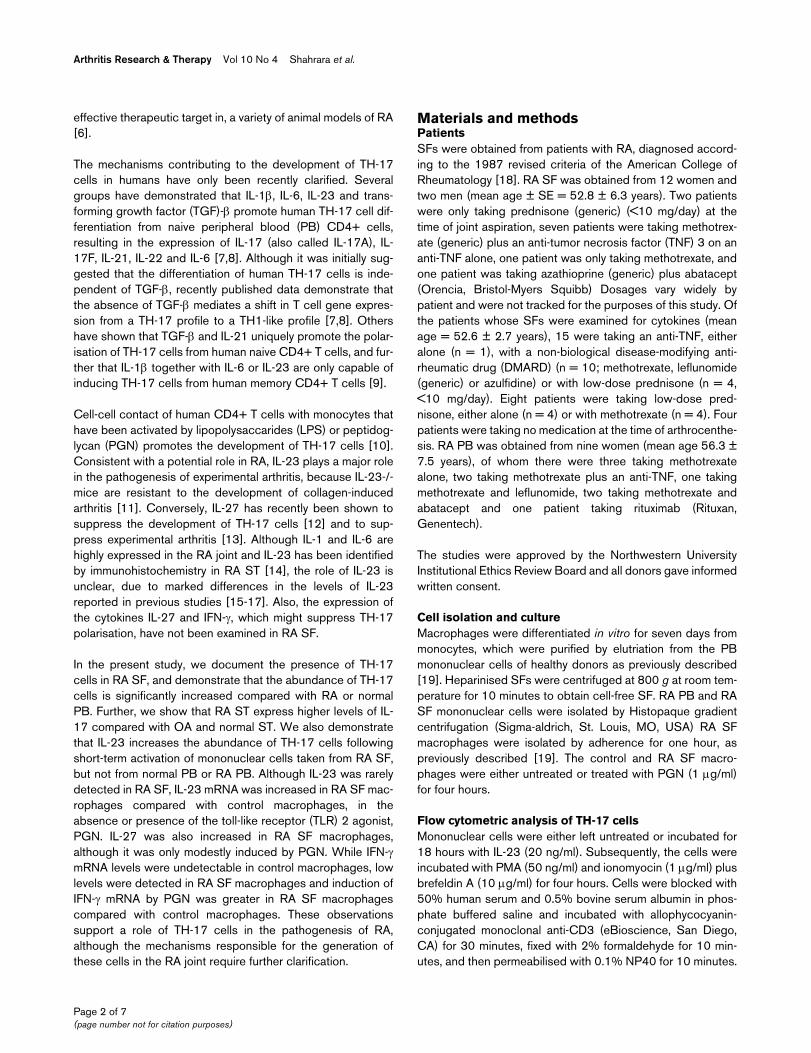
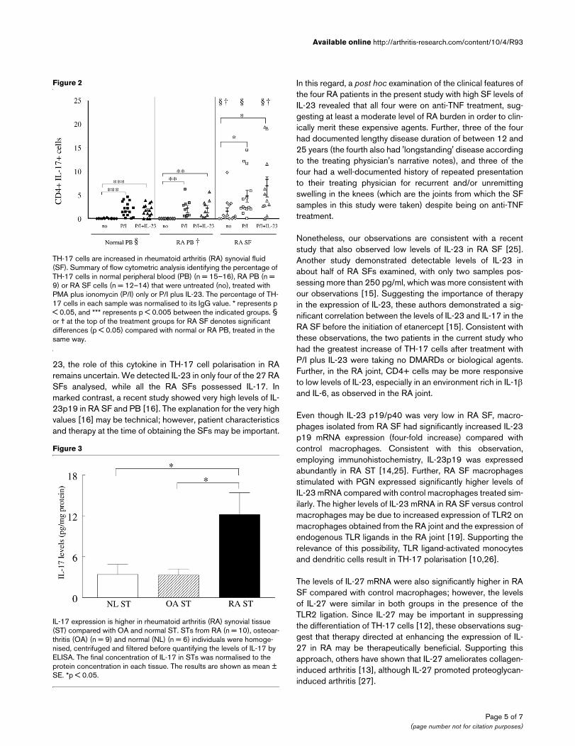
ResultsTH-17 cells expressed in RA SFIn the absence of stimulation, the percentage of TH-17 cells(CD3+CD4+IL-17+) was higher (p < 0.05) in RA SF (1.5 ±0.72%) compared with RA PB (0%) and normal PB (0.04 ±0.02%) (Figures 1 and 2). Short-term activation with PMA plusionomycin (P/I) was used to enhance detection of IL-17 withinthe CD4+ T cells, and not to promote polarisation. Treatmentwith P/I alone significantly increased the number of TH-17cells in normal PB (0.04% to 1.9 ± 0.4%, p < 0.005), RA PB(0% to 2.1 ± 0.6%, p < 0.05) and RA SF (1.5% to 4.7 ±1.24%, p < 0.05) (Figure 2). Mononuclear cells were alsoincubated with control medium or with IL-23 for 18 hoursbefore P/I was added. Pretreatment of RA SF mononuclearcells with IL-23 and P/I resulted in a significantly (p < 0.05)increased frequency of TH-17 cells (6.8 ± 1.93%) (Figures 1fand 2), compared with untreated RA SF (Figures 1e and 2).Employing normal PB (1.7 ± 0.3%) or RA PB (2.1 ± 0.6%),
pretreatment with IL-23 did not increase the number of TH-17cells compared with P/I alone; however, the percentage of TH-17 cells was significantly higher than in the untreated group.Following treatment with IL-23 plus P/I, the abundance of TH-17 cells in RA SF was significantly (p < 0.05) greater thanobserved with normal PB and RA PB treated in the same fash-ion (Figures 1b, 1f and 2). In conclusion, these results demon-strate the increased abundance of TH-17 cells in RA SFcompared with normal PB and RA PB.
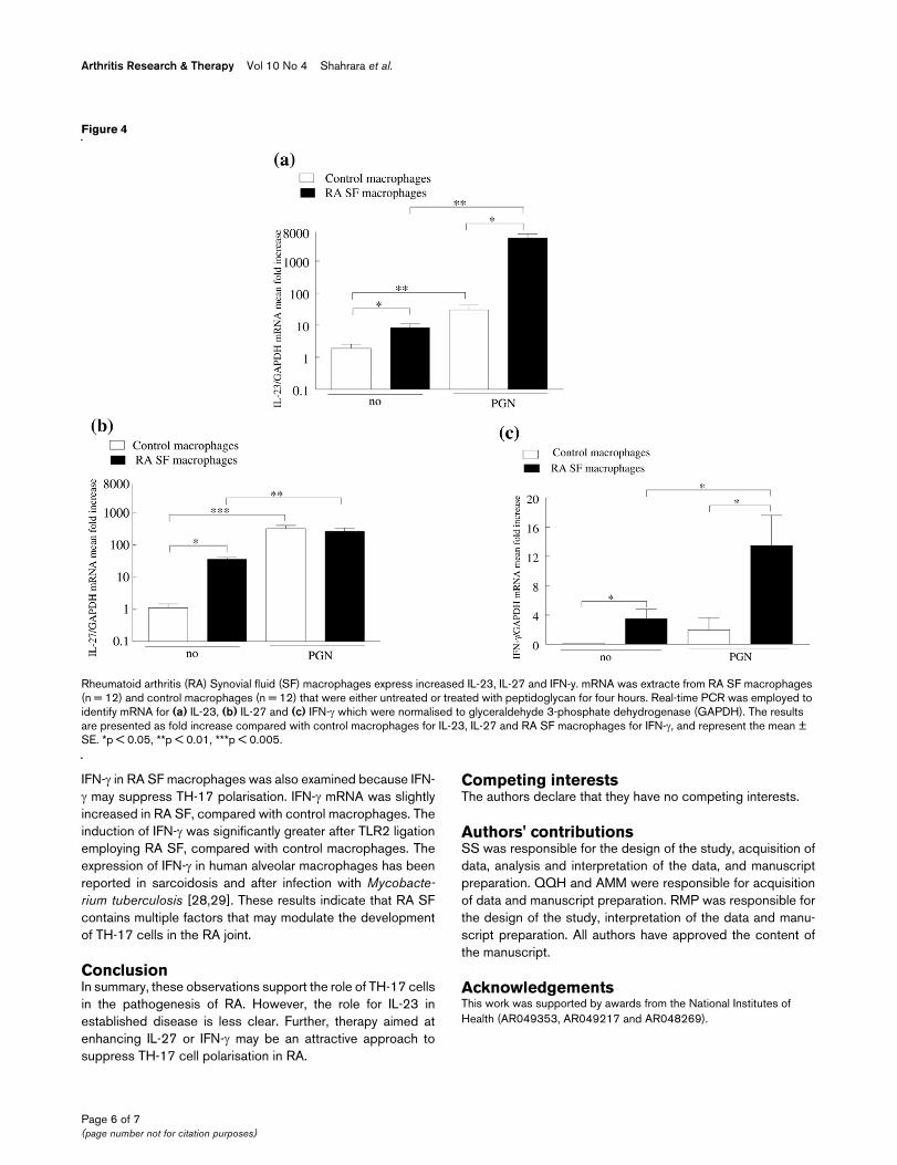
Level of IL-17 in RA, OA and normal synovial tissuesSince OA SFs do not contain enough cells to quantify TH-17cell number, we therefore measured the levels of IL-17 in RAST, OA ST and normal ST. Our results demonstrate that OAST (3.3 ± 0.9 pg/mg) and normal ST (3.4 ± 1.5 pg/mg) havecomparable levels of IL-17, and that the levels of IL-17 were3.5-fold increased (p < 0.05) in the RA ST (12.2 ± 3.2 pg/mg)(Figure 3).
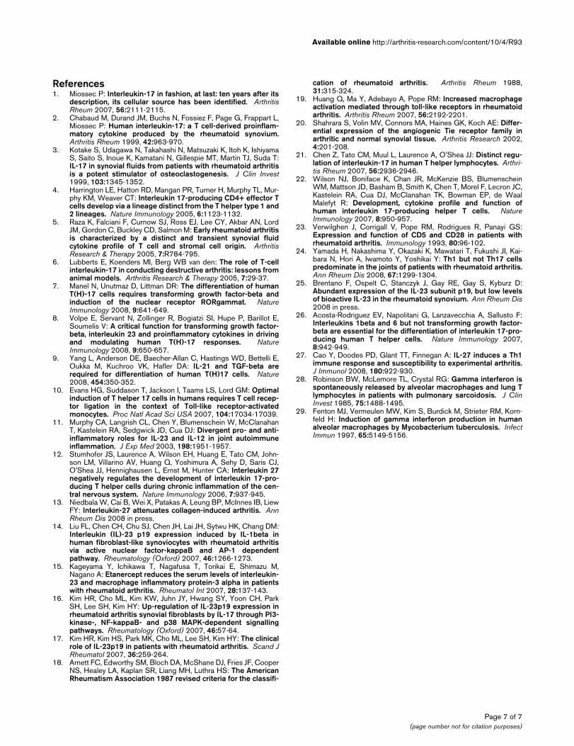
IL-23, IL-27 and IFN-γ in the RA jointRA SF T lymphocytes were responsive to IL-23, so the SFsfrom 28 patients with RA were examined for the presence ofIL-23, employing an assay specific for IL-23p19/p40. How-ever, IL-23 was only detected in four samples (20, 24, 30 and66 pg/ml), while none of the SFs from patients with OA werepositive (data not shown). In contrast, IL-17 was detected inall the RA SFs (233 ± 64 pg/ml) at levels significantly greaterthan those observed in OA SF (38 ± 18 pg/ml). Despite thepaucity of IL-23 in RA SF, macrophages from the joints ofpatients with RA expressed 4.3-fold (p < 0.05) more IL-23mRNA compared with control in vitro differentiated macro-phages (Figure 4a). Following activation with PGN, there wasa 15-fold increase of IL-23 mRNA in the control macrophages,but a significantly greater increase (p < 0.01) of 621-fold in theRA SF macrophages.
Since IL-27 and IFN-γ may suppress the development of TH-17 cells, macrophages were also examined for IL-27 and IFN-γ mRNA. Like IL-23, IL-27 and IFN-γ mRNA levels were signif-icantly higher in RA SF macrophages compared with controlmacrophages (Figures 4b, c). Following treatment with PGN,the IL-27 and IFN-γ mRNA increased in both RA SF (7.5-foldfor IL-27 and 3.8-fold for IFN-γ) and control macrophages(295-fold for IL-27 and from undetectable to 1.9-fold for IFN-γ). Similar to IL-23, the levels of IFN-γ were significantly higherafter PGN stimulation of macrophages from RA SF comparedwith control macrophages. In contrast to the results observedwith IL-23, after activation with PGN, there was no differencein IL-27 mRNA between the patients and the controls. Thesestudies demonstrate that although IL-23 is not plentiful in RASF, it is expressed in RA synovial macrophages and is greatlyincreased after TLR2 ligation. While IL-27 and IFN-γ areexpressed in RA SF macrophages, their induction after TLR2ligation was less compared with the induction of IL-23 in RASF macrophages.
Page 3 of 7(page number not for citation purposes)
Arthritis Research & Therapy Vol 10 No 4 Shahrara et al.
DiscussionIn the present study, we demonstrate that TH-17 cells aremore abundant in RA SF compared with normal PB and RAPB. IL-17 is produced by activated human CD4+CD45RO+memory T cells, while only low levels of IL-17 are secreted byCD4+CD45RA+ naive T cells [21,22]. The increased pres-ence of TH-17 cells in RA SF may be related to an increasedpercentage of memory CD4+ T cells in RA SF. However, thisis not likely to be the entire explanation because about 50% ofnormal PB and RA PB CD4+ cells are memory cells [23]. Inthe absence of IL-23 and P/I, TH-17 cells were 38-fold higherin RA SF compared with normal PB, whereas TH-17 cellswere essentially undetectable in RA PB. The reduction of TH-17 cells in RA compared with normal PB may be due to theenrichment of TH-17 cells in the RA joint, although differencesin disease activity or treatment may also be responsible. Our
observations contrast with a recently published study thatobserved a reduction of TH-17 cells in RA SF compared withRA PB [24]. The reason for the difference between that studyand other studies concerning IL-17 and TH-17 cells (includingthe work presented here) is not clear, although it is possiblethat differences in patient selection and therapy may have con-tributed, as well as technical differences such as the antibod-ies used or methods employed for cell fixation orpermeabilisation.
Treatment with IL-23 plus P/I resulted in increased numbers ofTH-17 cells in RA SF, which were greater than those observedin normal and RA PB. The expression of IL-23R is increasedon CD45RO memory T cells, and IL-23 may also induce theexpression of its own receptor [21,22]. Although our data sug-gest that RA SF CD4+ cells are capable of responding to IL-
Figure 1
Identification of TH-17 cells in rheumatoid arthritis (RA) synovial fluid (SF), RA peripheral blood (PB) and normal PBIdentification of TH-17 cells in rheumatoid arthritis (RA) synovial fluid (SF), RA peripheral blood (PB) and normal PB. (a, b) Normal PB, (c, d) RA PB or (e, f) RA SF mononuclear cells treated with control medium (a, c, e) or IL-23 (20 ng/ml) for 18 hours, followed by the addition of PMA (50 ng/ml) plus ionomycin (1 μg/ml) for four hours (b, d, f), before immunostaining. The values are presented as mean ± SE of % CD4+IL-17+ cells within the total CD4 population.
Page 4 of 7(page number not for citation purposes)
Available online http://arthritis-research.com/content/10/4/R93
23, the role of this cytokine in TH-17 cell polarisation in RAremains uncertain. We detected IL-23 in only four of the 27 RASFs analysed, while all the RA SFs possessed IL-17. Inmarked contrast, a recent study showed very high levels of IL-23p19 in RA SF and PB [16]. The explanation for the very highvalues [16] may be technical; however, patient characteristicsand therapy at the time of obtaining the SFs may be important.
In this regard, a post hoc examination of the clinical features ofthe four RA patients in the present study with high SF levels ofIL-23 revealed that all four were on anti-TNF treatment, sug-gesting at least a moderate level of RA burden in order to clin-ically merit these expensive agents. Further, three of the fourhad documented lengthy disease duration of between 12 and25 years (the fourth also had 'longstanding' disease accordingto the treating physician's narrative notes), and three of thefour had a well-documented history of repeated presentationto their treating physician for recurrent and/or unremittingswelling in the knees (which are the joints from which the SFsamples in this study were taken) despite being on anti-TNFtreatment.
Nonetheless, our observations are consistent with a recentstudy that also observed low levels of IL-23 in RA SF [25].Another study demonstrated detectable levels of IL-23 inabout half of RA SFs examined, with only two samples pos-sessing more than 250 pg/ml, which was more consistent withour observations [15]. Suggesting the importance of therapyin the expression of IL-23, these authors demonstrated a sig-nificant correlation between the levels of IL-23 and IL-17 in theRA SF before the initiation of etanercept [15]. Consistent withthese observations, the two patients in the current study whohad the greatest increase of TH-17 cells after treatment withP/I plus IL-23 were taking no DMARDs or biological agents.Further, in the RA joint, CD4+ cells may be more responsiveto low levels of IL-23, especially in an environment rich in IL-1βand IL-6, as observed in the RA joint.
Even though IL-23 p19/p40 was very low in RA SF, macro-phages isolated from RA SF had significantly increased IL-23p19 mRNA expression (four-fold increase) compared withcontrol macrophages. Consistent with this observation,employing immunohistochemistry, IL-23p19 was expressedabundantly in RA ST [14,25]. Further, RA SF macrophagesstimulated with PGN expressed significantly higher levels ofIL-23 mRNA compared with control macrophages treated sim-ilarly. The higher levels of IL-23 mRNA in RA SF versus controlmacrophages may be due to increased expression of TLR2 onmacrophages obtained from the RA joint and the expression ofendogenous TLR ligands in the RA joint [19]. Supporting therelevance of this possibility, TLR ligand-activated monocytesand dendritic cells result in TH-17 polarisation [10,26].
The levels of IL-27 mRNA were also significantly higher in RASF compared with control macrophages; however, the levelsof IL-27 were similar in both groups in the presence of theTLR2 ligation. Since IL-27 may be important in suppressingthe differentiation of TH-17 cells [12], these observations sug-gest that therapy directed at enhancing the expression of IL-27 in RA may be therapeutically beneficial. Supporting thisapproach, others have shown that IL-27 ameliorates collagen-induced arthritis [13], although IL-27 promoted proteoglycan-induced arthritis [27].
Figure 2
TH-17 cells are increased in rheumatoid arthritis (RA) synovial fluid (SF)TH-17 cells are increased in rheumatoid arthritis (RA) synovial fluid (SF). Summary of flow cytometric analysis identifying the percentage of TH-17 cells in normal peripheral blood (PB) (n = 15–16), RA PB (n = 9) or RA SF cells (n = 12–14) that were untreated (no), treated with PMA plus ionomycin (P/I) only or P/I plus IL-23. The percentage of TH-17 cells in each sample was normalised to its IgG value. * represents p < 0.05, and *** represents p < 0.005 between the indicated groups. § or † at the top of the treatment groups for RA SF denotes significant differences (p < 0.05) compared with normal or RA PB, treated in the same way.
Figure 3
IL-17 expression is higher in rheumatoid arthritis (RA) synovial tissue (ST) compared with OA and normal STIL-17 expression is higher in rheumatoid arthritis (RA) synovial tissue (ST) compared with OA and normal ST. STs from RA (n = 10), osteoar-thritis (OA) (n = 9) and normal (NL) (n = 6) individuals were homoge-nised, centrifuged and filtered before quantifying the levels of IL-17 by ELISA. The final concentration of IL-17 in STs was normalised to the protein concentration in each tissue. The results are shown as mean ± SE. *p < 0.05.
Page 5 of 7(page number not for citation purposes)
Arthritis Research & Therapy Vol 10 No 4 Shahrara et al.
IFN-γ in RA SF macrophages was also examined because IFN-γ may suppress TH-17 polarisation. IFN-γ mRNA was slightlyincreased in RA SF, compared with control macrophages. Theinduction of IFN-γ was significantly greater after TLR2 ligationemploying RA SF, compared with control macrophages. Theexpression of IFN-γ in human alveolar macrophages has beenreported in sarcoidosis and after infection with Mycobacte-rium tuberculosis [28,29]. These results indicate that RA SFcontains multiple factors that may modulate the developmentof TH-17 cells in the RA joint.
ConclusionIn summary, these observations support the role of TH-17 cellsin the pathogenesis of RA. However, the role for IL-23 inestablished disease is less clear. Further, therapy aimed atenhancing IL-27 or IFN-γ may be an attractive approach tosuppress TH-17 cell polarisation in RA.
Competing interestsThe authors declare that they have no competing interests.
Authors' contributionsSS was responsible for the design of the study, acquisition ofdata, analysis and interpretation of the data, and manuscriptpreparation. QQH and AMM were responsible for acquisitionof data and manuscript preparation. RMP was responsible forthe design of the study, interpretation of the data and manu-script preparation. All authors have approved the content ofthe manuscript.
AcknowledgementsThis work was supported by awards from the National Institutes of Health (AR049353, AR049217 and AR048269).
Figure 4
Rheumatoid arthritis (RA) synovial fluid (SF) macrophages express increased IL-23, IL-27 and IFN-γRheumatoid arthritis (RA) Synovial fluid (SF) macrophages express increased IL-23, IL-27 and IFN-y. mRNA was extracte from RA SF macrophages (n = 12) and control macrophages (n = 12) that were either untreated or treated with peptidoglycan for four hours. Real-time PCR was employed to identify mRNA for (a) IL-23, (b) IL-27 and (c) IFN-γ which were normalised to glyceraldehyde 3-phosphate dehydrogenase (GAPDH). The results are presented as fold increase compared with control macrophages for IL-23, IL-27 and RA SF macrophages for IFN-γ, and represent the mean ± SE. *p < 0.05, **p < 0.01, ***p < 0.005.
Page 6 of 7(page number not for citation purposes)
Available online http://arthritis-research.com/content/10/4/R93
References1. Miossec P: Interleukin-17 in fashion, at last: ten years after its
description, its cellular source has been identified. ArthritisRheum 2007, 56:2111-2115.
2. Chabaud M, Durand JM, Buchs N, Fossiez F, Page G, Frappart L,Miossec P: Human interleukin-17: a T cell-derived proinflam-matory cytokine produced by the rheumatoid synovium.Arthritis Rheum 1999, 42:963-970.
3. Kotake S, Udagawa N, Takahashi N, Matsuzaki K, Itoh K, IshiyamaS, Saito S, Inoue K, Kamatani N, Gillespie MT, Martin TJ, Suda T:IL-17 in synovial fluids from patients with rheumatoid arthritisis a potent stimulator of osteoclastogenesis. J Clin Invest1999, 103:1345-1352.
4. Harrington LE, Hatton RD, Mangan PR, Turner H, Murphy TL, Mur-phy KM, Weaver CT: Interleukin 17-producing CD4+ effector Tcells develop via a lineage distinct from the T helper type 1 and2 lineages. Nature Immunology 2005, 6:1123-1132.
5. Raza K, Falciani F, Curnow SJ, Ross EJ, Lee CY, Akbar AN, LordJM, Gordon C, Buckley CD, Salmon M: Early rheumatoid arthritisis characterized by a distinct and transient synovial fluidcytokine profile of T cell and stromal cell origin. ArthritisResearch & Therapy 2005, 7:R784-795.
6. Lubberts E, Koenders MI, Berg WB van den: The role of T-cellinterleukin-17 in conducting destructive arthritis: lessons fromanimal models. Arthritis Research & Therapy 2005, 7:29-37.
7. Manel N, Unutmaz D, Littman DR: The differentiation of humanT(H)-17 cells requires transforming growth factor-beta andinduction of the nuclear receptor RORgammat. NatureImmunology 2008, 9:641-649.
8. Volpe E, Servant N, Zollinger R, Bogiatzi SI, Hupe P, Barillot E,Soumelis V: A critical function for transforming growth factor-beta, interleukin 23 and proinflammatory cytokines in drivingand modulating human T(H)-17 responses. NatureImmunology 2008, 9:650-657.
9. Yang L, Anderson DE, Baecher-Allan C, Hastings WD, Bettelli E,Oukka M, Kuchroo VK, Hafler DA: IL-21 and TGF-beta arerequired for differentiation of human T(H)17 cells. Nature2008, 454:350-352.
10. Evans HG, Suddason T, Jackson I, Taams LS, Lord GM: Optimalinduction of T helper 17 cells in humans requires T cell recep-tor ligation in the context of Toll-like receptor-activatedmonocytes. Proc Natl Acad Sci USA 2007, 104:17034-17039.
11. Murphy CA, Langrish CL, Chen Y, Blumenschein W, McClanahanT, Kastelein RA, Sedgwick JD, Cua DJ: Divergent pro- and anti-inflammatory roles for IL-23 and IL-12 in joint autoimmuneinflammation. J Exp Med 2003, 198:1951-1957.
12. Stumhofer JS, Laurence A, Wilson EH, Huang E, Tato CM, John-son LM, Villarino AV, Huang Q, Yoshimura A, Sehy D, Saris CJ,O'Shea JJ, Hennighausen L, Ernst M, Hunter CA: Interleukin 27negatively regulates the development of interleukin 17-pro-ducing T helper cells during chronic inflammation of the cen-tral nervous system. Nature Immunology 2006, 7:937-945.
13. Niedbala W, Cai B, Wei X, Patakas A, Leung BP, McInnes IB, LiewFY: Interleukin-27 attenuates collagen-induced arthritis. AnnRheum Dis 2008 in press.
14. Liu FL, Chen CH, Chu SJ, Chen JH, Lai JH, Sytwu HK, Chang DM:Interleukin (IL)-23 p19 expression induced by IL-1beta inhuman fibroblast-like synoviocytes with rheumatoid arthritisvia active nuclear factor-kappaB and AP-1 dependentpathway. Rheumatology (Oxford) 2007, 46:1266-1273.
15. Kageyama Y, Ichikawa T, Nagafusa T, Torikai E, Shimazu M,Nagano A: Etanercept reduces the serum levels of interleukin-23 and macrophage inflammatory protein-3 alpha in patientswith rheumatoid arthritis. Rheumatol Int 2007, 28:137-143.
16. Kim HR, Cho ML, Kim KW, Juhn JY, Hwang SY, Yoon CH, ParkSH, Lee SH, Kim HY: Up-regulation of IL-23p19 expression inrheumatoid arthritis synovial fibroblasts by IL-17 through PI3-kinase-, NF-kappaB- and p38 MAPK-dependent signallingpathways. Rheumatology (Oxford) 2007, 46:57-64.
17. Kim HR, Kim HS, Park MK, Cho ML, Lee SH, Kim HY: The clinicalrole of IL-23p19 in patients with rheumatoid arthritis. Scand JRheumatol 2007, 36:259-264.
18. Arnett FC, Edworthy SM, Bloch DA, McShane DJ, Fries JF, CooperNS, Healey LA, Kaplan SR, Liang MH, Luthra HS: The AmericanRheumatism Association 1987 revised criteria for the classifi-
cation of rheumatoid arthritis. Arthritis Rheum 1988,31:315-324.
19. Huang Q, Ma Y, Adebayo A, Pope RM: Increased macrophageactivation mediated through toll-like receptors in rheumatoidarthritis. Arthritis Rheum 2007, 56:2192-2201.
20. Shahrara S, Volin MV, Connors MA, Haines GK, Koch AE: Differ-ential expression of the angiogenic Tie receptor family inarthritic and normal synovial tissue. Arthritis Research 2002,4:201-208.
21. Chen Z, Tato CM, Muul L, Laurence A, O'Shea JJ: Distinct regu-lation of interleukin-17 in human T helper lymphocytes. Arthri-tis Rheum 2007, 56:2936-2946.
22. Wilson NJ, Boniface K, Chan JR, McKenzie BS, BlumenscheinWM, Mattson JD, Basham B, Smith K, Chen T, Morel F, Lecron JC,Kastelein RA, Cua DJ, McClanahan TK, Bowman EP, de WaalMalefyt R: Development, cytokine profile and function ofhuman interleukin 17-producing helper T cells. NatureImmunology 2007, 8:950-957.
23. Verwilghen J, Corrigall V, Pope RM, Rodrigues R, Panayi GS:Expression and function of CD5 and CD28 in patients withrheumatoid arthritis. Immunology 1993, 80:96-102.
24. Yamada H, Nakashima Y, Okazaki K, Mawatari T, Fukushi JI, Kai-bara N, Hori A, Iwamoto Y, Yoshikai Y: Th1 but not Th17 cellspredominate in the joints of patients with rheumatoid arthritis.Ann Rheum Dis 2008, 67:1299-1304.
25. Brentano F, Ospelt C, Stanczyk J, Gay RE, Gay S, Kyburz D:Abundant expression of the IL-23 subunit p19, but low levelsof bioactive IL-23 in the rheumatoid synovium. Ann Rheum Dis2008 in press.
26. Acosta-Rodriguez EV, Napolitani G, Lanzavecchia A, Sallusto F:Interleukins 1beta and 6 but not transforming growth factor-beta are essential for the differentiation of interleukin 17-pro-ducing human T helper cells. Nature Immunology 2007,8:942-949.
27. Cao Y, Doodes PD, Glant TT, Finnegan A: IL-27 induces a Th1immune response and susceptibility to experimental arthritis.J Immunol 2008, 180:922-930.
28. Robinson BW, McLemore TL, Crystal RG: Gamma interferon isspontaneously released by alveolar macrophages and lung Tlymphocytes in patients with pulmonary sarcoidosis. J ClinInvest 1985, 75:1488-1495.
29. Fenton MJ, Vermeulen MW, Kim S, Burdick M, Strieter RM, Korn-feld H: Induction of gamma interferon production in humanalveolar macrophages by Mycobacterium tuberculosis. InfectImmun 1997, 65:5149-5156.
Page 7 of 7(page number not for citation purposes)