Embed Size (px)
Citation preview
Histopathology in fish: proposal for a protocol to assessaquatic pollution
D Bernet1, H Schmidt1, W Meier1, P Burkhardt-Holm1,2and T Wahli1
1 Centre for Fish and Wildlife Health,Institute of Veterinary Pathology,University of Berne,Berne,Switzerland2 Interdisciplinary Centre for GeneralEcology,University of Berne,Berne,Switzerland
Abstract
Waterpollution inducespathologicalchangesinfish.As an indicator of exposure to contaminants,histology represents a useful tool to assess the degreeof pollution, particularly for sub-lethal and chroniceffects.However,a standardized method forthedescription and assessment of histologicalchanges,mainly for use in freshwater fish, is still lacking.Inthis paper,the presentauthors propose a standar-dized tool for the assessment of histological findingswhich can beapplied to differentorgans.Themethodologyisbasedon two factors:(1)theextension ofa pathologicalchange is rated with a`score value'; and (2) the pathological importance ofthis alteration is defined as an `importance factor'.The sum of themultipliedscorevaluesandimportance factors of alldiagnosed changes resultsin differentindices.With these indices,statisticalanalysis can be carried out. Assessment methods forthe gills,liver,kidney and skin are described.
IntroductionThe dischargeof industrial,agriculturalanddomestic waste water into the environment resultsin the pollution ofaquatic systems.Fish are oftenexposed to highly contaminated water, especially inareas where the dilution rate of waste water is low.Thishasadverseeffects,particularlywhen con-
taminants:(1)arenotor onlyslightlydecom-posable;(2) exhibit a high biologicaleffectiveness;(3) possess a high potentialfor accumulation;and(4) influence each other in a synergistic or additiveway in the case ofmultiple contaminants.In fish,waterpollutioncan leadto differentchangesranging from biochemicalalterations in single cellsup to changes in whole populations.In the1990s,theconceptofbiomarkershas
become increasingly established (Hinton & LaureÂn1990;McCarthy& Shugart1990;Huggett,Kimerle,Mehrle & Bergman 1992;Myers,John-son,Olson,Stehr,Lomax,Horness,Anulacion,Willis,Collier,McCain,Stein & Varanasi1994).Accordingto Huggettet al.(1992),themostcommon usageoftheterm biomarkerhasbeenforbiochemical,physiologicalorhistologicalin-dicatorsofeitherexposureto ortheeffectsofxenobioticchemicalsat thesub-organismalororganismallevel.The advantage of histopathology as a biomarker
lies in its intermediate location with regard to thelevelofbiologicalorganization (Adams,Shepard,Greeley,Jimenez,Ryon,Shugart& McCarthy1989).Histologicalchangesappear asa medium-term response to sub-lethalstressors,and histologyprovidesa rapidmethodto detecteffectsofirritants,especially chronic ones,in various tissuesand organs (Johnson,Stehr,Olson,Myers,Pierce,Wigren,McCain & Varanasi1993).The exposureof fish to chemical contaminants is likely to inducea numberoflesionsin differentorgans(Sinder-mann 1979;Bucke,Vethaak,Lang & Mellergaard1996).Gills(Mallatt1985;Poleksic & Mitrovic-Tutundzic 1994),kidney (Oronsaye 1989;Bucher& Hofer1993),liver(Hinton & LaureÂn 1990;
Journal of Fish Diseases 1999,22,25±34
Correspondence D Bernet, Centre forFish and WildlifeHealth, Institute of Veterinary Pathology, University of Berne,Laenggass-Strasse 122, CH-3012 Berne, Switzerland
25Ó 1999BlackwellScience Ltd.
Myers et al.1994;ICES 1997) and skin (Vethaak1994) are suitable organs for histologicalexamina-tion in order to determine the effect of pollution.Theseorgansareprimary markersforaquatic
pollution: gills and skin exhibit large surfaces whichare in direct and permanent contact with potentialirritants.Furthermore,both organshave mucouscells.As thoroughly reviewed by Shephard (1994),mucus plays an important role in disease resistanceagainst pathogens and toxic substances, as well as awide range of other functions. The liver plays a keyrolein metabolism and subsequentexcretion ofxenobioticsand isalsothesiteof vitellogeninproduction. This protein is induced by endogenousoestrogensand isnormallyonlydetectableinfemales.As vitellogenin isinduced even in malesby an increasing number of man-made compoundswhich mimic the effects of oestrogens, the liver is ofadditionalinterest for the investigation of environ-mentalimpacts(Jobling & Sumpter1993).Thekidney is important for the maintenance of a stableinternal environment with respect to water and salt,excretion,and partially,forthemetabolism ofxenobiotics.In marine ecosystems, there exist some wide-scale
nationaland regionalmonitoringprogrammesdesigned to assessthe influence ofenvironmentalpollution on histologicalfeaturesin fish (Susani,Mearns & Long 1986; ICES 1989; Johnson, Stehr,Olson,Myers,Pierce,McCain & Varanasi1992;Johnson et al. 1993; Myers, Stehr, Olson, Johnson,McCain,Chan & Varanasi1993;Bylund&LoÈnnstroÈm 1994;ICES 1997).Neoplasmsandpre-neoplastic lesions,which are common findingsin bottom-dwelling fish from polluted areas, play amajor role in these monitoring programmes. Theselesions are specific, meaning that statistical analyseshave revealed an association between lesionsandexposureto irritantssuch aspolycyclicaromatichydrocarbons(PAHs),polychlorinated biphenyls(PCBs),DDT, dieldrin and chlordanes(Johnsonet al.1993;Myers et al.1994;Vethaak 1994).There is some evidence that neoplasms in young
fish living in contaminated areas are not as frequentasin olderfish and thatyoungfish areatasignificantlyhigherrisk ofsufferingfrom non-neoplastic lesions(Myers,Olson,Johnson,Stehr,Hom & Varanasi1992).With a quantification ofsuch non-neoplastic lesions, it might be possible toobtain an obviouslinkbetween thedegreeofpollutionand lesions.Therefore,theselesionsmight represent earlier indications for environmen-
talpollution than neoplastic lesions,butare lesswelldescribed.The above studiespointed outthe importance
of standardizingtechniquessuchas sampling,handlingsamples,preservationand laboratoryinvestigations.Standardizedmethodologiesforhistologicaltechniqueswere described by (Bucke1989,1994).Since there isnotyetany standar-dized method fordescribing and assessing histo-logicallesionsin freshwaterfish,a comparisonbetween examinations dealing with the same organis not possible.Furthermore,a quantitativecomparisonbetweenvariousorganscannotbemade because many changesare considered to beorgan-specificalterations,and therefore,arenotgenerally applicable.The present authors describe an assessment tool
which (1) is applicable on any given organ, (2) leadsto astandardized quantification,(3)allowsthepossibility oflegitimate comparison between dif-ferentstudies,and (4)with restrictions,betweendifferent organs as well.This toolshould lead to abetter understanding of the significance of histolo-gicalfindings after contaminant exposure.
Proposed methodologyHistologicaldescriptionFor each organ investigated,the respective patho-logicalchangesareclassified intofivereactionpatterns. These patterns represent a slight modifica-tion ofthe classification ofTakashima & Hibiya(1995),and arealsoin accordancewiththerecommendationsof Sindermann(1979),whoproposed this classification for the histopathologicalassessmentof experimentalstudies.A similarcategorization isfound in theNationalOceanicand Atmospheric Administration (NOAA) qualityassurance programme on marine fish histopathol-ogy (Susaniet al.1986),where,among others,theorgansincluded in the presentstudy were exam-ined.Each reaction pattern includes several alterations
which concern either functionalunits of the organ(e.g. epidermal and dermal parts of the skin) or anentire organ.An example is given in Table 1.
Reaction pattern 1 (rp1):circulatory disturbancesCirculatory disturbancesresultfrom a patholo-
gical condition of blood and tissue fluid flow. Fluidcontentalterationsin tissuesrelated to inflamma-
Journal of Fish Diseases 1999,22,25±34 D Bernet et al.Proposalfor a histopathologicalassessment protocol
26Ó 1999BlackwellScience Ltd.
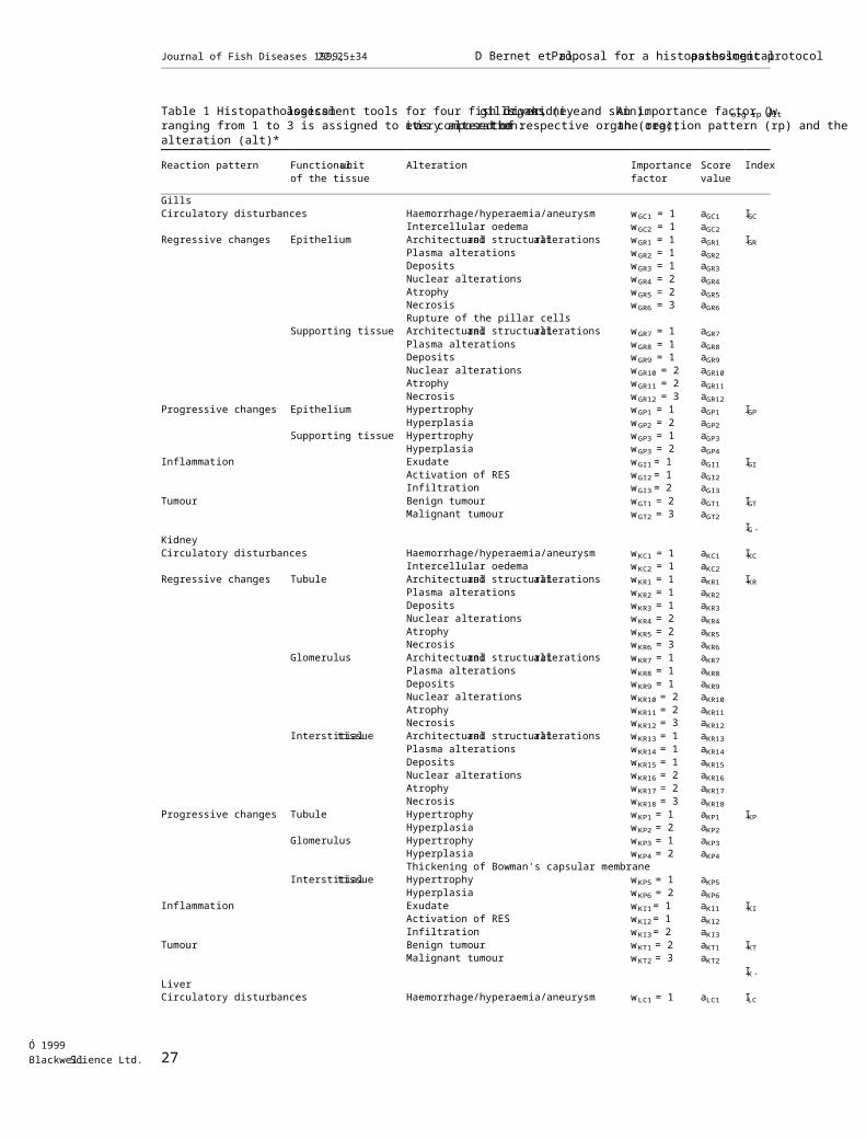
Table 1 Histopathologicalassessment tools for four fish organs (i.e.gills,liver,kidney and skin).An importance factor (worg rp alt)ranging from 1 to 3 is assigned to every alteration:itis composed ofthe respective organ (org),the reaction pattern (rp) and thealteration (alt)*
Reaction pattern Functionalunitof the tissue
Alteration Importancefactor
Scorevalue
Index
GillsCirculatory disturbances Haemorrhage/hyperaemia/aneurysm wGC1 = 1 aGC1 IGC
Intercellular oedema wGC2 = 1 aGC2Regressive changes Epithelium Architecturaland structuralalterations wGR1 = 1 aGR1 IGR
Plasma alterations wGR2 = 1 aGR2Deposits wGR3 = 1 aGR3Nuclear alterations wGR4 = 2 aGR4Atrophy wGR5 = 2 aGR5Necrosis wGR6 = 3 aGR6Rupture of the pillar cells
Supporting tissue Architecturaland structuralalterations wGR7 = 1 aGR7Plasma alterations wGR8 = 1 aGR8Deposits wGR9 = 1 aGR9Nuclear alterations wGR10 = 2 aGR10Atrophy wGR11 = 2 aGR11Necrosis wGR12 = 3 aGR12
Progressive changes Epithelium Hypertrophy wGP1 = 1 aGP1 IGPHyperplasia wGP2 = 2 aGP2
Supporting tissue Hypertrophy wGP3 = 1 aGP3Hyperplasia wGP3 = 2 aGP4
Inflammation Exudate wGI1 = 1 aGI1 IGIActivation of RES wGI2 = 1 aGI2Infiltration wGI3 = 2 aGI3
Tumour Benign tumour wGT1 = 2 aGT1 IGTMalignant tumour wGT2 = 3 aGT2
IG.KidneyCirculatory disturbances Haemorrhage/hyperaemia/aneurysm wKC1 = 1 aKC1 IKC
Intercellular oedema wKC2 = 1 aKC2Regressive changes Tubule Architecturaland structuralalterations wKR1 = 1 aKR1 IKR
Plasma alterations wKR2 = 1 aKR2Deposits wKR3 = 1 aKR3Nuclear alterations wKR4 = 2 aKR4Atrophy wKR5 = 2 aKR5Necrosis wKR6 = 3 aKR6
Glomerulus Architecturaland structuralalterations wKR7 = 1 aKR7Plasma alterations wKR8 = 1 aKR8Deposits wKR9 = 1 aKR9Nuclear alterations wKR10 = 2 aKR10Atrophy wKR11 = 2 aKR11Necrosis wKR12 = 3 aKR12
Interstitialtissue Architecturaland structuralalterations wKR13 = 1 aKR13Plasma alterations wKR14 = 1 aKR14Deposits wKR15 = 1 aKR15Nuclear alterations wKR16 = 2 aKR16Atrophy wKR17 = 2 aKR17Necrosis wKR18 = 3 aKR18
Progressive changes Tubule Hypertrophy wKP1 = 1 aKP1 IKPHyperplasia wKP2 = 2 aKP2
Glomerulus Hypertrophy wKP3 = 1 aKP3Hyperplasia wKP4 = 2 aKP4Thickening of Bowman's capsular membrane
Interstitialtissue Hypertrophy wKP5 = 1 aKP5Hyperplasia wKP6 = 2 aKP6
Inflammation Exudate wKI1= 1 aKI1 IKIActivation of RES wKI2= 1 aKI2Infiltration wKI3= 2 aKI3
Tumour Benign tumour wKT1 = 2 aKT1 IKTMalignant tumour wKT2 = 3 aKT2
IK.LiverCirculatory disturbances Haemorrhage/hyperaemia/aneurysm wLC1 = 1 aLC1 ILC
Journal of Fish Diseases 1999,22,25±34 D Bernet et al.Proposal for a histopathologicalassessment protocol
27Ó 1999BlackwellScience Ltd.

Table 1. Continued
Reaction pattern Functionalunitof the tissue
Alteration Importancefactor
Scorevalue
Index
Intercellular oedema wLC2 = 1 aLC2Regressive changes Liver tissue Architecturaland structuralalterations wLR1 = 1 aLR1 ILR
Plasma alterations wLR2 = 1 aLR2Deposits wLR3 = 1 aLR3Nuclear alterations wLR4 = 2 aLR4Atrophy wLR5 = 2 aLR5Necrosis wLR6 = 3 aLR6Vacuolar degeneration
Interstitialtissue Architecturaland structuralalterations wLR7 = 1 aLR7Plasma alterations wLR8 = 1 aLR8Deposits wLR9 = 1 aLR9Nuclear alterations wLR10 = 2 aLR10Atrophy wLR11 = 2 aLR11Necrosis wLR12 = 3 aLR12
Bile duct Architecturaland structuralalterations wLR13 = 1 aLR13Plasma alterations wLR14 = 1 aLR14Deposits wLR15 = 1 aLR15Nuclear alterations wLR16 = 2 aLR16Atrophy wLR17 = 2 aLR17Necrosis wLR18 = 3 aLR18
Progressive changes Liver tissue Hypertrophy wLP1 = 1 aLP1 ILPHyperplasia wLP2 = 2 aLP2
Interstitialtissue Hypertrophy wLP3 = 1 aLP3Hyperplasia wLP4 = 2 aLP4
Bile duct Hypertrophy wLP5 = 1 aLP5Hyperplasia wLP6 = 2 aLP6Wallproliferation of bile ducts or ductules
Inflammation Exudate wLI1= 1 aLI1 ILIActivation of RES wLI2= 1 aLI2Infiltration wLI3= 2 aLI3
Tumour Benign tumour wLT1 = 2 aLT1 ILTMalignant tumour wLT2 = 3 aLT2
IL.SkinCirculatory disturbances Haemorrhage/hyperaemia/aneurysm w SC1 = 1 aSC1 ISC
Intercellular oedema wSC2 = 1 aSC2Regressive changes Epidermis Architecturaland structuralalterations wSR1 = 1 aSR1 ISR
Plasma alterations wSR2 = 1 aSR2Deposits wSR3 = 1 aSR3Nuclear alterations wSR4 = 2 aSR4Atrophy wSR5 = 2 aSR5Necrosis wSR6 = 3 aSR6
Basement membrane Defect wSR7 = 2 aSR7Dermis Architecturaland structuralalterations wSR8 = 1 aSR8
Plasma alterations wSR9 = 1 aSR9Deposits wSR10 = 1 aSR10Nuclear alterations wSR11 = 2 aSR11Atrophy wSR12 = 2 aSR12Necrosis wSR13 = 3 aSR13
Progressive changes Epidermis Hypertrophy wSP1 = 1 aSP1 ISPHyperplasia wSP2 = 2 aSP2Hyperplasia of mucous cells
Dermis Hypertrophy wSP3 = 1 aSP3Hyperplasia wSP4 = 2 aSP4
Inflammation Exudate wSI1= 1 aSI1 ISIActivation of RES wSI2= 1 aSI2Infiltration wSI3= 2 aSI3
Tumour Benign tumour wST1 = 2 aST1 ISTMalignant tumour wST2 = 3 aST2
IS.*Abbreviations: (G) gills; (L) liver; (K) kidney; (S) skin; (C) circulatory disturbances; (R) regressive changes; (P) progressive changes; (I) inflammation; and(T) tumour. The alterations per reaction pattern are numbered beginning with 1. The score value is aorg rp alt. The composition corresponds to that of theimportance factor. The score value has to be rated for every alteration by histopathological assessment with a score ranging from 0 to 6. Iorg rpis the reactionindex of an organ,Iorg.the organ index.The sections of the table in italics are examples of the addition of supplementary alterations according to thespecific needs of a study or an investigator.However,these are not considered for the calculation of the indices.
Journal of Fish Diseases 1999,22,25±34 D Bernet et al.Proposalfor a histopathologicalassessment protocol
28Ó 1999BlackwellScience Ltd.
toryprocesses(e.g.exudate)areconsideredinreaction pattern 4.The alterationsincluded hereare:1 Haemorrhage/hyperaemia/aneurysm: blood leak-
ing from blood vessels (haemorrhage), congestion ofblood in an organ caused by venousaswellasarterialprocesses(hyperaemia),and well-outlineddilations of arterialblood vessels (aneurysm).2 Intercellular oedema: stagnant tissue fluid which
has leaked from capillaries into tissue.
Reaction pattern 2 (rp2):regressive changesRegressive changes are processes which terminate
in a functional reduction or loss of an organ. Theseinvolveatrophy,degeneration (malformation ordysfunction of cellular structures as a result of celldamage)and necrosis.Thisreactionpatterninvolves the following alterations:1 Architecturaland structuralalterations:changes
in tissuestructureas wellas in shapeandarrangement of cells.2 Plasma alterations:changesin cellular plasma
caused by hyaline droplets (granular degeneration),colloidaldroplets (colloid degeneration),degenera-tivefattyvacuolizationor hydropicglycogendroplets (glycogen degeneration), calcareous degen-eration,and thickeningof thefinefibresofconnective tissue (hyaline degeneration).3 Deposits:intercellularaccumulationsofsub-
stances primarily caused by degenerative processes.4 Nuclearalterations:changesin thenuclear
shape and structure of chromatin (e.g. karyopykno-sis and karyorrhexis).5 Atrophy:reduction in number and volume of
cellsand/ora decreasing amountofintercellularsubstances.6 Necrosis: morphological state of a cell or a tissue
which appears after irrevocable loss of cell function.
Reaction pattern 3 (rp3):progressive changesProgressive changes are processes which lead to
an increased activityofcellsor tissues.Typicallesions are:1 Hypertrophy:enlargementofcellvolumeor
tissue without increase in cellnumber.2 Hyperplasia: enlargement of tissue or organ by a
greater number of cells without change in volume ofthe cells.
Reaction pattern 4 (rp4):inflammationInflammatory changes are often associated with
processes belonging to other reaction patterns (e.g.
oedema). Therefore, it is often difficult to attributeinflammatorychangesto one singlereactionpattern.Hence,the presentauthorsuse the term`inflammation'in a very strict sense:1 Exudate:fluidcontaininga highprotein
concentration, and a large amount of cellular debrisexuded from blood and lymph vessels.2 Activationof thereticuloendothelialsystem
(RES):hypertrophy ofthe RES,which consists ofendothelialcellsand macrophagesthatline smallblood vessels.3 Infiltration:leucocytes penetrating the walls of
blood vessels and infiltrating the surrounding tissue.
Reaction pattern 5 (rp5):tumour (neoplasm)A tumourisan uncontrolled celland tissue
proliferation (autonomous proliferation).Tumoursare divided into two classes:1 Benigntumours:differentiatedcellswhich
replace or displace the original tissue; these tumourcells resemble the cells of the normaltissue.2 Malignanttumours:poorlydifferentiated,
rapidly multiplying cells which invade and destroyresident tissues;metastasis may be observed.
Individualdescription parametersIn addition to alterationswithin thereaction
patternsdescribed,itispossibleto designateindividualdescription parameters(seeTable 1).With these parameters, organ-specific lesions can beshown in an explicitway.Forthe index calcula-tions, these will not be considered since the changesare already covered by the standardized expressions(alterations)within the respective reaction patternas described above.
Histologicalevaluation
Importance factor (w)The relevance ofa lesion depends on its patholo-gicalimportance,i.e.how it affects organ functionand the ability of the fish to survive.This is takeninto accountby an importance factorassigned toevery alteration listed in the histological description.The alterationsare classified into three impor-
tance factors:1 minimal pathological importance, the lesion is
easily reversible as exposure to irritants ends;2 moderate pathological importance, the lesion is
reversible in most cases if the stressor is neutralized;and
Journal of Fish Diseases 1999,22,25±34 D Bernet et al.Proposal for a histopathologicalassessment protocol
29Ó 1999BlackwellScience Ltd.

3 marked pathologicalimportance,the lesion isgenerally irreversible, leading to partial or total lossof the organ function.
Score value (a)Every alteration is assessed using a score ranging
from 0 to 6, depending on the degree and extent ofthe alteration:(0) unchanged;(2) mild occurrence;(4) moderate occurrence; and (6) severe occurrence(diffuselesion).Intermediatevaluesarealsoconsidered.
Mathematicalcalculation of lesion indicesUsing importance factors and score values,four
different indices can be calculated (Table 2).If the lesions within one organ only are studied,
the two following indices are applicable:1 Organ index (Iorg.)
where:org = organ (constant);rp = reaction pat-tern;alt = alteration;a = score value;w = impor-tance factor.This index represents the degree of damage to an
organ.It is the sum ofthe multiplied importancefactors and score values of all changes found withinthe examined organ.A high index indicates a highdegreeof damage.Calculatingtheorgan indexallows a comparison between the degree of damageof the same organ in different individuals.2 Reaction index of an organ (Iorg rp)
where:org,rp = constant(forabbreviations,seeorgan index formula).The quality of the lesions in an organ is expressed
by the reaction index. It is calculated by the sum ofthe multiplied importance factors and score valuesofthealterationsofthecorrespondingreactionpattern.The sum of the five reaction indices of anorgan isequivalentto theorgan index(Iorg.).
Respectivereaction indicesofan organ (Iorgrp)from different individuals can be compared.The followingtwo indicescan beapplied if
severalorgans of a fish are examined:3 Totalindex (Tot-I)
(for abbreviations,see organ index formula).Thisindex representsa measure ofthe overall
health status based on the histologicallesions.It iscalculated by adding up allorgan indicesofanindividualfish.Asthe totalindex is calculated inthe same way for every fish,a comparison betweenindividuals is possible.4 Totalreaction index (I.rp)
where rp = constant(forabbreviations,see organindex formula).This index represents the quality of the histolo-
gical lesions in all examined organs of an individualfish.Itisthe sum ofthe corresponding reactionindices of allexamined organs of a fish.Using thisindexallowsa comparisonbetweendifferentindividuals.
Mathematicalcalculation of prevalencesBesidetheindicescalculated by extent(score
value)and pathologicalimportance(importancefactor) of lesions,a further point of interest is theprevalence ofhistopathologicalfeatures.The pre-valenceof everyalterationlistedherecan becalculatedas thepercentageoccurrenceof analteration within allanimalsof a sample.Thisallows an estimation of the occurrence of alterationsin an examined stock or a population.
Guidelines for field samplingAlongwithpollution,severalotherexogenousconditionsmay influencethehistopathologicalfeatures of an organ.To minimize the histological
Table 2 An example of lesion indices in one fish in which four organs were investigated
Reaction pattern
Organ rp1 rp2 rp3 rp4 rp5 S
org1 Iorg1 rp1 Iorg1 rp2 Iorg1 rp3 Iorg1 rp4 Iorg1 rp5 Iorg1.org2 Iorg2 rp1 Iorg2 rp2 Iorg2 rp3 Iorg2 rp4 Iorg2 rp5 Iorg2.org3 Iorg3 rp1 Iorg3 rp2 Iorg3 rp3 Iorg3 rp4 Iorg3 rp5 Iorg3.org4 Iorg4 rp1 Iorg4 rp2 Iorg4 rp3 Iorg4 rp4 Iorg4 rp5 Iorg4.S I.rp1 I.rp2 I.rp3 I.rp4 I.rp5 Tot-I
Journal of Fish Diseases 1999,22,25±34 D Bernet et al.Proposalfor a histopathologicalassessment protocol
30Ó 1999BlackwellScience Ltd.
changeswhich are caused by variablesotherthanirritants,a standardized selection ofthe sampledmaterialmust be takenintoaccount.Somevariables which could affect the histological appear-ance are briefly considered below. These reflect thepresentauthors'experience and are in accordancewith the recommendations ofICES (1997).For athorough interpretation of histopathological results,it is most important to take into account allthesefeatures.
Sample sizeThe sample size is a very important factor in everyhistopathologicalmonitoringprogramme.How-ever,there is no absolute recommendation for anoptimalsamplesizebecausethelattervariesaccording to the objectives ofa study.Ifitis thedetermination ofwhethera pathologicallesion ispresentwithin apopulation which isintended,statisticalrequirementsmust be fulfilled(95%confidenceof detectionof a certaindiseaseprevalencein apopulation).Iftheaim istodeterminewhetherhistopathologicalchangesinanimalsatpolluted sitesdiffersignificantly fromthose in fish from an unpolluted site,the requiredsample size dependson the detectable differencesbetween the sites:the smallerthe difference,thelargertherequired samplesizefora statisticalverification ofthisdifference (e.g.in a x2 test).Tablesto determine the required sample size aregiven in standard statisticaltexts.
SpeciesThe sensitivityto pollutants,as wellas thepollution-induced histopathologicalfeatures,mayvary within a wide range depending on the species(e.g.Braunbeck,Burkhardt-Holm,GoÈrge,Nagel,Negele & Storch 1992). Therefore, a comparison ofresultsfrom differentsitesshould bebased onsamples from the same species.
AgeThe age of all fish should be recorded since the ageofstockswillstrongly determinetherangeandnature ofpathologies(e.g.neoplasmsare signifi-cantly more frequent in older fish). The determina-tion ofthe age can be done by reading ofscales,otolithsor interopercularbones.However,thesetechniquesare time consuming and need experi-
ence.As a compromise,itisrecommended tosample fish of a standardised size range within onespecies.
Sex and stage of sexualmaturitySex and stage ofsexualmaturity should be notedsinceboth factorsareknown to potentiallyinfluencethehistologicalappearanceof certainorgans.
Sampling seasonSeasonality may play an importantrole in manypathological conditions of fish and can be explainedby the influence of temperature e.g. on the biologyofthe causative agentorthe immune system ofpoikilothermic animals or by the role of hormonalvariations in disease susceptibility.
MigrationMigrations during the life-cycle (e.g. for spawning),butalso quickflightreactionsto ashort-timepollutionpeak(Triebskorn,KoÈhler,Honnen,Schramm,Adams & MuÈller 1997),can affectthedistribution ofdiseased fish within a geographicalregion.To allow a comparison ofsamplesfromdifferentsites,itisrecommended thatsamplingwithin thesameseason isperformed,preferablywhen fish areon theirprimary residentfeedinggrounds.
DiscussionMany publicationshaveaddressed theissueofinduction of histologicallesions by irritants.How-ever,the methodsfor evaluating histopathologicallesions are rather divergent.In some publications,lesionshave only been described morphologically(Mitz& Giesy 1985;Hinton,Lantz,Hampton,McCuskey & McCuskey 1987).In these studies,the effectofwaterpollution hasbeen correlatedwith the abundance oflesions found in an organ.Other studies concentrated on a few alterations inan organ and assessed their extent by using a scale.Thisallowed statementson abundanceand in-tensity oflesions(Couillard,Berman & Panisset1988;Bucher & Hofer 1993;Haaparanta,Valto-nen & Hoffmann 1997).However,theuseofdifferentmethods and assessmentscales as wellasthe inclusion ofdifferenthistologicalchangeshas
Journal of Fish Diseases 1999,22,25±34 D Bernet et al.Proposal for a histopathologicalassessment protocol
31Ó 1999BlackwellScience Ltd.
alwaysmade itdifficultto comparedifferentstudies. Therefore, a standardized assessment meth-od is urgently needed.The standardized assessment method proposed in
the present study allows the quantification of organdamage,includingtheextentand pathologicalimportanceofchanges.Differentindicescan becalculatedwhichcharacterizea histology-basedhealth statusatdifferentlevels:indicesof theorganism (Tot-I),ofan organ (Iorg.) and ofthereaction pattern (Iorg rp and I.rp).The indicesrepresent the degree of damage. However, these alsoindicate the significance of the lesions.The degreeand significanceofdamageareimportantsincethese allow the assessmentofthe suitability ofanorgan or a lesion as an indicator for water pollution.With the quantification of the lesions,statistical
evaluation becomes practicable.The correspondingindices can be compared more easily than morpho-logicaldescriptionsof pathologicalchanges.Although histopathologyisa descriptivescienceand the assessment of the lesions will always dependon the investigator's experience and interpretation,thetoolallowsa more reliablecomparison ofdifferent studies.However,a directcomparison oftheorgan
indices (Iorg.) within the same fish to quantify theimpact of an irritant on the respective organ is notpossible. Since the number of functional units is notidenticalin allorgans,theattainablemaximumvalues for the organ indices differ. To overcome thisproblem,an indirectcomparison oftheorganindices(Iorg.)with those ofan unaffected controlgroup offish from an unpolluted water system isnecessary. The higher the proportional difference ofIorg.to the corresponding value in the unpollutedgroup,themoredamagehasbeen doneto theaffected organ.Effortsto standardize the judgementofhisto-
pathological lesions have already been made. Thereis agreement on the classification of liver alterationsin regional and national surveillance projects for theassessmentoftheinfluenceofcontamination incoastaland estuarinewaterson marinebottom-dwelling fish (Johnson et al.1992;Myerset al.1993; ICES 1997). The following classification hasbeen accepted:neoplasms,fociofcellularaltera-tions, unique (specific) degenerative lesions, general(non-specific)necrotic/degenerativechangesandnon-neoplasticproliferativelesions.Additionally,vascularabnormalitiesand anomalousstorageconditionshave been recommended asdiagnostic
criteriaby ICES (1997).In contrast,lesionsinkidney are categorized as necrosis, proliferation andsclerosis,asdescribed bytheNationalBenthicSurveillanceProjecton thePacific(Myerset al.1993)and theNorth-eastCoast(Johnson et al.1992).Thus,differing histologicalevaluationsofthe two organs prevent a direct comparison of thedegree and significance ofan organ damage.Byintroducing reaction indices (Iorg rp), as proposed inthe present study,a comparison is possible at leastbetween correspondingreaction patternsof thesame organ in different fish.A further advantage ofcategorizing histopatho-
logicallesionsliesin thepossibility ofassessingwhich organshavebeen damaged and to whatextentchangeshavebeen induced.Thisisanimportantprerequisite forsurveillance and mon-itoring projects.However,itmustbe appreciatedthatthecategorization ofpathologicalfindingsresults in a simplification,particularly because themethod hasto be applicable to differentorgans.Therefore, a thorough morphological description ofthelesionsisessential.Individualdescriptionparameters can be added to the assessment methodwhich,although not considered for the calculationofthe indices(see Table 1),allow additionalandmore detailed evaluations according to the specificneeds of an investigator.Recently,modern diagnostic methodologiesfor
the detection ofexposure to contamination havebeen wellestablished forthe liver.Mostofthesebiomarkersare designed to detectcellular/subcel-lular(e.g.lysosomalmembrane stability),orbiochemicaland molecularresponses[e.g.en-zyme-alteredfoci(G6PDH), proliferatingcellnuclearantigen(PCNA), CYP1A and DNAadducts].These are early indicatorsofbiologicaldamage and some can be induced experimentallywithin a few days (Collier & Varanasi 1991; Stein,Collier, Reichert, Casillas, Tom & Varanasi 1992).Although Myerset al.(1994)have shown in fishthata significantcorrelation between biochemicalchanges and histologicallesions in the liver can bedrawn,theseearly responsebiochemicalmarkerscannottotally substitute the assessmentofhisto-pathologicallesionsas a markerforchronicexposure to pollution.Therefore,in many regionaland nationalprogrammes,cellular/sub-cellular andbiochemicalbiomarkers,as wellas histopathology,are included in a decision-tree-type model(Adamset al.1989;ICES 1997;Triebskorn et al.1997).For monitoringof earlychanges,cellular/sub-
Journal of Fish Diseases 1999,22,25±34 D Bernet et al.Proposalfor a histopathologicalassessment protocol
32Ó 1999BlackwellScience Ltd.
cellularand biochemicalbiomarkersareused,followed by histopathology which partly considersendpointeffects.Theseconsiderationsfurthersupportthe importance ofa standardized assess-ment tool for histopathological lesions, as describedin the present study.
AcknowledgmentsWe thankDr D. Bucke,ConsultantFish andShellfish Pathologist,Weymouth,UK, and Dr E.Staub,Swiss Agency for Environment,Forests andLandscape(BUWAL), Berne,Switzerland,forhelpfulsuggestions.Dr A.Hemphillimproved theEnglish.This study was supported by grants fromtheSwissNationalScienceFoundation(31-45894.95),BUWAL, the Inspectorate ofFisheriesof Berne,and theWaterand SoilProtectionLaboratory of the District of Berne (GBL).
ReferencesAdams S.M.,Shepard K.L.,Greeley M.S.,Jr,Jimenez B.D.,
Ryon M.G., Shugart L.R. & McCarthy J.F. (1989) The useof bioindicators for assessing the effects of pollutant stress onfish.Marine EnvironmentalResearch 28, 459±464.
Braunbeck T., Burkhardt-Holm P., GoÈrge G., Nagel R., NegeleR.-D.& Storch V. (1992) Regenbogenforelle und Zebra-baÈrbling,zwei Modelle fuÈr verlaÈngerte ToxizitaÈtstests:Relative Empfindlichkeit,Art- und OrganspezifitaÈt in dercytopathologischen Reaktion von Leber und Darm aufAtrazin. Schriftenreihe Verein Wasser,Boden,Lufthygiene 89,109±145.
Bucher F.& Hofer R.(1993) The effects of treated domesticsewage on three organs (gills,kidney,liver) of brown trout(Salmo trutta).Water Research 27, 255±261.
Bucke D.(1989) Histology.In:Methods for the MicrobiologicalExamination of Fish and Shellfish (ed.by B.Austin & D.A.Austin),pp.69±97.Ellis Horwood Ltd,Chichester.
Bucke D. (1994) Methodologies for demonstrating pathologicalchanges in flounder (Platichthys flesus).In:Diseases andParasites of Flounder (Platichthys flesus) in the Baltic Sea (ed.by G.Bylund & L.-G.LoÈnnstroÈm),pp.131±145.BalticMarine Biologists Publication No.15,Turku/Abo.
Bucke D.,Vethaak D.,Lang T.& Mellergaard S.(1996)Common Diseases and Parasites of Fish in the North Atlantic:Training Guide for Identification.InternationalCouncilforthe Exploration of the Sea Techniques in Marine Environ-mentalSciences,Copenhagen.
Bylund G.& LoÈnnstroÈm L.-G.(1994) Diseases and Parasites ofFlounder (Platichthys flesus) in the Baltic Sea.Baltic MarineBiologists Publication No.15,Turku/Abo.
Collier T.K.& Varanasi U.(1991) Hepatic activities ofxenobiotic metabolizing enzymes and biliary levels ofxenobiotics in English sole (Parophrys vetulus) exposed to
organic-solvent extracts of marine sediments from contami-nated and reference areas.Comparative Biochemistry andPhysiology 84C, 291±298.
Couillard C.M.,Berman R.A.& Panisset J.C.(1988) Histo-pathology of rainbow trout exposed to a bleached kraft pulpmilleffluent. Archives of EnvironmentalContamination andToxicology 17, 319±323.
Haaparanta A.,Valtonen E.T.& Hoffmann R.W. (1997) Gillanomalies of perch and roach from four lakes differing inwater quality. Journalof Fish Biology 50, 575±591.
Hinton D.E.,Lantz R.C.,Hampton J.A.,McCuskey P.R.&McCuskey R.S.(1987) Normal versus abnormalstructure:considerations in morphologic responses of teleosts topollutants.EnvironmentalHealth Perspectives 71, 139±146.
Hinton D.E.& LaureÂn D.J.(1990) Liver structuralalterationsaccompanying chronic toxicity in fishes: potential biomarkersof exposure. In: Biomarkers of Environmental Contaminations(ed.by J.F.McCarthy & L.R.Shugart),pp.17±57.LewisPublisher,Boca Raton,FL.
Huggett R.J.,Kimerle R.A.,Mehrle P.M., Jr & Bergman H.L.(1992) Biomarkers.Lewis Publishers,Boca Raton,FL.
InternationalCouncilfor the Exploration of the Sea (ICES)(1989) Methodology of Fish Disease Surveys.CooperativeResearch Report of the InternationalCouncilfor theExploration of the Sea No.166,Copenhagen.
InternationalCouncilfor the Exploration of the Sea (ICES)(1997) Special Meeting on the Use of Liver Pathology of Flatfishfor Monitoring BiologicalEffects of Contaminants.Interna-tionalCouncilfor the Exploration of the Sea,Copenhagen.
Jobling S.& Sumpter J.P.(1993) Detergent components insewage effluent are weakly oestrogenic to fish:an in vitrostudy using rainbow trout (Oncorhynchus mykiss) hepatocytes.Aquatic Toxicology 27, 361±372.
Johnson L.L., Stehr C.M., Olson O.P., Myers M.S., Pierce S.M.,McCain B.B.& Varanasi U.(1992) Nationalstatus andtrends program,nationalbenthic surveillance project:Northeast Coast,fish histopathology and relationshipbetween lesions and chemicalcontaminants (1987±1989).US Department of Commerce, NOAA Technical MemorandumNMFS-NWFSC-4,96 pp.
Johnson L.L., Stehr C.M., Olson O.P., Myers M.S., Pierce S.M.,Wigren C.A., McCain B.B. & Varanasi U. (1993) Chemicalcontaminants and hepatic lesions in winter flounder(Pleuronectes americanus) from the Northeast Coast of theUnited States.EnvironmentalScience and Technology 27,2759±2771.
Mallatt J. (1985) Fish gill structural changes induced by toxicantsand other irritants:a statisticalreview.Canadian JournalofFisheries and Aquatic Sciences 42, 630±648.
McCarthy J.F.& Shugart L.R.(1990) Biomarkers of Environ-mentalContaminations.Lewis Publisher,Boca Raton,FL.
Mitz S.V. & Giesy J.P. (1985) Sewage effluent biomonitoring ±I.Survival, growth,and histopathologicaleffects in channelcatfish.Ecotoxicology and EnvironmentalSafety 10, 22±39.
Myers S.M.,Johnson L.L.,Olson O.P.,Stehr C.M.,LomaxD.P.,Horness B.H.,Anulacion B.F., Willis M.L.,CollierT.K.,McCain B.B.,Stein J.E.& VaranasiU.(1994)Toxicopathic hepatic lesions and other biomarkers of
Journal of Fish Diseases 1999,22,25±34 D Bernet et al.Proposal for a histopathologicalassessment protocol
33Ó 1999BlackwellScience Ltd.
exposure to chemicalcontaminants in marine bottomfishspecies from the northeast and Pacific coast,U.S.A.In:Diseases and Parasites of Flounder (Platichthys flesus) in theBaltic Sea (ed.by G.Bylund & L.-G.LoÈnnstroÈm),pp.81±98. Baltic Marine Biologists Publication No. 15, Turku/Abo.
Myers M.S.,Olson O.P., Johnson L.L.,Stehr C.S.,Hom T.&Varanasi U. (1992) Hepatic lesions other than neoplasms insubadult flatfish from Puget Sound,Washington: relation-ships with indices of contaminant exposure.MarineEnvironmentalResearch 34, 45±51.
Myers M.S.,Stehr C.M.,Olson O.P., Johnson L.L.,McCainB.B., Chan S.-L. & Varanasi U. (1993) National status andtrends program, national benthic surveillance project: PacificCoast,fish histopathology and relationships between tox-icopathic lesions and exposure to chemical contaminants forcycles I to V (1984±88). US Department of Commerce, NOAATechnical Memorandum NMFS-NWFSC-4,160 pp.
Oronsaye J.A.O. (1989) Histological changes in the kidneys andthe gills of the stickleback, Gasterosteus aculeatus L., exposedto dissolved cadmium in hard water.Ecotoxicology andEnvironmentalSafety 17, 279±290.
Poleksic V.& Mitrovic-Tutundzic V.(1994) Fish gills as amonitor of sublethaland chronic effects of pollution. In:Sublethal and Chronic Effects of Pollutants on Freshwater Fish(ed. by R. MuÈller & R. Lloyd), pp. 339±352. FAO, FishingNews Books,Oxford.
Shephard K.L. (1994) Functions for fish mucus. Reviews in FishBiology and Fisheries 4, 401±429.
Sindermann C.J.(1979) Pollution-associated diseases andabnormalities of fish and shellfish:a review.Fishery Bulletin76, 717±749.
Stein J.E.,Collier T.K.,Reichert W.L.,Casillas E.,Tom T.&Varanasi U.(1992) Bioindicators of contaminant exposureand sublethal effects:studies with benthic fish in PugetSound, Washington. Environmental Toxicology and Chemistry11, 701±714.
Susani L., Mearns A. & Long E. (1986) NOAA Quality AssuranceProgram Workshop on Marine Fish Histopathology.CEABPacific Office,Seattle,WA.
Takashima F.& Hibiya T.(1995) An Atlas of Fish Histology.Kodansha,Gustav Fischer-Verlag,Tokyo,Stuttgart.
Triebskorn R., KoÈhler H.-R., Honnen W., Schramm M., AdamsS.M. & MuÈller E.F. (1997) Induction of heat shock proteins,changes in liver ultrastructure and alterations of fishbehavior: are these biomarkers related and are they useful toreflect the state of pollution in the field? Journalof AquaticEcosystems Stress and Recovery 6, 57±73.
Vethaak A.D.(1994) The use of mesocosms to study disease inflounder (Platichthys flesus).In:Diseases and Parasites ofFlounder (Platichthys flesus) in the Baltic Sea (ed.by G.Bylund & L.-G. LoÈnnstroÈm),pp.121±129.Baltic MarineBiologists Publication No.15,Turku/Abo.
Journal of Fish Diseases 1999,22,25±34 D Bernet et al.Proposalfor a histopathologicalassessment protocol
34Ó 1999BlackwellScience Ltd.


