Embed Size (px)
Citation preview

A sex-ratio Meiotic Drive System in Drosophilasimulans. I: An Autosomal SuppressorYun Tao
1,2*, John P. Masly
3¤, Luciana Araripe
1, Yeyan Ke
1, Daniel L. Hartl
1
1 Department of Organismic and Evolutionary Biology, Harvard University, Cambridge, Massachusetts, United States of America, 2 Department of Biology, Emory University,
Atlanta, Georgia, United States of America, 3 Department of Biology, University of Rochester, Rochester, New York, United States of America
Sex ratio distortion (sex-ratio for short) has been reported in numerous species such as Drosophila, where distortioncan readily be detected in experimental crosses, but the molecular mechanisms remain elusive. Here we characterizean autosomal sex-ratio suppressor from D. simulans that we designate as not much yang (nmy, polytene chromosomeposition 87F3). Nmy suppresses an X-linked sex-ratio distorter, contains a pair of near-perfect inverted repeats of 345bp, and evidently originated through retrotransposition from the distorter itself. The suppression is likely mediated bysequence homology between the suppressor and distorter. The strength of sex-ratio is greatly enhanced by lowertemperature. This temperature sensitivity was used to assign the sex-ratio etiology to the maturation process of the Y-bearing sperm, a hypothesis corroborated by both light microscope observations and ultrastructural studies. It haslong been suggested that an X-linked sex-ratio distorter can evolve by exploiting loopholes in the meiotic machineryfor its own transmission advantage, which may be offset by other changes in the genome that control the selfishdistorter. Data obtained in this study help to understand this evolutionary mechanism in molecular detail and provideinsight regarding its evolutionary impact on genomic architecture and speciation.
Citation: Tao Y, Masly JP, Araripe L, Ke Y, Hartl DL (2007) A sex-ratio meiotic drive system in Drosophila simulans. I: An autosomal suppressor. PLoS Biol 5(11): e292. doi:10.1371/journal.pbio.0050292
Introduction
As a rule, most dioecious species produce equal numbers ofmale and female progeny because the rarer sex has a matingadvantage. R. A. Fisher in 1930, as well as Carl Dusing in 1884before him, framed arguments based on parental expenditurethat mandates an equilibrium sex ratio of 50% female [1,2].This so-called Fisher’s principle is based on the premise thatgenes have maximum representation in future generationsonly if they are equally transmitted through both sexes [3].However, sex ratios in nature sometimes show markeddepartures from 50%, and there are many ecological andgenetic situations where the assumptions of Fisher’s principledo not hold [4,5]. One prominent example is related to sex-linked genes of the heterogametic sex. Because of theirunisexual transmission, mutations that distort their meiotictransmission—thus the sex ratio—in their own favor can gainselective advantage [6]. This type of sex ratio meiotic drive,also called sex-ratio, conflicts with selection for equal trans-mission of autosomal genes and thus represents a typicalintragenomic conflict among genes when their transmissionoptima are not congruent.
A sex-ratio distorter can invade a population as long as itsdeleterious effects on viability and fertility are offset by thebiased segregation [7]. Invasion by a sex-ratio distorter createsa genetic context promoting strong selection for sex-linkedor autosomal suppressors that ameliorate the segregationdistortion and/or its deleterious pleiotropic effects [8]. Thistype of recurrent intragenomic conflict generates a dynamicof genetic ‘‘attack’’ and ‘‘defense,’’ even in the absence of anyexternal abiotic or biotic challenges [4,9,10]. As a result,genetic conflicts over sex ratio may be a key factor in drivingthe evolution of gametogenesis in the heterogametic sex,thereby leading to the nearly ubiquitous pattern of Haldane’srule observed in speciation [11–13]. Because the evolution of
genetic suppression would make sex-ratio transient on anevolutionary time scale, the uncovering of suppressed sex-ratiomutations requires sophisticated crossing schemes, oftenbetween individuals from different populations or incipientspecies. Perhaps it is because Drosophila geneticists have oftenstudied matings of this type that most of the known examplesof sex-ratio are found in Drosophila [6], and new cases continueto be reported regularly [14–16].At least three independent sex-ratio meiotic drive systems
have been uncovered in the species D. simulans [15,17–20]. Tofacilitate discussion, we will assign each sex-ratio system adifferent name according to the location where the originalstocks were collected or the research was carried out. In themost thoroughly analyzed case, which we will refer to as theParis sex-ratio, at least two X-linked distorters have beenmapped, and they are currently being characterized at themolecular level [19,21,22]. The Paris sex-ratio (SR) appears tohave originated relatively recently, and evolutionary signa-tures of a recent selective sweep are evident [23]. Multiplesuppressors on the autosomes and the Y chromosome havebeen reported [19,21,24]. Both the distorters and the
Academic Editor: Daniel Barbash, Cornell University, United States of America
Received April 12, 2007; Accepted September 17, 2007; Published November 6,2007
Copyright: � 2007 Tao et al. This is an open-access article distributed under theterms of the Creative Commons Attribution License, which permits unrestricteduse, distribution, and reproduction in any medium, provided the original authorand source are credited.
Abbreviations: ASO, allele-specific oligonucleotide; DAPI, 4’,6-diamidino-2-phenyl-indole; IC, individualization complex; IR, inverted repeats; RACE, rapid amplificationof cDNA ends; SD, Segregation Distortion; siRNA, small interfering RNA; TEM,transmission electron microscopy
* To whom correspondence should be addressed. E-mail: [email protected]
¤ Current address: Department of Molecular and Computational Biology, Universityof Southern California, Los Angeles, California, United States of America
PLoS Biology | www.plosbiology.org November 2007 | Volume 5 | Issue 11 | e2922560
PLoS BIOLOGY

suppressors are polymorphic across the worldwide distribu-tion of D. simulans [25–28]. Cytogenetic studies show that most(92%–96%) of the Y chromosomes in SR males do notundergo normal disjunction during meiosis II. As a con-sequence, the Y chromosome is often fragmented and lost.Sperm without a Y chromosome do participate in fertiliza-tion, and account for the 15%–30% of sterile F1 progenymales observed [29]; however most of the Y-bearing sperma-tids fail to mature in spermiogenesis [30]. The second sex-ratiosystem, which we call the Durham sex-ratio, was uncovered byintrogressing regions of the third chromosome from D.mauritiana into D. simulans. The gene responsible for thesuppression was named too much yin (tmy) [15]. An intriguingobservation is that among about 20 hybrid male sterility locion the third chromosome between these two species, tmy alsohad the strongest sterilizing effect on hybrid males. Thisobservation provides a direct link between meiotic drive andinterspecific hybrid male sterility [15]. The third sex-ratiosystem, which we call the Winters sex-ratio, was first revealed ininterspecific hybrids between D. simulans and D. sechellia, usinga D. simulans stock collected in Winters, California [20]. Thegene responsible for revealing the sex-ratio is autosomalrecessive. It was thought to have been introgressed from D.sechellia into a largely D. simulans genetic background and tohave replaced the dominant suppressing D. simulans allele;however, two alternative explanations could not be ruled out.In the first alternative, a cryptic meiotic drive system from D.sechellia may have been introgressed into the D. simulansbackground and became reactivated. In the second alter-native, a sex-ratio system may have been created de novo by aninteraction between D. simulans and D. sechellia genes, eventhough these genes are irrelevant to sex ratio control in theirnative genomes [20].
Two additional sex-ratio cases have been reported in D.simulans [17,18]. For a case discovered in Brazil, an autosomalrecessive mutation was implicated [18]. Similarly, in fliescollected in California in 1959 [17], a recessive mutation (sxr)was mapped on the third chromosome. Unfortunately, thestocks of these two cases are no longer available and hencetheir relationship to the above three systems cannot beascertained.
Given the frequent observations of sex-ratio and its
potential biological significance, it is surprising that no singlegene has as yet been identified for any sex-ratio system. Twodifficulties prevent a molecular genetic analysis. First, manysex-ratio cases have been found in the genus Drosophila but notyet in natural populations of the model species D. melanogast-er. Second, sex-ratio genes are often associated with inversions,making even conventional genetic mapping difficult. In ourstudy we took advantage of the genomic and geneticresources developed in recent years for Drosophila tocharacterize the Winters sex-ratio system at the molecularlevel. Evidence for the existence of the other two independ-ent sex-ratio meiotic drive systems in D. simulans will also beelaborated. The study of sex-ratio systems may help us gainnew insights into mechanisms of speciation, the maintenanceof Mendelian segregation, and the evolutionary principles ofgenome organization.
Results
A Recessive Mutation of the sex-ratio Suppressor on theThird ChromosomeWe first mapped a recessive gene responsible for uncover-
ing the Winters sex-ratio to the vicinity of the locus pe(polytene chromosome position 85A6) on the third chromo-some through various crosses by using stocks from a previousstudy, where the Winters sex-ratio was revealed [20] (Text S1;Figures S1–S5). We reasoned that the recessive allele is likely aloss-of-function mutation of a dominant sex-ratio suppressor,rather than corresponding to the distorter itself, because asex-ratio distorter has no transmission advantage unless it issex-linked (Figure 1A). A sex-ratio line SSR12-2-7 wasconstructed by extracting the third chromosome from thestock SSR12 into a pure D. simulans background (Figure S2).This stock was used throughout this study for genetic andphenotypic analyses.Further mapping of the sex-ratio suppressor was made
possible through a 2-P mapping scheme [15], where the P[wþ]-tagged D. mauritiana introgression stocks were constructed forother purposes (mapping hybrid male sterility between D.mauritiana and D. simulans) [31]. The P inserts here conven-iently functioned as semi-dominant markers (Figure S6).Their positions and the introgression lines with two P inserts(2-P lines) in the pe region are shown in Figure 1B. The resultsof the 2-P mapping clearly indicate that the target gene mustbe localized to a ;2,700-kb interval between P40 and P38(Figure S6). With allele-specific oligonucleotide (ASO)markers (Table S2), we genotyped 129 recombinants gener-ated from the 2P-10 line to narrow down the target to aninterval of 88 kb between CG10841 and CG31337 (Figure 1C).There are no obvious candidate genes for the sex-ratiosuppressor in this region.To pinpoint the target gene more precisely, we screened an
additional 3,100 recombinants within the P40 – P38 intervalwith 40 P38 and 50 P40 recombinants falling within the 88-kbinterval between CG10841 and CG31337 (Figure 1C). Analysisof 79 of these recombinants narrowed the target to the 7-kbinterval between CG14369 and K44 region (Figure 1C). Insuch a small region, it is unlikely that more than one gene isresponsible for uncovering the Winters sex-ratio. We namedthis gene not much yang (nmy). We use the symbol Nmy todenote the dominant suppressing allele in D. simulans and D.
PLoS Biology | www.plosbiology.org November 2007 | Volume 5 | Issue 11 | e2922561
sex-ratio Suppressor
Author Summary
Genetic conflicts among genes happen when their modes oftransmission differ. Genes in the heterogametic (XY) sex can begrouped as X-linked, Y-linked, autosomal, or cytoplasmic. Sex ratio inthe progeny greatly affects the transmission advantage of each ofthe four types of genes, with the optimal sex ratio for each typebeing respectively 100%, 0%, 50%, and 100% of females. Sex ratiocan often be biased from the normal 50% by genes that distort thesex ratio toward their own optimal transmission. Here we reportgenetic and molecular characterization of an autosomal gene thatfunctions as a suppressor of an X-linked sex-ratio distorter. Malemutants give rise to female-biased progeny. The cause of thedistortion was assigned to a failure of the Y-bearing sperm tomature. The DNA sequence of the suppressor gives clues tounderstanding the sex-ratio meiotic drive at the molecular level.More generally, the genetic conflict over sex ratio may be importantin determining the evolutionary dynamics and the architecture ofeukaryotic genomes.

Figure 1. Positional Cloning of not much yang, a Recessive Autosomal sex-ratio Suppressor on the Third Chromosome
(A) A recessive gene on the third chromosome was implicated for uncovering sex-ratio. Through various crossing schemes (see Text S1 for details),chromosomal combinations from sex-ratio (black) and wild-type (white) strains were tested for sex-ratio. In each genotype, bars from left to rightrepresent the X, the second, and the third chromosome, respectively. The bar with a hook represents the Y chromosome. The fourth chromosome wasnot followed. SR: sex-ratio; WT: wild type (not sex-ratio).(B) A major gene was mapped between P40 and P38 on the right arm of the third chromosome (3R). Shown here are the 2-P lines used in the mapping,with the blue bars representing the D. mauritiana portion of the introgression lines and the gray triangles representing the P[wþ] inserts (see Figure S6for details). The two bent arrows represent the 84F-93F inversion in D. simulans complex as compared to D. melanogaster. Dotted lines represent ASOmarkers. The target gene (*) falls within the green region that was subsequently mapped in more detail.(C) Fine mapping at 1-kb resolution. Fifty P40 and 40 P38 recombinants were found with their crossovers falling in the CG10841–CG31337 region. Thepositions of these crossovers were delimited with 13 ASO markers. The sex ratio (proportion of female) for each recombinant is shown here in rankorder within each ASO interval. Significant among-recombinant variation in sex ratio is indicated (one-way ANOVA: $, p , 5%; $$, p , 1%, and $$$, p ,0.1%). The target major gene (*) is mapped to the 7-kb interval of CG14369–K44.(D) Two insertions/deletions are responsible for the nmy phenotype. The parental sequences from D. mauritiana w (mau12 Nmy) and D. simulans SSR12-2-7 nmy were amplified by long PCR using the primers k50R30a and k44F30a (arrow heads, see Text S2 for sequences). Empty and filled circles representa subset of the informative substitutions. The filled rectangles represent the coding sequence of CG14370, which is a single exon gene in D.melanogaster. The sequences between the two filled rectangles, 2,791 bp in mau12 Nmy and 1,427 bp in SSR12-2-7 nmy, are missing in D. melanogaster.The arrow represents the transcription orientation. Dashed lines represent, from left to right, three large deletions of 664, 307, and 388 bp in SSR12-2-7nmy, respectively. The red arrows stand for inverted repeats (IR) of 380 bp in mau12 Nmy, and a 345-bp homolog in the SSR12-2-7 nmy allele. The green
PLoS Biology | www.plosbiology.org November 2007 | Volume 5 | Issue 11 | e2922562
sex-ratio Suppressor

mauritiana. The recessive loss-of-function allele that allows thesex-ratio phenotype to be expressed is denoted nmy.
We sequenced the parental chromosomes from D. maur-itiana mau12 (Nmy) and D. simulans SSR12-2-7 (nmy) across theCG14369–K44 interval (Figure 1D). From this region, sequen-ces homologous to CG14370 of D. melanogaster were recog-nized. Insertions of 2,791 bp and 1,427 bp were found in thecoding region of CG14370 in mau12 (Nmy) and SSR12-2-7(nmy), respectively. Notably, a pair of inverted repeats of 380bp was found in the 2.8-kb Nmy insert, and three largedeletions were found in the 1.4-kb nmy insert relative to the2.8-kb Nmy insert. Because of these deletions, only one repeatremains in the 1.4-kb nmy insert (Figure 1D). The targetregion was further narrowed down to the 2.3-kb interval ofnmy529–nmy2827 by genotyping the last 11 recombinants withseven additional ASO probes that mark the region fromnmy135 to nmy7123 (Figure 1D, Table S3). Sequencing showedthat the only difference between P40.B13 Nmy and P40.L12nmy is in the two deletions D307 and D388 (Figure 1D). Weconclude that the wild-type function of Nmy as a sex-ratiosuppressor will be lost if the pair of inverted repeats in theNmy insert are not intact. Hereafter, we use brackets todenote the species and length of the insert for the Nmy allele.For example, Nmy[mau2791] and nmy[sim1427] are used torepresent the alleles as well as the inserts found in mau12 andSSR12-2-7, respectively.
Gene Structure and Origin of NmyWe compared the CG14370/Nmy region from several species
of the D. melanogaster subgroup. The Nmy inserts are found inD. simulans (four strains) and D. mauritiana (one strain) but notin the other species (one strain each) (Figure 2A). Impor-tantly, we found no Nmy insert in the D. sechellia strain (3588)but two inserts (Nmy[sim2041] and nmy[sim1427]) in the D.simulans strain (sim2). These two strains were used inconstructing the D. simulans 3 D. sechellia recombinant inbredlines where the Winters sex-ratio was first observed [20].Evidently, the allele nmy[sim1427] does not come from D.sechellia. Rather, it is still segregating in the sim2 strain with afrequency of 6.1% (n ¼ 294). There is an intact inverted-repeat structure within the allele Nmy[sim2041] that has fullfunction as a sex-ratio suppressor (unpublished data). Inaddition, a null allele of nmy (i.e., CG14370 without anyinsert) was also found in a local Massachusetts population ofD. simulans (unpublished data). Taken together, the evidenceindicates that Nmy is a gain-of-function mutation thatsuppresses the Winters sex-ratio distorter.
The three Nmy inserts are compared in Figure 2B. Thelength of a single inverted repeat in Nmy[mau2791] is 35 bplonger than that in Nmy[sim2041]. The sequences between theinverted repeats in these two Nmy alleles are not homologous,except for a 93-bp element found in reverse orientation.There is a 664-bp fragment (D664 in Figure 2B) inNmy[mau2791] that is absent in the two D. simulans alleles.The allele nmy[sim1427] was derived from Nmy[sim2041] by theloss of one inverted repeat and the loss of most of the
sequence located between the inverted repeats, except for the93-bp element in reverse orientation. This observation againsuggests that the inverted-repeat structure is essential for thesex-ratio suppression function. Nmy does not appear to be aconventional gene (Figure 2B). Both ends of the transcriptsacross the Nmy[sim2041] or nmy[sim1427] region were deter-mined by 59 and 39 rapid amplification of cDNA ends (RACE)and they are identical to those of CG14370 found in D.melanogaster. The full length of the mRNA from nmy[sim1427]was determined, and two alternately spliced introns (I, II)were identified (Figure 2B). We failed, however, to obtain theinternal sequences of mRNA from Nmy[sim2041], because thereverse transcription was evidently stalled, most likely by astem-loop secondary structure formed between the invertedrepeats (see Text S3). The coding potential is limited fortranscripts from both nmy[sim1427] and Nmy[sim2041] becauseof the premature termination of the CG14370 open readingframe (ORF) (Figure 2B). Although we cannot rule out theexistence of other functional ORFs in the Nmy[sim2041]transcripts, it seems more plausible that the stem-loopstructure is functional and essential as a suppressor. Wepredict that small interfering RNAs (siRNAs) generated fromthis stem-loop structure would contain the information thatis necessary for the specificity of suppression.Further evidence for a possible siRNA mechanism comes
from the mapping and cloning of a sex-ratio distorter on the Xchromosome (Dox). Dox is a novel gene that is responsible forthe Winters sex-ratio distortion, whereas Nmy suppresses Doxand results in a normal sex ratio [32]. Sequence comparisonsstrongly suggest that Nmy originated from part of Dox throughretrotransposition (Figure 2C). The retrotransposition eventis evident by the following observations. First, there is atandem duplication of the dinucleotide (TA) at the insertionsite within CG14370, and 11 base pairs (TTGTTTAATTT) thatare proximal to the 59 end of the Dox cDNA are alsoduplicated in the 59 end of the Nmy insert. These twoduplications are the telltale signs of a retrotranspositionevent through a target-primed reverse transcription mecha-nism, which are possibly catalyzed by some non–long terminalrepeat type retrotransposon [33–35]. Second, the threeintrons of Dox, with lengths of 57, 63, and 63 bp, respectively,are not found in Nmy. On the other hand, it is unclearwhether the precursor mRNA in the retrotransposition eventwas a bona fide transcript of Dox, because the 39 end of theNmy inserts matches to genomic region 100–200 bp upstreamfrom Dox. There are several explanations for this discrepancy.First, the 59 end of an alternative Dox transcript may havebeen missed in our 59 RACE experiments. Second, theevolutionary precursor of the current Dox might have alonger transcript. Third, the Nmy insert may derive from alonger and aberrant transcript from Dox.Sequence comparisons support further inferences about
the molecular evolution of the Nmy gene. Other than the left-most inverted repeat (IR’ in Figure 2C), sequences of Nmy andthe cDNA from Dox are largely colinear, implying that IR’originated as a secondary duplication after the retrotrans-
lines represent sequences between the IRs. The last informative 11 recombinants were genotyped with seven ASO probes (nmy135 – nmy7123). Eight ofthe 11 recombinants (P40.C61 to P38.D29) are shown with their D. mauritiana portions (bars). The three most informative recombinants weresequenced with their crossovers precisely determined. The only difference between P40.B13 Nmy and P40.L12 nmy is the last two insertions/deletions(shaded box).doi:10.1371/journal.pbio.0050292.g001
PLoS Biology | www.plosbiology.org November 2007 | Volume 5 | Issue 11 | e2922563
sex-ratio Suppressor

Figure 2. The Molecular Structure and Evolution of Nmy
(A) An incomplete survey of Nmy/CG14370 alleles found in species of the D. melanogaster subgroup. CG14370 is a single-exon gene in most species, butvarious inserts are found in D. simulans and D. mauritiana that represent alleles of Nmy. Arrow: transcription orientation; rectangle: transcripts; filled:coding sequence; empty: untranslated region. The status of translation in D. simulans and D. mauritiana is unclear.(B) Comparison of the three Nmy inserts found in D. mauritiana and D. simulans. For each allele, solid lines indicate the genomic sequence with varioussequence components marked with symbols (e.g., an empty triangle for a 93-bp element). For Nmy[sim2041] and nmy[sim1427]. Partial transcripts ofvarious lengths are shown as rectangles (filled: coding sequence as in CG14370 but with earlier termination; dotted: alternative transcripts). Black arrowindicates transcription orientation. Two likely alternatively spliced introns (I and II) were identified. Possible spurious reverse-transcription products werealso indicated (curved dotted line, see Text S3 for more details).(C) Origin of Nmy inserts by retrotransposition. The cDNA of Dox, Nmy[sim2041], and Nmy[mau2791] are compared and the paralogous regions arehighlighted (parallelogram; its twisted form for reverse orientation). The positions of three introns of 57, 63, and 63 bp (vertical broken bar with intronsize) and an alternative intron of 91 bp (horizontal green bar) from Dox are shown. Nucleotides at the 59 ends of the two Nmy alleles and the cDNA ofDox are shown; also shown are the 39 end of Nmy[sim2041]. Eleven nucleotides, TTGTTTAATTT, near the 39 end of the Dox transcription were duplicatedduring the retrotransposition event. The insertion site within CG14370 for the retrotranspostion is also shown. The dinucleotide, TA, before the insertiontarget (^) was also duplicated, but the nearby tetranucleotides TTGT (underlined) might not be related to the other TTGT (framed). The 39 endsequences from both Nmy alleles fail to match to the extant cDNA of Dox, but the sequence does match to the genomic regions 114 and 225 bpupstream of Dox, respectively (dotted lines with the number of 114 or 225). The left-hand inverted repeat IR’ was most likely generated as a secondaryduplication after the retrotransposition event.(D) Both Nmy[sim2041] and Nmy[mau2791] evolved from a common retrotransposed sequence of Dox. Upper: Homologous sequences of 1,467 bpamong Dox, Nmy[sim2041], and Nmy[mau2791] were used to construct a star phylogeny with the number of single nucleotide substitutions as well asthe three insertions/deletions of 1–3 nucleotides (D3, D3, and D1) mapped on the branches. Lower: Homologous sequences of 345 bp among the twoinverted repeats of Nmy[sim2041], Nmy[mau2791], and Dox were used to construct another star phylogeny. The four inverted repeat sequences areessentially identical except for a 6-bp deletion (D6) in IR’ of Nmy[sim2041], and two nucleotide substitutions in IR’’ of Nmy[mau2791].doi:10.1371/journal.pbio.0050292.g002
PLoS Biology | www.plosbiology.org November 2007 | Volume 5 | Issue 11 | e2922564
sex-ratio Suppressor

position. Many nucleotide substitutions are shared byNmy[sim2041] and Nmy[mau2791], although each also has itsunique substitutes. This observation suggests that these twoalleles shared a common ancestor before the split between D.simulans and D. mauritiana. Even though there are some grosssequence changes along the two Nmy lineages, their homol-ogous sites have only minor differences (3/1,467bp) relative totheir divergence from the Dox sequence (Figure 2D).Furthermore, the inverted repeats from all alleles arevirtually identical, again suggesting the functional impor-tance of the stem-loop secondary structure (Figure 2D).
Etiology of the Winters sex-ratio: AbnormalSpermatogenesis
Distorted sex ratio can be caused by a number ofmechanisms, including male-specific lethality, phenotypicsex reversal, or a deficiency of functional Y-bearing sperm.We inferred abnormalities in spermatogenesis from a cross ofsex-ratio males with C(1)RM y w females, where the Y-bearingsperm are transmitted to female progeny. Progeny from thiscross show a male biased sex ratio, implying a deficiency offunctional Y-bearing sperm from sex-ratio males (Table 1). Inanother cross of sex-ratio males to females carrying the X-linked marker forked (f), no f female progeny were everobserved, ruling out the feminization of XY males tophenotypic females as the cause of the female-biased sexratio (Table 1). Additionally, survival rates from egg to adultin the progeny of sex-ratio males are not different fromcontrols, confirming earlier suggestion that male-specificlethality is not the cause of sex-ratio (Table S4) [20].
A lack of functional Y-bearing sperm could be caused byfailure in development, motility, or ability to fertilize. Todistinguish among these possibilities, we exploited thetemperature sensitivity of the Winters sex-ratio. In particular,at 18 8C, the sex ratio in progeny of nmy males can reach ashigh as 93%, whereas at 25 8C, the sex ratio decreases to about60% (Figure 3A). The age of the males has a much smaller butalso significant effect on the sex ratio. In experiments carriedout in such a way that each functional sperm was maximizedin its chance of fertilization, we estimated that a nmy maleproduces about as many sperm as a simB male does at roomtemperature or 25 8C, but produces less than half as manyfunctional sperm at 18 8C (Figure 3A). To assess whether anyreproductive process taking place in females contributes tothe sex ratio distortion, we carried out an experiment inwhich Nmy and nmy males and females reared at twotemperatures (18 8C and 25 8C) were mated, and the F1progeny were reared separately at these two temperatures
(Figure 3B). The resulting sex ratio depended only on thetemperature at which the parental males were reared,suggesting that spermatogenesis, not sperm competitionand/or fertilization, is the stage where the etiology of thesex-ratio happens.
Cytological Evidence for Failure of the Y-Bearing SpermMaturationAn ultrastructural study of spermatogenesis from Nmy and
nmy males further narrowed the etiology of sex-ratio toabnormal spermiogenesis. Normal spermiogenesis followsan elaborate program of nuclear elongation, microtubuleassembly and depolymerization, chromatin condensation,elimination of excess nuclear envelope and neoplasm, andindividualization at the final stage [36–38]. During thisprocess, a vast majority (.99%) of the nuclear content iseliminated as waste, and the microtubules perform essentialcytoskeletal and transport functions [38]. Abnormalities invarious stages of spermiogenesis in nmy males are evident(Figure 4). In early spermiogenesis, the individual mitochon-dria aggregate and fuse to form two giant interleavedstructures (the onion stage, or nebenkern). Normally, thenuclei appear to be filled with a homogeneously finegranulofibrillar material, punctuated with occasional coarsegranules and a prominent protein body [39]. However, themutant spermatid nuclei start to accumulate nucleoplasmicvacuoles at the late onion stage. This is the earliestmorphological abnormality that might be associated withmalfunctions in spermiogenesis (Figure 4A and 4B).In the subsequent elongation period, the nuclear envelope
has already been demarcated into two well-defined areas,fenestrated and nonfenestrated. In the early stage ofelongation, the fenestrated portion of the nuclear envelopeis apposed to rows of microtubules running parallel to theelongating axis (Figure 4C). In the following periods, themicrotubules migrate from the fenestrated to the non-fenestrated areas, at the same time that chromatin con-densation begins on the nonfenestrated side. Along with thecontinuing nuclear condensation, much of the nuclearenvelope and excess nucleoplasm are eliminated. The endproduct is a highly packed lanceolate nucleus, which is onlyabout 1/200 of the original size. It is believed that themicrotubules play essential roles in the process of chromatincondensation by performing transport and support functions[40] (Figure 4E). In nmy genotypes, some spermatid nucleidevelop nucleoplasmic vacuoles that become very large in theearly elongation stage. Because the rows of microtubulesappear to be normal (Figure 4D), the failed transport across
Table 1. Sex-ratio Is Not Caused by a Zygotic Problem
Crosses Number of Vials Male Female Proportion of Female (k 6 SEM)
SSR12-2-7 nmy 3 C(1)RM yw a 26 2610 736 0.210 6 0.014
simB Nmy 3 C(1)RM yw a 26 1845 2354 0.559 6 0.040
SSR12-2-7 nmy 3 f b 9 274 1257 0.827 6 0.025
simB Nmy 3 f b 8 593 587 0.487 6 0.022
a Crosses were set up as one male 3 five females per vial.b Crosses were set up as one male 3 three females per vial. No f F1 female was observed.doi:10.1371/journal.pbio.0050292.t001
PLoS Biology | www.plosbiology.org November 2007 | Volume 5 | Issue 11 | e2922565
sex-ratio Suppressor

Figure 3. The Expression of sex-ratio Is Temperature Sensitive during Spermatogenesis
(A) The expression of sex-ratio is temperature sensitive. The sex ratio and size of progeny of SSR12-2-7 nmy or simB Nmy (control) males at various agesare shown. ‘‘Sperm exhaustion’’ experiments were done at three temperatures: 18 (6 0.5)8C, room temperature (22 6 1.08C), and 25 (6 0.5)8C. Every 3d, single SSR12-2-7 nmy or simB Nmy males were provided with three w; e virgin females, and the mated w; e females were transferred to fresh vials. Theprocedure was repeated until the females were no longer fertile. In this fashion, progeny number (mean 6 SEM per male) serves as a proxy for thenumber of functional sperm. The overall sex ratio (k) trend with age (x) is also shown (k ¼ ax þ b; n ¼ males tested; p ¼ significance test for thehypothesis a¼0). A significant age effect on sex-ratio was detected at all three temperatures (k¼0.00383xþ0.771; p¼0.046 for SSR12-2-7 nmy at roomtemperature if the last data point is excluded). The expressivity of sex-ratio is inversely correlated with the temperature. SSR12-2-7 nmy males were asfertile as simB at room tempreature and 25 8C (t-test, p¼ 0.729 and 0.08, respectively), but they were significantly lower in fertility at 188C (p¼ 0.002).(B) The temperature sensitivity of sex-ratio is restricted to spermatogenesis. In this ‘‘temperature combination’’ experiment, pairs of 2-d-old virgin males(SSR12-2-7 nmy or simB Nmy) and females (w; e), reared at either 18 8C or 25 8C, were monitored for copulation up to 8 h at room temperature (22 6 1 8C).Immediately after copulation, the male was aspirated away while the female was cultured at either 18 8C or 25 8C. These mated females were transferredto fresh vials every 3 d until the females became sterile. Seven to 25 males were tested for each of the 16 combinations of factors (genotype andtemperature). The results (sex-ratio 6 SEM) are shown as columns, and error bars with the sample sizes are also indicated. A generalized linear model wasused for analysis of variance (SAS PROC GENMOD). Only the temperature at which males were reared had a significant effect on the observed sex-ratio (p, 0.0001), while the other two temperatures at which the females and the progeny were reared have no effect (p¼ 0.5703 and 0.3021, respectively).doi:10.1371/journal.pbio.0050292.g003
PLoS Biology | www.plosbiology.org November 2007 | Volume 5 | Issue 11 | e2922566
sex-ratio Suppressor

Figure 4. Abnormal Nuclear Transformation of the Y-Bearing Sperm
Ultrastructural studies of sperm nuclear transformation and individualization in wild-type simB Nmy males (A, C, E, G, I) as compared with SSR12-2-7 nmymales (B, D, F, H, J) at 16 8C. All scale bars represent 500 nm.(A and B) Onion stage. The earliest abnormalities are detected as nucleoplasmic vacuoles (*) at the onion stage in the nuclei (N) of nmy (B) but not ofNmy (A) males. The grainy protein body, likely the nucleolus (nu) and the spherical nebenkern (NK), appear to be normal.(C and D) Elongation period. Normally, the nucleoplasm (N) is homogenous, and several rows of microtubules are aligned along the fenestrated portionof the nuclear membrane (arrowheads). A sheath of endoplasmic reticulum (ER) surrounds the nucleus (arrow). In nmy males, some nuclei at this stagehave very pronounced nucleoplasmic vacuoles (*), as if the elimination of excess nucleoplasm through the fenestrated side has failed, although thealignment of microtubules is apparently normal (arrowheads, D). On the other hand, some nuclei appear normal, and show a reduced volumecompared to the abnormal nuclei (N in D), suggesting some nucleoplasm has already been eliminated.(E and F) Post-elongation period. By the end of this period, the chromatin has been homogeneously condensed and most of the nuclear envelope hasbeen eliminated (E). Meanwhile, arrays of microtubules (arrowheads in lower left insert) have become rigidly organized around the nuclei. However, thenuclear condensation process has been disrupted in many nmy nuclei, in which the nucleoplasmic vacuoles apparently block the condensationprogress (* for examples shown in F). Note that nuclear condensation is always accompanied by apposed rows of microtubules (arrowheads in lowerleft insert of F), lack of which seems to correspond to failed chromatin condensation and the subsequent rupture of nuclear envelope nearby.(G–J) Individualization process. A cystic bulge is initiated at the head region and traverses through the entire length of the spermatid cyst. During thisprocess, excess nucleoplasm, nuclear envelope, and syncytial bridges between spermatids are squeezed away into the waste bag (G). In a cross sectionthrough the cystic bulge near the head region, fenestrated (*), and nonfenestrated (#) nuclear envelopes are seen in the process of elimination. Oneaxonemal complex is marked with ‘‘v’’. For the nuclear head (N in G) that the cystic bulge has already passed, note the absence of surroundingmicrotubules. (I) In this cross section through the tail region after individualization, 62 spermatid tails are each invested in its own membrane and wellseparated. One abnormal tail is also seen (arrow). The other three of the 64 tails may have already been eliminated into the waste bag. (H)Nucleoplasmic vacuoles (*) within an abnormal spermatid are squeezed into the tail region, apparently causing physical difficulty for the cystic bulge topass through. This may explain why individualization cannot proceed through for a cluster of 13 tails (arrow in J), while the other 46 tails appear normal.The other five of the 64 tails were probably eliminated into the waste bag. Also shown in (G) and (H) are some notable structural components within anaxonemal complex including basal body (BB), major mitochondrial derivative (MM), minor mitochondrial derivative (mM), and paracrystalline body (PB).doi:10.1371/journal.pbio.0050292.g004
PLoS Biology | www.plosbiology.org November 2007 | Volume 5 | Issue 11 | e2922567
sex-ratio Suppressor
the nuclear envelope is not likely caused by microtubulemalfunctions. In other words, it is more likely a failed processwithin the nuclei (e.g., chromatin packaging) that underliesthe formation of the nucleoplasmic vacuoles. Later in thepost-elongation period, these vacuoles may cause rupture ofthe nuclear envelope when forces generated from themicrotubule arrays as well as from chromatin condensationexert a strong pressure on it (Figure 4F).
As an apparent consequence of the failed chromatincondensation, the sperm individualization process in nmymales is also perturbed. Normally, the individualizationcomplex (IC) is initiated at the head region of the spermatidbundle and traverses along the entire length of the bundle.Concomitant with the passing of the IC, excess nucleoplasm,nuclear envelope, syncytial bridges, and degenerated sper-matids are stripped off and collected in the waste bag. Eachindividual spermatid becomes invested in its own membrane[36] (Figure 4G and 4I). In nmy males, the nucleoplasmicvacuoles are prominently present in the IC, likely blocking itspassage through the tails (Figure 4H). These abnormal tailsremain in the syncytium, while the rest of the tails areindividualized and separated (Figure 4J).
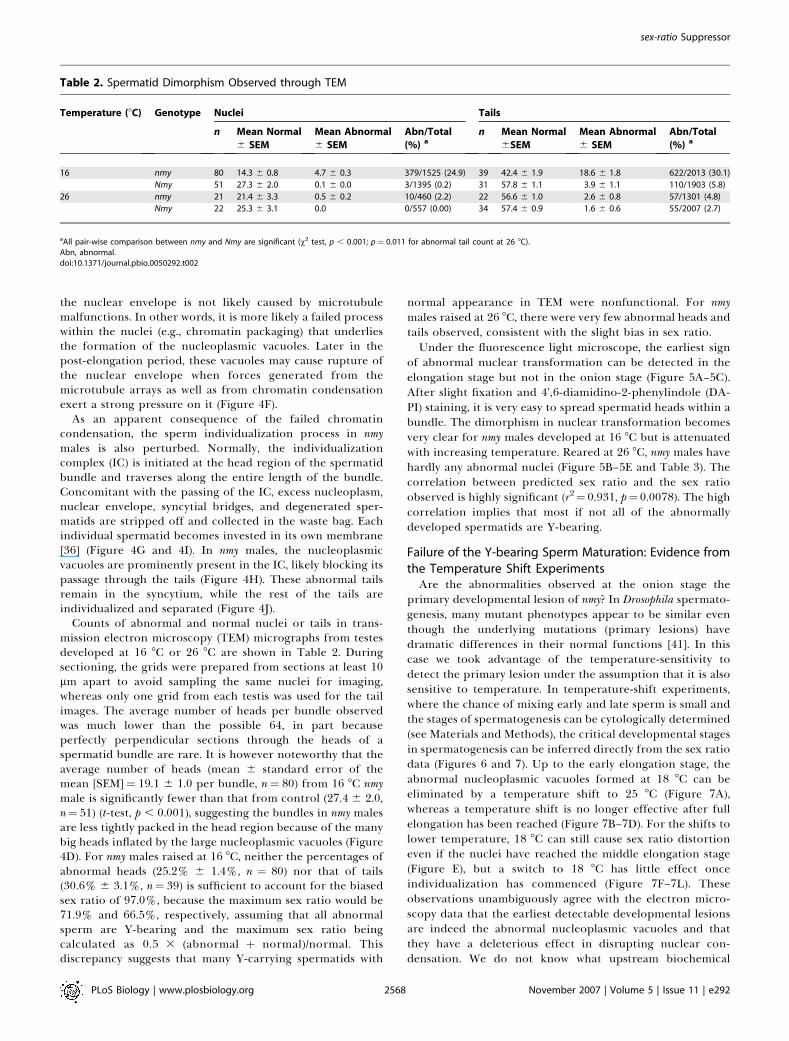
Counts of abnormal and normal nuclei or tails in trans-mission electron microscopy (TEM) micrographs from testesdeveloped at 16 8C or 26 8C are shown in Table 2. Duringsectioning, the grids were prepared from sections at least 10lm apart to avoid sampling the same nuclei for imaging,whereas only one grid from each testis was used for the tailimages. The average number of heads per bundle observedwas much lower than the possible 64, in part becauseperfectly perpendicular sections through the heads of aspermatid bundle are rare. It is however noteworthy that theaverage number of heads (mean 6 standard error of themean [SEM]¼ 19.1 6 1.0 per bundle, n¼ 80) from 16 8C nmymale is significantly fewer than that from control (27.4 6 2.0,n¼ 51) (t-test, p , 0.001), suggesting the bundles in nmy malesare less tightly packed in the head region because of the manybig heads inflated by the large nucleoplasmic vacuoles (Figure4D). For nmy males raised at 16 8C, neither the percentages ofabnormal heads (25.2% 6 1.4%, n ¼ 80) nor that of tails(30.6% 6 3.1%, n¼ 39) is sufficient to account for the biasedsex ratio of 97.0%, because the maximum sex ratio would be71.9% and 66.5%, respectively, assuming that all abnormalsperm are Y-bearing and the maximum sex ratio beingcalculated as 0.5 3 (abnormal þ normal)/normal. Thisdiscrepancy suggests that many Y-carrying spermatids with
normal appearance in TEM were nonfunctional. For nmymales raised at 26 8C, there were very few abnormal heads andtails observed, consistent with the slight bias in sex ratio.Under the fluorescence light microscope, the earliest sign
of abnormal nuclear transformation can be detected in theelongation stage but not in the onion stage (Figure 5A–5C).After slight fixation and 4’,6-diamidino-2-phenylindole (DA-PI) staining, it is very easy to spread spermatid heads within abundle. The dimorphism in nuclear transformation becomesvery clear for nmy males developed at 16 8C but is attenuatedwith increasing temperature. Reared at 26 8C, nmy males havehardly any abnormal nuclei (Figure 5B–5E and Table 3). Thecorrelation between predicted sex ratio and the sex ratioobserved is highly significant (r2¼ 0.931, p¼ 0.0078). The highcorrelation implies that most if not all of the abnormallydeveloped spermatids are Y-bearing.
Failure of the Y-bearing Sperm Maturation: Evidence fromthe Temperature Shift ExperimentsAre the abnormalities observed at the onion stage the
primary developmental lesion of nmy? In Drosophila spermato-genesis, many mutant phenotypes appear to be similar eventhough the underlying mutations (primary lesions) havedramatic differences in their normal functions [41]. In thiscase we took advantage of the temperature-sensitivity todetect the primary lesion under the assumption that it is alsosensitive to temperature. In temperature-shift experiments,where the chance of mixing early and late sperm is small andthe stages of spermatogenesis can be cytologically determined(see Materials and Methods), the critical developmental stagesin spermatogenesis can be inferred directly from the sex ratiodata (Figures 6 and 7). Up to the early elongation stage, theabnormal nucleoplasmic vacuoles formed at 18 8C can beeliminated by a temperature shift to 25 8C (Figure 7A),whereas a temperature shift is no longer effective after fullelongation has been reached (Figure 7B–7D). For the shifts tolower temperature, 18 8C can still cause sex ratio distortioneven if the nuclei have reached the middle elongation stage(Figure E), but a switch to 18 8C has little effect onceindividualization has commenced (Figure 7F–7L). Theseobservations unambiguously agree with the electron micro-scopy data that the earliest detectable developmental lesionsare indeed the abnormal nucleoplasmic vacuoles and thatthey have a deleterious effect in disrupting nuclear con-densation. We do not know what upstream biochemical
Table 2. Spermatid Dimorphism Observed through TEM
Temperature (8C) Genotype Nuclei Tails
n Mean Normal
6 SEM
Mean Abnormal
6 SEM
Abn/Total
(%) an Mean Normal
6SEM
Mean Abnormal
6 SEM
Abn/Total
(%) a
16 nmy 80 14.3 6 0.8 4.7 6 0.3 379/1525 (24.9) 39 42.4 6 1.9 18.6 6 1.8 622/2013 (30.1)
Nmy 51 27.3 6 2.0 0.1 6 0.0 3/1395 (0.2) 31 57.8 6 1.1 3.9 6 1.1 110/1903 (5.8)
26 nmy 21 21.4 6 3.3 0.5 6 0.2 10/460 (2.2) 22 56.6 6 1.0 2.6 6 0.8 57/1301 (4.8)
Nmy 22 25.3 6 3.1 0.0 0/557 (0.00) 34 57.4 6 0.9 1.6 6 0.6 55/2007 (2.7)
aAll pair-wise comparison between nmy and Nmy are significant (v2 test, p , 0.001; p¼ 0.011 for abnormal tail count at 26 8C).Abn, abnormal.doi:10.1371/journal.pbio.0050292.t002
PLoS Biology | www.plosbiology.org November 2007 | Volume 5 | Issue 11 | e2922568
sex-ratio Suppressor

processes are involved, but chromatin modulation and trafficacross the nuclear envelope are among the primary suspects.
Discussion
We have presented genetic, molecular, and cytological datacharacterizing the suppression of the Winters sex-ratio in D.simulans. We have shown that the Winters sex-ratio ispolymorphic within natural populations of D. simulans, andthat it has no relation to D. sechellia as previously thought. Theetiology of the aberrant sex-ratio is attributed to the failure ofnuclear condensation in the Y-bearing sperm. We mappedand cloned an autosomal suppressor that acts to suppress asex-ratio distorter on the X chromosome. The nucleotidesequence and structure of the suppressor strongly suggests amechanism involving siRNAs. Remarkably, the evolutionaryorigin of the suppressor is from a transcript of the sex-ratiodistorter gene itself. Our work supports the theory that
intragenomic conflicts are important evolutionary processesthat may ultimately underlie several seemingly unrelatedmajor biological phenomena including Mendelian segrega-tion, genome organization and speciation.
One Species, Three Systems (At Least)The cases of sex-ratio recorded in D. simulans can be
recognized as at least three independent systems, as high-lighted below.Paris versus Winters. For the Paris system, two epistatic
distorters have been implicated in the sn–lz region [22], whichis adjacent to but not overlapping the lz–v region where theWinters distorter (Dox) has been localized [32]. Otherevidence also suggests an older evolutionary age for theWinters system, as detailed in [32].Winters versus Durham. Tmy has been mapped to 89A4 and
cloned (YT, unpublished data). It is at different location andhas a different sequence than that of Nmy (87F3). Further-
Table 3. Spermatid Dimorphism Observed after DAPI Staining
Temperature (8C) Genotype n Mean Normal
6 SEM
Mean Abnormal
6 SEM
Abn/total (%) a Predicted
Sex RatiobReal Sex
Ratio
16 nmy 23 31.9 6 0.6 29.4 6 0.6 675/1408 (47.9) 0.960 0.970
Nmy 16 62.1 6 0.4 0.3 6 0.3 4/997 (0.40) 0.502 0.500
18 nmy 46 36.5 6 0.8 25.1 6 0.8 1153/2832 (40.7) 0.843 0.950
Nmy 24 60.6 6 0.5 0.3 6 0.1 6/1461 (0.41) 0.502 0.490
RT nmy 26 41.3 6 0.9 18.8 6 0.9 488/1563 (31.2) 0.727 0.870
Nmy 19 61.5 6 0.6 0.3 6 0.1 5/1174 (0.4) 0.502 0.510
25 nmy 23 57.4 6 0.9 4.0 6 0.6 91/1411 (6.4) 0.534 0.590
Nmy 20 62.5 6 0.4 0.2 6 0.1 4/1253 (0.3) 0.502 0.500
26 nmy 19 59.7 6 0.8 0.2 6 0.1 4/1139 (0.4) 0.502 0.540
Nmy 20 60.8 6 0.8 0.2 6 0.1 3/1218 (0.3) 0.501 0.510
aAll pair- wise comparison between nmy and Nmy are significant (v2 test, p , 0.001) except at 26 8C (p¼ 0.638).bCalculated as 0.5 3 (mean normalþmean abnormal)/mean normal.doi:10.1371/journal.pbio.0050292.t003
Figure 5. Abnormal Transformation of the Y-Bearing Sperm Detected by DAPI Staining
(A) Onion stage (SSR12-2-7 nmy, 16 8C) similar to that in Figure 4A and 4B. Abnormal nucleoplasmic vacuoles are not visible under the fluorescencemicroscope. Nucleus (N) and nebenkern (NK) are indicated. Scale bar: 20 lm.(B and C) Spermatids of SSR12-2-7 nmy have strong dimorphism in nuclear transformation at 16 8C (C) as compared with simB Nmy (B).(D and E) The nuclear dimorphism of SSR12-2-7 nmy disappears at 26 8C (E) as compared to simB Nmy (D). Scale bar for B-E: 10 lm.doi:10.1371/journal.pbio.0050292.g005
PLoS Biology | www.plosbiology.org November 2007 | Volume 5 | Issue 11 | e2922569
sex-ratio Suppressor

more, Tmy does not suppress Dox (crosses 37 versus 39 inTable S1).
Paris versus Durham. Neither does Tmy suppress SR6, an Xchromosome carrying the Paris distorters [32].
These observations suggest that all three sex-ratio systemsare distinct. We cannot exclude some evolutionary connec-tions among them that genetic tests so far have failed toreveal. Nonetheless, these observations strongly suggest thatgenetic variations of sex-ratio are not rare, at least in D.simulans. Although one search for genetic variation affectingsex ratio in D. melanogaster was negative [42], positive results inthe same species have been reported [43,44]. Some X-linkedmutations distorting sex ratio can be readily generatedthrough mutagenesis in D. melanogaster [45,46], but for reasonsthat are unknown, no clear case of sex-ratio has ever beenfound in natural populations of this species.
The recessive mutant (sxr) might well be allelic to nmy. Thisgene can be placed 26.6 cM distal to H (74.2) and 34.0 cMproximal to pe (121.4), i.e., in the vicinity of Ubx (90.3) on onegenetic map [47]. Thus sxr seems to fall in roughly the sameregion as nmy (Figure 1B, also see Table 6 in [17]). Thephenotypes of sxr also appear to be very similar to nmy inseveral respects, including temperature sensitivity and ab-normal morphology in spermiogenesis [17]. At the ultra-structural level, sxr spermatogenesis showed pronouncedsperm tail degeneration [48], as we report here for nmy(Figure 4J).
On the other hand, some phenotypes of sxr were describeddifferently from those of nmy reported here. One majordiscrepancy is in the temperature-sensitive stage, which wasinferred to be the primary spermatocyte for sxr and nucleartransformation for nmy. The determination of the sxrtemperature-sensitive stage could be somewhat discrepant,because the temperature-shift protocol used does not stagespermatogenesis with great precision. In addition, the timecourse of spermatogenesis inferred for D. simulans was notbased on direct cytological examination but was assumed tobe the same as that in D. melanogaster [17]. Under TEM, sxr
males showed abnormalities in the primary spermatocytesand in the axonemal complex before the individualizationstage [48], but nmy males did not show these defects, evenwhen special attention was given to these possibilities. In anyevent, if sxr and nmy are indeed the same gene, the mutationlikely has maintained stable frequency in natural populationsof California for many decades.
The Evolution of sex-ratio SuppressorIntragenomic conflicts are struggles within a genome over
hereditary transmission [49]. Meiotic drive is one type ofintragenomic conflict in that the driving allele or haplotypehas more than 50% representation in next generation. Fornuclear genes that freely recombine the evolutionary stablestrategy is exact Mendelian segregation [8]. Thus for most ofthe genome, there is strong evolutionary pressure forselecting modifiers that increase the fidelity of Mendeliansegregation. This explains why segregation in meiosis isusually Mendelian, and it also underlies Fisher’s argumentabout the sex ratio based on parental expenditure [8].However, this logic does not apply to cases where freerecombination is inhibited. For example, in the two classiccases of autosomal meiotic drive—Segregation Distortion (SD) inD. melanogaster [50] and the t- complex inMus musculus [51]— thedistorter(s) and insensitive responder(s) are locked togetherwithin complex inversions. Similarly, the evolution of sex-linked meiotic drive is facilitated by the lack of recombina-tion between the X and the Y chromosomes. It has beenreasoned that sex-ratio meiotic drive is a more potentevolutionary force than autosomal drive, based on twoarguments [11,12]. First, sex-ratio drive can evolve morereadily. When the sex chromosomes do not undergorecombination along most of their length, which includesmost cases of heteromorphic sex chromosomes, many sex-linked genes can potentially mutate to sex-ratio distorter. Thisis usually not true for an autosomal distorter, because theprecondition for its invasion is satisfied only in specialcircumstances, such as in the centromeric region with itsreduced recombination or within inversions. Second, sex-ratio
Figure 6. Temperature-Shift Experiment
At various time points during development from eggs to adults, vials of SSR12-2-7 (nmy) flies were shifted from 18 8C to 25 8C (A) or from 25 8C to 18 8C(B). Seven to 32 males eclosed from each of these vials were tested for sex-ratio, and the results (sex ratio and error bar¼ SEM) are shown at the timepoints of temperature shift. The stages of spermatogenesis at critical points (A–L) were examined cytologically (see Figure 7 for the correspondingimages).doi:10.1371/journal.pbio.0050292.g006
PLoS Biology | www.plosbiology.org November 2007 | Volume 5 | Issue 11 | e2922570
sex-ratio Suppressor

has a much greater effect on the rest of genome preciselybecause it affects the sex ratio, and thus, the transmissionrates of different genomic compartments. In addition tofavoring modifiers that reduce any fitness cost, sex-ratio favorsmodifiers that render the sex ratio more equal, as mandatedby Fisher’s principle [52]. The evolutionary cycle of distorterand suppressor could go on indefinitely as long as new sex-ratio mutations unaffected by existing suppressors can occur.When a sex-ratio mutation invades a population, its fate can
be fixation, stable polymorphism, or extinction, dependingon the configuration of fitness components (particularly thefitness of sex-ratio males) [53]. Polymorphisms of distortersand suppressors have been reported in several Drosophilaspecies, including D. simulans [25], D. quinaria [54,55], D. obscura[56], D. paramelanica [57], and D. mediopunctata [58–60]. Fixationof a suppressor is likely the case for the cryptic sex-ratiosuppressor Tmy in D. simulans [15]. However, no sex-ratiosuppressors have been found in D. pseudoobscura, and the causemay be related to high fitness cost of sex-ratio males [61,62]. Itis remarkable in itself that modifiers that increase the fitnessof sex-ratio males seem not to have been selected for the past0.7–1.3 My [63]. The answer may lie in the molecularmechanism of this sex-ratio distorter that is still unknown.Molecular characterization of the Winters sex-ratio systemmakes possible future studies on the molecular populationgenetics and ecological dynamics of suppressors in naturalpopulations.
The Molecular Mechanism of sex-ratio and Its EvolutionaryImpactWe have shown here that an RNAi mechanism is likely
involved in the Winters sex-ratio suppression. Remarkably, thesuppressor Nmy was generated through the use of thesequence information of the Dox distorter itself by means ofretrotransposition. An interesting observation is that theabnormal transformation of the Y-bearing sperm in Dox;nmymale is exacerbated at lower temperature. If a lack of smallRNAs encoded by the nmy allele is indeed the cause forderepressing the expression of Dox, then the moleculesencoded by Dox (RNA or protein) may be more harmful tothe Y-bearing sperm at lower temperature. Further in-depthmolecular analysis of this hypothesis is required. This Dox-Nmy system is reminiscent of a possible case of cryptic sex-ratiosystem Ste/Su(Ste) in D. melanogaster [64,65]. The distorter Steand the suppressor Su(Ste) share common sequences, and anRNAi mechanism has been convincingly shown to beresponsible for the suppression [66,67].The cytological defects in nmy are also reminiscent of
abnormal nuclear condensation in the classical meiotic drivesystem SD in D. melanogaster [50]. The gene Sd targets theResponder (Rsp) locus and causes degeneration of Rsp-bearingsperm [68]. The Sd-bearing sperm is thus transmitted to morethan 95% of the progeny from Sd/Rsp males. Sd is a truncatedduplication of RanGAP and is still enzymatically active
Figure 7. Critical Stage for Temperature Sensitivity
Five spermatid bundles of the most advanced stage from each of the fivegonads or testes were examined at each critical point of the temperatureshift experiment under phase contrast optics (A–F) or epifluorescence(G–L). Spermatid development stages (p to s) were classified accordingto the standard described in [41]. Scale bar: 10 lm (A–F); 100 lm (G–I); 20lm (J–L).(A–D) Spermatogenesis at 18 8C before the early middle stage ofelongation (stages p or q) can be rescued for sex-ratio by shifting to 25 8C(A; also see Figure 6). However, when spermatogenesis reaches latemiddle stage of elongation (stage r), the rescue is almost impossible (B–D). Thus, stage r is the late boundary of temperature sensitivity. Note thephase dark nucleoplasmic vacuoles in the abnormal nuclei (arrow) ascompared to normal nuclear transformation (arrowhead). (E and F)Spermatogenesis at 25 8C up to the middle stage of elongation (stages qand r) can be affected by 18 8C treatment to give full sex-ratio (E).However, the temperature sensitivity decreases once the spermato-genesis reaches the fully elongated stage (stage s) (F).(G–L) Individualization complex (IC) is viewed with Alexa Fluor 488
Phalloidin that binds to F-actin (green) while the nuclei are marked byDAPI (blue). By the beginning of individualization (arrow in I and J, whereJ is a closeup of I; compared to a younger bundle in which the IC has notformed yet, arrowhead), the 18 8C treatment can only cause a slight sexratio distortion (Figure 6B). On the 8th day of spermatogenesis at 25 8C,there are numerous spermatid bundles that have initiated individualiza-tion (K) or that have the IC traversing along the tails (L). The 18 8Ctreatment has no effect from this time on.doi:10.1371/journal.pbio.0050292.g007
PLoS Biology | www.plosbiology.org November 2007 | Volume 5 | Issue 11 | e2922571
sex-ratio Suppressor

[69,70]. It may cause problems in establishing a normalRanGTP–RanGDP gradient across the nuclear envelope [71],which is a critical condition for many cytological functionsincluding transport of small RNAs [72]. The Rsp locus consistsof a cluster of satellite repeats whose sensitivity is propor-tional to copy number [73]. One might speculate that aninterruption of heterochromatin condensation in andaround the Rsp cluster directly causes the degeneration ofRsp-bearing sperm, because normal heterochromatin regu-lation requires small RNAs homologous to the heterochro-matic region [74], whereas small RNAs originating and/ortargeting the Rsp cluster may be misregulated in Sd/Rsp males.Indeed, small RNAs originated from the Rsp locus have beenrecorded [75].
Segregation distortion in males may happen at eithermeiotic or post-meiotic stages. For example, the Y chromo-some can be fragmented or lost during meiosis II [29,76], orthe Y-carrying spermatids may not mature [77,78]. Post-meiotic failure in spermiogenesis can result from aberrationsin meiosis. For example, failure of X-Y pairing duringprophase of meiosis I has been shown to cause spermiogenicfailure in D. melanogaster [77,79]. For the Winters sex-ratio, it isunclear whether there is any pairing problem between the sexchromosomes, and whether chromosomal behavior duringmeiosis is normal. Through light and electron microscopy, wehave demonstrated that the primary lesion in nmymutants is adefect in nuclear condensation during spermiogenesis. In anycase, segregation distortion in males usually results from afailure to produce a class of sperm, or a failure of a class ofsperm to function, and there is a concomitant reduction infertility.
Whatever the actual molecular mechanisms or develop-mental stages involved, it is logical to make an evolutionarylink between sex-ratio meiotic drive and speciation. A meioticdrive distorter first exploits the meiotic and post-meioticmechanisms for the biased transmission of itself, then thegenome regains balanced transmission through the evolutionof suppressors that correct the meiotic or post-meioticaberrations. During each episode of distortion and suppres-sion, male fertility might be compromised and recovered.Because of these dynamics, the meiotic/post-meiotic geneshave an accelerated rate of evolution, thus promoting thereproductive isolation among isolated populations or incip-ient species that do not share common distorters andsuppressors [13]. Under this meiotic drive scenario, it is easyto explain two ubiquitously observed genetic patterns forpostzygotic reproductive isolation: a much faster accumu-lation of hybrid male sterility among Drosophila species, and a‘‘large X’’ effect for the distribution of the hybrid malesterility genes [13]. Two Drosophila cases where one geneexpresses both hybrid male sterility and sex-ratio lends directsupport to a role for meiotic drive in speciation [14,15]. Atestable prediction is that genes that are responsible for theinitial postzygotic reproductive isolation between speciesmost likely function in meiotic and post-meiotic processessuch as chromosome segregation and chromatin condensa-tion.
Materials and Methods
Fly stocks. Five NSR (normal sex ratio) lines (IG88, IG113, IG118,IG132, and IG143) and six SSR (skewed sex ratio) lines (IG12, IG33,
IG54, IG151, IG73, and Q15.3) were used from a previous D. simulans3D. schellia hybridization in which the parental D. simulans stock, sim2,had been collected in Winters, California [20]. The D. mauritiana 3 D.simulans introgression lines have been described before [13,31]. Thefollowing lines were used in this study: homozygous introgression lineP18.16 and P38.7 and heterozygous line P40.6. The heterozygous 2-Plines were made by recombining two P[wþ] inserts in cis from twointrogression lines: 2P-10 (P40.83P38.4), 2P-20 (P37.13P35.2), 2P-19(P37.3 3 P38.5), 2P-17 (P35.2 3 P46.17), and 2P-15 (P38.1 3 P46.17).The expression of the wþ allele affecting eye color in P[wþ] is sensitiveto its position and copy number. Essentially, these P[wþ] insertsprovide semi-dominant markers along the third chromosome in D.simulans.
A D. simulans stock with the multiply marked third chromosome jvst e pe has been described before [80]. These mutations and theirgenetic positions are javelin 3–19.2, scarlet 3–49.5, ebony 3–63.0, andpeach 3–104.9 [81]. They are allelic to D. melanogaster mutations javelin3–19.2, 65A5-E1 [82], scarlet 3–44.0, 73A3 [82], ebony 3–70.7, 93C7-D1[47], and pink 3–48.0, 85A6 [82], respectively. Note that because thereis a large inversion 84F-94F on 3R in D. simulans as compared with D.melanogaster, the map positions of e pe in D. simulans are reversed onthe map of D. melanogaster. Other D. simulans stocks simB (w; nt; III) andw; e have been described before [31]; and C(1)RM y w/lzS was kindlyprovided by J. Coyne.
Fly work. All flies were reared on cornmeal-molasses-agar mediumsprinkled with yeast grains at room temperature (22 6 1 8C) if nototherwise indicated. The sex-ratio phenotype of a male was scored bymating the male with three tester virgin females, usually of the stockw; e, for 7 d before clearing all adults. The progeny were sexed andcounted three times until the 19th day. The sex ratio (k) wascalculated as the proportion of females.
Molecular biology. The use of ASO markers was describedpreviously [31]. Some key techniques and the reagents/kits used areas follows: long PCR (Takara LA Taq); PCR product cloning (Topo TAand Topo XL PCR cloning kits, Invitrogen); sequencing of large DNAfragment (EZ-Tn5 Insertion Kit, Epicentre); phage genomic library ofD. simulans simB (Lambda ZAP II vector predigested with EcoR I,stratagene); RNA isolation (TRIZOL Reagent, Invitrogen); RT-PCR(reverse transcription polymerase chain reaction) (39 and 59- RACEkits and SuperScirpt II Reverse Transcriptase, Invitrogen).
Light microscopy. Testes or gonads from crawling larvae or youngadult males were dissected into saline (0.7% NaCl). Spermatids ormature sperm were released from gonads/testes with a fine tungstenneedle. Live specimens were observed directly under phase contrast.For a reliable count of abnormal spermatids, the specimens werefixed for 20 s to 1 min on a microscope slide with 10 ll 2%glutaraldehyde in phosphate-buffered saline (PBS) (2.7 mM KCl, 137mM NaCl, 8.0 mM KH2PO4); and rinsed with PBS for 5 min beforestaining with DAPI (100 ng/ml in PBST–0.1% Triton X-100 in PBS)for 5 min. The specimens were spread with vigorous tapping on thecover slip before being observed under epifluorescence. To visualizethe IC [83], an additional step was added in the above protocol: fixedspecimens were rinsed for 5 min in PBSTB (PBS with 0.1% Triton X-100 and 1% BSA) and stained 10 min with Alexa Fluor 488 Phalloidin(10 l/ml; Invitrogen). The specimens were rinsed again for 3 min withPBSTB before DAPI staining. For longer storage, specimens weremounted in SlowFade Gold (Invitrogen). Images were taken on anAxioskop2 or Leica DMRB with digital camera, and processed withAdobe Photoshop.
TEM. In a drop of chilled 0.067 M pH 7.4 phosphate buffer, testesand accessory glands were dissected from young males (1–3 d old)with a fine tungsten needle and were transferred immediately to 2%glutaraldehyde in 0.067 M phosphate buffer on ice. The specimenswere fixed for 2 h at 4 8C in 1% paraformaldehyde and 2%glutaraldehyde in 0.067 M phosphate buffer, followed by a post-fixation of 1 h in 2% OsO4 at 4 8C. The specimens were treated with1% uranyl acetate at room temperature for 1 h before processingthrough an ethanol series to dehydrate. The specimens were trimmedafter ethanol dehydration so that only one of each pair of testes wasused in embedding. The Epon resin was made by mixing Taab Epon(42.4 g), DDSA (19.8 g), and NMA (18.0 g) for 30 min before adding 1.9g DMP-30. Before final embedding, each testis was cut into 4–5segments and aligned on the bottom of the mold in a straight linewith the apical tip facing out. Sections were cut on a Reichertultracut-S microtome, followed by staining with uranyl acetate andlead citrate. The grids were observed with a Tecnai G2 SpiritBioTWIN electron microscope.
Temperature shift experiments. Vials with SSR12-2-7 (nmy) eggscollected at 25 8C were cultured at 18 8C or 25 8C and shifted to theother temperature at time points of 0.5 or 2 d apart (0.5 d for the
PLoS Biology | www.plosbiology.org November 2007 | Volume 5 | Issue 11 | e2922572
sex-ratio Suppressor

more critical stages) until adult flies emerged. The first few malesafter eclosion from each vial were singly mated to ten w; e 2-d-oldvirgins, which were adequate to exhaust the sperm of a single male(unpublished data). Each male was aspirated to new vials containing10 virgin females every day for 3–5 d. Offspring from each vial werecounted, but only those from the first vials were used in data analysisonce their total reached over 50. On average, 148 and 108 offspringper male were used for calculating the sex ratio for the 18 8C to 25 8Cshift and the 25 8C to 18 8C shift, respectively. In this way the firstbatch of sperm developing from the first few sperm bundles wereexhausted and assayed. Critical intervals that were sensitive totemperature were detected from the change in sex ratio. Further-more, the spermatogenetic stages of the first most mature bundles atthese sensitive intervals were determined cytologically, eitherthrough phase contrast optics as described in D. melanogaster [41], orusing Alexa Fluor 488 Phalloidin for viewing the individualizationcomplex [83].
Supporting Information
Figure S1. The Gene Responsible for sex-ratio Is Recessive and Maps tothe Third Chromosome
SSR12 expresses sex ratio distortion while Cy; Antp does not. However,this experiment does not rule out the possibility that the expressionof sex-ratio is conditioned on the presence of the X and/or the secondchromosomes from the SSR lines.
Found at doi:10.1371/journal.pbio.0050292.sg001 (245 KB PDF).
Figure S2. Extraction of the Third Chromosome into the simB w; nt; IIIBackground
(A) Five third chromosomes were extracted for each of the three SSRlines (IG12, IG54, and IG151) and three NSR lines (IG88, IG113, andIG118). Two different P-element inserts (P18 and P40), which aredistinguishable by eye color, were used as dominant markers in thiscross scheme. The introgressed D. mauritianamaterial is shown in gray[31] but was irrelevant to our purpose here. There might berecombination in the females, thus flies in G4 are not necessarily allhomozygous for the SSR or NSR alleles. However, the majority of thevials from G4 showed sex-ratio for the SSR lines, but only a few vialsshowed sex-ratio for the NSR lines (B). The overall average clearlyindicates that the responsible gene is located on the thirdchromosome. Several lines from each of the three SSR lines werestably expressing sex-ratio up to G10, but only one from each of theSSR lines (SSR12-2-7, SSR54-2-3, and SSR151-2-10) was kept forfurther analysis.
Found at doi:10.1371/journal.pbio.0050292.sg002 (288 KB PDF).
Figure S3. Extraction of the Second Chromosome into the simB w; nt;III Background(A) Five second chromosomes were extracted for each of the threeSSR lines (IG12, IG54, and IG151) and three NSR lines (IG88, IG113,and IG118). In G5, 1/3 of ntþ males should be þ/þ. The proportion ishigher from vials where no nt flies were found in their progeny. If thegenes on the second chromosome are responsible for sex-ratio, thereshould be sex-ratio in the ntþ class as compared to nt class (B). There isno indication of sex-ratio in any of these genotypes, suggesting that nogenes on the second chromosome are involved.
Found at doi:10.1371/journal.pbio.0050292.sg003 (326 KB PDF).
Figure S4. Extraction of the X Chromosome into the CM(1) y w/lzs
Background
(A) Five sublines were set up for SSR12 and NSR88 by mating singlemales to ten CM(1) y w females. Note that sex ratio in this generationwas calculated as the proportion of males. In the F1 and subsequentbackcrosses, three males were crossed to ten CM(1) y w females. In theF1, BC1, BC5, and BC10 generations, three males from each sublinewere singly mated to three w; e virgins, and the sex ratios of theirprogeny were scored (B). The X chromosome clearly does not differbetween SSR12 and NSR88 with respect to sex ratio.
Found at doi:10.1371/journal.pbio.0050292.sg004 (271 KB PDF).
Figure S5. Mapping the Gene on the Third Chromosome
(A) In all crosses, females are at the left-hand side. Crosses G1–G3were set up en masse (10–12 pairs of virgin males and females). CrossG4 was set up by singly mating to three sim2 virgin females. In G3,males and females of all the 16 possible genotypes of the four markers
were mated. In G4, some males are heterozygous for some markers,whereas their phenotype is not distinguishable from wild-typehomozygotes. Because the target gene (*) is recessive, homozygoteof closer ‘‘þ’’ markers will have a higher chance of expressing sex-ratio.A total of 1342 males were tested. Region surrounding pe is therebyimplicated with gene affecting sex-ratio (B).Found at doi:10.1371/journal.pbio.0050292.sg005 (284 KB PDF).
Figure S6. The 2-P Mapping Scheme
(A) The mapping target is determined by its association with either ofthe two P-elements, whose eye color phenotypes are easy todistinguish from each other and from flies with both P-elements.The sex-ratio phenotype of each recombinant was tested bycomplementation with SSR12-2-7 that is homozygous for themapping target, a recessive loss-of-function suppressor (*). The twoP inserts and the region of introgressed D. mauritiana of each 2-P linesare shown in Figure 1B. Recombinants with either (like P40 or P38) orboth (like 2P10) P inserts host the loss-of-function allele (*) if k . 0.5(B). Clearly, the target (*) is localized in between P40 and P38, aninterval of ;2,700 kb, as estimated by determining the exact P-element insertion sites through inverse PCR (Araripe L, Eckstrand N,Hartl DL, Tao Y, unpublished data).
Found at doi:10.1371/journal.pbio.0050292.sg006 (286 KB PDF).
Table S1. Sex Ratio Tested for Various Genotypes
Found at doi:10.1371/journal.pbio.0050292.st001 (97 KB DOC).
Table S2. New ASO Markers Developed in this Study
Found at doi:10.1371/journal.pbio.0050292.st002 (116 KB DOC).
Table S3. ASO Probes in the Last 7-kb Region of Fine Mapping
Found at doi:10.1371/journal.pbio.0050292.st003 (56 KB DOC).
Table S4. Viability from Egg to Adult
Found at doi:10.1371/journal.pbio.0050292.st004 (58 KB DOC).
Text S1. A Recessive sex-ratio Suppressor on the Third Chromosome
Found at doi:10.1371/journal.pbio.0050292.sd001 (43 KB DOC).
Text S2. Relevant Primers Used
Found at doi:10.1371/journal.pbio.0050292.sd002 (20 KB DOC).
Text S3. Transcripts of Nmy[sim2041] and nmy[sim1427]Found at doi:10.1371/journal.pbio.0050292.sd003 (30 KB DOC).
Accession Numbers
All sequences have been deposited in the GenBank (http://www.ncbi.nlm.nih.gov/Genbank/index.html) database and have been assignedthe accession numbers EF565211–EF565217.
Acknowledgments
We thank Andrew G. Clark for the original sex-ratio stocks and DavidBegun and Jerry Coyne for some other stocks that made this researchpossible; Justin Blumenstiel, Andy Clark, Steve Frank, Sarah Kingan,John Lucchesi, Collin Meiklejohn, Allen Orr, Robert Trivers,Jianming Zhang, and members of the Hartl lab for numerousstimulating discussions related to issues covered in this article;Bernado Carvalho for help to access Brazilian sources of literature;and Nathan Eckstrand, Kalsang Namgyal, and Ivan Nurminsky fortechnical support. We are grateful to Laurent Keller and twoanonymous reviewers whose comments greatly help us to improvethe manuscript. YT is grateful to Louise Trakimas of the ElectronMicroscopy Facility in Harvard Medical School for her guidanceduring the TEM study and to William M. Gelbart and Hans Hofmannfor allowing him use of their microscopes.
Author contributions. YT, JPM, and DLH conceived the research.YT, JPM, LA, and YK performed the experiments. YT and JPManalyzed the data. YT and DLH wrote the paper.
Funding. This work was funded by grants from the NationalInstitutes of Health to DLH and YT (GM65169), to JPM (GM51932through H. Allen Orr); and an Emory University Research Councilgrant to YT.
Competing interests. The authors have declared that no competinginterests exist.
PLoS Biology | www.plosbiology.org November 2007 | Volume 5 | Issue 11 | e2922573
sex-ratio Suppressor

References1. Fisher RA (1930) The genetical theory of natural selection. A complete
variorium edition. Oxford: Oxford University Press. 318 p.2. Edwards AWF (2000) Carl Dusing (1884) on The Regulation of the Sex-Ratio.
Theor Pop Biol 58: 255–257.3. Shaw RF, Mohler JD (1953) The selective significance of the sex ratio. Am
Nat 87: 337–342.4. Hamilton WD (1967) Extraordinary sex ratios. Science 156: 477–488.5. Bull JJ, Charnov EL (1988) How fundamental are Fisherian sex ratios? In:
Harvey PH, Partridge L, editors. Oxford surveys in evolutionary biology.New York: Oxford University Press. pp. 97–135.
6. Jaenike J (2001) Sex chromosome meiotic drive. Annu Rev Ecol Syst 32: 25–49.
7. Edwards AWF (1961) The population genetics of ‘‘sex-ratio’’ in Drosophilapseudoobscura. Heredity 16: 291–304.
8. Crow JF (1991) Why is Mendelian segregation so exact? BioEssays 13: 305–312.
9. Werren JH, Beukeboom LW (1998) Sex determination, sex ratios, andgenetic conflict. Ann Rev Ecol Syst 29: 233–261.
10. Cosmides LM, Tooby J (1981) Cytoplasmic inheritance and intragenomicconflict. J Theor Biol 89: 83–129.
11. Hurst LD, Pomiankowski A (1991) Causes of sex ratio bias may account forunisexual sterility in hybrids: a new explanation of Haldane’s rule andrelated phenomena. Genetics 128: 841–858.
12. Frank SA (1991) Divergence of meiotic drive-suppression systems as anexplanation for sex-biased hybrid sterility and inviability. Evolution 45:262–267.
13. Tao Y, Hartl DL (2003) Genetic dissection of hybrid incompatibilitiesbetween Drosophila simulans and D. mauritiana. III. Heterogeneous accumu-lation of hybrid incompatibilities, degree of dominance, and implicationsfor Haldane’s rule. Evolution 57: 2580–2598.
14. OrrHA, Irving S (2005) Segregation distortion in hybrids between theBogotaand USA subspecies of Drosophila pseudoobscura. Genetics 169: 671–682.
15. Tao Y, Hartl DL, Laurie CC (2001) Sex-ratio segregation distortionassociated with reproductive isolation in Drosophila. Proc Natl Acad Sci US A 98: 13183–13188.
16. Yang Y-Y, Lin F-J, Chang H-y (2004) Sex ratio distortion in hybrids ofDrosophila albomicans and D. nasuta. Zool Stud 43: 622–628.
17. Faulhaber SH (1967) An abnormal sex ratio in Drosophila simulans. Genetics56: 189–213.
18. De Magalhaes LE, Roveroni IM, Campos SHA (1985) Occurrence of the sex-ratio trait in natural populations of Drosophila simulans in Brazil. Rev BrasilGenet 8: 449–456.
19. Mercot H, Atlan A, Jacques M, Montchamp-Moreau C (1995) Sex-ratiodistortion in Drosophila simulans: co-occurence of a meiotic drive and asuppressor of drive. J Evol Biol 8: 283–300.
20. Dermitzakis ET, Masly JP, Waldrip HM, Clark AG (2000) Non-Mendeliansegregation of sex chromosomes in heterospecific Drosophila males.Genetics 154: 687–694.
21. Cazemajor M, Landre C, Montchamp-Moreau C (1997) The sex-ratio trait inDrosophila simulans: genetic analysis of distortion and suppression. Genetics147: 635–642.
22. Montchamp-Moreau C, Ogereau D, Chaminade N, Colard A, Aulard S(2006) Organization of the sex-ratio meiotic drive region in Drosophilasimulans. Genetics 174: 1365–1371.
23. Derome N, Metayer K, Montchamp-Moreau C, Veuille M (2004) Signatureof selective sweep associated with the evolution of sex-ratio drive inDrosophila simulans. Genetics 166: 1357–1366.
24. Atlan A, Capillon C, Derome N, Couvet D, Montchamp-Moreau C (2003)The evolution of autosomal suppressors of sex-ratio drive in Drosophilasimulans. Genetica 117: 47–58.
25. Atlan A, Mercot H, Landre C, Montchamp-Moreau C (1997) The sex-ratiotrait in Drosophila simulans: geographical distribution of distortion andresistance. Evolution 51: 1886–1895.
26. Montchamp-Moreau C, Cazemajor M (2002) Sex-ratio drive in Drosophilasimulans: variation in segregation ratio of X Chromosomes from a naturalpopulation. Genetics 162: 1221–1231.
27. Montchamp-Moreau C, Ginhoux V, Atlan A (2001) The Y chromosomes ofDrosophila simulans are highly polymorphic for their ability to suppress sex-ratio drive. Evolution 55: 728–737.
28. Jutier D, Derome N, Montchamp-Moreau C (2004) The sex-ratio trait and itsevolution in Drosophila simulans: a comparative approach. Genetica 120: 87–99.
29. Cazemajor M, Joly D, Montchamp-Moreau C (2000) Sex-ratio drive inDrosophila simulans is related to equational non-disjunction of the Ychromosome. Genetics 154: 229–236.
30. Montchamp-Moreau C, Joly D (1997) Abnormal spermiogenesis isassociated with the X-linked sex-ratio trait in Drosophila simulans. Heredity79: 24–30.
31. Tao Y, Chen S, Hartl DL, Laurie CC (2003) Genetic dissection of hybridincompatibilities between Drosophila simulans and D. mauritiana. I. Differ-ential accumulation of hybrid male sterility effects on the X and autosomes.Genetics 164: 1383–1397.
32. Tao Y, Araripe L, Kingan SB, Ke Y, Xiao H, et al. (2007) A sex-ratio system in
Drosophila simulans. II: An X-linked distorter. PLoS Biol 5: e293. doi:10.1371/journal.pbio.0050293.
33. Chaboissier M-C, Finnegan D, Bucheton A (2000) Retrotransposition of theI factor, a non-long terminal repeat retrotransposon of Drosophila,generates tandem repeats at the 3’ end. Nucleic Acid Res 28: 2467–2472.
34. Luan DD, Korman MH, Jakubczak JL, Eickbush TH (1993) Reversetranscription of R2Bm RNA is primed by a nick at the chromosomaltarget site: a mechanism for non-LTR retrotransposition. Cell 72: 595–605.
35. Esnault C, Maestre J, Heidmann T (2000) Human LINE retrotransposonsgenerate processed pseudogenes. Nat Genet 24: 363–367.
36. Tokuyasu KT, Peacock WJ, Hardy RW (1972) Dynamics of spermiogenesisin Drosophila melanogaster. I. Individualization Process. Z Zelorsch 124: 479–506.
37. Tokuyasu KT, Peacock WJ, Hardy RW (1972) Dynamics of spermiogenesisin Drosophila melanogaster. II. Coiling process. Z Zelorsch 127: 492–525.
38. Tokuyasu KT (1974) Dynamics of spermiogenesis in Drosophila melanogaster.IV. Nuclear transformation. J Ultrastruct Res 48: 284–303.
39. Stanley HP, Bowan JT, Romrell LJ, Reed SC, Wilkinson RF (1972) Finestructure of normal spermatid differentiation in Drosophila melanogaster. JUltrastruct Res 41: 433–466.
40. Tokuyasu KT (1975) Dynamics of spermiogenesis in Drosophila melanogaster.VI. Significance of ‘‘onion’’ nebenkern formation. J Ultrastruct Res 53: 93–112.
41. Fuller M (1993) Spermatogenesis. In: Martinez-Arias A, Bate M, editors. TheDevelopment of Drosophila melanogaster. New York: Cold Spring HarborLaboratory Press. pp. 71–147.
42. Toro MA, Charlesworth B (1982) An attempt to detect genetic variation insex ratio in Drosophila melanogaster. Heredity 49: 199–209.
43. Hanks GD (1965) Are deviant sex ratios in normal strains of Drosophilacaused by aberrant segregation? Genetics 52: 259–266.
44. Reed FA, Reeves RG, Aquadro CF (2005) Evidence of susceptibility andresistance to cryptic X-linked meiotic drive in natural populations ofDrosophila melanogaster. Evolution 59: 1280–1291.
45. Baker BS, Carpenter ATC (1972) Genetic analysis of sex chromosomalmeiotic mutants in Drosophila melanogaster. Genetics 71: 255–286.
46. Novitski E, Hanks GD (1961) Analysis of irradiated Drosophila populationsfor meiotic drive. Nature 190: 989–990.
47. Barker JSF, Moth JJ (2001) Linkage maps of D. simulans: An update ofSturtevant (1929) with additional loci. Dros Inf Serv 84: 205–206.
48. Ramamurthy G, Alfert M, Stern C (1980) Ultrastructural studies onspermatogenesis in a sex-ratio-mutant strain of Drosophila simulans. Am JAnat 157: 205–219.
49. Burt A, Trivers R (2006) Genes in conflict. Cambridge (Massachusetts):Harvard University Press. 602 p.
50. Temin RG, Ganetzky B, Powers PA, Lyttle TW, Pimpinelli S, et al. (1990)Segregation Distorter (SD) in Drosophila melanogaster: genetic and molecularanalysis. Am Nat 137: 287–331.
51. Lyon MF (2003) Transmission ratio distortion in mice. Annu Rev Gent 37:393–408.
52. Varandas FR, Sampaio MC, Carvalho AB (1997) Heritability of sexualproportion in experimental sex-ratio populations of Drosophila mediopunctata.Heredity 79: 104–112.
53. Vaz SC, Carvalho AB (2004) Evolution of autosomal suppression of the sex-ratio trait in Drosophila. Genetics 166: 265–277.
54. Jaenike J (1996) Sex-ratio meiotic drive in the Drosophila quinaria group. AmNat 148: 237–254.
55. Jaenike J (1999) Suppression of sex-ratio meiotic drive and the main-tenance of Y-chromosome polymorphism in Drosophila. Evolution 53: 164–174.
56. Hauschteck-Jungen E (1990) Postmating reproductive isolation andmodification of the ‘‘sex ratio’’ trait in Drosophila subobscura induced bythe sex chromosome gene arrangement A2þ3þ5þ7. Genetica 83: 31–44.
57. Stalker HD (1961) The genetic systems modifying meiotic drive inDrosophila paramelanica. Genetics 46: 177–202.
58. Carvalho AB, Vaz SC, Klaczko LB (1997) Polymorphism for Y-linkedsuppressors of sex-ratio in two natural populations of Drosophila mediopunc-tata. Genetics 146: 891–902.
59. Carvalho AB, Klaczko LB (1993) Autosomal suppressors of sex-ratio inDrosophila mediopunctata. Heredity 71: 546–551.
60. Carvalho AB, Klaczko LB (1994) Y-linked suppressors of the sex-ratio trait inDrosophila mediopunctata. Heredity 73: 573–579.
61. Policansky D, Dempsey B (1978) Modifiers and ‘‘sex-ratio’’ in Drosophilapseudoobscura. Evolution 32: 922–924.
62. Wu C-I (1983) The fate of autosomal modifiers of the sex-ratio trait inDrosophila and other sex-linked meiotic drive systems. Theor Pop Biol 24:121–135.
63. Babcock CS, Anderson WW (1996) Molecular evolution of the sex-ratioinversion complex in Drosophila pseudoobscura: analysis of the Esterase-5 generegion. Mol Biol Evol 13: 297–308.
64. Hurst LD (1992) Is Stellate a relict meiotic driver? Genetics 130: 229–230.65. Hurst LD (1996) Further evidence consistent with Stellate’s involvement in
meiotic drive. Genetics 142: 641–643.66. Aravin AA, Klenov MS, Vagin VV, Bentignies F, Cavalli G, et al. (2004)
Dissection of a natural RNA silencing process in the Drosophila melanogastergerm line. Mol Cell Biol 24: 6742–6750.
PLoS Biology | www.plosbiology.org November 2007 | Volume 5 | Issue 11 | e2922574
sex-ratio Suppressor

67. Aravin AA, Naumova NM, Tulin AV, Vagin VV, Rozovsky YM, et al. (2001)Double-stranded RNA-mediated silencing of genomic tandem repeats andtransposable elements in the D. melanogaster germline. Curr Biol 11: 1017–1027.
68. Tokuyasu KT, Peacock WJ, Hardy RW (1977) Dynamics of spermiogenesisin Drosophila melanogaster. VII. Effects of Segregation Distorter (SD) chromo-some. J Ultrastruct Res 58: 96–107.
69. Merrill C, Bayraktaroglu L, Kusano A, Ganetzky B (1999) TruncatedRanGAP encoded by the Segregation Distorter locus of Drosophila. Science 283:1742–1745.
70. Kusano A, Staber C, Ganetzky B (2001) Nuclear mislocalization ofenzymatically active RanGAP causes segregation distortion in Drosophila.Dev Cell 1: 351–361.
71. Kusano A, Staber C, Ganetzky B (2002) Segregation distortion induced bywild-type RanGAP in Drosophila. Proc Natl Acad Sci U S A 99: 6866–6870.
72. Ohrt T, Merkle D, Birkenfeld K, Echeverri CJ, Schwille P (2006) In situfluorescence analysis demonstrates active siRNA exclusion from thenucleus by Exportin 5. Nucleic Acids Res 34: 1369–1380.
73. Wu C-I, Lyttle TW, Wu M-L, Lin G-F (1988) Association between a satelliteDNA sequence and the Responder of Segregation Distorter in D.melanogaster. Cell 54: 179–189.
74. Pal-Bhadra M, Leibovitch BS, Gandi SG, Rao M, Bhadra U, et al. (2004)Heterochromatic silencing and HP1 localization in Drosophila are depend-ent on the RNAi machinery. Science 303: 669–672.
75. Saito K, Nashida KM, Mori T, Kawamura Y, Miyoshi K, et al. (2006) Specificassociation of Piwi with rasiRNAs derived from retrotransposon andheterochromatic regions in the Drosophila genome. Genes Dev 20: 2214–2222.
76. Novitski E, Peacock WJ, Engel J (1965) Cytological basis of ‘‘sex ratio’’ inDrosophila pseudoobscura. Science 148: 516–517.
77. Peacock WJ, Miklos GLG, Goodchild DJ (1975) Sex chromosome meioticdrive systems in Drosophila melanogaster. I. Abnormal spermatid developmentin males with a heterochromatin-deficient X chromosome (sc4sc8). Genetics79: 613–634.
78. Hauschteck-Jungen E, Maurer B (1976) Sperm dysfunction in sex-ratiomales of Drosophila subobscura. Genetica 46: 459–477.
79. McKee BD, Wilhelm K, Merrill C, Ren X-j (1998) Male sterility and meioticdrive associated with sex chromosome rearrangement in Drosophila: roleof X-Y pairing. Genetics 149: 143–155.
80. Jones CD (1998) The genetic basis of Drosophila sechellia’s resistance to a hostplant toxin. Genetics 149: 1898–1908.
81. Jones CD, Orr HA (1998) Test of a Drosophila simulans balancer and aremapping of chromosome 3. Dros Inf Serv 81: 137.
82. Sturtevant AH, Novitski E (1941) The homologies of the chromosomeelements in the genus Drosophila. Genetics 26: 517–541.
83. Fabrizio JJ, Hime G, Lemmon SK, Bazinet C (1998) Genetic dissection ofsperm individualization in Drosophila melanogaster. Development 125: 1833–1843.
PLoS Biology | www.plosbiology.org November 2007 | Volume 5 | Issue 11 | e2922575
sex-ratio Suppressor


