Embed Size (px)
Citation preview

Pergamon Advan. Enzyme Regul., Vol. 35, pp, 147-162, 1995
Copyright @ 1995 Else&x Science Ltd Printed in Great Britain. All rights reserved
0065-2571/95/$29.00
0065-2571(94)00020-4
A NEW FAMILY OF PROTEIN KINASES - THE MITOCHONDRIAL
PROTEIN KINASES
ROBERT A. HARRIS, KIRILL M. POPOV, YU ZHAO, NATALIA Y. KEDISHVILI, YOSHIHARU SHIMOMURA
and DAVID W. CRABB Departments of Biochemistry and Molecular Biology and Medicine, Indiana
University School of Medicine, Indianapolis, IN 46202
INTRODUCTION
The recent focus of this laboratory has been on the molecular cloning of the protein kinases known to exist in mitochondria, i.e., the enzymes responsible for phosphorylation and inactivation of the branched-chain a-ketoacid dehydrogenase (EC 1.2.4.1+ EC no number + EC 1.8.1.4) and pyruvate dehydrogenase (EC 1.2.4.1-t EC 2.3.1.12,+ EC 1.8.1.4) complexes. Stable changes in the activities of branched-chain a-ketoacid dehydrogenase kinase (EC 2.7.1.115) (l-5) and pyruvate dehydrogenase kinase (EC 2.7.1.99) (6-16) occur under various nutritional and hormonal states. Although the molecular mechanisms responsible for altering the activities of these kinases are not understood, it is apparent that changes in the steady state phosphorylation states of the branched-chain cu-ketoacid dehydrogenase and pyruvate dehydrogenase complexes occur as a consequence. Since the degree to which a complex is phosphorylated determines its enzyme activity, stable changes in the activities of the kinases are important factors in determining the rates at which branched chain amino acids and pyruvate are catabolized in tissues in various physiological and pathological conditions. The long-term goal of our studies is to define the molecular mechanisms responsible for the stable increase in branched- chain o-ketoacid dehydrogenase kinase that occurs in the liver of rats starved for protein as well as the stable increase in pyruvate dehydrogenase kinasc activity that occurs in livers of rats starved for calories. As a first step toward defining this mechanism, the mitochondrial protein kinases have been isolated (17, 18), partially characterized (17, 18) and their cDNAs cloned (19-21). These studies reveal for the first time that the protein kinases responsible for phosphorylation of the a-ketoacid dehydrogenase complexes are members of a unique protein kinase family not previously known to exist in eukaryotes. The tools provided by these studies have also been useful in demonstrating that an increase in the
147

148 R. A. HARRIS et al.
amount of branched-chain a-ketoacid dehydrogenase kinase associated with the branched-chain a-ketoacid dehydrogenase complex explains the increase in branched-chain a-ketoacid dehydrogenase kinase activity that occurs in protein malnutrition (22). As discussed in greater detail below, molecular cloning has revealed the existence of two isozymic forms of pyruvate dehydrogenase kinase, a finding that raises new questions about the structure/function of the pyruvate dehydrogenase complex and the mechanisms responsible for regulation of its activity.
MATERIALS AND METHODS
Sources of materials. Diets, formulated to contain various levels of protein, were obtained from ICN Nutritional Biochemicals. Enzymes and biochemicals were obtained from Sigma Chemical Company, molecular biology reagents from Bethesda Research Laboratories, chromatographic supplies from Pharmacia, polymerase chain reaction reagents from Perkin-Elmer Cetus, DNA-modifying enzymes from Life Technologies, Sequenase version 2 reagents from United States Biochemicals, and radiolabelled nucleotides from ICN Radiochemicals and DuPont NEN. The PET expression system was purchased from Novagen. cDNA libraries were obtained from Clontech Laboratories. Recombinant pyruvate dehydrogenase (the El component of the pyruvate dehydrogenase complex) was obtained as a kind gift from Dr Mulchand Patel.
Enzyme isolation and amino acid sequence analysis. Pyruvate dehydro- genase kinase was purified from the rat heart pyruvate dehydrogenase complex as described previously (18). Activity was measured either by the incorporation of radioactive phosphate into kinase-depleted porcine heart pyruvate dehydrogenase complex or by ATP-dependent inactivation of the dehydrogenase activity of the pyruvate dehydrogenase complex (18). Peptide sequencing was performed on an Applied Biosystems Model 477A Pulse Liquid Sequencer. The phenylthiohydantoin-derivatives were analyzed by reverse phase high pressure liquid chromatography with a Model 120A analyzer.
Cfoning strategy. For the cloning of the 48 kDa subunit (p48) of pyruvate dehydrogenase kinase (20), a primer with the sequence AA(A/G)AA(C/T)GC(N)ATGAG(A/G)GC(N)AC was designed acco- rding to the amino acid sequence of the branched-chain a-ketoacid dehydrogenase kinase within its conserved subdomain region II (19). This primer was then used for cDNA synthesis and polymerase chain reaction with rat heart mRNA according to the 3’ RACE (rapid amplification of cDNA ends technique) protocol. The 650nucleotide product was isolated,

NEW PROTEIN KINASE FAMILY 149
subcloned, sequenced, and used as a probe to screen a rat heart lambda gtl0 library by standard molecular biology protocols (23). Four inserts were obtained by screening 106 bacteriophage.
For the cloning of the 45 kDa subunit (p45) of pyruvate dehydrogenase kinase (21), the primer AA(A/G)TG(T/C)TC(A/G/T)AT(A/G)TA(C/T) TTIGGIGC) was designed according to the amino-terminal sequence of p45. Inosines were used in the third position of codons with a degeneracy of four. Primers were also synthesized to correspond to bases 4266-4289 and 4323-4352 of the E. coli Lac operon flanking the Eco RI site in lambda gtll phage. DNA purified from an amplified lambda gtll rat heart cDNA library was used as a template for the polymerase chain reaction which produced a product of 119 bp. A nondegenerate probe (CCTGCCAGGGACGCATKITC), corresponding to the sequence of the product the polymerase chain reaction, was used to screen a lambda gtl0 rat heart library. Two positive plaques were obtained.
DNA sequencing. Single-stranded Ml3 DNA was prepared and se- quenced by the dideoxy chain termination method with Sequenase version 2.0.
Expression of pyruvate dehydrogenase kinase in E. coli. The ~48 and ~45 subunits of pyruvate dehydrogenase kinase have been expressed as active recombinant proteins in E. coli. (20, 21). Sac1 and XhoI restriction sites flanking the coding regions of ~48 and ~45 cDNAs were constructed by polymerase chain reaction. The resulting SacI/XhoI fragments were ligated in PET-28a expression vectors (Novagene, Madison, WI), cut with Sac1 and XhoI and dephosphorylated with alkaline phosphatase to produce an in-frame amino-terminal fusion with His-Tag. The resulting plasmids were expressed in E. cofi HMS174(DE3) (Novagene) as described previously (20, 21). The recombinant proteins were purified by metal chelation chromatography.
RESULTS AND DISCUSSION
Molecular cloning and the primary structure of ~48 subunit of pyruvate dehydrogerme kinase. The branched-chain a-ketoacid dehydrogenasc kinase was first purified to homogeneity in this laboratory (17). It is a 43 kDa monomer that has significant similarity in amino acid sequence with the bacterial ‘sensor’- or ‘histidine’-protein kinases (19). In contrast, pyruvate dehydrogenase kinase has been reported to be a heterodimer of catalytic (M, 48 kDa) and regulatory (M, 45 kDa) subunits (24). The finding of a monomeric structure for branched-chain ol-ketoacid dehydrogenase kinase versus a dimeric structure of pyruvate
150 R. A. HARRIS et al.
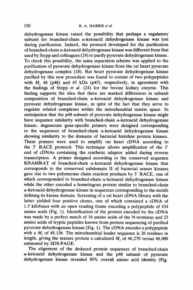
dehydrogenase kinase raised the possibility that perhaps a regulatory subunit for branched-chain cu-ketoacid dehydrogenase kinase was lost during purification. Indeed, the protocol developed for the purification of branched-chain o-ketoacid dehydrogenase kinase was different from that used by Stepp and colleagues (24) to purify pyruvate dehydrogenase kinase. To check this possibility, the same separation scheme was applied to the purification of pyruvate dehydrogenase kinase from the rat heart pyruvate dehydrogenase complex (18). Rat heart pyruvate dehydrogenase kinase purified by this new procedure was found to consist of two polypeptides with M, 48 (~48) and 45 kDa (p45), respectively, in agreement with the findings of Stepp et al. (24) for the bovine kidney enzyme. This finding supports the idea that there are marked differences in subunit composition of branched-chain a-ketoacid dehydrogenase kinase and pyruvate dehydrogenase kinase, in spite of the fact that they serve to regulate related complexes within the mitochondrial matrix space. In anticipation that the p48 subunit of pyruvate dehydrogenase kinase might have sequence similarity with branched-chain a-ketoacid dehydrogenase kinase, degenerate gene-specific primers were designed corresponding to the sequences of branched-chain cw-ketoacid dehydrogenase kinase showing similarity to the domains of bacterial histidine protein kinases. These primers were used to amplify rat heart cDNA according to the 3’ RACE protocol. This technique allows amplification of the 3’ end of cDNAs containing the synthetic adaptor added during reverse transcription. A primer designed according to the conserved sequence KNAMRAT of branched-chain a-ketoacid dehydrogenase kinase that corresponds to the conserved subdomain II of bacterial sensor kinases gave rise to two polymerase chain reaction products by 3’ RACE, one of which corresponded to branched-chain or-ketoacid dehydrogenase kinase while the other encoded a homologous protein similar to branched-chain cw-ketoacid dehydrogenase kinase in sequences corresponding to the motifs defining its kinase domain. Screening of a rat heart cDNA library with the latter yielded four positive clones, one of which contained a cDNA of 1.5 kilobases with an open reading frame encoding a polypeptide of 434 amino acids (Fig. 1). Identification of the protein encoded by the cDNA was made by a perfect match of 16 amino acids of the N-terminus and 23 amino acids of tryptic peptides known from protein sequencing of purified pyruvate dehydrogenase kinase (Fig. 1). The cDNA encodes a polypeptide with a M, of 49,136. The mitochondrial leader sequence is 26 residues in length, giving the mature protein a calculated M, of 46,270 versus 48,000 estimated by SDS-PAGE.
The alignment of the deduced protein sequences of branched-chain cw-ketoacid dehydrogenase kinase and the p48 subunit of pyruvate dehydrogenase kinase revealed 30% overall amino acid identity (Fig,
NEW PROTEIN KINASE FAMILY 151
HPCCOSAOCTCC coocaQo4mooT - 35 1 5 10 15 20 25 AToAooc-OoocA~~TeAaoc -EOTCCCCMEOCE*(KWT*OT _ 110 YnLAnLLnOOT8VnPLCAVPCISIB 26 30 35 40 65 SO C-ctCOO+Z -aJacEEocatcmAoAoc~ oouxAoQIOBLmtAw - 1e5 LAsnPAPoEoPAnr~Q VPOOVDPYA 51 55 60 65 70 7s CQCTtCTCQCC OtCOECIE~EI~O~OTARA~-mC0 - 360 nPSPEPLs3llaPLDP0svRACSSTS 76 80 85 90 96 100 TTcATsTTTctTcoAcAAoAot7oc-AutToacAMTA7AA7oMAoAM~ ERCCCDITAAT - 135 FYPLIQSLPVRLANINKLISLLPDl 101 ' 10s 110 115 120 125 C~cCCu~coTA~~w~ATA~~~~~~ - ‘10 LLRTPSVOLVOSNYIOSLOLLLD~S 126 130 135- 140- 165 150 OACAAQC7ATt7ATOAA-TAAOOA~CCOQCACMT - 485 DXSALPA~TIYC?TDTVIRIlRlWW 151 155 160 165 170 175 oAToTuTccccAcclmocccA~c7oMTAcM~Guo2twAte~ CCAOWM - 560 DVIPTYAQQVTCYKISPOVDPVTSQ 176 100 lS5 190 395 200 AAToTTeAoTActTTwmA- AUTWbOTCOCAtCtCAAYPAQMWACtCAA~OC-A - 635 NVOYPLDR?YYSRISIRYLLNQNSL 201 205 210 215 a30 225 ~oTccATcteA~~-TAAA-~AoTcQAo - 710 LPOQ~0#PINRKNIQIIWPHCDVV~ 326 230 335 260 245 250 oTrAttAAAoArew7AToAoMTocahoAcmcn7moA~TAtTAcott~TAoMcTY - 79s VISDOYSNAIILCDLYYVYSPSLBL 251 255 260 265 270 27s aAAQAAcTMA-TcAE cAoccAATAeAAoToa TTTATOYWC5TCCCATCYCTAYCACA7ODYO - 460 sxLNAKsPOQPIQVVYVPsxLfPYV 376 2#0 205 290 295 300 TlTMACTOt7CMOAA -ToR~ccATMAocAccAtou- oTTtA7cccccQATteAA - 935 FxLPx*A*nATYrPnADxaVYPPI~ 301 305 310 315 320 32s orrcAcoTcAcAcToowQAooA oMl7TaAcTotoAAoAToAoToA~ ccQcTQAoobb0 - 1010 VHVTLOlSDLTVKYSDROOOVPLRK 326 330 335 340 34s 350 ATcoIcIo1cTcczcMcTAcA~A~~~ec~~ rPuRoAcoTcecotocOccTo - 1005 IDRLPNYYYSTAPIPRVSTSRAVPL 351 355 360 365 370 375 OcMol~cC~AC~~CCUTA~AC~~~A~ACAOtA~~~~~~~~TA~~ - 1160 *cJPoYoLP1s1&YADYrDoDLSLYS 376 300 39s 390 395 400 TTAOlaOOCTACOOO1CO~~T~ATA~M~~~~C~~A?~ CTCCCW - 1135 LEOYOTDAVIYII WE R w 401 40s 410 415 420 425 TATMT1uJ1cCWF~A~~C~CCACMW~COACW~~~~M~~CO - 1310 -XAA”IRYRTNBIADD”~“PSR~P 426 430 435 )rAIMuTcAcMco~ccDo~oTM~~~c~~ mxbMTGToaTTTTTwcAAA - 1395 XDYTTPRSSmd CTTOOMOAQAMTOTA TPTOMACUUY’POTICETA~TUT~OTM~ ACMTCAQ~ITTGCT - 1460 MAW.TC!CTAAAGOATCMWAOCAACTTTATl’UMAAMMM 1507
Fig. 1. Nucleotide and deduced amino acid sequences of the ~48 subunit of pyruvate dehydrogenase kinase. The N-terminal and internal tryptic peptide sequences determined with purified native p48 protein are underlined. Reproduced with permission from Popov
e/ al. (20).
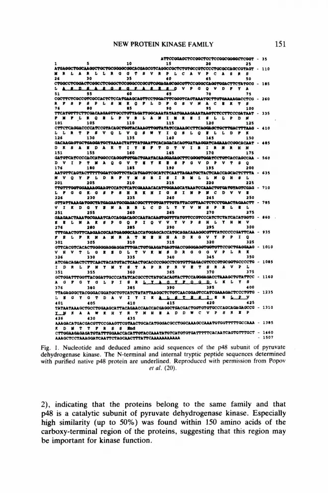
2), indicating that the proteins belong to the same family and that p48 is a catalytic subunit of pyruvate dehydrogenase kinase. Especially high similarity (up to 50%) was found within 150 amino acids of the carboxy-terminal region of the proteins, suggesting that this region may be important for kinase function.
152 R. A. HARRIS et al.
BCKDH KIN - YILTSVI6SOPRSQ6~WPLLQS8LSLRVRSTM TDTUWCLAMRSKTV - 50 Y L: ;L tat VR: A LA: t I
PDW KIK - URLMLLROUTs----------------VRPLCAVPCRTR8LASDSAS~S - 34
RCKDH KIM - TSI~SAIDWAIICP(~YSQMQ~~~-SCALP - 99 VA rD Y
- ~sAs;s~aQvA?Y~*PsPLsMQPLD KS rL QBLP
PDH KIN mavKAcKKT!lMLKQBLP - 84
BCKDK KIK - VRIAKKIKWVVFLS SLVATLPYCTWKLYIKAFQKLTDFPPIKDQADKA - 149 VRrA t t LrTr V YIrQLDP I :
PDH KIU - VKLAKIWISLLPDKLLKTPSVQLVQSUYIQSLQKLLDPKDKSAKDAKT - 134
RCKDH KIR - QYCQL--VKQLLDD ~LRMRKUISDBKL----VRYPLDKT - 193 Y v I * H Dvrt rArQr 1 I v YPLDI
PDH KIN - IYmTDTVIKIRllKmmIP SPt3VkTSQlNQYPLDM - 104
RCKDH KIR - LTIRLQIKXLA~ KPDPV-----OEISTRLSPKKIIEKUVDPA - 338 IRxtIPSL U L Kt a1 t :I I A
PDH KIN - YmSRIsIKMLLmQKSLLmQKoSPSHIaSIKPKcDwKV~KDQYKKA - a34
RCKDU KIU - KKwKKKYmmPRvKIIauuv~PFI------PnPLDYILPBrn - 181 RNCr YMrPtrr A I P L I?BLKmMK
PDH KIM - RRLCDLYYWSPELXLB~LUAKEPOQPIQWYVP8EL-BLP- - 184
RCKDH KIU - ATHKSKLDTPYKVPDVVITIAIIKIS~LDRVHDYK - 331 ATK&HDr P V tt DL r::6-rr I rDRr rY
PDX KIK - ATUERUADKQVYPPI~ -DLTVlWSDKQO(NPLKKIDKLFKYM - 333
RCKDB KIU - ?TTAKASTQDPRISPt aaQmPmI~aaPTsRAYAsYLaa - 301 rrTA I tt I PI OQtW 8R YAtY Q
PDB KIR - YSTAPRPRVBTSRA--------------VPLAWQYQLPIMLYAQYPQQ - 369
RCKDH KIM - SIQIQSq)aIaTDVYLRL _-_____---_-------_------------- - 400
L L SLIQ QTD : t PDR KIN - DLKLYSLKOYQTDAVIYIKALSTKSI~UKKYKTUKKAD~ - 419
RCKDII KIN - ---KnIDQKusPRI - 411 II
PDB KIN - vPsMPKDuTTPRss - 434
Fig. 2. Alignment of the deduced amino acid sequences of pyruvate dehydrogenase kinase and branched-chain a-ketoacid dehydrogenase kinase. PDH kin, pyruvate dehydrogenase kinase; BCKDH kin, branched-chain a-ketoacid dehydrogenase kinase. Identity is shown by (:), conserved residues (A,S,T; V,L,I,M; F,Y; D,E; K,R) are shown by (.). Reproduced
with permission from Popov et al. (20).
Molecular cloning and the primary structure of thep45 subunit of pyruvate dehydrogenase kinase. Molecular cloning of the 45 kDa (~45) subunit of pyruvate dehydrogenase kinase, long thought likely to correspond to a regulatory subunit for the catalytic subunit, became a priority following successful cloning of the ~48 subunit. The amino terminal sequence of ~45 (KNASLAGAPKYZEFSKFS-) allowed design of an oligonucleotide gene-specific primer with low degeneracy that was further decreased by the use of inosine in the third position of specific codons. A rat heart cDNA library was amplified by using this primer in combination with primers specific for lambda gtll DNA sequences of the library. One polymerase chain reaction product obtained yielded an open reading frame

NEW PROTEIN KINASE FAMILY 153
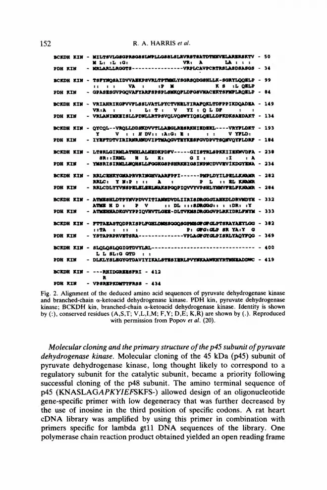
with a deduced amino acid sequence exactly matching the amino-terminal sequence of p45. This information was then used to clone a complete cDNA for p45 from a rat heart cDNA library (21). The 2.2 kb cDNA obtained has one open reading frame encoding for a poiypeptide of 408 amino acids with a M, of 46 kDa (Fig. 3). Identification of the protein encoded by the cDNA was made by a perfect match with the amino acid sequence of the N-terminus of p45 as well as matches with the sequences of two tryptic peptides derived from ~45. The cDNA encodes a mature protein with a calculated M, of 45,031, in good agreement with the M, of 45.000 estimated for ~45 by SDS-PAGE.
TLPPIKI*‘“ALOIIDt8rK”8D**QQ”PL* 161 110 110 190
111 330 3.0 150 TA?CT-3MWCCCWTCCAC QQAc7cmFOOIOCQCCmCC~~ m.~~oIccA~-QQcc -1156
ILK,LlTDO”I IL~VYMK~AWISIPTIP~~A
1.51 360 370 1@0 OQTO*CTWTQCQWCCCWChCA*WCCMQMCAC A-E~-~cc-~c~wwwA~ -1366
CD”C”9STCPKYT~TlRY8 391 390 399 QCTQCCAC~TECACAWQA l=lvmcmTcTATeccCwc~TwcQ&QAT -1436 cr-AwhK~~~-~~ - 1516 R~TQK*~ -1616 -l-cTQymp -1706
cmmMPAQT-- - 1’96 Nw~ocreclcmrmUferw~~- - l996 M-~C-QQcQk-~--~ - 1976 QcE-- - 10 66 Am3ccccA~~cAmQcUQTTTTvT M-w-~ccc~c ucLww*~QccQr0c~~~w - 1156 WCTWU~TIUU-~T~~~-~~QAT-~ -1215
Fig. 3. Nucleotide and deduced amino acid sequences of the p45 subunit of pyruvate dehydrogenase kinase. The N-terminal and internal tryptic peptide sequences determined with purified native ~45 protein are underlined. Reproduced with permission from Popov
et al. (21).

154 R. A. HARRIS ef al.
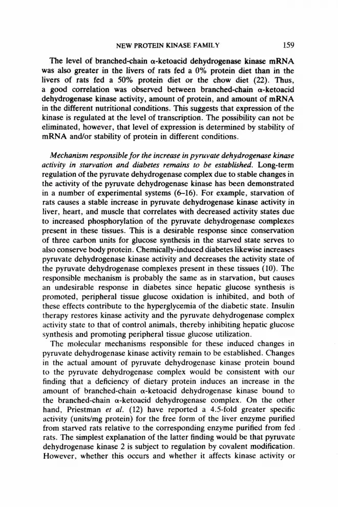
Alignment of the deduced protein sequences of p48 and ~45 revealed a surprisingly 70% amino acid identity between the two sequences, indicating that ~45 cDNA encodes another isoform of the catalytic subunit of pyruvate dehydrogenase kinase, rather than a regulatory subunit (Fig. 4). Although the similarity extends throughout these proteins, differences are also apparent throughout, particularly within the amino terminus. Since both ~45 and ~48 are catalytic subunits of pyruvate dehydrogenase kinase, the first isoform discovered (~48) has been named pyruvate dehydrogenase kinase 1 and the second (~45) pyruvate dehydrogenase kinase 2 (21).
The hypothetical structure of the kinase domain of mitochondrial protein kinases. The analysis of the primary structure of mitochondrial protein kinases clearly demonstrates that their catalytic domains are not conserved uniformly, but rather consist of five subdomains of highly conserved amino acids separated by regions of lower conservation (Fig. 5). These subdomains are likely important for catalytic function, either directly as
PDX II - ~--------ASLAQ*PltYI~S~SP~P~~POS~~~T~P :: .: : . ...::::::::::::::: . . . . . . . . . . . . . ..I
PDX I - ASD~QSQPM)~BaVPQSPE~~~~SP
PDX II - TPLXQELPVXLAH I~I*LLP~IVLBTPBVQLVQBWWPBLtOUlVLD :::::a::::::::::: :::: .: i::::::::::.::: . . . . . :
PDX I - NPLXQEL PVXLAHIWSI8LLPDHLLX TPSVQLVQWfYIQSLQXLLDPXD
PDX II - xDPzDimlzsQmDuNTI~QDDPvs~ .:. :::... :::a::::.::::::: :::...: :::. ::
POX I - ;&&I YuTDTVIRIXnXluDVIPsPQVDPVTSQw
PDX II - I~~DIVnSRISIWII~~I~~~~P~IQ~ID~~~~ . . . . . . . . . . . . . . . . ..t....t....*..i.x : :.: :::::: ::: : .:
PDX I - VQYPLDxPYnsxIBI~ SLLPQQxQsPmlxxxIQsIwPHcDwrv
PDX II - VKDAYDMAlU&CDKYYUSPDLBIQWMMWATQPI lmvYvPsHL- .:: :. :. ::a ::. ::.::. :.:: ::: . . . . . . . . . . . . . . . . . . . . . . .
PDX I - ImwmmxLaLYYvH8~ PQQPIQvvYvPsHLY3mw
PDX II - BLP xsaAmA~BPBBBLTLPPIxImvALQaBD L8ImemQQQvPLxxI . . . . . . . . . . . . . . . . . . . . . . : ::: . :.::::::..::::::::::::::
PDX I - II---PI ~LTVW#DXGQQVPLXXI
PDX II - LIwByyYST~pQWTaoT-P~~LPI~Y~~D~LP~ .t:: ::::::: : a:t::z::::::*:*: :::::: :.:.
PDX I -D-S%PXPXVB&l&~ TOYQLPISXLYAQYWQODLXLYSL
PDX II - EmoTDAvIILIULBTD~~~I~a~Px ::.:::::::.:::::.:.::::I::::.:a.:: : :: :::::: :::
PDX I - mYQTDAvIYIxALsTxsImL-TmnAD MCVPSXBPX
PDX II - wTsTYxv6 - 399 .:.: :
PDX I - DslTTPxS8 - 408
- 41
- 50
- 92
- 100
- 14a
- 150
- 191
- 200
- a42
- a50
- 292
- 300
- 341
- 350
- 391
- 400
Fig. 4. Alignment of the deduced amino acid sequences of p48 (PDK I) and p45 (PDK II). Identity is shown by (:), conserved residues (A,S,T; V,L,I,M; F,Y; D,E; K,R) are
shown by (.). Reproduced with permission from Popov et al. (21).

PDK
I -h
MVf
BLf
BTM
BhH-
-fs
llnYS
TApt
Pq-
PDK
II -h
MLf
BLf
IUUQ
1RRS
H-
- fn
YhaY
STlp
rPr-
BCKD
K -y
ILpB
Ll
-TM
BSE-
-m
dYhP
TTAe
aSt
-
BLK-
1
3 Q
POX
I -D
tVlrI
mrR
ndVi
ptM
ADG
- -D
LrVK
J&D~
Plkk
IDdL
- -d
LaG
POYC
LPiS
RlYA
gYf-
g
PDK
II -D
aLvt
IrnrH
ndW
ptM
agQ
- -D
LaIK
Ms0
1QO
OVP
lrkIE
rL-
-pLa
QPU
YCLP
iSRl
YAfY
f- s
BCKD
K -Q
lVrq
Lldd
fIkdW
tlLAS
Q-
-DLi
IRI
-IAhk
dLDr
V-
BLK-
1 -D
qLtv
Vlkr
Jfah
Wet
h-
-pM
hOPc
?EW
LPtS
RlYA
eYi-
52
E 2
NH2
I- Ki
I--
> lo
osa
2 5 -t
Fig
i Am
ino
acid
s co
nser
ved
in su
bdom
ains
I,
11, 1
11. I
V an
d V
of
mito
chon
dria
l pr
otei
n ki
nase
s.

156 R. A. HARRIS et al.
components of the active site or indirectly by contributing to the formation of the active site through constraints imposed on secondary structure. Most of the conserved domains lie in the carboxy-terminal part, whereas the amino-terminal part is quite variable, except for subdomain I. In the amino-terminal part of the proteins, a putative kinase domain is defined by an invariant histidine residue in subdomain I. It is separated by a spacer of more than 100 amino acids from subdomain II defined by an invariant asparagine occurring within a highly conserved region KNAMRAT. In the carboxyl-terminus the catalytic domain is defined by a glycine-rich loop GxGxG (subdomain V). The central core of the catalytic domain consists of subdomain III (consensus sequence DrGgG) and subdomain IV (defined by aromatic residue Y).
A similar arrangement for the kinase domain to that discussed above has been found for the bacterial histidine-protein kinases (for review see 25). Work completed thus far with protein kinases of this type from bacteria indicate that phosphorylation of the invariant histidine residue of subdomain I is involved in their catalytic mechanism. Subdomains III, IV and V may be involved in binding of ATP, whereas subdomain II may serve as a hinge controlling the interaction between the ATP-binding site and the amino-terminal region bearing the catalytic residue. In contrast to serine/threonine-specific protein kinases, bacterial enzymes use a mechanism of phosphotransfer that involves autophosphorylation on a histidine residue. The phosphohistidine intermediate serves as a donor of phosphate during subsequent phosphorylation of an aspartate residue of a target protein called a response regulator:
Protein kinase-His + ATP -+ Protein kinase-His-P -!- ADP Protein kinase-His-P + Response regulator-Asp Protein kinase-His + Response regular-Asp-P
Whether mitochondrial protein kinases use a similar mechanism of phosphotransfer recognition remains to be established.
Expression and purification of recombinant pyruvate dehydrogenase kinase isoforms. Recombinant pyruvate dehydrogenase kinase 1 and pyruvate dehydrogenase kinase 2 were expressed in E. coli (20, 21) and purified as described under Materials and Methods. Both kinases catalyze ATP-dependent inactivation of the dehydrogenase activity of the pyruvate dehydrogenase complex (depleted of endogenous pyruvate dehydrogenase kinase activity). Likewise, both kinases were found to phosphorylate and inactivate the pyruvate dehydrogenase (El) component of the pyruvate dehydrogenase complex. Interestingly, the specific activity of recombinant pyruvate dehydrogenase kinase 1 has been consistently found to be greater than the specific activity of recombinant pyruvate dehydrogenase kinase 2, suggesting that the relative levels of tissue expression of the two isoforms

NEW PROTEIN KINASE FAMILY 157
may be important in determining the total kinase activity for the pyruvate dehydrogenase complex in different tissues.
Relationship of pyruvate dehydrogenase kinase 2 to kinase activator protein. The possibility that two types of pyruvate dehydrogenase kinase may exist, one bound to the pyruvate dehydrogenase complex and one free within the mitochondrial matrix space, has been recognized for some time. Vigorous conditions are needed to release the bound form of the enzyme from the complex (18, 24) whereas the free form may be soluble in the mitochondrial matrix space (12). The free form was originally called kinase activator protein because it appeared to stimulate the activity of pyruvate dehydrogenase kinase bound to the pyruvate dehydrogenase complex (4-10). More recently, however, the kinase activator protein has clearly been shown to have intrinsic kinase activity towards the pyruvate dehydrogenase complex (11-13). It is uncertain whether the kinase activator protein is free in the mitochondrial matrix space or so loosely associated with the pyruvate dehydrogenase complex that release occurs readily during dilution required for purification of the complex. The kinase activator protein has been purified (12) and reported to correspond to a monomeric protein of 45 kDa (12). This stands in contrast to the bound form of pyruvate dehydrogenase kinase, which may be a dimer of 48 and 45 kDa subunits (24). Selective sensitivity of the p48 subunit to chymotryptic digestion has provided evidence that the catalytic activity resides in the ~48 subunit of enzymes isolated from both tissues (18,24). Selective sensitivity of the p45 subunit to tryptic digestion led Stepp et al. (24) to conclude that the p45 subunit lacks kinase activity and to speculate that it may function as a regulatory subunit. This could not be confirmed with the rat enzyme for want of selective sensitivity of the subunits to trypsin digestion (18).
Priestman et al. (12) recently reported amino acid sequence data for the amino-terminus of kinase activator protein. Two overlapping amino terminal sequences were apparent:
Amino terminus 1 - KNASLAGAIE- Amino terminus 2 - SLXGAPKY-. These sequences have remarkable similarity to the amino-terminal
sequence found for pyruvate dehydrogenase kinase 2 (21): Amino terminus - KNASLAGAPKYIE-. It is likely, therefore, that kinase activator protein and pyruvate
dehydrogenase kinase 2 correspond to the same protein. Since kinase activator protein appears to correspond to a free or soluble form of pyruvate dehydrogenase kinase and since pyruvate dehydrogenase kinase 2 was originally isolated as a component of the bound form of pyruvate dehydrogenase kinase, these findings suggest that pyruvate dehydrogenase kinase 2 may exist in a bound as well as a free form

158 R. A. HARRIS et al
within the mitochondrial matrix space. If this is the case, the binding and release of pyruvate dehydrogenase kinase 2 from the complex may be an important determinant of the phosphorylation and activity state of the pyruvate dehydrogenase complex.
Protein malnutrition increases the amount of branched-chain a-ketoacid dehydrogenase kinase bound to the branched-chain a-ketoacid dehydro- genase complex. Previous studies have established that the activity of branched-chain a-ketoacid dehydrogenase kinase associated with the branched-chain a-ketoacid dehydrogenase complex in liver depends on nutritional conditions (l-5). This phenomenon was investigated in more detail in a recent study from this laboratory (22). To determine whether the changes in total branched-chain a-ketoacid dehydrogenase kinase activity observed as a result of protein malnutrition was accompanied by changes in branched-chain a-ketoacid dehydrogenase kinase expression, the levels of the kinase protein and mRNA were analyzed by Western and Northern blotting, respectively. For Western blot analysis, the branched-chain a-ketoacid dehydrogenase complex was isolated from liver extracts by immunoaffinity chromatography and the amount of kinase associated with the complex was quantitated with antibodies prepared against a 10 residue peptide from the amino terminus of branched-chain a-ketoacid dehydrogenase kinase. Since branched-chain a-ketoacid dehydrogenase kinase is tightly bound to the branched-chain a-ketoacid dehydrogenase complex, this procedure allows quantitative measurement of the amount of the kinase relative to other subunits of the complex. Sample loading for polyacrylamide gel electrophoresis in the presence of sodium dodecyl sulfate and blotting was normalized on the basis of the El (a-ketoacid dehydrogenase subunits) and E2 (transacylase subunits) proteins of the branched-chain a-ketoacid dehydrogenase complex and was equal to approximately 2 l.r,g of total branched-chain a-ketoacid dehydrogenase complex protein. Antibodies against the amino terminal sequence of branched-chain a-ketoacid dehydrogenase kinase recognized a single polypeptide of approximately 43 kDa, corresponding to the molecular mass of branched-chain a-ketoacid dehydrogenase kinase as determined in previous experiments. Western blot analysis of branched-chain a-ketoacid dehydrogenase kinase immunoaffinity purified from liver extracts of rats on a 0% protein diet showed a significant increase in the amount of bound branched-chain a-ketoacid dehydrogenase kinase protein relative to the subunits of the branched-chain a-ketoacid dehydrogenase complex purified from liver extracts of animals on a chow diet (22). In good agreement with enzyme activity measurements, feeding rats a 50%-protein diet did not affect the level of branched-chain a-ketoacid dehydrogenase kinase protein (Fig. 6).

12 3456789
FIG. 6. Western blot analysis of branched-chain a-ketoacid dehydrogenase kinase in liver of chow and low-protein fed rats. Lanes l-3 correspond to samples from chow fed rats; lanes 4-6 to samples from rats fed a 0% protein diet for 14 days; lanes 7-9 to samples from rats fed a 50% protein diet for 14 days. Panel A shows Western blot analysis of branched-chain cu-ketoacid dehydrogenase kinase. Panel B shows a Western blot analysis of the E2, Ela and El@ to establish that sample loading of total branched-chain a-ketoacid
dehydrogenase complex protein was similar.
NEW PROTEIN KINASE FAMILY 1.59
The level of branched-chain a-ketoacid dehydrogenase kinase mRNA was also greater in the livers of rats fed a 0% protein diet than in the livers of rats fed a 50% protein diet or the chow diet (22). Thus, a good correlation was observed between branched-chain cr-ketoacid dehydrogenase kinase activity, amount of protein, and amount of mRNA in the different nutritional conditions. This suggests that expression of the kinase is regulated at the level of transcription. The possibility can not be eliminated, however, that level of expression is determined by stability of mFWA and/or stability of protein in different conditions.
Mechanism responsible for the increase in pyruvate dehydrogenase kinase activity in starvation and diabetes remains to be established. Long-term regulation of the pyruvate dehydrogenase complex due to stable changes in the activity of the pyruvate dehydrogenase kinase has been demonstrated in a number of experimental systems (6-16). For example, starvation of rats causes a stable increase in pyruvate dehydrogenase kinase activity in liver, heart, and muscle that correlates with decreased activity states due to increased phosphorylation of the pyruvate dehydrogenase complexes present in these tissues. This is a desirable response since conservation of three carbon units for glucose synthesis in the starved state serves to also conserve body protein. Chemically-induced diabetes likewise increases pyruvate dehydrogenase kinase activity and decreases the activity state of the pyruvate dehydrogenase complexes present in these tissues (10). The responsible mechanism is probably the same as in starvation, but causes an undesirable response in diabetes since hepatic glucose synthesis is promoted, peripheral tissue glucose oxidation is inhibited, and both of these effects contribute to the hyperglycemia of the diabetic state. Insulin therapy restores kinase activity and the pyruvate dehydrogenase complex activity state to that of control animals, thereby inhibiting hepatic glucose synthesis and promoting peripheral tissue glucose utilization.
The molecular mechanisms responsible for these induced changes in pyruvate dehydrogenase kinase activity remain to be established. Changes in the actual amount of pyruvate dehydrogenase kinase protein bound to the pyruvate dehydrogenase complex would be consistent with our finding that a deficiency of dietary protein induces an increase in the amount of branched-chain cx-ketoacid dehydrogenase kinase bound to the branched-chain ol-ketoacid dehydrogenase complex. On the other hand, Priestman et al. (12) have reported a 4.5fold greater specific activity (units/mg protein) for the free form of the liver enzyme purified from starved rats relative to the corresponding enzyme purified from fed rats. The simplest explanation of the latter finding would be that pyruvate dehydrogenase kinase 2 is subject to regulation by covalent modification. However, whether this occurs and whether it affects kinase activity or

160 R. A. HARRIS et al
isozyme-specific binding to the complex remain to be established. Clearly, our findings that isoforms of pyruvate dehydrogenase kinase exist and that one isoform may exist in a free form in the mitochondrial matrix space raise many new questions that need to be answered before regulation of the activity state of the pyruvate dehydrogenase complex is fully understood.
SUMMARY
Molecular cloning has provided evidence for a new family of protein kinases in eukaryotic cells. These kinases show no sequence similarity with other eukaryotic protein kinases, but are related by sequence to the histidine protein kinases found in prokaryotes. These protein kinases, responsible for phosphorylation and inactivation of the branched-chain ol-ketoacid dehydrogenase and pyruvate dehydrogenase complexes, are located exclusively in mitochondrial matrix space and have most likely evolved from genes originally present in respiration-dependent bacteria endocytosed by primitive eukaryotic cells.
Long-term regulatory mechanisms involved in the control of the activities of these two kinases are of considerable interest. Dietary protein deficiency increases the activity of branched-chain ol-ketoacid dehydrogenase kinase associated with the branched-chain a-ketoacid dehydrogenase complex. The amount of branched-chain cw-ketoacid dehydrogenase kinase protein associated with the branched-chain a-ketoacid dehydrogenase complex and the message level for branched-chain a-ketoacid dehydrogenase kinase are both greatly increased in the liver of rats starved for protein, suggesting increased expression of the gene encoding branched-chain a- ketoacid dehydrogenase kinase. The increase in branched-chain a-ketoacid dehydrogenase kinase activity results in greater phosphorylation and lower activity of the branched-chain cy-ketoacid dehydrogenase complex. The metabolic consequence is conservation of branched chain amino acids for protein synthesis during periods of dietary protein deficiency.
Two isoforms of pyruvate dehydrogenase kinase have been identified and cloned. Pyruvate dehydrogenase kinase 1, the first isoform cloned, corresponds to the 48 kDa subunit of the pyruvate dehydrogenase kinase isolated from rat heart tissue. Pyruvate dehydrogenase kinase 2, the second isoform cloned, corresponds to the 45 kDa subunit of this enzyme. In addition, it also appears to correspond to a possibly free or soluble form of pyruvate dehydrogenase kinase that was originally named kinase activator protein. Assuming that differences in kinetic and/or regulatory properties of these isoforms exist, tissue specific expression of these enzymes and/or control of their association with the complex will probably prove to be important for the long term regulation of the activity of the pyruvate dehydrogenase complex. Starvation and the diabetic state are known

NEW PROTEIN KINASE FAMILY 161
to greatly increase activity of the pyruvate dehydrogenase kinase in the liver, heart and muscle of the rat. This contributes in these states to the phosphorylation and inactivation of the pyruvate dehydrogenase complex and conservation of pyruvate and lactate for gluconeogenesis. Whether the mechanism responsible for the increase in activity of pyruvate dehydrogenase kinase involves an induced increase in protein amount of pyruvate dehydrogenase kinase (as found for the branched-chain OL- ketoacid dehydrogenase kinase in response to dietary protein deficiency), an isoenzyme shift, or activation of the pyruvate dehydrogenase kinases by unknown mechanisms remains to be established.
ACKNOWLEDGEMENTS
This investigation was supported by United States Public Health Service Grants DK 19259 and DK 47S44 (to RAH), Uehara Memorial Foundation Grant (to YS), Tsukuba Project Research Fund (to YS), Diabetes Research and Training Grant AM 20542, postdoctoral fellowship from the Indiana Affiliate of the American Heart Association (to NYK), Grant-in-Aid from the National American Heart Association (to KMP), predoctoral fellowship from the March of Dimes Foundation (to YZ), and the Grace M. Showalter Residuary Trust.
1.
2.
3.
4.
5.
6.
7.
x.
REFERENCES
J. ESPINAL, M. BEGGS, H. PATEL and P. J. RANDLE, Effects of low protein diet and starvation on the activity of branched-chain Z-oxoacid dehydrogenase kinase in rat liver and heart, Biochem /. 237, 285-288 (1986). M. BEGGS, H. PATEL, J. ESPINAL and P. J. RANDLE, Temporal relationships in the effects of protein-free diets on the activities of rat liver branched-chain keto acid dehydrogenase complex and kinase, FEBS Len. 215, 13-15 (1987). R. H. MILLER, R. S. EISENSTEIN and A. E. HARPER, Effects vf dietary protein intake on branched-chain keto acid dehydrogenase activity in the rat. Immunochemical analysis of the enzyme complex, J. Biol. Chem. 263, 3456-3461 (1988). Y. ZHAO, J. JASKIEWICZ and R. A. HARRIS, Effects of clofibric acid on the activity and activity state of the hepatic branched-chain 2-0~0 acid dehydrogenase complex, Biochem. J. 285, 167-172 (1992). R. A. HARRIS, K. M. POPOV, Y. SHIMOMURA, Y. ZHAO, J. A. JASKIEWICZ, N. NANAMURI and M. SUZUKI, Purification, characterization, regulation and molecular cloning of mitochondrial protein kinases, Advan. Enzyme Regul. 32, 267-284 ( 1992). A. L. KERBEY and P. J. RANDLE, Thermolabile factor accelerates pyruvate dehydrogenase kinase reaction in heart mitochondria of starved or alloxan-diabetic rats, FEBS Lerr. 127, 188-192, (1981). A. L. KERBEY and P. J. RANDLE, Pyruvate dehydrogenase kinase/activator in rat heart mitochondria: assay, effect of starvation, and effect of protein-synthesis inhibitors on starvation, Biochem. J. 206, 103-111 (1982). A. L. KERBEY, L. J. RICHARDSON and P. J. RANDLE, The roles of intrinsic kinase and of kinase activator protein in the enhanced phosphorylation of pyruvate dehydrogenase complex in starvation, FEBS Left. 176, 1 IS-119 (19841.

162 R. A. HARRIS ef al.
9. G. S. DENYER, A. L. KERBEY and P. J. RANDLE, Kinase activator protein mediates longer-term effects of starvation on activity of pyruvate dehydrogenasekinase in rat liver mitochondria. Biochem. J. 239, 347-354 (1986).
10. P. J. RANDLE, Fuel’ selection in animals, Biocheml Sot. Trans. 14, 799-806 (1986).
11. S. C. MISTRY, D. A. PRIESTMAN, A. L. KERBEY and P. J. RANDLE, Evidence that rat liver pyruvate dehydrogenase kinase activator protein is a pyruvate dehydrogenase kinase, Biochern. J. 275, 775-779 (1991).
12. D. A. PRIESTMAN, S. C. MISTRY, A. L. KERBEY and P. J. RANDLE, Purification and partial characterization of rat liver pyruvate dehydrogenase kinase activator protein (free pyruvate dehydrogenase kinase), FEB.9 Len. 308, 83-86, (1992).
13. B. S. JONES and S. J. YEAMAN, Long-term regulation of pyruvate dehydrogenase complex. Evidence that kinase-activator protein (KAP) is free pyruvate dehydrogenase kinase, Biochem. J. 275,781-784, (1991).
14. O.-J. PARK, D. CESAR, D. FAIX, K. WU, C. H. L. SHACKELTON and M. K. HELLERSTEIN, Mechanisms of fructose-induced hypertriglyceridaemia in the rat. Activation of hepatic pyruvate dehydrogenase through inhibition of pymvate dehydrogenase kinase, Biochem. J. 282, 753-757 (1992).
15. B. S. JONES, S. J. YEAMAN, M. C. SUGDEN and M. J. HOLNESS, Hepatic pyruvate dehydrogenase kinase activities during the starved-to-fed transition, Biochim. Biophys. Acfa 1134, 164-168 (1992).
16. M. C. SUGDEN, R. M. HOWARD, M. R. MUNDAY and M. J. HOLNESS, Mechanisms involved in the coordinate regulation of strategic enzymes of glucose metabolism. Advan. Enzyme Regul. 33, 71-95 (1993).
17. Y. SHIMOMURA, N. NANAUMI, M. SUZUKI, K. M. POPOV and R. A. HARRIS, Purification and partial characterization of branched chain a-ketoacid dehydrogenase kinase from rat liver and rat heart, Arch. Biochem. Biophys. 283, 293-299 (1990).
18. K. POPOV, Y. SHIMOMURA and R. A. HARRIS, Purification and comparative study of the kinases specific for branched-chain a-ketoacid dehydrogenase and pyruvate dehydrogenase, Protein Expression Purification 2, 278-286 (1991).
19. K. M. POPOV, Y. ZHAO, Y. SHIMOMURA, M. J. KUNTZ and R. A. HARRIS, Branched-chain a-ketoacid dehydrogenase kinase: molecular cloning, expression, and sequence similarity with histidine protein kinases, J. Biol. Chem. 267, 13127-13130 (1992).
20. K. M. POPOV, N. Y. KEDISHVILI, Y. ZHAO, Y. SHIMOMURA, D. W. CRABB and R. A. HARRIS, Primary structure of pyruvate dehydrogenase kinase establishes a new family of eukaryotic protein kinases, J. Biol. Chem. 268, 26602-26606 (1993).
21. K. M. POPOV, N. Y. KEDISHVILI, Y. ZHAO, R. GUDI and R. A. HARRIS, Molecular cloning of the p45 subunit of pyruvate dehydrogenase kinase, J. Biol. Chem. 269,29720-29724 (1994).
22. K. M. POPOV, Y. ZHAO, Y. SHIMOMURA, J. A. JASKIEWICZ, N. Y. KEDISHVILI, J. IRWIN, G. W. GOODWIN and R. A. HARRIS, Dietary control and tissue expression of branched-chain a-ketoacid dehydrogenase kinase, Arch. Biochem. Biophys., in press (1995).
23. J. SAMBROOK, E. F. FRITSCH, and T. MANIATIS, pp. 8.46-8.47, 17.38-17.41, and 18.40-18.41 in Molecular Cloning: A Laboratory Manual, Cold Spring Harbor Laboratory, Cold Spring Harbor, NY (1989).
24. L. R. STEPP, F. H. PETTIT, S. J. YEAMAN and L. J. REED, Purification and properties of pyruvate dehydrogenase kinase from bovine kidney, J. Biol. Chem. 258, 94569458 (1983).
25. J. B. STOCK, A. J. NINFA and A. M. STOCK, Protein phosphorylation and regulation of adaptive responses in bacteria, Microbial. Rev. 53, 450.490 (1989).


