Embed Size (px)
Citation preview

A Negative Feedback Regulatory Loop Associates theTyrosine Kinase Receptor ERBB2 and the TranscriptionFactor GATA4 in Breast Cancer Cells
Guoqiang Hua,1,2,3 Bing Zhu,1,2,3,4 Frederic Rosa,1,2,3 Nicolas Deblon,1,2,3,5 Jose Adelaıde,1,2,3
Brigitte Kahn-Perles,1,2,3 Daniel Birnbaum,1,2,3 and Jean Imbert1,2,3
1Universite de la Mediterranee; 2Institut Paoli-Calmettes; 3Institut National de la Sante et de la Recherche Medicale,U891, Centre de Recherche en Cancerologie de Marseille, Marseille, France; 4Department of Pathology,University of Texas Medical Branch, Galveston, Texas; and 5CMU/Dpt PHYM, Geneva, Switzerland
AbstractOverexpression of the ERBB2 gene, linked to genomic
and transcriptional amplifications, is a poor prognosis
indicator in 25% to 30% of breast cancers. In contrast
to some well-documented genomic amplifications,
molecular mechanisms leading to ERBB2 transcriptional
overexpression remain poorly characterized. Gene
expression analyses of breast cancer have characterized
distinct transcriptional signatures allowing a molecular
classification of breast carcinoma. Coexpression of the
ERBB2 and GATA4 genes was originally observed in
tumors. Both genes are essential for cardiovascular
development and GATA4 has been proposed to control
the transcription of critical genes for the differentiation
and the function of myocardium. We determined that
ERBB2-targeted small interfering RNA repressed both
ERBB2 and GATA4 genes, whereas GATA4-targeted
small interfering RNA repressed GATA4 and activated
ERBB2 transcription. Transfected GATA4-expressing
construct repressed ERBB2 promoter. Phylogenetic foot
printing revealed multiple putative GATA4 binding sites
conserved in mammals within the ERBB2 promoter
region. Chromatin immunoprecipitation showed that
GATA4 binds specifically to several ERBB2 gene
noncoding regions. Electrophoretic mobility shift assay
revealed GATA4 binding to a well-conserved consensus
motif. Site-directed mutagenesis confirmed the role of
this new regulatory element for the activity of the ERBB2
gene enhancer. In agreement with a repressor role of
GATA4 on ERBB2 gene expression balanced by ERBB2
activation of the GATA4 gene, a negative correlation
between the relative levels of ERBB2 and GATA4 mRNA
was observed in breast cancer cell lines and breast
tumor samples. We propose that the negative feedback
loop linking ERBB2 and GATA4 plays a role in the
transcriptional dysregulation of ERBB2 gene expression
in breast cancer. (Mol Cancer Res 2009;7(3):402–14)
IntroductionThe ERBB2 gene encodes a tyrosine kinase receptor
belonging to the epidermal growth factor receptor family (1).
Amplification and overexpression of this receptor is a poor
prognosis indicator observed in 25% to 30% of breast cancers
but also in other types of cancer with variable proportions (2).
Its detection might also predict resistance to chemotherapy. In
breast cancer with ERBB2 amplification, current treatments
include humanized monoclonal antibody trastuzumab (Hercep-
tin) directed against ERBB2 , which blocks the activity of the
receptor by poorly defined mechanisms. However, resistance to
trastuzumab is frequent and this drug can trigger some
cardiotoxicity (3-5).
Overexpression of ERBB2 was originally attributed to
genomic amplification, but it appeared rapidly that increased
transcription was observed in all analyzed tumor cells (6).
These observations indicate that overexpression at the tran-
scriptional level can precede gene amplification and contribute
to the severity of the disease (7). More recently, several reports
showed that various transcriptional and post-transcriptional
mechanisms contribute to increased levels of ERBB2 transcript
and protein in cancer cells (8-10). In this context, a precise and
complete definition of the regulatory elements and their cognate
transcription factors that control ERBB2 gene transcription is a
major challenge.
ERBB2 gene transcription is under the control of at least two
promoters separated by 12 kb (11). Although the distal promoter
remains poorly defined, several regulatory elements have been
characterized within the proximal promoter and its 5¶-flankingsequence up to the 6 kb upstream of the major transcription start
site (TSS; refs. 8, 12-16; Fig. 1A). A set of studies suggest that
several associated transactivators, or transrepressors such as
FOXP3 and PEA3 (10, 17), are involved in the increased
Received 4/9/08; revised 10/28/08; accepted 11/17/08; published OnlineFirst3/10/09.Grant support: Institut National de la Sante et de la Recherche Medicale, InstitutNational du Cancer grant PL06, and Ligue Nationale Contre le Cancer; Associationpour la Recherche sur le Cancer fellowship (G. Hua); Institut National de la Sante etde la RechercheMedicale, Association pour la Recherche sur le Cancer, and InstitutPaoli-Calmettes fellowships (B. Zhu); and Ligue Nationale Contre le Cancerfellowship (F. Rosa).The costs of publication of this article were defrayed in part by the payment of pagecharges. This article must therefore be hereby marked advertisement in accordancewith 18 U.S.C. Section 1734 solely to indicate this fact.Note: Current address for G. Hua, F. Rosa, B. Kahn-Perles, and J. Imbert: InstitutNational de la Sante et de la Recherche Medicale, U928, TAGC, Marseille Cedex09, F-13288, France.This work partially fulfills the requirement for the doctoral thesis of G. Hua andF. Rosa.Requests for reprints: Jean Imbert, Institut National de la Sante et de laRecherche Medicale, N928, TAGC 163, avenue de Luminy, case 928, MarseilleCedex 09, 13288 France. Phone: 33-491-828-703; Fax: 33-491-828-701. E-mail:[email protected] D 2009 American Association for Cancer Research.doi:10.1158/1541-7786.MCR-08-0175
Mol Cancer Res 2009;7(3). March 2009402

transcription of the ERBB2 gene in breast cancer cells (8, 12-14,
18-21). Among the sequence-specific transcription factors
bound specifically to the proximal ERBB2 promoter, only
AP-2 and ETS factor family members are required for a maximal
promoter activity in transient transfection assays and are
associated with ERBB2 gene overexpression in breast cancers
(7, 12, 22, 23). It was also proposed that the regulatory regions
of the ERBB2 gene involved in its overexpression in epithelial
breast cancer cells might be different from those contributing to
its overexpression in colon and ovarian cancers (8, 9).
Gene expression analyses of breast cancers with various
pathophysiologic and/or prognostic features have characterized
several sets of coregulated genes that define transcriptional
signatures, allowing a precise molecular classification of breast
cancers in subtypes (24). In one study, the gene encoding the
transcription factor GATA4 was identified as one of the 29
overexpressed genes differentially expressed in tumors associ-
ated with ERBB2 overexpression (24, 25). Interestingly,
ERBB2 and GATA4 are essential for cardiovascular develop-
ment (26-28) and it has been proposed that GATA4 controls
the transcription of critical genes for both differentiation and
function of the myocardium. The transcriptional activity of
GATA4 is activated via the mitogen-activated protein kinase
pathway in cardiomyocytes (29) and the ERBB2 receptor
triggers several transduction pathways including the mitogen-
activated protein kinase pathway (2). However, no direct
functional link has been established thus far between ERBB2
and GATA4 . Consequently, we have hypothesized the existence
of a direct functional interaction between these two major
regulators of mammalian development and cell biology. Using a
combination of functional and interaction studies, we show here
that the ERBB2 gene is a direct target of GATA4. We propose
that the GATA4 gene is activated by the ERBB2 receptor,
whereas the ERBB2 gene is repressed by the transcription factor
GATA4 through a negative feedback regulatory loop.
ResultsGATA4 Can Repress ERBB2, Whereas ERBB2 CanActivate GATA4
To investigate the functional relations between ERBB2 and
GATA4 , RNA interference (RNAi) assays were carried out in
the breast cancer cell line BT-474, which overexpresses both
ERBB2 and GATA4 . A 10- and 2-fold decrease of ERBB2 and
GATA4 RNA levels were observed, respectively, when human
ERBB2 Stealth select RNAi (Invitrogen) were transiently
transfected in BT-474 cells (Fig. 2A). Immunoblotting of
lysates from small interfering RNA (siRNA)-transfected cells
FIGURE 1. Schematic representation of the different promoter fragments of the human ERBB2 gene. A. Map of the region 5¶ upstream region [-5949,+104] of the human ERBB2 gene. Numbering above the map line refers to the major TSS according to RefSeq NM_000448. Known and putative regulatoryelements located on the map line (5¶-3¶ listing): 13 putative (open squares, [-4955, -4943], [-4769, -4757], [-4565, -4560], [-4551, -4539], [-3320, -3315],[-2727, -2715], [-2694, -2682], [-1221, -1216], [-1102, -1097], [-843, -831], [-466, -460], [-445, -440], and [-386, -381]) and 1 functional GATA (closed square,[-286, -281]); 4 AP-2 ([-4447, -4438], [-3946, -3938], [-439, -430], and [-158, -150]; refs. 7, 13, 14); 2 FOXP3/FKH ([-1924, -1908] and [-1114, -1098];ref. 10), 1 HTF binding site ([-460, -419]; ref. 18); 1 nuclear factor-nB ([-243, -234]); 3 Sp1/GC-box ([-82, -65], [-46, -28], and [-6, +8]; ref. 55); 1 CCAAT box([-15, -11]; ref. 56); 1 EBS ([+27, +32]; refs. 17, 57); 1 TATA box ([+34, +38]; ref. 55); and 1 ZO-1/ZONAB ([+60, +83]; ref. 58). Symbols corresponding to theregulatory elements are below the map line. B. Representation of the ERBB2 promoter regions inserted in the luciferase reporter vector used to generate thedata illustrated in Fig. 3 (13). Numbering relative to RefSeq entry NM_004448 differs from the original report, which refers to an alternative TSS (57): p215[-157, +104], p716 [-656, +104], p2209 [1967, +104], p3798 [-3731, +104], p6007 [-5949, +104]. C. Representation of the p3798 luciferase vector containinga wild-type or mutated GATA binding site [-286, -281] used to generate data illustrated in Fig. 6.
ERBB2/GATA4 Regulatory Loop in Breast Cancer Cells
Mol Cancer Res 2009;7(3). March 2009
403

confirmed the abolition of the ERBB2-specific signal in the
presence of ERBB2 siRNA and the dramatic reduction of
GATA4 compared with matched control (Fig. 2B, lanes 1
and 3). In contrast, GATA4 Stealth select RNAi induced a
2.5-fold increase of ERBB2 RNA level while producing a
2-fold inhibition of GATA4 RNA (Fig. 2C). Accordingly,
immunoblotting of lysates from GATA4 siRNA-transfected
cells revealed a significant decrease of GATA4 protein level,
whereas siRNA treatment significantly increased ERBB2
protein level (Fig. 2B, lane 2). Neither set of Stealth select
RNAi affected the expression of the gene coding for h-tubulinand TBP (Fig. 2B; data not shown). Because FOXP3 has been
recently identified as a major negative regulator of ERBB2 gene
expression in mammary tumors (10), we wondered whether this
transcription factor of the forkhead family might be involved in
GATA4-dependent ERBB2 gene repression. As shown in
Fig. 2D, GATA4 siRNA had no effect on FOXP3 gene
expression, whereas ERBB2 siRNA induced a 3-fold reduction
of FOXP3 RNA. These results excluded apparently the
hypothesis of an indirect inhibition of the ERBB2 gene by
GATA4 via the repression of the FOXP3 gene transcription and
added FOXP3 in the list of transcription factors activated by
ERBB2 signaling (30). Altogether, these results suggested the
presence of a regulatory loop linking the ERBB2 and GATA4
genes in BT-474 cells where GATA4 can directly repress
ERBB2 and ERBB2 can activate GATA4 .
GATA4 and ERBB2 mRNA Expression Levels AreNegatively Correlated
Results obtained with ERBB2 and GATA4 siRNA were
unexpected when one considers our original observation based
on transcriptome analysis using human cDNA microarrays. We
have indeed reported previously a transcriptional signature
including correlated ERBB2 and GATA4 overexpression in a set
of breast cancer cell lines and tumor samples (25). Therefore,
real-time quantitative reverse transcription-PCR (qRT-PCR)
analyses were done to determine the relative levels of ERBB2
and GATA4 mRNA in 17 breast cancer cell lines and 17 breast
carcinoma samples. The normal breast epithelial cell line
HME-1 was used as a reference to measure the relative gene
expression (RGE) of both genes. ERBB2 RGE was negatively
correlated to GATA4 RGE with a Spearman rank correlation
coefficient equal to -0.515 in breast cancer cell lines (Fig. 3A)
FIGURE 2. ERBB2 activates GATA4 and GATA4 represses ERBB2 in BT-474 cells. A. Effect of transfection of ERBB2 siRNA on GATA4 and ERBB2expression. Relative GATA4 and ERBB2 RNA levels on treatment with either human ERBB2 Stealth RNAi (HSS103333 and HSS103334) or matchednegative controls (Med and Lo GC, respectively) determined qRT-PCR (empty and gray columns , respectively). Y axis, RGE normalized against TBP levelsas described in Materials and Methods. Mean F SE of 6 independent assays. B. ERBB2 and GATA4 protein levels determined by immunoblotting of celllysates treated by ERBB2 (lane 1), GATA4 (lane 2), or control (lane 3 ) siRNA using GATA4 and ERBB2 antibodies. The amount of h-tubulin was used asloading control. C. Effect of transfection of GATA4 siRNA on GATA4 and ERBB2 expression. Relative GATA4 and ERBB2 RNA levels on treatment witheither human ERBB2 Stealth RNAi (HSS104005 and HSS104007) or matched negative controls (Lo and Med GC, respectively) determined by qRT-PCR(empty and gray columns , respectively). Y axis, RGE normalized against TBP level as described in Materials and Methods. Mean F SE of 12 independentassays. D. Effect of transfection of GATA4 and ERBB2 siRNA on FOXP3 gene expression. RNA samples tested were the same as used in A and C.
Hua et al.
Mol Cancer Res 2009;7(3). March 2009
404

and -0.517 in tumor samples (Fig. 3B). These significant
negative correlations (P = 0.035 and 0.034, respectively) were
in agreement with the increased level of ERBB2 when GATA4
was repressed by specific siRNA. They are also in agreement
with the hypothesis of a negative feedback loop of regulation
linking ERBB2 and GATA4 . The discrepancy with our original
cDNA microarray observations (25) is most probably due to the
difference in sensitivity between the two methods employed. It
is well documented indeed that qRT-PCR is more sensitive in
detecting a relative change than microarrays, which underes-
timate sometimes dramatically the actual expression change
(31, 32). This is particularly true for the low expressed genes
such as GATA4 in our experiments. Consequently, it was not a
posteriori unexpected to observe a coexpression of ERBB2 and
GATA4 because ERBB2 activates GATA4, although it was not
possible to determine precisely their relative changes using
DNA microarrays.
GATA4 Represses the Transcriptional Activity of theHuman ERBB2 Gene Promoter and Its 5¶-FlankingSequences
To evaluate whether GATA4 can directly act on ERBB2 gene
transcription, we used luciferase gene reporter vectors contain-
ing ERBB2 enhancer/promoter fragments of increasing size
(Fig. 1B; ref. 33) and a mammalian expression vector for
human GATA4 (34). The series of ERBB2 enhancer/promoter
fragments were first transiently transfected alone in two breast
cancer cell lines with ERBB2 gene amplification, BT-474
(RGE ERBB2 = 25 and GATA4 = 304) and HCC-1954 (RGE
ERBB2 = 22 and GATA4 = 0.01), and one without, HCC-1806
(RGE ERBB2 = 0.05 and GATA4 = 126). As shown in Fig. 4A,
the p3798 plasmid sustained the strongest luciferase activity
normalized to that sustained by p215 containing a human
ERBB2 minimal promoter fragment, which has been shown to
direct similar activity in a set of related breast cancer cell lines
FIGURE 3. Inverse corre-lation of GATA4 and ERBB2RNA levels in 17 breast can-cer cell lines (A) and in 17breast carcinoma samples (B)revealed by qRT-PCR. X andy axes, RGE levels of ERBB2and GATA4 RNA, respective-ly, in breast cancer cell lines ortumor samples normalized tothe levels of ERBB2 andGATA4 RNA measured in thenormal breast epithelial cellsHME-1 as described in Materi-als and Methods. Closed loz-enges, normal ized geneexpression values determinedfor each cell line as describedin Materials and Methods.Gray line, linear regression ofPearson. The coefficient ofcorrelation r was determinedand its statistical significancewas tested using the nonpara-metric Spearman rank corre-lation test.
ERBB2/GATA4 Regulatory Loop in Breast Cancer Cells
Mol Cancer Res 2009;7(3). March 2009
405
(13). However, our results differ from original reports (13, 33)
where p3798 displayed a very weak transcriptional activity
when compared with p716 or p6007 in the same breast cancer
cell line. Each luciferase vector was sequenced to eliminate
any mix-up (data not shown) and observations were highly
reproducible in at least three independent experiments. A
difference linked to the normalization used for our assays was
also excluded (dual luciferase assay versus protein concentra-
tion). It shall also be noted that some significant variations using
the same set of vectors were presented in at least two reports
(compare Fig. 2 in 33 and Fig. 1 in 13), but there is no clear
explanation at this stage for these discrepancies.
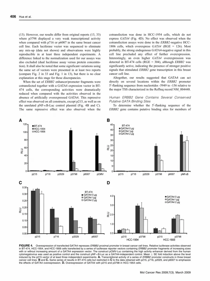
When the set of ERBB2 enhancer/promoter fragments were
cotransfected together with a GATA4 expression vector in BT-
474 cells, the corresponding activities were dramatically
reduced when compared with the activities observed in the
absence of artificially overexpressed GATA4. This repressive
effect was observed on all constructs, except p215, as well as on
the unrelated pNF-nB-Luc control plasmid (Fig. 4B and C).
The same repressive effect was also observed when the
cotransfection was done in HCC-1954 cells, which do not
express GATA4 (Fig. 4D). No effect was observed when the
cotransfection assays were done in the ERBB2-negative HCC-
1806 cells, which overexpress GATA4 (RGE = 126). Most
probably, the strong endogenous GATA4-negative signal in this
cell line precluded any effect of further overexpression.
Interestingly, an even higher GATA4 overexpression was
detected in BT-474 cells (RGE = 304), although ERBB2 was
significantly active, indicating the presence of stronger positive
signals that stimulated ERBB2 gene transcription in this breast
cancer cell line.
Altogether, our results suggested that GATA4 can act
directly on several locations within the ERBB2 promoter
5¶-flanking sequence from nucleotides -5949 to -156 relative to
the major TSS characterized in the RefSeq record NM_004448.
Human ERBB2 Gene Contains Several ConservedPutative GATA Binding Sites
To determine whether the 5¶-flanking sequence of the
ERBB2 gene contains putative binding sites for members of
FIGURE 4. Overexpression of transfected GATA4 represses ERBB2 proximal promoter in breast cancer cell lines. Relative luciferase activities observedin BT-474, HCC-1954, and HCC-1806 cells transfected by a series of luciferase reporter vectors containing ERBB2 promoter fragments of increasing sizeswith or without increasing amount of a GATA4 expression vector. The construct pCMV-Luc containing the high activity enhancer derived from the humancytomegalovirus was used as positive control and the construct pNF-nB-Luc as a GATA4-independent control. Mean F SE fold induction above the levelinduced by the p215 vector of at least three independent experiments. A. Transcriptional activity of a series of ERBB2 promoter constructs in three breastcancer cell lines. B and C. Same series of results in BT-474 cells but restricted in C to the data obtained with p215, p716, p2029, and p6007 to emphasizethe effects of GATA4 overexpression. D. Overexpression of GATA4 with p215 and p3798 in HCC-1954 cells.
Hua et al.
Mol Cancer Res 2009;7(3). March 2009
406

the GATA transcription factor family, a comparison between
human and mouse gene sequences was done combined with a
search for GATA binding sites. Eight conserved regions were
characterized by analysis of the corresponding sequences of the
human and murine ERBB2 genes using the online software
PipMaker6 (Fig. 5A). Several potential binding sites for GATA
factors were identified in seven of eight conserved regions by a
search combining several methods (consensus WGATAR,
Nucleotide Position Weight Matrix, etc.). Interestingly, the
nucleotide [-156, +100] region, corresponding to the minimal
promoter region included in the p215 construct, did not contain
any identifiable GATA consensus site in agreement with the
absence of effect of overexpressed GATA4 in transient
transfection assay. Similarly, the nucleotide [-5446, -5065]
CR3 region did not contain any GATA consensus site, whereas
six GATA putative binding sites were found within CR7 located
at nucleotide [-561, +310].
GATA4 Is Bound In vivo to Several Locations in theERBB2 Gene Enhancer/Promoter Region
In vivo recruitment of GATA4 was tested by chromatin
immunoprecipitation assay in BT-474 cells. After cross-linking
and sonication, the chromatin extract was immunoprecipitated
by a commercial, GATA4-specific mouse monoclonal antibody
and the corresponding DNA fragments were purified as
described previously (35). Portions of the seven characterized
conserved regions containing putative GATA binding sites plus
GATA-less CR3 were amplified by PCR using specific primer
pairs (Table 1) and quantified using SYBR Green chemistry and
a Light Cycler 2 (Roche Diagnostics). As illustrated in Fig. 5B
and C, several conserved regions were bound in vivo by
GATA4 including the proximal regulatory promoter region
included in CR7 and, to a less extent, CR4, CR5, and CR8. As
expected, no significant recruitment of GATA4 was observed
within CR3 devoid of any GATA consensus site but also in
CR1, CR2 and CR6, notwithstanding the presence of bona fide
GATA consensus sites. This confirms that not all consensus
binding sites are accessible in vivo for the recruitment of a
given sequence-specific transcription factor. Altogether, these
results identified in vivo binding of GATA4 in several locations
within the ERBB2 gene enhancer/promoter region, in accor-
dance with a direct role of this transcription factor in the
regulation of human ERBB2 gene transcription.
GATA4 Represses the ERBB2 Gene Enhancer/Promoterthrough a Positive Regulatory Element
Although chromatin immunoprecipitation assays revealed
in vivo recruitment of GATA4 at several locations within the
FIGURE 5. GATA4 is recruited in vivo at several locations of ERBB2 5¶-flanking sequence in BT-474 breast cancer cells. A. Representation of the 5¶-endflanking sequence of the human ERBB2 gene showing the eight conserved regions between Homo sapiens and Mus musculus . Putative GATA binding sitesfound within seven of eight conserved regions. Pointed lines below the schematic map correspond to the fragments amplified by the chromatinimmunoprecipitation-specific primers listed in Table 1. B. Quantitative chromatin immunoprecipitation assays done using a GATA4 monoclonal antibody asdescribed in Materials and Methods. Mean F SE of 3 to 6 independent experiments. C. Representative semiquantitative chromatin immunoprecipitationassays done with the GATA4-less CR3 region and the GATA4 -containing CR7 region using either a GATA4 monoclonal antibody or a matched IgG2a controlas indicated below the lanes.
6 pipmaker.bx.psu.edu
ERBB2/GATA4 Regulatory Loop in Breast Cancer Cells
Mol Cancer Res 2009;7(3). March 2009
407

ERBB2 gene enhancer promoter, they cannot define which
putative GATA binding sites are functional. For example, four
GATA potential binding sites were identified within the 338-bp
fragment nucleotide [-525, -187] amplified by PCR (Fig. 6A).
Among these four putative binding sites, the WGATAR motif
nucleotide [-286, -281] was strictly conserved between human
and mouse. In addition, this GATA consensus was almost
identical to a functional GATA4-specific binding site identified
in the human IL-5 gene promoter (Fig. 5B; ref. 36). Therefore,
we designed a 21-bp oligonucleotide probe covering this
WGATARmotif to perform electrophoretic mobility shift assays
(EMSA) using nuclear extracts from BT-474 cells (Fig. 6B).
Similar migration profiles were observed with both IL-5 and
ERBB2 GATA wild-type probes including five major retarded
protein-DNA complexes (Fig. 6C, lane 2 ; data not shown).
Competition using an excess of unlabeled wild-type double-
stranded oligonucleotides abolished the three C1, C2, and C3
complexes (Fig. 6C, lanes 3 and 5), which were not affected by
the corresponding mutant competitors (Fig. 6C, lanes 4 and 6).
This showed that the three C1, C2, and C3 complexes are specific
to the WGATAR motif present in the ERBB2 GATA probe.
The reaction mixtures were incubated with antibodies
directed against five members of the GATA family (GATA1-4
and GATA6) to determine which of these factors may
contribute to the formation of the specific protein-DNA
complexes. As shown in Fig. 6D (lane 7), only the GATA4-
specific antibodies retarded migration of C2 complex observed
with the wild-type ERBB2 GATA probe. Accordingly, only
GATA4 antibodies slowed down the migration of the
corresponding complex detected using the wild-type IL-5
GATA probe (Fig. 6D, lane 10; data not shown).
To determine whether the bona fide GATA4 binding site at
nucleotide [-286, -281] is transcriptionally activated, this site
was abrogated by site-directed mutagenesis in the luciferase
construct p3798 (Fig. 1C). Wild-type and mutated constructs
were then transiently transfected in BT-474. As illustrated in
Fig. 6E, the transcriptional activity was dramatically reduced in
the p3798 Mut construct. The lack of further repression in the
presence of coexpressed GATA4 evidenced that an intact
WGATAR motif is required for GATA4-mediated repression of
the ERBB2 enhancer/promoter region. No significant differ-
ences were observed when using a low-activity p716 wild-type
and p716 Mut plasmids (data not shown).
Altogether, these results strongly indicate that GATA4
mediates ERBB2 down-regulation through a bona fide
WGATAR motif embedded in a positive regulatory element
required for elevated expression of the ERBB2 gene.
DiscussionThe molecular mechanisms leading to ERBB2 overexpres-
sion in human cancers remain poorly characterized, except for
the well-documented genomic amplification. However, ERBB2
gene amplification alone is apparently not sufficient to explain
all cases of RNA and protein overexpression (6) because a
significant proportion of human cancers with increased
expression of ERBB2 do not exhibit gene amplification
(37-39). There are converging lines of evidence that an increase
of ERBB2 mRNA levels per gene copy depends on ERBB2
gene transcription. Several regulatory elements located near the
major TSS of the ERBB2 gene promoter have been character-
ized previously as well as some more distal elements up to 6 kb
in the 5¶-flanking sequence and in the ERBB2 gene first intron
(8, 12-14, 18-21). Altogether, data suggested their involvement
in the increased transcription of the ERBB2 gene observed in
breast cancer cells. Two of the cognate transcription factors
associated with the previously characterized regulatory ele-
ments have been identified as negative regulators of ERBB2
expression in breast cancer. The ETS DNA-binding protein
PEA3 targets specifically a DNA motif on the ERBB2 gene
promoter and down-regulates its promoter activity, but there is
no clear evidence that genetic lesions of PEA3 can cause
ERBB2 overexpression (17). The transcription factor FOXP3
recently characterized as a X-linked breast cancer tumor
suppressor in mice and humans represses transcription of the
ERBB2 gene via interaction with forkhead DNA-binding motifs
in the ERBB2 promoter (10). Although the functional relation-
ships between these negative and positive transacting factors
Table 1. qRT-PCR Oligonucleotide Primers Specific for the Eight Conserved Regions in ERBB2 Gene and an IrrelevantGenomic Control Region Analyzed by Chromatin Immunoprecipitation
ERBB2 gene Orientation Sequence PCR Product Size (bp)
CR1 Forward 5¶-ATCCTCTCCCTGCTCACCTC-3¶ 164Reverse 5¶-GGCTACTTCTTACTCATTCCAACC-3
CR2 Forward 5¶-TGGTGGAAGTGGGAGTAGAGA-3¶ 223Reverse 5¶-TGAAGCCAAATACAAGTTAGGAAG-3
CR3 Forward 5¶-GTTCCTCGTCTCCTCTTCCTTG-3¶ 324Reverse 5¶-TAGCTTTGCTTTGCCACCTG-3
CR4 Forward 5¶-CTGCCCCTTTGCTGTCCT-3¶ 167Reverse 5¶-AGGCTTGAGGTGTCCCTTTG-3
CR5 Forward 5¶-ATCTCAAGGCTCAAGGTTCCTC-3¶ 323Reverse 5¶-TCCAGGAGTCACTGGTTCATC-3
CR6 Forward 5¶-AGCACATGGAAGCAAGTTTAG-3¶ 163Reverse 5¶-CCCAGCCAAGAATGCAG-3
CR7 Forward 5¶-TCCTTTCGATGTGACTGTCTCC-3¶ 339Reverse 5¶-CTAAATGCAGAGGCTGGTGACT-3
CR8 Forward 5¶-GCTTAGGGACTGTGCTCTGTG-3¶ 220Reverse 5¶-TGGACAGATGGGTCAGGATAC-3
Control Forward 5¶-ATGGTTGCCACTGGGGATCT-3¶ 174Reverse 5¶-TGCCAAAGCCTAGGGGAAGA-3
Hua et al.
Mol Cancer Res 2009;7(3). March 2009
408

remain largely unexplored, these results illustrate altogether the
complexity of ERBB2 gene transcriptional regulation that
requires further investigation. In this context, we provide here
evidence that the transcription factor GATA4 can also act as a
transcriptional repressor of the ERBB2 gene in breast cancer
cells through its direct binding to ERBB2 regulatory sequences.
Interestingly, the three best characterized transrepressors of
the ERBB2 gene (FOXP3, PEA3, and GATA4) appear to act
through three positive regulatory elements embedding their
cognate binding sites (see Fig. 3E and G in ref. 10, Fig. 3A in
ref. 17, and Fig. 6D and E). This suggests the intervention of
unidentified transactivators acting on these bifunctional regula-
tory elements. Accordingly, our EMSA has evidenced the
presence of three specific protein-DNA complexes, whereas only
the C2 complex was displaced by GATA4-specific antibodies,
suggesting the presence of other sequence-specific transcription
FIGURE 6. GATA4 represses ERBB2 promoter activity though a bona fide GATA4 binding site acting as a positive regulatory DNA motif. A. Map of theregion CR7 located at nucleotide [-561, +310] showing the previously reported regulatory elements (see Fig. 1A for details) as well as the 6 putative GATAbinding sites (G) identified in silico . Closed square, conserved WGATAR motif located at nucleotide [-286, -281]. Major TSS (+1 ) and the first exon (graybox ). The fragment located at nucleotide [-525, -187] amplified by PCR for the chromatin immunoprecipitation assays and the oligonucleotide probenucleotide [-294, -274] used for the EMSA are shown below the map line as well as the symbols corresponding to the regulatory elements located on the mapline. B. Sequences of the double-stranded oligonucleotide probes used for EMSA corresponding to the conserved putative GATA4 motif of the ERBB2 gene(ERBB2-GATA4 -Wt) and the functional GATA4 binding site of the human IL-5 promoter described previously (ref. 36; IL5-GATA4 -Wt) and their matchedinactive substitution mutants (ERBB2-GATA4 -Mut and IL5-GATA4 -Mut, respectively). C. EMSA using BT-474 nuclear extracts and the radiolabeled ERBB2-GATA4 -Wt probe in the absence (lane 2) or presence of a 50-fold molar excess of wild-type (lanes 3 and 5) and mutated (lanes 4 and 6) unlabeled double-stranded competitors as indicated above the lanes. Lane 1, migration of the radiolabeled probe in the absence of nuclear cell extract. Right, migration of thethree specific protein-DNA complexes C1, C2, and C3 (closed arrowheads ). D. Characterization of the ERBB2 -GATA4 and IL-5 -GATA4 motif bindingproteins. BT-474 nuclear extracts were incubated 15 min at 4jC in the absence (lanes 3 and 9) or presence of GATA-specific antibodies (lanes 4-8 and 10)before addition of the radiolabeled ERBB2-GATA4 and IL5-GATA4 -Wt probes (lanes 1-8 and 9-10 , respectively) as indicated above the lanes. Lanes 1 and2, migration of the radiolabeled probe ERBB2-GATA4 -Wt without nuclear cell extract in the absence or presence of GATA4 monoclonal antibody. Right,migration of the GATA4-containing complex C2 and its shifted position in the presence of GATA4 antibody (closed and open arrowheads , respectively). E.Luciferase reporter gene activity driven by either wild-type or a mutated p3798 construct (Fig. 1C) transiently transfected in BT-474 cells in absence orpresence of overexpressed GATA4. Percentage F SE luciferase activity relative to the activity of the p3798T without overexpressed GATA4 defined as 100.
ERBB2/GATA4 Regulatory Loop in Breast Cancer Cells
Mol Cancer Res 2009;7(3). March 2009
409

factors. The elucidation of the underlying molecular mechanisms
requires further investigation to identify the transactivators
associated to these bifunctional regulatory elements and their
role in the dysregulations of ERBB2 in cancer cells.
Our siRNA assays excluded an indirect effect of GATA4 via
a putative action on the expression of the FOXP3 gene because
GATA4 siRNA did not affect significantly FOXP3 RNA levels.
We also wondered whether cofactors of the FOG family that
modulate the activity of the GATA transcription factors in
mammals (40) might be involved. Although a role of the
GATA4 partner FOG2/ZFPM2 (41) appears excluded in BT-
474 cell line, which does not overexpress the corresponding
gene (data not shown), an involvement of FOG1/ZFPM1 in
GATA4-dependent repression of ERBB2 is a worth investigat-
ing possibility (42).
A repressor role for GATA4 is in agreement with previous
observations on this transcription factor, which can act as both a
transcriptional activator and a repressor depending on various
factors such as the target gene, the cell type, the interactions
with other sequence-specific transcription factors, or even the
localization of its binding site relative to the core promoter. For
example, GATA4 activates the gene coding for P450c17 by
direct interaction with SP1 (34), the Grp78/BiP gene in
cooperation with ATF6 in embryonic heart (43), as well as the
IL-5 gene in T lymphocytes (36). In contrast, GATA4 acts as a
repressor of the a2(I) collagen (COL1A2) via its binding to
both proximal promoter and intronic elements (44, 45).
Furthermore, GATA4 binds two sites of the FGF3 promoter
acting as positive and negative regulatory elements (46).
Inhibition of GATA4 and FOXP3 by ERBB2-targeted siRNA
enlarges the list of transcription factors involved in ERBB2
signaling, which already include multiple factors such as FOS/
JUN, EGR1, MYC, ELK, and SP1 (30). Altogether, these
results and our own observations show that the transcriptional
control of ERBB2 is much more complex than expected and
require further detailed investigation before designing any
credible or risk-limited ‘‘transcriptional’’ therapy directed
against either ERBB2 regulatory elements and/or their cognate
sequence-specific transcription factors.
GATA4 transcriptional activity is activated by the mitogen-
activated protein kinase pathway in cardiomyocytes (29) and
the ERBB family triggers several signaling pathways including
this pathway (2). Although the activation of GATA4 by the
mitogen-activated protein kinase pathway subsequent to
ERBB2 overexpression in breast cancer cells requires further
exploration, it was reported that GATA4 gene expression is
induced by ERBB4 (47). Because GATA4 negatively regulates
ERBB2 expression, an ERBB2/ERBB4/GATA4 regulatory loop
might explain why co-overexpression of ERBB4 with ERBB2
in mammary carcinoma is associated with a more favorable
clinical outcome than overexpression of ERBB2 alone (48).
Similarly, the regulatory networks that associate the multiple
transcription factors activated by the ERBB2 receptor and the
ERBB2 gene itself need extensive investigation to determine
which are direct targets and what are the putative regulatory
loops linking these genes.
Trastuzumab, a humanized anti-ERBB2 antibody, is a major
therapeutic agent for patients with overexpressed ERBB2
receptor on cancer cells. This treatment blocks the activity of
the receptor by multiple and partially defined mechanisms, but
resistance to trastuzumab is frequent (3). Some secondary
cardiac effects have also been observed with this agent and
more particularly when associated with anthracyclines (4, 5,
49). Studies in animals and cell culture have provided some
insight into the mechanisms of trastuzumab-induced decrease of
cardiac contractile function (for review, see ref. 49). Gene
targeting studies and conditional deletion of ERBB2 in mice
have shown that ERBB2 is essential for maintenance of normal
cardiac structure and function. Collectively, published data
suggest that one role of ERBB2/4 signaling is to dynamically
regulate sarcomere structure. In contrast, overexpression of
the transcription factor GATA4, known to be important in the
regulation of cardiac sarcomeric protein expression, protects
the heart against anthracycline toxicity in a mouse model (50).
It remains to establish whether the ERBB2/GATA4 regulatory
loop, solely characterized here in a breast cancer cell model, has
any role in mature heart before trying to elucidate these
apparently opposite outcomes.
Considering the major role played by ERBB2 and GATA4
in the development of cardiovascular system (26, 51), our
results provide some new clues to further investigate the
molecular basis of trastuzumab cardiotoxicity. The complex
transcriptional regulation of the ERBB2 gene and the
transcriptional regulatory loop that associates this epidermal
growth factor receptor and the cardiac-specific transcription
factor GATA4 may indeed interfere with trastuzumab therapy.
A precise and complete definition of these regulatory
mechanisms is required for better understanding and fighting
resistance to treatments targeting ERBB2 as well as limiting
their cardiotoxicity.
Materials and MethodsCell Lines and Breast Carcinoma Samples
The nononcogenic human primary mammary epithelium
cells HME-1 (Clontech) and the breast cancer cell lines BT-474,
BT-483, HCC-202, HCC-1569, HCC-1806, HCC-1954, MDA-
MB-175, MDA-MB-361, MDA-MB-453, SK-BR-3, UACC-
812, ZR-75-30, and Br-Ca-Mz-01 (American Type Culture
Collection7) and SUM-185, SUM-190, SUM-206, and SUM-
225 (University of Michigan8; ref. 52) were grown according to
the recommendations of the supplier. Seventeen breast
carcinoma samples were obtained from women treated at
Institut Paoli-Calmettes. ERBB2 status of each sample was
previously measured by comparative genomic hybridization
array, transcriptome analysis, or immunohistochemistry
(Table 2). Twelve breast carcinoma samples were ERBB2
positive, whereas 5 samples were ERBB2 negative according to
these criteria.
Plasmids and Transient Transfection ExperimentsThe ERBB2 reporter vectors containing promoter fragments
of increasing sizes (13, 33) and GATA4 expression vector (34)
were kindly provided by Dr. Rosita Winkler (Molecular
Oncology Laboratory, University of Liege) and Dr. Walter L.
7 http://www.atcc.org8 http://www.cancer.med.umich.edu/breast_cell/production
Hua et al.
Mol Cancer Res 2009;7(3). March 2009
410

Miller (Department of Pediatrics and The Metabolic Research
Unit, University of California at San Francisco), respectively.
The pCMV-Luc and pNF-nB-Luc (Clontech) plasmids were
used as positive controls. Cells were transfected using FuGENE
6 reagent (Roche Diagnostics). Cells (1 � 105-3 � 105) were
seeded in six-well plates (Falcon 3046; BD Biosciences) and
treated with a FuGENE/DNA ratio of 3 AL/2 Ag for 48 h in
complete medium. Sample (1 Ag) of each reporter construct wascotransfected with 0.4 Ag Renilla plasmid (pTK-RL; Promega)
and 0.6 Ag pUC19. Cells were harvested and lysed and
luciferase activities were measured according to the manufac-
turer’s instructions (dual luciferase reporter gene assay kit;
Promega). ERBB2 reporter vector activity was calculated as the
ratio of firefly luciferase activity to Renilla luciferase activity
and then expressed for each construct as the ratio to the minimal
ERBB2 promoter vector (p215). GATA4 expression vector
(0-0.2 Ag) was cotransfected with 1 Ag sample of each reporter
construct, 0.4 Ag pTK-RL, 0.3 Ag pUC19, and 0.3 to 0.1 AgpNF-nB-Luc. Transfections were done in triplicate and repeatedat least in three independent experiments.
RNAi AssayThree 25-mer duplex siRNA to target ERBB2 and GATA4 ,
respectively, were obtained from a commercial source (Stealth
select RNAi; Invitrogen). All siRNA duplexes (10 nmol/L) and
Stealth RNAi Negative Control duplexes were transfected to
3 � 105 cells in six-well plates by Lipofectamine RNAiMAX
(Invitrogen) for 48 h at 37jC in a CO2 incubator according to
the manufacturer’s instructions. Gene knockdown was con-
firmed by qRT-PCR and immunoblotting. Inhibition effects
were observed from all siRNA duplexes and the siRNA that
induced the highest inhibition were used in three independent
experiments.
qRT-PCRTotal RNA from cell lines was extracted using RNeasy
Micro Kit (Qiagen) according to the manufacturer’s protocol.
Total RNA from tumor samples was extracted as described
previously (53). RNA was quantified using a Nanodrop 1000
spectrophotometer device. RT-PCR was done using SuperScript
II reverse transcriptase (Invitrogen) and random hexamer
primers. DNA was quantified using LC FastStart DNA Master
SYBR Green I and read with a Light Cycler 2 instrument
(Roche Diagnostics) according to the manufacturer’s instruc-
tions. The primers used for qRT-PCR analysis are listed in
Table 3. The precise amount of total cDNA added to each
reaction mix and its quality are both generally difficult to
Table 3. qRT-PCR Oligonucleotide Primers Used to Determine the Relative RNA Expression Level of TBP, GATA4, ERBB2, andFOXP3 Genes
mRNA Orientation Sequence Product Size (bp)
TBP Forward 5¶-CTTGTGCTCACCCACCAAC-3¶ 228Reverse 5¶-GGAGGCAAGGGTACATGAGA-3
GATA4 Forward 5¶-TCCAAACCAGAAAACGGAAG-3¶ 224Reverse 5¶-CATCGCACTGACTGAGAACG-3
ERBB2 Forward 5¶-GGGAAACCTGGAACTCACCT-3¶ 216Reverse 5¶-CAGGGGTGGTATTGTTCAGC-3
FOXP3 Forward 5¶-TACTTCAAGTTCCACAACATGCGACC-3¶ 202Reverse 5¶-CGCACAAAGCACTTGTGCAGACTCAG-3
Table 2. ERBB2 and GATA4 Gene Copy Numbers in Breast Cancer Carcinoma Biopsies and Cell Lines
Tumor Samples Comparative Genomic Hybridization Gene Copies Cell lines Comparative Genomic Hybridization Gene Copies
ERBB2 GATA4 ERBB2 GATA4
9934 7 2 BT-474 26 27462 7 1 BT-483 2 112710 9 1 MDA-MB-453 5 113591 8 2 HCC-1806 2 28035 2 1 ZR-75-30 30 12933 4 1 MDA-MB-175 2 17780 4 1 UACC-812 24 113008 17 1 HCC-1569 50 19948 21 1 SUM-225 104 112781 15 1 SUM-185 1 18584 7 2 SK-BR-3 17 110982 2 2 SUM-206 2 19983 6 1 MDA-MB-361 6 19725 4 2 SUM-190 72 16604S 2 1 HCC-202 14 19840 2 2 HCC-1954 71 19745 2 1 Br-Ca-Mz-01 2 2
HME-1 2 2
ERBB2/GATA4 Regulatory Loop in Breast Cancer Cells
Mol Cancer Res 2009;7(3). March 2009
411

assess. Therefore, the relative expression level of the gene of
interest was computed with respect to the internal standard TBP
to normalize for variations in RNA quality and the amount of
input cDNA. Threshold cycles (Ct) were determined for
quantification of the input target number.
For comparison between different cell lines and tumor
samples, the target gene expression level (ERBB2 or GATA4) in
each cell line and tumor samples was normalized using a
calibrator RNA sample extracted from the HME-1 cell line. For
RNAi assay, the RGE was calculated as a percentage of the
measured mRNA level in cells transfected with target siRNA
compared with cells transfected with matched control siRNA.
Data were normalized against the TBP signal and the 2-DDCt
method was used to calculate the relative expression level of a
given gene (54).
ImmunoblottingFor immunoblotting, 105 BT-474 cells were lysed in 10 AL
lysis buffer [25 mmol/LTris (pH 7.9), 1% (w/v) SDS, 1 mmol/L
DTT] and fractionated on a 7.5% SDS-polyacrylamide gel and
electrotransferred to a polyvinylidene fluoride membrane.
Specific polypeptides were revealed with the rabbit polyclonal
anti-ERBB2 (Santa Cruz Biotechnology), mouse monoclonal
anti-GATA4 (Santa Cruz Biotechnology) at a dilution of 1:200
in TBS-Tween 20 containing 5% nonfat milk, and mouse
monoclonal anti-h-tubulin (Abcam) at a dilution of 1:1,000 in
TBS-Tween 20 containing 5% bovine serum albumin and then
reacted with secondary antibodies at a dilution of 1:104 in the
same buffer.
Chromatin Immunoprecipitation AssayThe chromatin immunoprecipitation assay was done using
the Chromatin Immunoprecipitation Assay kit (Upstate). In
brief, 2 � 106 BT-474 cells were treated by 1% formaldehyde
for 15 min at 37jC, and the cross-linking reaction was stopped
by the addition of 125 mmol/L glycine incubating 10 min at
room temperature. Cells were rinsed twice with ice-cold
1� PBS containing protease inhibitors (Complete Protease
Inhibitor Cocktail; Roche Diagnostics), scraped, and collected.
After centrifugation, the cell pellet was lysed 10 min on ice in
SDS lysis buffer supplemented with protease inhibitor cocktail.
The chromatin was sheared by sonication to an average DNA
length of 200 to 1,000 bp using a Sonics ultrasonic processor
(750 W). The sonicated chromatin supernatant was diluted with
Dilution Buffer containing protease inhibitor cocktail and
subjected to a 30 min preclearing by incubation with salmon
sperm DNA/protein A agarose-50% slurry beads at 4jC under
rotation. The beads were pelleted, and the precleared chromatin
supernatant was immunoprecipitated with 3 Ag of either a
matched nonimmune IgG control (mouse IgG2a; BD Pharmin-
gen) or a specific antibody (mouse monoclonal anti-GATA4;
Santa Cruz Biotechnology) at 4jC overnight under rotation.
Immunocomplexes were collected by 1 h incubation with
salmon sperm DNA/protein A agarose-50% slurry at 4jC under
rotation. Complexes were washed once, sequentially, in a low-
salt buffer, high-salt buffer, and low LiCl buffer and twice in TE
buffer. The antibody-protein-DNA complexes were eluted twice
in 250 AL elution buffer (0.1 mol/L NaHCO3, 1% SDS) by
medium power vortexing and rotation for 15 min at room
temperature. The combined eluates as well as the input sample
(1% of the amount used in the immunoprecipitation procedure)
were cross-link reversed by heating at least 4 h at 65jC in the
presence of 200 mmol/L NaCl. After proteinase K digestion,
the DNA fragments were extracted using phenol/chloroform
and ethanol precipitated. qRT-PCR using LC FastStart DNA
Master SYBR Green I and Light Cycler 2 instrument (Roche
Diagnostics) and semiquantitative PCR were done with selected
primer pairs (Table 1). For each experimental sample, the
amount of target and endogenous reference was determined
from a standard curve. The standard curve was constructed with
4-fold serial dilutions of input from the BT-474. The results
from specific antibodies were reported to that observed in the
corresponding IgG samples. A region between the GAPDH and
CNAP1 genes was used as the negative control to standardize
the results from all the conserved regions (ChIP-It protocol;
Active Motif).
EMSAComplementary oligonucleotides carrying a GATA motif
related to the GATA4-specific binding site of the IL-5 gene
promoter (36) and corresponding to nucleotide [-294, -274] of
the wild-type ERBB2 promoter (5¶-AAAGTTTAAGA-
TAAAACCTGA-3¶ and 3¶-TTTCAAATTCTATATTGGACT-5¶) was labeled with [g-32P]ATP and T4 kinase. Labeled
oligonucleotides were annealed and purified by Chroma Spin
Columns (STE buffer; Clontech). Nuclear extracts from BT-474
cells (16.8 Ag proteins) were incubated for 5 min at 20jC with
0.25 Ag poly[(dI-dC)/(dI-dC)], 5 mmol/L HEPES, 1 mmol/L
KCl, 49 mmol/L NaCl, 1 mmol/L EDTA, 5 mmol/L DTT, 4%
glycerol, 1% Ficoll, 50 pg/mL pUC19, and 50-fold molar
excess of unlabeled wild-type or mutated GATA binding site
double-stranded competitors (5¶-AAAGTTTAGTCGA-
CAACCTGA-3¶ and 3¶-TTTCAAATCAGCTGTTGGACT-5¶).Radiolabeled probe (0.5 ng; 5 � 105 cpm) was then added to
the reaction mixture for 10 min at 20jC. The complexes
were resolved on a 5% polyacrylamide gel in TBE buffer
[0.025 mol/LTris base, 0.022 mol/L boric acid, and 0.5 mmol/L
EDTA (pH 8.3)] at 200 V. The gel was dried and analyzed by
autoradiography. Supershift assay was done by incubating
antibodies against GATA1 to GATA4 and GATA6, before the
addition of radiolabeled probe, with the reaction mixture for
15 min at 4jC.
Site-Directed MutagenesisSite-directed mutagenesis was carried out using QuikChange
II Site-Directed Mutagenesis Kit (Stratagene). PCR was done
in ERBB2 reporter vectors using following primers: forward
5¶-gatgcaagctccccaggaaagtttagtcgacaacctgagacttaaaagggtgt-3¶and reverse 5¶-acacccttttaagtctcaggttgtcgactaaactttcctggg-gagcttgcatc-3¶. PCR products were digested by DpnI restriction
enzyme and transformed in XL1-Blue supercompetent cells.
Mutated ERBB2 luciferase reporter vectors were confirmed by
sequencing.
Disclosure of Potential Conflicts of InterestNo potential conflicts of interest were disclosed.
Hua et al.
Mol Cancer Res 2009;7(3). March 2009
412

AcknowledgmentsWe thank Dr. Rosita Winkler for the luciferase reporter vectors containingERBB2 gene promoter regions and Dr. Walter L. Miller for the GATA4expression vector.
References1. Akiyama T, Sudo C, Ogawara H, Toyoshima K, Yamamoto T. The product ofthe human c-erbB-2 gene: a 185-kilodalton glycoprotein with tyrosine kinaseactivity. Science 1986;232:1644–6.
2. Yarden Y, Sliwkowski MX. Untangling the ErbB signalling network. Nat RevMol Cell Biol 2001;2:127– 37.
3. Nahta R, Esteva FJ. Herceptin: mechanisms of action and resistance. CancerLett 2006;232:123– 38.
4. Guarneri V, Lenihan DJ, Valero V, et al. Long-term cardiac tolerability oftrastuzumab in metastatic breast cancer: the M.D. Anderson Cancer Centerexperience. J Clin Oncol 2006;24:4107– 15.
5. Pugatsch T, Abedat S, Lotan C, Beeri R. Anti-erbB2 treatment inducescardiotoxicity by interfering with cell survival pathways. Breast Cancer Res 2006;8:R36.
6. Kraus MH, Popescu NC, Amsbaugh SC, King CR. Overexpression of theEGF receptor-related proto-oncogene erbB-2 in human mammary tumor cell linesby different molecular mechanisms. EMBO J 1987;6:605 –10.
7. Hurst HC. Update on HER-2 as a target for cancer therapy: the ERBB2promoter and its exploitation for cancer treatment. Breast Cancer Res 2001;3:395– 8.
8. Vernimmen D, Gueders M, Pisvin S, Delvenne P, Winkler R. Differentmechanisms are implicated in ERBB2 gene overexpression in breast and in othercancers. Br J Cancer 2003;89:899 –906.
9. Li M, Wang Y, Hung MC, Kannan P. Inefficient proteasomal-degradationpathway stabilizes AP-2A and activates HER-2/neu gene in breast cancer. Int JCancer 2006;118:802 –11.
10. Zuo T, Wang L, Morrison C, et al. FOXP3 is an X-linked breast cancersuppressor gene and an important repressor of the HER-2/ErbB2 oncogene. Cell2007;129:1275–86.
11. Nezu M, Sasaki H, Kuwahara Y, et al. Identification of a novel promoterand exons of the c-ERBB-2 gene. Biochem Biophys Res Commun 1999;258:499– 505.
12. Begon DY, Delacroix L, Vernimmen D, Jackers P, Winkler R. Yin Yang 1cooperates with activator protein 2 to stimulate ERBB2 gene expression inmammary cancer cells. J Biol Chem 2005;280:24428–34.
13. Delacroix L, Begon D, Chatel G, Jackers P, Winkler R. Distal ERBB2promoter fragment displays specific transcriptional and nuclear binding activitiesin ERBB2 overexpressing breast cancer cells. DNA Cell Biol 2005;24:582–94.
14. Vernimmen D, Begon D, Salvador C, Gofflot S, Grooteclaes M, Winkler R.Identification of HTF (HER2 transcription factor) as an AP-2 (activator protein-2)transcription factor and contribution of the HTF binding site to ERBB2 geneoverexpression. Biochem J 2003;370:323 –9.
15. Ishii S, Imamoto F, Yamanashi Y, Toyoshima K, Yamamoto T. Character-ization of the promoter region of the human c-erbB-2 protooncogene. Proc NatlAcad Sci U S A 1987;84:4374–8.
16. Tal M, King CR, Kraus MH, Ullrich A, Schlessinger J, Givol D. HumanHER2 (neu ) promoter: evidence for multiple mechanisms for transcriptionalinitiation. Mol Cell Biol 1987;7:2597 –601.
17. Xing X, Wang SC, Xia W, et al. The ets protein PEA3 suppresses HER-2/neuoverexpression and inhibits tumorigenesis. Nat Med 2000;6:189– 95.
18. Grooteclaes M, Vernimmen D, Plaza S, Pasleau F, Hodzic D, Winkler-Gol R.A new cis element is involved in the HER2 gene overexpression in human breastcancer cells. Cancer Res 1999;59:2527 –31.
19. Bates NP, Hurst HC. An intron 1 enhancer element mediates oestrogen-induced suppression of ERBB2 expression. Oncogene 1997;15:473– 81.
20. Bosher JM, Totty NF, Hsuan JJ, Williams T, Hurst HC. A family of AP-2proteins regulates c-erbB-2 expression in mammary carcinoma. Oncogene 1996;13:1701 –7.
21. Bosher JM, Williams T, Hurst HC. The developmentally regulatedtranscription factor AP-2 is involved in c-erbB-2 overexpression in humanmammary carcinoma. Proc Natl Acad Sci U S A 1995;92:744–7.
22. Scott GK, Chang CH, Erny KM, et al. Ets regulation of the erbB2 promoter.Oncogene 2000;19:6490 –502.
23. Bates NP, Hurst HC. Transcriptional regulation of type I receptor tyrosinekinases in the mammary gland. J Mammary Gland Biol Neoplasia 1997;2:153– 63.
24. Bertucci F, Viens P, Hingamp P, Nasser V, Houlgatte R, Birnbaum D. Breastcancer revisited using DNA array-based gene expression profiling. Int J Cancer2003;103:565 –71.
25. Bertucci F, Borie N, Ginestier C, et al. Identification and validationof an ERBB2 gene expression signature in breast cancers. Oncogene 2004;23:2564– 75.
26. Negro A, Brar BK, Lee KF. Essential roles of Her2/erbB2 in cardiacdevelopment and function. Recent Prog Horm Res 2004;59:1 –12.
27. Kuo CT, Morrisey EE, Anandappa R, et al. GATA4 transcription factor isrequired for ventral morphogenesis and heart tube formation. Genes Dev 1997;11:1048– 60.
28. Molkentin JD, Lin Q, Duncan SA, Olson EN. Requirement of thetranscription factor GATA4 for heart tube formation and ventral morphogenesis.Genes Dev 1997;11:1061–72.
29. Liang Q, Wiese RJ, Bueno OF, Dai YS, Markham BE, Molkentin JD. Thetranscription factor GATA4 is activated by extracellular signal-regulated kinase1- and 2-mediated phosphorylation of serine 105 in cardiomyocytes. Mol CellBiol 2001;21:7460 –9.
30. Citri A, Yarden Y. EGF-ERBB signalling: towards the systems level. Nat RevMol Cell Biol 2006;7:505 –16.
31. Yuen T, Wurmbach E, Pfeffer RL, Ebersole BJ, Sealfon SC. Accuracy andcalibration of commercial oligonucleotide and custom cDNA microarrays.Nucleic Acids Res 2002;30:e48.
32. Park PJ, Cao YA, Lee SY, et al. Current issues for DNA microarrays:platform comparison, double linear amplification, and universal RNA reference.J Biotechnol 2004;112:225 –45.
33. Grooteclaes M, Pasleau F, Dijkmans H, Berzi P, Albert A, Winkler-Gol R.The 6-kilobase c-erbB2 promoter contains positive and negative regulatoryelements functional in human mammary cell lines. Cancer Res 1994;54:4193– 9.
34. Fluck CE, Miller WL. GATA-4 and GATA-6 modulate tissue-specifictranscription of the human gene for P450c17 by direct interaction with Sp1. MolEndocrinol 2004;18:1144 –57.
35. Yeh JH, Lecine P, Nunes JA, et al. Novel CD28-responsive enhanceractivated by CREB/ATF and AP-1 families in the human interleukin-2 receptor a-chain locus. Mol Cell Biol 2001;21:4515 –27.
36. Yamagata T, Nishida J, Sakai R, et al. Of the GATA-binding proteins, onlyGATA-4 selectively regulates the human interleukin-5 gene promoter ininterleukin-5-producing cells which express multiple GATA-binding proteins.Mol Cell Biol 1995;15:3830 –9.
37. Bofin AM, Ytterhus B, Martin C, O’Leary JJ, Hagmar BM. Detection andquantitation of HER-2 gene amplification and protein expression in breastcarcinoma. Am J Clin Pathol 2004;122:110–9.
38. Jimenez RE, Wallis T, Tabasczka P, Visscher DW. Determination of Her-2/Neu status in breast carcinoma: comparative analysis of immunohistochemistryand fluorescent in situ hybridization. Mod Pathol 2000;13:37– 45.
39. Todorovic-Rakovic N, Jovanovic D, Neskovic-Konstantinovic Z, Nikolic-Vukosavljevic D. Comparison between immunohistochemistry and chromogenicin situ hybridization in assessing HER-2 status in breast cancer. Pathol Int 2005;55:318–23.
40. Cantor AB, Orkin SH. Coregulation of GATA factors by the friend of GATA(FOG) family of multitype zinc finger proteins. Semin Cell Dev Biol 2005;16:117–28.
41. Svensson EC, Tufts RL, Polk CE, Leiden JM. Molecular cloning of FOG-2: amodulator of transcription factor GATA-4 in cardiomyocytes. Proc Natl Acad SciU S A 1999;96:956–61.
42. Johnson KD, Boyer ME, Kang JA, Wickrema A, Cantor AB, Bresnick EH.Friend of GATA-1-independent transcriptional repression: a novel mode ofGATA-1 function. Blood 2007;109:5230–3.
43. Mao C, Tai WC, Bai Y, Poizat C, Lee AS. In vivo regulation of Grp78/BiPtranscription in the embryonic heart: role of the endoplasmic reticulum stressresponse element and GATA-4. J Biol Chem 2006;281:8877–87.
44. Antoniv TT, Tanaka S, Sudan B, et al. Identification of a repressor in the firstintron of the human a2(I) collagen gene (COL1A2). J Biol Chem 2005;280:35417 –23.
45. Wang L, Tanaka S, Ramirez F. GATA-4 binds to an upstream element of thehuman a2(I) collagen gene (COL1A2) and inhibits transcription in fibroblasts.Matrix Biol 2005;24:333 –40.
46. Murakami A, Ishida S, Dickson C. GATA-4 interacts distinctively withnegative and positive regulatory elements in the Fgf-3 promoter. Nucleic AcidsRes 2002;30:1056 –64.
47. Amin DN, Perkins AS, Stern DF. Gene expression profiling of ErbB receptorand ligand-dependent transcription. Oncogene 2004;23:1428 –38.
ERBB2/GATA4 Regulatory Loop in Breast Cancer Cells
Mol Cancer Res 2009;7(3). March 2009
413

48. Suo Z, Risberg B, Kalsson MG, et al. EGFR family expression in breastcarcinomas. c-erbB-2 and c-erbB-4 receptors have different effects on survival.J Pathol 2002;196:17–25.
49. Peng X, Chen B, Lim CC, Sawyer DB. The cardiotoxicology ofanthracycline chemotherapeutics: translating molecular mechanism into preven-tative medicine. Mol Interv 2005;5:163 –71.
50. Aries A, Paradis P, Lefebvre C, Schwartz RJ, Nemer M. Essential role ofGATA-4 in cell survival and drug-induced cardiotoxicity. Proc Natl Acad SciU S A 2004;101:6975–80.
51. Patient RK, McGhee JD. The GATA family (vertebrates and invertebrates).Curr Opin Genet Dev 2002;12:416– 22.
52. Ethier SP, Mahacek ML, Gullick WJ, Frank TS, Weber BL. Differentialisolation of normal luminal mammary epithelial cells and breast cancer cellsfrom primary and metastatic sites using selective media. Cancer Res 1993;53:627– 35.
53. Theillet C, Adelaide J, Louason G, et al. FGFRI and PLAT genes and DNA
amplification at 8p12 in breast and ovarian cancers. Genes Chromosomes Cancer1993;7:219– 26.
54. Livak KJ, Schmittgen TD. Analysis of relative gene expression datausing real-time quantitative PCR and the 2(-DDC(T)) method. Methods 2001;25:402 –8.
55. Chen Y, Gill GN. Positive and negative regulatory elements in the humanerbB-2 gene promoter. Oncogene 1994;9:2269– 76.
56. Hudson LG, Ertl AP, Gill GN. Structure and inducible regulation of thehuman c-erb B2/neu promoter. J Biol Chem 1990;265:4389–93.
57. Scott GK, Daniel JC, Xiong X, Maki RA, Kabat D, Benz CC. Bindingof an ETS-related protein within the DNase I hypersensitive site of theHER2/neu promoter in human breast cancer cells. J Biol Chem 1994;269:19848 –58.
58. Balda MS, Matter K. The tight junction protein ZO-1 and aninteracting transcription factor regulate ErbB-2 expression. EMBO J 2000;19:2024–33.
Hua et al.
Mol Cancer Res 2009;7(3). March 2009
414


