Embed Size (px)
Citation preview
A Mouse Model of Blast-Induced mild Traumatic Brain Injury
Vardit Rubovitch, PhDa, Meital Ten-Bosch, BSca, Ofer Zohar, PhDb, Catherine R. Harrison,PhDc, Catherine Tempel-Brami, PhDd, Elliot Stein, PhDe, Barry J. Hoffer, MD, PhDe, CareyD. Balaban, PhDf, Shaul Schreiber, MDg, Wen-Ta Chiu, MD, Ph.Dh, and Chaim G. Pick, PhDa
aDepartment of Anatomy and Anthropology, Sackler Faculty of Medicine, Tel-Aviv University, Tel-Aviv, 69978 IsraelbBlanchette Rockefeller Neurosciences Institute, at the Johns Hopkins University MontgomeryCounty Campus, 9601 Medical Center Drive, Rockville MD 20850 USAcAir Force Research Laboratory, 711th HPW/RHPA, Wright Patterson AFB, OH 45433 USAdAlfredo Federico Strauss Center for Computational Neuro-Imaging, Tel Aviv University, Tel-AvivUniversity, Tel-Aviv, 69978 IsraeleNational Institute on Drug Abuse, IRP, 251 Bayview Boulevard, Baltimore, MD 21224 USAfDepartments of Otolaryngology, Neurobiology, Communication Sciences & Disorders, 107 Eye &Ear Institute, 203 Lothrop Street, Pittsburgh, PA 15213 USAgDepartment of Psychiatry, Tel Aviv Sourasky Medical Center and Sackler Faculty of Medicine,Tel-Aviv University, Tel-Aviv, 64239 IsraelhDepartment of Neurosurgery, Taipei Medical University, 250 Wu-Hsing Street, Taipei, Taiwan
AbstractImprovised explosive devices (IEDs) are one of the main causes for casualties among civilians andmilitary personnel in the present war against terror. Mild traumatic brain injury from IEDs inducesvarious degrees of cognitive, emotional and behavioral disturbances but knowledge of the exactbrain pathophysiology following exposure to blast is poorly understood. The study was aimed atestablishing a murine model for a mild BI-TBI that isolates low-level blast pressure effects to thebrain without systemic injuries. An open-field explosives detonation was used to replicate, asclosely as possible, low-level blast trauma in the battlefield or at a terror-attack site. No alterationsin basic neurological assessment or brain gross pathology were found acutely in the blast-exposedmice. At 7 days post blast, cognitive and behavioral tests revealed significantly decreasedperformance at both 4 and 7 meters distance from the blast (5.5 and 2.5 PSI, respectively). At 30days post-blast, clear differences were found in animals at both distances in the object recognitiontest, and in the 7 m group in the Y maze test. Using MRI, T1 weighted images showed anincreased BBB permeability one month post-blast. DTI analysis showed an increase in fractionalanisotropy (FA) and a decrease in radial diffusivity. These changes correlated with sites of up-regulation of manganese superoxide dismutase 2 in neurons and CXC-motif chemokine receptor 3around blood vessels in fiber tracts. These results may represent brain axonal and myelinabnormalities. Cellular and biochemical studies are underway in order to further correlate the
Correspondence Address: Dr. Barry J Hoffer, Intramural Research Program, National Institute on Drug Abuse, National Institute ofHealth, 251 Bayview Boulevard, Baltimore, MD 21224, Phone:443-386-9096 (cell), Fax:443-740-2855, [email protected]'s Disclaimer: This is a PDF file of an unedited manuscript that has been accepted for publication. As a service to ourcustomers we are providing this early version of the manuscript. The manuscript will undergo copyediting, typesetting, and review ofthe resulting proof before it is published in its final citable form. Please note that during the production process errors may bediscovered which could affect the content, and all legal disclaimers that apply to the journal pertain.
NIH Public AccessAuthor ManuscriptExp Neurol. Author manuscript; available in PMC 2012 December 1.
Published in final edited form as:Exp Neurol. 2011 December ; 232(2): 280–289. doi:10.1016/j.expneurol.2011.09.018.
NIH
-PA Author Manuscript
NIH
-PA Author Manuscript
NIH
-PA Author Manuscript

blast-induced cognitive and behavioral changes and to identify possible underlying mechanismsthat may help develop treatment- and neuroprotective modalities.
IntroductionMechanisms and consequences of brain injuries have always been a matter of debate andspeculation (Ratiu et al., 2004), and even more so when the injury may not easily beidentified by technologies used in clinical practice such as in the case of mild traumaticbrain injury (mTBI; Sterr et al., 2006). Since the actual mechanisms of injury are stillunclear (Bigler 2008; Ruff, 2005), there is controversy regarding diagnostic criteria formTBI as well as the relationship between mTBI and some manifestations of posttraumaticstress disorder (PTSD; Andreasen, 2004).. Routine and extended laboratory and clinicalevaluation of mTBI patients fail to show any clear morphological brain defects, but thepatients frequently suffer lasting cognitive and emotional difficulties, including variousdegrees of amnesia, difficulty in concentration and executive functions, depression, apathyand anxiety (Arciniegas et al., 1999; Kibby and Long, 1996; Levin et al. 1987). Althoughclinical signs and symptoms of mTBI usually resolve within the first year after injury,persistent ‘post-concussive syndrome’ in not uncommon (Finset et al., 1999; Margulies,2002). For blast mTBI, signs and symptoms differ in the acute (< 72 hours), sub-acute (72hours-30 days) and chronic (30–360 days) periods after initial exposure (Hoffer et al. 2010).Specifically, there was an elevated prevalence of vertigo, post-blast exercise induceddizziness and post-blast induced dizziness (with or without vertigo) in the subacute andchronic periods, and the new presentation of balance disorders tended to be more severewith latency from injury. To further complicate the issue, different spectra of clinicalmanifestations can appear with mTBI after a direct mechanical blow to the head (bluntinjury induced mTBI; Ryan and Warden, 2003), or by a blast wave (blast induced mTBI;Thompson et al., 2008) or mixed injuries (Hoffer et al., 2009).
There is a need to develop reliable and “real world” animal models of blast-induced braininjury, in order to identify pathophysiological processes in blast-induced mTBI. The existingmodels, including shock-tubes from which widespread fiber degeneration was found thatwas at least partially due to cardiovascular homeostatic mechanisms (Long et al, 2009),electro-magnetic blast waves (Cheng et al., 2010) that demonstrated changes of brain tissuewater content and neuron-specific enolase (NSE) expression along with vascular damage inthe cortex or the use of explosives (reviewed in Cernak and Noble-Haeusslein, 2010), onlypartially imitate real life conditions as most of these studies tested much higher blast levelsthat only partially imitate real life conditions. Our previous work has utilized a mouse modelof mechanical mTBI (Tashlykov et al., 2007; Tashlykov et al., 2009; Zohar et al., 2003) tostudy neural bases for behavioral alterations. This communication describes findings in arodent model for low level, open field blast exposures that replicate features of our clinicalexperience with civilian survivors of suicide bombings (Dolberg et al., 2007; Schreiber etal., 2007).
Materials and methodsExperimental design and animal procedures
Male ICR mice weighing 25–30 g were kept five per cage under a constant 12-h light/darkcycle at room temperature. Food and water were available ad libitum. For each time point ineach experimental group, at least 10 different mice were used (except the 7m group, 30d,n=8). Each mouse was used for one time point only. The ethics committee of the SacklerFaculty of Medicine approved the experimental protocol (M-07-055), in compliance withthe guidelines for animal experimentation of the National Institutes of Health.
Rubovitch et al. Page 2
Exp Neurol. Author manuscript; available in PMC 2012 December 1.
NIH
-PA Author Manuscript
NIH
-PA Author Manuscript
NIH
-PA Author Manuscript

Blast trauma model—The blast-induced procedures took place in the experimental site of“Tamar Explosives” and of “Sadwin Consultancy” in central Israel. Mice (both blast andsham) were anesthetized with an i.p. mixture of ketamine (100mg/kg) and xylazine (10mg/ml), a combination that induces deep anesthesia and still enables spontaneous respiration.Anesthetized mice were placed in individual compartments on a platform and covered with aplastic net (Figure 1a), which fixed their position but did not shield them from the blastwave. Free-Field ICP® Blast Pressure Sensor “pencil gauges” (PCB Piezoelectronics,Depew, NY, USA; Model 137) at the ends of each platform (D) measured ‘side-on’ blastoverpressure (in PSI) and temperature. The platforms were clamped to a stand that wasweighted with sandbags (Figure 1b). The platforms were raised 1 meter above the ground(Figure 1b) and were positioned either 4 m (B) or 7 m (C) from a cast of 500g TNT (A).which was also elevated 1 m above the ground level.. . The pressure-time curves wererecorded for each experiment at a sampling rate was 61.25 kHz (16 microseconds/sample)for each recording channel. The recorded blast waves showed the “direct” shock wave andthe reflected wave from the ground, as expected. The mice at a distance of 7 m wereexposed to 2.5 PSI peak over-pressure; at a distance of 4 m they were exposed to 5.5 PSIpeak over-pressure. . . . .
Acute pathological assessment—A total of 12 mice were euthanized for standardgross and histopathological assessment at one hour and 24 hours post blast (n= 6 mice, 2from the 2.5 PSI blast group, 2 from the 5 PSI blast group and 2 from the sham group foreach time point). Paraffin-embedded tissue blocks of the kidney, liver, lung, spleen, intestineand brain were examined. Formalin-fixed hematoxylin-eosin stained 6-micron slices wereexamined by light microscopy by a pathologist, who was “blinded” as to animal status.
Neurological and cognitive/behavioral procedure for acute and chronicassessment—The neurological status of each of the experimental mice was assessed 1, 24hours and 1 week post blast. The tests examined hind-leg flexion reflex, righting reflex,corneal reflex, secretory signs, strength, beam balance performance, a beam walkingcoordination task, exploration, and locomotor activity, as described previously (Zohar et al.,2003). Behavioral testing was performed at a subacute (7 day) and chronic (30 day) timepoint. Half of the mice in each exposure group (sham, 2.5 PSI and 5 PSI) were given thestaircase, object recognition and Y maze tests 7 days post-blast; the other half were tested at30 days post-blast.
A staircase was constructed as described previously (Simiand et al., 1984). A step wasconsidered climbed only if the mouse placed all four paws on it. Rearing was recorded whenthe mouse rose on its hind legs either on the step or against the wall. At the end of 3minutes, the mouse was removed and the staircase was cleaned to eliminate any residualodors.
An object recognition test was used to evaluate recognition memory (Messier 1997). Thistask is based on the tendency of rodents to discriminate between a familiar object and a newobject. Data was analyzed and expressed as time spent exploring the novel object comparedwith the familiar object. The Aggelton discrimination index (ADI) was used as an outcomemeasure.
Spatial memory was assessed by using a Y-maze (Dellu et al., 1992), a task requiringhippocampal function and spatial memory (Conrad et al., 1996). Data were analyzed andexpressed as the time spent at each arm and as a modified ADI (Aggleton et al., 1997).
In vivo MRI—MRI assessments were performed at the same times as the behavioral testing(7 or 30 days post blast). The mice were given inhalational isoflurane (1%–2%; Nicholas
Rubovitch et al. Page 3
Exp Neurol. Author manuscript; available in PMC 2012 December 1.
NIH
-PA Author Manuscript
NIH
-PA Author Manuscript
NIH
-PA Author Manuscript

Piramal) anesthesia in 98% oxygen. Body temperature was maintained by circulating waterat 37°C. Respiration was kept at 60–80 breath cycles/min. The imaging procedures wereconducted on a 7T BioSpec 70/30 USR system (Bruker, Rheinstetten, Germany) using avolume coil for excitation and a dedicated quadrature coil for acquisition. Three MRIprotocols were used: (1) Contrast enhanced T1 weighted images for assessing the bloodbrain barrier (BBB) were acquired before and 3, 6 and 9 min post i.p. injection of 0.5 mmol/kg Gadopentetate Dimeglumine (Gd-DTPA), 150 μl, Magnetol, Soreq, Israel) using a spin-echo pulse sequence: TR 600 ms, TE 12 ms, matrix size 256 × 128 (zero filled to 256 ×256), two averages, field of view (FOV) 2cm, 12 axial contiguous slices of 1mm thickness,spatial resolution 78μm2, acquisition time 2min33s. (2) Diffusion Tensor Imaging (DTI)with a diffusion-weighted echo planar (DWI-EPI) protocol. The parameters were TR/TE =3000/25 ms, 4 EPI segments, Δ/δ = 10/4.5 ms, b0 of 1.7 s/mm2 and maximal b values of1000 s/mm2 acquired at 15 non-collinear gradient directions, matrix size 128×128 and thesame geometry as the T1w images. (3) Anatomical T2 weighted, with a multi echo spin echoprotocol. The parameters were TR 3000ms and 16 TE incremented from 10 to 160ms,matrix dimension of 256×128 (zero filled to 256×256), two averages, scan time 9.5 min andsame geometry as T1w images. On each MRI session, both treated and control mice werescanned
BBB analyses: A brain barrier permeability index (BBPi) was defined as the number ofvoxels with contrast enhancement larger than 0.1% multiplied by the mean of theenhancement (in %) divided by the area of the region of interest (ROI). Contrastenhancement maps were calculated as post-contrast image minus pre-contrast image dividedby the pre-contrast image. ROIs were extracted by performing manual segmentation on 3consecutive brain slices, from approximately bregma 0.26 to −1.7. Each mouse BBPi isrepresented as the mean value of the 3 slices. Data (pre vs. 9 min post Gd-DTPA injection, atime that shows maximal difference between groups) were normalized by multiplying with a‘signal factor’ (mean of all the controls divided by the mean of the control on the sameexperimental day).
The DTI data were analyzed using in-house software to calculate fractional anisotropy (FA)and apparent diffusion (ADC) maps as well as axial (λ1) and radial (λ2 and λ3) diffusivityindexed maps (Basser et al., 1996). The calculated maps were then co-registered andnormalized with one of the experimental brains using SPM8b (Wellcome Department ofImaging Neuroscience, University College London).
Histopathological Analysis of Cranial Tissues in the Acute and Subacute TimeFrames—Three mice from each blast exposure (Sham, 2.5 PSI and 5.5 PSI) and wereeuthanized and perfused transcardially at survival times of 2 hr, 24 hr and 72 hr withphosphate-buffered saline (PBS; 0.9% NaCl in 50 mM phosphate buffer, pH 7.3), followedby phosphate-buffered formalin. The heads were removed, skinned and post-fixed in 10%neutral buffered formalin. The heads were decalcified in 10% formic acid then neutralizedovernight in 5% sodium sulfite by standard methods prior to trimming and paraffinembedding. Paraffin embedded sections were cut in the horizontal plane with a rotarymicrotome at 8–10 μm and mounted on subbed slides.
Every 25th section was de-paraffinized and stained with hematoxylin and eosin forhistopathological evaluation. Additional sets of sections were treated for 10 min with 30%H2O2 in dH2O and then rinsed thoroughly with distilled H2O. They were heated for 20 minat 90–98 °C in a low pH (0.80–3.06) sodium citrate–citric acid buffer (antigen retrieval) andrinsed thoroughly with PBS. Sections were incubated overnight with a primary polyclonalantibody raised to either (1) full length rat manganese superoxide dismutase 2 (SOD2)protein (Abcam Inc., ab13534) at 1:1000 dilution or (2) amino acids 1–95 of human CXC
Rubovitch et al. Page 4
Exp Neurol. Author manuscript; available in PMC 2012 December 1.
NIH
-PA Author Manuscript
NIH
-PA Author Manuscript
NIH
-PA Author Manuscript

Chemokine Receptor 3 (CXCR3) protein (Santa Cruz Biotechnology, sc-13951, 1:1000dilution). The SOD2 antibody has a broad spectrum of species reactivity (Drosophila andvertebrate) and its specificity has been confirmed by western blotting at 1/2000 dilution to asingle band (Saha et al., 2007; Bidmon et al., 1998; Liu et al., 1993). A biotinylatedsecondary anti-rabbit antibody and standard ABC-peroxidase reagents (Vector Laboratories)were applied. After buffer rinses, the slides were incubated in diaminobenzidine chromogen.Normal serum was substituted for primary antibody as negative controls. Bone marrowstaining was used as a positive control in each section.
Statistical analysis—Behavioral results were analyzed with Statistica 8 software(StatSoft, Tulsa, USA). Two-Way ANOVA was used to compare the blast and sham groups.P values of post hoc tests were adjusted using the LSD test.
For the MRI experiments, the permeability index of the control group and the time course ofboth blast distances (4 and 7 meters) were compared between and within the groups withone-way ANOVA (SPSS 11 Software, Genius Systems, Petah Tikva, Israel), followed bypost-hoc Fischer LSD comparisons where appropriate. Whole brain, voxel-wise DTIanalyses were performed using SPM5. Regions that displayed a statistical difference after aone-way-ANOVA (p<0.05 with false discovery rate [FDR] correction for multiplecomparisons) between groups were highlighted on the FA template image. Highlightedregions of interest were evaluated by Post-hoc Fischer LSD comparison using SPSS as notedabove. A one-way ANOVA test was used since there is only one pooled control group.Statistical significance was set at p<0.05 throughout and data are presented as mean ± SEM.
ResultsA total of 126 male ICR mice were subjected to a blast (Fig 1; 64 mice at 7m (2.5 PSI peakoverpressure) and 62 mice at 4m (5.5 PSI peak overpressure)). Less then 3% of the animalsdied as a consequence of the procedure. The blast animals survived and recovered fromanesthesia in a similar manner as the sham group. The survival rate was not different fromthat of the blunt mTBI model in our lab. However, potential causes of death includeanesthesia complications and climate conditions in the field, in addition to the blast effects.
General pathology in the acute time frame—No gross pathological visual damagewas found in any internal organ 1-hour post blast, mice (n=6; 2 control and 2 from eachblast group). Further, a thorough histopathological examination that included the brain,lungs, stomach, liver, kidney and the GI tract 24 hours post blast again revealed no visiblepathological damage in any of the blast-exposed mice (n=6; 2 control and 2 from each blastgroup).
Neurological scoresNo differences were found in neurological scores (NSS, Zohar et al., 2003) between theblast-exposed groups and sham animals at either 1 hour, 24 hours or 7 days after exposure.Hence, any neurological effects of these low level exposures are subclinical in theseanimals.
The staircase testThe staircase test was used to assess the effects of mild blast exposure on motor and anxiety-like behaviors. The the number of rearing events (NR) and steps ascended (NSA) weremeasured over a 3-min period in separate groups of animal at 7 days and 30 days after blastexposure (Fig. 2). Both blast exposures produced a similar, significant elevation in the NRoutcome measure at at both assessment times. Seven days post blast, a significant elevation
Rubovitch et al. Page 5
Exp Neurol. Author manuscript; available in PMC 2012 December 1.
NIH
-PA Author Manuscript
NIH
-PA Author Manuscript
NIH
-PA Author Manuscript
in NR was found in mice that were either 7 meters (2.5 PSI, n=15) or 4 meters (5 PSI, n=12)from the explosion site. The NR measure remained significantly elevated at 30 days afterblast exposure: Two-way ANOVA of the NR data revealed a significant effect of blastintensity (F (2,90)=14.21; p<0.01), but not of time after exposure (F (1,90)=3.017; n.s). Nointensity x time interaction was found.
By contrast, only the 2.5 PSI exposure group showed a significant NSA elevation at 7 dayspost blast. There was no effect of blast intensity (either 2,5 or 5 PSI peak exposure meters)in number of steps ascended (NSA) at 30 days. Two-way ANOVA revealed a significantmain effect of blast intensity (F (2,90)=3.090; p<0.01), but not of time (F (1,90)=3.109; n.s).No group x time interaction was found. Since the NSA was only slightly altered, weconclude that motor activity remained relatively intact after blast exposure. Because micewere anesthetized during the blast, this suggests that these behavioral changes inexploration/anxiety behavior were likely to physiological changes rather than a consequenceof exposure to fear.
The novel object recognition task—The relative preference of a novel objectcompared with a familiar one was used to assess the effect of blast on visual memory (Fig.3). The preference for novel objects by the mice was abolished in mice 7 and 30 days postblast. Two-way ANOVA revealed a significant effect of blast intensity: (F (2,55)=12.9;p<0.01) and time after exposure (F (2,55)=12.9; p≪0.01). No interaction effect was found.This finding suggests a persistent deficit in the blast animals’ visual memory and/or recallthat was of similar magnitude at both blast intensities.
Y-maze test—The Y-maze test was used to quantify the preference for a new path/locationbased on memory (Fig. 4). The preference for the new maze arm was reduced significantlyfor both the 2.5 and 5 PSI blast exposures after 7 days. The same tendency was found 30days post blast but reached statistical significance only after the 5 PSI exposure group.;Two-way ANOVA revealed a significant main effect of blast intensity [(F (2,62)=7.209;p<0.01) but not of time after exposure (F (1,62)=1.389; n.s). No interaction effect wasfound. The Y maze performance deficit persisted longer for the 5 PSI (4 m) exposure thanthe 2.5 PSI (7 m) exposure. In combination with the novel object recognition test results, theY maze data suggest a long-term cognitive/recall deficit in the 5 PSI peak blast waveexposure group. This deficit varied with the blast intensity. The 2.5 PSI peak blast waveexposure, produced less impairment, which reached statistical significance only with theobject recognition test, and a was a decrease of 31% for the Y maze, which did not reachsignificance.
In vivo MRIa) Contrast enhanced T1 weighted images—The average Brain Barrier permeabilityindex (BBpi) of each mouse was calculated for blast-exposed and control mice at 7 and 30days after exposure. The control mice from all groups were pooled together for theseanalyses because one-way ANOVA yielded no significant difference between them (F(4,12)= 0.1, p= 0.98). One-way ANOVA showed a main effect of blast intensity between allexperimental groups (F(4,40)=5.9, p<0.001). However, the BBPi increased significantlyonly in the blast group placed 7 meters (2.5 PSI peak exposure) from the explosion site 30days post blast (Figs 5 & 6), suggesting a delayed effect.
b) Diffusion Tensor Imaging (DTI)—Fractional anisotropy (FA) values increased inblast mice compared with controls, particularly in the hypothalamus (Fig. 7A, C, D). Fivecontrol cohorts were pooled in one group for this analysis because the control FA values didnot differ significantly in the analyzed regions (F(4,9)= 0.92, p= 0.5). The ventral
Rubovitch et al. Page 6
Exp Neurol. Author manuscript; available in PMC 2012 December 1.
NIH
-PA Author Manuscript
NIH
-PA Author Manuscript
NIH
-PA Author Manuscript
hypothalamus (including median eminence and mammillary bodies) showed significant FAalteration patterns (F(4,42)=7.2, p<0.0002) that varied with blast intensity (Figure 7C). Forthe lower intensity exposure (7 m distance, 2.5 PSI peak), there was a significant increase inFA at 7 days after blast exposure (Fisher LSD comparison, p<0.01), which resolved by 30days post-exposure (n.s. re: controls). For the higher intensity exposure group (7 m distance,5 PSI peak), by contrast, the significant FA increase at 7 days persisted (or was exacerbated)at 30 days post-blast exposure (Fisher LSD comparison, p<0.01), The dorsal hypothalamusshowed a similar pattern. The only significant FA increase (re:control values) in ratsassessed 30 days after a 5 PSI (4 m distance) blast exposure (p<0.0001). One-way ANOVAshowed a main effect of blast intensity (F(4,42)=6.08, P<0.0006). These effects suggested atemporally evolving structural alteration in these hypothalamic regions, with increasedseverity at the 5 PSI peak blast exposure. Diffuse cortical regions revealed a significant FAincrease between control and the 4m 30 days group (p<0.0005).
The λ3 values also showed temporal evolving alterations with increasing blast intensity.These values differed significantly from control values for both exposure intensities and atboth assessment times. The control group cohorts were pooled for these analyses becausethey did not differ significantly (one-way ANOVA between the λ3 values in the five controlcohort groups (F(4,9)= 0.28, NS). λ3 values decreased in blast exposed mice (2.5 PSI and 5PSI exposures at both 7 and 30 days) compared with the control group, especially in thehypothalamus (post-hoc LSD comparisons, p<0.02) (Fig 7B, E). One-way ANOVA showeda main effect of the exposure and survival groups (F(4,41)=12.591, p<0.00001). Nosignificant differences were found in ADC, λ1 or λ2 maps between the control or blast-exposed groups at either assessment time.
Since the number of animals in each of the groups was relatively small, we performedexploratory analyses to identify trends in the data that might usefully inform future DTIstudies. Setting an uncorrected threshold of p<0.01, revealed widespread, nonlocalizedincreases in FA and decreases in λ3 between groups, consistent with the more localizedeffect reported above after correcting for multiple comparisons and suggesting a potentiallymore global, nonspecific damage profile following blast injury (Fig. 8A-C).
Histological evaluation of cranial contentsIn addition to the brain and upper cervical spinal cord, the sectioned heads permittedhistological evaluation of the status of blood vessels in the subarachnoid space, particularlyin regions of the hypothalamus that displayed imaging alterations after blast exposure. Therewere no signs of subdural or subarachnoid hemorrhage from these low level blasts. Forexample, intact small veins and arteries are shown in the interpeducular fossa 72 hr after a5.5 PSI (4 m distance) exposure in Figure 9. The brain parenchyma appeared normal in theseregions that showed BBpi, FA and λ3 changes in MRI. However, immunohistochemistryrevealed a prominent up-regulation of mitochondrial superoxide dismutase 2 expression inthe mammillary bodies at 72 hr post-exposure in the group exposed to a 5.5 PSI wave at 4 mfrom the explosive charge (Figure 9), but the shorter survival times and lower blast exposuregroups showed no difference from tissues in the control (unexposed) mice. This finding issuggestive a localized mitochondrial response to oxidative stress.
Expression of the CXC motif chemokine receptor 3 (CXCR3) was also assessed as oneindicator of vascular remodeling activity. Immunoreactivity for CXCR3 was upregulatedwidely in the regions showing in vivo imaging changes after blast exposure. Theimmunoreactivity appeared to be associated with blood vessels in fiber tracts within 72hours after blast exposure at either 2.5 PSI (7 m distance, not shown) or 5.5 PSI (4 mdistance, Figure 10). In horizontal sections through the ventral aspect of the hypothalamus,this increased staining was prominent in the crus cerebri lateral to the interpeduncular fossa,
Rubovitch et al. Page 7
Exp Neurol. Author manuscript; available in PMC 2012 December 1.
NIH
-PA Author Manuscript
NIH
-PA Author Manuscript
NIH
-PA Author Manuscript
the fornix, and in the optic tract ventral to the lateral geniculate body. These features aresuggestive of recruitment of vascular mechanisms associated with angiostasis andangiogenesis after low-level blast exposure, in the absence of brain parenchymal changes.
DiscussionThe complex neurological and neuropsychiatric clinical picture presented by injured soldiersand civilians exposed to IEDs far exceed the physical injuries following exposure to low-level blast waves (Crabtree, 2006; Mekel et al., 2009). The spectrum of moderate to severebrain injury is easily detectable, both clinically by focal neurological signs and byneuroimaging (Mintz et al., 2002) as well as by altered (deficient functionalsynchronization) EEG activity (Sponheim et al., 2011). However, the affective, cognitive,and behavioral changes that frequently follow mild, or mild-to moderate brain injury aremore problematic, particularly in cases with absent or transient focal neurological signs andneuroimaging studies are show no evidence of abnormalities. In these cases, the mereexistence of a clinical entity has been controversial (Brenner et al., 2009; Pietrzak et al.,2009). As a consequence, symptom reports can be attributed erroneously to motives forsecondary gain (Hoge et al., 2009). Various laboratory models of blast induced mild TBIhave addressed this gap in knowledge, but none of them used “real-world” conditions. Inorder to mimic the exposure to a mild blast in as-realistically-as-possible, found that miceexposed to very low intensity explosions exhibit both behavioral and MRI resultscompatible with diffuse mild brain injury.
Study of the pathophysiological mechanisms that underlie blast-induced brain injury isneeded both for a better understanding of the clinical entity, and for the further developmentof possible treatments. This can be done best in an animal model that controls for as manyvariables as possible as well as resembles, as much as possible, “real life” conditions. Werecognize that inferences from an animal-model are limited by factors that include thestructural differences between murine and human brains and biophysical factors related tohead morphology (including skull thickness). However, the mechanisms of injury andresponses can provide insights for rational translational research in humans.
Our mouse-model of blast-induced brain injury fulfills the requirements of replicating keyfeatures of clinical blast-related mTBI. Almost all mice exposed to blast (64 mice for a 7mdistance/2.5PSI and 62 mice for a 4m distance/5.5PSI) survived and recovered fromanesthesia in a manner similar to the control mice. Moreover, upon visual and lightmicroscopic examination, no gross anatomical damage was found acutely in the blast-exposed mice. By seven days after blast exposure, there was only a marginal trend towardalteration of blood-brain barrier integrity. However, the neurological score test, nodifferences between the blast exposed and the sham group during the first week after blastexposure. These findings suggest that the degree of injury in this model is comparable withhuman ‘mild’ blast induced brain injury.
The results from mice in our open field model are consistent with findings from otherspecies that show damage after low overpressure exposures in shock tube models of brainblast injury. Pioneering studies by Saljo and co-workers (Saljo et al., 2001, 2002a, 2002b,2003) documented hippocampal and cerebral cortical signs after single relatively high levelshock wave exposure (198–202 dB peak overpressure re: 20 μ Pa; 25–35 PSI) to rats in ablast tube. These changes included evidence of apoptotic neurons, persistent changes inphosphorylated neurofilament proteins and transcription factors and a proliferation ofmicroglia and astrocytes for at least 3 weeks after exposure. The mouse model did notexhibit small parenchymal hemorrhages that have reported in the occipital cortex, cerebellarcortex and medulla of 30–40% pigs exposed to impulse noise in the 1.3–6 PSI range (Saljo
Rubovitch et al. Page 8
Exp Neurol. Author manuscript; available in PMC 2012 December 1.
NIH
-PA Author Manuscript
NIH
-PA Author Manuscript
NIH
-PA Author Manuscript
et al., 2008). However, the behavioral findings from our mouse model are consistent withthe 174 dB SPL (1.5 PSI) over-pressure threshold for abnormal Morris water maze behaviorfrom single blast exposure in rats (Saljo et al. 2009), Despite potential differences due tospecies and experimental design and, the mouse, rat and pig data all indicate that low blastexposures have persistent and evolving neurobehavioral consequences.
The combination of the behavioral studies and the MRI findings suggests diffuse but subtleCNS damage. This might be expected to appear following the double impact of the blastwave: a high-pressure phase, followed closely by a low-pressure phase. (Courtney andCourtney, 2009; Elder and Cristian, 2009; Moss et al., 2009). Behavioral and cognitivetesting at one week and at one-month post exposure, suggest that there are both persistentand progressive deficits after only a single, low-level blast exposure. The rearing behavioron the staircase test and the discrimination index in the novel object recognition test showeddeficits at 7 days that persisted at 30 days after blast exposure, but did not vary in severitybetween the two low level (2.5 and 5 PSI) blast exposures. However, the discriminationindex for Y-maze performance varies with blast exposure. After the lower 2.5 PSI peakexposure a deficit at 7 days resolved partially by 30 days after the blast. A single 5 PSI peakexposure, though, produced a deficit at 7 days that persisted at 30 days.
We suggest the following interpretation of these abnormal tests. A recent evaluation of thenovel object recognition test (McTighe et al., 2010) has indicated that,, rather than a loss ofmemory for the familiar object or deficit in executive function, there is a “false memory” ofthe novel object as familiar after perirhinal cortex damage. Furthermore, although no motorimpairments were found at the blast group (as seen in the “steps ascended” parameter of thestaircase test), the equivalent of a typical clinical picture of restlessness/agitation recognizedin people with blast induced mTBI, was evident in the blasted mice from the “rearing”parameter of this test. The fact that the blast-induced changes in these two different testswere not identical might be due to the fact that these are two separate systems. Together,these results suggest a clear and pronounced, combined cognitive and behavioral deficit inthe blast-exposed mice that develops during a subacute to chronic post-exposure period.
In the MRI study, significant alterations were found with T1 weighted images showing anincreased BBB permeability one month post-blast. BBB rupture is a common consequenceof traumatic brain injury and a leading cause for secondary brain damage immediately afterinjury (Beaumont et al., 2000; Vajtr et al., 2009). However, we did not see BBB disruptionuntil one month after these very low intensity blast exposures, and only in the mice exposedto the lower blast level (2.5 PSI/7 m). Because factors such as inflammatory processes caninterfere with repair of leakage in some neuroinflammatory conditions (de Vries et al.,1997), an explanation may come from elucidation the long- term time course ofinflammatory mechanisms after similar exposures.
Diffusion Tensor Imaging (DTI) has been suggested as a diagnostic tool for microstructuralalteration in brain tissue, especially for axonal and myelin pathologies (Jiang, Q et al., 2006;Jiang, Y et al., 2010; Knake et al., 2010; Mori et al., 2006; Song et al., 2002). DTIinvestigates tissue microstructure of the brain and allows extraction of FA indices (fractionalanisotropy), which relates to the level of white matter organization; ADC map (apparentdiffusion coefficient), which is related to the isotropic mobility of water; and axial λ1 andradial λ2, λ3, are related to the diffusivity along the long and short axes of the axons,respectively.
Our data revealed a significant increase in FA values with a concomitant decrease in λ3values in blasted mice compared with controls, particularly in the hypothalamus,. Theseresults suggest blast-induced microstructural changes. Elevated FA values are correlated
Rubovitch et al. Page 9
Exp Neurol. Author manuscript; available in PMC 2012 December 1.
NIH
-PA Author Manuscript
NIH
-PA Author Manuscript
NIH
-PA Author Manuscript
with axonal cytotoxicity and edema (Bazarian et al., 2007; Wilde et al., 2008). Reduced λ3values are compatible with myelin abnormalities. Both FA and λ3 values exhibit a morepronounced change over time (30 days>/< 7 days, respectively). This may reflect a timedependent process. These DTI results are in agreement with some of the cognitive andbehavioral results, where more deficits are found at 30 days post blast. These findingscorrelate with Sponheim et al., (2011) who recently published an elegant study where blastinjured patients exhibited diminished EEG phase synchrony of lateral frontal sites withcontralateral frontal brain regions, suggesting diminished inter-hemispheric coordination ofbrain activity following the blast injury. A similar trend in the DTI test was recently found inmTBI patients after blunt force (Chu et al., 2010) and blast trauma (MacDonald et al., 2011),suggesting a possible apoptosis of synapses as the underlying mechanism of damage(Coleman and Perry, 2002). However, a similar study with blast mTBI patients showing nochanges in DTI parameters, possibly due to the length of time (29 months) from the injury(Levin et al., 2010). Reduced λ3 values might reflect an increased affinity of myelin to theaxon (due to alteration in ganglioside composition) thus preventing its regenerationfollowing injury. It is noteworthy that an abnormal brain condition is known as “axonaloutgrowth- inhibition” suggested previously in other models of TBI (Schnaar 2010; Vyas etal., 2002).
Examination of histological sections of decalcified heads showed normal structure of thebrain, spinal cord, vasculature and meninges at survival times up to 72 hours. However,immunohistochemical observations revealed changes suggestive of early inflammatory andoxidative stress responses in the hypothalamic regions that showed later changes in theimaging studies. Neuronal immunoreactivity for the mitochondrial anti-oxidant enzyme,manganese superoxide dismutase 2, was augmented markedly in the ventral aspect of thehypothalamus at 72 hours after the higher blast exposure. Increased immunoreactivity forthe CXC chemokine receptor 3 (CXCR3) was also increased in association with bloodvessel profiles in the fornix, optic tract and crus cerebri. The apparent upregulation ofCXCR3 is of interest because this receptor has been linked to both vascular remodeling anddevelopment of autoimmune responses, such as endocrine autoimmunity and multiplesclerosis (Omari et al 2005; Rotondi et al. 2007; Lacotte et al. 2009). Because the primaryligands for CXCR3 (CXCL9, CXCL10 and CXCL11) are induced by interferon-γ, it will beimportant to explore the interplay between inflammatory and vascular remodelingmechanisms as factors in the chronic behavioral and cognitive consequences of mild blastTBI., The importance of such studies for longer-term neurological disorders, such asdementia and Alzheimer’s disease, has recently been emphasized (DeKosky et al, 2010).
CONCLUSIONIn conclusion, our initial study shows long term cognitive and affective behavioralabnormalities after low-level blasts in a “real life” environment. There are correlative BBBchanges and changes in expression of some biomarkers oxidative stress (MnSOD2) andendovascular remodeling (CXCR3) that may presage chronic neurologic complications, butno gross pathological alterations in brain or periphery. Moreover, no short-term neurologicalassessment changes were seen. We thus conclude that blast-induced mTBI may represent adistinct neurological disorder that requires considerable further research to definepathophysiology.
AcknowledgmentsWe thank Dr. Letizia Schreiber, Dr. Galia Tsarfati and Dr. Ronit Satchi-Fainaro for their help. In addition, wewould like to express thanks to Mr. Boaz Hayun, Mr. Avi Icar, Mr. Lippe Sadwin. Ms. Gloria Limetti and Ms.Cynthia Stone for their expert technical assistance and support. This study was partially supported by the Intramural
Rubovitch et al. Page 10
Exp Neurol. Author manuscript; available in PMC 2012 December 1.
NIH
-PA Author Manuscript
NIH
-PA Author Manuscript
NIH
-PA Author Manuscript

Research Programs at the NIH (National Institute on Drug Abuse), by Taiwan NSC grants NSC98-2321-B-038-003-MY3 and NSC98-2314-B-038-012-MY3, and by DOD – EOARD grant FA8655-08-1-3010.
ReferencesAggleton JP, Keen S, Warburton EC, Bussey TJ. Extensive cytotoxic lesions involving both the rhinal
cortices and area TE impair recognition but spare spatial alternation in the rat. Brain ResearchBulletin. 1997; 43:279–287. [PubMed: 9227838]
Andreasen NC. Acute and delayed posttraumatic stress disorders: a history and some issues. TheAmerican Journal of Psychiatry. 2004; 161:1321–1323. [PubMed: 15285955]
Arciniegas D, et al. Attention and memory dysfunction after traumatic brain injury: cholinergicmechanisms, sensory gating, and a hypothesis for further investigation. Brain Inj. 1999; 13:1–13.[PubMed: 9972437]
Barnum CJ, Tansey MG. (in press) The duality of TNF signaling outcomes in the brain: Potentialmechanisms? Experimental Neurology. 2011 (in press).
Basser PJ, Pierpaoli C. Microstructural and physiological features of tissues elucidated by quantitative-diffusion-tensor MRI. Journal of Magnetic Resonance. 1996; 111:209–219. [PubMed: 8661285]
Bazarian JJ, Zhong J, Blyth B, et al. Diffusion tensor imaging detects clinically important axonaldamage after mild traumatic brain injury: a pilot study. Journal of Neurotrauma. 2007; 24:1447–1459. [PubMed: 17892407]
Beaumont A, Marmarou A, Hayasaki K, et al. The permissive nature of blood brain barrier (BBB)opening in edema formation following traumatic brain injury. Acta Neurochirurgica. 2000; 76:125–129. [PubMed: 11449990]
Bigler ED. Neuropsychology and clinical neuroscience of persistent post-concussive syndrome. J IntNeuropsychol Soc. 2008; 14:1–22. [PubMed: 18078527]
Brenner LA, Vanderploeg RD, Terrio H. Assessment and diagnosis of mild traumatic brain injury,posttraumatic stress disorder, and other polytrauma conditions: burden of adversity hypothesis.Rehabilitation Psychology. 2009; 54:239–246. [PubMed: 19702422]
Cernak I, Noble-Haeusslein LJ. Traumatic brain injury: an overview of pathobiology with emphasis onmilitary populations. J Cereb Blood Flow Metab. 2010; 30:255–266. [PubMed: 19809467]
Cheng J, Gu J, Ma Y, et al. Development of a rat model for studying blast induced traumatic braininjury. Journal of the Neurological sciences. 2010; 294:23–28. [PubMed: 20478573]
Chu Z, Wilde EA, Hunter JV, et al. Voxel-based analysis of diffusion tensor imaging in mild traumaticbrain injury in adolescents. AJNR. 2010; 31:340–346. [PubMed: 19959772]
Coleman MP, Perry VH. Axon pathology in neurological disease: a neglected therapeutic target.Trends in Neuroscience. 2002; 25:53.
Conrad CD, Galea LA, Kuroda Y, McEwen BS. Chronic stress impairs rat spatial memory on the Ymaze, and this effect is blocked by tianeptine pretreatment. Behavioral Neuroscience. 1996;110:1321–1334. [PubMed: 8986335]
Courtney AC, Courtney MW. A thoracic mechanism of mild traumatic brain injury due to blastpressure waves. Medical Hypotheses. 2009; 72:76–83. [PubMed: 18829180]
Crabtree J. Terrorist homicide bombings: a primer for preparation. J Burn Care Res. 2006; 27:576–588. [PubMed: 16998388]
DeKosky ST, Ikonomovic MD, Gandy S. Traumatic Brain Injury - Football, Warfare, and Long-TermEffects. N Engl Med. 2010; 363:14.
Dellu F, Mayo W, Cherkaoui J, Le Moal M, Simon H. A two-trial memory task with automatedrecording: study in young and aged rats. Brain Research. 1992; 588:132–139. [PubMed: 1393562]
de Vries HE, Kuiper J, de Boer AG, et al. The blood-brain barrier in neuroinflammatory diseases.Pharmacological Reviews. 1997; 49:143–155. [PubMed: 9228664]
Dolberg OT, Barkai G, Leor A, et al. Injured civilian survivors of suicide bomb attacks: From partialPTSD to recovery or to traumatisation. Where is the turning point? World J Biol Psychiatry.2007:1–8.
Elder GA, Cristian A. Blast-related mild traumatic brain injury: mechanisms of injury and impact onclinical care. The Mount Sinai Journal of Medicine, New York. 2009; 76:111–118.
Rubovitch et al. Page 11
Exp Neurol. Author manuscript; available in PMC 2012 December 1.
NIH
-PA Author Manuscript
NIH
-PA Author Manuscript
NIH
-PA Author Manuscript

Finset A, et al. Cognitive performance in multiple trauma patients 3 years after injury. PsychosomMed. 1999; 61:576–583. [PubMed: 10443768]
Hoffer ME, Donaldson C, Gottshall KR, Balaban CD, Balough BJ. Blunt and blast trauma: differententities. International Tinnitus J. 2009; 15:115–118.
Hoffer ME, Balaban C, Gottshall KR, et al. Blast exposure: vestibular consequences and associatedcharacteristics. Otol Neurotol. 2010; 31:232–236. [PubMed: 20009782]
Hoge CW, Goldberg HM, Castro CA. Care of war veterans with mild traumatic brain injury--flawedperspectives. The New England Journal of Medicine. 2009; 360:1588–1591. [PubMed: 19369664]
Jiang Q, Zhang ZG, Ding GL, et al. MRI detects white matter reorganization after neural progenitorcell treatment of stroke. NeuroImage. 2006; 32:1080–1089. [PubMed: 16860575]
Jiang Y, Johnson GA. Microscopic diffusion tensor imaging of the mouse brain. NeuroImage. 2010;50:465–471. [PubMed: 20034583]
Kibby MY, Long CJ. Minor head injury: attempts at clarifying the confusion. Brain Inj. 1996; 10:159–186. [PubMed: 8777389]
Knake S, Belke M, Menzler K, et al. In vivo demonstration of microstructural brain pathology inprogressive supranuclear palsy: a DTI study using TBSS. Mov Disord. 2010 March 10.
Kurrelmeyer KM, Lloyd H, Michael LH, Baumgarten G, Taffet GE, Peschon JJ, Sivasubramanian N,Entman ML, Mann DL. Endogenous tumor necrosis factor protects the adult cardiac myocyteagainst ischemic-induced apoptosis in a murine model of acute myocardial infarction. PNAS.2000; 97(10):5456–5461. [PubMed: 10779546]
Levin HS, et al. Neurobehavioral outcome following minor head injury: a three-center study. JNeurosurg. 1987; 66:234–243. [PubMed: 3806205]
Levin HS, Wilde E, Troyanskaya M, et al. Diffusion tensor imaging of mild to moderate blast-relatedtraumatic brain injury and its sequelae. Journal of Neurotrauma. 2010; 27:683–694. [PubMed:20088647]
Long JB, Bentley TL, Wessner KA, Cerone C, Sweeney S, Bauman RA. Blast overpressure in rats:recreating a battlefield injury in the laboratory. Journal of Neurotrauma. 2009; 26(6):827–840.[PubMed: 19397422]
Lacotte S, Brun S, Muller S, et al. CXCR3, inflammation and autoimmune diseases. ContemporaryChallenges in Autoimmunity: Annals of the New York Academy of Sciences. 2009; 1173:310–317.
MacDonald CL, Johnson AM, Cooper D, Nelson EC, Werner NJ, Shimony JS, Snyder AZ, RaichleME, Witherow JR, Fang R, Flaherty SF, Brody DL. Detection of blast related traumatic braininjury in U.S.military personnel. NEngl J Med. 2011; 364:2091–2100.
Margulies S. The postconcussion syndrome after mild head trauma: is brain damage over diagnosed? JClin Neurosci. 2002; 7:400–408. [PubMed: 10942660]
McTighe, Stephanie M.; Cowell, Rosemary A.; Winters, Boyer D., et al. Paradoxical False Memoryfor Objects After Brain Damage. Science. 2010; 330:1408–1410. [PubMed: 21127256]
Mekel M, Bumenfeld A, Feigenberg Z, et al. Terrorist suicide bombings: lessons learned inMetropolitan Haifa from September 2000 to January 2006. American Journal of DisasterMedicine. 2009; 4:233–248. [PubMed: 19860166]
Messier C. Object recognition in mice: improvement of memory by glucose. Neurobiology ofLearning and Memory. 1997; 67:172–175. [PubMed: 9075246]
Mintz Y, Shapira SC, Pikarsky AJ, et al. The experience of one institution dealing with terror: the ElAqsa Intifada riots. Isr Med Assoc J. 2002; 4:554–556. [PubMed: 12120471]
Mori S, Zhang J, Bulte JW. Magnetic resonance microscopy of mouse brain development. Methods inMolecular Medicine. 2006; 124:129–147. [PubMed: 16506420]
Moss WC, King MJ, Blackman EG. Skull flexure from blast waves: a mechanism for brain injury withimplications for helmet design. Physical Review Letters. 2009; 103:108702. [PubMed: 19792349]
Omari KM, John GR, Sealfon SC, et al. CXC chemokine receptors on human oligodendrocytes:implications for multiple sclerosis. Brain. 2005; 128:1003–1015. [PubMed: 15774504]
Pietrzak RH, Johnson DC, Goldstein MB, et al. Posttraumatic stress disorder mediates the relationshipbetween mild traumatic brain injury and health and psychosocial functioning in veterans of
Rubovitch et al. Page 12
Exp Neurol. Author manuscript; available in PMC 2012 December 1.
NIH
-PA Author Manuscript
NIH
-PA Author Manuscript
NIH
-PA Author Manuscript

Operations Enduring Freedom and Iraqi Freedom. The Journal of Nervous and Mental Disease.2009; 197:748–753. [PubMed: 19829203]
Ratiu P, Talos IF, Haker S, et al. The tale of Phineas Gage, digitally remastered. Journal ofNeurotrauma. 2004; 21:637–643. [PubMed: 15165371]
Rotondi M, Chiovato L, Romagnani S, Serio M, Romagnani P. Role of chemokines in endocrineautoimmune diseases. Endocrine Reviews. 2007; 28:492–520. [PubMed: 17475924]
Ruff R. Two decades of advances in understanding of mild traumatic brain injury. The Journal of HeadTrauma Rehabilitation. 2005; 20:5–18. [PubMed: 15668567]
Ryan LM, Warden DL. Post concussion syndrome. International Review of Psychiatry, Abingdon,England. 2003; 15:310–316.
Saljo A, Bao F, Haglid KG, Hansson HA. Blast exposure causes a redistribution of phosphorylatedneurofilament subunits in neurons of the adult rat brain. J Neurotrauma. 2000; 17:719–726.[PubMed: 10972247]
Saljo A, Bao F, Hamberger A, Haglid KG, Hansson HA. Exposure to short-lasting impulse noisecauses microglial and astroglial cell activation in the adult rat brain. Pathophysiology. 2001;8:105–111. [PubMed: 11720806]
Saljo A, Bao F, Shi J, Hamberger A, Hansson HA, Haglid KG. Expression of c-Fos and c-Myc anddeposition of beta-APP in neurons in the adult rat brain as a result of exposure to short-lastingimpulse noise. J Neurotrauma. 2002a; 19:379–385. [PubMed: 11939505]
Saljo A, Jingshan S, Hamberger A, Hansson HA, Haglid KG. Exposure to short-lasting impulse noisecauses neuronal c-Jun expression and induction of apoptosis in the adult rat brain. J Neurotrauma.2002b; 19:985–991. [PubMed: 12225658]
Saljo A, Huang YL, Hansson HA. Impulse noise transiently increased the permeability of nerve andglial cell membranes, an effect accentuated by a recent brain injury. J Neurotrauma. 2003; 20:787–794. [PubMed: 12965057]
Saljo A, Arrhen F, Bolouri H, Mayorga M, Hamberger A. Neuropathology and pressure in the pigbrain resulting from low-impulse noise exposure. J Neurotrauma. 2008; 25:1397–1406. [PubMed:19146459]
Saljo A, Svensson B, Mayorga M, Hamberger A, Bolouri H. Low-level blasts raise intracranialpressure and impair cognitive function in rats. J Neurotrauma. 2009; 26:1345–1352. [PubMed:19317610]
Schnaar RL. Brain gangliosides in axon-myelin stability and axon regeneration. FEBS Letters. 2010;584:1741–1747. [PubMed: 19822144]
Schreiber S, Dolberg OT, Barkai, et al. Primary intervention for memory structuring and meaningacquisition (PIMSMA): study of a mental health first-aid intervention in the ED with injuredsurvivors of suicide bombing attacks. American Journal of Disaster Medicine. 2007; 2:307–320.[PubMed: 18297951]
Simiand J, Keane PE, Morre M. The staircase test in mice: a simple and efficient procedure forprimary screening of anxiolytic agents. Psychopharmacology. 1984; 84:48–53. [PubMed:6149594]
Song SK, Sun SW, Ramsbottom MJ, et al. Dysmyelination revealed through MRI as increased radial(but unchanged axial) diffusion of water. NeuroImage. 2002; 17:1429–1436. [PubMed: 12414282]
Sponheim SR, McGuire KA, Kang SS, et al. Evidence of disrupted functional connectivity in the brainafter combat-related blast injury. NeuroImage. 2011; 54:S21–S29. [PubMed: 20851190]
Sterr A, Herron KA, Hayward C, Montaldi D. Are mild head injuries as mild as we think?Neurobehavioral concomitants of chronic post-concussion syndrome. BMC Neurology. 2006; 6:7.[PubMed: 16460567]
Tashlykov V, Katz Y, Gazit, et al. Apoptotic changes in the cortex and hippocampus followingminimal brain trauma in mice. Brain Research. 2007; 1130:197–205. [PubMed: 17174280]
Tashlykov V, Katz Y, Volkov A, et al. Minimal traumatic brain injury induce apoptotic cell death inmice. J Mol Neurosci. 2009; 37:16–24. [PubMed: 18651249]
Thompson JM, Scott KC, Dubinsky L. Battlefield brain: unexplained symptoms and blast-related mildtraumatic brain injury. Canadian Family Physician Medecin de famille canadien. 2008; 54:1549–1551. [PubMed: 19005124]
Rubovitch et al. Page 13
Exp Neurol. Author manuscript; available in PMC 2012 December 1.
NIH
-PA Author Manuscript
NIH
-PA Author Manuscript
NIH
-PA Author Manuscript

Vajtr D, Benada O, Kukacka J, et al. Correlation of ultrastructural changes of endothelial cells andastrocytes occurring during blood brain barrier damage after traumatic brain injury withbiochemical markers of BBB leakage and inflammatory response. Physiological Research/Academia Scientiarum Bohemoslovaca. 2009; 58:263–268. [PubMed: 18380546]
Vyas AA, Patel HV, Fromholt SE, et al. Gangliosides are functional nerve cell ligands for myelin-associated glycoprotein (MAG), an inhibitor of nerve regeneration. Proceedings of the NationalAcademy of Sciences of the United States of America. 2002; 99:8412–8417. [PubMed: 12060784]
Wilde EA, McCauley SR, Hunter JV, et al. Diffusion tensor imaging of acute mild traumatic braininjury in adolescents. Neurology. 2008; 70:948–955. [PubMed: 18347317]
Zohar O, Schreiber S, Getslev V, et al. Closed-head minimal traumatic brain injury produces long-termcognitive deficits in mice. Neuroscience. 2003; 118:949–955. [PubMed: 12732240]
Rubovitch et al. Page 14
Exp Neurol. Author manuscript; available in PMC 2012 December 1.
NIH
-PA Author Manuscript
NIH
-PA Author Manuscript
NIH
-PA Author Manuscript
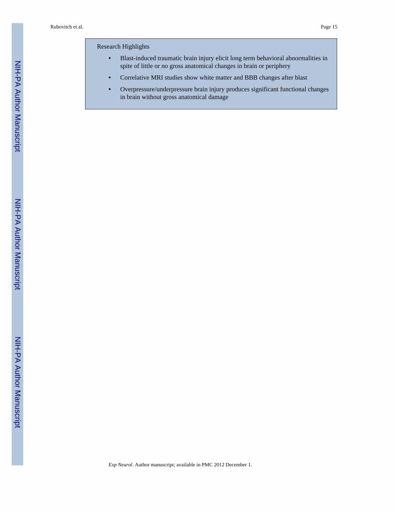
Research Highlights
• Blast-induced traumatic brain injury elicit long term behavioral abnormalities inspite of little or no gross anatomical changes in brain or periphery
• Correlative MRI studies show white matter and BBB changes after blast
• Overpressure/underpressure brain injury produces significant functional changesin brain without gross anatomical damage
Rubovitch et al. Page 15
Exp Neurol. Author manuscript; available in PMC 2012 December 1.
NIH
-PA Author Manuscript
NIH
-PA Author Manuscript
NIH
-PA Author Manuscript

Fig. 1. Blast experiment setting(a) The anesthetized mice were placed in a loose restraint device on a platform, which wascovered with white plastic mesh. Each platform had space for 12 anesthetized mice. (b)Photograph of the explosive charge (A) and mice immediately prior to detonation. A cast of500g TNT (A) was placed on a pedestal 1 meter above the ground. The 1 meter highplatforms constraining the anesthetized mice were situated 4 meters (B) and 7 meters (C)meters from the TNT charge). Two pressure gauges were mounted at the ends of eachplatform (D).
Rubovitch et al. Page 16
Exp Neurol. Author manuscript; available in PMC 2012 December 1.
NIH
-PA Author Manuscript
NIH
-PA Author Manuscript
NIH
-PA Author Manuscript

Fig. 2. Staircase test- the effect of blast on the number of rearing events (NR) and steps ascended(NSA) in a 3-min periodA. Rearing events (NR): Significant elevation NR was found in blasted mice both after 7days (24.8±1.73 for 7m and 26.4±1.96 for 4m, compared with 18±1.83 in sham mice) and30 days (28.8±6 for 7 m and 31.2±8.66 for 4 m, compared to 18.2±5.5 in sham mice). B.Steps ascended (NSA): The difference in the number of NSA reached statistical significanceonly in the 7 m group 7 days post blast (39±1.9 compared with 29.6±2.44 in sham mice).*p<0.05, **p<0.01 or ***p<0.001.
Rubovitch et al. Page 17
Exp Neurol. Author manuscript; available in PMC 2012 December 1.
NIH
-PA Author Manuscript
NIH
-PA Author Manuscript
NIH
-PA Author Manuscript

Fig. 3. The effect of blast on visual memory as assessed by the novel object recognition testThe preference for novel objects was significantly reduced in all the blast groups both at 7days(−0.02±0.01 for 7 m group, −0.008±0.009 for 4 m group and 0.45±0.09 for shamgroup) and 30 days (0.007±0.07 for 7 m group, −0.008±0.01 for 4 m group and 0.42±0.06for sham group). *p<0.05,**p<0.01 or ***p<0.001.
Rubovitch et al. Page 18
Exp Neurol. Author manuscript; available in PMC 2012 December 1.
NIH
-PA Author Manuscript
NIH
-PA Author Manuscript
NIH
-PA Author Manuscript

Fig. 4. The effect of blast on spatial memory as assessed by the Y-maze testPreference for the new arm was significantly reduced in mice 7 days post blast in bothgroups (0.2±0.07 for 7 m group and 0.22±0.07 for 4 m in comparison with the sham group0.51±0.15). Similar impaired memory was found after 30 days for the 4 m group(0.033±0.01 for the 4 m group and 0.361±0.09 for the sham group). *p<0.05,**p<0.01 or***p<0.001.
Rubovitch et al. Page 19
Exp Neurol. Author manuscript; available in PMC 2012 December 1.
NIH
-PA Author Manuscript
NIH
-PA Author Manuscript
NIH
-PA Author Manuscript
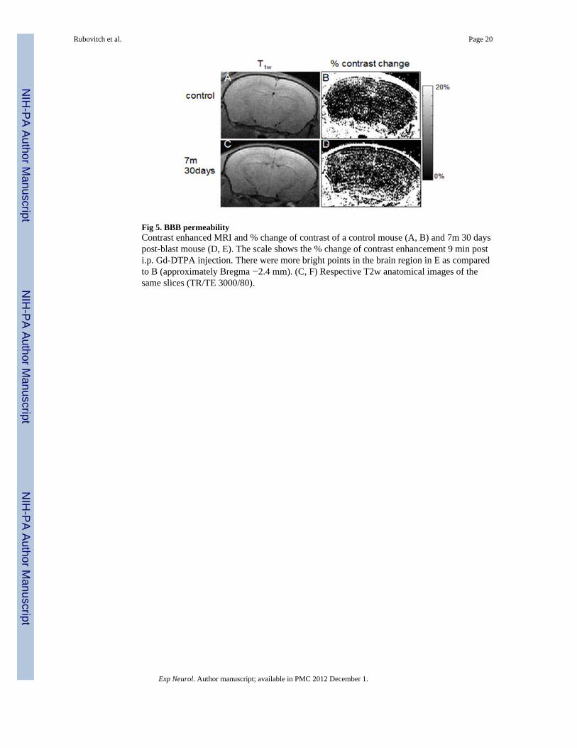
Fig 5. BBB permeabilityContrast enhanced MRI and % change of contrast of a control mouse (A, B) and 7m 30 dayspost-blast mouse (D, E). The scale shows the % change of contrast enhancement 9 min posti.p. Gd-DTPA injection. There were more bright points in the brain region in E as comparedto B (approximately Bregma −2.4 mm). (C, F) Respective T2w anatomical images of thesame slices (TR/TE 3000/80).
Rubovitch et al. Page 20
Exp Neurol. Author manuscript; available in PMC 2012 December 1.
NIH
-PA Author Manuscript
NIH
-PA Author Manuscript
NIH
-PA Author Manuscript

Fig. 6. Blast effect on BBB permeability index (BBPi)Permeability increased after blast-explosion at 30 days at 7 m compared with control group(F(4,40)=5.9, P<0.001). *** indicates significant difference between the 7m 30 days post-blast group compared to the control group, p<0.001.
Rubovitch et al. Page 21
Exp Neurol. Author manuscript; available in PMC 2012 December 1.
NIH
-PA Author Manuscript
NIH
-PA Author Manuscript
NIH
-PA Author Manuscript

Figure 7. Voxel-wise statistical analysis of the whole brain DTIRepresentative statistical maps overlaid on the fractional anisotropy (FA) (A) and radialdiffusivity (λ3) (B) (approximately Bregma −1.3 mm). The color coded clusters showregions that present significant differences between control and blast-exposed mice (p<0.05,FDR corrected). (C-E) Quantitative values at the indicated regions of interest for control andblast-exposed mice groups of FA (C&D) at the hypothalamus and thalamus, respectivelyand λ3 (E) at the hypothalamus. Significant difference between the blast group and thecontrol group *p<0.05, **p<0.01, ***p<0.001(one-way ANOVA). Control n=19, 4m 7 daysn=12, 4m 7d n=7, 4m 30d n=8 and 7m 30d n=12.
Rubovitch et al. Page 22
Exp Neurol. Author manuscript; available in PMC 2012 December 1.
NIH
-PA Author Manuscript
NIH
-PA Author Manuscript
NIH
-PA Author Manuscript

Figure 8. FA statistical maps overlaid on the fractional anisotropy (FA) mapsApparent regions of FA abnormality are diffusely spread over most of the brain includingthe cortex and the thalamus (approximately Bregma 1 mm, −1.4 mm and −2.3 mm - fromleft to right A-C). One-way ANOVA with significance of p<0.01 uncorrected.
Rubovitch et al. Page 23
Exp Neurol. Author manuscript; available in PMC 2012 December 1.
NIH
-PA Author Manuscript
NIH
-PA Author Manuscript
NIH
-PA Author Manuscript

Figure 9. Histopathology and distribution of MnSOD2 in the interpeduncular region andmammillary body after blast exposureThe left column shows low magnification photomicrographs of horizontal sections throughthe hypothalamus and interpeduncular nucleus (IP) of mice exposed 72h earlier to a single5.5 PSI overpressure blast. The left upper panel is stained with hematoxylin and eosin andshows normal vasculature and brain parenchyma. The lower left panels are stainedimmunohistochemically for MnSOD2. Note the strong up-regulation of MnSOD2 in themammillary body (Mm) after blast exposure, shown at higher magnification in the rightpanels. The calibration bar is 500 microns for the left panels and 50 microns for the rightpanels. Abbreviations: IP-interpeduncular nucleus, CC-crus cerebri, Mm-mammillary body,f-fornix.
Rubovitch et al. Page 24
Exp Neurol. Author manuscript; available in PMC 2012 December 1.
NIH
-PA Author Manuscript
NIH
-PA Author Manuscript
NIH
-PA Author Manuscript

Figure 10.Expression of C-X-C motif chemokine receptor 3 in fiber tracts after blast exposure.Photomicrographs show the emergence of immunoreactivity in association with bloodvessels within 72h of a single low level blast exposure in the crus cerebri (upper row), fornix(middle row) and optic tract (lower row). The positive control staining of bone marrow fromthe same blast exposed and control sections are shown in the insert. The calibration barrepresents 100 microns.
Rubovitch et al. Page 25
Exp Neurol. Author manuscript; available in PMC 2012 December 1.
NIH
-PA Author Manuscript
NIH
-PA Author Manuscript
NIH
-PA Author Manuscript


