Embed Size (px)
Citation preview

A ketogenic diet rescues the murine succinic semialdehydedehydrogenase deficient phenotype
Kirk Nylen1,3,4, Jose Luis Perez Velazquez1,2,4, Sergei S. Likhodii4,5, Miguel A.Cortez1,2,4, Lily Shen1,2, Yevgen Leshchenko1,2, Khosrow Adeli5, K. Michael Gibson6, W.M.Burnham3,4, and O.Carter Snead III1,2,3,4
1Program in Neuroscience and Mental Health, Hospital for Sick Children, Toronto, Ontario.
2Division of Neurology, Hospital for Sick Children, Toronto.
3Department of Pharmacology, University of Toronto.
4University of Toronto Epilepsy Research Program.
5Department of Pediatric Laboratory Medicine, Hospital for Sick Children.
6Division of Medical Genetics, Departments of Pediatrics, Pathology and Human Genetics, Children’sHospital, University of Pittsburgh School of Medicine, Pittsburgh, PA, USA.
AbstractSuccinic semialdehyde dehydrogenase (SSADH) deficiency is an heritable disorder of GABAdegradation characterized by ataxia, psychomotor retardation and seizures. To date, there is noeffective treatment for SSADH deficiency. We tested the hypothesis that a ketogenic diet (KD) wouldimprove outcome in an animal model of SSADH deficiency, the SSADH knockout mouse(Aldh5a1−/−). Using a 4:1 ratio of fat to combined carbohydrate and protein KD we set out to comparethe general phenotype, in vivo and in vitro electrophysiology and [35S]TBPS binding in bothAldh5a1−/− mice and control (Aldh5a1+/+) mice. We found that the KD prolonged the lifespan ofmutant mice by >300% with normalization of ataxia, weight gain and EEG compared to mutants feda control diet. Aldh5a1−/− mice showed significantly reduced mIPSC frequency in CA1 hippocampalneurons as well as significantly decreased [35S]TBPS binding in all brain areas examined. In KD fedmutants, mIPSC activity normalized and [35S]TBPS binding was restored in the cortex andhippocampus. The KD appears to reverse toward normal the perturbations seen in Aldh5a1−/− mice.Our data suggest that the KD may work in this model by restoring GABAergic inhibition. These datademonstrate a successful experimental treatment for murine SSADH deficiency using a KD, givingpromise to the idea that the KD may be successful in the clinical treatment of SSADH deficiency.
KeywordsSuccinic semialdehyde dehydrogenase deficiency; ketogenic diet; mouse model; GABA; beta-hydroxybutyrate; glucose; electrophysiology; chloride channel
Correspondence: Kirk Nylen ([email protected]) Neuroscience and Mental Health / Division of Neurology, 6535 Hill Wing,Hospital for Sick Children, 555 University Avenue, Toronto, ON, Canada, M5G 1X8.Publisher's Disclaimer: This is a PDF file of an unedited manuscript that has been accepted for publication. As a service to our customerswe are providing this early version of the manuscript. The manuscript will undergo copyediting, typesetting, and review of the resultingproof before it is published in its final citable form. Please note that during the production process errors may be discovered which couldaffect the content, and all legal disclaimers that apply to the journal pertain.
NIH Public AccessAuthor ManuscriptExp Neurol. Author manuscript; available in PMC 2009 April 1.
Published in final edited form as:Exp Neurol. 2008 April ; 210(2): 449–457.
NIH
-PA Author Manuscript
NIH
-PA Author Manuscript
NIH
-PA Author Manuscript

IntroductionSuccinic semialdehyde dehydrogenase (SSADH) deficiency is a rare, autosomal recessivedisorder of γ-aminobutyric acid (GABA) biotransformation (Gibson et al., 1983; Gibson andJakobs, 2001). The absence of SSADH leads to significantly elevated γ-hydroxybutyrate(GHB) and GABA levels throughout the body (Gibson and Jakobs, 2001; Pearl et al., 2003).Elevation of these neuroactive compounds is thought to play a role in the psychomotorretardation, ataxia and epilepsy (Gordon, 2004)— with seizure types ranging from absenceseizures to convulsive status epilepticus— seen in patients with this disorder (Pearl et al., 2003;Gibson et al., 1998). Currently, there is no effective treatment for human SSADH deficiency(Gropman, 2003; Gibson and Jakobs, 2001).
A murine analog of SSADH deficiency, Aldh5a1−/−, was developed to study thepathophysiology and treatment of this disorder (Hogema et al., 2001). Aldh5a1−/− mice exhibitdevelopmental delay, ataxia and a seizure disorder that progresses from absence seizures(~post-natal day, P, 15) to lethal status epilepticus (~P25) (Cortez et al., 2004). A variety ofpharmacological treatments were attempted to rescue Aldh5a1−/− mice with limited success(Hogema et al., 2001; Gupta et al., 2002; Cortez et al., 2004). However, it has been reportedthat pups that continue to suckle beyond weaning age (~P20) live longer than their weanlingcounterparts, suggesting that the high fat dam’s milk may have beneficial effects in this disorder(Hogema et al., 2001). This observation led us to pose the question whether administration ofa ketogenic diet (KD)— a high fat, low carbohydrate and adequate protein diet generally usedin the treatment of drug-resistant epilepsy and other neurological disorders (Vining, 1999;Thiele, 2003; Kossoff, 2004; Gasior et al., 2006; Hartman and Vining, 2007; Hartman et al.,2007)— might be successful in treating SSADH deficiency.
To test this hypothesis, we examined the effect of the KD on the Aldh5a1−/− mouse phenotype.Here we show that the KD normalizes lifespan, ataxia, weight gain and EEG in Aldh5a1−/−
mice. Previous research has shown a loss of GABAA receptor-associated chloride channelbinding and resultant neural hyperexcitation to be a factor in the SSADH null phenotype (Wuet al., 2006). We therefore examined [35S]TBPS binding and recorded excitatory and inhibitoryminiature post synaptic currents in KD and CD fed mutant mice. Our data suggest that the KDmay work towards rescuing the SSADH null phenotype by restoring GABAergic activity inthe brains of Aldh5a1−/− mice.
MethodsSubjects
The SSADH null mice with C57/129Sv background were first generated in the Oregon Healthand Science University in Portland (Hogema et al., 2001). Five pairs of heterozygous(Aldh5a1+/−) mice were transported to the Hospital for Sick Children in Toronto. The mutantmouse line was maintained by inbreeding. The absence of SSADH was confirmed by two-allele three-primer polymerase chain reaction using tail genomic DNA (Hogema et al., 2001).All animals were maintained in a controlled environment at 12h light-12h dark cycle with lightson at 06:00 h and given ad lib access to food and water. All animal work was approved by theHospital for Sick Children Division of Laboratory Animal Services and was performed inaccordance with the guidelines of the Canadian Council on Animal Care (CCAC).
DietsIn KD treated mutants, the KD replaced normal mouse chow when pups were P12. The youngmice fed primarily by suckling until ~P20. The KD was introduced earlier, however, to ensurethat any non-suckling feeding was ketogenic in nature. The 4:1 (fat:carbohydrate + protein, by
Nylen et al. Page 2
Exp Neurol. Author manuscript; available in PMC 2009 April 1.
NIH
-PA Author Manuscript
NIH
-PA Author Manuscript
NIH
-PA Author Manuscript

weight) KD was obtained from Harlan Teklad (Madison, WI; TD.03490) (Likhodii, 2001;Nylen et al., 2005; Nylen et al., 2006). Standard mouse chow served as the CD. Table 1summarizes the composition of both diets.
Weights and AtaxiaWeights and ataxia scores were determined for all mice 3–5 times per week. Ataxia scoreswere scored according to Loscher’s scale of sedation (Loscher et al., 1987). This scale has 6levels of ataxia: 1—slight ataxia of hindlegs, 2—dragging hindlegs, 3—ataxia and dragginghindlegs, 4—marked ataxia loss of balance, 5—marked ataxia no balance, and 6—loss ofrighting reflex with attempts to move forward.
Electrocorticography (ECoG)Mice (P20–25) were implanted with four epidural monopolar electrodes with their tips placedbilaterally over the frontal and parietal cortices under pentobarbital anesthesia. The electrodeswere placed 1mm deep, 2mm anterior to bregma and 2mm lateral from midline. Recordingswere made 1, 24 and 48 hours after recovery from anesthesia. Each animal was placed in anindividual Plexiglas chamber for a 20-min adaptation period prior to ECoG recordings in orderto minimize movement artifact. ECoG recordings were made on paper using a GrassPolysomnograph machine (Grass Instruments, Quincy, MA) (Cortez et al., 2004).
Determination of Serum AnalytesAll analytes were measured using methods implemented as User Defined Assays using reagentsfrom Randox Laboratories Ltd. (Antrim, United Kingdom). A Vitros Chemistry System 5,1Fusion Series (Ortho-Clinical Diagnostics, N.J.) was used to analyze all samples. Glucose wasdetermined using the conversion of β-D-glucose gluconate and H2O2 via glucose oxidase. Thegenerated hydrogen peroxide oxidizes the 4-aminoantipyrine, 1,7 dihydroxynaphthalenechromogen system in a horseradish peroxidase catalyzed reaction. The density of the resultingdye complex is related to the concentration of glucose in the specimen and is measured byreflectance spectrophotometry at 540nm (User Defined Assay Reference Guide for Vitros 5,1FS Chemistry System. Ortho-Clinical Diagnostics.). Beta-hydroxybutyrate (βOHB) wasmeasured by the oxidation of βOHB to acetoacetate via βOHB-dehydrogenase. This reactioncan be detected by measuring absorbance at 340nm caused by the conversion of NAD+ toNADH (Randox Ranbut D-3-Hydroxybutyrate Manual, Randox Laboratories Ltd.; UserDefined Assay Reference Guide for Vitros 5,1 FS Chemistry System. Ortho-ClinicalDiagnostics.). Determination of non-esterified fatty acids (NEFA, or free fatty acids) wasperformed by the acylation of coenzyme A (CoA) by the fatty acids in the presence of acyl-CoA synthetase. The acyl-CoA produced in the reaction is further oxidized by acyl-CoAoxidase, which generated H2O2 as a by-product. Hydrogen peroxide, in the presence ofperoxidase permits the oxidative condensation of N-ethyl-N-(2hydroxy-3-sulphopropyl)-m-toluidine with 4-aminoantipyrine (4-AAP) to form a purple coloured adduct. This colour wasmeasured spectrophotometrically at 550nm (Randox NEFA Manual, Randox LaboratoriesLtd.; User Defined Assay Reference Guide for Vitros 5,1 FS Chemistry System. Ortho-ClinicalDiagnostics.).
Electrophysiology: Brain Slices and SolutionsBrains were taken from P14 to P25 mice under halothane anesthesia. Transverse brain slices(450µm) were obtained by a Vibratome (Series 1000; St. Louis, MO) and maintained inartificial cerebrospinal fluid (aCSF) containing (mM) 125 NaCl, 2.5 KCl, 1.25 NaH2PO4, 2MgSO4, 2 CaCl2, 25 NaHCO3, and 10 glucose. aCSF was bubbled with carbogen (95% O2,5% CO2) and gravity fed to the recording chamber at a rate of 3–4ml/min.
Nylen et al. Page 3
Exp Neurol. Author manuscript; available in PMC 2009 April 1.
NIH
-PA Author Manuscript
NIH
-PA Author Manuscript
NIH
-PA Author Manuscript

The internal solution for recording mPSCs consisted of (mM) 20 cesium methanesulfonate, 2Mg-ATP, 10 HEPES, 0.3 GTP, 0.1 EGTA, 130 CsCl2. Osmolarity was 300±5mOsm and pHwas adjusted to 7.2 using cesium hydroxide. For mIPSC recordings, D-2-amino-5-phosphopentanoic acid (D-AP5; 20µM, made of 50mM stock solution in distilled water), 6-cyano-7-nitroquinoxaline-2,3-dione (CNQX; 100µM, made of 50mM stock indimethylsulfoxide) and tetrodotoxin (TTX; 1µM, made of 1mM stock in dH2O) were addedto the aCSF superfusate. For mEPSC recordings, bicuculline methiodide (BMI; 10µM madeof 10mM stock in dH2O) was added to aCSF superfusate along with TTX. Chemicals wereobtained from Sigma and diluted daily.
Electrophysiology: Whole Cell RecordingsNeuronal recordings were obtained from CA1 hippocampal pyramidal neurons using thewhole-cell configuration patch-clamp technique. Electrodes had tip resistances between 5 and8MΩ. Neuronal responses were recorded with the use of a Multiclamp 700B (MolecularDevices, Sunnyvale, CA).
Analysis of mPSCsmPSCs were analyzed using MiniAnalysis software (Synaptosoft, Decatur, GA).
[35S] Tert-butylbicyclophosphorothionate AutoradiographyMice were decapitated and their brains were removed and immediately immersed in isopentaneat −35°C. Coronal sections were cut from the anterior to posterior boundaries of the cerebralcortex at 20um at −20°C and thaw-mounted onto gelatin-coated slides that were dried andstored at −80°C until used. The regions analyzed were the frontoparietal cortex, the ventrobasalthalamus, the CA region of the hippocampus and the amygdala (Wu et al., 2006).
Statistical AnalysisLifespan and weight gain data were analyzed using two-tailed t-tests. Ataxia data were analyzedusing a repeated measures two-way analysis of variance (ANOVA). [35S]TBPS binding dataand serum analyte were analyzed using a two-way ANOVA with Bonferroni post-hoc tests.All other data were analyzed using a one-way ANOVA with Tukey’s post-hoc tests.Significance was considered at p<0.05; numerical values are expressed as means±standarderror (s.e.m.).
ResultsLifespan, Ataxia and Weights
Mutant mice weaned onto the CD invariably demonstrated convulsions, which culminated inlethal status epilepticus around P25. Although status epilepticus was the eventual cause ofdeath in all mice, the onset of status epilepticus was significantly delayed in the KD fed mutants,as evidenced by their significantly longer lifespan.
The average lifespan of KD fed mutants was 93.5±20.4 days, almost 4-fold higher than thelifespan of CD fed mutants who had an average lifespan of 24±3.7 days (Fig. 1a; n=9, p<0.0001,two-tailed t-test). All mice were weighed and assessed for ataxia daily. Beyond P12, CD fedmutants exhibited an average daily weight loss of 0.003±0.013g while KD fed mutants gainedan average of 0.130±0.010g per day (Fig. 1b; p=0.0002, two-tailed t-test). Ataxia scores wereassessed using a 6-point rating scale (Loscher et al., 1987). CD fed Aldh5a1−/− mice began todevelop ataxia at ~P18 (stage 2 and higher). Onset of ataxia was significantly delayed in KDfed mutants as they only began to develop stage 2 ataxia at ~P70 (Fig. 1c; p=0.0002, repeated-
Nylen et al. Page 4
Exp Neurol. Author manuscript; available in PMC 2009 April 1.
NIH
-PA Author Manuscript
NIH
-PA Author Manuscript
NIH
-PA Author Manuscript
measures two-way ANOVA, post-hoc analysis revealed significant differences between groupsfor days P17–40).
In Vivo ElectrocorticographyElectrocorticography (ECoG) was performed on freely moving CD and KD fed Aldh5a1−/−
mice to determine whether the above-mentioned changes corresponded to changes on theelectroencephalogram (EEG). The background ECoG baseline in CD fed Aldh5a1−/− mice(n=4) consisted of 35 to 60uV cortical activity at 4 to 7 Hz activity with intermingled 3–5 Hz,often associated with fast frequency oscillations at the frequency of 28 Hz during restful wakingconditions. There were numerous interruptions of the background activity by intermittent highamplitude 250–300uV bursts of spontaneous, recurrent spike and wave discharges (SWD),whose onset/offset was time-locked with absence-like ictal behavior which consisted of frozenimmobility, facial myoclonus, and vibrissal twitching. In contrast, the baseline of the KD fedAldh5a1−/− mice showed remarkable normalization with a fairly well regulated low amplitude35 to 50uV electrical fields at 4 Hz, with complete absence either slower frequencies in thedelta range or fast frequency oscillations within the frontal and parietal cortices recorded withsimultaneous standard paper EEG and digital systems (Grass Instrumnets, Quincy, MA)recorded at 6, 15 and 30 mm/sec (Fig. 2a). We also recoded the number of seizures withcorresponding convulsions. KD fed mutants experienced significantly fewer convulsionscompared to CD fed mutants (Fig 2b; p=0.031, t-test).
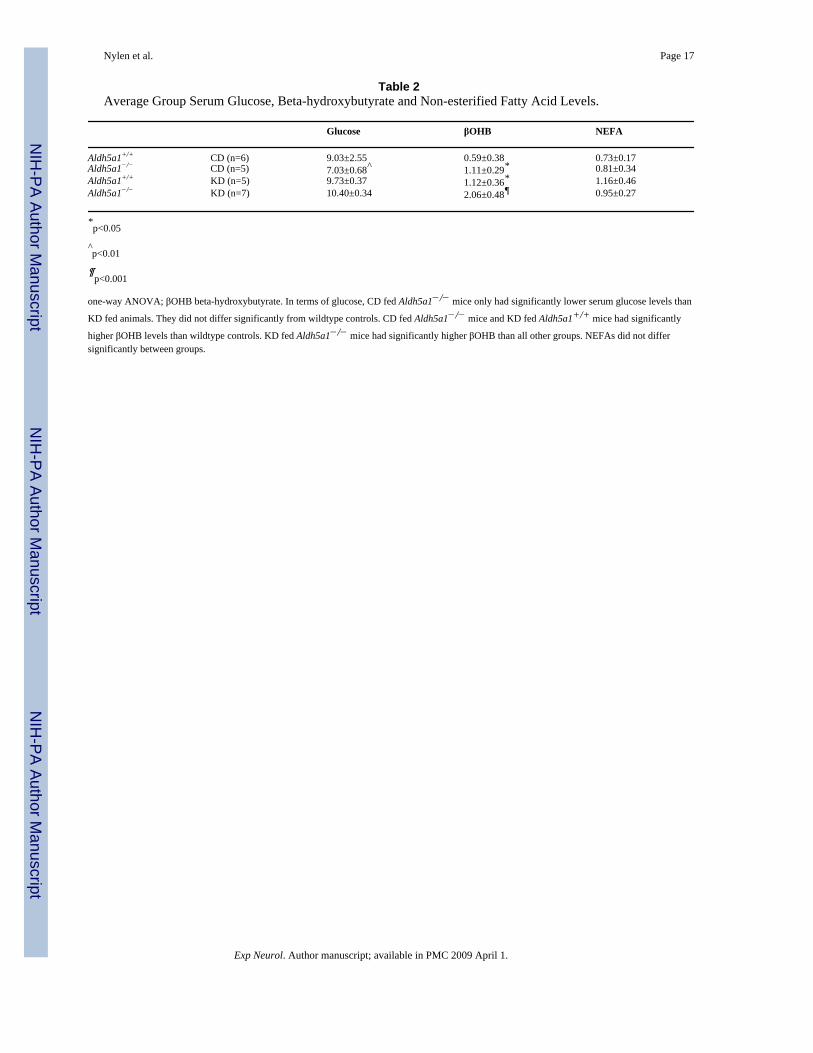
Serum Glucose, Beta-hydroxybutyrate, and Non-esterified Fatty AcidsTo determine the biochemical correlates of KD induced changes, we measured serum levelsof glucose, beta-hydroxybutyrate (βOHB) and non-esterified fatty acid (NEFA) concentrations(see Table 2). For this experiment we added a KD fed Aldh5a1+/+ group. Analysis of serumglucose, βOHB and NEFAs was performed given that these analytes are among the moststudied blood analytes in the literature investigating the potential mechanism of the KD.
Serum glucose levels were significantly different between groups (p=0.006, one-wayANOVA). Post hoc tests revealed that serum glucose was significantly elevated in KD fedmice when compared to CD fed Aldh5a1−/− mice (p<0.05). Serum glucose levels were onlysignificantly different between CD fed Aldh5a1−/− and KD fed mice— no groups differedsignificantly from CD fed Aldh5a1+/+ control mice.
Examination of βOHB levels demonstrated significant differences between groups (p=0.0004,one-way ANOVA). CD fed Aldh5a1−/− mice had significantly elevated βOHB when comparedto CD fed Aldh5a1+/+ control mice (p<0.05, post-hoc). βOHB levels were significantlyelevated in KD fed wildtype mice when compared to CD fed wildtype mice, which wasexpected due to the ketogenic nature of the diet (p<0.01). βOHB levels were highest in KD fedAldh5a1−/− mice and they differed significantly from CD fed wildtype mice (p<0.001).
We measured serum NEFAs in all mice. There were no statistically significant differencesbetween groups.
In Vitro Miniature Post-Synaptic Current RecordingsWhole cell voltage clamp studies were performed to record miniature inhibitory (mIPSC) andexcitatory (mEPSC) postsynaptic currents from CA1 pyramidal cells of hippocampal slices.A one-way ANOVA showed that mIPSC activity in slices from Aldh5a1−/− mice wassignificantly diminished compared to wildtype mice (Fig. 3a–b; p=0.04, ANOVA; p<0.05,post-hoc test). This decrease represented a >2.5 fold decrease in mIPSC frequency. There wereno statistically significant differences between groups in terms of mEPSC activity (Fig. 3c–d;p=0.46, one-way ANOVA). The decrease in mIPSC activity observed in the Aldh5a1−/− was
Nylen et al. Page 5
Exp Neurol. Author manuscript; available in PMC 2009 April 1.
NIH
-PA Author Manuscript
NIH
-PA Author Manuscript
NIH
-PA Author Manuscript

completely restored in KD fed mutants when compared to wildtype controls (Fig. 3a–b; p>0.05,post-hoc test).
[35S]tert-butylbicyclophosphorothionate Binding[35S]Tert-butylbicyclophosphorothionate ([35S]TBPS) binding in Aldh5a1−/− mice has beenshown to be significantly reduced in an age-dependent manner when compared with wildtypemice (Wu et al., 2006). The [35S]TBPS binding becomes increasingly diminished until thethird postnatal week of life in the mutant animals, reaching a nadir prior to the onset ofgeneralized convulsive seizures (Wu et al., 2006). Here we confirm that [35S]TBPS binding issignificantly reduced in Aldh5a1−/− mice compared to wildtype controls (Fig. 3d; p=0.002,two-way ANOVA). Significantly decreased [35S]TBPS binding in Aldh5a1−/− mice wasobserved in all brain regions studied (hippocampus, cortex, amygdala and thalamus; all regionsp<0.001, post-hoc test). Post-hoc analysis demonstrated that [35S]TBPS binding is restoredfully in hippocampus and frontoparietal cortex of KD fed Aldh5a1−/− mice when compared toCD fed Aldh5a1+/+ mice (p>0.05). The KD did not fully restore [35S]TBPS binding inamygdala and thalamus of Aldh5a1−/− mice when compared to CD fed wildtype mice (p>0.05,both cases). The reduction of [35S]TBPS binding seen in amygdala and thalamus of KD fedmutants was less severe than the reduction seen in CD fed mutants, suggesting that the KDinduced partial restoration of [35S]TBPS binding in these areas (Fig. 2d).
DiscussionOur data show that the KD eliminates the premature lethality seen in Aldh5a1−/− mutants andincreases the lifespan by >300%. KD fed mutants also showed significantly less ataxia, weightloss and fewer abnormalities on the ECoG. These effects were paralleled by a significantrestoration of GABAergic spontaneous synaptic activity and region-specific restoration ofGABAA receptor-associated chloride channel binding.
LifespanThe cause of death in Aldh5a1−/− mice is status epilepticus (Cortez et al., 2004). Around P18–22, untreated mutants experience tonic-clonic convulsions followed by status epilepticusaround P25. KD fed mutants did not experience status epilepticus at this age, but began todemonstrate susceptibility to auditory evoked convulsions around P70. These convulsionsinvolved a tonic-clonic component with jumping and running fits. Eventually, KD fedAldh5a1−/− mice succumbed to status epilepticus, albeit significantly later in life than theiruntreated counterparts.
To our knowledge, our experiments represent the longest any mouse has been maintained ona KD. The longest-living KD fed mutant was maintained on the KD for 130 days. The long-term effects of a high-fat diet in mice have not been thoroughly investigated. In humans, theKD has been associated with increased risk of coronary heart disease (Best et al., 2000;Bergqvist et al., 2003; Dashti et al., 2003; Kwiterovich et al., 2003) among other adverseeffects. It is unclear whether such adverse events played a role in the eventual death of KD fedAldh5a1−/− mice.
Ataxia and WeightsThe KD significantly delayed the onset of ataxia in Aldh5a1−/− mice. CD fed Aldh5a1−/− micedeveloped marked ataxia by P15–17, as was evidenced by significant dragging of the hindlimbs (stage 2 ataxia). Ataxia in these animals quickly progressed to involve completeimmobility for prolonged periods with unsuccessful attempts to walk. By P25, untreatedmutants had little control over their locomotion. KD fed mutants, however, took significantlylonger to develop ataxia. Stage 2 ataxia did not occur until ~P70.
Nylen et al. Page 6
Exp Neurol. Author manuscript; available in PMC 2009 April 1.
NIH
-PA Author Manuscript
NIH
-PA Author Manuscript
NIH
-PA Author Manuscript

Weights were assessed in KD and CD fed Aldh5a1−/− as well as CD fed wildtype mice. Wedid not measure weight gain in KD fed Aldh5a1+/+ mice. There are several studies, both clinicaland experimental, that report KD fed subjects being lighter than normal diet fed subjects (Rhoet al, 1999; Bough & Eagles, 2001; Vining et al, 2002; Zhao et al, 2004; Nylen et al, 2005).Despite having significantly stunted growth, rodents fed a KD do not experience anywherenear the stunting seen in SSADH mutants fed either a KD or a CD.
KD fed mutants showed significantly less stunting of growth compared to CD fed mutants.This was evidenced by significantly higher daily weight gain in the KD fed mutants. Clinically,the KD is associated with significantly attenuated growth compared to humans consuming anon-KD (Vining et al., 2002; Liu et al., 2003; Papandreou et al., 2006). This has also beenshown in rats fed a KD (Zhao et al, 2004; Nylen et al, 2005; Nylen et al, 2006). This held truein the KD fed mutants as they showed significant stunting of growth when compared to CDfed wildtype mice. When compared to CD fed Aldh5a1−/− mice, however, KD fedAldh5a1−/− mice gained significantly more weight.
It is possible that the cause of this weight gain is simply because the KD fed mutants consumedmore food. Previous research from our group, however, has measured food intake in rats onthe KD and found that animals regulate their caloric intake (Likhodii et al., 2000). Because theKD is almost twice as calorie-dense as the control diet, this means that KD fed animals tendto eat about half as much food.
We believe significant weight loss in untreated Aldh5a1−/− mice may be caused by impairedglucose metabolism (Chowdhury et al., in press) and subsequent metabolism of fat and proteinstores. Consumption of a high fat KD provides an alternative energy source for Aldh5a1−/−
mice, allowing some growth to take place.
Serum AnalytesWe examined serum levels of glucose, βOHB and NEFAs. Glucose levels only differedsignificantly between CD fed Aldh5a1−/− mice and KD fed mice. None of the groups differedsignificantly from CD fed wildtype controls, however.
βOHB levels were significantly elevated in CD fed Aldh5a1−/− mice when compared to CDfed wildtype animals. This supports a previous finding showing that impaired glucose oxidationin SSADH mutants leads to a significant elevation of βOHB levels (Chowdhury et al., inpress). If Aldh5a1−/− mice are unable to oxidize dietary carbohydrate effectively then they willbegin to oxidize their fat stores for energy, causing ketosis. This has important implicationsfor the mechanism of the KD in this model. It is possible that giving the mice a high-fat dietsimply gives them an energy source that they wouldn’t otherwise be able to get fromcarbohydrate rich mouse chow.
As expected, serum βOHB levels were significantly elevated in KD fed animals due to theketogenic nature of the diet. βOHB levels in KD fed mutants were very high, possibly acompounded effect of elevated ketosis due to impaired glucose oxidation coupled with theketogenic nature of the diet.
An unexpected finding was an absence of increased blood NEFA during KD intervention.Similar findings have been reported by Klepper and colleagues (2004), however, using the KDto treat human glucose transporter deficiency. Whereas βOHB rose significantly in theirpatients, there was no corresponding increase in absolute plasma NEFA. In our study, weassume no differences between genotypes for lipoprotein lipase activity or its ability to activatein response to diet. One explanation for the absence of increased plasma NEFA may reside inthe capacity of Aldh5a1−/− mice to more effectively convert NEFA to βOHB. The ratios of
Nylen et al. Page 7
Exp Neurol. Author manuscript; available in PMC 2009 April 1.
NIH
-PA Author Manuscript
NIH
-PA Author Manuscript
NIH
-PA Author Manuscript

mean NEFA to βOHB (Table 2) were: Aldh5a1+/+ (CD), 1.20; Aldh5a1−/− (CD), 0.73;Aldh5a1+/+ (KD), 1.04; Aldh5a1−/− (KD), 0.46. While the ratio for Aldh5a1+/+ mice wascomparable despite diet, the same value in Aldh5a1−/− mice was lower on CD and decreasedand additional 40% with KD intervention, suggesting enhanced β-oxidation of fatty acids inthe Aldh5a1−/− mice.
ECoGKD fed mice show remarkable normalization of the ECoG. This normalization might be dueto the KD’s ability to elevate ketone bodies. In our model, we show that the KD significantlyelevates βOHB (acetone and acetoacetate have not yet been measured). The ketone bodiesacetone, βOHB and acetoacetate—all elevated by the KD— have all been shown to haveanticonvulsant effects in animal seizure models (Rho et al., 2002; Likhodii et al., 2003; Gasioret al., 2007). Elevation of βOHB may, in part, explain why the ECoG of KD fed Aldh5a1−/−
mice show less seizure activity, and why the mice have fewer convulsions. Anotherconsideration, discussed below, is that the KD restored spontaneous GABAergic synapticactivity and GABAA receptor-associated chloride channel binding.
[35S]TBPS BindingWe confirmed that CD fed Aldh5a1−/− mice exhibit significantly reduced [35S]TBPS bindingcompared to wildtype controls. This reduction was seen in all brain areas studied, with thelargest decrease (36%) seen in the thalamus. [35S]TBPS binding was completely restored inhippocampus and cortex and partially restored in amygdala and thalamus of KD fed mutants.[35S]TBPS is a specific ligand for the GABAA receptor-gated chloride channel (Behrends,2000). These data likely reflect a reduction—and KD induced rescue—of GABAA receptorbinding and/or C1− channel activity. Interestingly, the KD had no effect on [35S]TBPS bindingin wildtype mice.
mPSC RecordingsHere we report a significant decrease in mIPSC frequency in Aldh5a1−/− mice. Reductions inmIPSC activity have been implicated as a potential mechanism of epileptogenesis (Hirsch etal., 1999; Shao et al., 2005). mIPSCs are known to play an important role in synapticmaintenance and function (Hartman et al., 2006). A decrease in mIPSC frequency with acorresponding decrease in mIPSC amplitude can suggest postsynaptic changes to GABAAreceptors, however, mIPSC amplitudes were not reduced in KD fed Aldh5a1−/− mice (data notshown). The decrease in mIPSC frequency might therefore reflect a decrease in the number ofpresynaptic inhibitory interneuron terminals in the stratum radiatum. These interneuronssynapse on the CA1 pyramidal cells from which we recorded. Decreased input from theseinterneurons might explain the neural hyperactivity observed in CD fed mutants (Wu et al.,2006). It is possible that the KD may be acting to preserve or repair the interneurons. Thereexists considerable evidence for the neuro-protective effects of the KD and ketone bodies(Massieu et al., 2003; Noh et al., 2003; Gasior et al., 2006; Noh et al., 2006; Maalouf et al.,2007). This hypothesis requires further studies to determine the exact cause for the mIPSCreduction in CD fed Aldh5a1−/− mice.
Role of GABA and GHB in KD’s MechanismWe propose that the KD plays a role in restoring hippocampal mIPSC activity and thehippocampal [35S]TBPS binding in parallel. Although we have not shown the relationshipbetween these events to be causal, there is some evidence to suggest that changes in GABAAreceptor binding could underlie changes in hippocampal mIPSC frequency (Hartman et al.,2006; Swanwick et al., 2006). In untreated mutants, both of these GABAergic componentswere significantly compromised when compared to wildtype control mice. KD stimulated
Nylen et al. Page 8
Exp Neurol. Author manuscript; available in PMC 2009 April 1.
NIH
-PA Author Manuscript
NIH
-PA Author Manuscript
NIH
-PA Author Manuscript

restoration of GABAergic activity may have played a role in forstalling the evolution of lethalstatus epilepticus in these mutant animals, thus significantly prolonging their lifespan.
GHB exerts it pharmacological activity via the GABAB receptor (Snead & Gibson, 2005). Inmammalian brain GHB levels approximate 1–4 µM (Doherty et al., 1978; Snead, 1991), whilein Aldh5a1−/− mice this level rises to ~250 µM (Hogema et al., 2001). This concentration ofGHB is still well below that which is believed to activate GABAB receptors (~5 mM;Lingenhoehl et al., 1999; Wu et al., 2004). On the other hand, some of this GHB mayinterconvert to GABA, as the enzymes responsible for this are still operative (Rumigny et al.,1981), hence elevating the excess GABA due to the block in Aldh5a1 even more (Snead &Gibson, 2005). The resultant elevated GABA would be predicted to down-regulate bothGABAA (Wu et al., 2006) and GABAB (Buzzi et al., 2006) receptors. Whether elevated GABApreferentially modulates pre-/postsynaptic GABAB receptors is unknown, although selectedstudies using baclofen in cultured hippocampal neurons indicate a preferential desensitizationof postsynaptic GABAB receptors (Wetherington & Lambert, 2002). There is some evidencethat GHB may preferentially act on presynaptic GABABR (Banerjee et al., 1995; Snead,1996).
ConclusionTo our knowledge, this is the first evidence that a KD is effective in the treatment of murineSSADH deficiency. Our data provide support for the potential usefulness of the KD in thetreatment of human SSADH deficiency. We speculate that detailed characterization of KDmechanisms in SSADH null mice will provide novel insight into the utility of this diet in thetreatment of refractory epilepsy.
Acknowledgements
We would like to thank Dr.Tim Moran and Natalia Fedianina for their technical advice and support with theelectrophysiological recordings. KJN is the recipient of a SickKids Foundation Graduate Scholarship (Restracomp)and the Van Gelder-Savoy Studentship. Supported by NIH NS 40270 (KMG, OCS).
ReferencesBanerjee PK, Snead OC. Presynaptic γ-hydroxybutyric acid (GHB) and γ-aminobutyric acid B
(GABAB) receptor-mediated release of GABA and glutamate (GLU) in rat thalamic ventrobasalnucleus (VB): a possible mechanism for the generation of absence-like seizures induced by GHB. J.Pharmacol. Exp. Ther 1995;273:1534–1543. [PubMed: 7791129]
Behrends JC. Modulation by bicuculline and penicillin of the block by t-butyl-bicyclophosphorothionate(TBPS) of GABA(A)-receptor mediated Cl(‒)-current responses in rat striatal neurones. Br. J.Pharmacol 2000;129:402–408. [PubMed: 10694249]
Bergqvist AG, Chee CM, Lutchka L, Rychik J, Stallings VA. Selenium deficiency associated withcardiomyopathy: a complication of the ketogenic diet. Epilepsia 2003;44:618–620. [PubMed:12681013]
Best TH, Franz DN, Gilbert DL, Nelson DP, Epstein MR. Cardiac complications in pediatric patients onthe ketogenic diet. Neurology 2000;54:2328–2330. [PubMed: 10881264]
Bough KJ, Eagles DA. A ketogenic diet increases resistance to pentylenetetrazole-induced seizures inthe rat. Epilepsia 1999;40:138–143. [PubMed: 9952258]
Bough KJ, Eagles DA. Comparison of the anticonvulsant efficacies and neurotoxic effects of valproicacid, phenytoin, and the ketogenic diet. Epilepsia 2001;42:1345–1353. [PubMed: 11737171]
Buzzi A, Wu Y, Frantseva MV, Perez-Velazquez J-L, Cortez MA, Liu CC, Shen LQ, Gibson KM, SneadOC. Succinic semialdehyde dehydrogenase deficiency: GABAB receptor mediated function. BrainRes 2006;1090:15–22. [PubMed: 16647690]
Nylen et al. Page 9
Exp Neurol. Author manuscript; available in PMC 2009 April 1.
NIH
-PA Author Manuscript
NIH
-PA Author Manuscript
NIH
-PA Author Manuscript
Cortez MA, Wu Y, Gibson KM, Snead OC 3rd. Absence seizures in succinic semialdehydedehydrogenase deficient mice: a model of juvenile absence epilepsy. Pharmacol. Biochem. Behav2004;79:547–553. [PubMed: 15582027]
Chowdhury GMI, Gupta M, Gibson KM, Patel AB, Behar KL. Altered glucose and acetate metabolismin succinic semialdehyde dehydrogenase (SSADH) deficient mice: evidence for glial dysfunction andreduced glutamate/glutamine cycling. J. Neurochem. (in press)
Dashti HM, Bo-Abbas YY, Asfar SK, Mathew TC, Hussein T, Behbahani A, Khoursheed MA, Al-SayerHM, Al-Zaid NS. Ketogenic diet modifies the risk factors of heart disease in obese patients. Nutrition2003;19:901–902. [PubMed: 14559328]
Doherty JD, Hattox SE, Snead OC, Roth RH. Identification of endogenous gamma-hydroxybutyrate inhuman and bovine brain and its regional distribution in human, guinea pig and rhesus monkey brain.J. Pharmacol. Exp. Ther 1978;207:130–139. [PubMed: 100596]
Gasior M, French A, Joy MT, Tang RS, Hartman AL, Rogawski MA. The anticonvulsant activity ofacetone, the major ketone body in the ketogenic diet, is not dependent on its metabolites acetol, 1,2-propanediol, methylglyoxal, or pyruvic acid. Epilepsia 2007;48:793–800. [PubMed: 17386058]
Gasior M, Rogawski MA, Hartman AL. Neuroprotective and disease-modifying effects of the ketogenicdiet. Behav. Pharmacol 2006;17:431–439. [PubMed: 16940764]
Gibson KM, Gupta M, Pearl PL, Tuchman M, Vezina LG, Snead OC, Smit LME, Jakobs C. Significantbehavioral disturbances in succinic semialdehyde dehydrogenase (SSADH) deficiency (Gamma-hydroxybutyric aciduria). Biol. Psych 2003;54:763–768.
Gibson KM, Hoffmann GF, Hodson AK, Bottiglieri T, Jakobs C. 4-Hydroxybutyric acid and the clinicalphenotype of succinic semialdehyde dehydrogenase deficiency, an inborn error of GABAmetabolism. Neuropediatrics 1998;29:14–22. [PubMed: 9553943]
Gibson, KM.; Jakobs, C. Disorders of beta- and gamma amino acids in free and peptide-linked forms.In: Scriver, CR.; Beaudet, AL.; Sly, WS.; Valle, D., editors. The metabolic and molecular bases ofinherited disease. New York: McGraw-Hill; 2001. p. 2079-2105.
Gibson KM, Schor DS, Gupta M, Guerand WS, Senephansiri H, Burlingame TG, Bartels H, HogemaBM, Bottiglieri T, Froestl W, Snead OC, Grompe M, Jakobs C. Focal neurometabolic alterations inmice deficient for succinate semialdehyde dehydrogenase. J. Neurochem 2002;81:71–79. [PubMed:12067239]
Gibson KM, Sweetman L, Nyhan WL, Jakobs C, Rating D, Siemes H, Hanefeld F. Succinic semialdehydedehydrogenase deficiency: an inborn error of gamma-aminobutyric acid metabolism. Clin. Chim.Acta 1983;133:33–42. [PubMed: 6627675]
Gordon N. Succinic semialdehyde dehydrogenase deficiency (SSADH) (4-hydroxybutyric aciduria,gamma-hydroxybutyric aciduria). Eur. J. Paediatr. Neurol 2004;8:261–265. [PubMed: 15341910]
Gropman A. Vigabatrin and newer interventions in succinic semialdehyde dehydrogenase deficiency.Ann. Neurol 2003;54:S66–S72. [PubMed: 12891656]
Gupta M, Greven R, Jansen EE, Jakobs C, Hogema BM, Froestl W, Snead OC, Bartels H, Grompe M,Gibson KM. Therapeutic intervention in mice deficient for succinate semialdehyde dehydrogenase(gamma-hydroxybutyric aciduria). J. Pharmacol. Exp. Ther 2002;302:180–187. [PubMed:12065715]
Hartman AL, Gasior M, Vining EP, Rogawski MA. The neuropharmacology of the ketogenic diet.Pediatr. Neurol 2007;36:281–292. [PubMed: 17509459]
Hartman AL, Vining EP. Clinical aspects of the ketogenic diet. Epilepsia 2007;48:31–42. [PubMed:17241206]
Hartman KN, Pal SK, Burrone J, Murthy VN. Activity-dependent regulation of inhibitory synaptictransmission in hippocampal neurons. Nat. Neurosci 2006;9:642–649. [PubMed: 16582905]
Hirsch JC, Agassandian C, Merchan-Perez A, Ben-Ari Y, DeFelipe J, Esclapez M, Bernard C. Deficit ofquantal release of GABA in experimental models of temporal lobe epilepsy. Nat. Neurosci1999;2:499–500. [PubMed: 10448211]
Hogema BM, Gupta M, Senephansiri H, Burlingame TG, Taylor M, Jakobs C, Schutgens RB, FroestlW, Snead OC, Diaz-Arrastia R, Bottiglieri T, Grompe M, Gibson KM. Pharmacologic rescue of lethalseizures in mice deficient in succinate semialdehyde dehydrogenase. Nat. Genet 2001;72:218–222.
Nylen et al. Page 10
Exp Neurol. Author manuscript; available in PMC 2009 April 1.
NIH
-PA Author Manuscript
NIH
-PA Author Manuscript
NIH
-PA Author Manuscript

Klepper J, Diefenbach S, Kohlschütter A, Voit T. Effects of the ketogenic diet in the glucose transporter1 deficiency syndrome. Prostaglandins. Leukot. Essent. Fatty Acids 2004;70:321–327. [PubMed:14769490]
Kossoff EH. More fat and fewer seizures: dietary therapies for epilepsy. Lancet Neurol 2004;3:415–420.[PubMed: 15207798]
Kwiterovich PO Jr, Vining EP, Pyzik P, Skolasky R Jr, Freeman JM. Effect of a high-fat ketogenic dieton plasma levels of lipids, lipoproteins, and apolipoproteins in children. JAMA 2003;290:912–920.[PubMed: 12928468]
Likhodii SS, Musa K, Mendonca A, Dell C, Burnham WM, Cunnane SC. Dietary fat, ketosis, and seizureresistance in rats on the ketogenic diet. Epilepsia 2000;41:1400–1410. [PubMed: 11077453]
Likhodii SS. Experiments in the rat pentylenetetrazole infusion threshold model of the ketogenic diet.Epilesy Res 2001;44:83–86.
Likhodii SS, Serbanescu I, Cortez MA, Murphy P, Snead OC III, Burnham WM. Anticonvulsantproperties of acetone, a brain ketone elevated by the ketogenic diet. Ann. Neurol 2003;54:219–226.[PubMed: 12891674]
Lingenhoehl K, Brom R, Heid J, Beck P, Froestl W, Kaupmann K, Bettler B, Mosbacher J. Gamma-hydroxybutyrate is a weak agonist at recombinant GABA(B) receptors. Neuropharmacology1999;38:1667–1673. [PubMed: 10587082]
Liu YM, Williams S, Basualdo-Hammond C, Stephens D, Curtis R. A prospective study: growth andnutritional status of children treated with the ketogenic diet. J. AM. Diet Assoc 2003;103:707–712.[PubMed: 12778041]
Loscher W, Honack D, Hashem A. Anticonvulsant efficacy of clonazepam and the beta-carboline ZK93423 during chronic treatment in amygdala-kindled rats. Eur. J. Pharmacol 1987;143:403–414.[PubMed: 3691663]
Maalouf M, Sullivan PG, Davis L, Kim DY, Rho JM. Ketones inhibit mitochondrial production of reactiveoxygen species production following glutamate excitotoxicity by increasing NADH oxidation.Neuroscience 2007;145:256–264. [PubMed: 17240074]
Massieu L, Haces ML, Montiel T, Hernandez-Fonseca K. Acetoacetate protects hippocampal neuronsagainst glutamate-mediated neuronal damage during glycolysis inhibition. Neuroscience2003;120:365–378. [PubMed: 12890508]
Noh HS, Kim YS, Lee HP, Cheng KM, Kim DW, Kang SS, Cho GJ, Choi WS. The protective effect ofa ketogenic diet on kainic acid-induced hippocampal cell death in the male ICR mice. Epilepsy Res2003;53:119–128. [PubMed: 12576173]
Noh HS, Kim DW, Kang SS, Kim YH, Cho GJ, Choi WS. Ketogenic diet decreases the level ofproenkephalin mRNA induced by kainic acid in the mouse hippocampus. Neurosci. Lett2006;395:87–92. [PubMed: 16300887]
Nylen K, Likhodii S, Abdelmalik PA, Clarke J, Burnham WM. A comparison of the ability of a 4:1ketogenic diet and a 6.3:1 ketogenic diet to elevate seizure thresholds in adult and young rats.Epilepsia 2005;46:1198–1204. [PubMed: 16060928]
Nylen K, Likhodii SS, Hum KM, Burnham WM. A ketogenic diet and diallyl sulfide do not elevateafterdischarge thresholds in adult kindled rats. Epilepsy Res 2006;71:23–31. [PubMed: 16782309]
Papandreou D, Pavlou E, Kalimeri E, Mavromichalis I. The ketogenic diet in children with epilepsy. Br.J. Nutr 2006;95:5–13. [PubMed: 16441911]
Peral PL, Gibson KM, Acosta MT, Vezina LG, Theodore WH, Rogawski MA, Novotny EJ, GropmanA, Conry JA, Berry GT, Tuchman M. Clinical spectrum of succinic semialdehyde dehydrogenasedeficiency. Neurology 2003;60:1413–1417. [PubMed: 12743223]
Randox. Ranbut D-3-Hydroxybutyrate Manual. Randox Laboratories Ltd;Rho JM, Anderson GD, Donevan SD, White HS. Acetoacetate, acetone, and dibenzylamine (a
contaminant in l-(+)-beta-hydroxybutyrate) exhibit direct anticonvulsant actions in vivo. Epilepsia2002;43:358–361. [PubMed: 11952765]
Rho JM, Kim DW, Robbins CA, Anderson GD, Schwartzkroin PA. Age-dependent differences influrothyl seizure sensitivity in mice treated with a ketogenic diet. Epilepsy Res 1999;37:233–240.[PubMed: 10584973]
Nylen et al. Page 11
Exp Neurol. Author manuscript; available in PMC 2009 April 1.
NIH
-PA Author Manuscript
NIH
-PA Author Manuscript
NIH
-PA Author Manuscript

Rumigny JF, Cash C, Mandel P, Vincendon G, Maitre M. Evidence that a specific succinic semialdehydereductase is responsible for gamma-hydroxybutyrate synthesis in brain tissue slices. FEBS Lett1981;134:96–98. [PubMed: 9222333]
Shao LR, Dudek FE. Input to dentate granule cells and possible compensatory responses. J. Neurophysiol2005;94:952–960. [PubMed: 15772233]
Snead OC. Presynaptic GABAB− and γ-hydroxybutyric acid mediated mechanisms in generalizedabsence seizures. Neuropharmacology 1996;35:259–367.
Snead OC 3rd. The gamma-hydroxybutyrate model of absence seizures: correlation of regional brainlevels of gamma-hydroxybutyric acid and gamma-butyrolactone with spike wave discharges.Neuropharmacology 1999;30:161–167. [PubMed: 2030821]
Snead OC 3rd, Gibson KM. Gamma-hydroxybutyric acid. N. Engl. J. Med 2005;352:2721–2732.[PubMed: 15987921]
Swanwick CC, Murthy NR, Mtchedlishvili Z, Sieghart W, Kapur J. Development of gamma-aminobutyric acidergic synapses in cultured hippocampal neurons. J. Comp. Neurol 2006;495:497–510. [PubMed: 16498682]
Thiele E. Assessing the efficacy of antiepileptic treatments: The ketogenic diet. Epilepsia 2003;44:26–69. [PubMed: 12919336]
User Defined Assay Reference Guide for Vitros 5,1 FS Chemistry System. Ortho-Clinical Diagnostics;Vining E. Clinical efficacy of the ketogenic diet. Epilepsy Res 1999;37:181–190. [PubMed: 10584968]Vining EP, Pyzik P, McGrogan J, Hladky H, Anand A, Kriegler S, Freeman JM. Growth of children on
the ketogenic diet. Dev. Med. Child Neurol 2002;44:796–802. [PubMed: 12455855]Wetherington JP, Lambert NA. GABA(B) receptor activation desensitizes postsynaptic GABA(B) and
A(1) adenosine responses in rat hippocampal neurones. J. Physiol 2002;544:459–467. [PubMed:12381818]
Wu Y, Ali S, Ahmadian G, Liu CC, Wang YT, Gibson KM, Calver AR, Francis J, Pangalos MN, SneadOC 3rd. Gamma-hydroxybutyric acid (GHB) and gamma-aminobutyric acidB receptor (GABABR)binding sites are distinctive from one another: molecular evidence. Neuropharmacology2004;47:1146–1156. [PubMed: 15567424]
Wu Y, Buzzi A, Frantseva M, Perez Velazquez JL, Cortez MA, Liu C, Shen L, Gibson KM, Snead OCIII. Status Epilepticus in Mice Deficient for Succinate Semialdehyde Dehydrogenase: GABAAReceptor-Mediated Mechanisms. Ann. Neurol 2006;59:42–52. [PubMed: 16240371]
Zhao Q, Stafstrom CE, Fu DD, Hu Y, Holmes GL. Detrimental effects of the ketogenic diet on cognitivefunction in rats. Pediatr. Res 2004;55:498–506. [PubMed: 14711901]
Nylen et al. Page 12
Exp Neurol. Author manuscript; available in PMC 2009 April 1.
NIH
-PA Author Manuscript
NIH
-PA Author Manuscript
NIH
-PA Author Manuscript

Figure 1. Phenotype data (lifespan, ataxia, weights)This figure outlines the KD’s effects on behavior in SSADH deficient mice. a) CD fed mutantslived an average of 23.78±2.70 days (n=9, range: 18–43 days) whereas KD fed mutants lived93.50±13.39 days (n=9, range: 58–165 days). Not only did the KD fed mutant mice live longer,but they appeared much healthier— as evidenced by improved weight gain and reduced ataxia.b) All mice were weighed daily from P12 onward. CD fed mutants lost an average of 0.003±0.013g per day (n=9) while KD fed mutants gained 0.13±0.01g per day (n=9). CD fed wildtypemice showed normal weight gain for a mouse, which was significantly higher than both KDand CD fed mutants. The KD is generally associated with stunted growth (both in animals andhumans), however, in Aldh5a1−/− mice it allows for significantly more weight gain than CD.c) Ataxia was assessed daily. CD fed mutants progressed very quickly to high levels of ataxia(n=9). KD fed mutants eventually reached similar levels of ataxia, but took significantly longerto arrive at that stage (n=9). ***p<0.0001, d=days, g=grams.
Nylen et al. Page 13
Exp Neurol. Author manuscript; available in PMC 2009 April 1.
NIH
-PA Author Manuscript
NIH
-PA Author Manuscript
NIH
-PA Author Manuscript

Figure 2. ECoG Recordings in CD and KD fed Aldh5a1−/− Micea) The baseline ECoG in CD fed Aldh5a1−/− (n=4) consisted of 35 to 60uV cortical activityat 4 to 7 Hz activity with intermingled 3–5 Hz often interrupted by intermittent 250–300uVbursts of spontaneous, recurrent spike and wave discharges (SWD), which onset/offset wastime-locked with absence-like ictal behavior which consisted of frozen immobility, facialmyoclonus, and vibrissal twitching. There were also runs of SWD associated with ictal forelimbclonic movements. The baseline of the KD fed Aldh5a1−/− mice showed a fairly well regulated35 to 50uV electrical fields at 4 Hz, with decreased interictal or ictal bursts of SWD, recordedat 6, 15 and 30 mm/sec. Time / voltage scale indicate 50uV/ 1 second. Sensitivity= 30uV/mm;LFF= 1 Hz; HHF= 100 Hz; Notched Filter (60 Hz) on; LF= Left Frontal; RF= Right frontal;LP= Left Parietal; RP= Right Parietal. b) The number of convulsions per hour were calculatedin CD and KD fed mutants. A convulsion was defined as ictal activity with correspondingclonus, tonus, running or jumping.
Nylen et al. Page 14
Exp Neurol. Author manuscript; available in PMC 2009 April 1.
NIH
-PA Author Manuscript
NIH
-PA Author Manuscript
NIH
-PA Author Manuscript
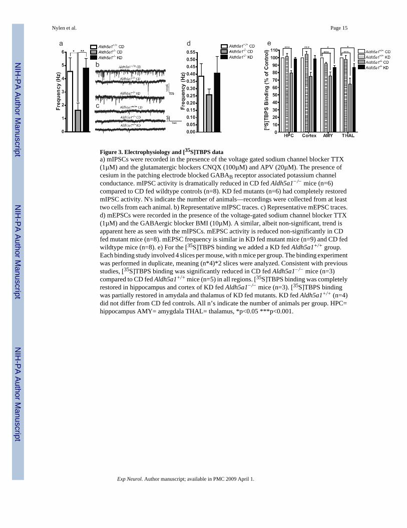
Figure 3. Electrophysiology and [35S]TBPS dataa) mIPSCs were recorded in the presence of the voltage gated sodium channel blocker TTX(1µM) and the glutamatergic blockers CNQX (100µM) and APV (20µM). The presence ofcesium in the patching electrode blocked GABAB receptor associated potassium channelconductance. mIPSC activity is dramatically reduced in CD fed Aldh5a1−/− mice (n=6)compared to CD fed wildtype controls (n=8). KD fed mutants (n=6) had completely restoredmIPSC activity. N's indicate the number of animals—recordings were collected from at leasttwo cells from each animal. b) Representative mIPSC traces. c) Representative mEPSC traces.d) mEPSCs were recorded in the presence of the voltage-gated sodium channel blocker TTX(1µM) and the GABAergic blocker BMI (10µM). A similar, albeit non-significant, trend isapparent here as seen with the mIPSCs. mEPSC activity is reduced non-significantly in CDfed mutant mice (n=8). mEPSC frequency is similar in KD fed mutant mice (n=9) and CD fedwildtype mice (n=8). e) For the [35S]TBPS binding we added a KD fed Aldh5a1+/+ group.Each binding study involved 4 slices per mouse, with n mice per group. The binding experimentwas performed in duplicate, meaning (n*4)*2 slices were analyzed. Consistent with previousstudies, [35S]TBPS binding was significantly reduced in CD fed Aldh5a1−/− mice (n=3)compared to CD fed Aldh5a1+/+ mice (n=5) in all regions. [35S]TBPS binding was completelyrestored in hippocampus and cortex of KD fed Aldh5a1−/− mice (n=3). [35S]TBPS bindingwas partially restored in amydala and thalamus of KD fed mutants. KD fed Aldh5a1+/+ (n=4)did not differ from CD fed controls. All n’s indicate the number of animals per group. HPC=hippocampus AMY= amygdala THAL= thalamus, *p<0.05 ***p<0.001.
Nylen et al. Page 15
Exp Neurol. Author manuscript; available in PMC 2009 April 1.
NIH
-PA Author Manuscript
NIH
-PA Author Manuscript
NIH
-PA Author Manuscript

NIH
-PA Author Manuscript
NIH
-PA Author Manuscript
NIH
-PA Author Manuscript
Nylen et al. Page 16
Table 1Composition of Experimental Diets.
Nutrient DietKD (g) CD (g)
Protein 146.97 234Carbohydrate 30 490Fat 707.83 100Vitamins 4.6 54Minerals 44.47 69Fibre 66.09 53
4:1 KD. The AIN-93G vitamin mix used in the 4:1 KD contains no carbohydrates. Soybean oil came from the vitamin mix where it serves as a carrier forfat-soluble vitamins. The type of fat (i.e. butter or lard) can be varied. Adapted from Likhodii, 2001, modified by reversing quantities of lard and butter.CD. The ash content of the CD is derived from formula AIN-76 mineral mix #F8505. The vitamin mix is derived from formula AIN-76 vitamin mix#F8000. Adapted from Bough et al., 1999. g: grams; KD: ketogenic diet; CD: control diet.
Exp Neurol. Author manuscript; available in PMC 2009 April 1.
NIH
-PA Author Manuscript
NIH
-PA Author Manuscript
NIH
-PA Author Manuscript
Nylen et al. Page 17
Table 2Average Group Serum Glucose, Beta-hydroxybutyrate and Non-esterified Fatty Acid Levels.
Glucose βOHB NEFA
Aldh5a1+/+ CD (n=6) 9.03±2.55 0.59±0.38 0.73±0.17Aldh5a1−/− CD (n=5) 7.03±0.68^ 1.11±0.29* 0.81±0.34Aldh5a1+/+ KD (n=5) 9.73±0.37 1.12±0.36* 1.16±0.46Aldh5a1−/− KD (n=7) 10.40±0.34 2.06±0.48¶ 0.95±0.27
*p<0.05
^p<0.01
¶p<0.001
one-way ANOVA; βOHB beta-hydroxybutyrate. In terms of glucose, CD fed Aldh5a1−/− mice only had significantly lower serum glucose levels than
KD fed animals. They did not differ significantly from wildtype controls. CD fed Aldh5a1−/− mice and KD fed Aldh5a1+/+ mice had significantly
higher βOHB levels than wildtype controls. KD fed Aldh5a1−/− mice had significantly higher βOHB than all other groups. NEFAs did not differsignificantly between groups.
Exp Neurol. Author manuscript; available in PMC 2009 April 1.


![[National consensus on the ketogenic diet]](https://img.dokumen.tips/doc/110x75/635c6467095e4caf22053447/national-consensus-on-the-ketogenic-diet.jpg)


