Embed Size (px)
Citation preview
Molecular Biology of the CellVol. 9, 2093–2106, August 1998
A Developmentally Regulated Kinesin-related MotorProtein from Dictyostelium discoideumEugenio L. de Hostos,*† Gretchen McCaffrey,‡§ Richard Sucgang,|Daniel W. Pierce,‡ and Ronald D. Vale‡¶
*Department of Biochemistry and Cell Biology, Rice University, Houston Texas 77005; ‡Department ofMolecular and Cellular Pharmacology and ¶Howard Hughes Medical Institute, University ofCalifornia, San Francisco, California 94143; and \Department of Biochemistry, Baylor College ofMedicine, Houston Texas 77030
Submitted August 22, 1997; Accepted April 20, 1998Monitoring Editor: J. Richard McIntosh
The cellular slime mold Dictyostelium discoideum is an attractive system for studying theroles of microtubule-based motility in cell development and differentiation. In this work,we report the first molecular characterization of kinesin-related proteins (KRPs) inDictyostelium. A PCR-based strategy was used to isolate DNA fragments encoding sixKRPs, several of which are induced during the developmental program that is initiatedby starvation. The complete sequence of one such developmentally regulated KRP(designated K7) was determined and found to be a novel member of the kinesinsuperfamily. The motor domain of K7 is most similar to that of conventional kinesin, butunlike conventional kinesin, K7 is not predicted to have an extensive a-helical coiled-coildomain. The nonmotor domain is unusual and is rich in Asn, Gln, and Thr residues;similar sequences are found in other developmentally regulated genes in Dictyostelium.K7, expressed in Escherichia coli, supports plus end–directed microtubule motility in vitroat a speed of 0.14 mm/s, indicating that it is a bona fide motor protein. The K7 motor isfound only in developing cells and reaches a peak level of expression between 12 and16 h after starvation. By immunofluorescence microscopy, K7 localizes to a membranousperinuclear structure. To examine K7 function, we prepared a null cell line but found thatthese cells show no gross developmental abnormalities. However, when cultivated in thepresence of wild-type cells, the K7-null cells are mostly absent from the prestalk zone ofthe slug. This result suggests that in a population composed largely of wild-type cells, theabsence of the K7 motor protein interferes either with the ability of the cells to localize tothe prestalk zone or to differentiate into prestalk cells.
INTRODUCTION
Since the prototype kinesin was purified from squidaxoplasm (Lasek and Brady, 1985; Schroer et al., 1985;Vale et al., 1985), dozens of related proteins (herereferred to as kinesins or kinesin-related proteins[KRPs]) have been identified in organisms rangingfrom fungi to mammals (Moore and Endow, 1996).KRPs share a conserved mechanochemical motor do-
main responsible for ATP hydrolysis and microtubulebinding. Outside of the 350-amino acid-long motordomain, KRPs show great sequence diversity, but re-gions predicted to mediate homodimerization via for-mation of coiled-coil structures are often present(Goldstein, 1993). Sequence diversity among KRPsoutside of the motor domain is thought to play a keyrole in their functional diversity by specifying whichproteins and cargo associate with the motor (Gold-stein, 1993). Kinesins have been shown to drive themovement of vesicles in fast axonal transport (Halland Hedgecock, 1991; Hurd and Saxton, 1996; Hurd etal., 1996), the movement of vesicles between the Golgi
†Corresponding Author. E-mail address: [email protected].§Present Address: Department of Microbiology and Genetics,Massey University, Palmerston North, New Zealand.
© 1998 by The American Society for Cell Biology 2093
and the ER (Lippincott-Schwartz and Cole, 1995; Lip-pincott-Schwartz et al., 1995), and the movement ofother organelles, including secretory vesicles, lyso-somes, mitochondria, and nuclei (Hirokawa, 1996;Lopez, 1996; Cottingham and Hoyt, 1997; DeZwaan, etal., 1997; Pereira et al., 1997). Kinesins have also beenshown to be involved in the organization, assembly,and operation of the mitotic spindle (Barton and Gold-stein, 1996; Walczak and Mitchison, 1996) as well as inthe function of the kinetochores, which connect chro-mosomes to microtubules (Brown et al., 1996).
Several studies have also implicated kinesins in de-velopmental processes. Microtubules are known to benecessary for the proper localization of Vg1 RNA inXenopus oocytes (Yisraeli et al., 1990), and it has beenproposed that microtubule-based motors are involvedin this and other cases of RNA localization (for review,see Wilhelm and Vale, 1993). A role in developmenthas been demonstrated for the kinesin-related proteinXklp1 in Xenopus, which is required for both spindleassembly and for the aggregation of germ plasm inearly Xenopus embryos (Robb et al., 1996). In Drosoph-ila, COS2 is a distant kinesin relative, which is thoughtto tether a signaling complex consisting of the fusedkinase and the cubitus interruptus transcription factorto microtubules (Robbins, et al., 1997; Sisson, et al.,1997). The access of the complex to the nucleus is inturn believed to be regulated by the hedgehog geneproduct, which may reverse the binding of COS2 tomicrotubules.
Dictyostelium has many advantages as a model sys-tem for the study of the cytoskeleton (for reviews, seeSchleicher and Noegel, 1992; Schleicher et al., 1995)and developmental processes (for reviews, see Firtel,1996; Loomis, 1996). Like yeast, it offers the possibilityof combining biochemical and genetic approaches, butin addition, it shows a full spectrum of cellular func-tions, including chemotaxis and multicellular devel-opment. Dictyostelium has been used extensively in thestudy of actin-based myosin motors (Novak et al.,1995; Jung et al., 1996; Temesvari et al., 1996; Wessels etal., 1996). Work on microtubule-based motors has in-cluded the characterization of cytoplasmic dynein(Koonce and McIntosh, 1990; Koonce et al., 1992, 1994)and the purification of a protein with the properties ofa kinesin motor from vegetative cells (McCaffrey andVale, 1989). The following is the first report on themolecular biology of kinesin motors in this organism.
Here, we describe the identification of six KRPs,several of which are induced during the Dictyosteliumdevelopmental program. One motor (termed K7) wasextensively characterized; its complete sequence, de-velopmental expression pattern, motor activity, intra-cellular localization, and null phenotype are reportedin this work. K7 is a novel plus end–directed kinesinmotor with an unusual structure in the nonmotordomain. Transcription is developmentally regulated,
and the protein localizes to what appears to be amembranous perinuclear structure. In pure culture,the mutant null cells show no obvious phenotype.However, when mixed with wild-type cells, the K7-null cells are absent from the prestalk zone of the slug.This result reveals a developmental defect in an envi-ronment where mutant and wild-type cells are bothpresent.
MATERIALS AND METHODS
Cloning of Dictyostelium KRPsFully degenerate oligonucleotides (kindly provided by Dr. MarkRose, Princeton University, Princeton, NJ) corresponding to thehighly conserved kinesin peptides IFAYGQT and LVDLAGSE(Stewart et al., 1991) were used to prime a PCR using genomicDictyostelium DNA as a template. Two PCR products (K2 and K4)were cloned and used as probes to screen a cDNA l-gt11 library(Clontech, Palo Alto, CA) made from cells that had been starved for4 h (developmental time points are referred to as hours after star-vation, e.g., “T4”). Two KRP cDNAs were isolated using each probe:K6 and K8 using K2, and K3 and K7 using K4. Southern andNorthern blots were hybridized at 65°C in phosphate buffer (250mM NaPO4, 250 mM NaCl, 10% wt/vol PEG 8000, 1 mM EDTA, pH7.2) as described (Amasino, 1986).
The entire 3.7-kb K7 insert was sequenced, and it was found to bea mixed clone of two unrelated cDNAs: one end of the insertencodes a KRP, whereas the other end encodes a protein with a highdegree of homology to the mitochondrial ribosomal protein S14.This insert is thought to be the product of a cloning artifact. Thebreakpoint between the two parts of the insert was located in aregion containing repeats of the trinucleotide AAC (nucleotides2640–2700).
To find additional cDNAs encoding K7, a l-Zap cDNA librarymade from T8–T12 cells (kindly provided by Dr. R. Firtel, Univer-sity of California, San Diego, CA) was screened using the fragmentof the original insert corresponding to the KRP and ending at anEcoRV site upstream of the breakpoint. A cDNA was obtained thatspanned the breakpoint in the original K7 clone but still appeared toterminate prematurely. The final piece of the K7 39 end was ob-tained by RT-PCR using a primer based on the 39 end of the l-Zapclone. Total RNA was extracted from T12 cells (RNeasy kit; Qiagen,Santa Clarita, CA) and used as a template for RT-PCR (39-Ampli-FINDER rapid amplification of cDNA ends 39 RT-PCR kit; Clontech,Palo Alto, CA) which yielded a product corresponding to the 39 endof the K7 transcript. Percent identity between protein sequences wascalculated using the program Bestfit of the Wisconsin Package (Ge-netics Computer Group, Madison, WI). Multiple sequence align-ment was done with the program Pileup (Genetics ComputerGroup). Coiled-coil predictions were made using the worldwideweb version of the program Paircoil (Berger et al., 1995) available athttp://ostrich.lcs.mit.edu/cgi-bin/score.
Gene DisruptionThe K7 gene was disrupted following a previously described strat-egy (de Hostos et al., 1993) designed to replace the endogenous copyof the gene with a modified copy interrupted by an antibioticresistance cassette. Using the first 2 kb of the K7 cDNA as a tem-plate, 59 and 39 end fragments (corresponding to nucleotides 32–1073 and 1502–2723, respectively) containing appropriate restrictionsites were generated by PCR and subcloned. A cassette conferringblasticidin resistance (kindly provided by Dr. H. Adachi, Universityof Tokyo, Tokyo, Japan; Adachi et al., 1994) was cloned between thetwo K7 fragments to complete the gene disruption construct. Plas-mid DNA was digested to release the gene disruption construct
E.L. de Hostos et al.
Molecular Biology of the Cell2094
from the vector and transformed into cells by electroporation asdescribed (de Hostos et al., 1993). Transformants were selected inDD-broth 20 media (Manstein et al., 1995) containing 5 mg/mlblasticidin (ICN, Costa Mesa, CA).
AntibodiesA PCR-generated fragment encoding the first 520 amino acids of K7was cloned into the EcoRI site of the maltose-binding protein (MBP)expression vector pMAL-p2 (New England BioLabs, Beverly, MA).The K7/MBP fusion protein was expressed in Escherichia coli andpurified by affinity chromatography over an amylose matrix asspecified by the manufacturer (New England BioLabs). Recombi-nant protein was eluted with maltose in PBS and used for immu-nization of a rabbit (Cocalico Biologicals, Reamstown, PA). Anti-serum was used at a dilution of 1:1000–3000 for Western blotting,using alkaline phosphatase-coupled secondary antibodies (Harlowand lane, 1988). To reduce background labeling in later Westernblots and immunofluorescence labeling experiments, anti-K7/MBPantiserum was preadsorbed with strips of nitrocellulose previouslysoaked in a mixture of an extract of E. coli expressing MBP andDictyostelium T12 K7-knock-out (KO) cells.
For immunofluorescence, cells were grown on glass coverslips.The coverslips with cells were rinsed in Soerensen phosphate buffer(Malchow et al., 1972) and then submerged in buffer to a depth of ;2mm for 6–12 h. After development, the coverslips were tapped dryand submerged in dehydrated methanol containing 1% formalde-hyde at 215°C (Fukui et al., 1987). After 10 min, the cover slips wereremoved, allowed to dry, rinsed with 0.5% Tween in Tris-bufferedsaline (TNT; Sambrook et al., 1989), and blocked with 5% BSA inTNT. Preadsorbed antiserum diluted 1:30–100 in BSA-TNT wasadded and incubated for 30 min at 37°C. Cells were washed twicewith TNT for 10 min and then incubated with FITC-labeled anti-rabbit antibodies (Sigma, St. Louis, MO) under the same conditionsas the primary antiserum. After labeling the cells were washed asbefore, except that DAPI (1 mg/ml; Sigma) was included in anadditional 5-min wash. The coverslips were rinsed in distilled wa-ter, air dried, and mounted using Vectashield medium (VectorLaboratories, Burlingame, CA). Cells were observed in a Zeiss(Oberkochen, Germany) Axiophot microscope, and images werecaptured on slide film (Sensia ASA 400; Fuji, Tokyo, Japan). In theexperiments involving cells expressing green fluorescent protein(GFP) targeted to the ER, the cells were fixed under an agar overlay(Fukui et al., 1987), and anti-K7/MBP antibodies were followed byTexas Red-labeled anti-rabbit antibodies (Vector Laboratories). An-ti-Kar2p antibodies (Ng and Walter, 1996) were kindly provided byDrs. Davis Ng and Peter Walter (University of California, SanFancisco, CA). Antinuclear pore antibodies (Snow et al., 1987) werekindly provided by Drs. R. Mahajan and L. Gerace (Scripps Re-search Institute, La Jolla, CA).
Developmental Time CourseCells were grown in DD-broth 20 media to a density of 5 3 106
cells/ml and then collected by centrifugation, washed once in phos-phate buffer (Malchow et al., 1972), and resuspended in the samebuffer at a density of 2 3 108 cells/ml. One milliliter of cells wasplaced on a phosphate buffer-agar plate for development. Develop-ing cells were collected from the plates at the appropriate timepoints and either lysed in SDS gel-loading buffer for protein elec-trophoresis or subjected to RNA extraction (RNeasy). Protein sam-ples containing 1 3 106 cell equivalents were subjected to SDS-PAGE (Laemmli, 1970) through 8% gels and then transferred tonitrocellulose using a semidry blotting apparatus (E & K, Saratoga,CA). Ten micrograms of RNA for each time point were analyzed byNorthern blotting as described above. As a hybridization probe, theEcoRV fragment of the K7 cDNA corresponding to the N terminusof the protein was radioactively labeled by random priming usingthe RediPrime system (Amersham, Arlington Heights, IL).
Expression of K7-GFP in E. coliThe same K7 fragment that was used for the generation of a K7/MBP antigen (residues 1–520) was cloned into the EcoRI site of thehistidine tag expression vector pET23a (Novagen, Madison, WI).The coding sequence of the Aequorea victoria green fluorescent pro-tein (S65T GFP mutant; Heim et al., 1995) was cloned into the HincIIand XhoI sites of the vector. GFP was included in the construct tomake the fusion protein suitable for future single-molecule fluores-cence experiments (see Pierce et al., 1997); the green color of thefusion protein also serves as a convenient marker during purifica-tion. BL21(DE3) E. coli cells transformed with the expression con-struct were grown to an OD600 of 1–2 at 37°C in low-salt TPMmedium (amounts per liter: 20 g tryptone, 15 g yeast extract, 4 gNaCl, 2 g Na2HPO4, 1 g NaH2PO4, 2 g glucose, 0.1 g ampicillin, pH7.0). Cultures were cooled to 23°C, induced by the addition of 0.2mM isopropyl-1-thio-b-d-galactopyranoside, and grown for an ad-ditional 14 h at 23°C. Cells were harvested by centrifugation andstored at 280°C. Cell pellets from 1 l of culture were thawed andresuspended in 25 ml of lysis buffer (50 mM NaPO4, pH 8.0, 250 mMNaCl, 20 mM imidazole, 250 mM NaCl, 1 mM MgCl2, 0.5 mM ATP,10 mM b-mercaptoethanol [b-ME]) containing leupeptin (1 mg/ml),pepstatin (1 mg/ml), chymostatin (1 mg/ml), aprotinin (1 mg/ml),and 0.25 mg/ml Pefabloc (Boehringer Mannheim, Indianapolis, IN)and disrupted in a French press. The lysate was clarified by centrif-ugation for 30 min at 28,000 3 g, and the supernatant was incubatedwith Ni-nitrilotriacetic acid resin (Qiagen) for 1 h at 4°C (1.5-ml bedvolume of resin per 50 ml of supernatant). The mixture was thentransferred to a disposable column, and the resin was washed with50 ml of 50 mM NaPO4 (pH 6), 250 mM NaCl, 1 mM MgCl2, 0.1 mMATP, and 10 mM b-ME. K7-GFP was eluted with 50 mM NaPO4, 500mM imidazole, 250 mM NaCl, 1 mM MgCl2, 0.1 mM ATP, and 10mM b-ME (pH 7.2). The peak fractions were then diluted 20-foldinto column buffer [25 mM Na-piperazine-N,N9-bis(2-ethanesul-fonic acid), pH 6.8, 2 mM MgCl2, 1 mM EGTA, 1 mM DTT, and 0.1mM ATP] and further purified by FPLC chromatography on amono-Q column (Pharmacia, Piscataway, NJ) using a 20-ml 0–1.0 MNaCl gradient in column buffer. K7-GFP eluted as a sharp bandcentered at 290 mM NaCl.
In Vitro Motility AssayPolarity-marked microtubules were prepared and an in vitro motil-ity assay was performed essentially as described (Howard andHyman, 1993), except that microtubules were added in BRB12 buffer[12 mM K-Na-piperazine-N,N9-bis(2-ethanesulfonic acid) pH 6.8, 2mM MgCl2, 1 mM EGTA, 1 mM ATP[ containing 2 mg/ml caseinand 20 mM taxol (Molecular Probes, Eugene, OR) and a coupledenzyme oxygen scavenger system (Harada et al., 1990). Recombi-nant motor protein eluted from the mono-Q purification step wasloaded directly into a flow cell and allowed to bind to the glasssurface. Microtubules were introduced into the chamber and im-aged with a Zeiss Axiophot microscope. Images were obtainedusing a silicon intensifier target camera (Hamamatsu Photonics,Bridgewater, NJ) and recorded to sVHS tape. Individual frameswere grabbed from the video using an Apple Power Macintoshrunning Adobe (San Diego, CA) Premiere. Velocities declined onprolonged illumination when using fluorescent microtubules; there-fore velocity measurements reported were performed using unla-beled microtubules imaged by differential interference contrast andcontrast enhanced using an Argus 20 image processor(Hamamatsu).
Preparation of Dictyostelium MembranesCells were starved in suspension culture for 12 h at a density of 1 3107 cells/ml of phosphate buffer. Crude membranes were preparedessentially as described (Goodloe-Holland and Luna, 1987). Thecells were resuspended in lysis buffer supplemented with a proteaseinhibitor mix (Complete, Boehringer Mannheim) and 1 mM DTT.
A Developmentally Regulated Kinesin
Vol. 9, August 1998 2095
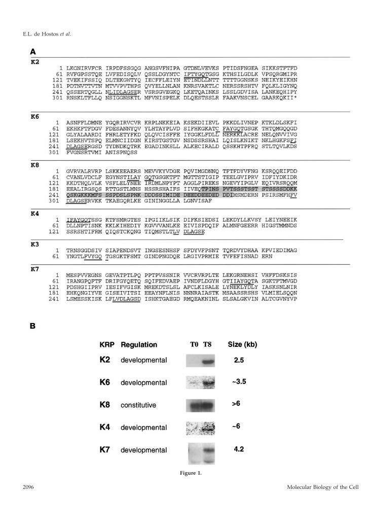
Figure 1.
E.L. de Hostos et al.
Molecular Biology of the Cell2096
The cells were broken by passage though a BioNeb cell disrupter(Glas-Col, Terre Haute, IN) at 150 psi of N2. The homogenate wascentrifuged for 20 min at 38,000 3 g to obtain a supernatant andcrude membrane pellet. Protein samples containing 1 3 106 cellequivalents were subjected to SDS-PAGE (Laemmli, 1970) through8% gels and Western blotting.
Construction of a GFP Marker for the ERThe coding sequence of the GFP S65T mutant was modified usingPCR by the addition to its 39 end of codons coding for the yeast ERretention signal HDEL (Pelham et al., 1988) and cloned into the KpnIand XbaI sites of the vector pDXA-3H (Manstein et al., 1995), whichcarries the marker neoR and expresses cloned inserts under thecontrol of the constitutive actin-15 promoter. To provide a transla-tion start site and a leader peptide, a DNA fragment encoding thefirst 38 amino acids of contact site A (Noegel et al., 1986) wasamplified by PCR from a cDNA library (de Hostos, unpublishedresults) made in the vector l-YES (Elledge et al., 1991) and clonedinto HindIII and KpnI sites upstream of the modified GFP. Theconstruct was cotransformed by electroporation (de Hostos et al.,1993) into AX2 cells with the pREP helper plasmid (Manstein et al.,1995), and transformants were selected and maintained in DD-broth20 media containing 20 mg/ml Geneticin (Life Technologies, Gaith-ersburg, MD).
Cell-tracking ExperimentsMixing experiments with fluorescently labeled tracer cells wereconducted essentially as described (Knecht and Shelden, 1995; Xu, etal., 1996). K7-null and wild-type AX2 cells (106 cells each) werelabeled by resuspending in 200 ml of phosphate buffer containing 50mM Cell Tracker Green (Molecular Probes) and shaking at roomtemperature for 20 min. The cells were washed twice in phosphatebuffer and added to 5% of a mixture with unlabeled AX2 cells. Cells(4 3 106) from the mixture were spread at high density on phos-phate agar plates containing 2% activated charcoal to provide con-trast during microscopy. Cell aggregates were photographed at T12.
RESULTS
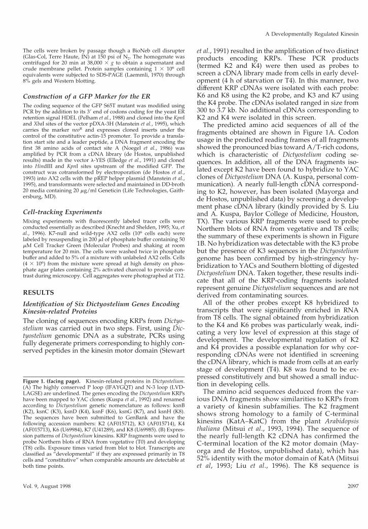
Identification of Six Dictyostelium Genes EncodingKinesin-related ProteinsThe cloning of sequences encoding KRPs from Dictyo-stelium was carried out in two steps. First, using Dic-tyostelium genomic DNA as a substrate, PCRs usingfully degenerate primers corresponding to highly con-served peptides in the kinesin motor domain (Stewart
et al., 1991) resulted in the amplification of two distinctproducts encoding KRPs. These PCR products(termed K2 and K4) were then used as probes toscreen a cDNA library made from cells in early devel-opment (4 h of starvation or T4). In this manner, twodifferent KRP cDNAs were isolated with each probe:K6 and K8 using the K2 probe, and K3 and K7 usingthe K4 probe. The cDNAs isolated ranged in size from300 to 3.7 kb. No additional cDNAs corresponding toK2 and K4 were isolated in this screen.
The predicted amino acid sequences of all of thefragments obtained are shown in Figure 1A. Codonusage in the predicted reading frames of all fragmentsshowed the pronounced bias toward A/T-rich codons,which is characteristic of Dictyostelium coding se-quences. In addition, all of the DNA fragments iso-lated except K2 have been found to hybridize to YACclones of Dictyostelium DNA (A. Kuspa, personal com-munication). A nearly full-length cDNA correspond-ing to K2, however, has been isolated (Mayorga andde Hostos, unpublished data) by screening a develop-ment phase cDNA library (kindly provided by S. Liuand A. Kuspa, Baylor College of Medicine, Houston,TX). The various KRP fragments were used to probeNorthern blots of RNA from vegetative and T8 cells;the summary of these experiments is shown in Figure1B. No hybridization was detectable with the K3 probebut the presence of K3 sequences in the Dictyosteliumgenome has been confirmed by high-stringency hy-bridization to YACs and Southern blotting of digestedDictyostelium DNA. Taken together, these results indi-cate that all of the KRP-coding fragments isolatedrepresent genuine Dictyostelium sequences and are notderived from contaminating sources.
All of the other probes except K8 hybridized totranscripts that were significantly enriched in RNAfrom T8 cells. The signal obtained from hybridizationto the K4 and K6 probes was particularly weak, indi-cating a very low level of expression at this stage ofdevelopment. The developmental regulation of K2and K4 provides a possible explanation for why cor-responding cDNAs were not identified in screeningthe cDNA library, which is made from cells at an earlystage of development (T4). K8 was found to be ex-pressed constitutively and but showed a small induc-tion in developing cells.
The amino acid sequences deduced from the var-ious DNA fragments show similarities to KRPs froma variety of kinesin subfamilies. The K2 fragmentshows strong homology to a family of C-terminalkinesins (KatA–KatC) from the plant Arabidopsisthaliana (Mitsui et al., 1993, 1994). The sequence ofthe nearly full-length K2 cDNA has confirmed theC-terminal location of the K2 motor domain (May-orga and de Hostos, unpublished data), which has52% identity with the motor domain of KatA (Mitsuiet al, 1993; Liu et al., 1996). The K8 sequence is
Figure 1. (facing page). Kinesin-related proteins in Dictyostelium.(A) The highly conserved P loop (IFAYGQT) and N-3 loop (LVD-LAGSE) are underlined. The genes encoding the Dictyostelium KRPshave been mapped to YAC clones (Kuspa et al., 1992) and renamedaccording to Dictyostelium genetic nomenclature as follows: ksnB(K2), ksnC (K3), ksnD (K4), ksnF (K6), ksnG (K7), and ksnH (K8).The sequences have been submitted to GenBank and have thefollowing accession numbers: K2 (AF015712), K3 (AF015714), K4(AF015713), K6 (U69984), K7 (U41289), and K8 (U69985). (B) Expres-sion patterns of Dictyostelium kinesins. KRP fragments were used toprobe Northern blots of RNA from vegetative (T0) and developing(T8) cells. Exposure times varied from blot to blot. Transcripts areclassified as “developmental” if they are expressed primarily in T8cells and “constitutive” when comparable amounts are detectable atboth time points.
A Developmentally Regulated Kinesin
Vol. 9, August 1998 2097
unusual in that it contains a segment ;67 residueslong, which has no counterpart in other kinesins.This segment is located in a region that correspondsto a surface loop (L10) in the crystal structure ofkinesin motor domains (Kull et al., 1996; Sablin et al.,1996), but its function is unknown.
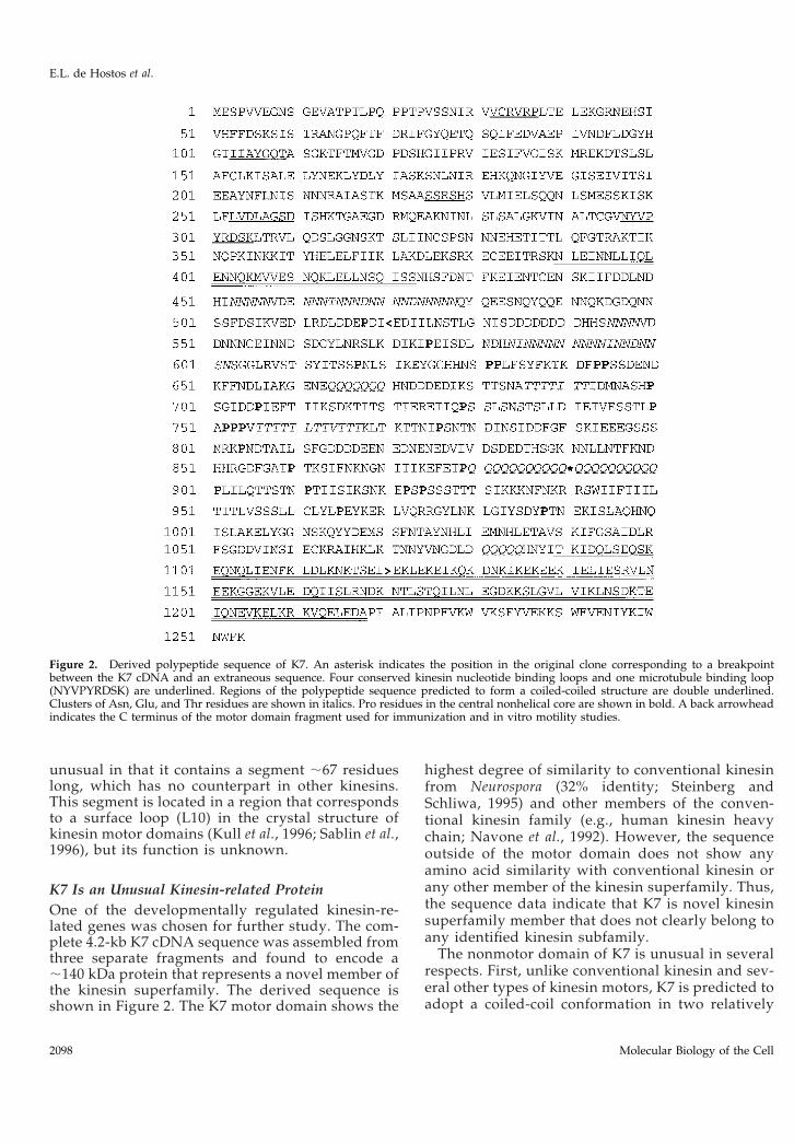
K7 Is an Unusual Kinesin-related ProteinOne of the developmentally regulated kinesin-re-lated genes was chosen for further study. The com-plete 4.2-kb K7 cDNA sequence was assembled fromthree separate fragments and found to encode a;140 kDa protein that represents a novel member ofthe kinesin superfamily. The derived sequence isshown in Figure 2. The K7 motor domain shows the
highest degree of similarity to conventional kinesinfrom Neurospora (32% identity; Steinberg andSchliwa, 1995) and other members of the conven-tional kinesin family (e.g., human kinesin heavychain; Navone et al., 1992). However, the sequenceoutside of the motor domain does not show anyamino acid similarity with conventional kinesin orany other member of the kinesin superfamily. Thus,the sequence data indicate that K7 is novel kinesinsuperfamily member that does not clearly belong toany identified kinesin subfamily.
The nonmotor domain of K7 is unusual in severalrespects. First, unlike conventional kinesin and sev-eral other types of kinesin motors, K7 is predicted toadopt a coiled-coil conformation in two relatively
Figure 2. Derived polypeptide sequence of K7. An asterisk indicates the position in the original clone corresponding to a breakpointbetween the K7 cDNA and an extraneous sequence. Four conserved kinesin nucleotide binding loops and one microtubule binding loop(NYVPYRDSK) are underlined. Regions of the polypeptide sequence predicted to form a coiled-coiled structure are double underlined.Clusters of Asn, Glu, and Thr residues are shown in italics. Pro residues in the central nonhelical core are shown in bold. A back arrowheadindicates the C terminus of the motor domain fragment used for immunization and in vitro motility studies.
E.L. de Hostos et al.
Molecular Biology of the Cell2098
short segments that are widely separated from oneanother (Figure 2, double underlined). Based on astructure prediction using the program Paircoil(Berger et al., 1995), the nonmotor domain (begin-ning approximately after residue 350) can be di-vided into a central non-a helical core (residues424-1089) that has 22 scattered Pro residues, flankedby two segments predicted to have a largely coiled-coil conformation (residues 390 – 423 and1090 –1218). The nonhelical central domain of K7 isalso different from any other known kinesin in thatit contains clustered repeats of Asn, Thr, or Glnresidues (Figure 2, italicized). Most of the poly-Glnrepeats and many of the residues in the clusters ofAsn and Thr are encoded by the trinucleotide AACin the alternate reading frames CAA, AAC, andACA, respectively. The significance of the nucleo-tide repeats and the amino acid clusters is notknown, but similar repeats have been identified inother developmentally regulated genes in Dictyoste-lium (Shaw et al., 1989). The structures adopted bythese repeat elements are unknown.
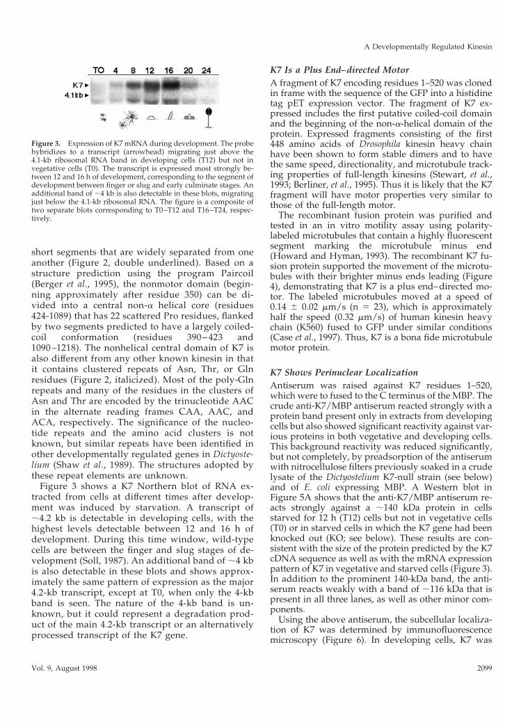
Figure 3 shows a K7 Northern blot of RNA ex-tracted from cells at different times after develop-ment was induced by starvation. A transcript of;4.2 kb is detectable in developing cells, with thehighest levels detectable between 12 and 16 h ofdevelopment. During this time window, wild-typecells are between the finger and slug stages of de-velopment (Soll, 1987). An additional band of ;4 kbis also detectable in these blots and shows approx-imately the same pattern of expression as the major4.2-kb transcript, except at T0, when only the 4-kbband is seen. The nature of the 4-kb band is un-known, but it could represent a degradation prod-uct of the main 4.2-kb transcript or an alternativelyprocessed transcript of the K7 gene.
K7 Is a Plus End–directed MotorA fragment of K7 encoding residues 1–520 was clonedin frame with the sequence of the GFP into a histidinetag pET expression vector. The fragment of K7 ex-pressed includes the first putative coiled-coil domainand the beginning of the non-a-helical domain of theprotein. Expressed fragments consisting of the first448 amino acids of Drosophila kinesin heavy chainhave been shown to form stable dimers and to havethe same speed, directionality, and microtubule track-ing properties of full-length kinesins (Stewart, et al.,1993; Berliner, et al., 1995). Thus it is likely that the K7fragment will have motor properties very similar tothose of the full-length motor.
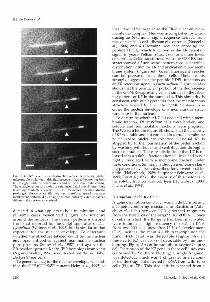
The recombinant fusion protein was purified andtested in an in vitro motility assay using polarity-labeled microtubules that contain a highly fluorescentsegment marking the microtubule minus end(Howard and Hyman, 1993). The recombinant K7 fu-sion protein supported the movement of the microtu-bules with their brighter minus ends leading (Figure4), demonstrating that K7 is a plus end–directed mo-tor. The labeled microtubules moved at a speed of0.14 6 0.02 mm/s (n 5 23), which is approximatelyhalf the speed (0.32 mm/s) of human kinesin heavychain (K560) fused to GFP under similar conditions(Case et al., 1997). Thus, K7 is a bona fide microtubulemotor protein.
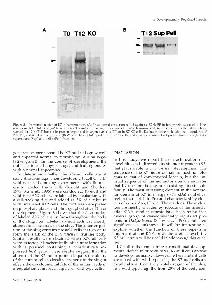
K7 Shows Perinuclear LocalizationAntiserum was raised against K7 residues 1–520,which were to fused to the C terminus of the MBP. Thecrude anti-K7/MBP antiserum reacted strongly with aprotein band present only in extracts from developingcells but also showed significant reactivity against var-ious proteins in both vegetative and developing cells.This background reactivity was reduced significantly,but not completely, by preadsorption of the antiserumwith nitrocellulose filters previously soaked in a crudelysate of the Dictyostelium K7-null strain (see below)and of E. coli expressing MBP. A Western blot inFigure 5A shows that the anti-K7/MBP antiserum re-acts strongly against a ;140 kDa protein in cellsstarved for 12 h (T12) cells but not in vegetative cells(T0) or in starved cells in which the K7 gene had beenknocked out (KO; see below). These results are con-sistent with the size of the protein predicted by the K7cDNA sequence as well as with the mRNA expressionpattern of K7 in vegetative and starved cells (Figure 3).In addition to the prominent 140-kDa band, the anti-serum reacts weakly with a band of ;116 kDa that ispresent in all three lanes, as well as other minor com-ponents.
Using the above antiserum, the subcellular localiza-tion of K7 was determined by immunofluorescencemicroscopy (Figure 6). In developing cells, K7 was
Figure 3. Expression of K7 mRNA during development. The probehybridizes to a transcript (arrowhead) migrating just above the4.1-kb ribosomal RNA band in developing cells (T12) but not invegetative cells (T0). The transcript is expressed most strongly be-tween 12 and 16 h of development, corresponding to the segment ofdevelopment between finger or slug and early culminate stages. Anadditional band of ;4 kb is also detectable in these blots, migratingjust below the 4.1-kb ribosomal RNA. The figure is a composite oftwo separate blots corresponding to T0–T12 and T16–T24, respec-tively.
A Developmentally Regulated Kinesin
Vol. 9, August 1998 2099
detected in what appears to be a membranous andin some cases vesiculated (Figure 6a) structurearound the nucleus. The overall pattern is distinctfrom that reported for the Golgi apparatus of Dic-tyostelium (Weiner, et al., 1993) but is similar to thatexpected for the nuclear envelope. To determinewhether the structure labeled could be the nuclearenvelope, antibodies against mammalian nuclearpore proteins (Snow et al., 1987) and against theER-resident protein Kar2p (BiP homolog) from yeast(Ng and Walter, 1996) were tested but did not labelDictyostelium cells.
To generate a tag for the nuclear envelope, we mod-ified the GFP (GFP S65T mutant; Heim et al., 1995) so
that it would be targeted to the ER–nuclear envelopemembrane complex. This was accomplished by intro-ducing an N-terminal signal sequence derived fromthe contact site A cell adhesion glycoprotein (Noegel etal., 1986) and a C-terminal sequence encoding thepeptide HDEL, which functions as the ER retentionsignal in yeast (Pelham et al., 1988) and other lowereukaryotes. Cells transformed with the GFP-ER con-struct showed a fluorescence pattern consistent with adistribution within the ER and nuclear envelope mem-brane system (Figure 6d). Green fluorescent vesiclescan be prepared from these cells. These resultsstrongly suggest that the peptide HDEL functions asan ER retention signal in Dictyostelium. Figure 6d alsoshows that the perinuclear portion of the fluorescencein the GFP-ER–expressing cells is similar to the label-ing pattern of K7 in the same cells. This similarity isconsistent with our hypothesis that the membranousstructure labeled by the anti-K7/MBP antiserum iseither the nuclear envelope or a membranous struc-ture close to the nucleus.
To determine whether K7 is associated with a mem-brane fraction, Dictyostelium cells were broken, andsoluble and sedimentable fractions were prepared.The Western blot in Figure 5b shows that the majorityof K7 is soluble and not enriched in a crude membranepellet where nuclei are expected. Residual K7 isstripped by further purification of the pellet fractionby washing with buffer and centrifugation through asucrose gradient. These results indicate that K7 is re-leased into a soluble fraction after cell lysis and is nottightly associated with a membrane fraction underthese conditions. Similarly, although membrane stain-ing patterns have been described for conventional ki-nesin (Hollenbeck, 1989; Lippincott-Schwartz et al.,1995; Lin et al., 1996), the majority of this motor is inthe soluble fraction after cell lysis (Hollenbeck, 1989;Niclas et al., 1994).
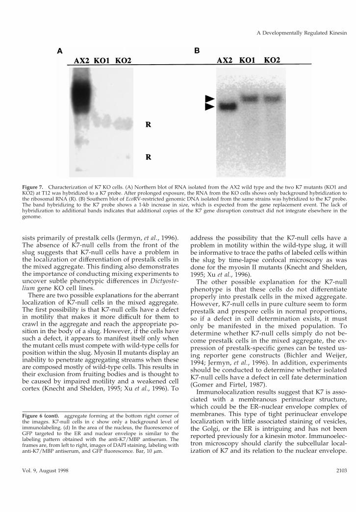
Disruption of the K7 GeneA gene disruption construct was made by insertinga cassette conferring resistance to blasticidin (Ada-chi et al., 1994) between PCR-generated fragmentsfrom the first 2 kb of the original K7 cDNA. Clonesof cells in which the K7 gene had been inactivatedwere found at a high frequency (.80%). In RNAfrom two KO cell lines after 12 h of development(T12), neither the main 4.2-kb transcript nor theminor 4-kb band was detectable (Figure 7A). Inthese cells, K7 was also not detectable by immuno-blotting (Figure 5A) or immunofluorescence (Figure6c). Disruption of the K7 gene in these cell lines wasconfirmed by Southern blotting; a single fragmentwas detected, which was 1 kb greater in size com-pared the fragment detected in DNA from wild-typecells (Figure 7B). This size shift is expected from a
Figure 4. K7 is a plus end–directed motor. A polarity-labeledmicrotubule is shown in this fluorescence image to be moving fromleft to right, with the bright minus end of the microtubule leading.The triangle serves as a point of reference. Bar, 1 mm. Frames weretaken approximately every 10 s, but velocities declined duringprolonged fluorescence illumination; therefore, speed measure-ments were performed by imaging microtubules by video-enhanceddifferential interference contrast.
E.L. de Hostos et al.
Molecular Biology of the Cell2100
gene replacement event. The K7-null cells grew welland appeared normal in morphology during vege-tative growth. In the course of development, thenull cells formed fingers, slugs, and fruiting bodieswith a normal appearance.
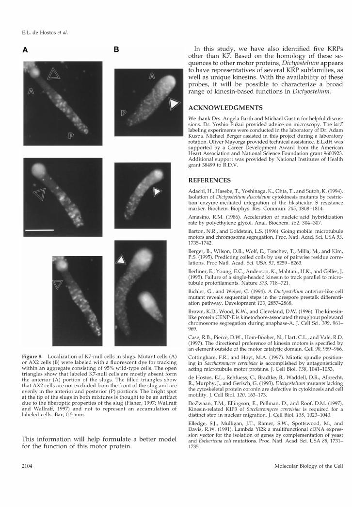
To determine whether the K7-null cells are atsome disadvantage when developing together withwild-type cells, mixing experiments with fluores-cently labeled tracer cells (Knecht and Shelden,1995; Xu et al., 1996) were conducted. K7-null andwild-type AX2 cells were labeled by incubation witha cell-tracking dye and added as 5% of a mixturewith unlabeled AX2 cells. The mixtures were platedon phosphate plates and photographed after 12 h ofdevelopment. Figure 8 shows that the distributionof labeled AX2 cells is uniform throughout the bodyof the slugs, but labeled K7-null cells are mostlyabsent from the front of the slug. The anterior por-tion of the slug contains prestalk cells that go on toform the stalk of the Dictyostelium fruiting body.Similar results were obtained when K7-null cellswere detected histochemically after transformationwith a plasmid containing a constitutively ex-pressed lacZ gene. These results suggest that theabsence of the K7 motor protein impairs the abilityof the mutant cells to localize properly in the slug oraffects the developmental fate of the mutant cells ina population composed largely of wild-type cells.
DISCUSSION
In this study, we report the characterization of anovel plus end– directed kinesin motor protein (K7)that plays a role in Dictyostelium development. Thesequence of the K7 motor domain is most homolo-gous to that of conventional kinesin, but the un-usual sequence of the nonmotor domain indicatesthat K7 does not belong to an existing kinesin sub-family. The most intriguing element in the nonmo-tor domain of K7 is a large (;74 kDa) nonhelicalregion that is rich in Pro and characterized by clus-ters of either Asn, Gln, or Thr residues. These clus-ters are mostly encoded by repeats of the trinucle-otide CAA. Similar repeats have been found in adiverse group of developmentally regulated pro-teins in Dictyostelium (Shaw et al., 1989), but theirsignificance is unknown. It will be interesting toexplore whether the function of these repeats isimportant at the RNA or at the protein level; theK7-null strain will be useful in addressing this ques-tion.
K7-null cells demonstrate a conditional develop-mental defect. In pure cultures, K7-null cells appearto develop normally. However, when mutant cellsare mixed with wild-type cells, the K7-null cells aremostly absent from the prestalk region of the slug.In a wild-type slug, the front 20% of the body con-
Figure 5. Immunodetection of K7 in Western blots. (A) Preadsorbed antiserum raised against a K7/MBP fusion protein was used to labela Western blot of total Dictyostelium proteins. The antiserum recognizes a band of ;140 kDa (arrowhead) in proteins from cells that have beenstarved for 12 h (T12) but not in proteins expressed in vegetative cells (T0) or in K7 KO cells. Dashes indicate molecular mass standards of205, 116, and 66 kDa, respectively. (B) Western blot of total proteins from T12 cells, and equivalent amounts of protein found in 38,000 3 gsupernatant (Sup) and pellet (Pell) fractions.
A Developmentally Regulated Kinesin
Vol. 9, August 1998 2101
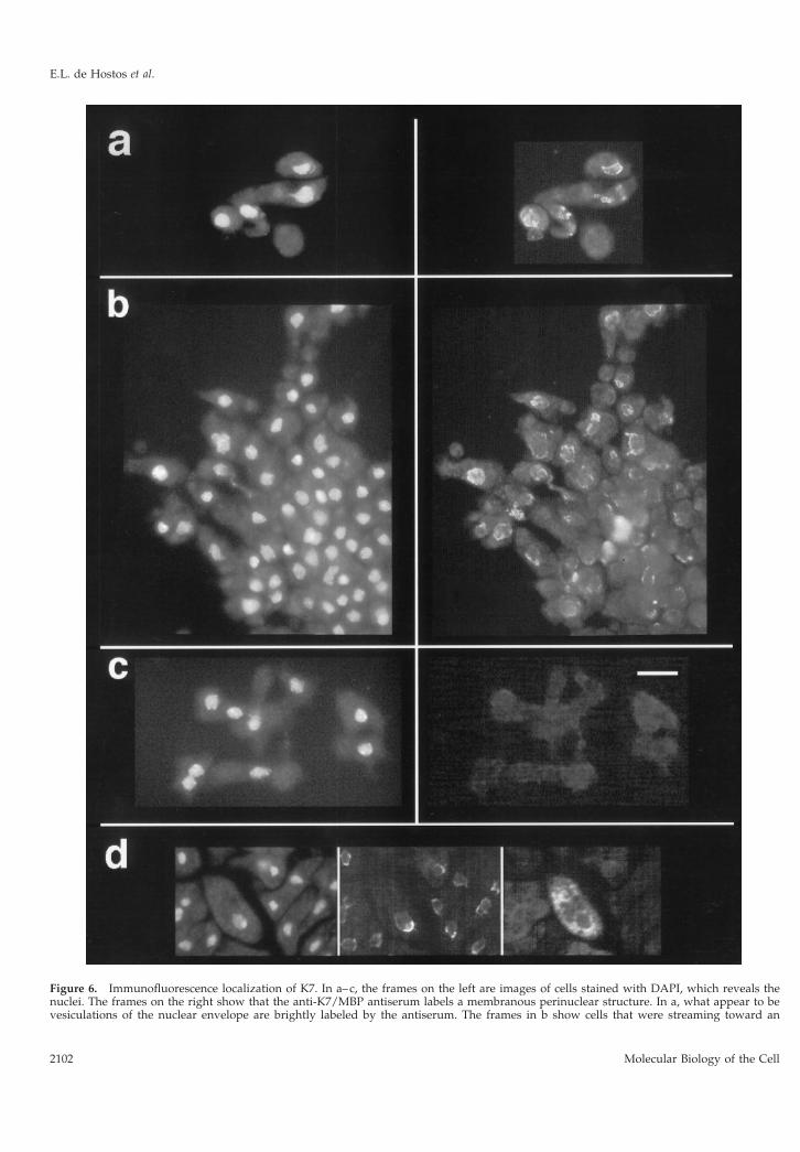
Figure 6. Immunofluorescence localization of K7. In a–c, the frames on the left are images of cells stained with DAPI, which reveals thenuclei. The frames on the right show that the anti-K7/MBP antiserum labels a membranous perinuclear structure. In a, what appear to bevesiculations of the nuclear envelope are brightly labeled by the antiserum. The frames in b show cells that were streaming toward an
E.L. de Hostos et al.
Molecular Biology of the Cell2102
sists primarily of prestalk cells (Jermyn, et al., 1996).The absence of K7-null cells from the front of theslug suggests that K7-null cells have a problem inthe localization or differentiation of prestalk cells inthe mixed aggregate. This finding also demonstratesthe importance of conducting mixing experiments touncover subtle phenotypic differences in Dictyoste-lium gene KO cell lines.
There are two possible explanations for the aberrantlocalization of K7-null cells in the mixed aggregate.The first possibility is that K7-null cells have a defectin motility that makes it more difficult for them tocrawl in the aggregate and reach the appropriate po-sition in the body of a slug. However, if the cells havesuch a defect, it appears to manifest itself only whenthe mutant cells must compete with wild-type cells forposition within the slug. Myosin II mutants display aninability to penetrate aggregating streams when theseare composed mostly of wild-type cells. This results intheir exclusion from fruiting bodies and is thought tobe caused by impaired motility and a weakened cellcortex (Knecht and Shelden, 1995; Xu et al., 1996). To
address the possibility that the K7-null cells have aproblem in motility within the wild-type slug, it willbe informative to trace the paths of labeled cells withinthe slug by time-lapse confocal microscopy as wasdone for the myosin II mutants (Knecht and Shelden,1995; Xu et al., 1996).
The other possible explanation for the K7-nullphenotype is that these cells do not differentiateproperly into prestalk cells in the mixed aggregate.However, K7-null cells in pure culture seem to formprestalk and prespore cells in normal proportions,so if a defect in cell determination exists, it mustonly be manifested in the mixed population. Todetermine whether K7-null cells simply do not be-come prestalk cells in the mixed aggregate, the ex-pression of prestalk-specific genes can be tested us-ing reporter gene constructs (Bichler and Weijer,1994; Jermyn, et al., 1996). In addition, experimentsshould be conducted to determine whether isolatedK7-null cells have a defect in cell fate determination(Gomer and Firtel, 1987).
Immunolocalization results suggest that K7 is asso-ciated with a membranous perinuclear structure,which could be the ER–nuclear envelope complex ofmembranes. This type of tight perinuclear envelopelocalization with little associated staining of vesicles,the Golgi, or the ER is intriguing and has not beenreported previously for a kinesin motor. Immunoelec-tron microscopy should clarify the subcellular local-ization of K7 and its relation to the nuclear envelope.
Figure 7. Characterization of K7 KO cells. (A) Northern blot of RNA isolated from the AX2 wild type and the two K7 mutants (KO1 andKO2) at T12 was hybridized to a K7 probe. After prolonged exposure, the RNA from the KO cells shows only background hybridization tothe ribosomal RNA (R). (B) Southern blot of EcoRV-restricted genomic DNA isolated from the same strains was hybridized to the K7 probe.The band hybridizing to the K7 probe shows a 1-kb increase in size, which is expected from the gene replacement event. The lack ofhybridization to additional bands indicates that additional copies of the K7 gene disruption construct did not integrate elsewhere in thegenome.
Figure 6 (cont). aggregate forming at the bottom right corner ofthe images. K7-null cells in c show only a background level ofimmunolabeling. (d) In the area of the nucleus, the fluorescence ofGFP targeted to the ER and nuclear envelope is similar to thelabeling pattern obtained with the anti-K7/MBP antiserum. Theframes are, from left to right, images of DAPI staining, labeling withanti-K7/MBP antiserum, and GFP fluorescence. Bar, 10 mm.
A Developmentally Regulated Kinesin
Vol. 9, August 1998 2103
This information will help formulate a better modelfor the function of this motor protein.
In this study, we have also identified five KRPsother than K7. Based on the homology of these se-quences to other motor proteins, Dictyostelium appearsto have representatives of several KRP subfamilies, aswell as unique kinesins. With the availability of theseprobes, it will be possible to characterize a broadrange of kinesin-based functions in Dictyostelium.
ACKNOWLEDGMENTS
We thank Drs. Angela Barth and Michael Gustin for helpful discus-sions. Dr. Yoshio Fukui provided advice on microscopy. The lacZlabeling experiments were conducted in the laboratory of Dr. AdamKuspa. Michael Berger assisted in this project during a laboratoryrotation. Oliver Mayorga provided technical assistance. E.L.dH wassupported by a Career Development Award from the AmericanHeart Association and National Science Foundation grant 9600923.Additional support was provided by National Institutes of Healthgrant 38499 to R.D.V.
REFERENCES
Adachi, H., Hasebe, T., Yoshinaga, K., Ohta, T., and Sutoh, K. (1994).Isolation of Dictyostelium discoideum cytokinesis mutants by restric-tion enzyme-mediated integration of the blasticidin S resistancemarker. Biochem. Biophys. Res. Commun. 205, 1808–1814.
Amasino, R.M. (1986). Acceleration of nucleic acid hybridizationrate by polyethylene glycol. Anal. Biochem. 152, 304–307.
Barton, N.R., and Goldstein, L.S. (1996). Going mobile: microtubulemotors and chromosome segregation. Proc. Natl. Acad. Sci. USA 93,1735–1742.
Berger, B., Wilson, D.B., Wolf, E., Tonchev, T., Milla, M., and Kim,P.S. (1995). Predicting coiled coils by use of pairwise residue corre-lations. Proc Natl. Acad. Sci. USA 92, 8259–8263.
Berliner, E., Young, E.C., Anderson, K., Mahtani, H.K., and Gelles, J.(1995). Failure of a single-headed kinesin to track parallel to micro-tubule protofilaments. Nature 373, 718–721.
Bichler, G., and Weijer, C. (1994). A Dictyostelium anterior-like cellmutant reveals sequential steps in the prespore prestalk differenti-ation pathway. Development 120, 2857–2868.
Brown, K.D., Wood, K.W., and Cleveland, D.W. (1996). The kinesin-like protein CENP-E is kinetochore-associated throughout polewardchromosome segregation during anaphase-A. J. Cell Sci. 109, 961–969.
Case, R.B., Pierce, D.W., Hom-Booher, N., Hart, C.L., and Vale, R.D.(1997). The directional preference of kinesin motors is specified byan element outside of the motor catalytic domain. Cell 90, 959–966.
Cottingham, F.R., and Hoyt, M.A. (1997). Mitotic spindle position-ing in Saccharomyces cerevisiae is accomplished by antagonisticallyacting microtubule motor proteins. J. Cell Biol. 138, 1041–1053.
de Hostos, E.L., Rehfuess, C., Bradtke, B., Waddell, D.R., Albrecht,R., Murphy, J., and Gerisch, G. (1993). Dictyostelium mutants lackingthe cytoskeletal protein coronin are defective in cytokinesis and cellmotility. J. Cell Biol. 120, 163–173.
DeZwaan, T.M., Ellingson, E., Pellman, D., and Roof, D.M. (1997).Kinesin-related KIP3 of Saccharomyces cerevisiae is required for adistinct step in nuclear migration. J. Cell Biol. 138, 1023–1040.
Elledge, S.J., Mulligan, J.T., Ramer, S.W., Spottswood, M., andDavis, R.W. (1991). Lambda YES: a multifunctional cDNA expres-sion vector for the isolation of genes by complementation of yeastand Escherichia coli mutations. Proc. Natl. Acad. Sci. USA 88, 1731–1735.
Figure 8. Localization of K7-null cells in slugs. Mutant cells (A)or AX2 cells (B) were labeled with a fluorescent dye for trackingwithin an aggregate consisting of 95% wild-type cells. The opentriangles show that labeled K7-null cells are mostly absent formthe anterior (A) portion of the slugs. The filled triangles showthat AX2 cells are not excluded from the front of the slug and areevenly in the anterior and posterior (P) portions. The bright spotat the tip of the slugs in both mixtures is thought to be an artifactdue to the fiberoptic properties of the slug (Fisher, 1997; Wallraffand Wallraff, 1997) and not to represent an accumulation oflabeled cells. Bar, 0.5 mm.
E.L. de Hostos et al.
Molecular Biology of the Cell2104
Firtel, R.A. (1996). Interacting signaling pathways controlling mul-ticellular development in Dictyostelium. Curr. Opin. Genet Dev. 6,545–554.
Fisher, P.R. (1997). Genetics of phototaxis in a model eukaryote,Dictyostelium discoideum. Bioessays 19, 397–407.
Fukui, Y., Yumura, S., and Yumura, T.K. (1987). Agar-overlay im-munofluorescence: high-resolution studies of cytoskeletal compo-nents and their changes during chemotaxis. Methods Cell Biol. 28,347–356.
Goldstein, L.S. (1993). With apologies to Scheherazade: tails of 1001kinesin motors. Annu. Rev Genet. 27, 319–351.
Gomer, R.H., and Firtel, R.A. (1987). Cell-autonomous determina-tion of cell-type choice in Dictyostelium development by cell-cyclephase. Science 237, 758–762.
Goodloe-Holland, C.M., and Luna, E.J. (1987). Purification and char-acterization of Dictyostelium discoideum plasma membranes. Meth-ods Cell Biol. 28, 103–128.
Hall, D.H., and Hedgecock, E.M. (1991). Kinesin-related gene unc-104 is required for axonal transport of synaptic vesicles in C. elegans.Cell 65, 837–847.
Harada, Y., Sakurada, K., Aoki, T., Thomas, D.D., and Yanagida, T.(1990). Mechanochemical coupling in actomyosin energy transduc-tion studied by in vitro movement assay. J. Mol. Biol. 216, 49–68.
Harlow, E., and Lane, D. (1988). Antibodies: A Laboratory Manual,Cold Spring Harbor, NY: Cold Spring Harbor Laboratory.
Heim, R., Cubitt, A.B., and Tsien, R.Y. (1995). Improved greenfluorescence. Nature 373, 663–664.
Hirokawa, N. (1996). The molecular mechanism of organelle trans-port along microtubules: the identification and characterization ofKIFs (kinesin superfamily proteins). Cell Struct. Funct. 21, 357–367.
Hollenbeck, P.J. (1989). The distribution, abundance and subcellularlocalization of kinesin. J. Cell Biol. 108, 2335–2342.
Howard, J., and Hyman, A.A. (1993). Preparation of marked micro-tubules for the assay of the polarity of microtubule-based motors byfluorescence microscopy. Methods Cell Biol. 39, 105–113.
Hurd, D.D., and Saxton, W.M. (1996). Kinesin mutations causemotor neuron disease phenotypes by disrupting fast axonal trans-port in Drosophila. Genetics 144, 1075–1085.
Hurd, D.D., Stern, M., and Saxton, W.M. (1996). Mutation of theaxonal transport motor kinesin enhances paralytic and suppressesShaker in Drosophila. Genetics 142, 195–204.
Jermyn, K., Traynor, D., and Williams, J. (1996). The initiation ofbasal disc formation in Dictyostelium discoideum is an early event inculmination. Development 122, 753–760.
Jung, G., Wu, X., and Hammer, J.A., III (1996). Dictyostelium mu-tants lacking multiple classic myosin I isoforms reveal combinationsof shared and distinct functions. J. Cell Biol. 133, 305–323.
Knecht, D.A., and Shelden, E. (1995). Three-dimensional localizationof wild-type and myosin II mutant cells during morphogenesis ofDictyostelium. Dev. Biol. 170, 434–444.
Koonce, M.P., Grissom, P.M., Lyon, M., Pope, T., and McIntosh, J.R.(1994). Molecular characterization of a cytoplasmic dynein fromDictyostelium.J. Euk. Microbiol. 41, 645–651.
Koonce, M.P., Grissom, P.M., and McIntosh, J.R. (1992). Dyneinfrom Dictyostelium—primary structure comparisons between a cy-toplasmic motor enzyme and flagellar dynein. J. Cell Biol. 119,1597–1604.
Koonce, M.P., and McIntosh, J.R. (1990). Identification and immu-nolocalization of cytoplasmic dynein in Dictyostelium. Cell Motil.Cytoskeleton 15, 51–62.
Kull, F.J., Sablin, E.P., Lau, R., Fletterick, R.J., and Vale, R.D. (1996).Crystal structure of the kinesin motor domain reveals a structuralsimilarity to myosin. Nature 380, 550–555.
Kuspa, A., Maghakian, D., Bergesch, P., and Loomis, W.F. (1992).Physical mapping of genes to specific chromosomes in Dictyosteliumdiscoideum. Genomics 13, 49–61.
Laemmli, U.K. (1970). Cleavage of structural proteins during theassembly of the head of bacteriophage T4. Nature 227, 680–685.
Lasek, R.J., and Brady, S.T. (1985). Attachment of transported vesi-cles to microtubules in axoplasm is facilitated by AMP-PNP. Nature316, 645–647.
Lin, S.X., Pfister, K.K., and Collins, C.A. (1996). Comparison of theintracellular distribution of cytoplasmic dynein and kinesin in cul-tured cells: motor protein location does not reliably predict function.Cell Motil. Cytoskeleton 34, 299–312.
Lippincott-Schwartz, J., and Cole, N.B. (1995). Roles for microtu-bules and kinesin in membrane traffic between the endoplasmicreticulum and the Golgi complex. Biochem. Soc. Trans. 23, 544–548.
Lippincott-Schwartz, J., Cole, N.B., Marotta, A., Conrad, P.A., andBloom, G.S. (1995). Kinesin is the motor for microtubule-mediatedGolgi-to-ER membrane traffic. J. Cell Biol. 128, 293–306.
Liu, B., Cyr, R.J., and Palevitz, B.A. (1996). A kinesin-like protein,KatAp, in the cells of Arabidopsis and other plants. Plant Cell 8,119–132.
Loomis, W.F. (1996). Genetic networks that regulate development inDictyostelium cells. Microbiol. Rev. 60, 135–150.
Lopez, L.A. (1996). Factors regulating organelles transport alongmicrotubules. Biocell 20, 313–316.
Malchow, D., Nagele, B., Schwartz, H., and Gerisch, G. (1972).Membrane-bound cyclic AMP phosphodiesterase in chemotacticallyresponding cells of Dictyostelium discoideum. Eur. J. Biochem. 28,136–142.
Manstein, D.J., Schuster, H.P., Morandini, P., and Hunt, D.M. (1995).Cloning vectors for the production of proteins in Dictyostelium dis-coideum. Gene 162, 129–134.
Mitsui, H., Nakatani, K., Yamaguchi-Shinozaki, K., Shinozaki, K.,Nishikawa, K., and Takahashi, H. (1994). Sequencing and charac-terization of the kinesin-related genes katB and katC of Arabidopsisthaliana. Plant Mol. Biol. 25, 865–786.
Mitsui, H., Yamaguchi-Shinozaki, K., Shinozaki, K., Nishikawa, K.,and Takahashi, H. (1993). Identification of a gene family (kat) en-coding kinesin-like proteins in Arabidopsis thaliana and the charac-terization of secondary structure of KatA. Mol. Gen. Genet. 238,362–368.
McCaffrey, G., and Vale, R.D. (1989). Identification of a kinesin-likemicrotubule-based motor protein in Dictyostelium discoideum. EMBOJ. 8, 3229–3234.
Moore, J.D., and Endow, S.A. (1996). Kinesin proteins: a phylum ofmotors for microtubule-based motility. Bioessays 18, 207–219.
Navone, F., Niclas, J., Hom-Booher, N., Sparks, L., Bernstein, H.D.,McCaffrey, G., and Vale, R.D. (1992). Cloning and expression of ahuman kinesin heavy chain gene: interaction of the COOH-terminaldomain with cytoplasmic microtubules in transfected CV-1 cells.J. Cell Biol. 117, 1263–1275.
Niclas, J., Navone, F., Hom-Booher, N., and Vale, R.D. (1994). Clon-ing and localization of a conventional kinesin motor expressedexclusively in neurons. Neuron 12, 1059–1072.
Ng, D.T., and Walter, P. (1996). ER membrane protein complexrequired for nuclear fusion. J. Cell Biol. 132, 499–509.
A Developmentally Regulated Kinesin
Vol. 9, August 1998 2105
Noegel, A., Gerisch, G., Stadler, J., and Westphal, M. (1986). Com-plete sequence and transcript regulation of a cell adhesion proteinfrom aggregating Dictyostelium cells. EMBO J. 5, 1473–1476.
Novak, K.D., Peterson, M.D., Reedy, M.C., and Titus, M.A. (1995).Dictyostelium myosin I double mutants exhibit conditional defects inpinocytosis. J. Cell Biol. 131, 1205–1221.
Pelham, H.R., Hardwick, K.G., and Lewis, M.J. (1988). Sorting ofsoluble ER proteins in yeast. EMBO J. 7, 1757–1762.
Pereira, A.J., Dalby, B., Stewart, R.J., Doxsey, S.J., and Goldstein, L.S.(1997). Mitochondrial association of a plus end-directed microtu-bule motor expressed during mitosis in Drosophila. J. Cell Biol. 136,1081–1090.
Pierce, D.W., Hom-Booher, N., and Vale, R.D. (1997). Imaging indi-vidual green fluorescent proteins. Nature 388, 338.
Robb, D.L., Heasman, J., Raats, J., and Wylie, C. (1996). A kinesin-like protein is required for germ plasm aggregation in Xenopus. Cell87, 823–831.
Robbins, D.J., Nybakken, K.E., Kobayashi, R., Sisson, J.C., Bishop,J.M., and Therond, P.P. (1997). Hedgehog elicits signal transductionby means of a large complex containing the kinesin-related proteincostal2. Cell 90, 225–234.
Sablin, E.P., Kull, F.J., Cooke, R., Vale, R.D., and Fletterick, R.J.(1996). Crystal structure of the motor domain of the kinesin-relatedmotor ncd. Nature 380, 555–559.
Sambrook, J., Fritsch, E.F., and Maniatis, T. (1989). Molecular Clon-ing: A Laboratory Manual, Cold Spring Harbor, NY: Cold SpringHarbor Laboratory.
Schleicher, M., Andre, B., Andreoli, C., Eichinger, L., Haugwitz, M.,Hofmann, A., Karakesisoglou, J., Stockelhuber, M., and Noegel,A.A. (1995). Structure/function studies on cytoskeletal proteins inDictyostelium amoebae as a paradigm. FEBS Lett. 369, 38–42.
Schleicher, M., and Noegel, A.A. (1992). Dynamics of the Dictyoste-lium cytoskeleton during chemotaxis. New Biol. 4, 461–472.
Schroer, T.A., Brady, S.T., and Kelly, R.B. (1985). Fast axonal trans-port of foreign synaptic vesicles in squid axoplasm. J. Cell Biol. 101,568–752.
Shaw, D.R., Richter, H., Giorda, R., Ohmachi, T., and Ennis, H.L.(1989). Nucleotide sequences of Dictyostelium discoideum develop-mentally regulated cDNAs rich in (AAC) imply proteins that con-tain clusters of asparagine, glutamine, or threonine. Mol. Gen. Genet218, 453–459.
Sisson, J.C., Ho, K.S., Suyama, K., and Scott, M.P. (1997). Costal2, anovel kinesin-related protein in the Hedgehog signaling pathway.Cell 90, 235–245.
Snow, C.M., Senior, A., and Gerace, L. (1987). Monoclonal antibod-ies identify a group of nuclear pore complex glycoproteins. J. CellBiol. 104, 1143–1156.
Soll, D.R. (1987). Methods for manipulating and investigating de-velopmental timing in Dictyostelium discoideum. Methods Cell Biol.28, 413–431.
Steinberg, G., and Schliwa, M. (1995). The Neurospora organellemotor: a distant relative of conventional kinesin with unconven-tional properties. Mol. Biol. Cell 6, 1605–1618.
Stewart, R.J., Pesavento, P.A., Woerpel, D.N., and Goldstein, L.S.(1991). Identification and partial characterization of six members ofthe kinesin superfamily in Drosophila. Proc. Natl. Acad. Sci. USA 88,8470–8474.
Stewart, R.J., Thaler, J.P., and Goldstein, L.S. (1993). Direction ofmicrotubule movement is an intrinsic property of the motor do-mains of kinesin heavy chain and Drosophila ncd protein. Proc. Natl.Acad. Sci. USA 90, 5209–5213.
Temesvari, L.A., Bush, J.M., Peterson, M.D., Novak, K.D., Titus,M.A., and Cardelli, J.A. (1996). Examination of the endosomal andlysosomal pathways in Dictyostelium discoideum myosin I mutants.J. Cell Sci. 109, 663–673.
Vale, R.D., Reese, T.S., and Sheetz, M.P. (1985). Identification of anovel force-generating protein, kinesin, involved in microtubule-based motility. Cell 42, 39–50.
Walczak, C.E., and Mitchison, T.J. (1996). Kinesin-related proteins atmitotic spindle poles: function and regulation. Cell 85, 943–946.
Wallraff, E., and Wallraff, H.G. (1997). Migration and bidirectionalphototaxis in Dictyostelium discoideum slugs lacking the actin cross-linking 120 kDa gelation factor. J. Exp. Biol. 200, 3213–3220.
Weiner, O.H., Murphy, J., Griffiths, G., Schleicher, M., and Noegel,A.A. (1993). The actin-binding protein comitin (p24) is a componentof the Golgi apparatus. J. Cell Biol. 123, 23–34.
Wessels, D., Titus, M., and Soll, D.R. (1996). A Dictyostelium myosinI plays a crucial role in regulating the frequency of pseudopodsformed on the substratum. Cell Motil. Cytoskeleton 33, 64–79.
Wilhelm, J.E., and Vale, R.D. (1993). RNA on the move: the mRNAlocalization pathway. J. Cell Biol. 123, 269–274.
Xu, X.S., Kuspa, A., Fuller, D., Loomis, W.F., and Knecht, D.A.(1996). Cell-cell adhesion prevents mutant cells lacking myosin IIfrom penetrating aggregation streams of Dictyostelium. Dev. Biol.175, 218–226.
Yisraeli, J.K., Sokol, S., and Melton, D.A. (1990). A two-step modelfor the localization of maternal mRNA in Xenopus oocytes: involve-ment of microtubules and microfilaments in the translocation andanchoring of Vg1 mRNA. Development 108, 289–298.
E.L. de Hostos et al.
Molecular Biology of the Cell2106


