Embed Size (px)
Citation preview
Molecular Biology of the CellVol. 18, 2795–2804, August 2007
A Developmentally Regulated Chaperone Complex for theEndoplasmic Reticulum of Male Haploid Germ CellsMarcel van Lith,* Anna-Riikka Karala,† Dave Bown,* John A. Gatehouse,*Lloyd W. Ruddock,† Philippa T.K. Saunders,‡ and Adam M. Benham*
*Department of Biological and Biomedical Sciences, University of Durham, Durham, DH1 3LE, UnitedKingdom; †Biocenter Oulu and Department of Biochemistry, University of Oulu, 90014 Oulu, Finland; and‡Medical Research Council Human Reproductive Sciences Unit, The Queen’s Medical Research Institute,Edinburgh, EH16 4TJ, United Kingdom
Submitted February 20, 2007; Revised April 23, 2007; Accepted May 4, 2007Monitoring Editor: Sean Munro
Glycoprotein folding is mediated by lectin-like chaperones and protein disulfide isomerases (PDIs) in the endoplasmicreticulum. Calnexin and the PDI homologue ERp57 work together to help fold nascent polypeptides with glycans locatedtoward the N-terminus of a protein, whereas PDI and BiP may engage proteins that lack glycans or have sugars towardthe C-terminus. In this study, we show that the PDI homologue PDILT is expressed exclusively in postmeiotic male germcells, in contrast to the ubiquitous expression of many other PDI family members in the testis. PDILT is induced duringpuberty and represents the first example of a PDI family member under developmental control. We find that PDILT is notactive as an oxido-reductase, but interacts with the model peptide �-somatostatin and nonnative bovine pancreatic trypsininhibitor in vitro, indicative of chaperone activity. In vivo, PDILT forms a tissue-specific chaperone complex with thecalnexin homologue calmegin. The identification of a redox-inactive chaperone partnership defines a new system oftestis-specific protein folding with implications for male fertility.
INTRODUCTION
Protein disulfide isomerase (PDI) catalyzes the formation,isomerization and reduction of disulfide bonds in the endo-plasmic reticulum (ER). More than 17 mammalian PDI ho-mologues have been identified all characterized by the pres-ence of one or more thioredoxin folds (Ellgaard andRuddock, 2005). PDI itself contains four of these thioredoxindomains: two with the thioredoxin redox active site CXXC(designated a-type domains) and two domains lacking theactive site (designated b-type domains). The b-type domainslack redox activity, but have either a structural function orare involved in substrate recognition and binding (Klappa etal., 1998a). In PDI, these domains have an a-b-b�-a� organi-zation. PDIp, ERp57, and PDILT share the same domainstructure with PDI, but various other configurations of thi-oredoxin domains exist in other PDI family members, withmost PDIs containing at least one a-type domain with anintact active site motif (Ellgaard and Ruddock, 2005).
PDI homologues are not functionally equivalent, asshown by differences in their ability to complement a yeaststrain deficient for PDI (Gunther et al., 1993; Kramer et al.,2001). Furthermore, the different PDI family members inter-
act with discrete sets of substrates. PDI catalyzes in vitroredox reactions in a wide variety of substrates, includingbovine pancreatic trypsin inhibitor (BPTI), insulin, and ribo-nuclease (Weissman and Kim, 1993; Zheng and Gilbert,2001). PDI also displays chaperone activity toward nondis-ulfide bond–containing substrates (Cai et al., 1994) andforms the noncatalytic subunit in prolyl 4-hydroxylase(Koivu et al., 1987) and microsomal triglyceride transfercomplex (Wetterau et al., 1990). ERp57 interacts with a spe-cific set of glycosylated proteins that are recruited via itsinteraction with the lectins calnexin/calreticulin (Oliver etal., 1997; Jessop et al., 2007) and is a component of the majorhistocompatibility complex (MHC) class I loading complex(Lindquist et al., 1998; Dick et al., 2002). ERp72 has beenfound in association with a number of immature or unfoldedproteins, including MHC class II and thyroglobulin (Schaiffet al., 1992; Kuznetsov et al., 1994) and has been implicated inER retention of misfolded proteins (Forster et al., 2006). Thepancreas-specific PDIp binds unfolded proteins and specif-ically recognizes tryptophan and tyrosine residues in sub-strates (Klappa et al., 1998b; Ruddock et al., 2000). Therefore,multiple PDI homologues may be required for substrate-specific folding.
Two PDI-like proteins, ERp44 and PDILT, have ER lume-nal a-type domains, but lack the canonical active site motif,whereas ERp27 and ERp28 are devoid of a-type domains(Ferrari et al., 1998; Anelli et al., 2002; van Lith et al., 2005;Alanen et al., 2006). Most PDI family members are ubiqui-tously expressed, although a few PDI family members suchas endoPDI, PDIp, and PDILT have a restricted tissue dis-tribution. PDIp expression is mostly limited to the pancreas(Desilva et al., 1996; Dias-Gunasekara et al., 2005). AlthoughPDIp may have overlapping substrate specificities withPDI, it could serve as a specific folding catalyst for a
This article was published online ahead of print in MBC in Press(http://www.molbiolcell.org/cgi/doi/10.1091/mbc.E07–02–0147)on May 16, 2007.
Address correspondence to: Adam Benham ([email protected]).
Abbreviations used: BPTI, bovine pancreatic trypsin inhibitor; en-doH, endoglycosidase H; IAA, iodo-acetic acid; MHC, major histo-compatibility complex; PDI, protein disulfide isomerase; PDILT,protein disulfide isomerase-like protein of the testis.
© 2007 by The American Society for Cell Biology 2795
subset of pancreatic secretory proteins (Ruddock et al.,2000). EndoPDI/ERp46, with three CxxC thioredoxin mo-tifs, is highly expressed in endothelial cells where it has aprotective role during hypoxia (Sullivan et al., 2003). Thethird PDI family member with restricted tissue expression isPDILT.
We have recently identified PDILT as a PDI homologuethat is specifically expressed in testis (van Lith et al., 2005).The testis has two distinct compartments: the seminiferoustubules, which contain differentiating germ cells supportedby somatic Sertoli cells, and an interstitium containing Ley-dig cells, peritubular cells, and blood vessels. Germ cellmaturation (spermatogenesis) is a complex process; duringthe first phase, diploid spermatogonia undergo rounds ofmitotic replication before entering a second, lengthy, meioticprophase that culminates in two rounds of cell division toyield haploid spermatids. During the third phase, known asspermiogenesis, the spermatids undergo extensive remodel-ing characterized by loss of somatic histones, DNA compac-tion, cessation of DNA transcription, and development of aflagellum (Hess, 1990). Within the seminiferous epitheliumthe germ cells at different phases of maturation are arrangedin constant associations known as “stages” with the diploidspermatogonia at the periphery of the tubules and the mostmature, haploid, elongate spermatids proximal to the lumen(Hess, 1990; Russell et al., 1990).
In the present study, we set out to determine PDILTexpression and function in the testis. Although calnexininteracts with ERp57 to support disulfide bond formation innewly synthesized glycoproteins in somatic cells (Oliver etal., 1997), we identify PDILT as a partner for the testis-specific calnexin homologue calmegin in postmeiotic malegerm cells. Our data suggest that PDILT-calmegin com-plexes are required as a specialized chaperone system forspermatogenesis-specific proteins during the final stages ofgerm cell maturation.
MATERIALS AND METHODS
Cell Lines, Tissues, and AntibodiesHuman cervical carcinoma HeLa cells were maintained in minimal Eagle’smedium (Invitrogen, Carlsbad, CA) supplemented with 8% fetal calf serum(Sigma, St. Louis, MO), 2 mM glutamax, 100 U/ml penicillin, and 100 �g/mlstreptomycin (Invitrogen).
Tissues for immunoprecipitation/Western blot analysis and immunohisto-chemistry were obtained from rats (SD) and mice (C57/B6 or CD1). Sectionsof testes from human, stump-tailed macaque, and common marmoset werefrom blocks of fixed tissue held in an archive at the Human ReproductiveSciences Unit Edinburgh.
The anti-HA (HA-7; Sigma), anti-myc (9B11; Cell Signaling, Beverly, MA),anti-ERp72 (Calbiochem, La Jolla, CA) and anti-BiP (H-129; Santa Cruz Biotech-nology, Santa Cruz, CA) antibodies were commercially available. The PDILTantiserum was raised against purified recombinant His-tagged PDILT; this anti-serum was used in all experiments, except where noted. The anti-peptide PDILTantiserum was raised against the PDILT peptide RQKLINDSTNKQELN coupledto hemocyanin and was a kind gift from Dr. L. Ellgaard (University of Copen-hagen, Denmark). The polyclonal antiserum against PDI has been described(Benham et al., 2000), and the mAb against PDI was obtained from AffinityBioReagents (Golden, CO). The anti-ERp57 antiserum was a kind gift from Prof.N. Bulleid (University of Manchester, England). The calmegin antiserum was akind gift from Prof. Y. Nishimune (The Research Institute of Microbial Diseases,Osaka, Japan). The calnexin antiserum was a kind gift from Prof. I. Braakman(University of Utrecht, The Netherlands).
ConstructsConstruction of human PDILT with a C-terminal myc-tag has been de-scribed previously (van Lith et al., 2005). The PDILT cysteine-to-alaninemutants were generated from this construct using Quik Change Site-Directed Mutagenesis kit (Stratagene, La Jolla, CA) using primers GTCA-GAGCCCATCAGCGCCAAAGGAGTGGTTGAATC/GATTCAACCACTC-CTTTGGCGCTGATGGGCTCTGAC (PDILT C135A) and GCACCCTG-GTCTAAAAAGGCCAAGATGCTGTTCCCAC/GTGGGAACAGCATCTT-
GGCCTTTTTAGACCAGGGTGC (PDILT C420A). For biophysical analysis,mature PDILT (S21-L584) was cloned into pLWRP51, a modified pET23b vector(Novagen, Madison, WI), which encodes an N-terminal His tag (MHHHHHHM)before the first amino acid of the protein sequence, using the primers TTTTTTT-TCATATGTCACCAGAGGTTAACGCCGGTG and TTTTTTTTGGATCCTTATTAAAGTTCTTCCTTGACTTTTGGTTTCTTCTTTTGC. The PDILT-HA constructwas a kind gift from Dr. L. Ellgaard. All plasmids generated were verified bysequencing.
TransfectionsTransfections with Lipofectamine 2000 (Invitrogen) were done according tothe manufacturer’s instructions. Subconfluent cells in 6-cm dishes werewashed with Hanks’ balanced salt solution and Optimem and transfectedwith 1 �g of DNA for 6 h in the presence of Optimem serum-free medium.After 6 h the cells were washed and returned to normal growth medium. Thecells were analyzed 24 h after transfection.
RNA Isolation and RT-PCRTotal RNA from rat tissues was extracted using TRI reagent (Sigma) accord-ing to the manufacturer’s instructions. The RNA concentration was deter-mined by spectrophotometry to ensure equal total RNA input. RT-PCR wasperformed with the AccessQuick kit (Promega, Madison, WI) using 50 ngRNA on a Peltier Thermal Cycler (MJ Research, Waltham, MA). Sense andantisense primers were designed on different exons to exclude amplificationof contaminating genomic DNA: PDILT (set 1), CATCGTTGGCTTCTTCCAGand TATTTTGGAATCTCTTCACTGG; PDILT (set 2), CCATGTGCTCAAG-CAGGAAC and CACTCAGAAACTGATGTCAGGAC; calmegin, CACCT-CAACCTATAGGAGAAG and CTTCATCTCATCCTCTGATCC; PDI, AAG-GAATATACAGCTGGCAG and CCTTCTTCAGGCCAAAGAAC; and actin,CCACACCTTCTACAATGAGC and ACTCCTGCTTGCTGATCCAC.
PCR products were analyzed on 1% agarose gels.
Immunoprecipitations and Western BlottingCells and tissues were lysed in 1% Triton X-100, 50 mM Tris (pH 8.0), 150 mMNaCl, and 5 mM EDTA, supplemented with 20 mM iodo-acetic acid (IAA)and protease inhibitors. Postnuclear lysates were incubated with protein ASepharose beads (Sigma) and antisera for 1–2 h at 4°C. After extensivewashing of the beads, immunoprecipitated proteins were eluted by boiling insample buffer and analyzed by 8% SDS-PAGE. Proteins were transferred topolyvinylidene difluoride membranes (Millipore, Bedford, MA) at 150 mA for2 h. The membranes were blocked in Tris-buffered saline Tween (TBST) with8% milk, followed by incubation with primary antibody at the followingconcentrations: 1:2000 anti-myc and anti-HA, 1:10,000 anti-PDILT, and 1:1000anti-PDI and anti-calmegin. After washing three times with TBST, the mem-branes were incubated with secondary antibodies (DAKO, High Wycombe,Bucks., United Kingdom), washed, and visualized by electrochemilumines-cence (ECL; Amersham/GE Healthcare, Little Chalfont, United Kingdom)and exposure to film (Eastman Kodak, Rochester, NY). Protein markers werefrom Bio-Rad.
Endoglycosidase H TreatmentSamples were treated with endoglycosidase H (endoH) according to themanufacturer’s protocol (New England Biolabs, Beverly, MA). Briefly, lysatescleared by centrifugation were �2-mercaptoethanol–reduced and SDS-dena-tured followed by incubation without or with endoH in 50 mM sodium citrate(pH 5.5) at 37°C for 16 h and analyzed by SDS-PAGE.
ImmunohistochemistryTesticular tissues were fixed in Bouins solution, processed into paraffin waxaccording to standard procedures and cut in 4-�m-thick sections onto poly-lysine slides (VWR Scientific Products, West Chester, PA). Wax was removedwith Histoclear (Agar Scientific, Stansted, United Kingdom), and the tissuewas rehydrated in sequential washing steps from 100 to 70% ethanol. Endog-enous peroxidase activity was blocked with 1% hydrogen peroxide in meth-anol. Antigen retrieval was done by incubating the slides in 10 mM sodiumcitrate at 90°C for 30 min. After a blocking step in phosphate-buffered saline(PBS) with 5% goat serum and 0.2% bovine serum albumin (BSA), the sectionswere incubated with primary antibodies in PBS with 0.2% BSA. Primaryantibodies were detected with the ABC kit (DAKO) followed by developingwith 3,3�-diaminobenzidine (DAB; Sigma). Sections were counterstained withhematoxylin. Slides were mounted with 1,3-diethyl-8-phenylxanthine (DPX;Agar Scientific) and analyzed with an inverted microscope (Axiovert 10,Zeiss, Thornwood, NY).
Purification of Human Recombinant PDILTHis-tagged PDILT was expressed in the Escherichia coli strain BL21 (DE3)pLysS in LB medium at 37°C by inducing with 1 mM isopropyl �-d-thiogal-actoside at an OD600 of 0.3 for 4 h. After pelleting at 8000 rpm for 10 min, thepellet was resuspended in one-tenth volume of 20 mM sodium phosphate (pH7.3) with 10 �g/ml DNase (Roche, Indianapolis, IN). The cells were lysed by
M. van Lith et al.
Molecular Biology of the Cell2796
freeze-thawing twice, and the insoluble material was collected by centrifuga-tion (9000 rpm for 20 min). The pellet was washed twice with 20 ml of 50 mMTris, 10 mM EDTA, 0.5% Triton X-100 (pH 8.0), twice with distilled water, andcentrifuged at 9000 rpm for 20 min between each wash. PDILT was recoveredfrom the washed pellet by solubilizing in 20 ml of 5 M guanidine hydrochlo-ride/50 mM Tris (pH 8.75), with the insoluble material removed by centrif-ugation at 9000 rpm for 20 min. The soluble fraction was incubated in 10 mMdithiothreitol (DTT) at room temperature for 30 min. Excess DTT was re-moved by gel filtration using a PD-10 column (GE Healthcare) which hadbeen pre-equilibrated in 5 M guanidine hydrochloride, 0.2 M sodium phos-phate (pH 7.0). Solubilized His-PDILT was loaded onto a His-Trap column(GE Healthcare) and refolded on-column by a linear buffer exchange from 3M guanidine/0.2 M sodium phosphate (pH 7.0) to 0.2 M sodium phosphate(pH 7.0) over 4 h. PDILT was eluted from the column with 50 mM EDTA, 20mM sodium phosphate (pH 7.0).
Purified Rat PDI was a gift from N. Bulleid.
Biophysical AnalysisFar UV circular dichroism spectra were obtained on a Jasco J810 Spectropo-larimeter (Easton, MD). Data were collected using 0.1 mg/ml PDILT at 25°Cas an average of eight scans, using a cell with a path length of 0.1 cm,measured at a scan speed of 20 nm/min, a spectral bandwidth of 1.0 nm, anda time constant of 0.5 s. The maximum HT voltage was below 600 V.
Fluorescence spectra were collected on a PerkinElmer Life Sciences LS50spectrophotometer (Boston, MA) using a 1-ml cuvette. Data were collected at25°C as an average of four scans, excitation at 280 nm, emission at 300–400nm, slit widths of 5 nm, and a scan speed of 200 nm/min. Protein stocks werediluted �20-fold to a final concentration of 2 �M into 0.2 M phosphate buffer,pH 7.0, containing 0–6 M guanidine hydrochloride and equilibrated for 5 minat 25°C before fluorescence spectra were recorded. All spectra were correctedfor the blank spectra with no protein added. The fluorescence parameterexamined to observe the effects of guanidine hydrochloride on protein struc-ture was the ratio of the average fluorescence intensity 2 nm either side of the�max for native protein to the average fluorescence intensity over the range320–400 nm. This parameter was chosen because it is independent of con-centration and less dependent on the direct effects of guanidine hydrochlorideon tryptophan fluorescence.
Oxido-reductase AssaysFor the insulin-reduction assay, human insulin (Sigma) at a final concentra-tion of 0.17 mM was incubated with 1 �M PDILT or PDI in the presence of 5mM DTT. Precipitation of the insulin B chain was monitored by measuringOD600 at 5-min intervals after addition of insulin.
To monitor protein oxidation and isomerization, 70 �M of recombinantbovine pancreatic trypsin inhibitor (BPTI) in 0.1 M Tris buffer, pH 7.5, or 0.1M phosphate buffer, pH 6.0, both containing 1 mM EDTA, 2 mM reducedglutathione (GSH), and 0.5 mM oxidized glutathione was incubated with andwithout 10 �M of PDILT. Disulfide bond formation of BPTI was monitored bytrapping thiol-disulfide exchange by treating with 1.1 M iodoacetamide,desalting the sample, and analyzing it by electrospray mass spectrometry(Micromass, Manchester, United Kingdom).
The protein oxidation assay using a fluorescent decapeptide with 3.2 �Mpeptide substrate and 0.2 �M PDILT has been described (Ruddock et al.,1996). Deglutathionylation activity was monitored as described previously(Peltoniemi et al., 2006), using 0.2 �M PDILT in the presence of 1 mM GSHand 5 �M substrate peptide. Glutathionylation activity was determined asdescribed previously (Peltoniemi et al., 2006), using 0.2 �M PDILT and 5.4 �Msubstrate peptide.
Peptide-binding AssayCell extracts from E. coli BL21 (DE3) pLysS were prepared by repeated freeze-thawing. Bolton-Hunter 125I-labeling of �-somatostatin (AGSKNFFWKTFSS)was performed as recommended by the manufacturer (Amersham PharmaciaBiotech, Piscataway, NJ). Cross-linking was performed using the homobifunc-tional cross-linking reagent disuccinimidyl glutarate (Sigma) as described(Klappa et al., 1998a). As a positive control an E. coli lysate expressing ERp27(Alanen et al., 2006) was used. This lysate was prepared as detailed for thepurification of PDILT, except that because ERp27 is solubly expressed, thesoluble postlysis material was used.
RESULTS
Biophysical Characterization of PDILTPDILT shares almost 50% sequence similarity with PDI, withthe most notable differences being the altered active sitemotifs in PDILT (SXXS and SXXC) and a free cysteine atposition 135 in the first catalytic domain. C135 is conservedin PDILT between species, but is not found in other PDIfamily members. As PDI has both redox and chaperone-like
properties, we wanted to see whether these functions wereretained in PDILT. Therefore, we purified and refolded aHis-tagged version of PDILT from a bacterial lysate to near-purity as shown on a Coomassie-stained SDS-PAGE gel(Figure 1A). Mass spectrometry analysis of the purified pro-tein gave the correct mass (65612.5 Da) for a single sodiumadduct of the protein. The far-UV circular dichroism spec-trum suggested that PDILT was structured and contained asignificant amount of �-helix and �-sheet (Figure 1B). Ma-ture PDILT contains three tryptophans, and the fluorescencespectra of PDILT under nondenaturing conditions gave a�max of 334 nm (Figure 1C), indicative of a tryptophanresidue being in a hydrophobic environment. On addition of6 M guanidinium chloride the fluorescence spectra of PDILThad a �max of 354 nm, indicative of a denatured protein.Fluorescence-based denaturation curves for PDILT showeda multiple-phase transition from the native to the denaturedstate (Figure 1D), similar to that observed for PDI (Morjanaet al., 1993). The midpoint of the fluorescence change be-tween 0 and 6 M guanidinium chloride was around 3.5 M,higher than that of any domain of PDI, indicating that thepurified PDILT was structured.
Human PDILT contains two cysteine residues: one in thecatalytic site of the second catalytic domain (C420) and oneaway from the catalytic site in the first catalytic domain, butat a position suitable for PDI-substrate interactions (C135).Under nondenaturing conditions PDILT has a free sulfhy-dryl content of 2.1, as determined by the Ellman’s reagentassay (data not shown), implying that both cysteines aresolvent accessible. In addition, when lysates from HeLa cellstransfected with PDILT were analyzed by nonreducing SDS-PAGE, PDILT was engaged in disulfide-linked complexes(Figure 1E, lane 1). Replacement of either cysteine withalanine resulted in the loss of some, but not all complexes,showing that each cysteine mediates distinct disulfide-linked complexes (Figure 1E, lanes 2 and 3). As expected,mutation of both cysteines abolished all disulfide-linkedPDILT complexes (Figure 1E, lane 4). Thus, the PDILT cys-teines are solvent exposed, both in vitro and in vivo.
Because both cysteines of PDILT are solvent exposed, wenext examined if PDILT had oxido-reductase activity. PDIcatalyzes a wide range of thiol-disulfide exchange reactions,including reduction of disulfides, oxidation of dithiols to adisulfide, and reductive removal of a glutathione moietyfrom a protein-glutathione mixed disulfide (Ruddock et al.,1996; Peltoniemi et al., 2006). First, we used an insulin re-duction assay (Holmgren, 1979) to establish whether PDILThad reductase activity. When PDI was incubated with insu-lin in the presence of DTT, the insulin B chain rapidlyprecipitated as expected (Figure 1F, PDI). In contrast, PDILTdid not display any reductase activity over the nonenzy-matic reduction of insulin alone (Figure 1F, compare PDILTvs. insulin), suggesting that PDILT is not a conventionaldisulfide oxidoreductase. Similarly, PDILT does not showany peptide oxidation activity toward a decapeptide sub-strate (Ruddock et al., 1996), nor does it show any degluta-thionylation activity toward a glutathionylated octapeptide(Peltoniemi et al., 2006) nor glutathionylation activity towarda nonglutathionylated version of the same octapeptide (datanot shown).
In contrast to these results, PDILT-substrate interactionswere observed in a BPTI refolding assay. BPTI has beenwidely used to study PDI-catalyzed protein refolding(Weissman and Kim, 1993). Reduced BPTI is soluble, as ispurified PDILT, but when they were combined, a precipitateformed rapidly. Analysis of the precipitate by SDS-PAGEshowed that it contained both proteins, indicating a PDILT-
Male Germ Cell Function of PDILT
Vol. 18, August 2007 2797

BPTI interaction (data not shown). Because PDI is able tointeract with peptides and proteins that do not contain cys-teines, we next examined whether PDILT engages in similarinteractions by using a �-somatostatin cross-linking assay.The 14-amino acid peptide �-somatostatin has previouslybeen used to examine peptide/nonnative protein-bindingsites in other PDI family members, including PDI (Klappa etal., 1998a), PDIp (Klappa et al., 1998b), and ERp27 (Alanen etal., 2006). When radiolabeled �-somatostatin is added tosuitable lysates, specific cross-linking with these PDI familymembers can be observed by autoradiography of SDS-PAGEgels. When PDILT was used in the �-somatostatin assay, aweak band was observed at the expected position of PDILTin an E. coli lysate that had 2 �g of purified PDILT added toit (Figure 1G, lane 1). These bacteria expressed humanERp27 as an internal control. Because the PDILT band wasnot visible in the control samples (Figure 1G, lane 2), thisshows that PDILT interacts with �-somatostatin indepen-dently of substrate cysteines.
Expression of PDILT Is Restricted to a Subset of GermCellsTo gain further insight into the function of PDILT in vivo,we raised a polyclonal antiserum against recombinant puri-fied human PDILT. The antiserum specifically recognizedboth transfected PDILT in HeLa cells (Figure 2A) and en-dogenous PDILT in mouse testis (Figure 2B). The antiserumdid not detect PDILT in nontesticular tissues (Figure 2B, top
panel), whereas PDI and calnexin were detected in all tissuesexamined (Figure 2B, bottom panel), corroborating the tes-tis-specific expression of PDILT reported previously (vanLith et al., 2005).
Having demonstrated the specificity of the PDILT anti-serum, we used immunohistochemistry on rat testis sectionscounterstained with hematoxylin to determine the cellular
Figure 1. PDILT has chaperone-like proper-ties. (A) Purified human His-tagged PDILTand purified rat PDI were analyzed by SDS-PAGE and Coomassie staining. (B) Far-UVcircular dichroism spectrum of purified re-combinant human PDILT. (C) Fluorescencespectra of purified human His-tagged PDILTunder native (�Gnd) and denaturing condi-tions (�Gnd). (D) Guanidine hydrochloridedenaturing curve of purified human His-tagged PDILT. Shown is the ratio of the aver-age fluorescence intensity at 332–336 nm(peak of native protein) to that at 320–400 nm.(E) HeLa cells were mock-transfected or trans-fected with myc-tagged wild-type PDILT orthe PDILT cysteine mutants as indicated. Ly-sates of these transfectants were analyzed bynonreducing (lanes 1–5) and reducing (lanes6–10) SDS-PAGE. Monomeric PDILT (PDILT-SH) and PDILT in disulfide-linked complexes(PDILT-SS-*) are indicated. Molecular weightsin kDa are indicated on the right-hand side.(F) Human insulin, 0.17 mM, was incubatedwith DTT only (insulin) or DTT with 1 �Menzyme (PDI or PDILT) in PBS. Insulin with-out DTT (insulin-DTT) was indistinguishablefrom PBS. The OD600 was monitored over 30min to follow precipitation of the insulin Bchain. (G) Extracts of E. coli expressing ERp27were incubated with 125I-labeled �-somatosta-tin with (lane 1) or without (lane 2) 2 �grecombinant PDILT. After cross-linking withdisuccinimidyl glutarate, the samples wereanalyzed by SDS-PAGE.
Figure 2. Specificity of the PDILT antiserum. (A) Lysates fromHeLa cells transfected with PDILT-myc or an irrelevant myc con-struct were analyzed by Western blotting with a rabbit antiserumraised against purified recombinant His-tagged human PDILT. (B)Equal amounts (in mg of tissue) of murine tissue lysates wereanalyzed by Western blotting with a rabbit antiserum raised againstpurified recombinant His-tagged human PDILT (top), anti-PDI(middle), and anti-calnexin (bottom).
M. van Lith et al.
Molecular Biology of the Cell2798

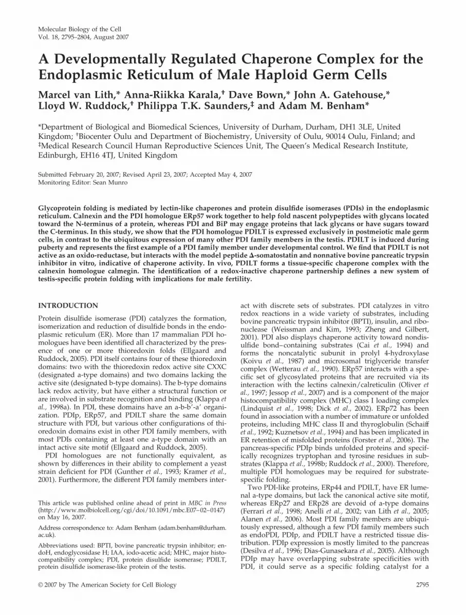
site of PDILT expression. The preimmune serum showedminor staining in the interstitial areas of rat testis due toresidual trapped serum (Figure 3A, rat-NRS) and was indis-tinguishable from secondary antibody alone (data notshown). In contrast, the anti-PDILT serum intensely stainedpopulations of germ cells in the seminiferous tubules (Figure3A, rat-�PDILT). The immunostaining pattern was essen-tially the same for mouse testis (Figure 3A, mouse-�PDILT)and when using the antiserum raised against a PDILT pep-tide [Figure 3A, mouse-�PDILT (pep)]. PDILT expressionwas also restricted to germ cells in testis from human, ma-caque, and marmoset (Figure 3B). Although in rat, mouse,and macaque, cross-sections of individual tubules are char-acterized as containing a single grouping (spermatogenicstage) of germ cells (Hess, 1990), the organization of sper-matogenesis in human and the common marmoset is morecomplex, and tubule cross-sections contain germ cell com-plements typical of a mixture of stages (Schulze et al., 1986;Millar et al., 2000). The restricted expression pattern ofPDILT to a subset of germ cells was in stark contrast with theexpression patterns of other PDI family members PDI,ERp57, and ERp72 or the ER chaperone BiP. Although ex-pression levels varied between the different cell types withinthe seminiferous tubules, expression of these other PDI fam-ily members was ubiquitous throughout the seminiferousepithelium (Figure 3C). Thus, PDILT is expressed in testis ofmultiple mammalian species and in contrast to other PDIfamily members, its expression is restricted to a subset oftesticular germ cells.
PDILT Is Expressed in Postmeiotic Germ CellsCloser examination of adult rat testis sections showed thatPDILT was present in the most mature germ cells, locatedaround the seminiferous tubule lumen (Figure 4A). Whenevaluated using the standard spermatogenic staging scheme(Figure 4B), it was apparent that immunopositive stainingwas first detectable in haploid round spermatids found atstage VII. In all species examined, the onset of PDILT ex-pression commenced upon completion of meiotic prophase.Thereafter, in the rat PDILT was present in the cell body ofall subsequent round and elongating spermatids (Figure 4A,stages IX, XI, and XIV). PDILT was not detected in sperma-tozoa in the epididymis or in the ejaculate (data not shown;Ellerman et al., 2006), consistent with the finding that PDILTwas not found in the nuclei of the spermatozoa but could bedetected in apical and basal residual bodies (Figure 4A,stage XI) that are left behind when spermatozoa are released(Clermont et al., 1987). The germ cells to which PDILT waslocalized are summarized on a diagram that shows the germcells associations found in the different stages of spermato-genesis in the rat (Figure 4B).
PDILT Expression Is under Developmental ControlAs PDILT was first seen at the round spermatid stage, weasked whether the onset of PDILT expression coincides withthe appearance of differentiating germ cells during the firstwave of spermatogenesis in development. Total RNA, iso-lated from rat testes at different ages, was used for RT-PCRto determine the expression of PDILT with age. A cDNAcontaining the coding region of PDILT gave a product ofexpected size (Figure 5A, PDILT 1). No product was ob-served when using RNA derived from testes of 2- and15-d-old rats, demonstrating that PDILT was not expressedin somatic cells that make up the bulk of the testes at theseages. PDILT mRNA was detected in testis from day 30 andreached adult levels from day 45 onward. RNA from liver ofa 58-d-old rat served as negative control (Figure 5A, PDILT
Figure 3. Expression of PDILT is germ cell specific. (A) Sectionsfrom rat testis counterstained with hematoxylin (blue) were immu-nostained with normal rabbit serum (NRS) or an anti-PDILT serumraised against recombinant PDILT at a dilution of 1:8000 (top pan-els). The PDILT staining pattern (brown) is similar for mouse testis(bottom left) or when using a serum raised against an internalPDILT peptide at 1:1000 (bottom right). Scale bar, 200 �m. (B) Testissections from human, macaque, and marmoset were immuno-stained for PDILT. Although a single population of PDILT-positivegerm cells occupied the full circumference of the tubule in macaque,the staining on human and marmoset was more heterogenous con-sistent with the existence of tubules containing more than one stageof spermatogenesis in a single cross section. Scale bar, 100 �m. (C)Rat testis sections were immunostained for ERp57, ERp72, PDI, andBiP (brown). Expression of these proteins was observed throughoutthe seminiferous epithelium. Scale bar, 200 �m.
Male Germ Cell Function of PDILT
Vol. 18, August 2007 2799
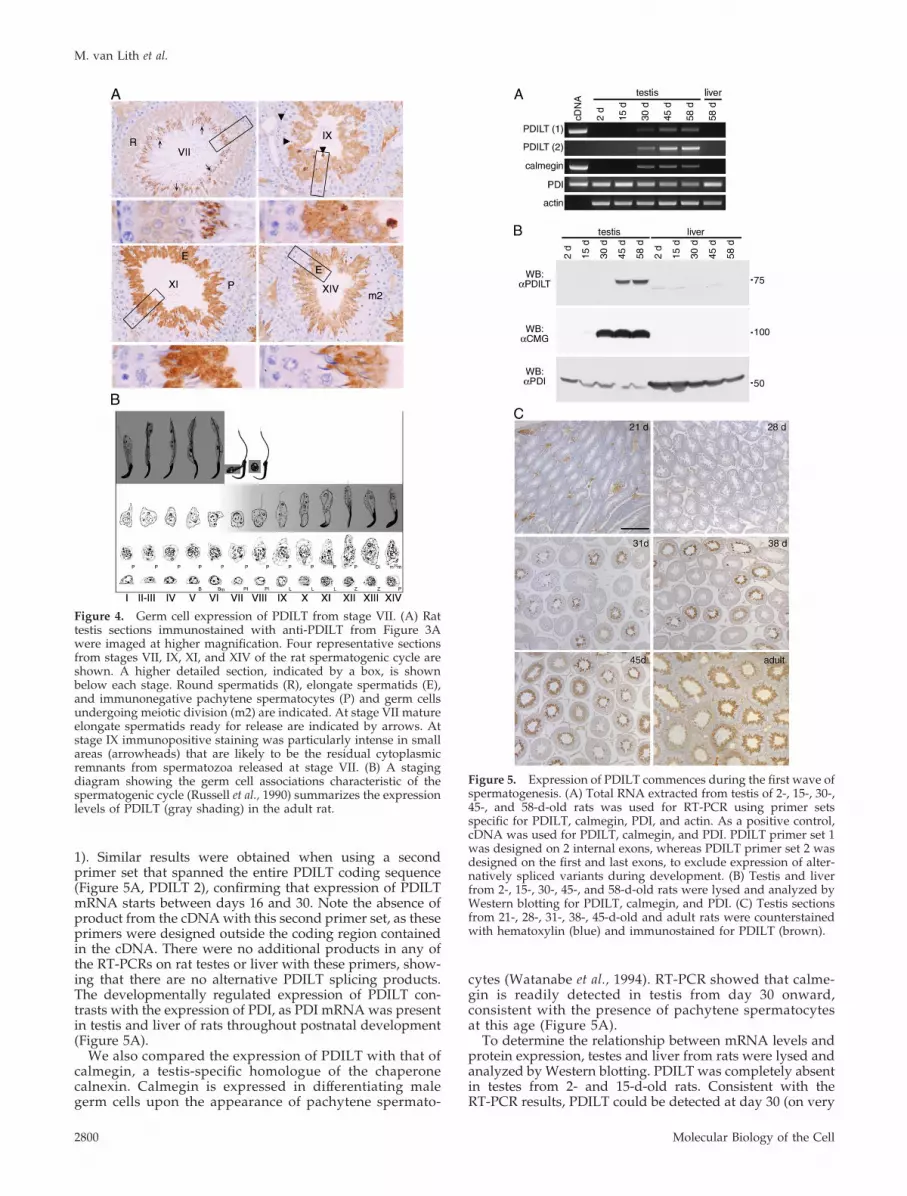
1). Similar results were obtained when using a secondprimer set that spanned the entire PDILT coding sequence(Figure 5A, PDILT 2), confirming that expression of PDILTmRNA starts between days 16 and 30. Note the absence ofproduct from the cDNA with this second primer set, as theseprimers were designed outside the coding region containedin the cDNA. There were no additional products in any ofthe RT-PCRs on rat testes or liver with these primers, show-ing that there are no alternative PDILT splicing products.The developmentally regulated expression of PDILT con-trasts with the expression of PDI, as PDI mRNA was presentin testis and liver of rats throughout postnatal development(Figure 5A).
We also compared the expression of PDILT with that ofcalmegin, a testis-specific homologue of the chaperonecalnexin. Calmegin is expressed in differentiating malegerm cells upon the appearance of pachytene spermato-
cytes (Watanabe et al., 1994). RT-PCR showed that calme-gin is readily detected in testis from day 30 onward,consistent with the presence of pachytene spermatocytesat this age (Figure 5A).
To determine the relationship between mRNA levels andprotein expression, testes and liver from rats were lysed andanalyzed by Western blotting. PDILT was completely absentin testes from 2- and 15-d-old rats. Consistent with theRT-PCR results, PDILT could be detected at day 30 (on very
Figure 4. Germ cell expression of PDILT from stage VII. (A) Rattestis sections immunostained with anti-PDILT from Figure 3Awere imaged at higher magnification. Four representative sectionsfrom stages VII, IX, XI, and XIV of the rat spermatogenic cycle areshown. A higher detailed section, indicated by a box, is shownbelow each stage. Round spermatids (R), elongate spermatids (E),and immunonegative pachytene spermatocytes (P) and germ cellsundergoing meiotic division (m2) are indicated. At stage VII matureelongate spermatids ready for release are indicated by arrows. Atstage IX immunopositive staining was particularly intense in smallareas (arrowheads) that are likely to be the residual cytoplasmicremnants from spermatozoa released at stage VII. (B) A stagingdiagram showing the germ cell associations characteristic of thespermatogenic cycle (Russell et al., 1990) summarizes the expressionlevels of PDILT (gray shading) in the adult rat.
Figure 5. Expression of PDILT commences during the first wave ofspermatogenesis. (A) Total RNA extracted from testis of 2-, 15-, 30-,45-, and 58-d-old rats was used for RT-PCR using primer setsspecific for PDILT, calmegin, PDI, and actin. As a positive control,cDNA was used for PDILT, calmegin, and PDI. PDILT primer set 1was designed on 2 internal exons, whereas PDILT primer set 2 wasdesigned on the first and last exons, to exclude expression of alter-natively spliced variants during development. (B) Testis and liverfrom 2-, 15-, 30-, 45-, and 58-d-old rats were lysed and analyzed byWestern blotting for PDILT, calmegin, and PDI. (C) Testis sectionsfrom 21-, 28-, 31-, 38-, 45-d-old and adult rats were counterstainedwith hematoxylin (blue) and immunostained for PDILT (brown).
M. van Lith et al.
Molecular Biology of the Cell2800
long exposures, data not shown) and reached adult levels ofexpression by day 45. (Figure 5B). Calmegin reached adultlevels of expression by day 30, whereas PDI was expressedthroughout postnatal development (Figure 5B). To examinewhether the onset of PDILT expression corresponds with thefirst appearance of round spermatids in development, sec-tions of testes from rats of different ages were taken forimmunohistochemistry. PDILT was not detected in any so-matic cells or germ cells in testis from 21- and 28-d-old rats.On day 31, some of the seminiferous tubules containedPDILT-positive cells close to the tubule lumen. The propor-tion of seminiferous tubules containing PDILT-positive cellsdramatically increased with age and by day 45, all of theseminiferous tubules were positive for PDILT (Figure 5C).Taken together, these data show that PDILT expression isdetected in testis from 30-d-old rats, consistent with theappearance of round spermatids during the first spermato-genic wave at puberty. This strongly suggests that PDILT isunder developmental control and has a role in sperm differ-entiation.
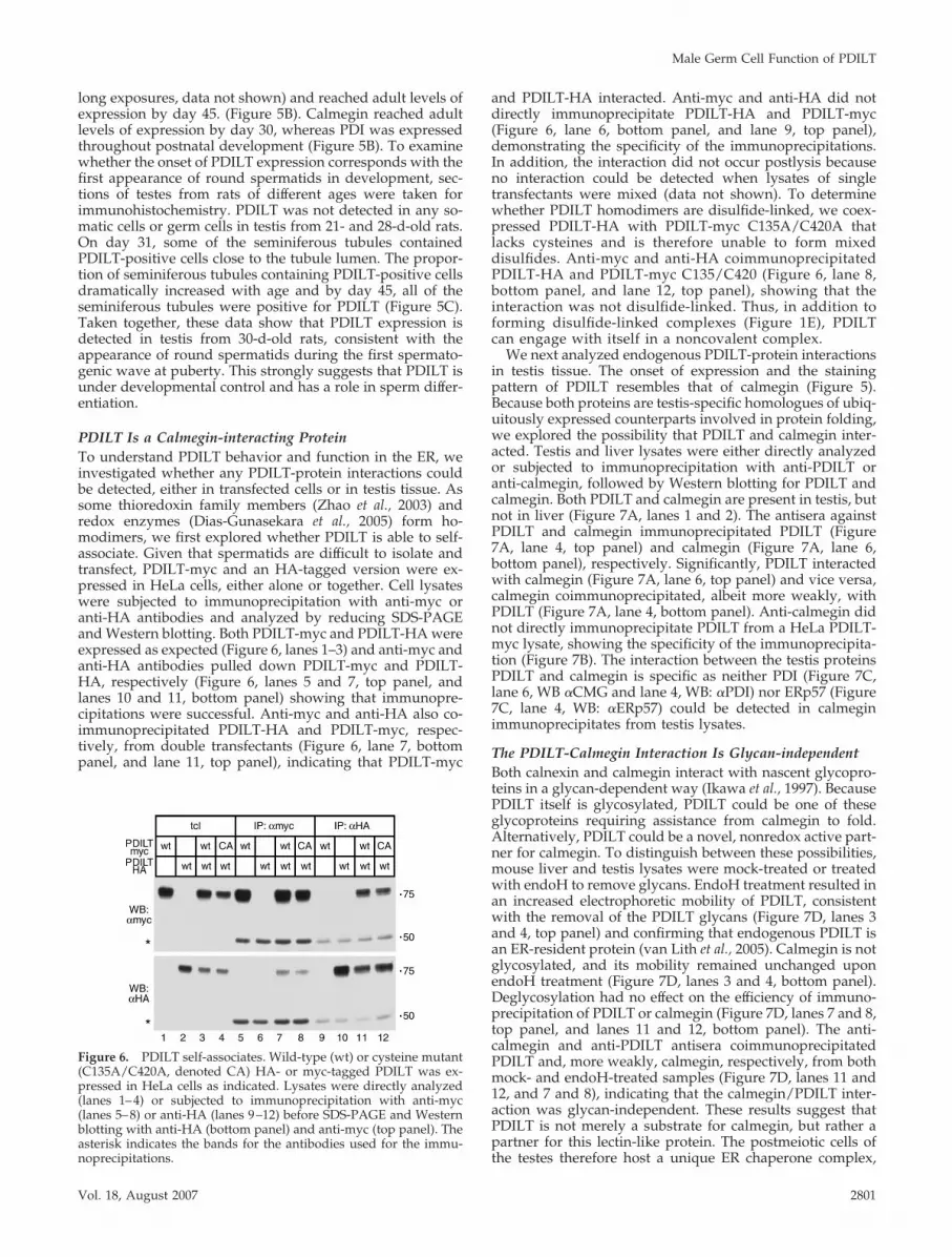
PDILT Is a Calmegin-interacting ProteinTo understand PDILT behavior and function in the ER, weinvestigated whether any PDILT-protein interactions couldbe detected, either in transfected cells or in testis tissue. Assome thioredoxin family members (Zhao et al., 2003) andredox enzymes (Dias-Gunasekara et al., 2005) form ho-modimers, we first explored whether PDILT is able to self-associate. Given that spermatids are difficult to isolate andtransfect, PDILT-myc and an HA-tagged version were ex-pressed in HeLa cells, either alone or together. Cell lysateswere subjected to immunoprecipitation with anti-myc oranti-HA antibodies and analyzed by reducing SDS-PAGEand Western blotting. Both PDILT-myc and PDILT-HA wereexpressed as expected (Figure 6, lanes 1–3) and anti-myc andanti-HA antibodies pulled down PDILT-myc and PDILT-HA, respectively (Figure 6, lanes 5 and 7, top panel, andlanes 10 and 11, bottom panel) showing that immunopre-cipitations were successful. Anti-myc and anti-HA also co-immunoprecipitated PDILT-HA and PDILT-myc, respec-tively, from double transfectants (Figure 6, lane 7, bottompanel, and lane 11, top panel), indicating that PDILT-myc
and PDILT-HA interacted. Anti-myc and anti-HA did notdirectly immunoprecipitate PDILT-HA and PDILT-myc(Figure 6, lane 6, bottom panel, and lane 9, top panel),demonstrating the specificity of the immunoprecipitations.In addition, the interaction did not occur postlysis becauseno interaction could be detected when lysates of singletransfectants were mixed (data not shown). To determinewhether PDILT homodimers are disulfide-linked, we coex-pressed PDILT-HA with PDILT-myc C135A/C420A thatlacks cysteines and is therefore unable to form mixeddisulfides. Anti-myc and anti-HA coimmunoprecipitatedPDILT-HA and PDILT-myc C135/C420 (Figure 6, lane 8,bottom panel, and lane 12, top panel), showing that theinteraction was not disulfide-linked. Thus, in addition toforming disulfide-linked complexes (Figure 1E), PDILTcan engage with itself in a noncovalent complex.
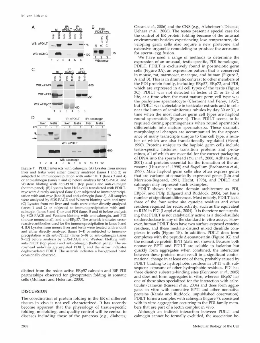
We next analyzed endogenous PDILT-protein interactionsin testis tissue. The onset of expression and the stainingpattern of PDILT resembles that of calmegin (Figure 5).Because both proteins are testis-specific homologues of ubiq-uitously expressed counterparts involved in protein folding,we explored the possibility that PDILT and calmegin inter-acted. Testis and liver lysates were either directly analyzedor subjected to immunoprecipitation with anti-PDILT oranti-calmegin, followed by Western blotting for PDILT andcalmegin. Both PDILT and calmegin are present in testis, butnot in liver (Figure 7A, lanes 1 and 2). The antisera againstPDILT and calmegin immunoprecipitated PDILT (Figure7A, lane 4, top panel) and calmegin (Figure 7A, lane 6,bottom panel), respectively. Significantly, PDILT interactedwith calmegin (Figure 7A, lane 6, top panel) and vice versa,calmegin coimmunoprecipitated, albeit more weakly, withPDILT (Figure 7A, lane 4, bottom panel). Anti-calmegin didnot directly immunoprecipitate PDILT from a HeLa PDILT-myc lysate, showing the specificity of the immunoprecipita-tion (Figure 7B). The interaction between the testis proteinsPDILT and calmegin is specific as neither PDI (Figure 7C,lane 6, WB �CMG and lane 4, WB: �PDI) nor ERp57 (Figure7C, lane 4, WB: �ERp57) could be detected in calmeginimmunoprecipitates from testis lysates.
The PDILT-Calmegin Interaction Is Glycan-independentBoth calnexin and calmegin interact with nascent glycopro-teins in a glycan-dependent way (Ikawa et al., 1997). BecausePDILT itself is glycosylated, PDILT could be one of theseglycoproteins requiring assistance from calmegin to fold.Alternatively, PDILT could be a novel, nonredox active part-ner for calmegin. To distinguish between these possibilities,mouse liver and testis lysates were mock-treated or treatedwith endoH to remove glycans. EndoH treatment resulted inan increased electrophoretic mobility of PDILT, consistentwith the removal of the PDILT glycans (Figure 7D, lanes 3and 4, top panel) and confirming that endogenous PDILT isan ER-resident protein (van Lith et al., 2005). Calmegin is notglycosylated, and its mobility remained unchanged uponendoH treatment (Figure 7D, lanes 3 and 4, bottom panel).Deglycosylation had no effect on the efficiency of immuno-precipitation of PDILT or calmegin (Figure 7D, lanes 7 and 8,top panel, and lanes 11 and 12, bottom panel). The anti-calmegin and anti-PDILT antisera coimmunoprecipitatedPDILT and, more weakly, calmegin, respectively, from bothmock- and endoH-treated samples (Figure 7D, lanes 11 and12, and 7 and 8), indicating that the calmegin/PDILT inter-action was glycan-independent. These results suggest thatPDILT is not merely a substrate for calmegin, but rather apartner for this lectin-like protein. The postmeiotic cells ofthe testes therefore host a unique ER chaperone complex,
Figure 6. PDILT self-associates. Wild-type (wt) or cysteine mutant(C135A/C420A, denoted CA) HA- or myc-tagged PDILT was ex-pressed in HeLa cells as indicated. Lysates were directly analyzed(lanes 1–4) or subjected to immunoprecipitation with anti-myc(lanes 5–8) or anti-HA (lanes 9–12) before SDS-PAGE and Westernblotting with anti-HA (bottom panel) and anti-myc (top panel). Theasterisk indicates the bands for the antibodies used for the immu-noprecipitations.
Male Germ Cell Function of PDILT
Vol. 18, August 2007 2801
distinct from the redox-active ERp57-calnexin and BiP-PDIpartnerships observed for glycoprotein folding in somaticcells (Molinari and Helenius, 2000).
DISCUSSION
The coordination of protein folding in the ER of differenttissues in vivo is not well characterized. It has recentlybecome apparent that the physiology of tissue-specificfolding, misfolding, and quality control will be central todiseases including those of the pancreas (e.g., diabetes;
Ozcan et al., 2006) and the CNS (e.g., Alzheimer’s Disease;Uehara et al., 2006). The testes present a special case forthe control of ER protein folding because of the unusualenvironment; besides experiencing low temperature, de-veloping germ cells also require a new proteome andextensive organelle remodeling to produce the acrosomefor sperm– egg fusion.
We have used a range of methods to determine theexpression of an unusual, testis-specific, PDI homologue,PDILT. PDILT is exclusively found in postmeiotic germcells (Figure 3A), an expression pattern that is conservedin mouse, rat, marmoset, macaque, and human (Figure 3,A and B). This is in dramatic contrast to other members ofthe PDI protein family, including ERp57, ERp72, and PDI,which are expressed in all cell types of the testis (Figure3C). PDILT was not detected in testes at 21 or 28 d oflife, at a time when the most mature germ cell type wasthe pachytene spermatocyte (Clermont and Perey, 1957),but PDILT was detectable in testicular extracts and in cellsnear the lumen of seminiferous tubules by day 30 or 31, atime when the most mature germ cell types are haploidround spermatids (Figure 4). Thus PDILT seems to berequired during spermiogenesis when round spermatidsdifferentiate into mature spermatozoa. These dramaticmorphological changes are accompanied by the appear-ance of many transcripts unique to this cell type, a num-ber of which are also translationally regulated (Hecht,1990). Proteins unique to the haploid germ cells includetestis-specific histones, transition proteins and prota-mines, all of which are essential for the correct packagingof DNA into the sperm head (Yu et al., 2000; Adham et al.,2001) and proteins essential for the formation of the ac-rosome (Hurst et al., 1998) and flagellum (Brohmann et al.,1997). Male haploid germ cells also often express genesthat are variants of somatically expressed genes (Lin andMorrison-Bogorad, 1991; Hecht, 1998), and PDILT andcalmegin may represent such examples.
PDILT shows the same domain architecture as PDI,ERp57, and PDIp (Ellgaard and Ruddock, 2005), but has anumber of significant differences. Most notably, PDILT lacksthree of the four active site cysteine residues and otherresidues required for redox activity, such as the equivalentto R120 in PDI (Lappi et al., 2004). It is therefore not surpris-ing that PDILT is not catalytically active as a thiol-disulfideoxidoreductase in any of the standard in vitro assays. How-ever, human PDILT does have two surface-exposed cysteineresidues, and these mediate distinct mixed disulfide com-plexes in cells (Figure 1E). In addition, PDILT does formcomplexes with the peptide �-somatostatin (Figure 1G) andthe nonnative protein BPTI (data not shown). Because bothnonnative BPTI and PDILT are soluble in isolation butquickly form aggregates when combined, the interactionbetween these proteins must result in a significant confor-mational change in at least one of them, probably caused byPDILT binding to hydrophobic residues in BPTI with sub-sequent exposure of other hydrophobic residues. PDI hasthree distinct substrate-binding sites (Koivunen et al., 2005)and does not form aggregates in vitro, whereas ERp57 hasone of these sites specialized for the interaction with calre-ticulin/calnexin (Russell et al., 2004) and does form aggre-gates in vitro with nonnative BPTI and other nonnativeproteins (Karala and Ruddock, unpublished observation).PDILT forms a complex with calmegin (Figure 7), consistentwith in vitro aggregation occurring to the PDI-family mem-bers that are part of a lectin complex in vivo.
Although an indirect interaction between PDILT andcalmegin cannot be formally excluded, the association be-
Figure 7. PDILT interacts with calmegin. (A) Lysates from mouseliver and testis were either directly analyzed (lanes l and 2) orsubjected to immunoprecipitation with anti-PDILT (lanes 3 and 4)or anti-calmegin (lanes 5 and 6) before analysis by SDS-PAGE andWestern blotting with anti-PDILT (top panel) and anti-calmegin(bottom panel). (B) Lysates from HeLa cells transfected with PDILT-myc were directly analyzed (lane 1) or subjected to immunoprecipi-tation with anti-myc (lane 2) and anti-calmegin (lane 3). All sampleswere analyzed by SDS-PAGE and Western blotting with anti-myc.(C) Lysates from rat liver and testis were either directly analyzed(lanes 1 and 2) or subjected to immunoprecipitation with anti-calmegin (lanes 3 and 4) or anti-PDI (lanes 5 and 6) before analysisby SDS-PAGE and Western blotting with anti-calmegin, anti-PDI(mouse monoclonal), and anti-ERp57. The asterisk indicates cross-reactive antibodies used for the immunoprecipitation in lanes 3 and4. (D) Lysates from mouse liver and testis were treated with endoHand either directly analyzed (lanes 1–4) or subjected to immuno-precipitation with anti-PDILT (lanes 5–8) or anti-calmegin (lanes9–12) before analysis by SDS-PAGE and Western blotting withanti-PDILT (top panel) and anti-calmegin (bottom panel). The ar-rowhead indicates glycosylated PDILT, and the arrow indicatesdeglycosylated PDILT. The asterisk indicates a background bandoccasionally observed.
M. van Lith et al.
Molecular Biology of the Cell2802
tween the two proteins in the ER of spermatids is glycan-independent (Figure 7). Thus, PDILT is not just a glycosy-lated substrate for calmegin, but is more likely to be apartner in a unique protein-folding system. Our inability todetect calmegin-ERp57 and calmegin-PDI complexes in tes-tis suggests that the PDILT-calmegin complex performs aunique role in ER quality control, although some caution isrequired in interpreting negative immunoprecipitation data(Figure 7C). Because PDILT has no oxido-reductase activityin vitro and lacks the classical WCGHC motif of standardoxidoreductases, it is likely that PDILT operates as a foldingassistant for spermatid-specific glycoproteins that have beenensnared by calmegin. This is supported by the observationthat calmegin interacts with nascent glycoproteins (Ikawa etal., 1997) and our finding that PDILT interacts in vitro withthe model peptide �-somatostatin and the nonnative proteinBPTI (Figure 1). The fact that PDILT can be recovered indisulfide dependent complexes from transfected cells (Fig-ure 1E) and as complexes from testes (van Lith et al., 2005)suggests that both PDILTs solvent-exposed cysteine resi-dues may trap free single cysteines in substrates, perhapsusing them as a molecular handle to facilitate protein fold-ing of the peptide backbone. Whether noncovalent PDILT-PDILT complexes (Figure 6) have an independent functionalrole in vivo, or perhaps operate as an inactive chaperonereservoir, requires further investigation. Together, our workidentifies a new quality control hub unique to the ER ofspermatids, alongside the ERp57/calnexin and BiP/PDIpartnerships found in somatic cells (Molinari and Helenius,2000). Although calnexin presents nonnative glycoproteinsto the redox-active ERp57, the glycan recognition is de-coupled from the redox activity for the PDILT/calmeginpair. It will now be important to determine whether calme-gin and PDILT work on similar or independent substrates toERp57 and PDI, given that both PDI and ERp57 are alsoexpressed in postmeiotic cells (Figure 3C). Now that ap-proaches to determine the substrate specificity of ERp57have been successful (Jessop et al., 2007), it should be possi-ble to identify the range of endogenous PDILT substrates byproteomics and the in vivo role of PDILT homodimerization(Figure 6), once the limitations of transfecting and culturinggerm cells have been overcome.
Another interesting possibility is that PDILT is requiredfor specific events during meiosis, such as remodeling theER in preparation for acrosome formation and the expulsionof the cytoplasmic body. Both PDI and ERp29 are involvedin ERAD of microbial proteins (Tsai and Rapoport, 2002;Magnuson et al., 2005), and it may be that PDILT is requiredfor unfolding and retrotranslocation of ER proteins duringthe programmed disposal of the residual body. Further-more, ERp57 is found at the surface of sperm and is requiredfor disulfide bond exchange in preparation for gamete fu-sion (Ellerman et al., 2006). The C-terminal QEDL motif ofERp57 is inefficiently retrieved by the KDEL receptors,meaning that ERp57 may be more reliant on its associa-tion with ER-resident proteins for its retention than otherPDIs (Raykhel, Alananen, and Ruddock, unpublisheddata). Thus one possibility is that PDILT regulates ERp57export to the cell surface by competing with ERp57 forcalmegin and calnexin and allowing monomeric ERp57 tobe exported from the ER. In vitro experiments with thepurified, glycosylated components will be required to deter-mine the relative affinities of these proteins for each other.
Calmegin is required for fertility (Ikawa et al., 1997),and it will be important to determine whether PDILT-deficient mice are also infertile. With many human maleinfertility cases unexplained, identifying genes and path-
ways associated with infertility remains a public healthpriority.
ACKNOWLEDGMENTS
We thank Laura Walters, David Dixon, and Helen McPhee for technicalassistance and Neil Bulleid, Lars Ellgaard, Ineke Braakman, and YoshitakeNishimune for reagents.
REFERENCES
Adham, I. M., Nayernia, K., Burkhardt-Gottges, E., Topaloglu, O., Dixkens,C., Holstein, A. F., and Engel, W. (2001). Teratozoospermia in mice lacking thetransition protein 2 (Tnp2). Mol. Hum. Reprod. 7, 513–520.
Alanen, H. I., Williamson, R. A., Howard, M. J., Hatahet, F. S., Salo, K. E.,Kauppila, A., Kellokumpu, S., and Ruddock, L. W. (2006). ERp27, a newnon-catalytic endoplasmic reticulum-located human protein disulfide isomer-ase family member, interacts with ERp57. J. Biol. Chem. 281, 33727–33738.
Anelli, T., Alessio, M., Mezghrani, A., Simmen, T., Talamo, F., Bachi, A., andSitia, R. (2002). ERp44, a novel endoplasmic reticulum folding assistant of thethioredoxin family. EMBO J. 21, 835–844.
Benham, A. M., Cabibbo, A., Fassio, A., Bulleid, N., Sitia, R., and Braakman,I. (2000). The CXXCXXC motif determines the folding, structure and stabilityof human Ero1-Lalpha. EMBO J. 19, 4493–4502.
Brohmann, H., Pinnecke, S., and Hoyer-Fender, S. (1997). Identification andcharacterization of new cDNAs encoding outer dense fiber proteins of ratsperm. J. Biol. Chem. 272, 10327–10332.
Cai, H., Wang, C. C., and Tsou, C. L. (1994). Chaperone-like activity of proteindisulfide isomerase in the refolding of a protein with no disulfide bonds.J. Biol. Chem. 269, 24550–24552.
Clermont, Y., Morales, C., and Hermo, L. (1987). Endocytic activities of Sertolicells in the rat. Ann. NY Acad. Sci. 513, 1–15.
Clermont, Y., and Perey, B. (1957). Quantitative study of the cell population ofthe seminiferous tubules in immature rats. Am. J. Anat. 100, 241–267.
Desilva, M. G., Lu, J., Donadel, G., Modi, W. S., Xie, H., Notkins, A. L., andLan, M. S. (1996). Characterization and chromosomal localization of a newprotein disulfide isomerase, PDIp, highly expressed in human pancreas. DNACell Biol. 15, 9–16.
Dias-Gunasekara, S., Gubbens, J., van Lith, M., Dunne, C., Williams, J. A.,Kataky, R., Scoones, D., Lapthorn, A., Bulleid, N. J., and Benham, A. M. (2005).Tissue-specific expression and dimerization of the endoplasmic reticulumoxidoreductase Ero1beta. J. Biol. Chem. 280, 33066–33075.
Dick, T. P., Bangia, N., Peaper, D. R., and Cresswell, P. (2002). Disulfide bondisomerization and the assembly of MHC class I-peptide complexes. Immunity16, 87–98.
Ellerman, D. A., Myles, D. G., and Primakoff, P. (2006). A role for spermsurface protein disulfide isomerase activity in gamete fusion: evidence for theparticipation of ERp57. Dev. Cell. 10, 831–837.
Ellgaard, L., and Ruddock, L. W. (2005). The human protein disulphideisomerase family: substrate interactions and functional properties. EMBORep. 6, 28–32.
Ferrari, D. M., Nguyen Van, P., Kratzin, H. D., and Soling, H. D. (1998).ERp28, a human endoplasmic-reticulum-lumenal protein, is a member of theprotein disulfide isomerase family but lacks a CXXC thioredoxin-box motif.Eur. J. Biochem. 255, 570–579.
Forster, M. L., Sivick, K., Park, Y. N., Arvan, P., Lencer, W. I., and Tsai, B.(2006). Protein disulfide isomerase-like proteins play opposing roles duringretrotranslocation. J. Cell Biol. 173, 853–859.
Gunther, R., Srinivasan, M., Haugejorden, S., Green, M., Ehbrecht, I. M., andKuntzel, H. (1993). Functional replacement of the Saccharomyces cerevisiaeTrg1/Pdi1 protein by members of the mammalian protein disulfide isomerasefamily. J. Biol. Chem. 268, 7728–7732.
Hecht, N. B. (1990). Regulation of ‘haploid expressed genes’ in male germcells. J. Reprod. Fertil. 88, 679–693.
Hecht, N. B. (1998). Molecular mechanisms of male germ cell differentiation.Bioessays 20, 555–561.
Hess, R. A. (1990). Quantitative and qualitative characteristics of the stagesand transitions in the cycle of the rat seminiferous epithelium: light micro-scopic observations of perfusion-fixed and plastic-embedded testes. Biol.Reprod. 43, 525–542.
Holmgren, A. (1979). Thioredoxin catalyzes the reduction of insulin disulfidesby dithiothreitol and dihydrolipoamide. J. Biol. Chem. 254, 9627–9632.
Male Germ Cell Function of PDILT
Vol. 18, August 2007 2803
Hurst, S., Howes, E. A., Coadwell, J., and Jones, R. (1998). Expression of atestis-specific putative actin-capping protein associated with the developingacrosome during rat spermiogenesis. Reprod. Dev. 49, 81–91.
Ikawa, M., Wada, I., Kominami, K., Watanabe, D., Toshimori, K., Nishimune,Y., and Okabe, M. (1997). The putative chaperone calmegin is required forsperm fertility. Nature 387, 607–611.
Jessop, C. E., Chakravarthi, S., Garbi, N., Hammerling, G. J., Lovell, S., andBulleid, N. J. (2007). ERp57 is essential for efficient folding of glycoproteinssharing common structural domains. EMBO J. 26, 28–40.
Klappa, P., Ruddock, L. W., Darby, N. J., and Freedman, R. B. (1998a). The b�domain provides the principal peptide-binding site of protein disulfide isomer-ase but all domains contribute to binding of misfolded proteins. EMBO J. 17,927–935.
Klappa, P., Stromer, T., Zimmermann, R., Ruddock, L. W., and Freedman,R. B. (1998b). A pancreas-specific glycosylated protein disulphide-isomerasebinds to misfolded proteins and peptides with an interaction inhibited byoestrogens. Eur. J. Biochem. 254, 63–69.
Koivu, J., Myllyla, R., Helaakoski, T., Pihlajaniemi, T., Tasanen, K., andKivirikko, K. I. (1987). A single polypeptide acts both as the beta subunit ofprolyl 4-hydroxylase and as a protein disulfide-isomerase. J. Biol. Chem. 262,6447–6449.
Koivunen, P., Salo, K. E., Myllyharju, J., and Ruddock, L. W. (2005). Threebinding sites in protein-disulfide isomerase cooperate in collagen prolyl 4-hy-droxylase tetramer assembly. J. Biol. Chem. 280, 5227–5235.
Kramer, B., Ferrari, D. M., Klappa, P., Pohlmann, N., and Soling, H. D. (2001).Functional roles and efficiencies of the thioredoxin boxes of calcium-bindingproteins 1 and 2 in protein folding. Biochem. J. 357, 83–95.
Kuznetsov, G., Chen, L. B., and Nigam, S. K. (1994). Several endoplasmicreticulum stress proteins, including ERp72, interact with thyroglobulin dur-ing its maturation. J. Biol. Chem. 269, 22990–22995.
Lappi, A. K., Lensink, M. F., Alanen, H. I., Salo, K. E., Lobell, M., Juffer, A. H.,and Ruddock, L. W. (2004). A conserved arginine plays a role in the catalyticcycle of the protein disulphide isomerases. J. Mol. Biol. 335, 283–295.
Lin, S. C., and Morrison-Bogorad, M. (1991). Cloning and characterization ofa testis-specific thymosin beta 10 cDNA. Expression in post-meiotic malegerm cells. J. Biol. Chem. 266, 23347–23353.
Lindquist, J. A., Jensen, O. N., Mann, M., and Hammerling, G. J. (1998). ER-60,a chaperone with thiol-dependent reductase activity involved in MHC class Iassembly. EMBO J. 17, 2186–2195.
Magnuson, B., Rainey, E. K., Benjamin, T., Baryshev, M., Mkrtchian, S., andTsai, B. (2005). ERp29 triggers a conformational change in polyomavirus tostimulate membrane binding. Mol. Cell 20, 289–300.
Millar, M. R., Sharpe, R. M., Weinbauer, G. F., Fraser, H. M., and Saunders,P. T. (2000). Marmoset spermatogenesis: organizational similarities to thehuman. Int. J. Androl. 23, 266–277.
Molinari, M., and Helenius, A. (2000). Chaperone selection during glycopro-tein translocation into the endoplasmic reticulum. Science 288, 331–333.
Morjana, N. A., McKeone, B. J., and Gilbert, H. F. (1993). Guanidine hydro-chloride stabilization of a partially unfolded intermediate during the revers-ible denaturation of protein disulfide isomerase. Proc. Natl. Acad. Sci. USA90, 2107–2111.
Oliver, J. D., van der Wal, F. J., Bulleid, N. J., and High, S. (1997). Interactionof the thiol-dependent reductase ERp57 with nascent glycoproteins. Science275, 86–88.
Ozcan, U., Yilmaz, E., Ozcan, L., Furuhashi, M., Vaillancourt, E., Smith, R. O.,Gorgun, C. Z., and Hotamisligil, G. S. (2006). Chemical chaperones reduce ERstress and restore glucose homeostasis in a mouse model of type 2 diabetes.Science 313, 1137–1140.
Peltoniemi, M. J., Karala, A. R., Jurvansuu, J. K., Kinnula, V. L., and Ruddock,L. W. (2006). Insights into deglutathionylation reactions. Different intermedi-
ates in the glutaredoxin and protein disulfide isomerase catalyzed reactionsare defined by the gamma-linkage present in glutathione. J. Biol. Chem. 281,33107–33114.
Ruddock, L. W., Freedman, R. B., and Klappa, P. (2000). Specificity in sub-strate binding by protein folding catalysts: tyrosine and tryptophan residuesare the recognition motifs for the binding of peptides to the pancreas-specificprotein disulfide isomerase PDIp. Protein Sci. 9, 758–764.
Ruddock, L. W., Hirst, T. R., and Freedman, R. B. (1996). pH-dependence ofthe dithiol-oxidizing activity of DsbA (a periplasmic protein thiol:disulphideoxidoreductase) and protein disulphide-isomerase: studies with a novel sim-ple peptide substrate. Biochem. J. 315(Pt 3), 1001–1005.
Russell, L. D., Ettlin, R., Sinha, A., and Clegg, E. (1990). Histological andHistopathological Evaluation of the Testis, Miami, FL: Cache River Press.
Russell, S. J., Ruddock, L. W., Salo, K. E., Oliver, J. D., Roebuck, Q. P.,Llewellyn, D. H., Roderick, H. L., Koivunen, P., Myllyharju, J., and High, S.(2004). The primary substrate binding site in the b� domain of ERp57 isadapted for endoplasmic reticulum lectin association. J. Biol. Chem. 279,18861–18869.
Schaiff, W. T., Hruska, K. A., Jr., McCourt, D. W., Green, M., and Schwartz,B. D. (1992). HLA-DR associates with specific stress proteins and is retainedin the endoplasmic reticulum in invariant chain negative cells. J. Exp. Med.176, 657–666.
Schulze, W., Riemer, M., Rehder, U., and Hohne, K. H. (1986). Computer-aided three-dimensional reconstructions of the arrangement of primary sper-matocytes in human seminiferous tubules. Cell Tissue Res. 244, 1–7.
Sullivan, D. C., Huminiecki, L., Moore, J. W., Boyle, J. J., Poulsom, R.,Creamer, D., Barker, J., and Bicknell, R. (2003). EndoPDI, a novel protein-disulfide isomerase-like protein that is preferentially expressed in endothelialcells acts as a stress survival factor. J. Biol. Chem. 278, 47079–47088.
Tsai, B., and Rapoport, T. A. (2002). Unfolded cholera toxin is transferred tothe ER membrane and released from protein disulfide isomerase upon oxi-dation by Ero1. J. Cell Biol. 159, 207–216.
Uehara, T., Nakamura, T., Yao, D., Shi, Z. Q., Gu, Z., Ma, Y., Masliah, E.,Nomura, Y., and Lipton, S. A. (2006). S-nitrosylated protein-disulphideisomerase links protein misfolding to neurodegeneration. Nature 441, 513–517.
van Lith, M., Hartigan, N., Hatch, J., and Benham, A. M. (2005). PDILT, adivergent testis-specific protein disulfide isomerase with a non-classical SXXCmotif that engages in disulfide-dependent interactions in the endoplasmicreticulum. J. Biol. Chem. 280, 1376–1383.
Watanabe, D., Yamada, K., Nishina, Y., Tajima, Y., Koshimizu, U., Nagata, A.,and Nishimune, Y. (1994). Molecular cloning of a novel Ca(2�)-bindingprotein (calmegin) specifically expressed during male meiotic germ cell de-velopment. J. Biol. Chem. 269, 7744–7749.
Weissman, J. S., and Kim, P. S. (1993). Efficient catalysis of disulphide bondrearrangements by protein disulphide isomerase. Nature 365, 185–188.
Wetterau, J. R., Combs, K. A., Spinner, S. N., and Joiner, B. J. (1990). Proteindisulfide isomerase is a component of the microsomal triglyceride transferprotein complex. J. Biol. Chem. 265, 9800–9807.
Yu, Y. E., Zhang, Y., Unni, E., Shirley, C. R., Deng, J. M., Russell, L. D., Weil,M. M., Behringer, R. R., and Meistrich, M. L. (2000). Abnormal spermatogen-esis and reduced fertility in transition nuclear protein 1-deficient mice. Proc.Natl. Acad. Sci. USA 97, 4683–4688.
Zhao, Z., Peng, Y., Hao, S. F., Zeng, Z. H., and Wang, C. C. (2003). Dimer-ization by domain hybridization bestows chaperone and isomerase activities.J. Biol. Chem. 278, 43292–43298.
Zheng, J., and Gilbert, H. F. (2001). Discrimination between native and non-native disulfides by protein-disulfide isomerase. J. Biol. Chem. 276, 15747–15752.
M. van Lith et al.
Molecular Biology of the Cell2804


