Embed Size (px)
Citation preview
A Complex Gene Regulatory Mechanism that Operates at theNexus of Multiple RNA Processing Decisions
David S. McPheeters1, Nicole Cremona1, Sham Sunder1, Huei-Mei Chen2, NicoleAverbeck2, Janet Leatherwood2, and Jo Ann Wise1
1Center for RNA Molecular Biology and Department of Molecular Biology & Microbiology, CaseWestern Reserve University, School of Medicine, Cleveland, Ohio 44106-49602Department of Molecular Genetics and Microbiology, State University of New York, Stony Brook,NY 11794-5222
AbstractExpression of crs1 pre-mRNA, encoding a meiotic cyclin, is blocked in actively growing fissionyeast cells by a multifaceted mechanism. The most striking feature is that crs1 transcripts arecontinuously synthesized in vegetative cells, but are targeted for degradation rather than splicingand polyadenylation. Turnover of crs1 RNA requires the exosome, similar to previously describednuclear surveillance and silencing mechanisms, but does not involve a non-canonical poly(A)polymerase. Instead, crs1 transcripts are targeted for destruction by a factor previously implicatedin turnover of meiotic RNAs in growing cells. Like exosome mutants, mmi1 mutants splice andpolyadenylate vegetative crs1 transcripts. Two regulatory elements are located at the 3′ end of thecrs1 gene, consistent with the increased accumulation of spliced RNA in polyadenylation factormutants. This highly integrated regulatory strategy may ensure a rapid response to adverseconditions, thereby guaranteeing survival.
INTRODUCTIONMeiosis is a highly conserved cellular differentiation pathway in which one round of DNAsynthesis is followed by two successive rounds of division, leading to the formation ofhaploid cells from diploid precursors1. Although the meiotic cell cycle is similar amongeukaryotes, the underlying regulatory mechanisms vary widely. In multicellular organisms,extrinsic cues from surrounding cells stimulate germ cells to enter the differentiationpathway, whereas in unicellular organisms such as the budding yeast Saccharomycescerevisiae and the fission yeast Schizosaccharomyces pombe, entry into meiosis is triggeredby nutrient deprivation. In either case, the decision to initiate sexual differentiation is tightlyregulated to prevent execution of this specialized cell cycle under inappropriate conditionsor in improper developmental contexts.
Correspondence should be addressed to J.A.W. ([email protected]) Phone: (216)-368-1876, FAX (216)-368-3055.AUTHOR CONTRIBUTIONSDavid McPheeters performed the TRO experiments, designed the real-time PCR assay, and participated in development of the modelas well as writing of the manuscript. Nicole Cremona constructed and analyzed the chimeric and mutant alleles of crs1 to define theregulatory element, performed the RNA analyses on trans-acting factor mutants, qPCR, and expertly proofread the manuscript. ShamSunder analyzed processing of crs1 RNA over a meiotic time course and conducted initial experiments to map the crs1 regulatoryelement. Nicole Averbeck mapped the crs1 RNA termini by RACE and constructed the crs1 deletion strain. Huei-Mei Chen and JanetLeatherwood discovered the splicing defect in the pfs2-11 mutant. Jo Ann Wise wrote the manuscript and contributed to the designand interpretation of all experiments.
Note: Supplementary Information accompanies this article.
NIH Public AccessAuthor ManuscriptNat Struct Mol Biol. Author manuscript; available in PMC 2009 November 12.
Published in final edited form as:Nat Struct Mol Biol. 2009 March ; 16(3): 255–264. doi:10.1038/nsmb.1556.
NIH
-PA Author Manuscript
NIH
-PA Author Manuscript
NIH
-PA Author Manuscript
At the molecular level, meiosis involves a complex cascade of sequential changes in geneexpression1, which in S. pombe are due in part to an extensive program of meiosis-specificpre-mRNA splicing2. In contrast to budding yeast, where the transcripts spliced exclusivelyduring meiosis encode proteins involved in chromosome transactions3,4, meiosis-specificsplicing controls production of a wide variety of gene products in fission yeast2,5.Moreover, at least for crs1 and rem1 RNAs, which encode meiotic cyclins, the block tosplicing is biologically relevant, as over-expression in mitotic S. pombe cells is toxic2,6.
Although regulated splicing in the form of intron retention precludes production of full-length meiotic proteins in actively growing cells of both yeasts, the underlying controlmechanisms appear to be distinct. In S. cerevisiae, meiotically spliced RNAs containintronic enhancer elements that promote splicing via binding to a KH domain RNA bindingprotein expressed only during meiosis4. In contrast, the three meiotically spliced S. pombetranscripts examined to date (mes1, crs1, and rem1) are regulated by non-intronic sequencesoutside the coding regions, which in both rem1 and mes1 reside upstream2,6-8. Theseobservations were reminiscent of the changes in alternative splicing patterns upon switchingpromoters in metazoans2,8-10. However, the crs1 regulatory mechanism appeared to bedistinct, as the flanking regions were unable to prevent splicing of heterologous introns inmitotically growing fission yeast cells, whereas the rem1 flanking regions sufficed to imposemeiosis-specific splicing on an otherwise constitutively spliced transcript2.
Here, we describe a series of experiments in S. pombe designed to illuminate the intricatemolecular mechanism underlying crs1 regulation. As the regulatory sequences lie at the 3′end of the gene, the mechanism clearly does not involve promoter-driven splicing. The mostsurprising finding was that increased RNA accumulation during sexual differentiation is notdue to up-regulation of crs1 transcription, but rather mirrors alterations in nuclear RNAprocessing and turnover. Another unexpected feature of the crs1 control mechanism iscoupling of polyadenylation and splicing, which was previously believed to occur only inmammals11. We propose that the highly integrated crs1 regulatory strategy serves to“prime” the gene expression pump, allowing a rapid response to adverse conditions thatensures survival.
RESULTSAccumulation of crs1 RNA does not reflect transcription
To follow up on our finding that critical splicing regulatory element(s) lie outside the crs1coding region2, we set out to determine if transcription of the gene changes during meiosis.To this end, we directly measured RNA synthesis using transcriptional run-on (TRO)assays12 in the temperature-sensitive pat1–114 mutant, which undergoes ectopic meiosis atthe restrictive temperature even as a haploid13. Remarkably, crs1 transcription was highestin vegetative cells (0 time point), followed by a steady decline to background levels 5 hrafter meiotic induction (Fig. 1a, top). We conclude that the increased accumulation duringmeiosis must in fact reflect decreased turnover of the RNA. This inference is at odds withthe central conclusion from genome-wide microarray and deep-sequencing analyses, whichattributed changes in RNA levels between mitotic and meiotic fission yeast cells totranscriptional regulation14-16.
As controls, we performed TRO on two intronless genes. The pta1 gene, encoding thefission yeast orthologue of the polyadenylation factor symplekin17, displayed a transcriptionprofile similar to crs1 (Fig. 1a, middle). However, in contrast to the spike in the crs1microarray signal, pta1 RNA accumulation remained constant throughout meiosis14, apattern typical of constitutive transcripts (including those encoding other core 3′ processingfactors). The transcription profile of meu4, a meiotic gene of unknown function18, was
McPheeters et al. Page 2
Nat Struct Mol Biol. Author manuscript; available in PMC 2009 November 12.
NIH
-PA Author Manuscript
NIH
-PA Author Manuscript
NIH
-PA Author Manuscript
distinct from both crs1 and pta1, with undetectable synthesis in mitotic cells and a dramaticpeak during mid-meiosis (Fig. 1a, bottom).
To determine whether the discrepancy between synthesis and accumulation of crs1 RNAreflected the altered pattern of splicing observed previously2, we used quantitative real-timePCR (qPCR) with primers designed to distinguish spliced transcripts from those thatretained introns (see Supplementary Methods, online). Notably, the 74-fold difference intotal steady-state crs1 RNA levels between vegetative cells and the peak observed duringmeiosis (∼3 hr post-induction) was attributable almost entirely to an increase in spliced crs1RNA (Fig. 1b), consistent with processing, not transcription, determining increasedexpression during meiosis.
Polyadenylation is activated concurrently with splicingIn light of the unexpected discrepancy between the kinetics of transcription and splicing, weexamined 3′ processing of crs1 RNA over a meiotic time course (Fig. 2a). Remarkably,vegetative transcripts (0 time point) were not only unspliced, but also lacked poly(A) tails.In meiotic cells, two polyadenylated crs1 species were detected; the larger RT-PCR productpeaked at 3 hr, while the smaller one was approximately equal in intensity at 2-4 hr (Fig.2a). Sequence analysis revealed that the distal crs1 3′ end, which predominates, is locatedwithin an element that closely resembles mammalian cleavage and polyadenylationsignals19,20, while the proximal, minor 3′ end is specified by an element that contains acritical deviation (AAUcAA) from the consensus (AAUAAA) mammalian hexamer21 (Fig.2b). Intriguingly, the two polyadenylation signals are contiguous (Fig. 2b & c), and use ofthe wild-type proximal signal was increased by mutating the signature hexanucleotide toconsensus (Supplementary Fig. 1 online). In contrast, 5′ RACE indicated that thetranscription start site does not change between mitotic and meiotic cells (Fig. 2c).
To confirm that the kinetics of crs1 polyadenylation and splicing precisely coincide duringmeiosis, we assayed the same RNA preparations for intron removal (Fig. 2d). Likepolyadenylation, splicing peaked at ∼3 hr post-meiotic induction, suggesting that the twoRNA processing reactions might be coupled. This inference was confirmed by RT-PCRanalysis with a 5′ primer complementary to the first exon and oligo(dT) as the 3′ primer,which demonstrated that individual crs1 transcripts had either undergone both RNAprocessing reactions or were neither spliced nor polyadenylated (Fig. 2e). In further supportof coupling between 3′ processing and splicing, unspliced read-through transcripts, but notspliced read-through transcripts, were detected at early time points following meioticinduction (Supplementary Fig. 2 online).
The nearly undetectable level of partially spliced crs1 RNAs throughout meiosis (Fig. 2d)suggested that splicing of all four crs1 introns might be coordinately regulated. Consistentwith a control mechanism extending throughout the gene, the predominant species detectedwhen splicing was assayed at more frequent intervals was either full-length precursor orfully spliced RNA (Supplementary Fig. 3a online). Partially spliced products were alsovirtually undetectable when subsets of the crs1 introns were assayed (Supplementary Fig.3b-i online). Taken together, these data support a regulatory mechanism that combinescontinuous transcription of the crs1 gene with suppression of RNA processing in vegetativecells and coupled activation of polyadenylation and splicing during meiosis.
crs1 transcripts are actively turned over in growing cellsAs mature mRNAs are generally more stable than unprocessed precursors22,23, it wasconceivable that the increased accumulation of crs1 RNA during meiosis was due solely torelief of the block to splicing and polyadenylation. However, the dramatic difference
McPheeters et al. Page 3
Nat Struct Mol Biol. Author manuscript; available in PMC 2009 November 12.
NIH
-PA Author Manuscript
NIH
-PA Author Manuscript
NIH
-PA Author Manuscript
between the transcription and accumulation profiles of crs1 RNA in mitotic cells (Fig. 1)suggested that an active turnover mechanism might be at work. Based on the increasedlevels of meiotic transcripts in vegetative cells carrying a deletion of the non-essential cid14gene, encoding the fission yeast orthologue of the budding yeast Trf4/5 non-canonical RNApolymerase24, we tested this strain for effects on processing and accumulation of crs1 RNA.Notably, crs1 transcripts remained largely unspliced and unpolyadenylated in the cid14Δstrain, as in wild-type cells (Fig. 3a). As Cid14/Trf4 functions in the context of the TRAMP(Trf4, Air2 and Mtr4 polyadenylation) complex to add short poly(A) tails to nuclearsubstrates destined for destruction25-27, these results are consistent with our finding thatcrs1 RNA is not polyadenylated in actively growing S. pombe cells (Fig. 2a).
The exosome, an assemblage of 3′-5′ exonucleases, has also been implicated in nuclearquality control mechanisms that affect multiple classes of RNA25. To investigate a potentialrole for the exosome in determining the fate of crs1 RNA, we assayed splicing andpolyadenylation in fission yeast cells harboring a deletion of the rrp6 gene. Although crs1transcript accumulation was higher in this strain than in the wild type or cid14Δ mutant(Fig. 3a), the absence of Rrp6 also alleviated the block to both splicing and polyadenylationthat normally occurs in vegetative cells (Fig. 3a). Further evidence that the nuclear exosomeplays either a direct or indirect role in processing as well as turnover decisions for crs1 isprovided by the increased splicing and RNA accumulation observed in the cold-sensitivedis3-54 mutant28 (Supplementary Fig. 4 online), encoding the catalytically active subunit29.Regulation of crs1 thus contrasts with heterochromatic silencing, where the nuclear exosomeand Cid14 function together30.
Mmi1 regulates crs1 RNA processing as well as turnoverIn seeking to identify the mechanism by which crs1 RNA is targeted to the nuclear exosome,we noted that crs1 was among the dozen transcripts reported to yield stronger microarraysignals at the non-permissive temperature in the mmi1 mutant, the only known trans-actingfactor required for selective elimination of meiotic transcripts from vegetatively growingfission yeast cells31. To test whether Mmi1-mediated turnover and regulated processing ofcrs1 RNA might be manifestations of the same underlying control mechanism, we assayedboth available temperature-sensitive mutants for relief of the block to splicing andpolyadenylation in vegetative cells. Consistent with our hypothesis, crs1 RNA was fullyprocessed at the non-permissive temperature in mmi1-ts6 (Fig. 3b, left panel) and mmi1-ts3(Supplementary Fig. 5 online). The bypass of negative regulation was not due to elevatedtemperature alone, as the RNA was neither spliced nor polyadenylated in an isogenic wild-type strain subjected to the same growth regimen (Fig. 3b, right panel). Moreover, qPCRrevealed that the increased accumulation of crs1 RNA in the mmi1-ts6 strain largelyreflected an elevation of spliced RNA levels (Fig. 3c), similar to meiotic induction (Fig. 1b).The less dramatic difference (∼8.5- vs. 74-fold) may reflect residual Mmi1 activity in theconditional mutants and/or the existence of additional layers of regulation.
Mutating polyadenylation factors disrupts crs1 regulationBased on the presence of two differentially utilized 3′ processing signals in crs1, wehypothesized that one or more polyadenylation factors might also function in the regulatorymechanism. An excellent candidate was Pfs2, a WD repeat-containing protein functionallyrelated to mammalian CstF-5032, in which a temperature-sensitive mutation (pfs2-11)conferred a chromosome segregation defect similar to the one observed in mitotic cells over-producing spliced crs1 mRNA2,33. Consistent with a role for Pfs2 in crs1 regulation,shifting pfs2-11 (Fig. 4a, left panel), but not an isogenic wild-type strain (Fig. 4a, rightpanel), to the non-permissive temperature led to splicing of crs1 RNA even in non-meiotic
McPheeters et al. Page 4
Nat Struct Mol Biol. Author manuscript; available in PMC 2009 November 12.
NIH
-PA Author Manuscript
NIH
-PA Author Manuscript
NIH
-PA Author Manuscript
cells. Futhermore, real-time PCR indicated that crs1 RNA accumulation is highly elevated inthis strain (Fig. 4b), comparable to the mmi1-ts6 strain (Fig. 3c).
The increased accumulation of crs1 RNA in the pfs2-11 strain was surprising in that 3′processing mutants generally led to mRNA destabilization in budding yeast34. Nevertheless,in vivo assays indicated that this mutation is defective for constitutive polyadenylation33,consistent with our inability to detect a poly(A) tail on the crs1 RNA produced in this strain(Fig. 4c, lane 1). Instead, read-through transcripts indicative of polyadenyation/transcriptiontermination failure35 were observed, in most cases at a level comparable to the 3′ UTRcontrol (Fig 4c, compare lane 2 with lanes 3-7). A second conditional mutant with achromosome segregation defect similar to crs1 over-expression, dhp1-136, also led tosplicing of crs1 RNA combined with read-through transcription (Supplementary Fig. 6online). The dhp1 gene encodes the fission yeast orthologue of Rat1/XRN2, a 5′ to 3′exonuclease implicated in coupling cleavage/polyadenylation to RNA polymerase IItranscription termination in both budding yeast and mammals37,38. Consistent with thesimilar effects of dhp1-1 and pfs2-11 on crs1 RNA processing and accumulation, recent datasuggest an additional, earlier function for Rat1 in enhancing recruitment of 3′ processingfactors39.
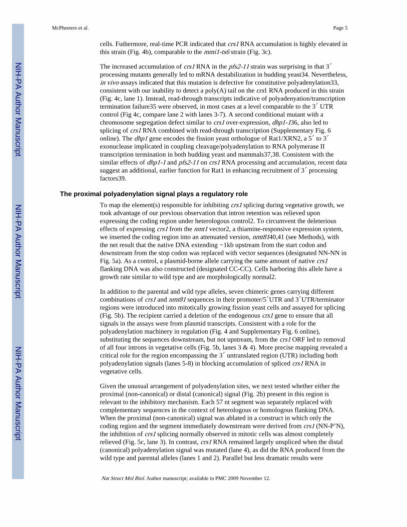
The proximal polyadenylation signal plays a regulatory roleTo map the element(s) responsible for inhibiting crs1 splicing during vegetative growth, wetook advantage of our previous observation that intron retention was relieved uponexpressing the coding region under heterologous control2. To circumvent the deleteriouseffects of expressing crs1 from the nmt1 vector2, a thiamine-responsive expression system,we inserted the coding region into an attenuated version, nmt8140,41 (see Methods), withthe net result that the native DNA extending ∼1kb upstream from the start codon anddownstream from the stop codon was replaced with vector sequences (designated NN-NN inFig. 5a). As a control, a plasmid-borne allele carrying the same amount of native crs1flanking DNA was also constructed (designated CC-CC). Cells harboring this allele have agrowth rate similar to wild type and are morphologically normal2.
In addition to the parental and wild type alleles, seven chimeric genes carrying differentcombinations of crs1 and nmt81 sequences in their promoter/5′UTR and 3′UTR/terminatorregions were introduced into mitotically growing fission yeast cells and assayed for splicing(Fig. 5b). The recipient carried a deletion of the endogenous crs1 gene to ensure that allsignals in the assays were from plasmid transcripts. Consistent with a role for thepolyadenylation machinery in regulation (Fig. 4 and Supplementary Fig. 6 online),substituting the sequences downstream, but not upstream, from the crs1 ORF led to removalof all four introns in vegetative cells (Fig. 5b, lanes 3 & 4). More precise mapping revealed acritical role for the region encompassing the 3′ untranslated region (UTR) including bothpolyadenylation signals (lanes 5-8) in blocking accumulation of spliced crs1 RNA invegetative cells.
Given the unusual arrangement of polyadenylation sites, we next tested whether either theproximal (non-canonical) or distal (canonical) signal (Fig. 2b) present in this region isrelevant to the inhibitory mechanism. Each 57 nt segment was separately replaced withcomplementary sequences in the context of heterologous or homologous flanking DNA.When the proximal (non-canonical) signal was ablated in a construct in which only thecoding region and the segment immediately downstream were derived from crs1 (NN-P’N),the inhibition of crs1 splicing normally observed in mitotic cells was almost completelyrelieved (Fig. 5c, lane 3). In contrast, crs1 RNA remained largely unspliced when the distal(canonical) polyadenylation signal was mutated (lane 4), as did the RNA produced from thewild type and parental alleles (lanes 1 and 2). Parallel but less dramatic results were
McPheeters et al. Page 5
Nat Struct Mol Biol. Author manuscript; available in PMC 2009 November 12.
NIH
-PA Author Manuscript
NIH
-PA Author Manuscript
NIH
-PA Author Manuscript
obtained with a series of constructs in which the polyadenylation signals were mutated in thecontext of native crs1 flanking DNA (Supplementary Fig. 7 online).
Based on the indistinguishable kinetics of increased crs1 RNA accumulation and activationof splicing during meiosis (Fig. 2a & d), we asked whether substituting or mutating eitherpolyadenylation signal increased the steady-state level of crs1 RNA in vegetative cells.Strikingly, the proximal (non-canonical) polyadenylation signal mutant (NN-P’N) producedconsiderably more RNA than either the wild type (CC-CC) or parental alleles (NN-CN; Fig.5d, compare c3 with b1 & b8). Moreover, as during meiosis (Fig. 1b), the increase was duealmost entirely to a higher level of spliced RNA. In contrast, ablating the distal (canonical)polyadenylation signal depressed the level of both spliced and unspliced RNA (Fig. 5d,compare b8 & c4). We conclude that the proximal (non-canonical) polyadenylation signal isimportant not only for preventing splicing, but also contributes to restricting accumulation ofmature crs1 RNA. This conclusion is bolstered by quantitative comparisons of splicing andRNA accumulation between chimeras that differ in only a single segment (SupplementaryTables 1 and 2 online).
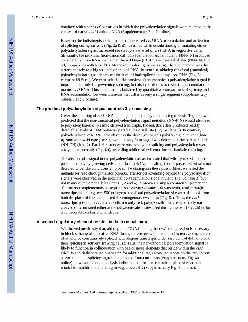
The proximal polyadenylation signal controls 3′ processingGiven the coupling of crs1 RNA splicing and polyadenylation during meiosis (Fig. 2e), wepredicted that the non-canonical polyadenylation signal mutation (NN-P’N) would also leadto polyadenylation of plasmid-derived transcripts. Indeed, this allele produced readilydetectable levels of RNA polyadenylated at the distal site (Fig. 6a, lane 3). In contrast,polyadenylated crs1 RNA was absent in the distal (canonical) poly(A) signal mutant (lane4), similar to wild type (lane 1), while a very faint signal was detected in the parental allele(NN-CN) (lane 2). Parallel results were observed when splicing and polyadenylation wereassayed concurrently (Fig. 6b), providing additional evidence for mechanistic coupling.
The absence of a signal in the polyadenylation assay indicated that wild-type crs1 transcriptspresent in actively growing cells either lack poly(A) tails altogether or possess short tails notdetected under the conditions employed. To distinguish these possibilities, we tested themutants for read-through transcription35. Transcripts extending beyond the polyadenylationsignals were observed in the proximal polyadenylation signal mutant (Fig. 6c, lane 3) butnot in any of the other alleles (lanes 1, 2 and 4). Moreover, using a common 5′ primer and3′ primers complementary to sequences at varying distances downstream, read-throughtranscripts extending over 500 nt beyond the distal polyadenylation site were detected fromboth the plasmid-borne allele and the endogenous crs1 locus (Fig. 6c). Thus, the crs1transcripts present in vegetative cells not only lack poly(A) tails, but are apparently notcleaved or terminated either at the polyadenylation sites used during meiosis (Fig. 2b) or fora considerable distance downstream.
A second regulatory element resides in the terminal exonWe showed previously that, although the DNA flanking the crs1 coding region is necessaryto block splicing of the native RNA during mitotic growth, it is not sufficient, as expressionof otherwise constitutively spliced heterologous transcripts under crs1 control did not blocktheir splicing in actively growing cells2. Thus, the non-canonical polyadenylation signal islikely to function in collaboration with one or more elements that reside within the crs1ORF. We initially focused our search for additional regulatory sequences on the crs1 introns,as each contains splicing signals that deviate from consensus (Supplementary Fig. 8aonline); however, deletion analysis indicated that the non-canonical splice sites are notcrucial for inhibition of splicing in vegetative cells (Supplementary Fig. 8b online).
McPheeters et al. Page 6
Nat Struct Mol Biol. Author manuscript; available in PMC 2009 November 12.
NIH
-PA Author Manuscript
NIH
-PA Author Manuscript
NIH
-PA Author Manuscript
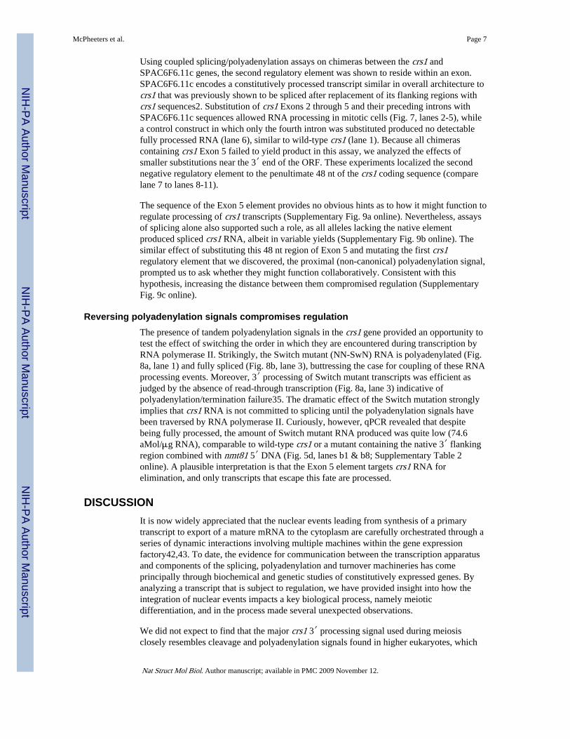
Using coupled splicing/polyadenylation assays on chimeras between the crs1 andSPAC6F6.11c genes, the second regulatory element was shown to reside within an exon.SPAC6F6.11c encodes a constitutively processed transcript similar in overall architecture tocrs1 that was previously shown to be spliced after replacement of its flanking regions withcrs1 sequences2. Substitution of crs1 Exons 2 through 5 and their preceding introns withSPAC6F6.11c sequences allowed RNA processing in mitotic cells (Fig. 7, lanes 2-5), whilea control construct in which only the fourth intron was substituted produced no detectablefully processed RNA (lane 6), similar to wild-type crs1 (lane 1). Because all chimerascontaining crs1 Exon 5 failed to yield product in this assay, we analyzed the effects ofsmaller substitutions near the 3′ end of the ORF. These experiments localized the secondnegative regulatory element to the penultimate 48 nt of the crs1 coding sequence (comparelane 7 to lanes 8-11).
The sequence of the Exon 5 element provides no obvious hints as to how it might function toregulate processing of crs1 transcripts (Supplementary Fig. 9a online). Nevertheless, assaysof splicing alone also supported such a role, as all alleles lacking the native elementproduced spliced crs1 RNA, albeit in variable yields (Supplementary Fig. 9b online). Thesimilar effect of substituting this 48 nt region of Exon 5 and mutating the first crs1regulatory element that we discovered, the proximal (non-canonical) polyadenylation signal,prompted us to ask whether they might function collaboratively. Consistent with thishypothesis, increasing the distance between them compromised regulation (SupplementaryFig. 9c online).
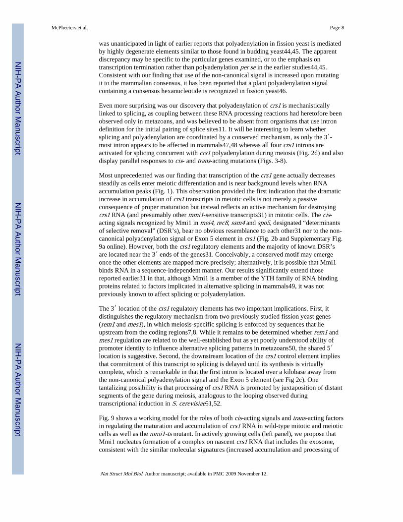
Reversing polyadenylation signals compromises regulationThe presence of tandem polyadenylation signals in the crs1 gene provided an opportunity totest the effect of switching the order in which they are encountered during transcription byRNA polymerase II. Strikingly, the Switch mutant (NN-SwN) RNA is polyadenylated (Fig.8a, lane 1) and fully spliced (Fig. 8b, lane 3), buttressing the case for coupling of these RNAprocessing events. Moreover, 3′ processing of Switch mutant transcripts was efficient asjudged by the absence of read-through transcription (Fig. 8a, lane 3) indicative ofpolyadenylation/termination failure35. The dramatic effect of the Switch mutation stronglyimplies that crs1 RNA is not committed to splicing until the polyadenylation signals havebeen traversed by RNA polymerase II. Curiously, however, qPCR revealed that despitebeing fully processed, the amount of Switch mutant RNA produced was quite low (74.6aMol/μg RNA), comparable to wild-type crs1 or a mutant containing the native 3′ flankingregion combined with nmt81 5′ DNA (Fig. 5d, lanes b1 & b8; Supplementary Table 2online). A plausible interpretation is that the Exon 5 element targets crs1 RNA forelimination, and only transcripts that escape this fate are processed.
DISCUSSIONIt is now widely appreciated that the nuclear events leading from synthesis of a primarytranscript to export of a mature mRNA to the cytoplasm are carefully orchestrated through aseries of dynamic interactions involving multiple machines within the gene expressionfactory42,43. To date, the evidence for communication between the transcription apparatusand components of the splicing, polyadenylation and turnover machineries has comeprincipally through biochemical and genetic studies of constitutively expressed genes. Byanalyzing a transcript that is subject to regulation, we have provided insight into how theintegration of nuclear events impacts a key biological process, namely meioticdifferentiation, and in the process made several unexpected observations.
We did not expect to find that the major crs1 3′ processing signal used during meiosisclosely resembles cleavage and polyadenylation signals found in higher eukaryotes, which
McPheeters et al. Page 7
Nat Struct Mol Biol. Author manuscript; available in PMC 2009 November 12.
NIH
-PA Author Manuscript
NIH
-PA Author Manuscript
NIH
-PA Author Manuscript
was unanticipated in light of earlier reports that polyadenylation in fission yeast is mediatedby highly degenerate elements similar to those found in budding yeast44,45. The apparentdiscrepancy may be specific to the particular genes examined, or to the emphasis ontranscription termination rather than polyadenylation per se in the earlier studies44,45.Consistent with our finding that use of the non-canonical signal is increased upon mutatingit to the mammalian consensus, it has been reported that a plant polyadenylation signalcontaining a consensus hexanucleotide is recognized in fission yeast46.
Even more surprising was our discovery that polyadenylation of crs1 is mechanisticallylinked to splicing, as coupling between these RNA processing reactions had heretofore beenobserved only in metazoans, and was believed to be absent from organisms that use introndefinition for the initial pairing of splice sites11. It will be interesting to learn whethersplicing and polyadenylation are coordinated by a conserved mechanism, as only the 3′-most intron appears to be affected in mammals47,48 whereas all four crs1 introns areactivated for splicing concurrent with crs1 polyadenylation during meiosis (Fig. 2d) and alsodisplay parallel responses to cis- and trans-acting mutations (Figs. 3-8).
Most unprecedented was our finding that transcription of the crs1 gene actually decreasessteadily as cells enter meiotic differentiation and is near background levels when RNAaccumulation peaks (Fig. 1). This observation provided the first indication that the dramaticincrease in accumulation of crs1 transcripts in meiotic cells is not merely a passiveconsequence of proper maturation but instead reflects an active mechanism for destroyingcrs1 RNA (and presumably other mmi1-sensitive transcripts31) in mitotic cells. The cis-acting signals recognized by Mmi1 in mei4, rec8, ssm4 and spo5, designated “determinantsof selective removal” (DSR’s), bear no obvious resemblance to each other31 nor to the non-canonical polyadenylation signal or Exon 5 element in crs1 (Fig. 2b and Supplementary Fig.9a online). However, both the crs1 regulatory elements and the majority of known DSR’sare located near the 3′ ends of the genes31. Conceivably, a conserved motif may emergeonce the other elements are mapped more precisely; alternatively, it is possible that Mmi1binds RNA in a sequence-independent manner. Our results significantly extend thosereported earlier31 in that, although Mmi1 is a member of the YTH family of RNA bindingproteins related to factors implicated in alternative splicing in mammals49, it was notpreviously known to affect splicing or polyadenylation.
The 3′ location of the crs1 regulatory elements has two important implications. First, itdistinguishes the regulatory mechanism from two previously studied fission yeast genes(rem1 and mes1), in which meiosis-specific splicing is enforced by sequences that lieupstream from the coding regions7,8. While it remains to be determined whether rem1 andmes1 regulation are related to the well-established but as yet poorly understood ability ofpromoter identity to influence alternative splicing patterns in metazoans50, the shared 5′location is suggestive. Second, the downstream location of the crs1 control element impliesthat commitment of this transcript to splicing is delayed until its synthesis is virtuallycomplete, which is remarkable in that the first intron is located over a kilobase away fromthe non-canonical polyadenylation signal and the Exon 5 element (see Fig 2c). Onetantalizing possibility is that processing of crs1 RNA is promoted by juxtaposition of distantsegments of the gene during meiosis, analogous to the looping observed duringtranscriptional induction in S. cerevisiae51,52.
Fig. 9 shows a working model for the roles of both cis-acting signals and trans-acting factorsin regulating the maturation and accumulation of crs1 RNA in wild-type mitotic and meioticcells as well as the mmi1-ts mutant. In actively growing cells (left panel), we propose thatMmi1 nucleates formation of a complex on nascent crs1 RNA that includes the exosome,consistent with the similar molecular signatures (increased accumulation and processing of
McPheeters et al. Page 8
Nat Struct Mol Biol. Author manuscript; available in PMC 2009 November 12.
NIH
-PA Author Manuscript
NIH
-PA Author Manuscript
NIH
-PA Author Manuscript
crs1 RNA) that we observe in the rrp6Δ and mmi1-ts mutants, as well as a two-hybridMmi1-Rrp6 interaction31. In that deletion of a TRAMP complex subunit has no discernibleimpact on crs1 RNA processing or accumulation, the most parsimonious explanation is thatin the specialized turnover pathway that blocks production of meiotic mRNAs in mitotic S.pombe cells, Mmi1 plays a role analogous to TRAMP-mediated polyadenylation in othernuclear surveillance and silencing pathways27,30.
Although we have not definitively proven that the crs1 regulatory mechanism operates co-transcriptionally, several lines of circumstantial evidence support this idea. By analogy tothe known association of the exosome with RNA polymerase II in Drosophila53, it seemsplausible to suggest this mode of delivery in crs1. Co-transcriptional assembly of thecomplex that regulates turnover and processing is also consistent with the effect of reversingthe order of the polyadenylation signals, as well as the involvement of Pfs2, apolyadenylation factor functionally related to mammalian CstF-50, which is known to bindthe Pol II CTD42. The involvement of dhp1 in crs1 regulation (Supplementary Fig 6.)provides perhaps the most compelling evidence for a co-transcriptional mechanism, as itsorthologues rat1/XRN2 link transcription termination with polyadenylation37,38. Finally,several meiotic transcripts were spliced upon lowering the gene dosage of a fission yeastcyclophilin that modulates Pol II CTD phosphorylation54, although crs1 was not among theRNAs examined.
The middle panel of Fig. 9 depicts the proposed molecular events in wild-type S. pombecells undergoing sexual differentiation. While it remains to be determined whether Mmi1influences the delivery, stabilization or activation of the exosome in mitotic cells, in meioticcells it cannot perform this role due to sequestration by the Mei2 protein, a key regulator ofsexual development in fission yeast1,31. The functional inactivation of Mmi1 indifferentiating cells renders it unavailable to bind nascent crs1 transcripts, thus allowingthem to undergo normal 3′ maturation and splicing, presumably followed by export of themature mRNA to the cytoplasm and translation to produce the Crs1 cyclin. In mmi1-tsmutant cells (right panel), we propose a similar sequence of events except that a mutationrather than sequestration prevents Mmi1 from associating with newly synthesized crs1RNA. The molecular events in the Switch mutant are more speculative, but a plausibleinterpretation of the low yield of fully processed RNA is a bifurcating pathway in whichdestruction of the RNA competes with processing (Supplementary Fig. 10 online).
The precise role of Pfs2 is even more enigmatic, as the pfs2-11 mutation affects constitutiveas well as regulated polyadenylation, and in the context of crs1 uncouples this RNAprocessing event from splicing. One possible explanation is that splicing in pfs2-11 (anddhp1-1/rat1, which behaves similarly) occurs post- rather than co-transcriptionally. Theinvolvement of Pfs2 in crs1 regulation suggests a link to control mechanisms discovered inmore complex eukaryotes, as putative orthologues of this protein (CstF-50 paralogues) havebeen implicated in the regulation of Arabidopsis flowering55 and display increasedexpression during mouse spermatogenesis56. A key difference, however, is that previousstudies of regulated 3′ processing, including two (non-meiotic) examples in S.pombe20,57-59 described switching between sites rather than the differential activationimplied by our data (Fig. 2a). Based on these precedents, we fully expected a positivecontrol mechanism in which a meiosis-specific factor would allow use of the proximal (non-canonical) polyadenylation signal; however, all of our data point instead to a negativemechanism that blocks the use of both sites in vegetative cells.
The complex, and to our knowledge unprecedented, mechanism that up-regulates crs1 geneexpression independent of transcriptional induction raises an obvious question: why dovegetative fission yeast cells continuously synthesize crs1 RNA, only to destroy it? We
McPheeters et al. Page 9
Nat Struct Mol Biol. Author manuscript; available in PMC 2009 November 12.
NIH
-PA Author Manuscript
NIH
-PA Author Manuscript
NIH
-PA Author Manuscript
propose that this regulatory strategy, in addition to preventing the deleterious production ofa meiotic protein in growing cells2, serves to “prime” the gene expression pump, ensuring avery rapid response to adverse conditions in which mating, meiosis and sporulation are theorganism’s sole means of survival. Given that some elements of the crs1 regulatorymechanism have antecedents in other organisms, it is tempting to speculate that selectiveRNA turnover pathways may contribute more generally to changes in gene expression inresponse to environmental or developmental cues.
METHODSS. pombe manipulations
See Supplementary Table 3 (online) for complete genotypes and sources for the strains usedin this study. Fission yeast media and growth conditions were as described earlier2. Weinduced ectopic meiosis in the pat1-114 mutant (Figs. 1 & 2) by shifting the fission yeaststrain F90 to the non-permissive temperature (34°C) as described previously2. Growthconditions for other strains are described in the figure legends.
OligonucleotidesSee Supplementary Table 4 (online) for sequences of all oligonucleotides used in this study;these are grouped according to the purpose for which they were used.
Mapping the crs1 terminiTo determine the 5′ and 3′ ends of crs1 RNA, we performed RACE (Rapid Amplificationof cDNA Ends) on RNA extracted from vegetatively growing or meiotic (4 h at 34°C) F90cells. Products visible by ethidium bromide staining were cloned and sequenced by thefacility at SUNY, Stony Brook.
Construction of chimeric and mutant plasmidsDue to the large number of constructs analyzed and their complexity, the relevantinformation is provided in Supplementary Methods online.
RNA processing assaysProcessing of crs1 RNA was assayed by both standard and real-time (quantitative) PCR.Standard (semi-quantitative) RT-PCR splicing assays were performed as described earlier2using crs1-SAF (oligo #23) and crs1-SAR3 (oligo #68) as primers. Real-time PCR (qPCR)splicing assays were performed and the data analyzed as described in SupplementaryMethods online. RT-PCR assays of polyadenylation were performed similarly except that,depending on the experimental question, two different 5′ primers (crs1-RC3′, oligo #28, orcrs1-SAF, oligo #23) were used in combination with oligo(dT)24 (oligo #69) as the 3′primer. The 3′ UTR primer used to detect crs1 RNA independent of polyadenylation statewas crs1-nmtTRNew (oligo #17). To assay for read-through transcription, the 3′ primerswere crs1-CER (oligo #70) or nmt-SEQ B2. All RNA processing assays were repeated 2-4times using independent transformants in the case of plasmid-based experiments.
Transcription Run-On (TRO) analysisS. pombe TRO analysis was performed essentially as described13 with the followingmodifications. Cells were permeabilized in 0.6% Sarcosyl (w/v) for 25 minutes withconstant mixing on ice. Following this step, transcription was carried out at 30°C for 6minutes. Equal numbers of micrograms of 32P-UTP-labeled RNA from each time point washybridized to filters on which 1.4 pmole of each denatured PCR fragment had beenimmobilized. The probes, which correspond to the 3′ ends of the coding region for each
McPheeters et al. Page 10
Nat Struct Mol Biol. Author manuscript; available in PMC 2009 November 12.
NIH
-PA Author Manuscript
NIH
-PA Author Manuscript
NIH
-PA Author Manuscript
gene analyzed, were made by PCR using the following oligonucleotide primers(Supplementary Table 4 online): for crs1, crs1-nmtTR3New (oligo #19 and crs-SPAC6F-Ex5(N) (oligo #59); for pta1, Pta1-PF (oligo #83) and Pta1-PR (oligo #84); and for meu4,Meu4-DF (oligo #85) and Meu4-DR (oligo #86).
Supplementary MaterialRefer to Web version on PubMed Central for supplementary material.
AcknowledgmentsThe authors would like to thank Masayuki Yamamoto (Univ. Tokyo), Chris Norbury (Oxford Univ.) and RichMaraia (NIH) for strains (see Supplementary Table 3 online). We are grateful to our colleagues Kristian Baker,Jonatha Gott, Hua Lou, Tim Nilsen, Cathy Patterson, Helen Salz and Steve Sanders for helpful discussions andcritical reading of the manuscript. This work was funded by NIH grants R01-GM073217, awarded jointly to J. A.W. and J. L.; R01-GM064682, awarded to D. S. M.; and R01-GM38070, awarded to J. A. W.
REFERENCES1. Harigaya Y, Yamamoto M. Molecular mechanisms underlying the mitosis-meiosis decision.
Chromosome Res. 2007; 15:523–37. [PubMed: 17674143]
2. Averbeck N, Sunder S, Sample N, Wise JA, Leatherwood J. Negative control contributes to anextensive program of meiotic splicing in fission yeast. Mol Cell. 2005; 18:491–8. [PubMed:15893732]
3. Juneau K, Palm C, Miranda M, Davis RW. High-density yeast-tiling array reveals previouslyundiscovered introns and extensive regulation of meiotic splicing. Proc Natl Acad Sci U S A. 2007;104:1522–7. [PubMed: 17244705]
4. Spingola M, Ares M Jr. A yeast intronic splicing enhancer and Nam8p are required for Mer1p-activated splicing. Mol Cell. 2000; 6:329–38. [PubMed: 10983980]
5. Kishida M, Nagai T, Nakaseko Y, Shimoda C. Meiosis-dependent mRNA splicing of the fissionyeast Schizosaccharomyces pombe mes1+ gene. Curr Genet. 1994; 25:497–503. [PubMed:8082199]
6. Malapeira J, et al. A meiosis-specific cyclin regulated by splicing is required for proper progressionthrough meiosis. Mol Cell Biol. 2005; 25:6330–7. [PubMed: 16024772]
7. Shimoseki M, Shimoda C. The 5′ terminal region of the Schizosaccharomyces pombe mes1 mRNAis crucial for its meiosis-specific splicing. Mol Genet Genomics. 2001; 265:673–82. [PubMed:11459187]
8. Moldon A, et al. Promoter-driven splicing regulation in fission yeast. Nature. 2008; 455:997–1000.[PubMed: 18815595]
9. Cramer P, Pesce CG, Baralle FE, Kornblihtt AR. Functional association between promoter structureand transcript alternative splicing. Proc Natl Acad Sci U S A. 1997; 94:11456–60. [PubMed:9326631]
10. Tasic B, et al. Promoter choice determines splice site selection in protocadherin alpha and gammapre-mRNA splicing. Mol Cell. 2002; 10:21–33. [PubMed: 12150904]
11. Niwa M, Rose SD, Berget SM. In vitro polyadenylation is stimulated by the presence of anupstream intron. Genes Dev. 1990; 4:1552–9. [PubMed: 1701407]
12. Hansen K, Birse CE, Proudfoot NJ. Nascent transcription from the nmt1 and nmt2 genes ofSchizosaccharomyces pombe overlaps neighbouring genes. Embo J. 1998; 17:3066–77. [PubMed:9606189]
13. Iino Y, Yamamoto M. Negative control for the initiation of meiosis in Schizosaccharomycespombe. Proc Natl Acad Sci U S A. 1985; 82:2447–2451. [PubMed: 16593556]
14. Mata J, Lyne R, Burns G, Bahler J. The transcriptional program of meiosis and sporulation infission yeast. Nat Genet. 2002; 32:143–7. [PubMed: 12161753]
15. Mata J, Wilbrey A, Bahler J. Transcriptional regulatory network for sexual differentiation infission yeast. Genome Biol. 2007; 8:R217. [PubMed: 17927811]
McPheeters et al. Page 11
Nat Struct Mol Biol. Author manuscript; available in PMC 2009 November 12.
NIH
-PA Author Manuscript
NIH
-PA Author Manuscript
NIH
-PA Author Manuscript
16. Wilhelm BT, et al. Dynamic repertoire of a eukaryotic transcriptome surveyed at single-nucleotideresolution. Nature. 2008; 453:1239–43. [PubMed: 18488015]
17. Takagaki Y, Manley JL. Complex protein interactions within the human polyadenylationmachinery identify a novel component. Mol Cell Biol. 2000; 20:1515–25. [PubMed: 10669729]
18. Watanabe T, et al. Comprehensive isolation of meiosis-specific genes identifies novel proteins andunusual non-coding transcripts in Schizosaccharomyces pombe. Nucleic Acids Res. 2001;29:2327–37. [PubMed: 11376151]
19. Gilmartin GM. Eukaryotic mRNA 3′ processing: a common means to different ends. Genes Dev.2005; 19:2517–21. [PubMed: 16264187]
20. Liu D, et al. Systematic variation in mRNA 3′-processing signals during mouse spermatogenesis.Nucleic Acids Res. 2007; 35:234–46. [PubMed: 17158511]
21. Wilusz J, Pettine SM, Shenk T. Functional analysis of point mutations in the AAUAAA motif ofthe SV40 late polyadenylation signal. Nucleic Acids Res. 1989; 17:3899–908. [PubMed: 2543957]
22. Bousquet-Antonelli C, Presutti C, Tollervey D. Identification of a regulated pathway for nuclearpre-mRNA turnover. Cell. 2000; 102:765–75. [PubMed: 11030620]
23. Wilusz CJ, Wilusz J. Bringing the role of mRNA decay in the control of gene expression intofocus. Trends Genet. 2004; 20:491–7. [PubMed: 15363903]
24. Wang SW, Stevenson AL, Kearsey SE, Watt S, Bahler J. Global role for polyadenylation-assistednuclear RNA degradation in posttranscriptional gene silencing. Mol Cell Biol. 2008; 28:656–65.[PubMed: 18025105]
25. Houseley J, LaCava J, Tollervey D. RNA-quality control by the exosome. Nat Rev Mol Cell Biol.2006; 7:529–39. [PubMed: 16829983]
26. LaCava J, et al. RNA degradation by the exosome is promoted by a nuclear polyadenylationcomplex. Cell. 2005; 121:713–24. [PubMed: 15935758]
27. Win TZ, et al. Requirement of fission yeast Cid14 in polyadenylation of rRNAs. Mol Cell Biol.2006; 26:1710–21. [PubMed: 16478992]
28. Kinoshita N, Goebl M, Yanagida M. The fission yeast dis3+ gene encodes a 110-kDa essentialprotein implicated in mitotic control. Mol Cell Biol. 1991; 11:5839–47. [PubMed: 1944266]
29. Dziembowski A, Lorentzen E, Conti E, Seraphin B. A single subunit, Dis3, is essentiallyresponsible for yeast exosome core activity. Nat Struct Mol Biol. 2007; 14:15–22. [PubMed:17173052]
30. Buhler M, Haas W, Gygi SP, Moazed D. RNAi-dependent and - independent RNA turnovermechanisms contribute to heterochromatic gene silencing. Cell. 2007; 129:707–21. [PubMed:17512405]
31. Harigaya Y, et al. Selective elimination of messenger RNA prevents an incidence of untimelymeiosis. Nature. 2006; 442:45–50. [PubMed: 16823445]
32. Ohnacker M, Barabino SM, Preker PJ, Keller W. The WD-repeat protein pfs2p bridges twoessential factors within the yeast pre-mRNA 3′-end-processing complex. Embo J. 2000; 19:37–47.[PubMed: 10619842]
33. Wang SW, Asakawa K, Win TZ, Toda T, Norbury CJ. Inactivation of the pre-mRNA cleavage andpolyadenylation factor Pfs2 in fission yeast causes lethal cell cycle defects. Mol Cell Biol. 2005;25:2288–96. [PubMed: 15743824]
34. Hilleren P, McCarthy T, Rosbash M, Parker R, Jensen TH. Quality control of mRNA 3′-endprocessing is linked to the nuclear exosome. Nature. 2001; 413:538–42. [PubMed: 11586364]
35. Proudfoot N. New perspectives on connecting messenger RNA 3′ end formation to transcription.Curr Opin Cell Biol. 2004; 16:272–8. [PubMed: 15145351]
36. Shobuike T, Tatebayashi K, Tani T, Sugano S, Ikeda H. The dhp1(+) gene, encoding a putativenuclear 5′-->3′ exoribonuclease, is required for proper chromosome segregation in fission yeast.Nucleic Acids Res. 2001; 29:1326–33. [PubMed: 11238999]
37. West S, Gromak N, Proudfoot NJ. Human 5′ --> 3′ exonuclease Xrn2 promotes transcriptiontermination at co-transcriptional cleavage sites. Nature. 2004; 432:522–5. [PubMed: 15565158]
38. Kim M, et al. The yeast Rat1 exonuclease promotes transcription termination by RNA polymeraseII. Nature. 2004; 432:517–22. [PubMed: 15565157]
McPheeters et al. Page 12
Nat Struct Mol Biol. Author manuscript; available in PMC 2009 November 12.
NIH
-PA Author Manuscript
NIH
-PA Author Manuscript
NIH
-PA Author Manuscript
39. Luo W, Johnson AW, Bentley DL. The role of Rat1 in coupling mRNA 3′- end processing totranscription termination: implications for a unified allosterictorpedo model. Genes Dev. 2006;20:954–65. [PubMed: 16598041]
40. Basi G, Schmid E, Maundrell K. TATA box mutations in the Schizosaccharomyces pombe nmt1promoter affect transcription efficiency but not the transcription start point or thiaminerepressibility. Gene. 1993; 123:131–6. [PubMed: 8422997]
41. Maundrell K. nmt1 of fission yeast. A highly transcribed gene completely repressed by thiamine. JBiol Chem. 1990; 265:10857–64. [PubMed: 2358444]
42. Fong N, Bentley DL. Capping, splicing, and 3′ processing are independently stimulated by RNApolymerase II: different functions for different segments of the CTD. Genes Dev. 2001; 15:1783–95. [PubMed: 11459828]
43. Maniatis T, Reed R. An extensive network of coupling among gene expression machines. Nature.2002; 416:499–506. [PubMed: 11932736]
44. Aranda A, Proudfoot NJ. Definition of transcriptional pause elements in fission yeast. Mol CellBiol. 1999; 19:1251–61. [PubMed: 9891059]
45. Birse CE, Lee BA, Hansen K, Proudfoot NJ. Transcriptional termination signals for RNApolymerase II in fission yeast. Embo J. 1997; 16:3633–43. [PubMed: 9218804]
46. Chakraborty S, Sarmah B, Chakraborty N, Datta A. Premature termination of RNA polymerase IImediated transcription of a seed protein gene in Schizosaccharomyces pombe. Nucleic Acids Res.2002; 30:2940–9. [PubMed: 12087180]
47. Cooke C, Hans H, Alwine JC. Utilization of splicing elements and polyadenylation signal elementsin the coupling of polyadenylation and last-intron removal. Mol Cell Biol. 1999; 19:4971–9.[PubMed: 10373547]
48. Niwa M, Berget SM. Mutation of the AAUAAA polyadenylation signal depresses in vitro splicingof proximal but not distal introns. Genes Dev. 1991; 5:2086–95. [PubMed: 1657710]
49. Stoilov P, Rafalska I, Stamm S. YTH: a new domain in nuclear proteins. Trends Biochem Sci.2002; 27:495–7. [PubMed: 12368078]
50. Kornblihtt AR. Promoter usage and alternative splicing. Curr Opin Cell Biol. 2005; 17:262–8.[PubMed: 15901495]
51. Ansari A, Hampsey M. A role for the CPF 3′-end processing machinery in RNAP II-dependentgene looping. Genes Dev. 2005; 19:2969–78. [PubMed: 16319194]
52. O’Sullivan JM, et al. Gene loops juxtapose promoters and terminators in yeast. Nat Genet. 2004;36:1014–8. [PubMed: 15314641]
53. Andrulis ED, et al. The RNA processing exosome is linked to elongating RNA polymerase II inDrosophila. Nature. 2002; 420:837–41. [PubMed: 12490954]
54. Gullerova M, Barta A, Lorkovic ZJ. Rct1, a nuclear RNA recognition motifcontaining cyclophilin,regulates phosphorylation of the RNA polymerase II C-terminal domain. Mol Cell Biol. 2007;27:3601–11. [PubMed: 17339332]
55. Quesada V, Macknight R, Dean C, Simpson GG. Autoregulation of FCA pre-mRNA processingcontrols Arabidopsis flowering time. Embo J. 2003; 22:3142–52. [PubMed: 12805228]
56. Henderson IR, Liu F, Drea S, Simpson GG, Dean C. An allelic series reveals essential roles for FYin plant development in addition to flowering-time control. Development. 2005; 132:3597–607.[PubMed: 16033802]
57. Beaudoing E, Gautheret D. Identification of alternate polyadenylation sites and analysis of theirtissue distribution using EST data. Genome Res. 2001; 11:1520–6. [PubMed: 11544195]
58. Jang YK, et al. Differential expression of the rhp51+ gene, a recA and RAD51 homolog from thefission yeast Schizosaccharomyces pombe. Gene. 1996; 169:125–30. [PubMed: 8635736]
59. Munoz MJ, Daga RR, Garzon A, Thode G, Jimenez J. Poly(A) site choice during mRNA 3′-endformation in the Schizosaccharomyces pombe wos2 gene. Mol Genet Genomics. 2002; 267:792–6.[PubMed: 12207226]
60. Moreno S, Klar A, Nurse P. Molecular genetic analysis of fission yeast Schizosaccharomycespombe. Methods Enzymol. 1991; 194:795–823. [PubMed: 2005825]
McPheeters et al. Page 13
Nat Struct Mol Biol. Author manuscript; available in PMC 2009 November 12.
NIH
-PA Author Manuscript
NIH
-PA Author Manuscript
NIH
-PA Author Manuscript
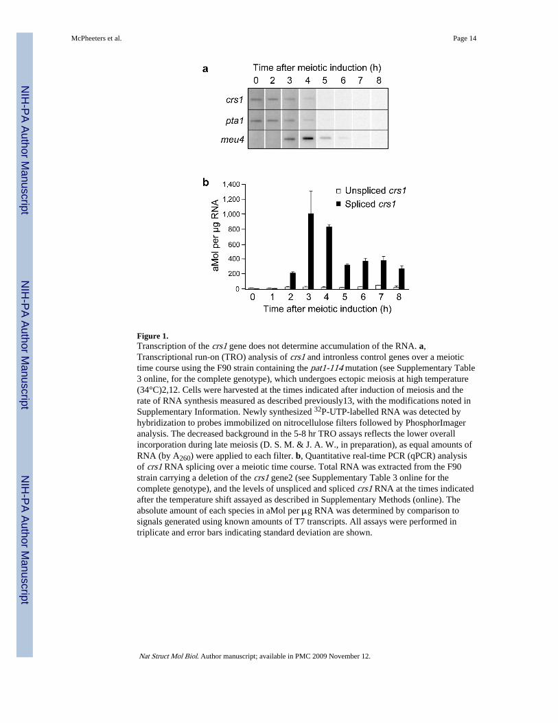
Figure 1.Transcription of the crs1 gene does not determine accumulation of the RNA. a,Transcriptional run-on (TRO) analysis of crs1 and intronless control genes over a meiotictime course using the F90 strain containing the pat1-114 mutation (see Supplementary Table3 online, for the complete genotype), which undergoes ectopic meiosis at high temperature(34°C)2,12. Cells were harvested at the times indicated after induction of meiosis and therate of RNA synthesis measured as described previously13, with the modifications noted inSupplementary Information. Newly synthesized 32P-UTP-labelled RNA was detected byhybridization to probes immobilized on nitrocellulose filters followed by PhosphorImageranalysis. The decreased background in the 5-8 hr TRO assays reflects the lower overallincorporation during late meiosis (D. S. M. & J. A. W., in preparation), as equal amounts ofRNA (by A260) were applied to each filter. b, Quantitative real-time PCR (qPCR) analysisof crs1 RNA splicing over a meiotic time course. Total RNA was extracted from the F90strain carrying a deletion of the crs1 gene2 (see Supplementary Table 3 online for thecomplete genotype), and the levels of unspliced and spliced crs1 RNA at the times indicatedafter the temperature shift assayed as described in Supplementary Methods (online). Theabsolute amount of each species in aMol per μg RNA was determined by comparison tosignals generated using known amounts of T7 transcripts. All assays were performed intriplicate and error bars indicating standard deviation are shown.
McPheeters et al. Page 14
Nat Struct Mol Biol. Author manuscript; available in PMC 2009 November 12.
NIH
-PA Author Manuscript
NIH
-PA Author Manuscript
NIH
-PA Author Manuscript
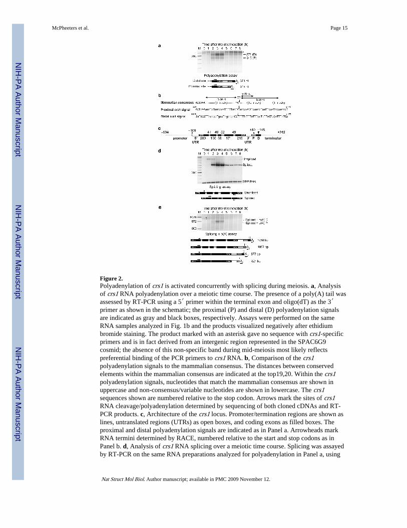
Figure 2.Polyadenylation of crs1 is activated concurrently with splicing during meiosis. a, Analysisof crs1 RNA polyadenylation over a meiotic time course. The presence of a poly(A) tail wasassessed by RT-PCR using a 5′ primer within the terminal exon and oligo(dT) as the 3′primer as shown in the schematic; the proximal (P) and distal (D) polyadenylation signalsare indicated as gray and black boxes, respectively. Assays were performed on the sameRNA samples analyzed in Fig. 1b and the products visualized negatively after ethidiumbromide staining. The product marked with an asterisk gave no sequence with crs1-specificprimers and is in fact derived from an intergenic region represented in the SPAC6G9cosmid; the absence of this non-specific band during mid-meiosis most likely reflectspreferential binding of the PCR primers to crs1 RNA. b, Comparison of the crs1polyadenylation signals to the mammalian consensus. The distances between conservedelements within the mammalian consensus are indicated at the top19,20. Within the crs1polyadenylation signals, nucleotides that match the mammalian consensus are shown inuppercase and non-consensus/variable nucleotides are shown in lowercase. The crs1sequences shown are numbered relative to the stop codon. Arrows mark the sites of crs1RNA cleavage/polyadenylation determined by sequencing of both cloned cDNAs and RT-PCR products. c, Architecture of the crs1 locus. Promoter/termination regions are shown aslines, untranslated regions (UTRs) as open boxes, and coding exons as filled boxes. Theproximal and distal polyadenylation signals are indicated as in Panel a. Arrowheads markRNA termini determined by RACE, numbered relative to the start and stop codons as inPanel b. d, Analysis of crs1 RNA splicing over a meiotic time course. Splicing was assayedby RT-PCR on the same RNA preparations analyzed for polyadenylation in Panel a, using
McPheeters et al. Page 15
Nat Struct Mol Biol. Author manuscript; available in PMC 2009 November 12.
NIH
-PA Author Manuscript
NIH
-PA Author Manuscript
NIH
-PA Author Manuscript
primers complementary to the terminal exons. SRP RNA served as an internal loadingcontrol. e, Concurrent assays of crs1 RNA splicing and polyadenylation over a meiotic timecourse. The coincidence of the two RNA processing reactions on individual crs1 transcriptswas assessed as indicated on the diagram beneath the gel using the same 5′ primer as inPanel d and oligo(dT) as the 3′ primer. Assays employed the same RNA preparationsanalyzed in Panels a and d. The bands marked with asterisks migrate close to the predictedsizes of unspliced polyadenylated crs1 RNA but gave no sequence with crs1-specificprimers.
McPheeters et al. Page 16
Nat Struct Mol Biol. Author manuscript; available in PMC 2009 November 12.
NIH
-PA Author Manuscript
NIH
-PA Author Manuscript
NIH
-PA Author Manuscript
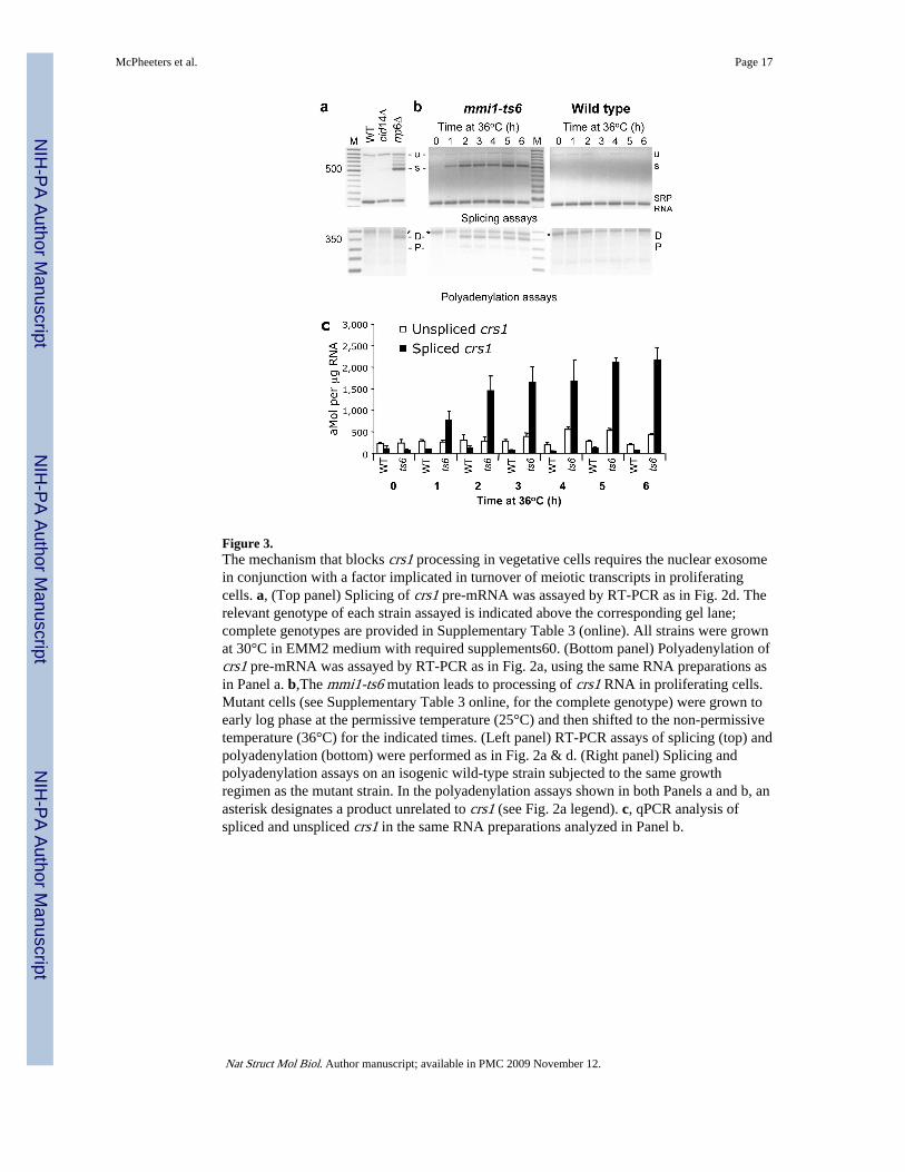
Figure 3.The mechanism that blocks crs1 processing in vegetative cells requires the nuclear exosomein conjunction with a factor implicated in turnover of meiotic transcripts in proliferatingcells. a, (Top panel) Splicing of crs1 pre-mRNA was assayed by RT-PCR as in Fig. 2d. Therelevant genotype of each strain assayed is indicated above the corresponding gel lane;complete genotypes are provided in Supplementary Table 3 (online). All strains were grownat 30°C in EMM2 medium with required supplements60. (Bottom panel) Polyadenylation ofcrs1 pre-mRNA was assayed by RT-PCR as in Fig. 2a, using the same RNA preparations asin Panel a. b,The mmi1-ts6 mutation leads to processing of crs1 RNA in proliferating cells.Mutant cells (see Supplementary Table 3 online, for the complete genotype) were grown toearly log phase at the permissive temperature (25°C) and then shifted to the non-permissivetemperature (36°C) for the indicated times. (Left panel) RT-PCR assays of splicing (top) andpolyadenylation (bottom) were performed as in Fig. 2a & d. (Right panel) Splicing andpolyadenylation assays on an isogenic wild-type strain subjected to the same growthregimen as the mutant strain. In the polyadenylation assays shown in both Panels a and b, anasterisk designates a product unrelated to crs1 (see Fig. 2a legend). c, qPCR analysis ofspliced and unspliced crs1 in the same RNA preparations analyzed in Panel b.
McPheeters et al. Page 17
Nat Struct Mol Biol. Author manuscript; available in PMC 2009 November 12.
NIH
-PA Author Manuscript
NIH
-PA Author Manuscript
NIH
-PA Author Manuscript
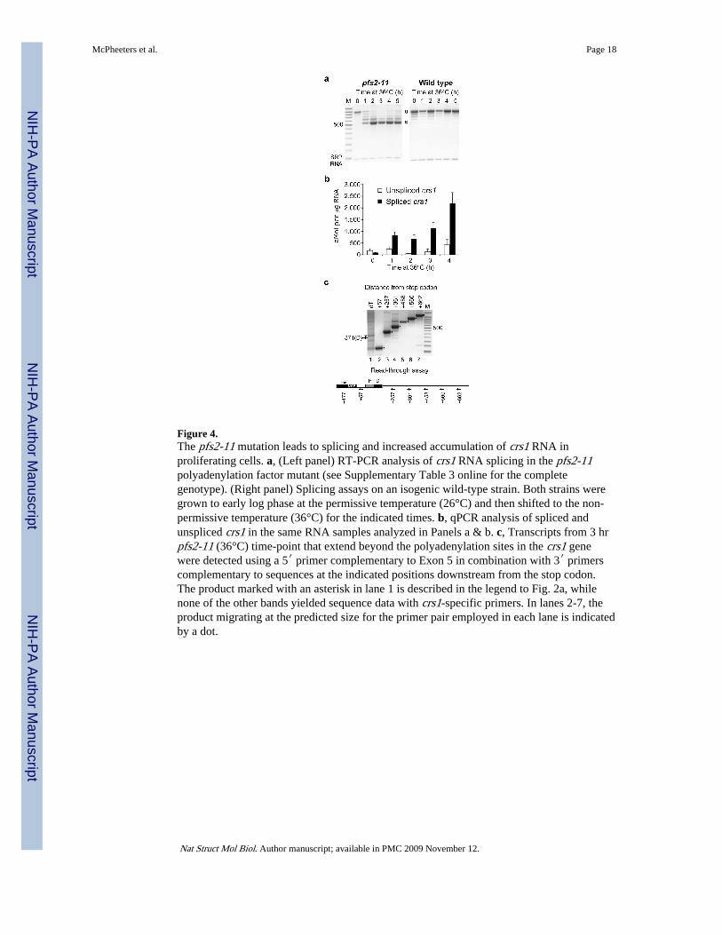
Figure 4.The pfs2-11 mutation leads to splicing and increased accumulation of crs1 RNA inproliferating cells. a, (Left panel) RT-PCR analysis of crs1 RNA splicing in the pfs2-11polyadenylation factor mutant (see Supplementary Table 3 online for the completegenotype). (Right panel) Splicing assays on an isogenic wild-type strain. Both strains weregrown to early log phase at the permissive temperature (26°C) and then shifted to the non-permissive temperature (36°C) for the indicated times. b, qPCR analysis of spliced andunspliced crs1 in the same RNA samples analyzed in Panels a & b. c, Transcripts from 3 hrpfs2-11 (36°C) time-point that extend beyond the polyadenylation sites in the crs1 genewere detected using a 5′ primer complementary to Exon 5 in combination with 3′ primerscomplementary to sequences at the indicated positions downstream from the stop codon.The product marked with an asterisk in lane 1 is described in the legend to Fig. 2a, whilenone of the other bands yielded sequence data with crs1-specific primers. In lanes 2-7, theproduct migrating at the predicted size for the primer pair employed in each lane is indicatedby a dot.
McPheeters et al. Page 18
Nat Struct Mol Biol. Author manuscript; available in PMC 2009 November 12.
NIH
-PA Author Manuscript
NIH
-PA Author Manuscript
NIH
-PA Author Manuscript
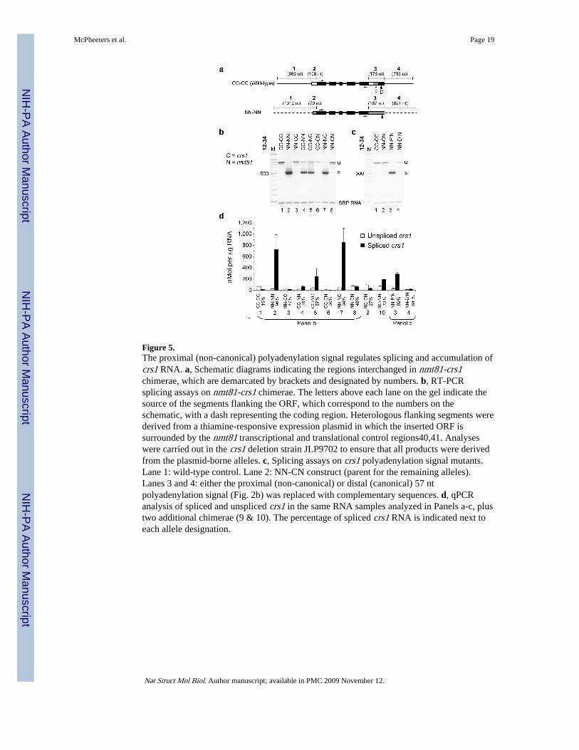
Figure 5.The proximal (non-canonical) polyadenylation signal regulates splicing and accumulation ofcrs1 RNA. a, Schematic diagrams indicating the regions interchanged in nmt81-crs1chimerae, which are demarcated by brackets and designated by numbers. b, RT-PCRsplicing assays on nmt81-crs1 chimerae. The letters above each lane on the gel indicate thesource of the segments flanking the ORF, which correspond to the numbers on theschematic, with a dash representing the coding region. Heterologous flanking segments werederived from a thiamine-responsive expression plasmid in which the inserted ORF issurrounded by the nmt81 transcriptional and translational control regions40,41. Analyseswere carried out in the crs1 deletion strain JLP9702 to ensure that all products were derivedfrom the plasmid-borne alleles. c, Splicing assays on crs1 polyadenylation signal mutants.Lane 1: wild-type control. Lane 2: NN-CN construct (parent for the remaining alleles).Lanes 3 and 4: either the proximal (non-canonical) or distal (canonical) 57 ntpolyadenylation signal (Fig. 2b) was replaced with complementary sequences. d, qPCRanalysis of spliced and unspliced crs1 in the same RNA samples analyzed in Panels a-c, plustwo additional chimerae (9 & 10). The percentage of spliced crs1 RNA is indicated next toeach allele designation.
McPheeters et al. Page 19
Nat Struct Mol Biol. Author manuscript; available in PMC 2009 November 12.
NIH
-PA Author Manuscript
NIH
-PA Author Manuscript
NIH
-PA Author Manuscript
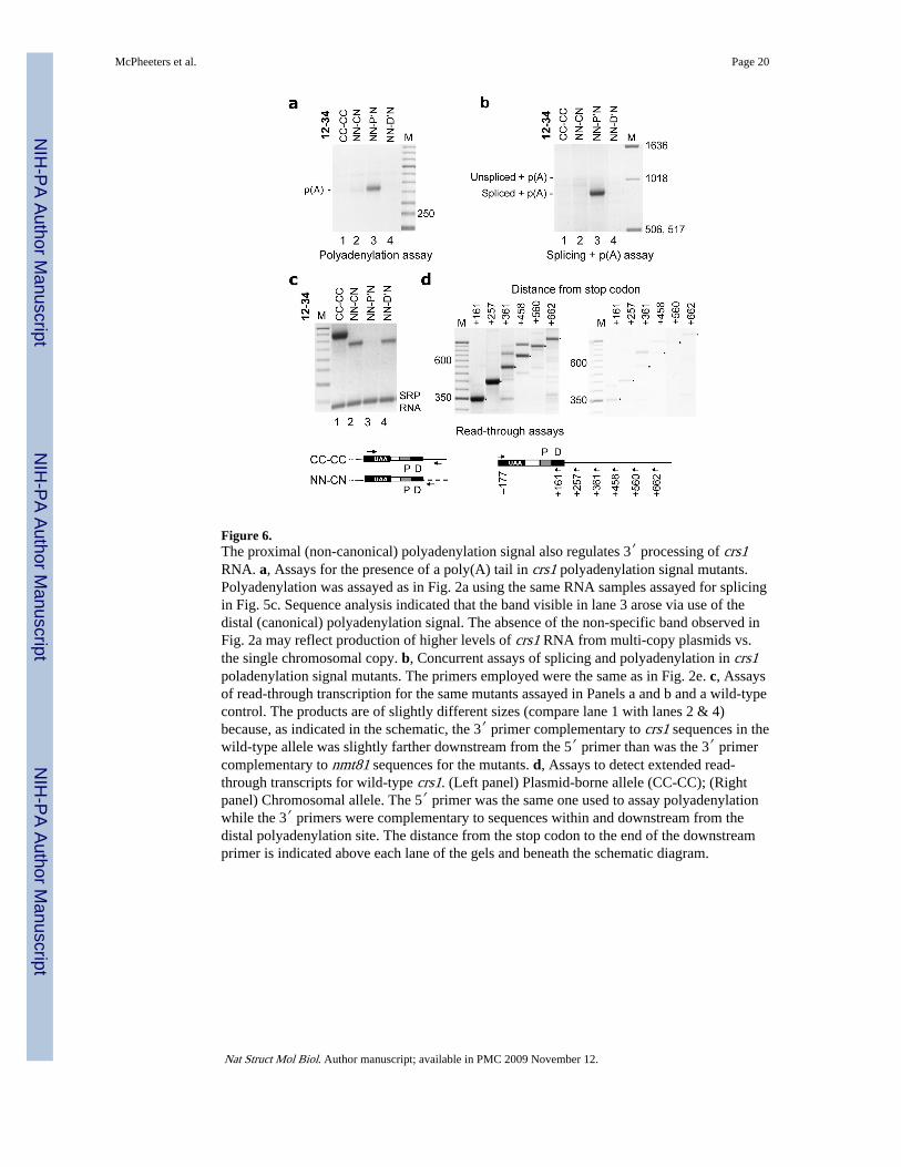
Figure 6.The proximal (non-canonical) polyadenylation signal also regulates 3′ processing of crs1RNA. a, Assays for the presence of a poly(A) tail in crs1 polyadenylation signal mutants.Polyadenylation was assayed as in Fig. 2a using the same RNA samples assayed for splicingin Fig. 5c. Sequence analysis indicated that the band visible in lane 3 arose via use of thedistal (canonical) polyadenylation signal. The absence of the non-specific band observed inFig. 2a may reflect production of higher levels of crs1 RNA from multi-copy plasmids vs.the single chromosomal copy. b, Concurrent assays of splicing and polyadenylation in crs1poladenylation signal mutants. The primers employed were the same as in Fig. 2e. c, Assaysof read-through transcription for the same mutants assayed in Panels a and b and a wild-typecontrol. The products are of slightly different sizes (compare lane 1 with lanes 2 & 4)because, as indicated in the schematic, the 3′ primer complementary to crs1 sequences in thewild-type allele was slightly farther downstream from the 5′ primer than was the 3′ primercomplementary to nmt81 sequences for the mutants. d, Assays to detect extended read-through transcripts for wild-type crs1. (Left panel) Plasmid-borne allele (CC-CC); (Rightpanel) Chromosomal allele. The 5′ primer was the same one used to assay polyadenylationwhile the 3′ primers were complementary to sequences within and downstream from thedistal polyadenylation site. The distance from the stop codon to the end of the downstreamprimer is indicated above each lane of the gels and beneath the schematic diagram.
McPheeters et al. Page 20
Nat Struct Mol Biol. Author manuscript; available in PMC 2009 November 12.
NIH
-PA Author Manuscript
NIH
-PA Author Manuscript
NIH
-PA Author Manuscript
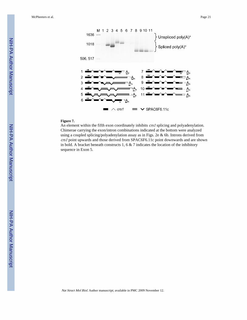
Figure 7.An element within the fifth exon coordinately inhibits crs1 splicing and polyadenylation.Chimerae carrying the exon/intron combinations indicated at the bottom were analyzedusing a coupled splicing/polyadenylation assay as in Figs. 2e & 6b. Introns derived fromcrs1 point upwards and those derived from SPAC6F6.11c point downwards and are shownin bold. A bracket beneath constructs 1, 6 & 7 indicates the location of the inhibitorysequence in Exon 5.
McPheeters et al. Page 21
Nat Struct Mol Biol. Author manuscript; available in PMC 2009 November 12.
NIH
-PA Author Manuscript
NIH
-PA Author Manuscript
NIH
-PA Author Manuscript
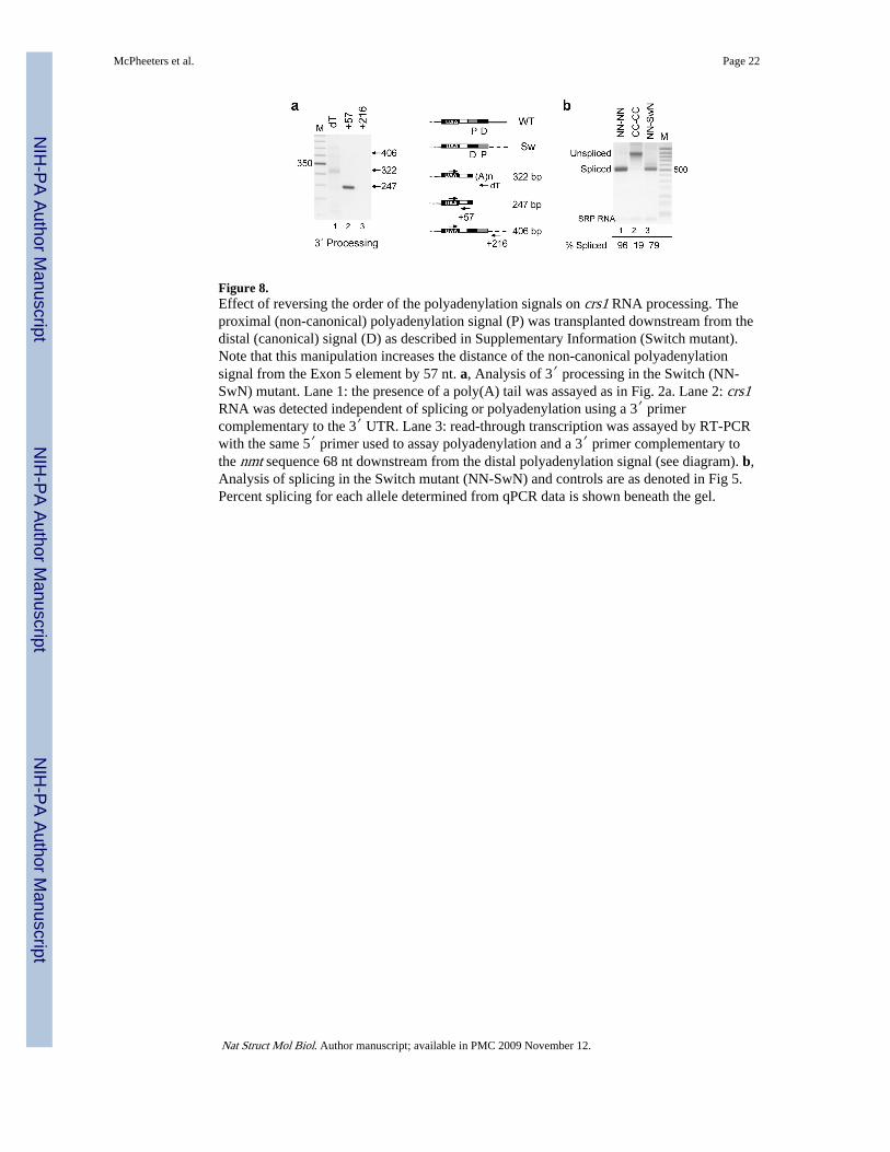
Figure 8.Effect of reversing the order of the polyadenylation signals on crs1 RNA processing. Theproximal (non-canonical) polyadenylation signal (P) was transplanted downstream from thedistal (canonical) signal (D) as described in Supplementary Information (Switch mutant).Note that this manipulation increases the distance of the non-canonical polyadenylationsignal from the Exon 5 element by 57 nt. a, Analysis of 3′ processing in the Switch (NN-SwN) mutant. Lane 1: the presence of a poly(A) tail was assayed as in Fig. 2a. Lane 2: crs1RNA was detected independent of splicing or polyadenylation using a 3′ primercomplementary to the 3′ UTR. Lane 3: read-through transcription was assayed by RT-PCRwith the same 5′ primer used to assay polyadenylation and a 3′ primer complementary tothe nmt sequence 68 nt downstream from the distal polyadenylation signal (see diagram). b,Analysis of splicing in the Switch mutant (NN-SwN) and controls are as denoted in Fig 5.Percent splicing for each allele determined from qPCR data is shown beneath the gel.
McPheeters et al. Page 22
Nat Struct Mol Biol. Author manuscript; available in PMC 2009 November 12.
NIH
-PA Author Manuscript
NIH
-PA Author Manuscript
NIH
-PA Author Manuscript
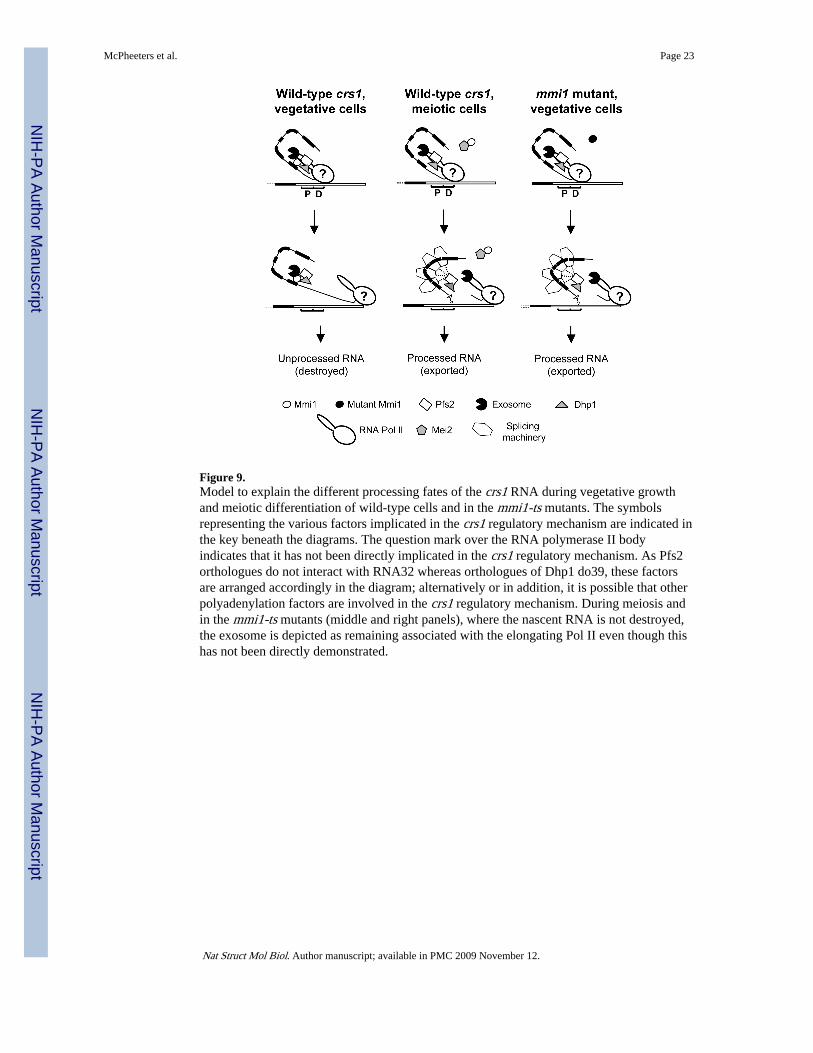
Figure 9.Model to explain the different processing fates of the crs1 RNA during vegetative growthand meiotic differentiation of wild-type cells and in the mmi1-ts mutants. The symbolsrepresenting the various factors implicated in the crs1 regulatory mechanism are indicated inthe key beneath the diagrams. The question mark over the RNA polymerase II bodyindicates that it has not been directly implicated in the crs1 regulatory mechanism. As Pfs2orthologues do not interact with RNA32 whereas orthologues of Dhp1 do39, these factorsare arranged accordingly in the diagram; alternatively or in addition, it is possible that otherpolyadenylation factors are involved in the crs1 regulatory mechanism. During meiosis andin the mmi1-ts mutants (middle and right panels), where the nascent RNA is not destroyed,the exosome is depicted as remaining associated with the elongating Pol II even though thishas not been directly demonstrated.
McPheeters et al. Page 23
Nat Struct Mol Biol. Author manuscript; available in PMC 2009 November 12.
NIH
-PA Author Manuscript
NIH
-PA Author Manuscript
NIH
-PA Author Manuscript


