Embed Size (px)
Citation preview
Varus and Valgus Alignment and Incident and Progressive KneeOsteoarthritis
Leena Sharma, MD1, Jing Song, MS1, Dorothy Dunlop, PhD1, David Felson, MD, MPH2,Cora E. Lewis, MD3, Neil Segal, MD4, James Torner, PhD4, T. Derek V. Cooke, MB, BChir,FRCS5, Jean Hietpas, LCSW, OTR6, John Lynch, PhD6, and Michael Nevitt, PhD61Northwestern University, Chicago, IL2Boston University, Boston, MA3University of Alabama at Birmingham, Birmingham, AL4University of Iowa, Iowa City, IA5Queens University, Kingston, ON, Canada, and OAISYS, Inc6University of California at San Francisco, San Francisco, CA
AbstractObjective—Varus and valgus alignment increase, respectively, medial and lateral tibiofemoralload. Alignment was associated with tibiofemoral osteoarthritis progression in previous studies; aneffect on risk of incident osteoarthritis is less certain. We tested whether alignment influences therisk of incident and progressive radiographic tibiofemoral osteoarthritis.
Methods—In an observational, longitudinal study of the MOST (Multicenter OsteoarthritisStudy) cohort, full-limb x-rays to measure alignment were acquired at baseline and knee x-rayswere acquired at baseline and 30 months. Varus alignment was defined as ≤ 178° and valgus as ≥182°. Using logistic regression and GEE, we examined the association of baseline alignment andincident osteoarthritis at 30 months (in knees without osteoarthritis at baseline), and alignment andosteoarthritis progression (in knees with baseline osteoarthritis). All analyses were adjusted forage, gender, BMI, knee injury, laxity, and extensor strength, with neutral knees as referent.
Results—2958 knees (1752 participants) were without osteoarthritis at baseline. Varus (adj. OR1.49, 95% CI 1.06, 2.10) but not valgus alignment was associated with incident osteoarthritis.1307 knees (950 participants) had osteoarthritis at baseline. Varus alignment was associated with agreater risk of medial osteoarthritis progression (adj. OR 3.59, 95% CI 2.62, 4.92) and a reducedrisk of lateral progression, and valgus with a greater risk of lateral progression (adj. OR 4.85, 95%CI 3.17, 7.42) and a reduced risk of medial progression.
Conclusion—Varus but not valgus alignment increased the risk of incident tibiofemoralosteoarthritis. In knees with osteoarthritis, varus and valgus alignment each increased the risk ofprogression in the biomechanically stressed compartment and reduced the risk of progression inthe unloaded compartment.
Corresponding author: Leena Sharma MD, Division of Rheumatology, Feinberg School of Medicine, Northwestern University, 240East Huron, McGaw M300, Chicago, IL 60611, [email protected], 312-908-5597 or 312-503-8003.The Corresponding Author has the right to grant on behalf of all authors and does grant on behalf of all authors, an exclusive license(or non exclusive for government employees) on a worldwide basis to the BMJ Publishing Group Ltd to permit this article (ifaccepted) to be published in ARD and any other BMJPGL products and sublicenses such use and exploit all subsidiary rights, as setout in our license (http://ARD.bmjjournals.com/ifora/licence.pdf).
NIH Public AccessAuthor ManuscriptAnn Rheum Dis. Author manuscript; available in PMC 2011 November 1.
Published in final edited form as:Ann Rheum Dis. 2010 November ; 69(11): 1940–1945. doi:10.1136/ard.2010.129742.
NIH
-PA Author Manuscript
NIH
-PA Author Manuscript
NIH
-PA Author Manuscript

INTRODUCTIONThe load-bearing axis of the lower limb can be represented by a line extending from femoralhead center to ankle joint center. In a varus (bow-leg) knee, this line passes medial to thecenter of the knee, increasing force across the medial tibiofemoral compartment. In a valgus(knock-knee) knee, the axis passes lateral to knee center, increasing force across the lateralcompartment. Animal studies and human studies of complicated fractures provided someearly evidence that alignment may influence development and progression of kneeosteoarthritis (OA) (1).
In recent years, natural history studies of primary knee OA have revealed a link betweenalignment and subsequent OA progression (2–6). The effect of varus and valgus alignmenton risk of incident knee OA is less certain (5,7). Alignment was found to have a strongereffect in knees with moderate tibiofemoral OA than in knees with mild OA, presumablyrelating to greater vulnerability of more diseased knees to altered load distribution (8). Inview of this, it seems likely that any alignment effect on risk of incident knee OA is smaller,and possibly more difficult to detect, than the effect on progression.
Most paradigms of knee OA development and progression posit a central role for localmechanical factors acting within a systemic milieu. There is a particularly compellingbiomechanical rationale to support a role for varus and valgus alignment. However, fewlongitudinal cohort studies have examined the alignment effect on risk of incident knee OA.To advance understanding of the pathways to knee OA development and to informdevelopment of non-invasive prevention strategies, it is important to clarify the impact ofalignment in knees without established OA.
In a prospective ancillary study to the Multicenter Osteoarthritis Study (MOST), we testedthe hypotheses:
1. varus and valgus alignment increase the odds of incident radiographic tibiofemoralOA, in knees without tibiofemoral OA at baseline;
2. varus alignment increases the odds of medial OA progression, and valgusalignment increases the odds of lateral OA progression, in knees with OA atbaseline.
METHODSSample
MOST is an observational cohort study of incident and progressive knee OA in 3026community-dwelling men and women, ages 50–79 years. Participants were recruited usingmass letter and brochure mailings and community outreach campaigns and enrolled at IowaCity, Iowa, or Birmingham, Alabama. To be eligible for MOST, persons were required tohave symptomatic knee OA or characteristics that placed them at increased risk fordeveloping it during the study (9,10). Exclusion criteria were: bilateral total kneereplacement or plan for this within the next year; inability to walk without the aid of anotherperson or a walker; serious health condition that would limit longitudinal studyparticipation; ankylosing spondylitis, psoriatic arthritis, reactive arthritis, or rheumatoidarthritis; dialysis; cancer other than nonmelanoma skin cancer; a plan to move from the areawithin three years.
The study protocol was approved by the institutional review boards at each participatingsite.
Sharma et al. Page 2
Ann Rheum Dis. Author manuscript; available in PMC 2011 November 1.
NIH
-PA Author Manuscript
NIH
-PA Author Manuscript
NIH
-PA Author Manuscript

Measurement of Varus-Valgus Alignment and other Factors at BaselineAlignment was assessed from full-limb radiographs, including hip and tibio-talar joints,acquired at baseline using a previously described protocol (2). Participants stood with thetibial tubercle facing forward. The x-ray beam was centered at the knee at a distance of 2.4m. A setting of 100 to 300 mA/s and 80–90 kV was used, depending on limb size and tissuecharacteristics. One AP radiograph of both limbs was obtained. The full limb of tallparticipants was included by using a 51 × 14 inch graduated grid cassette (Iowa) and a CR-based system of overlapping cassettes and simultaneously exposed subimages forming astitched image (Birmingham).
Alignment (the hip-knee-ankle angle) was measured as the angle at the intersection of theline connecting femoral head and intercondylar notch centers with the line connecting ankletalar surface center and tibial interspinous sulcus base. Image analysis (11) was completed inbatches of 50 by one of three trained readers using a customized program (Surveyor 3OAISYS Inc., Kingston, Ontario) and blinded to all other data. Each batch was reviewed bya manager prior to transmission. In a reliability study of 200 full-limb pairs assessed by thethree readers, the inter-reader and intra-reader ICCs for the hip-knee-ankle angle were 0.95and 0.96, respectively (12). In analyses, varus alignment was defined as ≤ 178°, valgus as ≥182°, and neutral as 179–181°.
Concentric knee extensor strength was measured for each lower limb with a Cybex 350isokinetic dynamometer (Avocent, Huntsville AL) at 60° per second (13). The averagetorque (Nm) of 4 maximum effort repetitions was analyzed. Medial-lateral laxity (°) wasmeasured using a protocol and device previously described (14), consisting of a bench andattached arc-shaped track, and providing thigh and ankle immobilization, a stable kneeflexion angle, and fixed medial and lateral load. Weight (kg) without shoes or heavy clotheswas measured on a balance beam scale and height without shoes using a stadiometer. Injurywas defined as any knee injury severe enough to limit ability to walk without a gait aid for atleast two days.
Knee X-Ray Acquisition and AssessmentAt baseline and 30 months, knee radiographs were acquired using the posteroanterior (PA)“fixed-flexion” weightbearing protocol (15), in which knees are flexed to 20–30° and feetinternally rotated 10° using a plexiglass positioning frame (SynaFlexer™). The right and leftknees were imaged together on 14 × 17 film with a 72 inch film-to-focus distance. Lateralweight-bearing films were also obtained, following a Framingham Osteoarthritis Studyprotocol (16).
An experienced rheumatologist and musculoskeletal radiologist independently assessed eachPA film for Kellgren and Lawrence (K/L) grade and each PA and lateral film for medial andlateral joint space narrowing grade. The readers were blinded to clinical data and knew thetime sequence of the images (17). Joint space narrowing grade was scored (0–3) separatelyfor the medial and lateral compartments using a modified version of the OARSI scale(18,19). Previous studies (3) revealed worsening of joint space narrowing over time notsufficient to move one full OARSI grade. When this occurred in knees with joint spacenarrowing at baseline, readers were instructed to use ½ grades, an approach which has beenvalidated (19). If readers disagreed on whether incident OA had developed or joint spacenarrowing grade had worsened, the reading was adjudicated by a panel of three readers.Weighted kappas for agreement between the two readers were: K/L grade 0.79; medial andlateral joint space narrowing grade 0.81 and 0.86, respectively.
Sharma et al. Page 3
Ann Rheum Dis. Author manuscript; available in PMC 2011 November 1.
NIH
-PA Author Manuscript
NIH
-PA Author Manuscript
NIH
-PA Author Manuscript

Definition of Key OutcomesAll outcomes were knee-based and assessed from baseline and 30-month radiographicimages. Using the established and widely applied approach, radiographic knee OA requireddefinite osteophyte presence (K/L ≥ 2) at standard image size; incident OA was defined asthe new onset of K/L 2 or greater at 30 months, in knees graded K/L 0 or 1 at baseline.
OA progression was assessed in knees K/L 2 or greater at baseline using a compartment-specific approach. Medial OA progression was defined as any worsening of modifiedOARSI grade of medial joint space narrowing, and lateral OA progression as any worseningof lateral joint space narrowing grade. Knees with advanced OA that could not progressfurther (K/L grade 4 or joint space narrowing grade 3) were excluded from analyses.
Statistical AnalysisBoth knees from each person were examined. Knee characteristics were calculatedseparately among knees without radiographic knee OA (K/L < 2) at baseline and knees withknee OA (K/L ≥ 2) at baseline by alignment group [varus (≤ 178°), valgus (≥ 182°), andneutral (179–181°)]. Multiple logistic regression with generalized estimating equations(GEE), to account for potentially correlated observations for knees from the same person,was used to evaluate:
1. the relationship of varus and valgus alignment at baseline to incident knee OA at 30months, among knees without tibiofemoral OA at baseline and therefore at risk forincident OA;
2. the relationship of varus and valgus alignment to medial and lateral tibiofemoralOA progression at 30 months, among knees with OA at baseline and therefore atrisk for progression.
Neutral knees constituted the reference group in all analyses. All analyses were adjusted forage (continuous), gender, BMI (continuous), knee injury (dichotomous), laxity (continuous),and extensor strength (continuous). Results from each model are reported as adjusted oddsratios (ORs) with associated 95% confidence intervals (CIs); a 95% CI excluding 1represents a statistically significant association. In secondary analyses, severity of varus andvalgus alignment were analyzed as continuous variables. Sensitivity analyses were runseparately for men and women to determine if results were consistent across gender.Analyses were performed using SAS software version 9.2 (SAS Institute Inc., Cary NC).
The funding source played no role in: study design and conduct; collection, management,analysis, and interpretation of data; and preparation, review, or approval of the manuscript.
RESULTSOf 3026 persons enrolled in MOST, 30-month follow-up contact occurred in 2969 (seeFigure 1). Of the 57 with no 30-month contact, 33 had died and 24 could not be reached. Ofthe 2969 persons with 30-month contact: 2713 completed both telephone interview andclinic visit; 215 completed only the telephone interview and a missed clinic visit telephoneinterview; and 41 completed only the telephone interview. Reasons for not completing the30-month clinic visit were: too busy (77 persons); health problems (70); caregivingresponsibilities (31); deceased (30); clinic too far (21); moved out of area (20); not satisfiedwith study (19); unable to contact (16); refused to give reason (8); personal problems (7);and other reasons in the remaining 14. Those not completing the 30-month clinic visit didnot differ in age, gender, or alignment distribution in the dominant knee (32% neutral, 47%varus, 21% valgus) but had a higher BMI (32.0 ± 6.9, S.D. vs. 30.6 ± 5.8) than those whocompleted this visit.
Sharma et al. Page 4
Ann Rheum Dis. Author manuscript; available in PMC 2011 November 1.
NIH
-PA Author Manuscript
NIH
-PA Author Manuscript
NIH
-PA Author Manuscript

In the 2713 persons who completed the 30-month clinic visit, 621 knees were excluded fromanalysis for a total knee replacement at baseline or at follow-up or for advanced OA atbaseline that could not progress further. In these 2713 persons, 1614 right knees and 1692left knees did not have OA at baseline and were at risk to develop incident OA. Of these3306 knees, 348 were excluded for missing data (predominantly strength or laxity), resultingin a sample of 2958 knees for analyses of incident tibiofemoral OA. OA was present atbaseline in 797 right and 702 left knees; these knees were at risk for OA progression. Ofthese 1499 knees, 192 were excluded for missing data, resulting in a sample of 1307 kneesfor analyses of OA progression.
The 1752 participants who contributed 2958 knees for analyses of incident OA had a meanage of 61.3 years (± 7.8, SD), a mean BMI of 29.5 kg/m2 (± 5.1), and included 1034 (59%)women. Baseline characteristics of these knees are summarized in Table 1. The 950participants who contributed the 1307 knees for analyses of OA progression had a mean ageof 63.6 years (± 7.8), a mean BMI of 31.7 kg/m2 (± 5.9), and included 592 (62%) women.Characteristics of these knees are summarized in Table 2. In total, 4265 knees in 2287persons were analyzed; 415 persons contributed to both analysis samples.
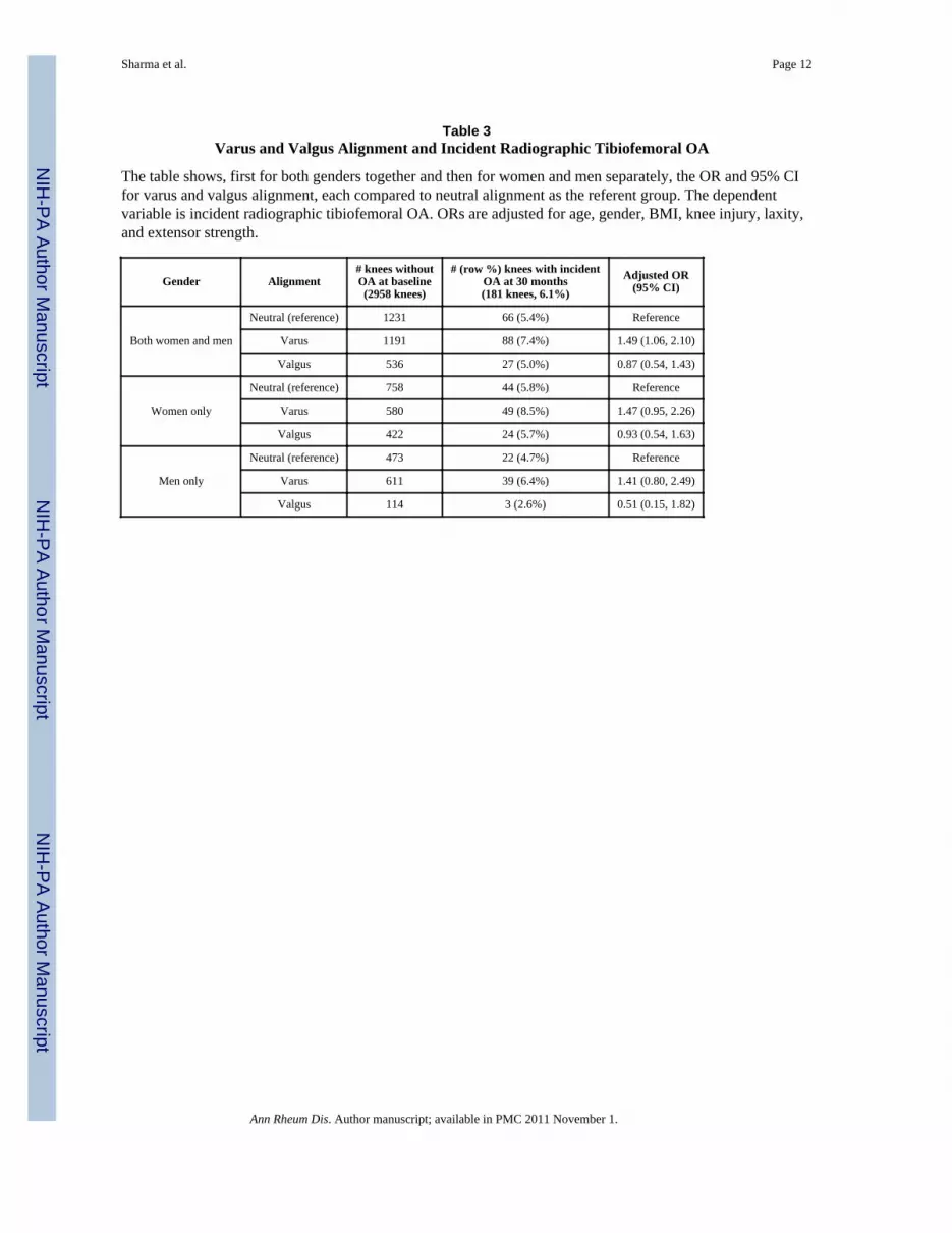
As shown in Table 3, the odds of developing incident tibiofemoral OA were significantlyelevated in knees with varus, but not in knees with valgus alignment at baseline, comparedto neutral knees, in analyses adjusting for age, gender, BMI, knee injury, laxity, andstrength. The magnitude of the OR was comparable in the smaller strata of women and menconsidered separately. We secondarily analyzed alignment as a continuous variable.Including only neutral and varus knees, greater severity of varus at baseline was associatedwith greater odds of incident OA approaching significance [adjusted OR 1.07/1° varus (95%CI 0.99, 1.16)]. Among valgus and neutral knees, greater severity of valgus was notassociated with greater odds of incident OA [adjusted OR 0.98/1° valgus (95% CI 0.85,1.14)].
As shown in Table 4, the odds of medial OA progression were significantly elevated inknees varus at baseline. Medial OA progression risk was significantly reduced in valgusknees. On the other hand, the odds of lateral OA progression were significantly elevated inknees valgus at baseline and significantly reduced in varus knees (see Table 4). The resultswere similar in men and women considered separately. The finding for valgus alignment andlateral progression in men may reflect that there were only 28 valgus knees with OA in men.We secondarily analyzed alignment as a continuous variable. Including only neutral andvarus knees, greater severity of varus at baseline was significantly associated with greaterodds of medial OA progression [adjusted OR 1.29/1° varus (95% CI 1.22, 1.37)]. Amongvalgus and neutral knees, greater severity of valgus was significantly associated with greaterodds of lateral OA progression [adjusted OR 1.47/1° valgus (95% CI 1.30, 1.65)].
Further adjustment for baseline K/L grade, baseline knee pain severity, and concurrentchange in BMI had minimal impact on these results.
DISCUSSIONVarus but not valgus alignment increased the risk of incident radiographic tibiofemoral OA.In the more vulnerable milieu of the knee with established OA, varus and valgus alignmenteach increased the risk of OA progression in the biomechanically stressed compartment andreduced the risk of progression in the unloaded compartment. A substantial proportion ofknees were varus or valgus: 41% and 19%, respectively, in knees without tibiofemoral OA;58% and 18% in knees with OA.
Sharma et al. Page 5
Ann Rheum Dis. Author manuscript; available in PMC 2011 November 1.
NIH
-PA Author Manuscript
NIH
-PA Author Manuscript
NIH
-PA Author Manuscript

The pattern of results in the stressed and unloaded tibiofemoral compartments furthersupports that the mechanism of action of malalignment relates to its effect on loaddistribution. Varus alignment shifts the load-bearing axis medial to knee center, creating amoment arm that increases forces across the medial compartment and reduces lateral load;the lateral shift of the load-bearing axis due to valgus alignment increases forces across thelateral compartment and reduces medial load.
We found that varus but not valgus alignment increased the risk of incident OA. Similarly,Brouwer et al (5) observed that varus had a significant effect, while valgus had a borderlineeffect, in a study using a comparable definition of varus and valgus and defining, as we did,neutral knees as reference. The relationship between alignment and knee OA developmentwas also examined in a case-control study involving Framingham cohort members; in thisstudy, the most varus (1–7° varus) was compared not to neutral knees but to the most valgusquartile (5–10° valgus) as referent (7), modeling a different question than what we posed.
From a biomechanical perspective, a stronger finding for varus alignment is not surprising.Due to a stance phase knee adduction moment, greater load passes medially than laterallyeven in neutrally aligned, healthy knees (20,21). The adduction moment magnitudeincreases as varus alignment increases (22). Adduction moment magnitude predicted kneeOA progression (23); an adduction moment increase may lie in the causal pathway betweenvarus alignment and knee OA progression. Varus alignment further increases total loadpassing medially (24). Although valgus alignment is associated with an increase in lateralcompartment peak pressures (25), the medial compartment often continues to bear more loaduntil more severe valgus is present (26,27).
Alternatively, the inability to detect an association between valgus alignment and incidentOA may reflect a lower sensitivity of the measure of incident OA vs. the measure of OAprogression. In the vast majority of knees, osteophyte development precedes joint spacenarrowing. The definition of incident OA hinges upon these osteophytes. Becauseosteophyte formation is neither specific to the involved nor to the spared compartment, atthe earliest stage of OA (K/L 2), radiographs cannot reveal whether a knee has medial orlateral OA. It is possible that the greater overall frequency of medial vs. lateral OA dilutesthe detected valgus effect upon a non-compartment specific measure of incident OA. Incontrast, the measure of progression allows specific examination of the compartmentstressed by the alignment (medial for varus, lateral for valgus). As such, in the analysis ofthe impact of alignment, the measure of incident OA, which is not compartment-specific, isgenerally inferior to the measure of progression
In the analyses of knees with OA at baseline, varus and valgus alignment were associatedwith medial and lateral OA progression, respectively, in keeping with previous studies (2–6). Brouwer et al also found significant results for varus only; the association betweenvalgus and lateral progression was significant only for those who were obese (5). As theauthors note, the inability to detect the valgus effect may relate to use of K/L worsening todefine progression and the relatively small numbers with progression (5).
The source of malalignment predating knee OA may be genetic, developmental, ortraumatic. Prior to the study by Brouwer et al (5), evidence that malalignment maycontribute to OA development came from animal models and human fracture studies. Ourfindings and those of Brouwer et al support that, in knees without radiographic tibiofemoralOA, varus alignment increases the risk of OA development. The prevalence of varus andvalgus alignment in knees without radiographic OA – i.e., before any loss of bone andcartilage height that could contribute to malalignment – support that not all malalignment isa consequence of disease. And, whatever the original cause of the alignment, varus and
Sharma et al. Page 6
Ann Rheum Dis. Author manuscript; available in PMC 2011 November 1.
NIH
-PA Author Manuscript
NIH
-PA Author Manuscript
NIH
-PA Author Manuscript

valgus alignment each increase the risk of subsequent knee OA progression. It seems likelythat worsening of tibiofemoral OA in turn increases malalignment at least in some knees.With only one time point of alignment measurement, we could not explore this. Theexistence of a vicious cycle does not lessen the impact of our findings; strategies thatinterrupt a vicious cycle may be a potent means of delaying progression. These resultssupport further development and testing of non-invasive modalities to improve tibiofemoralload distribution in varus-aligned and valgus-aligned knees.
It is important to acknowledge that MOST participants without knee OA were at higher riskto develop it. Those at higher risk to develop knee OA are of particular public healthimportance (9), and it is crucial to understand the relationship of alignment to incident OAin them. Varus alignment was associated with incident OA by the established definition. Welacked sufficient power to separately examine incidence within K/L 0 and 1 strata. With orwithout these analyses, it cannot be concluded from any radiographic study that any factorinitiates knee OA, given the insensitivity of x-rays to early OA pathology and the inabilityto identify the point of OA onset. Although there is no consensus as yet about what kneeMRI feature(s) constitute OA, future studies should explore the relationship betweenalignment and OA development in knees without MRI-based measures of OA pathology.We were not able to assess change in alignment between baseline and follow-up. Those whodid not complete the 30-month visit had a higher BMI; it is uncertain what impact this mayhave had on the results.
In conclusion, varus but not valgus alignment increased the risk of incident radiographictibiofemoral OA. In knees with established OA, varus and valgus alignment each increasedthe risk of OA progression in the biomechanically stressed compartment and reduced therisk of progression in the unloaded compartment.
AcknowledgmentsSupport:
NIH NICHD RO1 HD43500
NIA U01 AG18820, AG18832, AG18947, AG19069
REFERENCES1. Tetsworth K, Paley D. Malalignment and degenerative arthropathy. Orthop Clin North Am
1994;25:367–377. [PubMed: 8028880]2. Sharma L, Song J, Felson DT, et al. The role of knee alignment in disease progression and
functional decline in knee osteoarthritis. JAMA 2001;286:188–195. [PubMed: 11448282]3. Felson DT, McLaughlin S, Goggins J, et al. Bone marrow edema and its relation to progression of
knee osteoarthritis. Ann Intern Med 2003;139:330–336. [PubMed: 12965941]4. Cicuttini F, Wluka A, Hankin J, et al. Longitudinal study of the relationship between knee angle and
tibiofemoral cartilage volume in subjects with knee osteoarthritis. Rheumatology (Oxford)2004;43:321–324. [PubMed: 14963201]
5. Brouwer GM, van Tol AW, Bergink AP, et al. Association between Valgus and Varus Alignmentand the Development and Progression of Radiographic Osteoarthritis of the Knee. Arthritis Rheum2007;56:1204–1211. [PubMed: 17393449]
6. Sharma L, Eckstein F, Song J, et al. The relationship of meniscal damage, meniscal extrusion,malalignment, and joint laxity to subsequent cartilage loss in osteoarthritic knees. Arthritis Rheum2008;58:1716–1726. [PubMed: 18512777]
7. Hunter DJ, Niu J, Felson DT, et al. Knee alignment does not predict incident osteoarthritis: theFramingham Osteoarthritis Study. Arthritis Rheum 2007;56:1212–1218. [PubMed: 17393450]
Sharma et al. Page 7
Ann Rheum Dis. Author manuscript; available in PMC 2011 November 1.
NIH
-PA Author Manuscript
NIH
-PA Author Manuscript
NIH
-PA Author Manuscript

8. Cerejo R, Dunlop DD, Cahue S, et al. The influence of alignment on risk of knee osteoarthritisprogression according to baseline stage of disease. Arthritis Rheum 2002;46:2632–2636. [PubMed:12384921]
9. Felson DT, Nevitt MC. Epidemiologic studies for osteoarthritis: new versus conventional studydesign approaches. Rheum Dis Clin North Am 2004;30:783–797. [PubMed: 15488693]
10. Felson DT, Anderson JJ, Mainmark A, et al. Obesity and knee osteoarthritis: The FraminghamStudy. Ann Intern Med 1988;109:18–24. [PubMed: 3377350]
11. Cooke TD, Sled EA, Scudamore RA. Frontal plane knee alignment: a call for standardizedmeasurement. J Rheumatol 2007;34:1796–1801. [PubMed: 17787049]
12. Sled EA, Sheehy LM, Felson DT, et al. Reliability of lower limb alignment measures using anestablished landmark-based method with a customized computer software program.Rheumatology :in press.
13. Segal NA, Torner JC, Felson D, et al. Effect of thigh strength on incident radiographic andsymptomatic knee osteoarthritis in a longitudinal cohort. Arthritis Rheum 2009;61:1210–1217.[PubMed: 19714608]
14. Sharma L, Lou C, Felson DT, et al. Laxity in healthy and osteoarthritic knees. Arthritis Rheum1999;42:861–870. [PubMed: 10323441]
15. Peterfy C, Li J, Zaim S, et al. Comparison of fixed-flexion positioning with fluoroscopic semi-flexed positioning for quantifying radiographic joint-space width in the knee: test-retestreproducibility. Skeletal Radiol 2003;32:128–132. [PubMed: 12605275]
16. LaValley MP, McLaughlin S, Goggins J, et al. The lateral view radiograph for assessment of thetibiofemoral joint space in knee osteoarthritis: its reliability, sensitivity to change, and longitudinalvalidity. Arthritis Rheum 2005;52:3542–3547. [PubMed: 16255043]
17. Felson DT, Nevitt MC. Blinding images to sequence in osteoarthritis: evidence from otherdiseases. Osteoarthritis Cartilage 2009;17:281–283. [PubMed: 18977156]
18. Altman R, Hochberg M, Murphy W, et al. Atlas of individual radiographic features inosteoarthritis. Osteoarthritis Cartilage 1995;3(A):3–70. [PubMed: 8581752]
19. Felson DT, Nevitt MC, Yang M, et al. A new approach yields high rates of x-ray progression inknee osteoarthritis. J Rheumatol 2008;35:2047–2054. [PubMed: 18793000]
20. Andriacchi TP. Dynamics of knee malalignment. Orthop Clin North Am 1994;25:395–403.[PubMed: 8028883]
21. Morrison JB. The mechanics of the knee joint in relation to normal walking. J Biomech 1970;3:51–61. [PubMed: 5521530]
22. Hurwitz DE, Ryals AB, Case JP, et al. The knee adduction moment during gait in subjects withknee osteoarthritis is more closely correlated with static alignment than radiographic diseaseseverity, toe out angle and pain. J Orthop Res 2002;20:101–107. [PubMed: 11853076]
23. Miyazaki T, Wada M, Kawahara H, et al. Dynamic load at baseline can predict radiographicdisease progression in medial compartment knee osteoarthritis. Ann Rheum Dis 2002;61:617–622.[PubMed: 12079903]
24. Hsu RWW, Himeno S, Coventry MB, et al. Normal axial alignment of the lower extremity andload-bearing distribution at the knee. Clin Orthop 1990;255:215–227. [PubMed: 2347155]
25. Bruns J, Volkmer M, Luessenhop S. Pressure distribution at the knee joint. Influence of varus andvalgus deviation without and with ligament dissection. Arch Orthop Trauma Surg 1993;133:12–19. [PubMed: 8117504]
26. Johnson F, Leitl S, Waugh W. The distribution of load across the knee. A comparison of static anddynamic measurements. J Bone Joint Surg 1980;62-B:346–349.
27. Harrington IJ. Static and dynamic loading patterns in knee joints with deformities. J Bone JointSurg 1983;65-A:247–259. [PubMed: 6822587]
Sharma et al. Page 8
Ann Rheum Dis. Author manuscript; available in PMC 2011 November 1.
NIH
-PA Author Manuscript
NIH
-PA Author Manuscript
NIH
-PA Author Manuscript

Figure 1.The figure illustrates how the samples of 2958 knees for analyses of incident knee OA and1307 knees for analyses of knee OA progression were derived.
Sharma et al. Page 9
Ann Rheum Dis. Author manuscript; available in PMC 2011 November 1.
NIH
-PA Author Manuscript
NIH
-PA Author Manuscript
NIH
-PA Author Manuscript

NIH
-PA Author Manuscript
NIH
-PA Author Manuscript
NIH
-PA Author Manuscript
Sharma et al. Page 10
Table 1Characteristics of Knees without Radiographic Tibiofemoral OA
The table includes characteristics of knees without tibiofemoral OA at baseline. There were 2958 kneeswithout tibiofemoral OA from 1752 persons. In the Table, data from only one knee per person is included, i.e.the right knee. (If only the left knee was without tibiofemoral OA, then data from the left knee is included.)
Neutral alignment688/1752 knees (39.2%)
Varus alignment725/1752 knees (41.4%)
Valgus alignment339/1752 knees (19.4%)
Number (%) knees in group with injury 127 (18%) 141 (19%) 72 (21%)
Laxity, mean (S.D.), ° 4.1 (2.6) 3.9 (2.5) 3.8 (2.7)
Extensor strength, mean (S.D.), Nm 86.4 (39.5) 95.2 (42.9) 71.9 (35.2)
K/L grade 0, number (%) 490 (71%) 466 (64%) 246 (73%)
K/L grade 1, number (%) 198 (29%) 259 (36%) 93 (27%)
Ann Rheum Dis. Author manuscript; available in PMC 2011 November 1.

NIH
-PA Author Manuscript
NIH
-PA Author Manuscript
NIH
-PA Author Manuscript
Sharma et al. Page 11
Table 2Characteristics of Knees with Radiographic Tibiofemoral OA
The table includes characteristics of knees with tibiofemoral OA at baseline. There were 1307 knees withouttibiofemoral OA from 950 persons. In the Table, data from only one knee per person is included, i.e. the rightknee. (If only the left knee had tibiofemoral OA, then data from the left knee is included.)
Neutral alignment232/950 knees (24%)
Varus alignment550/950 knees (58%)
Valgus alignment168/950 knees (18%)
Number (%) knees in group with injury 72 (31%) 212 (39%) 42 (25%)
Laxity, mean (S.D.), ° 3.9 (2.5) 3.8 (2.5) 4.4 (2.6)
Extensor strength, mean (S.D.), Nm 72.2 (35.1) 80.0 (42.0) 54.5 (30.8)
K/L grade 2, number (%) 165 (71%) 208 (38%) 95 (57%)
K/L grade 3, number (%) 67 (29%) 342 (62%) 73 (43%)
Ann Rheum Dis. Author manuscript; available in PMC 2011 November 1.
NIH
-PA Author Manuscript
NIH
-PA Author Manuscript
NIH
-PA Author Manuscript
Sharma et al. Page 12
Table 3Varus and Valgus Alignment and Incident Radiographic Tibiofemoral OA
The table shows, first for both genders together and then for women and men separately, the OR and 95% CIfor varus and valgus alignment, each compared to neutral alignment as the referent group. The dependentvariable is incident radiographic tibiofemoral OA. ORs are adjusted for age, gender, BMI, knee injury, laxity,and extensor strength.
Gender Alignment# knees withoutOA at baseline(2958 knees)
# (row %) knees with incidentOA at 30 months(181 knees, 6.1%)
Adjusted OR(95% CI)
Both women and men
Neutral (reference) 1231 66 (5.4%) Reference
Varus 1191 88 (7.4%) 1.49 (1.06, 2.10)
Valgus 536 27 (5.0%) 0.87 (0.54, 1.43)
Women only
Neutral (reference) 758 44 (5.8%) Reference
Varus 580 49 (8.5%) 1.47 (0.95, 2.26)
Valgus 422 24 (5.7%) 0.93 (0.54, 1.63)
Men only
Neutral (reference) 473 22 (4.7%) Reference
Varus 611 39 (6.4%) 1.41 (0.80, 2.49)
Valgus 114 3 (2.6%) 0.51 (0.15, 1.82)
Ann Rheum Dis. Author manuscript; available in PMC 2011 November 1.

NIH
-PA Author Manuscript
NIH
-PA Author Manuscript
NIH
-PA Author Manuscript
Sharma et al. Page 13
Table 4Varus and Valgus Alignment and Radiographic Tibiofemoral OA Progression
The table shows, first for both genders together and then for women and men separately, the OR and 95% CIfor varus and valgus alignment, each compared to neutral alignment as the referent group. The dependentvariable is medial tibiofemoral OA progression in the top half of the table, and lateral tibiofemoral OAprogression in the bottom half of the table. ORs are adjusted for age, gender, BMI, knee injury, laxity, andextensor strength.
Gender Alignment # knees withOA at baseline(1307 knees)
# (row %) knees withMEDIAL OA progression
at 30 months(558 knees, 42.7%)
Adjusted* OR(95% CI)
Both women and men
Neutral (reference) 317 88 (27.8%) Reference
Varus 758 443 (58.4%) 3.59 (2.62, 4.92)
Valgus 232 27 (11.6%) 0.34 (0.21, 0.55)
Women only
Neutral (reference) 221 56 (25.3%) Reference
Varus 408 242 (59.3%) 4.21 (2.84, 6.24)
Valgus 204 23 (11.3%) 0.37 (0.21, 0.65)
Men only
Neutral (reference) 96 32 (33.3%) Reference
Varus 350 201 (57.4%) 2.75 (1.63, 4.66)
Valgus 28 4 (14.3%) 0.34 (0.11, 1.03)
Alignment Gender # knees with OA atbaseline (1307 knees)
# (row %) knees with LATERAL OAprogression at 30 months (163 knees,
12.5%)
Adjusted* OR (95% CI)
Both women and men
Neutral (reference) 317 46 (14.5%) Reference
Varus 758 17 (2.2%) 0.12 (0.07. 0.21)
Valgus 232 100 (43.1%) 4.85 (3.17, 7.42)
Women only
Neutral (reference) 221 24 (10.9%) Reference
Varus 408 8 (2.0%) 0.15 (0.06, 0.33)
Valgus 204 88 (43.1%) 6.35 (3.76, 10.75)
Men only
Neutral (reference) 96 22 (22.9%) Reference
Varus 350 9 (2.6%) 0.08 (0.04, 0.19)
Valgus 28 12 (42.9%) 1.88 (0.69, 5.12)
Ann Rheum Dis. Author manuscript; available in PMC 2011 November 1.


