Embed Size (px)
Citation preview

2019 ESC Guidelines for the diagnosis and
management of chronic coronary syndromes
The Task Force for the diagnosis and management of chroniccoronary syndromes of the European Society of Cardiology (ESC)
Authors/Task Force Members: Juhani Knuuti* (Finland) (Chairperson),
William Wijns* (Ireland) (Chairperson), Antti Saraste (Finland), Davide Capodanno
(Italy), Emanuele Barbato (Italy), Christian Funck-Brentano (France),
Eva Prescott (Denmark), Robert F. Storey (United Kingdom), Christi Deaton
(United Kingdom), Thomas Cuisset (France), Stefan Agewall (Norway),
Kenneth Dickstein (Norway), Thor Edvardsen (Norway), Javier Escaned (Spain),
Bernard J. Gersh (United States of America), Pavel Svitil (Czech Republic),
Martine Gilard (France), David Hasdai (Israel), Robert Hatala (Slovak Republic),
Felix Mahfoud (Germany), Josep Masip (Spain), Claudio Muneretto (Italy),
Marco Valgimigli (Switzerland), Stephan Achenbach (Germany), Jeroen J. Bax
(Netherlands)
Document Reviewers: Franz-Josef Neumann (Germany) (CPG Review Coordinator), Udo Sechtem(Germany) (CPG Review Coordinator), Adrian Paul Banning (United Kingdom), Nikolaos Bonaros(Austria), Hector Bueno (Spain), Raffaele Bugiardini (Italy), Alaide Chieffo (Italy), Filippo Crea (Italy),
* Corresponding authors: Juhani Knuuti, Department of Clinical Physiology, Nuclear Medicine and PET and Turku PET Centre, Turku University Hospital, Kiinamyllynkatu 4-8, FI-20520 Turku, Finland. Tel: þ358 500 592 998, Email: [email protected]. William Wijns, The Lambe Institute for Translational Medicine and Curam, National University ofIreland, Galway, University Road, Galway, H91 TK33, Ireland. Tel: þ353 91 524411, Email: [email protected].
Author/Task Force Member Affiliations: listed in the Appendix.
ESC Committee for Practice Guidelines (CPG) and National Cardiac Societies document reviewers: listed in the Appendix.
ESC entities having participated in the development of this document:
Associations: Acute Cardiovascular Care Association (ACCA), Association of Cardiovascular Nursing & Allied Professions (ACNAP), European Association of CardiovascularImaging (EACVI), European Association of Preventive Cardiology (EAPC), European Association of Percutaneous Cardiovascular Interventions (EAPCI), European Heart RhythmAssociation (EHRA), Heart Failure Association (HFA).
Councils: Council for Cardiology Practice.
Working Groups: Atherosclerosis and Vascular Biology, Cardiovascular Pharmacotherapy, Cardiovascular Surgery, Coronary Pathophysiology and Microcirculation,Thrombosis.
The content of these ESC Guidelines has been published for personal and educational use only. No commercial use is authorized. No part of the ESC Guidelines may be trans-lated or reproduced in any form without written permission from the ESC. Permission can be obtained upon submission of a written request to Oxford University Press, thepublisher of the European Heart Journal and the party authorized to handle such permissions on behalf of the ESC ([email protected]).
Disclaimer. The ESC Guidelines represent the views of the ESC and were produced after careful consideration of the scientific and medical knowledge, and the evidence availableat the time of their publication. The ESC is not responsible in the event of any contradiction, discrepancy, and/or ambiguity between the ESC Guidelines and any other official recom-mendations or guidelines issued by the relevant public health authorities, in particular in relation to good use of healthcare or therapeutic strategies. Health professionals are encour-aged to take the ESC Guidelines fully into account when exercising their clinical judgment, as well as in the determination and the implementation of preventive, diagnostic, ortherapeutic medical strategies; however, the ESC Guidelines do not override, in any way whatsoever, the individual responsibility of health professionals to make appropriate andaccurate decisions in consideration of each patient’s health condition and in consultation with that patient and, where appropriate and/or necessary, the patient’s caregiver. Nor dothe ESC Guidelines exempt health professionals from taking into full and careful consideration the relevant official updated recommendations or guidelines issued by the competentpublic health authorities, in order to manage each patient’s case in light of the scientifically accepted data pursuant to their respective ethical and professional obligations. It is also thehealth professional’s responsibility to verify the applicable rules and regulations relating to drugs and medical devices at the time of prescription.
VC The European Society of Cardiology 2019. All rights reserved. For permissions please email: [email protected].
European Heart Journal (2019) 00, 1�71
ESC GUIDELINES
doi:10.1093/eurheartj/ehz425
Dow
nloaded from https://academ
ic.oup.com/eurheartj/advance-article-abstract/doi/10.1093/eurheartj/ehz425/5556137 by guest on 31 August 2019

..
..
..
..
..
..
..
..
..
..
..
..
..
..
..
..
..
..
..
..
..
..
..
..
..
..
..
..
..
..
..
..
..
..
..
..
..
..
..
..
..
..
..
..
..
..
..
.
Martin Czerny (Germany), Victoria Delgado (Netherlands), Paul Dendale (Belgium),Frank Arnold Flachskampf (Sweden), Helmut Gohlke (Germany), Erik Lerkevang Grove (Denmark),Stefan James (Sweden), Demosthenes Katritsis (Greece), Ulf Landmesser (Germany), Maddalena Lettino(Italy), Christian M. Matter (Switzerland), Hendrik Nathoe (Netherlands), Alexander Niessner (Austria),Carlo Patrono (Italy), Anna Sonia Petronio (Italy), Steffen E. Pettersen (United Kingdom), Raffaele Piccolo(Italy), Massimo Francesco Piepoli (Italy), Bogdan A. Popescu (Romania), Lorenz R€aber (Switzerland),Dimitrios J. Richter (Greece), Marco Roffi (Switzerland), Franz X. Roithinger (Austria), Evgeny Shlyakhto(Russian Federation), Dirk Sibbing (Germany), Sigmund Silber (Germany), Iain A. Simpson(United Kingdom), Miguel Sousa-Uva (Portugal), Panos Vardas (Greece), Adam Witkowski (Poland),Jose Luis Zamorano (Spain)
The disclosure forms of all experts involved in the development of these Guidelines are available on theESC website www.escardio.org/guidelines
For the Supplementary Data which include background information and detailed discussion of the datathat have provided the basis for the Guidelines see https://academic.oup.com/eurheartj/article-lookup/doi/10.1093/eurheartj/ehz425#supplementary-data
...................................................................................................................................................................................................Keywords Guidelines • chronic coronary syndromes • angina pectoris • myocardial ischaemia • coronary artery
disease • diagnostic testing • imaging • risk assessment • lifestyle modifications • anti-ischaemic drugs •antithrombotic therapy • lipid-lowering drugs • myocardial revascularization • microvascular angina •vasospastic angina • screening
Table of contents
1. Preamble . . . . . . . . . . . . . . . . . . . . . . . . . . . . . . . . . . . . . . . . . . . . . . . . . . . . . . . . 5
2. Introduction . . . . . . . . . . . . . . . . . . . . . . . . . . . . . . . . . . . . . . . . . . . . . . . . . . . . . 7
2.1 What is new in the 2019 Guidelines? . . . . . . . . . . . . . . . . . . . . . . . . . . 8
3. Patients with angina and/or dyspnoea, and suspected
coronary artery disease . . . . . . . . . . . . . . . . . . . . . . . . . . . . . . . . . . . . . . . . . . . . 10
3.1 Basic assessment, diagnosis, and risk assessment . . . . . . . . . . . . . . 10
3.1.1 Step 1: symptoms and signs . . . . . . . . . . . . . . . . . . . . . . . . . . . . . . 11
3.1.1.1 Stable vs. unstable angina . . . . . . . . . . . . . . . . . . . . . . . . . . . . 12
3.1.1.2 Distinction between symptoms caused by
epicardial vs. microvascular/vasospastic disease . . . . . . . . . . . . . 13
3.1.2 Step 2: comorbidities and other causes of symptoms . . . . . . 13
3.1.3 Step 3: basic testing . . . . . . . . . . . . . . . . . . . . . . . . . . . . . . . . . . . . . . 13
3.1.3.1 Biochemical tests . . . . . . . . . . . . . . . . . . . . . . . . . . . . . . . . . . . 13
3.1.3.2 Resting electrocardiogram and ambulatory
monitoring . . . . . . . . . . . . . . . . . . . . . . . . . . . . . . . . . . . . . . . . . . . . . . . . 14
3.1.3.3 Echocardiography and magnetic resonance
imaging at rest . . . . . . . . . . . . . . . . . . . . . . . . . . . . . . . . . . . . . . . . . . . . . 14
3.1.3.4 Chest X-ray . . . . . . . . . . . . . . . . . . . . . . . . . . . . . . . . . . . . . . . . 15
3.1.4 Step 4: assess pre-test probability and clinical likelihood
of coronary artery disease . . . . . . . . . . . . . . . . . . . . . . . . . . . . . . . . . . . . 15
3.1.5 Step 5: select appropriate testing . . . . . . . . . . . . . . . . . . . . . . . . . 16
3.1.5.1 Functional non-invasive tests . . . . . . . . . . . . . . . . . . . . . . . . 16
3.1.5.2 Anatomical non-invasive evaluation . . . . . . . . . . . . . . . . . 17
3.1.5.3 Role of the exercise electrocardiogram . . . . . . . . . . . . . . 17
3.1.5.4 Selection of diagnostic tests . . . . . . . . . . . . . . . . . . . . . . . . . 18
3.1.5.5 The impact of clinical likelihood on the selection
of a diagnostic test . . . . . . . . . . . . . . . . . . . . . . . . . . . . . . . . . . . . . . . . . 18
3.1.5.6 Invasive testing . . . . . . . . . . . . . . . . . . . . . . . . . . . . . . . . . . . . . 19
3.1.6 Step 6: assess event risk . . . . . . . . . . . . . . . . . . . . . . . . . . . . . . . . . . 21
3.1.6.1 Definition of levels of risk . . . . . . . . . . . . . . . . . . . . . . . . . . . 22
3.2 Lifestyle management . . . . . . . . . . . . . . . . . . . . . . . . . . . . . . . . . . . . . . . . 23
3.2.1 General management of patients with coronary artery
disease . . . . . . . . . . . . . . . . . . . . . . . . . . . . . . . . . . . . . . . . . . . . . . . . . . . . . . . 23
3.2.2 Lifestyle modification and control of risk factors . . . . . . . . . . 23
3.2.2.1 Smoking . . . . . . . . . . . . . . . . . . . . . . . . . . . . . . . . . . . . . . . . . . . 23
3.2.2.2 Diet and alcohol . . . . . . . . . . . . . . . . . . . . . . . . . . . . . . . . . . . . 24
3.2.2.3 Weight management . . . . . . . . . . . . . . . . . . . . . . . . . . . . . . . 24
3.2.2.4 Physical activity . . . . . . . . . . . . . . . . . . . . . . . . . . . . . . . . . . . . . 24
3.2.2.5 Cardiac rehabilitation . . . . . . . . . . . . . . . . . . . . . . . . . . . . . . . 24
3.2.2.6 Psychosocial factors . . . . . . . . . . . . . . . . . . . . . . . . . . . . . . . . 24
3.2.2.7 Environmental factors . . . . . . . . . . . . . . . . . . . . . . . . . . . . . . 25
3.2.2.8 Sexual activity . . . . . . . . . . . . . . . . . . . . . . . . . . . . . . . . . . . . . . 25
3.2.2.9 Adherence and sustainability . . . . . . . . . . . . . . . . . . . . . . . . 25
3.2.2.10 Influenza vaccination . . . . . . . . . . . . . . . . . . . . . . . . . . . . . . . 25
3.3 Pharmacological management . . . . . . . . . . . . . . . . . . . . . . . . . . . . . . . . 26
3.3.1 Anti-ischaemic drugs . . . . . . . . . . . . . . . . . . . . . . . . . . . . . . . . . . . . 26
3.3.1.1 General strategy . . . . . . . . . . . . . . . . . . . . . . . . . . . . . . . . . . . . 26
3.3.1.2 Available drugs . . . . . . . . . . . . . . . . . . . . . . . . . . . . . . . . . . . . . 26
3.3.1.3 Patients with low blood pressure . . . . . . . . . . . . . . . . . . . . 29
3.3.1.4 Patients with low heart rate . . . . . . . . . . . . . . . . . . . . . . . . . 29
3.3.2 Event prevention . . . . . . . . . . . . . . . . . . . . . . . . . . . . . . . . . . . . . . . . 30
3.3.2.1 Antiplatelet drugs . . . . . . . . . . . . . . . . . . . . . . . . . . . . . . . . . . 30
3.3.2.2 Anticoagulant drugs in sinus rhythm . . . . . . . . . . . . . . . . . 30
3.3.2.3 Anticoagulant drugs in atrial fibrillation . . . . . . . . . . . . . . . 31
3.3.2.4 Proton pump inhibitors . . . . . . . . . . . . . . . . . . . . . . . . . . . . . 31
3.3.2.5 Cardiac surgery and antithrombotic therapy . . . . . . . . . 31
3.3.2.6 Non-cardiac surgery and antithrombotic therapy . . . . 32
3.3.3 Statins and other lipid-lowering drugs . . . . . . . . . . . . . . . . . . . . 34
3.3.4 Renin-angiotensin-aldosterone system blockers . . . . . . . . . . 34
3.3.5 Hormone replacement therapy . . . . . . . . . . . . . . . . . . . . . . . . . . 35
2 ESC GuidelinesD
ownloaded from
https://academic.oup.com
/eurheartj/advance-article-abstract/doi/10.1093/eurheartj/ehz425/5556137 by guest on 31 August 2019

..
..
..
..
..
..
..
..
..
..
..
..
..
..
..
..
..
..
..
..
..
..
..
..
..
..
..
..
..
..
..
..
..
..
..
..
..
..
..
..
..
..
..
..
..
..
..
..
..
..
..
..
..
..
..
..
..
..
..
..
..
..
..
..
..
..
..
..
..
..
..
..
..
..
..
..
..
..
..
..
..
..
..
..
..
..
.3.4 Revascularization . . . . . . . . . . . . . . . . . . . . . . . . . . . . . . . . . . . . . . . . . . . . 35
4. Patients with new onset of heart failure or reduced left
ventricular function . . . . . . . . . . . . . . . . . . . . . . . . . . . . . . . . . . . . . . . . . . . . . . . . 36
5. Patients with a long-standing diagnosis of chronic coronary
syndromes . . . . . . . . . . . . . . . . . . . . . . . . . . . . . . . . . . . . . . . . . . . . . . . . . . . . . . . . 38
5.1 Patients with stabilized symptoms <1 year after an acute
coronary syndrome or patients with recent revascularization . . . . . 38
5.2 Patients >1 year after initial diagnosis or revascularization . . . . . 38
6. Angina without obstructive disease in the epicardial
coronary arteries . . . . . . . . . . . . . . . . . . . . . . . . . . . . . . . . . . . . . . . . . . . . . . . . . . 40
6.1 Microvascular angina . . . . . . . . . . . . . . . . . . . . . . . . . . . . . . . . . . . . . . . . 41
6.1.1 Risk stratification . . . . . . . . . . . . . . . . . . . . . . . . . . . . . . . . . . . . . . . . 41
6.1.2 Diagnosis . . . . . . . . . . . . . . . . . . . . . . . . . . . . . . . . . . . . . . . . . . . . . . . 41
6.1.3 Treatment . . . . . . . . . . . . . . . . . . . . . . . . . . . . . . . . . . . . . . . . . . . . . . 41
6.2 Vasospastic angina . . . . . . . . . . . . . . . . . . . . . . . . . . . . . . . . . . . . . . . . . . . 42
6.2.1 Diagnosis . . . . . . . . . . . . . . . . . . . . . . . . . . . . . . . . . . . . . . . . . . . . . . . 42
6.2.2 Treatment . . . . . . . . . . . . . . . . . . . . . . . . . . . . . . . . . . . . . . . . . . . . . . 42
7. Screening for coronary artery disease in asymptomatic subjects . . . 43
8. Chronic coronary syndromes in specific circumstances . . . . . . . . . . . 44
8.1 Cardiovascular comorbidities . . . . . . . . . . . . . . . . . . . . . . . . . . . . . . . . 44
8.1.1 Hypertension . . . . . . . . . . . . . . . . . . . . . . . . . . . . . . . . . . . . . . . . . . . 44
8.1.2 Valvular heart disease (including planned transcatheter
aortic valve implantation) . . . . . . . . . . . . . . . . . . . . . . . . . . . . . . . . . . . . . 44
8.1.3 After heart transplantation . . . . . . . . . . . . . . . . . . . . . . . . . . . . . . . 44
8.2 Non-cardiovascular comorbidities . . . . . . . . . . . . . . . . . . . . . . . . . . . 45
8.2.1 Cancer . . . . . . . . . . . . . . . . . . . . . . . . . . . . . . . . . . . . . . . . . . . . . . . . . 45
8.2.2 Diabetes mellitus . . . . . . . . . . . . . . . . . . . . . . . . . . . . . . . . . . . . . . . . 45
8.2.3 Chronic kidney disease . . . . . . . . . . . . . . . . . . . . . . . . . . . . . . . . . . 46
8.2.4 Elderly . . . . . . . . . . . . . . . . . . . . . . . . . . . . . . . . . . . . . . . . . . . . . . . . . . 46
8.3 Sex . . . . . . . . . . . . . . . . . . . . . . . . . . . . . . . . . . . . . . . . . . . . . . . . . . . . . . . . . 46
8.4 Patients with refractory angina . . . . . . . . . . . . . . . . . . . . . . . . . . . . . . . 47
9. Key messages . . . . . . . . . . . . . . . . . . . . . . . . . . . . . . . . . . . . . . . . . . . . . . . . . . . 48
10. Gaps in the evidence . . . . . . . . . . . . . . . . . . . . . . . . . . . . . . . . . . . . . . . . . . . 49
10.1 Diagnosis and assessment . . . . . . . . . . . . . . . . . . . . . . . . . . . . . . . . . . . 49
10.2 Assessment of risk . . . . . . . . . . . . . . . . . . . . . . . . . . . . . . . . . . . . . . . . . 49
10.3 Lifestyle management . . . . . . . . . . . . . . . . . . . . . . . . . . . . . . . . . . . . . . 49
10.4 Pharmacological management . . . . . . . . . . . . . . . . . . . . . . . . . . . . . . 49
10.5 Revascularization . . . . . . . . . . . . . . . . . . . . . . . . . . . . . . . . . . . . . . . . . . . 49
10.6 Heart failure and left ventricular dysfunction . . . . . . . . . . . . . . . . . 49
10.7 Patients with long-standing diagnosis of chronic
coronary syndromes . . . . . . . . . . . . . . . . . . . . . . . . . . . . . . . . . . . . . . . . . . . . 49
10.8 Angina without obstructive coronary artery disease . . . . . . . . . 49
10.9 Screening in asymptomatic subjects . . . . . . . . . . . . . . . . . . . . . . . . . 49
10.10 Comorbidities . . . . . . . . . . . . . . . . . . . . . . . . . . . . . . . . . . . . . . . . . . . . 50
10.11 Patients with refractory angina . . . . . . . . . . . . . . . . . . . . . . . . . . . . . 50
11. ’What to do’ and ’what not to do’ messages from the
Guidelines . . . . . . . . . . . . . . . . . . . . . . . . . . . . . . . . . . . . . . . . . . . . . . . . . . . . . . . . 50
12. Supplementary data . . . . . . . . . . . . . . . . . . . . . . . . . . . . . . . . . . . . . . . . . . . . 54
13. Appendix . . . . . . . . . . . . . . . . . . . . . . . . . . . . . . . . . . . . . . . . . . . . . . . . . . . . . . 54
14. References . . . . . . . . . . . . . . . . . . . . . . . . . . . . . . . . . . . . . . . . . . . . . . . . . . . . 55
Recommendations
2019 New major recommendations . . . . . . . . . . . . . . . . . . . . . . . . . . . . . . . . . 8
Changes in major recommendations . . . . . . . . . . . . . . . . . . . . . . . . . . . . . . . 10
Basic biochemistry testing in the initial diagnostic management
of patients with suspected coronary artery disease . . . . . . . . . . . . . . . . . . 13
Resting electrocardiogram in the initial diagnostic management
of patients with suspected coronary artery disease . . . . . . . . . . . . . . . . . . 14
Ambulatory electrocardiogram monitoring in the initial
diagnostic management of patients with suspected coronary
artery disease . . . . . . . . . . . . . . . . . . . . . . . . . . . . . . . . . . . . . . . . . . . . . . . . . . . . . 14
Resting echocardiography and cardiac magnetic resonance in
the initial diagnostic management of patients with suspected
coronary artery disease . . . . . . . . . . . . . . . . . . . . . . . . . . . . . . . . . . . . . . . . . . . . 15
Chest X-ray in the initial diagnostic management of patients
with suspected coronary artery disease . . . . . . . . . . . . . . . . . . . . . . . . . . . . . 15
Use of diagnostic imaging tests in the initial diagnostic
management of symptomatic patients with suspected coronary
artery disease . . . . . . . . . . . . . . . . . . . . . . . . . . . . . . . . . . . . . . . . . . . . . . . . . . . . . 20
Performing exercise electrocardiogram in the initial diagnostic
management of patients with suspected coronary artery disease . . . . . 20
Recommendations for risk assessment . . . . . . . . . . . . . . . . . . . . . . . . . . . . . 22
Recommendations on lifestyle management . . . . . . . . . . . . . . . . . . . . . . . . 25
Recommendations on anti-ischaemic drugs in patients with
chronic coronary syndromes . . . . . . . . . . . . . . . . . . . . . . . . . . . . . . . . . . . . . . 29
Recommendations for event prevention I . . . . . . . . . . . . . . . . . . . . . . . . . . . 32
Recommendations for event prevention II . . . . . . . . . . . . . . . . . . . . . . . . . . 35
General recommendations for the management of patients with
cnronic coronary syndromes and symptomatic heart failure
due to ischaemic cardiomyopathy and left ventricular systolic
dysfunction . . . . . . . . . . . . . . . . . . . . . . . . . . . . . . . . . . . . . . . . . . . . . . . . . . . . . . . 37
Recommendations for patients with a long-standing diagnosis
of chronic coronary syndromes . . . . . . . . . . . . . . . . . . . . . . . . . . . . . . . . . . . . 40
Investigations in patients with suspected coronary microvascular
angina . . . . . . . . . . . . . . . . . . . . . . . . . . . . . . . . . . . . . . . . . . . . . . . . . . . . . . . . . . . . . 42
Recommendations for investigations in patients with suspected vasospas-
tic angina . . . . . . . . . . . . . . . . . . . . . . . . . . . . . . . . . . . . . . . . . . . . . . . . . . . . . . . . . . 42
Recommendations for screening for coronary artery disease in
asymptomatic subjects . . . . . . . . . . . . . . . . . . . . . . . . . . . . . . . . . . . . . . . . . . . . . 43
Recommendations for hypertension treatment in chronic
coronary syndromes . . . . . . . . . . . . . . . . . . . . . . . . . . . . . . . . . . . . . . . . . . . . . . 44
Recommendations for valvular disease in chronic coronary
syndromes . . . . . . . . . . . . . . . . . . . . . . . . . . . . . . . . . . . . . . . . . . . . . . . . . . . . . . . . 44
Recommendations for active cancer in chronic coronary
syndromes . . . . . . . . . . . . . . . . . . . . . . . . . . . . . . . . . . . . . . . . . . . . . . . . . . . . . . . . 45
Recommendations for diabetes mellitus in chronic coronary
syndromes . . . . . . . . . . . . . . . . . . . . . . . . . . . . . . . . . . . . . . . . . . . . . . . . . . . . . . . . 45
Recommendations for chronic kidney disease in chronic
coronary syndromes . . . . . . . . . . . . . . . . . . . . . . . . . . . . . . . . . . . . . . . . . . . . . . 46
Recommendations for elderly patients with chronic
coronary syndromes . . . . . . . . . . . . . . . . . . . . . . . . . . . . . . . . . . . . . . . . . . . . . . 46
Recommendation for sex issues and chronic coronary
syndromes . . . . . . . . . . . . . . . . . . . . . . . . . . . . . . . . . . . . . . . . . . . . . . . . . . . . . . . . 47
Recommendations for treatment options for refractory angina . . . . . . 48
Recommendations: ’what to do’ and ’what not to do’ . . . . . . . . . . . . . . . . 50
List of tables
Table 1 Classes of recommendations . . . . . . . . . . . . . . . . . . . . . . . . . . . . . . . . 6
Table 2 Levels of evidence . . . . . . . . . . . . . . . . . . . . . . . . . . . . . . . . . . . . . . . . . . 6
ESC Guidelines 3D
ownloaded from
https://academic.oup.com
/eurheartj/advance-article-abstract/doi/10.1093/eurheartj/ehz425/5556137 by guest on 31 August 2019

..
..
..
..
..
..
..
..
..
..
..
..
..
..
..
..
..
..
..
..
..
..
..
..
..
..
..
..
..
..
..
..
..
..
..
..
..
..
..
..
..
..
..
..
..
..
..
..
..
..
..
..
..
..
..
..
..
..
..
..
..
..
..
..
..
..
..
..
..
..
..
..
..
..
..
..
..
..
..
..
..
.Table 3 Traditional clinical classification of suspected anginal
symptoms . . . . . . . . . . . . . . . . . . . . . . . . . . . . . . . . . . . . . . . . . . . . . . . . . . . . . . . . . 12
Table 4 Grading of effort angina severity according to the
Canadian Cardiovascular Society . . . . . . . . . . . . . . . . . . . . . . . . . . . . . . . . . . . 12
Table 5 Pre-test probabilities of obstructive coronary artery
disease in 15 815 symptomatic patients according to age, sex,
and the nature of symptoms in a pooled analysis of contemporary
data . . . . . . . . . . . . . . . . . . . . . . . . . . . . . . . . . . . . . . . . . . . . . . . . . . . . . . . . . . . . . . . 16
Table 6 Definitions of high event risk for different test modalities
in patients with established chronic coronary syndromes . . . . . . . . . . . . 21
Table 7 Lifestyle recommendations for patients with chronic
coronary syndromes. . . . . . . . . . . . . . . . . . . . . . . . . . . . . . . . . . . . . . . . . . . . . . . 23
Table 8 Healthy diet characteristics . . . . . . . . . . . . . . . . . . . . . . . . . . . . . . . . . 24
Table 9 Treatment options for dual antithrombotic therapy in
combination with aspirin 75-100 mg daily in alphabetical order in
patients who have a high or moderate risk of ischaemic events,
and do not have a high bleeding risk. . . . . . . . . . . . . . . . . . . . . . . . . . . . . . . . . 34
Table 10 Blood pressure thresholds for definition of hypertension
with different types of blood pressure measurement . . . . . . . . . . . . . . . . 44
Table 11 Potential treatment options for refractory angina and
summary of trial data . . . . . . . . . . . . . . . . . . . . . . . . . . . . . . . . . . . . . . . . . . . . . . 47
List of figures
Figure 1 Schematic illustration of the natural history of chronic
coronary syndromes . . . . . . . . . . . . . . . . . . . . . . . . . . . . . . . . . . . . . . . . . . . . . . . 7
Figure 2 Approach for the initial diagnostic management of patients
with angina and suspected coronary artery disease . . . . . . . . . . . . . . . . . . 11
Figure 3 Determinants of clinical likelihood of obstructive coronary
artery disease . . . . . . . . . . . . . . . . . . . . . . . . . . . . . . . . . . . . . . . . . . . . . . . . . . . . . 17
Figure 4 Main diagnostic pathways in symptomatic patients
with suspected obstructive coronary artery disease . . . . . . . . . . . . . . . . . 18
Figure 5 Ranges of clinical likelihood of coronary artery disease
in which the test can rule-in or rule-out obstructive coronary
artery disease . . . . . . . . . . . . . . . . . . . . . . . . . . . . . . . . . . . . . . . . . . . . . . . . . . . . . 19
Figure 6 Comparison of risk assessments in asymptomatic
apparently healthy subjects (primary prevention) and patients with
established chronic coronary syndromes (secondary prevention) . . . . 21
Figure 7 The five As of smoking cessation . . . . . . . . . . . . . . . . . . . . . . . . . . . 24
Figure 8 Suggested stepwise strategy for long-term
anti-ischaemic drug therapy in patients with chronic coronary
syndromes and specific baseline characteristics . . . . . . . . . . . . . . . . . . . . . 28
Figure 9 Decision tree for patients undergoing invasive coronary
angiography . . . . . . . . . . . . . . . . . . . . . . . . . . . . . . . . . . . . . . . . . . . . . . . . . . . . . . . 36
Figure 10 Proposed algorithm according to patient types
commonly observed at chronic coronary syndrome outpatient
clinics . . . . . . . . . . . . . . . . . . . . . . . . . . . . . . . . . . . . . . . . . . . . . . . . . . . . . . . . . . . . . 39
Abbreviations and acronyms
ABI Ankle-brachial indexACE Angiotensin-converting enzyme
ACS Acute coronary syndrome(s)ACTION A Coronary disease Trial Investigating Outcome
with Nifedipine gastrointestinal therapeutic systemAF Atrial fibrillationARB Angiotensin receptor blockerAUGUSTUS An Open-label, 2� 2 Factorial, Randomized
Controlled, Clinical Trial to Evaluate the Safety ofApixaban vs. Vitamin K Antagonist and Aspirin vs.Aspirin Placebo in Patients With Atrial Fibrillationand Acute Coronary Syndrome or PercutaneousCoronary Intervention
BARI-2D Bypass Angioplasty Revascularization Investigation2 Diabetes
BEAUTIFUL If Inhibitor Ivabradine in Patients with CoronaryArtery Disease and Left Ventricular Dysfunction
b.i.d. Bis in die (twice a day)BMI Body mass indexBP Blood pressureb.p.m. Beats per minuteCABG Coronary artery bypass graftingCAD Coronary artery diseaseCAPRIE Clopidogrel vs. Aspirin in Patients at Risk of
Ischaemic EventsCASS Coronary Artery Surgery StudyCCB Calcium channel blockerCCS Chronic coronary syndrome(s)CFR Coronary flow reserveCHA2DS2-VASc
Cardiac failure, Hypertension, Age >_75[Doubled], Diabetes, Stroke [Doubled] �Vascular disease, Age 65�74 and Sex category[Female]
CHD Coronary heart diseaseCI Confidence intervalCKD Chronic kidney diseaseCMR Cardiac magnetic resonanceCOMPASS Cardiovascular Outcomes for People Using
Anticoagulation StrategiesCOURAGE Clinical Outcomes Utilizing Revascularization and
Aggressive Drug EvaluationCPG Committee for Practice GuidelinesCRT Cardiac resynchronization therapyCT Computed tomographyCTA Computed tomography angiographyCVD Cardiovascular diseaseDAPT Dual antiplatelet therapyDES Drug-eluting stent(s)DHP DihydropyridineECG ElectrocardiogrameGFR Estimated glomerular filtration rateESC European Society of CardiologyFAME 2 Fractional Flow Reserve versus Angiography for
Multivessel Evaluation 2FFR Fractional flow reserve
4 ESC GuidelinesD
ownloaded from
https://academic.oup.com
/eurheartj/advance-article-abstract/doi/10.1093/eurheartj/ehz425/5556137 by guest on 31 August 2019

..
..
..
..
..
..
..
..
..
..
..
..
..
..
..
..
..
..
..
..
..
..
..
..
..
..
..
..
..
..
..
..
..
..
..
..
..
..
..
..
..
..
..
..
..
..
..
..
..
..
..
..
..
..
..
..
..
..
..
..
..
..
..
..
..
..
..
..
..
..
..
..
..
..
..
..
..
..
..
..
..
.FFRCT Computed tomography-based fractional flow
reserveGEMINI-ACS
A Study to Compare the Safety of RivaroxabanVersus Acetylsalicylic Acid in Addition to EitherClopidogrel or Ticagrelor Therapy in ParticipantsWith Acute Coronary Syndrome
GFR Glomerular filtration rateGLS Global longitudinal strainGOSPEL Global secondary prevention strategies to limit
event recurrence after myocardial infarctionHbA1c Glycated haemoglobinHF Heart failureICA Invasive coronary angiographyIMR Index of microcirculatory resistanceIMT Intima-media thicknessIONA Impact Of Nicorandil in AnginaiwFR Instantaneous wave-free ratio (instant flow
reserve)LAD Left anterior descendingLBBB Left bundle branch blockLDL-C Low-density lipoprotein cholesterolLM Left main (coronary artery)LV Left ventricularLVEF Left ventricular ejection fractionMI Myocardial infarctionMRA Mineralocorticoid receptor antagonistNOAC Non-vitamin K antagonist oral anticoagulantNT-proBNP N-terminal pro-B-type natriuretic peptideOAC Oral anticoagulanto.d. Omni die (once a day)ORBITA Objective Randomised Blinded Investigation with
optimal medical Therapy of Angioplasty in stableangina
PAD Peripheral artery diseasePCI Percutaneous coronary interventionPCSK9 Proprotein convertase subtilisin-kexin type 9PEGASUS-TIMI 54
Prevention of Cardiovascular Events in Patientswith Prior Heart Attack Using TicagrelorCompared to Placebo on a Background ofAspirin�Thrombolysis in Myocardial Infarction 54
PET Positron emission tomographyPROMISE Prospective Multicenter Imaging Study for
Evaluation of Chest PainPTP Pre-test probabilityRAS Renin-angiotensin systemRCT Randomized clinical trialREACH Reduction of Atherothrombosis for Continued
HealthRIVER-PCI Ranolazine for Incomplete Vessel Revascularization
Post-Percutaneous Coronary InterventionSCORE Systematic COronary Risk EvaluationSCOT-HEART
Scottish Computed Tomography of the HEART
SIGNIFY Study Assessing the Morbidity�Mortality Benefitsof the If Inhibitor Ivabradine in Patients withCoronary Artery Disease
SPECT Single-photon emission computed tomographyVKA Vitamin K antagonist
1 Preamble
Guidelines summarize and evaluate available evidence with the aim ofassisting health professionals in proposing the best managementstrategies for an individual patient with a given condition. Guidelinesand their recommendations should facilitate decision making ofhealth professionals in their daily practice. However, the final deci-sions concerning an individual patient must be made by the responsi-ble health professional(s) in consultation with the patient andcaregiver as appropriate.
A great number of guidelines have been issued in recent years bythe European Society of Cardiology (ESC), as well as by other soci-eties and organizations. Because of their impact on clinical practice,quality criteria for the development of guidelines have been estab-lished in order to make all decisions transparent to the user. The rec-ommendations for formulating and issuing ESC Guidelines can befound on the ESC website (http://www.escardio.org/Guidelines-&-Education/Clinical-Practice-Guidelines/Guidelines-development/Writing-ESC-Guidelines). The ESC Guidelines represent the offi-cial position of the ESC on a given topic and are regularly updated.
The ESC carries out a number of registries which are essential toassess, diagnostic/therapeutic processes, use of resources and adher-ence to Guidelines. These registries aim at providing a better under-standing of medical practice in Europe and around the world, basedon data collected during routine clinical practice.
The guidelines are developed together with derivative educationalmaterial addressing the cultural and professional needs for cardiolo-gists and allied professionals. Collecting high-quality observationaldata, at appropriate time interval following the release of ESCGuidelines, will help evaluate the level of implementation of theGuidelines, checking in priority the key end points defined with theESC Guidelines and Education Committees and Task Force membersin charge.
The Members of this Task Force were selected by the ESC,including representation from its relevant ESC sub-specialtygroups, in order to represent professionals involved with themedical care of patients with this pathology. Selected experts inthe field undertook a comprehensive review of the published evi-dence for management of a given condition according to ESCCommittee for Practice Guidelines (CPG) policy. A critical evalua-tion of diagnostic and therapeutic procedures was performed,including assessment of the riskbenefit ratio. The level of evidenceand the strength of the recommendation of particular manage-ment options were weighed and graded according to predefinedscales, as outlined in Tables 1 and 2.
The experts of the writing and reviewing panels provided declara-tion of interest forms for all relationships that might be perceived as
ESC Guidelines 5D
ownloaded from
https://academic.oup.com
/eurheartj/advance-article-abstract/doi/10.1093/eurheartj/ehz425/5556137 by guest on 31 August 2019

..
..
..
..
..
..
..
..
..
..
..
..real or potential sources of conflicts of interest. These forms werecompiled into one file and can be found on the ESC website (http://www.escardio.org/guidelines). Any changes in declarations of interestthat arise during the writing period were notified to the ESC andupdated. The Task Force received its entire financial support fromthe ESC without any involvement from the healthcare industry.
The ESC CPG supervises and coordinates the preparation ofnew Guidelines. The Committee is also responsible for the
endorsement process of these Guidelines. The ESC Guidelinesundergo extensive review by the CPG and external experts. Afterappropriate revisions the Guidelines are approved by all theexperts involved in the Task Force. The finalized document isapproved by the CPG for publication in the European HeartJournal. The Guidelines were developed after careful considera-tion of the scientific and medical knowledge and the evidenceavailable at the time of their dating.
Table 1 Classes of recommendations
©ES
C 2
019
Cla
sses
of r
ecom
men
datio
ns
Class I Evidence and/or general agreement that a given treatment or procedure is
Is recommended or is indicated
Wording to use
Class III Evidence or general agreement that the given treatment or procedure is not useful/effective, and in some cases may be harmful.
Is not recommended
Class IIbestablished by evidence/opinion.
May be considered
Class IIa Weight of evidence/opinion is in Should be considered
Class II
Table 2 Levels of evidence
©ES
C 2
019
Level of evidence A
Data derived from multiple randomized clinical trials or meta-analyses.
Level of evidence B
Data derived from a single randomized clinical trialor large non-randomized studies.
Level of evidence C
Consensus of opinion of the experts and/or small studies, retrospective studies, registries.
6 ESC GuidelinesD
ownloaded from
https://academic.oup.com
/eurheartj/advance-article-abstract/doi/10.1093/eurheartj/ehz425/5556137 by guest on 31 August 2019

..
..
..
..
..
..
..
..
..
..
..
..
..
..
..
..
..
..
..
..
..
..
..
..
..
..
..
..
..
..The task of developing ESC Guidelines also includes the crea-tion of educational tools and implementation programmes for therecommendations including condensed pocket guideline versions,summary slides, booklets with essential messages, summary cardsfor non-specialists and an electronic version for digital applications(smartphones, etc.). These versions are abridged and thus, formore detailed information, the user should always access to thefull text version of the Guidelines, which is freely available via theESC website and hosted on the EHJ website. The NationalSocieties of the ESC are encouraged to endorse, translate andimplement all ESC Guidelines. Implementation programmes areneeded because it has been shown that the outcome of diseasemay be favourably influenced by the thorough application of clini-cal recommendations.
Health professionals are encouraged to take the ESC Guidelinesfully into account when exercising their clinical judgment, as well as inthe determination and the implementation of preventive, diagnosticor therapeutic medical strategies. However, the ESC Guidelines donot override in any way whatsoever the individual responsibility ofhealth professionals to make appropriate and accurate decisions in
consideration of each patient’s health condition and in consultationwith that patient or the patient’s caregiver where appropriate and/ornecessary. It is also the health professional’s responsibility to verifythe rules and regulations applicable in each country to drugs and devi-ces at the time of prescription.
2 Introduction
Coronary artery disease (CAD) is a pathological process character-ized by atherosclerotic plaque accumulation in the epicardial arteries,whether obstructive or non-obstructive. This process can be modi-fied by lifestyle adjustments, pharmacological therapies, and invasiveinterventions designed to achieve disease stabilization or regression.The disease can have long, stable periods but can also become unsta-ble at any time, typically due to an acute atherothrombotic eventcaused by plaque rupture or erosion. However, the disease ischronic, most often progressive, and hence serious, even in clinicallyapparently silent periods. The dynamic nature of the CAD processresults in various clinical presentations, which can be conveniently
Time
Car
diac
ris
k (d
eath
, MI)
Higher risk with insufficiently controlled risk factors, suboptimal lifestyle modifications and/or medical therapy, large area at risk of myocardial ischaemia
Subclinical phase
Recent diagnosis or revascularization
Long-standing diagnosis
Lower risk with optimally controlled risk factors, lifestyle changes, adequate therapy for secondary prevention (e.g. aspirin, statins, ACE inhibitors) and appropriate revascularization
Revascularization
12 monthpost ACS
ACS
ACS
12 monthpost ACS
12 monthpost ACS
Revascularization
RevascularizationRevascularizationRevascularization
ACS
Higher risk with insufficiently controlled risk factors, suboptimal lifestyle modifications and/or medical therapy, large area at risk of myocardial ischaemia
Subclinical phase
Recent diagnosis or revascularization
(≤12 months)
Long-standing diagnosis
Lower risk with optimally controlled risk factors, lifestyle changes, adequate therapy for secondary prevention (e.g. aspirin, statins, ACE inhibitors) and appropriate revascularization
Revascularization
12 monthpost ACS
ACS
ACS
12 monthpost ACS
12 monthpost ACS
Revascularization
Revascularization
ACS
?
©ES
C 2
019
Figure 1 Schematic illustration of the natural history of chronic coronary syndromes. ACE = angiotensin-converting enzyme; ACS = acute coronarysyndromes; CCS = chronic coronary syndromes; MI = myocardial infarction.
ESC Guidelines 7D
ownloaded from
https://academic.oup.com
/eurheartj/advance-article-abstract/doi/10.1093/eurheartj/ehz425/5556137 by guest on 31 August 2019
..
..
..
..
..
..
..
..
..
..
..
..
..
..
..
..
..
..
..
..
..
..
..
..
..
..
..
..categorized as either acute coronary syndromes (ACS) or chroniccoronary syndromes (CCS). The Guidelines presented here refer tothe management of patients with CCS. The natural history of CCS isillustrated in Figure 1.
The most frequently encountered clinical scenarios in patientswith suspected or established CCS are: (i) patients with suspectedCAD and ‘stable’ anginal symptoms, and/or dyspnoea (see section 3);(ii) patients with new onset of heart failure (HF) or left ventricular(LV) dysfunction and suspected CAD (see section 4); (iii) asympto-matic and symptomatic patients with stabilized symptoms <1 yearafter an ACS, or patients with recent revascularization (see section5.1); (iv) asymptomatic and symptomatic patients >1 year after initialdiagnosis or revascularization (see section 5.2); (v) patients withangina and suspected vasospastic or microvascular disease (see sec-tion 6); and (vi) asymptomatic subjects in whom CAD is detected atscreening (see section 7).
All of these scenarios are classified as a CCS but involve different risksfor future cardiovascular events [e.g. death or myocardial infarction
(MI)], and the risk may change over time. Development of an ACS mayacutely destabilize each of these clinical scenarios. The risk may increaseas a consequence of insufficiently controlled cardiovascular risk factors,suboptimal lifestyle modifications and/or medical therapy, or unsuccess-ful revascularization. Alternatively, the risk may decrease as a conse-quence of appropriate secondary prevention and successfulrevascularization. Hence, CCS are defined by the different evolutionaryphases of CAD, excluding situations in which an acute coronary arterythrombosis dominates the clinical presentation (i.e. ACS).
In the present Guidelines, each section deals with the main clinicalscenarios of CCS. This structure aims to simplify the use of theGuidelines in clinical practice. Additional information, tables, figures,and references are available in the Supplementary Data on the ESCwebsite (www.escardio.org) as well as in The ESC Textbook ofCardiovascular Medicine.
2.1 What is new in the 2019 Guidelines?
New/revised concepts in 2019
The Guidelines have been revised to focus on CCS instead of stable CAD.
This change emphasizes the fact that the clinical presentations of CAD can be categorized as either ACS or CCS. CAD is a dynamic process of atheroscler-
otic plaque accumulation and functional alterations of coronary circulation that can be modified by lifestyle, pharmacological therapies, and revascularization,
which result in disease stabilization or regression.
In the current Guidelines on CCS, six clinical scenarios most frequently encountered in patients are identified: (i) patients with suspected CAD and ‘stable’ anginal
symptoms, and/or dyspnoea; (ii) patients with new onset of HF or LV dysfunction and suspected CAD; (iii) asymptomatic and symptomatic patients with stabilized
symptoms <1 year after an ACS or patients with recent revascularization; (iv) asymptomatic and symptomatic patients >1 year after initial diagnosis or revasculariza-
tion; (v) patients with angina and suspected vasospastic or microvascular disease; (vi) asymptomatic subjects in whom CAD is detected at screening.
The PTP of CAD based on age, gender and nature of symptoms have undergone major revisions. In addition, we introduced a new phrase ’Clinical likelihood of CAD’
that utilizes also various risk factors of CAD as PTP modifiers. The application of various diagnostic tests in different patient groups to rule-in or rule-out CAD have been
updated.
The Guidelines emphasize the crucial role of healthy lifestyle behaviours and other preventive actions in decreasing the risk of subsequent cardiovascular
events and mortality.
ACS = acute coronary syndromes; CAD = coronary artery disease; CCS = chronic coronary syndromes; HF = heart failure; LV = left ventricular; PTP = pre-test probability.
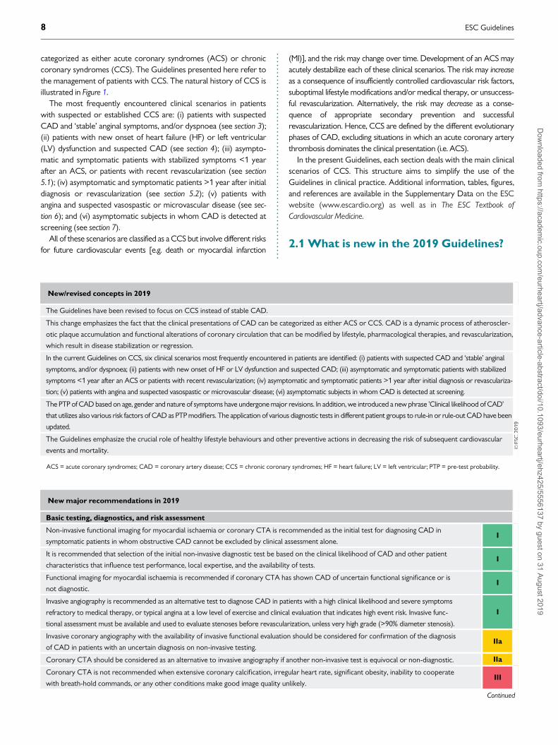
New major recommendations in 2019
Basic testing, diagnostics, and risk assessment
Non-invasive functional imaging for myocardial ischaemia or coronary CTA is recommended as the initial test for diagnosing CAD in
symptomatic patients in whom obstructive CAD cannot be excluded by clinical assessment alone.I
It is recommended that selection of the initial non-invasive diagnostic test be based on the clinical likelihood of CAD and other patient
characteristics that influence test performance, local expertise, and the availability of tests.I
Functional imaging for myocardial ischaemia is recommended if coronary CTA has shown CAD of uncertain functional significance or is
not diagnostic.I
Invasive angiography is recommended as an alternative test to diagnose CAD in patients with a high clinical likelihood and severe symptoms
refractory to medical therapy, or typical angina at a low level of exercise and clinical evaluation that indicates high event risk. Invasive func-
tional assessment must be available and used to evaluate stenoses before revascularization, unless very high grade (>90% diameter stenosis).
I
Invasive coronary angiography with the availability of invasive functional evaluation should be considered for confirmation of the diagnosis
of CAD in patients with an uncertain diagnosis on non-invasive testing.IIa
Coronary CTA should be considered as an alternative to invasive angiography if another non-invasive test is equivocal or non-diagnostic. IIa
Coronary CTA is not recommended when extensive coronary calcification, irregular heart rate, significant obesity, inability to cooperate
with breath-hold commands, or any other conditions make good image quality unlikely.III
Continued
8 ESC GuidelinesD
ownloaded from
https://academic.oup.com
/eurheartj/advance-article-abstract/doi/10.1093/eurheartj/ehz425/5556137 by guest on 31 August 2019

Antithrombotic therapy in patients with CCS and sinus rhythm
Addition of a second antithrombotic drug to aspirin for long-term secondary prevention should be considered in patients with a high
risk of ischaemic events and without high bleeding risk (see options in section 3.3.2).IIa
Addition of a second antithrombotic drug to aspirin for long-term secondary prevention may be considered in patients with at least a
moderately increased risk of ischaemic events and without high bleeding risk (see options in section 3.3.2).IIb
Antithrombotic therapy in patients with CCS and AF
When oral anticoagulation is initiated in a patient with AF who is eligible for a NOAC, a NOAC is recommended in preference to a
VKA.I
Long-term OAC therapy (a NOAC or VKA with time in therapeutic range >70%) is recommended in patients with AF and a CHA2DS2-
VASc score >_2 in males and >_3 in females.I
Long-term OAC therapy (a NOAC or VKA with time in therapeutic range >70%) should be considered in patients with AF and a
CHA2DS2-VASc score of 1 in males and 2 in females.IIa
Antithrombotic therapy in post-PCI patients with AF or another indication for OAC
In patients who are eligible for a NOAC, it is recommended that a NOAC (apixaban 5 mg b.i.d., dabigatran 150 mg b.i.d., edoxaban 60
mg o.d., or rivaroxaban 20 mg o.d.) is used in preference to a VKA in combination with antiplatelet therapy.I
When rivaroxaban is used and concerns about high bleeding risk prevail over concerns about stent thrombosis or ischaemic stroke, rivar-
oxaban 15 mg o.d. should be considered in preference to rivaroxaban 20 mg o.d. for the duration of concomitant single or dual antiplate-
let therapy.
IIa
When dabigatran is used and concerns about high bleeding risk prevail over concerns about stent thrombosis or ischaemic stroke, dabi-
gatran 110 mg b.i.d. should be considered in preference to dabigatran 150 mg b.i.d. for the duration of concomitant single or dual antipla-
telet therapy
IIa
After uncomplicated PCI, early cessation (<_1 week) of aspirin, and continuation of dual therapy with OAC and clopidogrel, should be
considered if the risk of stent thrombosis is low or if concerns about bleeding risk prevail over concerns about risk of stent thrombosis,
irrespective of the type of stent used.
IIa
Triple therapy with aspirin, clopidogrel, and an OAC for >_1 month should be considered when the risk of stent thrombosis outweighs
the bleeding risk, with the total duration (<_6 months) decided upon according to the assessment of these risks and clearly specified at
hospital discharge.
IIa
In patients with an indication for a VKA in combination with aspirin and/or clopidogrel, the dose intensity of the VKA should be carefully
regulated with a target international normalized ratio in the range of 2.0 - 2.5 and with time in therapeutic range >70%.IIa
Dual therapy with an OAC and either ticagrelor or prasugrel may be considered as an alternative to triple therapy with an OAC, aspirin,
and clopidogrel in patients with a moderate or high risk of stent thrombosis, irrespective of the type of stent used.IIb
Other pharmacological therapy
Concomitant use of a proton pump inhibitor is recommended in patients receiving aspirin monotherapy, DAPT, or OAC monotherapy
who are at high risk of gastrointestinal bleeding.I
Lipid-lowering drugs: if goals are not achieved with the maximum tolerated dose of statin, combination with ezetimibe is recommended. I
Lipid-lowering drugs: for patients at very high risk who do not achieve their goals on a maximum tolerated dose of statin and ezetimibe,
combination with a PCSK9 inhibitor is recommended.I
ACE inhibitors should be considered in CCS patients at very high risk of cardiovascular adverse events. IIa
The sodium-glucose co-transporter 2 inhibitors empagliflozin, canagliflozin, or dapagliflozin are recommended in patients with diabetes
mellitus and CVD.I
A glucagon-like peptide-1 receptor agonist (liraglutide or semaglutide) is recommended in patients with diabetes mellitus and CVD. I
Screening for CAD in asymptomatic subjects
Carotid ultrasound IMT for cardiovascular risk assessment is not recommended. III
Recommendations for treatment options for refractory angina
A reducer device for coronary sinus constriction may be considered to ameliorate symptoms of debilitating angina refractory to optimal
medical and revascularization strategies.IIb
aClass of recommendation.ACE = angiotensin-converting enzyme; ACS = acute coronary syndromes; AF = atrial fibrillation; b.i.d. = bis in die (twice a day); CAD = coronary artery disease; CCS = chroniccoronary syndromes; CHA2DS2-VASc = Cardiac failure, Hypertension, Age >_75 [Doubled], Diabetes, Stroke [Doubled] � Vascular disease, Age 65�74 and Sex category[Female]; CTA = computed tomography angiography; CVD = cardiovascular disease; HF = heart failure; IMT = intima-media thickness; LV = left ventricular; NOAC = non-vita-min K antagonist oral anticoagulant; OAC = oral anticoagulant; o.d. = omni die (once a day); PCI = percutaneous coronary intervention; PCSK9 = proprotein convertase subtili-sin-kexin type 9; VKA = vitamin K antagonist.
ESC Guidelines 9D
ownloaded from
https://academic.oup.com
/eurheartj/advance-article-abstract/doi/10.1093/eurheartj/ehz425/5556137 by guest on 31 August 2019
..
..
..
..
..
..
..
..
..
..
..
..
..
..
..
..
..
..
..3 Patients with angina and/ordyspnoea, and suspected coronaryartery disease
3.1 Basic assessment, diagnosis, and riskassessmentThe general approach for the initial diagnostic management ofpatients with angina and suspected obstructive CAD is presentedin Figure 2. The diagnostic management approach includes sixsteps. The first step is to assess the symptoms and signs, toidentify patients with possible unstable angina or other forms of
ACS (step 1). In patients without unstable angina or other ACS,the next step is to evaluate the patient’s general condition andquality of life (step 2). Comorbidities that could potentially influ-ence therapeutic decisions are assessed and other potentialcauses of the symptoms are considered. Step 3 includes basic test-ing and assessment of LV function. Thereafter, the clinical likeli-hood of obstructive CAD is estimated (step 4) and, on this basis,diagnostic testing is offered to selected patients to establish thediagnosis of CAD (step 5). Once a diagnosis of obstructive CADhas been confirmed, the patient’s event risk will be determined(step 6) as it has a major impact on the subsequent therapeuticdecisions.
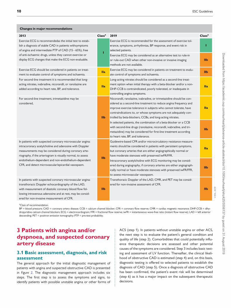
Changes in major recommendations
2013 Classa 2019 Classa
Exercise ECG is recommendedas the initial test to estab-
lish a diagnosis of stable CAD in patients withsymptoms
of angina and intermediate PTP of CAD (15�65%), free
of anti-ischaemic drugs, unless they cannot exercise or
display ECG changes that make the ECG non-evaluable.
I
Exercise ECG is recommended for the assessment of exercise tol-
erance, symptoms, arrhythmias, BP response, and event risk in
selected patients.
I
Exercise ECG may be considered as an alternative test to rule-in
or rule-out CAD when other non-invasive or invasive imaging
methods are not available.
IIb
Exercise ECG should be considered in patients on treat-
ment to evaluate control of symptoms and ischaemia.IIa
Exercise ECG may be considered in patients on treatment to evalu-
ate control of symptoms and ischaemia.IIb
For second-line treatment it is recommended that long-
acting nitrates, ivabradine, nicorandil, or ranolazine are
added according to heart rate, BP, and tolerance.IIa
Long-acting nitrates should be considered as a second-line treat-
ment option when initial therapy with a beta-blocker and/or a non-
DHP-CCB is contraindicated, poorly tolerated, or inadequate in
controlling angina symptoms.
IIa
For second-line treatment, trimetazidine may be
considered,
IIb
Nicorandil, ranolazine, ivabradine, or trimetazidine should be con-
sidered as a second-line treatment to reduce angina frequency and
improve exercise tolerance in subjects who cannot tolerate, have
contraindications to, or whose symptoms are not adequately con-
trolled by beta-blockers, CCBs, and long-acting nitrates.
IIa
In selected patients, the combination of a beta-blocker or a CCB
with second-line drugs (ranolazine, nicorandil, ivabradine, and tri-
metazidine) may be considered for first-line treatment according
to heart rate, BP, and tolerance.
IIb
In patients with suspected coronary microvascular angina:
intracoronary acetylcholine and adenosine with Doppler
measurements may be considered during coronary arte-
riography, if the arteriogram is visually normal, to assess
endothelium-dependent and non-endothelium-dependent
CFR, and detect microvascular/epicardial vasospasm.
IIb
Guidewire-based CFR and/or microcirculatory resistance measure-
ments should be considered in patients with persistent symptoms,
but coronary arteries that are either angiographically normal or
have moderate stenoses with preserved iwFR/FFR.
IIa
Intracoronary acetylcholine with ECG monitoring may be consid-
ered during angiography, if coronary arteries are either angiograph-
ically normal or have moderate stenoses with preserved iwFR/FFR,
to assess microvascular vasospasm.
IIb
In patients with suspected coronary microvascular angina:
transthoracic Doppler echocardiography of the LAD,
with measurement of diastolic coronary blood flow fol-
lowing intravenous adenosine and at rest, may be consid-
ered for non-invasive measurement of CFR.
IIb
Transthoracic Doppler of the LAD, CMR, and PET may be consid-
ered for non-invasive assessment of CFR.
IIb
aClass of recommendation.BP = blood pressure; CAD = coronary artery disease; CCB = calcium channel blocker; CFR = coronary flow reserve; CMR = cardiac magnetic resonance; DHP-CCB = dihy-dropyridine calcium channel blockers; ECG = electrocardiogram; FFR = fractional flow reserve; iwFR = instantaneous wave-free ratio (instant flow reserve); LAD = left anteriordescending; PET = positron emission tomography; PTP = pre-test probability.
10 ESC GuidelinesD
ownloaded from
https://academic.oup.com
/eurheartj/advance-article-abstract/doi/10.1093/eurheartj/ehz425/5556137 by guest on 31 August 2019

..
..
..
..
..
..
..
..
..
..
..
..
..
..
..
..
..
..
..
..
..
..
..
..
..
..
..
.After these steps, appropriate therapies are to be initiated,
which include lifestyle management (see section 3.2), medicaltherapy (see section 3.3), and revascularization when indicated(see section 3.4).
3.1.1. Step 1: Symptoms and signs
A careful history is the cornerstone of the diagnosis of angina. Itis possible to achieve a high degree of certainty on a diagnosisbased on history alone, although physical examination and objec-tive tests are most often necessary to confirm the diagnosis,exclude alternative diagnoses, and assess the severity of underly-ing disease. The history should include any manifestation of cardi-ovascular disease (CVD) and risk factors (i.e. family history ofCVD, dyslipidaemia, diabetes, hypertension, smoking, and otherlifestyle factors).
The characteristics of discomfort related to myocardial ischae-mia (angina pectoris) may be divided into four categories: location,character, duration, and relationship to exertion, and other
exacerbating or relieving factors. The discomfort caused by myo-cardial ischaemia is usually located in the chest, near the sternum,but may be felt anywhere from the epigastrium to the lower jawor teeth, between the shoulder blades, or in either arm to thewrist and fingers. The discomfort is often described as pressure,tightness, or heaviness; sometimes strangling, constricting, orburning. It may be useful to ask the patient directly about the pres-ence of ‘discomfort’ as many do not feel ‘pain’ or ‘pressure’ in theirchest. Shortness of breath may accompany angina, and chest dis-comfort may also be accompanied by less-specific symptoms suchas fatigue or faintness, nausea, burning, restlessness, or a sense ofimpending doom. Shortness of breath may be the sole symptom ofCAD and it may be difficult to differentiate this from shortness ofbreath caused by other conditions.
The duration of the discomfort is brief—<_10 min in the majorityof cases, and more commonly just a few minutes or less—andchest pain lasting for seconds is unlikely to be due to CAD. Animportant characteristic is the relationship to exercise. Symptoms
Assess symptoms and perform clinical investigations
Consider comorbidities and quality of life
Resting ECG, biochemistry, chest X-ray in selectedpatients, echocardiography at restb
Assess pre-test probability and clinical likelihood of CADc
STEP 1
STEP 2
STEP 3
STEP 4
Unstable angina?
Revascularizationfutile
LVEF <50%
Cause of chest painother than CAD?
Follow ACS guidelines
Medical therapya
See section 4
Treat as appropriate orinvestigate other causes
Offer diagnostic testing
No diagnostic
testing mandated
Coronary CTAf
STEP 5
Clinical likelihood of obstructive CAD Very highVery low
Choose appropriate therapy based on symptoms and event riskg STEP 6
Invasiveangiography
(with iwFR/FFR)e
Testing for ischaemia(imaging testing preferred)
Choice of the test based on clinicallikelihood, patient characteristics
and preference, availability, as well as local expertised
©ES
C 2
019
Figure 2 Approach for the initial diagnostic management of patients with angina and suspected coronary artery disease. ACS = acute coronarysyndrome; BP = blood pressure; CAD = coronary artery disease; CTA = computed tomography angiography; ECG = electrocardiogram; FFR = frac-tional flow reserve; iwFR = instantaneous wave-free ratio; LVEF = left ventricular ejection fraction. aIf the diagnosis of CAD is uncertain, establishinga diagnosis using non-invasive functional imaging for myocardial ischaemia before treatment may be reasonable. bMay be omitted in very young andhealthy patients with a high suspicion of an extracardiac cause of chest pain, and in multimorbid patients in whom the echocardiography result hasno consequence for further patient management. cConsider exercise ECG to assess symptoms, arrhythmias, exercise tolerance, BP response, andevent risk in selected patients. dAbility to exercise, individual test-related risks, and likelihood of obtaining diagnostic test result. eHigh clinical likeli-hood and symptoms inadequately responding to medical treatment, high event risk based on clinical evaluation (such as ST-segment depression,combined with symptoms at a low workload or systolic dysfunction indicating CAD), or uncertain diagnosis on non-invasive testing. fFunctionalimaging for myocardial ischaemia if coronary CTA has shown CAD of uncertain grade or is non-diagnostic. gConsider also angina without obstruc-tive disease in the epicardial coronary arteries (see section 6).
ESC Guidelines 11D
ownloaded from
https://academic.oup.com
/eurheartj/advance-article-abstract/doi/10.1093/eurheartj/ehz425/5556137 by guest on 31 August 2019

..
..
..
..
..
..
..
..
..
..
..
..
..
..
..
..
..
..
..
..
..
..
..
..
..
..
..
..
..
..
..
..
..
..
..
..
..
..
..
..
..
..
..
..
..
..
..
..
..
..
..
..
..
..
..
..
..classically appear or become more severe with increased levels ofexertion—such as walking up an incline or against a breeze, or incold weather—and rapidly disappear within a few minutes whenthese causal factors abate. Exacerbations of symptoms after aheavy meal or after waking up in the morning are classic featuresof angina. Angina may paradoxically be reduced with further exer-cise (walk-through angina) or on second exertion (warm-upangina).1 Sublingual nitrates rapidly relieve angina. Symptoms areunrelated to respiration or position. The angina threshold, andhence symptoms, may vary considerably from day to day and evenduring the same day.
Definitions of typical and atypical angina are summarized inTable 3. The classification, although subjective, is practical and ofproven value in determining the likelihood of obstructive CAD.2,3
Studies published since 2015 have reported that the majority ofpatients suspected of having CAD present with atypical or non-anginal chest pain,4�6 with as few as 10 - 15% presenting with typi-cal angina.3,7,8 The Canadian Cardiovascular Society classificationis still widely used as a grading system for angina,9 to quantify thethreshold at which symptoms occur in relation to physical activ-ities (Table 4).
Physical examination of a patient with suspected CAD is importantto assess the presence of anaemia, hypertension, valvular heart dis-ease, hypertrophic cardiomyopathy, or arrhythmias. It is also recom-
mended that practitioners obtain the body mass index (BMI) andsearch for evidence of non-coronary vascular disease, which may beasymptomatic [includes palpation of peripheral pulses, and ausculta-tion of carotid and femoral arteries, as well as assessment of theankle-brachial index (ABI)], and other signs of comorbid conditionssuch as thyroid disease, renal disease, or diabetes. This shouldbe used in the context of other clinical information, such as the pres-ence of cough or stinging pain, making CAD more unlikely. Oneshould also try to reproduce the symptoms by palpation10 and testthe effect of sublingual nitroglycerin in order to classify the symptoms(Table 3).
3.1.1.1 Stable vs. unstable anginaUnstable angina may present in one of three ways: (i) as restangina, i.e. pain of characteristic nature and location occurring atrest and for prolonged periods (>20 min); (ii) new-onset angina,i.e. recent (2 months) onset of moderate-to-severe angina(Canadian Cardiovascular Society grade II or III); or (iii) crescendoangina, i.e. previous angina, which progressively increases inseverity and intensity, and at a lower threshold, over a shortperiod of time. Management of angina fulfilling these criteria isdealt with in the ESC Guidelines for the management of ACS.11,12
New-onset angina is generally regarded as unstable angina; how-ever, if angina occurs for the first time with heavy exertion andsubsides at rest, the suspected condition falls under the definitionof CCS rather than unstable angina. In patients with unstableangina identified as being at low risk, it is recommended that thediagnostic and prognostic algorithms presented in theseGuidelines be applied once the period of instability has subsided.11
Low-risk patients with unstable angina are characterized by norecurrence of angina, no signs of HF, no abnormalities in the initialor subsequent electrocardiogram (ECG), and no rise in troponinlevels.11 In this setting, a non-invasive diagnostic strategy is recom-mended before deciding on an invasive strategy. Based on the defi-nition above, stable and unstable angina may overlap, and manyCCS patients pass through a period of experiencing unstableangina.
Table 4 Grading of effort angina severity according to the Canadian Cardiovascular Society
Grade Description of angina severity
I Angina only with strenuous exertion Presence of angina during strenuous, rapid, or prolonged ordinary
activity (walking or climbing the stairs).
II Angina with moderate exertion Slight limitation of ordinary activities when they are performed
rapidly, after meals, in cold, in wind, under emotional stress, or
during the first few hours after waking up, but also walking uphill,
climbing more than one flight of ordinary stairs at a normal pace,
and in normal conditions.
III Angina with mild exertion Having difficulties walking one or two blocks, or climbing one
flight of stairs, at normal pace and conditions.
IV Angina at rest No exertion needed to trigger angina.
Table 3 Traditional clinical classification of suspectedanginal symptoms
Typical angina Meets the following three characteristics:
(i) Constricting discomfort in the front of the chest or
in the neck, jaw, shoulder, or arm;
(ii) Precipitated by physical exertion;
(iii) Relieved by rest or nitrates within 5 min.
Atypical angina Meets two of these characteristics.
Non-anginal
chest pain
Meets only one or none of these characteristics.
12 ESC GuidelinesD
ownloaded from
https://academic.oup.com
/eurheartj/advance-article-abstract/doi/10.1093/eurheartj/ehz425/5556137 by guest on 31 August 2019
..
..
..
..
..
..
..
..
..
..
..
..
..
..
..
..
..
..
..
..
..
..
..
..
..
..
..
..
..
..
..
..
..
..
..
..
..
..
..
..
..
..
..
..
..
..
..
..
..
..
..
..
..
..
..
..3.1.1.2 Distinction between symptoms caused by epicardial vs. microvas-cular/vasospastic diseaseA distinction between symptoms caused by an epicardial stenosis andsymptoms caused by microvascular or vasospastic disease cannot bemade with reasonable certainty. Reliance on ischaemia testing ordepiction of the coronary anatomy is often unavoidable to excludeobstructive CAD, which can be absent in symptomatic patients.13,14
A diagnostic workup for microvascular or vasospastic disease is dis-cussed in section 6 of these Guidelines.
3.1.2 Step 2: Comorbidities and other causes of
symptoms
Before any testing is considered, one must assess the patient’s generalhealth, comorbidities, and quality of life. If revascularization is unlikelyto be an acceptable option, further testing may be reduced to a clini-cally indicated minimum and appropriate therapy should be insti-tuted, which may include a trial of antianginal medication even if adiagnosis of CAD has not been fully demonstrated. Non-invasivefunctional imaging for ischaemia may be an option if there is need toverify the diagnosis (Figure 2).
If the pain is clearly non-anginal, other diagnostic testing may beindicated to identify gastrointestinal, pulmonary, or musculoskeletalcauses of chest pain. Nevertheless, these patients should alsoreceive Guideline-based risk-factor modification based on commonlyapplied risk charts such as SCORE (Systematic COronary RiskEvaluation) (www.heartscore.org).15
3.1.3 Step 3: Basic testing
Basic (first-line) testing in patients with suspected CAD includes stand-ard laboratory biochemical testing, a resting ECG, possible ambulatoryECG monitoring, resting echocardiography, and, in selected patients, achest X-ray. Such testing can be done on an outpatient basis.
3.1.3.1 Biochemical testsLaboratory investigations are used to identify possible causes ofischaemia, to establish cardiovascular risk factors and associated
conditions, and to determine prognosis. Haemoglobin as part of afull blood count and—where there is a clinical suspicion of a thy-roid disorder—thyroid hormone levels provide informationrelated to possible causes of ischaemia. Fasting plasma glucose andglycated haemoglobin (HbA1c) should be measured in everypatient with suspected CAD. If both are inconclusive, an additionaloral glucose tolerance test is recommended.16 Knowledge of glu-cose metabolism is important because of the well-recognized asso-ciation between diabetes and adverse cardiovascular outcome.Patients with diabetes should be managed according to specificGuidelines.15,16 A lipid profile, including total cholesterol, high-density lipoprotein cholesterol, low-density lipoprotein cholesterol(LDL-C), and triglycerides, should also be evaluated in any patientwith suspected CAD to establish the patient’s risk profile andascertain the need for treatment.15,17 To characterize severe dysli-pidaemia or follow-up on high triglyceridaemia, fasting values arerecommended.17
Peripheral artery disease (PAD) and renal dysfunction increase thelikelihood of CAD, and have a negative impact on prognosis.18�20
Hence, baseline renal function should be evaluated with estimation ofthe glomerular filtration rate (GFR). It may also be reasonable tomeasure the uric acid level, as hyperuricaemia is a frequent comorbidcondition and may also affect renal function.
If there is a clinical suspicion of CAD instability, biochemicalmarkers of myocardial injury—such as troponin T or troponin I—should be measured, preferably using high-sensitivity assays, andmanagement should follow the Guidelines for ACS without persis-tent ST-segment elevation.11 If high-sensitivity assays are employed,low levels of troponin can be detected in many patients with stableangina. Increased troponin levels are associated with adverse out-come21�25 and small studies have indicated a possible incrementalvalue in diagnosing CAD,26,27 but larger trials are needed to verifythe utility of systematic assessment in patients suspected of CAD.While multiple biomarkers may be useful for prognostication(see section 5), they do not yet have a role in diagnosing obstructiveCAD.
Basic biochemistry testing in the initial diagnostic management of patients with suspected coronary artery disease
Recommendations Classa Levelb
If evaluation suggests clinical instability or ACS, repeated measurements of troponin, preferably using high-sensitivity or
ultrasensitive assays, are recommended to rule-out myocardial injury associated with ACS.28,29 I A
The following blood tests are recommended in all patients:
• Full blood count (including haemoglobin);30 I B
• Creatinine measurement and estimation of renal function;31,32 I A
• A lipid profile (including LDL-C).33,34 I A
It is recommended that screening for type 2 diabetes mellitus in patients with suspected and established CCS is imple-
mented with HbA1c and fasting plasma glucose measurements, and that an oral glucose tolerance test is added if HbA1c
and fasting plasma glucose results are inconclusive.16,35
I B
Assessment of thyroid function is recommended in case of clinical suspicion of thyroid disorders. I C
ACS = acute coronary syndromes; CAD = coronary artery disease; CCS = chronic coronary syndromes; HbA1c = glycated haemoglobin; LDL-C = low-density lipoproteincholesterol.aClass of recommendation.bLevel of evidence.
ESC Guidelines 13D
ownloaded from
https://academic.oup.com
/eurheartj/advance-article-abstract/doi/10.1093/eurheartj/ehz425/5556137 by guest on 31 August 2019

..
..
..
..
..
..
..
..
..
..
..
..
..
..
..
..
..
..
..
..
..
..
..
..
..
..
..
..
..
..
..
..
..
..
..
..
..
..
..
..
..
..
..
..
..
..
..
..
..
..
..
..
3.1.3.2 Resting electrocardiogram and ambulatory monitoringThe paradigm of diagnosing myocardial ischaemia has, for almost acentury, been based on the detection of repolarization abnormalities,mainly in the form of ST-segment depressions. Thus, the resting 12lead ECG remains an indispensable component of the initial evalua-tion of a patient with chest pain without an obviously non-cardiaccause. Two scenarios of clinical evaluation are encountered: (i) apatient without symptoms of chest pain or discomfort, and (ii) apatient with ongoing anginal symptoms.
The former situation is far more prevalent and a normal restingECG is frequently recorded. However, even in the absence of repola-rization abnormalities, an ECG can demonstrate indirect signs ofCAD, such as signs of previous MI (pathological Q waves) or conduc-tion abnormalities [mainly left bundle branch block (LBBB) andimpairment of atrioventricular conduction]. Atrial fibrillation (AF) is afrequent finding in patients with chest pain (usually atypical). ST-segment depression during supraventricular tachyarrhythmias is notpredictive of obstructive CAD.36�39
The ECG can be crucial for diagnosing myocardial ischaemia ifdynamic ST-segment changes are recorded during ongoing angina.The diagnosis of Prinzmetal and vasospastic angina is based on thedetection of typical transient ST-segment elevation or depressionduring an angina attack (usually at rest).
Long-term ambulatory ECG monitoring and recording should notbe used to replace exercise testing; however, 12 lead ECG monitor-ing can be considered in selected patients to detect anginal episodesunrelated to physical exercise. Ambulatory ECG monitoring mayreveal evidence of silent myocardial ischaemia in patients withCCS, but rarely adds relevant diagnostic or prognostic informationthat cannot be derived from stress testing.40 ECG changes suggestingischaemia on ambulatory ECG monitoring are very frequent inwomen, but do not correlate with findings during stress testing.41
Most importantly, therapeutic strategies targeting silent ischaemiadetected by ambulatory monitoring have not demonstrated clearsurvival benefits.42,43
3.1.3.3 Echocardiography and magnetic resonance imaging at restAn echocardiographic study will provide important information aboutcardiac function and anatomy. LV ejection fraction (LVEF) is often nor-mal in patients with CCS.44 A decreased LV function and/or regionalwall motion abnormalities may increase the suspicion of ischaemicmyocardial damage,45 and a pattern of LV dysfunction following thetheoretical distribution territory of the coronary arteries is typical inpatients who have already had an MI.46,47 The detection of regionalwall motion abnormalities can challenging by visual assessment, anddetection of early systolic lengthening, decreased systolic shortening,or post-systolic shortening by strain imaging techniques might be help-ful in patients with apparently normal LV function but with clinical sus-picion of CCS.48�50 Decreased diastolic LV function has beenreported to be an early sign of ischaemic myocardial dysfunction andcould also be indicative of microvascular dysfunction.51,52
Echocardiography is an important clinical tool for the exclusion ofalternative causes of chest pain and also aids in diagnosing concurrentcardiac diseases, such as valvular heart diseases, HF, and most cardio-myopathies,53 but it is important to remember that these diseasesoften coexist with obstructive CAD. The use of an echocardio-graphic contrast agent can be useful in patients with poor acousticwindows.54
Cardiac magnetic resonance (CMR) may be considered inpatients with suspected CAD when the echocardiogram (havingused contrast) is inconclusive.55 CMR will provide useful informa-tion on cardiac anatomy and systolic cardiac function, similar tothat from an echocardiogram, in patients with no contraindicationsfor CMR. CMR can assess global and regional function,56 and theuse of late gadolinium enhancement CMR can reveal a typical pat-tern of scarred myocardium in patients who have already experi-enced an MI.57
Assessment of LV function is important in all patients for risk strati-fication (see Supplementary Data section 3.2) and should therefore beperformed in all symptomatic patients with suspected CAD.
Resting electrocardiogram in the initial diagnostic man-agement of patients with suspected coronary arterydisease
Recommendations Classa Levelb
A resting 12 lead ECG is recommended in all
patients with chest pain without an obvious
non-cardiac cause.
I C
A resting 12 lead ECG is recommended in all
patients during or immediately after
an episode of angina suspected to be indicative
of clinical instability of CAD.
I C
ST-segment alterations recorded during
supraventricular tachyarrhythmias should not
be used as evidence of CAD.
III C
CAD = coronary artery disease; CCS = chronic coronary syndromes; ECG =electrocardiogram.aClass of recommendation.bLevel of evidence.
Ambulatory electrocardiogram monitoring in the initialdiagnostic management of patients with suspected coro-nary artery disease
Recommendations Classa Levelb
Ambulatory ECG monitoring is recommended
in patients with chest pain and
suspected arrhythmias.
I C
Ambulatory ECG recording, preferably moni-
toring with 12 lead ECG, should be consid-
ered in patients with suspected vasospastic
angina.
IIa C
Ambulatory ECG monitoring should not be
used as a routine examination in patients with
suspected CCS.
III C
CAD = coronary artery disease; CCS = chronic coronary syndromes; ECG =electrocardiogram.aClass of recommendation.bLevel of evidence.
14 ESC GuidelinesD
ownloaded from
https://academic.oup.com
/eurheartj/advance-article-abstract/doi/10.1093/eurheartj/ehz425/5556137 by guest on 31 August 2019

..
..
..
..
..
..
..
..
..
..
..
..
..
..
..
..
..
..
..
..
..
..
..
..
..
..
..
..
..
..
..
..
..
..
..
..
..
..
..
..
..
..
..
..
..
..
..
..
..
..
..
..
..
..
..
..
..
..
..
..
..
..
..
..
..
..
..
..
..
..
..
..
..
..
..
..
..
..
..
..
..
..
..
..
..
..
.Management of patients with either angina or HF symptoms, withreduced LVEF <40% or a mid-range reduced LVEF of 40-49%, isdescribed in section 4 of the Guidelines.
3.1.3.4 Chest X-rayChest X-ray is frequently used in the assessment of patients withchest pain. However, in CCS, it does not provide specific informationfor diagnosis or event risk stratification. The test may occasionally behelpful in assessing patients with suspected HF. Chest X-ray may alsobe useful in patients with pulmonary problems, which often accom-pany CAD, or to rule-out another cause of chest pain in atypicalpresentations.
3.1.4 Step 4: Assessment of pre-test probability and clini-
cal likelihood of coronary artery disease
The performance of the available methods in diagnosing obstructiveCAD (i.e. the likelihood that the patient has disease if the test isabnormal, and the likelihood that the patient does not have disease ifthe test is normal) depends on the prevalence of disease in the
population studied and, thus, the likelihood that a given patient willactually have CAD. Diagnostic testing is most useful when the likeli-hood is intermediate. When likelihood is high, a large number ofpatients need to be studied to identify the few patients that do nothave disease, and a negative test result can seldom rule out the pres-ence of obstructive CAD (i.e. the negative predictive value is low).When the likelihood is low, a negative test can rule out the disease,but the lower the likelihood, the higher the likelihood of a false-positive test (i.e. a positive test in the absence of obstructive CAD).In patients at the extreme ends of the probability range, it is thereforereasonable to refrain from diagnostic testing, and assume that thepatient does or does not have obstructive CAD based on clinicalevaluation alone.
The likelihood of obstructive CAD is influenced by the prevalenceof the disease in the population studied, as well as by clinical featuresof an individual patient. A simple predictive model can be used to esti-mate the pre-test probability (PTP) of obstructive CAD based onage, sex, and the nature of symptoms.59 In the previous version ofthese Guidelines,60 estimation of the PTP was based on data gatheredby Genders et al.,61 which updated previous data from Diamond andForrester.59 Notably, the prevalence of disease for a given constella-tion of age, sex, and nature of symptoms was lower than inthe Diamond and Forrester data. Since the previous version of theGuidelines was published, several studies have indicated thatthe prevalence of obstructive disease among patients with suspectedCAD is lower than in the previous update.7,8,62,63
A pooled analysis64 of three contemporary study cohorts, includ-ing patients evaluated for suspected CAD,7,8,62 has indicated that thePTP based on age, sex, and symptoms is approximately one-third ofthat predicted by the model used in the previous version of theGuidelines.57,62 Overestimation of PTP is an important contributoryfactor to a low diagnostic yield of non-invasive and invasive testing.The new set of PTPs presented in Table 5 may substantially reducethe need for non-invasive and invasive tests in patients with suspectedstable CAD. The table now also includes patients presenting withdyspnoea as their main symptom. However, it should be noted thatthe PTPs presented in Table 5 (as well as the PTP table in the previousversion of the Guidelines) are based mainly on patients from coun-tries with low CVD risk, and may vary between regions andcountries.
Application of the new PTPs (Table 5) has important conse-quences for the referral of patients for diagnostic testing. If diag-nostic testing was deferred in patients with a new PTP <15%, thiswould result in a large increase in the proportion of patients forwhom diagnostic testing was not recommended, because morepatients are classified as having a PTP <15%. In data derived fromthe PROMISE (Prospective Multicenter Imaging Study forEvaluation of Chest Pain) trial, 50% of patients previously classifiedas having an intermediate likelihood of obstructive CAD werereclassified to a PTP <15% according to the new PTP.62 In dataderived from the pooled analysis64 (Table 5), 57% of all patientswere classified to a PTP <15%.
Studies have shown that outcomes in patients classified withthe new PTP <15% is good (annual risk of cardiovascular deathor MI is <1%).7,62 Hence, it is safe to defer routine testing inpatients with PTP <15%, thus reducing unnecessary proceduresand costs.
Resting echocardiography and cardiac magnetic reso-nance in the initial diagnostic management of patientswith suspected coronary artery disease
Recommendations Classa Levelb
A resting transthoracic echocardiogram is rec-
ommended in all patients for:
(1) Exclusion of alternative causes of angina;
(2) Identification of regional wall motion
abnormalities suggestive of CAD;
(3) Measurement of LVEF for risk stratifica-
tion; and
(4) Evaluation of diastolic function.44,45,52,58
I B
Ultrasound of the carotid arteries should be
considered, and be performed by adequately
trained clinicians, to detect plaque in patients
with suspected CCS without known athero-
sclerotic disease.
IIa C
CMR may be considered in patients with an
inconclusive echocardiographic test.IIb C
CAD = coronary artery disease; CCS = chronic coronary syndromes; CMR =cardiac magnetic resonance imaging; LVEF = left ventricular ejection fraction.aClass of recommendation.bLevel of evidence.
Chest X-ray in the initial diagnostic management ofpatients with suspected coronary artery disease
Recommendation Classa Levelb
Chest X-ray is recommended for patients
with atypical presentation, signs and symptoms
of HF, or suspicion of pulmonary disease.
I C
HF = heart failure.aClass of recommendation.bLevel of evidence.
ESC Guidelines 15D
ownloaded from
https://academic.oup.com
/eurheartj/advance-article-abstract/doi/10.1093/eurheartj/ehz425/5556137 by guest on 31 August 2019

..
..
..
..
..
..
..
..
..
..
..
..
..
..
..
..
..
..
..
..
..
..
..
..
..
..
..
..
..
..
..
..
..
..
..
..
..
..
..
..
..
..
..
..
..
..
..
..
..
..
..
..
..
..
.Recent studies have also demonstrated that, when tested, the
true observed prevalence of obstructive CAD has been <5% inpatients who had a PTP <15% according to the 2013 version ofthese Guidelines.7,63 Therefore, this Task Force recognizes thatthe performance of diagnostic testing in patients with a new PTPof 5 - 15% more closely reflects current clinical practice and maybe considered, particularly if symptoms are limiting and requireclarification.7,63 Patient preference, local resources and the avail-ability of tests, clinical judgement, and appropriate patient informa-tion remain important when making a decision to proceed withnon-invasive diagnostic testing for an individual patient when thePTP is 5 - 15%, and the higher likelihood of a false-positive testmust be considered. Patients with a PTP <_5% can be assumed tohave such a low probability of disease that diagnostic testingshould be performed only for compelling reasons. Implementationof the new PTPs also indicates that patients should not be rou-tinely referred directly to invasive assessment unless clinical orother data indicate a high likelihood of obstructive CAD.
Clinical models that incorporate information on risk factors forCVD, resting ECG changes, or coronary calcification have improvedthe identification of patients with obstructive CAD compared withage, sex, and symptoms alone.3,7,60,65�68 Therefore, the presence ofrisk factors for CVD (such as family history of CVD, dyslipidaemia,diabetes, hypertension, smoking, and other lifestyle factors) thatincrease the probability of obstructive CAD can be used as modifiersof the PTP estimate. If available, Q-wave, ST-segment, or T-wavechanges on the ECG, LV dysfunction suggestive of ischaemia, andfindings on exercise ECG, as well as information on coronary calciumobtained by computed tomography (CT), can be used to improveestimations of the PTP of obstructive CAD.3,69 In particular, theabsence of coronary calcium (Agatston score = 0) is associated witha low prevalence of obstructive CAD (<5%), and low risk of death ornon-fatal MI (<1% annual risk).69,70 However, it should be noted thatcoronary calcium imaging does not exclude coronary stenosis causedby a non-calcified atherosclerotic lesion,70 and the presence of coro-nary calcium is a weak predictor of obstructive CAD.69 Although the
optimal use of these factors in improving PTP assessment has not yetbeen established, they should be considered in addition to the PTPbased on sex, age, and the nature of symptoms to determine theoverall clinical likelihood of obstructive CAD, as summarized inFigure 3. This is particularly important in refining the likelihood ofCAD patients with a PTP of 5�15% based on age, sex, and the natureof symptoms.
3.1.5 Step 5: Selecting appropriate testing
In patients in whom revascularization is futile due to comorbiditiesand overall quality of life, the diagnosis of CAD can be made clini-cally and only medical therapy is required. If the diagnosis of CADis uncertain, establishing a diagnosis using non-invasive functionalimaging for myocardial ischaemia before treatment is reasonable(Figure 2).
In a patient with a high clinical likelihood of CAD, symptoms unre-sponsive to medical therapy or typical angina at a low level of exer-cise, and an initial clinical evaluation (including echocardiogram and,in selected patients, exercise ECG) that indicates a high event risk,proceeding directly to invasive coronary angiography (ICA) withoutfurther diagnostic testing is a reasonable option. Under such circum-stances, the indication for revascularization should be based onappropriate invasive confirmation of the haemodynamic significanceof a stenosis.71,72
In other patients in whom CAD cannot be excluded byclinical assessment alone, non-invasive diagnostic tests are recom-mended to establish the diagnosis and assess the event risk.The current Guidelines recommend the use of either non-invasive functional imaging of ischaemia or anatomical imaging usingcoronary CT angiography (CTA) as the initial test for diagnosing CAD.
3.1.5.1 Functional non-invasive testsFunctional non-invasive tests for the diagnosis of obstructive CADare designed to detect myocardial ischaemia through ECGchanges, wall motion abnormalities by stress CMR or stress echo-cardiography, or perfusion changes by single-photon emission CT
Table 5 Pre-test probabilities of obstructive coronary artery disease in 15 815 symptomatic patients according to age,sex, and the nature of symptoms in a pooled analysis64 of contemporary data7,8,62
Typical Atypical Non-anginal
Age Men Women Men Women Men Women
30–39 3% 5% 4% 3% 1% 1%
40–49 22% 10% 10% 6% 3% 2%
50–59 32% 13% 17% 6% 11% 3%
60–69 44% 16% 26% 11% 22% 6%
70+ 52% 27% 34% 19% 24% 10%
Dyspnoeaa
Men Women
0% 3%
12% 3%
20% 9%
27% 14%
32% 12%
©ES
C 2
019
CAD = coronary artery disease; PTP = pre-test probability.aIn addition to the classic Diamond and Forrester classes,59 patients with dyspnoea only or dyspnoea as the primary symptom are included. The regions shaded dark greendenote the groups in which non-invasive testing is most beneficial (PTP >15%). The regions shaded light green denote the groups with PTPs of CAD between 5�15%, in whichtesting for diagnosis may be considered after assessing the overall clinical likelihood based on the modifiers of PTPs presented in Figure 3.
16 ESC GuidelinesD
ownloaded from
https://academic.oup.com
/eurheartj/advance-article-abstract/doi/10.1093/eurheartj/ehz425/5556137 by guest on 31 August 2019

..
..
..
..
..
..
..
..
..
..
..
..
..
..
..
..
..
..
..
..
..
..
..
..
..
..
..
..
..
..
..
..
..
..
..
..
..
..
..
..
..
..
..
..
..
..
..
..
..
.(SPECT), positron emission tomography (PET), myocardial con-trast echocardiography, or contrast CMR. Ischaemia can be pro-voked by exercise or pharmacological stressors, either byincreased myocardial work and oxygen demand, or by heteroge-neity in myocardial perfusion by vasodilatation. Non-invasive func-tional tests are associated with high accuracy for the detection offlow-limiting coronary stenosis compared with invasive functionaltesting [fractional flow reserve (FFR)].73 However, lower-gradecoronary atherosclerosis not linked with ischaemia remains unde-tected by functional testing and, in the presence of a negative func-tional test, patients should receive risk-factor modification basedon commonly applied risk charts and recommendations.
3.1.5.2 Anatomical non-invasive evaluationAnatomical non-invasive evaluation, by visualizing the coronary arterylumen and wall using an intravenous contrast agent, can be performedwith coronary CTA, which provides high accuracy for the detectionof obstructive coronary stenoses defined by ICA,73 because both testsare based on anatomy. However, stenoses estimated to be 50�90%by visual inspection are not necessarily functionally significant, i.e. theydo not always induce myocardial ischaemia.73,74 Therefore, eithernon-invasive or invasive functional testing is recommended for furtherevaluation of angiographic stenosis detected by coronary CTA orinvasive angiography, unless a very high-grade (>90% diameter steno-sis) stenosis is detected via invasive angiography. The presence orabsence of non-obstructive coronary atherosclerosis on coronaryCTA provides prognostic information, and can be used to guide pre-ventive therapy.75 The SCOT-HEART (Scottish ComputedTomography of the HEART) trial demonstrated a significantly lowerrate of the combined endpoint of cardiovascular death or non-fatal MI(2.3 vs. 3.9% during 5 year follow-up) in patients in whom coronaryCTA was performed in addition to routine testing, which consisted
predominantly of exercise ECG.6 Other randomized, prospective clin-ical trials have demonstrated that diagnostic testing with coronaryCTA is associated with clinical outcomes similar to those for func-tional imaging in patients with suspected CAD.4,6,76 In patients withextensive CAD, coronary CTA complemented by CT-based FFR wasnon-inferior to ICA and FFR for decision-making, and the identificationof targets for revascularization.77
3.1.5.3 Role of the exercise electrocardiogramExercise ECG has inferior diagnostic performance compared withdiagnostic imaging tests, and has limited power to rule-in or rule-outobstructive CAD.73 Since the publication of the previous version ofthese Guidelines, randomized clinical trials (RCTs) have comparedthe effects of diagnostic strategies based on exercise ECG or an imag-ing diagnostic test6,78,79 on clinical outcomes. These studies haveshown that the addition of coronary CTA5,6,78,80 or functionalimaging79 clarifies the diagnosis, enables the targeting of preventivetherapies and interventions, and potentially reduces the risk of MIcompared with an exercise ECG. Some, although not all, registrystudies have also shown similar benefits regarding the use of an imag-ing diagnostic test in patients treated in everyday clinical practice.81,82
Therefore, these Guidelines recommend the use of an imaging diag-nostic test instead of exercise ECG as the initial test for to diagnoseobstructive CAD.
An exercise ECG alone may be considered as an alternative todiagnose obstructive CAD if imaging tests are not available, keepingin mind the risk of false-negative and false-positive test results.73,83
An exercise ECG is of no diagnostic value in patients with ECGabnormalities that prevent interpretation of the ST-segment changesduring stress (i.e. LBBB, paced rhythm, Wolff-Parkinson-White syn-drome, >_0.1 mV ST-segment depression on resting ECG, or who arebeing treated with digitalis). An exercise ECG provides
©ES
C 2
019
PTP based on sex, age and nature of symptoms (Table 5)
Clinical likelihood of CAD
Decreases likelihood• Normal exercise ECGa
• No coronary calcium by CT (Agatston score = 0)a
Increases likelihood• Risk factors for CVD
(dyslipidaemia, diabetes, hypertension, smoking, family history of CVD)
• Resting ECG changes(Q-wave or ST-segment/T-wave changes)
• LV dysfunction suggestive of CAD
• Abnormal exercise ECGa
• Coronary calcium by CTa
Figure 3 Determinants of the clinical likelihood of obstructive coronary artery disease. CAD = coronary artery disease; CT = computed tomography,CVD = cardiovascular disease, ECG = electrocardiogram, LV = left ventricular; PTP = pre-test probability. aWhen available.
ESC Guidelines 17D
ownloaded from
https://academic.oup.com
/eurheartj/advance-article-abstract/doi/10.1093/eurheartj/ehz425/5556137 by guest on 31 August 2019

..
..
..
..
..
..
..
..
..
..
..
..
..
..
..
..
..
..
..
..
..
..
..
..
..
..
..
..
..
..
..
..
..
..
..
..
..
..complementary clinically useful information beyond ECG changesand valuable prognostic information. Therefore, application of anexercise ECG may be considered in selected patients to complementclinical evaluation for the assessment of symptoms, ST-segmentchanges, exercise tolerance, arrhythmias, blood pressure (BP)response, and event risk.
3.1.5.4 Selection of diagnostic testsEither a functional or anatomical test can be used to establish a diag-nosis of obstructive CAD. A summary of the main diagnostic path-ways is displayed in Figure 4. For revascularization decisions,information on both anatomy and ischaemia is needed.
3.1.5.5 The impact of clinical likelihood on the selection of a diagnostic testEach non-invasive diagnostic test has a particular range of clinical like-lihood of obstructive CAD where the usefulness of its application ismaximal. The likelihood ratios of the tests constitute useful parame-ters of their abilities to correctly classify patients, and can be used tofacilitate the selection of the most useful test in any given patient.73,84
Given a clinical likelihood of obstructive CAD and the likelihood ratioof a particular test, one can assess the post-test probability ofobstructive CAD after performing such a test. Using this approach,one can estimate the optimal ranges of clinical likelihood for eachtest, where they can reclassify patients from intermediate to eitherlow or high post-test probability of CAD (Figure 5).73
Coronary CTA is the preferred test in patients with a lower rangeof clinical likelihood of CAD, no previous diagnosis of CAD, andcharacteristics associated with a high likelihood of good image quality.It detects subclinical coronary atherosclerosis, but can also accu-rately rule out both anatomically and functionally significant CAD(Figure 5). It has higher accuracy values when low clinical likelihoodpopulations are subjected to examination.85 Trials evaluating out-comes after coronary CTA to date have mostly included patientswith a low clinical likelihood.4,5
The non-invasive functional tests for ischaemia typically have bet-ter rule-in power. In outcome trials, functional imaging tests havebeen associated with fewer referrals for downstream ICA comparedwith a strategy relying on anatomical imaging.55,76,86 Before revascula-rization decisions can be made, functional evaluation of ischaemia(either non-invasive or invasive) is required in most patients.Therefore, functional non-invasive testing may be preferred inpatients at the higher end of the range of clinical likelihood if revascu-larization is likely or the patient has previously diagnosed CAD.
Patients in whom CAD is suspected, but who have a very low clini-cal likelihood (<_5%) of CAD, should have other cardiac causes ofchest pain excluded and their cardiovascular risk factors adjusted,based on a risk-score assessment. In patients with repeated, unpro-voked attacks of anginal symptoms mainly at rest, vasospastic anginashould be considered, diagnosed, and treated appropriately(see section 6).
©ES
C 2
019
Non-invasivetesting forischaemia
Preferentially considered if:� High clinical likelihood� Revascularization likely� Local expertise and availability� Viability assessment also required
Preferentially considered if:� High clinical likelihood and severe
symptoms refractory to medical therapy� Typical angina at low level of exercise and
clinical evaluation including exercise ECG indicates high-risk of events
� LV dysfunction suggestive of CAD
Preferentially considered if:� Low clinical likelihood� Patient characteristics suggest
high image quality� Local expertise and availability� Information on atherosclerosis
desired� No history of CAD
Invasivecoronary
angiography
Revascularization
CoronaryCTA
Drug therapyb
Functionalassessment
Drugtherapyb
Ongoing symptomsa
Stenosis >90% or with established
correlation to ischaemia
Figure 4 Main diagnostic pathways in symptomatic patients with suspected obstructive coronary artery disease. Depending on clinical conditions andthe healthcare environment, patient workup can start with either of three options: non-invasive testing, coronary computed tomography angiography, orinvasive coronary angiography. Through each pathway, both functional and anatomical information is gathered to inform an appropriate diagnostic andtherapeutic strategy. Risk-factor modification should be considered in all patients. CAD = coronary artery disease; CTA = computed tomography angiog-raphy; ECG = electrocardiogram; LV = left ventricular. aConsider microvascular angina. bAntianginal medications and/or risk-factor modification.
18 ESC GuidelinesD
ownloaded from
https://academic.oup.com
/eurheartj/advance-article-abstract/doi/10.1093/eurheartj/ehz425/5556137 by guest on 31 August 2019
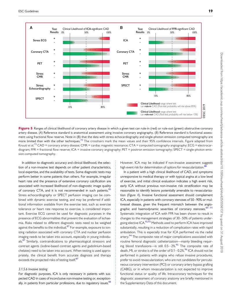
..
..
..
..
..
..
..
..
..
..
..
..
..
..
..
..
..
..
..
..
..
..
..
..
..
..
..
..
..
..
..
..
..
..
..
..
..
..
..
..
..In addition to diagnostic accuracy and clinical likelihood, the selec-tion of a non-invasive test depends on other patient characteristics,local expertise, and the availability of tests. Some diagnostic tests mayperform better in some patients than others. For example, irregularheart rate and the presence of extensive coronary calcification areassociated with increased likelihood of non-diagnostic image qualityof coronary CTA, and it is not recommended in such patients.85
Stress echocardiography or SPECT perfusion imaging can be com-bined with dynamic exercise testing, and may be preferred if addi-tional information available from the exercise test, such as exercisetolerance or heart rate response to exercise, is considered impor-tant. Exercise ECG cannot be used for diagnostic purposes in thepresence of ECG abnormalities that prevent the evaluation of ischae-mia. Risks related to different diagnostic tests need to be weighedagainst the benefits to the individual.87 For example, exposure to ion-izing radiation associated with coronary CTA and nuclear perfusionimaging needs to be taken into account, especially in young individu-als.87 Similarly, contraindications to pharmacological stressors andcontrast agents (iodine-based contrast agents and gadolinium-basedchelates) need to be taken into account. When testing is used appro-priately, the clinical benefit from accurate diagnosis and therapyexceeds the projected risks of testing itself.87
3.1.5.6 Invasive testingFor diagnostic purposes, ICA is only necessary in patients with sus-pected CAD in cases of inconclusive non-invasive testing or, exception-ally, in patients from particular professions, due to regulatory issues.88
However, ICA may be indicated if non-invasive assessment suggestshigh event risk for determination of options for revascularization.88
In a patient with a high clinical likelihood of CAD, and symptomsunresponsive to medical therapy or with typical angina at a low levelof exercise, and initial clinical evaluation indicates a high event risk,early ICA without previous non-invasive risk stratification may bereasonable to identify lesions potentially amenable to revasculariza-tion (Figure 4). Invasive functional assessment should complementICA, especially in patients with coronary stenoses of 50 - 90% or mul-tivessel disease, given the frequent mismatch between the angio-graphic and haemodynamic severities of coronary stenoses.89�91
Systematic integration of ICA with FFR has been shown to result inchanges to the management strategies of 30 - 50% of patients under-going elective ICA.92,93 Methods used to perform ICA have improvedsubstantially, resulting in a reduction of complication rates with rapidambulation. This is especially true for ICA performed via the radialartery.94 The composite rate of major complications associated withroutine femoral diagnostic catheterization—mainly bleeding requir-ing blood transfusions—is still 0.5�2%.95 The composite rate ofdeath, MI, or stroke is of the order of 0.1�0.2%.96 ICA should not beperformed in patients with angina who refuse invasive procedures,prefer to avoid revascularization, who are not candidates for percuta-neous coronary intervention (PCI) or coronary artery bypass grafting(CABG), or in whom revascularization is not expected to improvefunctional status or quality of life. Intracoronary techniques for thediagnostic assessment of coronary anatomy are briefly mentioned inthe Supplementary Data of this document.
©ES
C 2
019
TestResults
Stress ECG
Coronary CTA
StressCMR
SPECT
StressEchocardiography
PET
A TestResults
ICA
Coronary CTA
StressCMR
SPECT
Clinical Likelihood range where testcan rule-in CAD (Post-test probability will rise above 85%)
Clinical Likelihood range where testcan rule-out CAD (Post-test probability will rise below 15%)
PET
BClinical Likelihood of ICA-significant CAD0%
15% 85%
100%50%Clinical Likelihood of FFR-significant CAD
0%
15% 85%
100%50%
Figure 5 Ranges of clinical likelihood of coronary artery disease in which a given test can rule-in (red) or rule-out (green) obstructive coronaryartery disease. (A) Reference standard is anatomical assessment using invasive coronary angiography. (B) Reference standard is functional assess-ment using fractional flow reserve. Note in (B) that the data with stress echocardiography and single-photon emission computed tomography aremore limited than with the other techniques.73 The crosshairs mark the mean values and their 95% confidence intervals. Figure adapted fromKnuuti et al.73 CAD = coronary artery disease; CMR = cardiac magnetic resonance; CTA = computed tomography angiography; ECG = electrocar-diogram; FFR = fractional flow reserve; ICA = invasive coronary angiography; PET = positron emission tomography; SPECT = single-photon emis-sion computed tomography.
ESC Guidelines 19D
ownloaded from
https://academic.oup.com
/eurheartj/advance-article-abstract/doi/10.1093/eurheartj/ehz425/5556137 by guest on 31 August 2019

Use of diagnostic imaging tests in the initial diagnostic management of symptomatic patients with suspected coronaryartery disease
Recommendations Classa Levelb
Non-invasive functional imaging for myocardial ischaemiac or coronary CTA is recommended as the initial test to diagnose
CAD in symptomatic patients in whom obstructive CAD cannot be excluded by clinical assessment alone.4,5,55,73,78�80 I B
It is recommended that selection of the initial non-invasive diagnostic test is done based on the clinical likelihood of CAD
and other patient characteristics that influence test performance,d local expertise, and the availability of tests.I C
Functional imaging for myocardial ischaemia is recommended if coronary CTA has shown CAD of uncertain functional sig-
nificance or is not diagnostic.4,55,73 I B
Invasive coronary angiography is recommended as an alternative test to diagnose CAD in patients with a high clinical likeli-
hood, severe symptoms refractory to medical therapy or typical angina at a low level of exercise, and clinical evaluation
that indicates high event risk. Invasive functional assessment must be available and used to evaluate stenoses before revas-
cularization, unless very high grade (>90% diameter stenosis).71,72,74
I B
Invasive coronary angiography with the availability of invasive functional evaluation should be considered for confirmation
of the diagnosis of CAD in patients with an uncertain diagnosis on non-invasive testing.71,72 IIa B
Coronary CTA should be considered as an alternative to invasive angiography if another non-invasive test is equivocal or
non-diagnostic.IIa C
Coronary CTA is not recommended when extensive coronary calcification, irregular heart rate, significant obesity, inabil-
ity to cooperate with breath-hold commands, or any other conditions make obtaining good image quality unlikely.III C
Coronary calcium detection by CT is not recommended to identify individuals with obstructive CAD. III C
CAD = coronary artery disease; CT = computed tomography; CTA = computed tomography angiography.aClass of recommendation.bLevel of evidencecStress echocardiography, stress cardiac magnetic resonance, single-photon emission CT, or positron emission tomography.dCharacteristics determining ability to exercise, likelihood of good image quality, expected radiation exposure, and risks or contraindications.
Use of exercise electrocardiogram in the initial diagnostic management of patients with suspected coronary arterydisease
Recommendations Classa Levelb
Exercise ECG is recommended for the assessment of exercise tolerance, symptoms, arrhythmias, BP response, and event
risk in selected patients.cI C
Exercise ECG may be considered as an alternative test to rule-in and rule-out CAD when non-invasive imaging is not
available.73,83 IIb B
Exercise ECG may be considered in patients on treatment to evaluate control of symptoms and ischaemia. IIb C
Exercise ECG is not recommended for diagnostic purposes in patients with >_0.1 mV ST-segment depression on resting
ECG or who are being treated with digitalis.III C
BP = blood pressure; CAD = coronary artery disease; ECG = electrocardiogram.aClass of recommendation.bLevel of evidence.cWhen this information will have an impact on diagnostic strategy or management.
20 ESC GuidelinesD
ownloaded from
https://academic.oup.com
/eurheartj/advance-article-abstract/doi/10.1093/eurheartj/ehz425/5556137 by guest on 31 August 2019

..
..
..
..
..
..
..
..
..
..
..
..3.1.6 Step 6: Assessment of event risk
Assessment of event risk is recommended in every patient being eval-uated for suspected CAD or with a newly diagnosed CAD, as it hasmajor impacts on therapy decisions. The process of risk stratificationserves to identify patients at high event risk who will benefit fromrevascularization beyond the amelioration of symptoms. Event riskstratification is usually based on the assessments used to make a diag-nosis of CAD. All patients should undergo cardiovascular event risk
stratification using clinical evaluation, the assessment of LV function byresting echocardiography, and, in the majority of cases, non-invasiveassessment of ischaemia or coronary anatomy. Although the diagnos-tic value of an exercise ECG is limited,73 the occurrence of ST-segment depression at a low workload combined with exertionalsymptoms (angina or dyspnoea), low exercise capacity, complex ven-tricular ectopy, or arrhythmias and abnormal BP response aremarkers of a high risk of cardiac mortality.97�100 Patients with typical
Table 6 Definitions of high event risk for different test modalities in patients with established chronic coronary syndro-mesa 102�104
Exercise ECG Cardiovascular mortality >3% per year according to Duke Treadmill Score
SPECT or PET perfusion imaging Area of ischaemia >_10% of the left ventricle myocardium
Stress echocardiography >_3 of 16 segments with stress-induced hypokinesia or akinesia
CMR >_2 of 16 segments with stress perfusion defects or >_3 dobutamine-induced dysfunctional segments
Coronary CTA or ICA Three-vessel disease with proximal stenoses, LM disease, or proximal anterior descending disease
Invasive functional testing FFR <_0.8, iwFR <_0.89
CTA = computed tomography angiography; CMR = cardiac magnetic resonance; ECG = electrocardiogram; FFR = fractional flow reserve; ICA = invasive coronary angiography;iwFR = instantaneous wave-free ration (instant flow reserve); LM = left main; PET = positron emission tomography; SPECT; single-photon emission computed tomography.aFor detailed explanations, refer to the Supplementary Data.
©ES
C 2
019
15% and over
10%–14%
5%–9%
3%–4% 2% 1%
<1%
Very high-risk
High-risk
Low-to-moderate risk
Symptomatic patients with established CCSAsymptomatic apparently healthy subjects
Annual risk of cardiac mortality10 year risk of cardiovascular mortality
3% and over
1%–2.9%
0%–0.9%
High-risk
Moderate risk
Low-risk
0
5
10
15
20
25
30
35
40
0
0,5
1
1,5
2
2,5
3
3,5
4
PRIM
ARY
PR
EVEN
TIO
NSEC
ON
DA
RY PR
EVEN
TIO
N
Figure 6 Comparison of risk assessments in asymptomatic apparently healthy subjects (primary prevention) and patients with established chronic coro-nary syndromes (secondary prevention). Note that in asymptomatic subjects (left panel), SCORE estimates 10 year cardiovascular mortality, while insymptomatic patients (right panel), annual cardiac mortality is estimated. CCS = chronic coronary syndromes; SCORE = Systematic COronary RiskEvaluation.
ESC Guidelines 21D
ownloaded from
https://academic.oup.com
/eurheartj/advance-article-abstract/doi/10.1093/eurheartj/ehz425/5556137 by guest on 31 August 2019

..
..
..
..
..
..
..
..
..
..
..
..
..
..
..
..
..
..
..
..
..angina and LV systolic dysfunction in a pattern that indicates CAD arealso at high risk of cardiac mortality.101 ICA for risk stratification willonly be required in a selected subgroup of patients and additional FFRmay be required for event risk stratification as appropriate (Figure 4).Risk assessment in patients with HF and LV dysfunction, asymptomaticpatients with known CAD, and patients with recurrent symptomsafter previous coronary intervention is discussed in sections 4 and 5.
3.1.6.1 Definition of levels of riskIn patients with established CCS, the risk of annual cardiac mortality isused to describe the event risk. As in the previous version of theGuidelines,60 high event risk is defined as a cardiac mortality rate >3%per year and low event risk as a cardiac mortality rate <1% per year.
The definitions of high event risk based on findings of diagnostic tests insymptomatic patients or in patients with established CCS are shown inTable 6.
Notably, the level of risk is different from the risk assessment basedon SCORE in asymptomatic individuals without diabetes who areapparently healthy (see section 7). SCORE defines 10 year cardiovas-cular mortality in asymptomatic subjects. Differences in these risk-assessment tools and the scales are illustrated in Figure 6. The findingsof different test modalities that correspond to high event risk are pre-sented in Table 6 and are discussed in more detail in theSupplementary Data (sections 1.1 and 1.2).102�104 For all non-invasivetests presented in Table 6, a normal test result is associated with a lowevent risk.105
Recommendations on risk assessment
Recommendations Classa Levelb
Risk stratification is recommended based on clinical assessment and the result of the diagnostic test initially employed to
diagnose CAD.6,75,102,103 I B
Resting echocardiography is recommended to quantify LV function in all patients with suspected CAD. I C
Risk stratification, preferably using stress imaging or coronary CTA (if permitted by local expertise and availability), or
alternatively exercise stress ECG (if significant exercise can be performed and the ECG is amenable to the identification
of ischaemic changes), is recommended in patients with suspected or newly diagnosed CAD.6,75,102,106
I B
In symptomatic patients with a high-risk clinical profile, ICA complemented by invasive physiological guidance (FFR) is rec-
ommended for cardiovascular risk stratification, particularly if the symptoms are responding inadequately to medical treat-
ment and revascularization is considered for improvement of prognosis.104,107
I A
In patients with mild or no symptoms, ICA complemented by invasive physiological guidance (FFR/iwFR) is recommended
for patients on medical treatment, in whom non-invasive risk stratification indicates a high event risk and revascularization
is considered for improvement of prognosis.104,107
I A
ICA complemented by invasive physiological guidance (FFR) should be considered for risk-stratification purposes in
patients with inconclusive or conflicting results from non-invasive testing.74 IIa B
If coronary CTA is available for event risk stratification, additional stress imaging should be performed before the referral
of a patient with few/no symptoms for ICA.108,109 IIa B
Echocardiographic assessment of global longitudinal strain provides incremental information to LVEF and may be consid-
ered when LVEF is >35%.110�114 IIb B
Intravascular ultrasound may be considered for the risk stratification of patients with intermediate LM stenosis.115,116 IIb B
ICA is not recommended solely for risk stratification. III C
CAD = coronary artery disease; CTA = computed tomography angiography; ECG = electrocardiogram; FFR = fractional flow reserve; ICA = invasive coronary angiography;iwFR = instantaneous wave-free ratio; LM = left main; LV = left ventricular; LVEF = LV ejection fraction.aClass of recommendation.bLevel of evidence.
22 ESC GuidelinesD
ownloaded from
https://academic.oup.com
/eurheartj/advance-article-abstract/doi/10.1093/eurheartj/ehz425/5556137 by guest on 31 August 2019
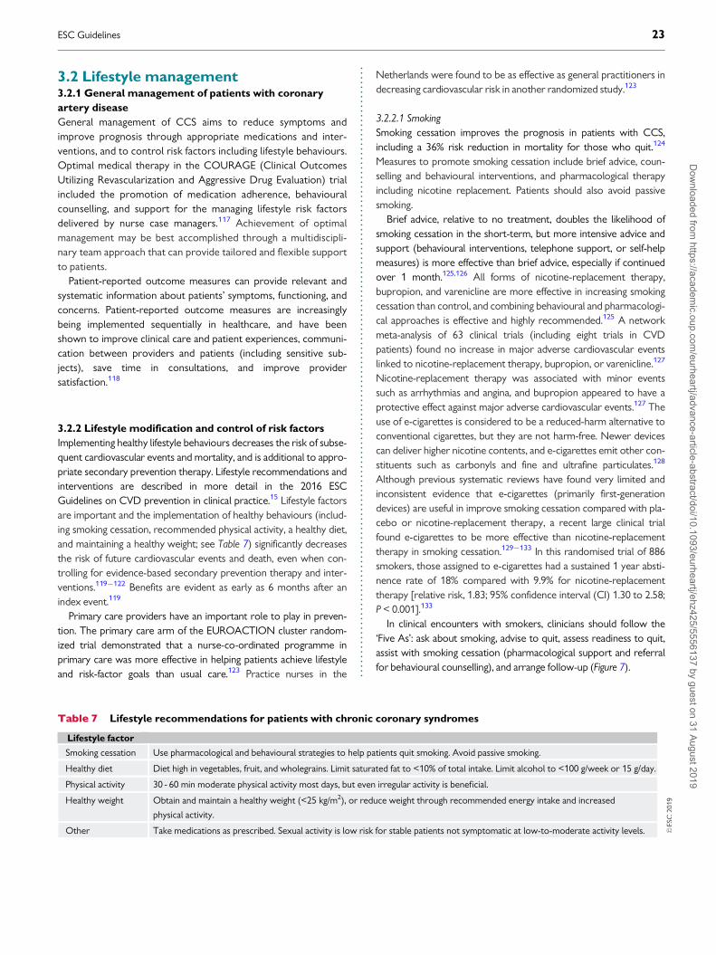
..
..
..
..
..
..
..
..
..
..
..
..
..
..
..
..
..
..
..
..
..
..
..
..
..
..
..
..
..
..
..
..
..
..
..
..
..
..
..
..
..
..
..
..
..
..
..
..
..
..
..
..
..
..
..
..
..
..
..
..
..
..
..
..
.3.2 Lifestyle management3.2.1 General management of patients with coronary
artery disease
General management of CCS aims to reduce symptoms andimprove prognosis through appropriate medications and inter-ventions, and to control risk factors including lifestyle behaviours.Optimal medical therapy in the COURAGE (Clinical OutcomesUtilizing Revascularization and Aggressive Drug Evaluation) trialincluded the promotion of medication adherence, behaviouralcounselling, and support for the managing lifestyle risk factorsdelivered by nurse case managers.117 Achievement of optimalmanagement may be best accomplished through a multidiscipli-nary team approach that can provide tailored and flexible supportto patients.
Patient-reported outcome measures can provide relevant andsystematic information about patients’ symptoms, functioning, andconcerns. Patient-reported outcome measures are increasinglybeing implemented sequentially in healthcare, and have beenshown to improve clinical care and patient experiences, communi-cation between providers and patients (including sensitive sub-jects), save time in consultations, and improve providersatisfaction.118
3.2.2 Lifestyle modification and control of risk factors
Implementing healthy lifestyle behaviours decreases the risk of subse-quent cardiovascular events and mortality, and is additional to appro-priate secondary prevention therapy. Lifestyle recommendations andinterventions are described in more detail in the 2016 ESCGuidelines on CVD prevention in clinical practice.15 Lifestyle factorsare important and the implementation of healthy behaviours (includ-ing smoking cessation, recommended physical activity, a healthy diet,and maintaining a healthy weight; see Table 7) significantly decreasesthe risk of future cardiovascular events and death, even when con-trolling for evidence-based secondary prevention therapy and inter-ventions.119�122 Benefits are evident as early as 6 months after anindex event.119
Primary care providers have an important role to play in preven-tion. The primary care arm of the EUROACTION cluster random-ized trial demonstrated that a nurse-co-ordinated programme inprimary care was more effective in helping patients achieve lifestyleand risk-factor goals than usual care.123 Practice nurses in the
Netherlands were found to be as effective as general practitioners indecreasing cardiovascular risk in another randomized study.123
3.2.2.1 SmokingSmoking cessation improves the prognosis in patients with CCS,including a 36% risk reduction in mortality for those who quit.124
Measures to promote smoking cessation include brief advice, coun-selling and behavioural interventions, and pharmacological therapyincluding nicotine replacement. Patients should also avoid passivesmoking.
Brief advice, relative to no treatment, doubles the likelihood ofsmoking cessation in the short-term, but more intensive advice andsupport (behavioural interventions, telephone support, or self-helpmeasures) is more effective than brief advice, especially if continuedover 1 month.125,126 All forms of nicotine-replacement therapy,bupropion, and varenicline are more effective in increasing smokingcessation than control, and combining behavioural and pharmacologi-cal approaches is effective and highly recommended.125 A networkmeta-analysis of 63 clinical trials (including eight trials in CVDpatients) found no increase in major adverse cardiovascular eventslinked to nicotine-replacement therapy, bupropion, or varenicline.127
Nicotine-replacement therapy was associated with minor eventssuch as arrhythmias and angina, and bupropion appeared to have aprotective effect against major adverse cardiovascular events.127 Theuse of e-cigarettes is considered to be a reduced-harm alternative toconventional cigarettes, but they are not harm-free. Newer devicescan deliver higher nicotine contents, and e-cigarettes emit other con-stituents such as carbonyls and fine and ultrafine particulates.128
Although previous systematic reviews have found very limited andinconsistent evidence that e-cigarettes (primarily first-generationdevices) are useful in improve smoking cessation compared with pla-cebo or nicotine-replacement therapy, a recent large clinical trialfound e-cigarettes to be more effective than nicotine-replacementtherapy in smoking cessation.129�133 In this randomised trial of 886smokers, those assigned to e-cigarettes had a sustained 1 year absti-nence rate of 18% compared with 9.9% for nicotine-replacementtherapy [relative risk, 1.83; 95% confidence interval (CI) 1.30 to 2.58;P < 0.001].133
In clinical encounters with smokers, clinicians should follow the‘Five As’: ask about smoking, advise to quit, assess readiness to quit,assist with smoking cessation (pharmacological support and referralfor behavioural counselling), and arrange follow-up (Figure 7).
Table 7 Lifestyle recommendations for patients with chronic coronary syndromes
Lifestyle factor
Smoking cessation Use pharmacological and behavioural strategies to help patients quit smoking. Avoid passive smoking.
Healthy diet Diet high in vegetables, fruit, and wholegrains. Limit saturated fat to <10% of total intake. Limit alcohol to <100 g/week or 15 g/day.
Physical activity 30 - 60 min moderate physical activity most days, but even irregular activity is beneficial.
Healthy weight Obtain and maintain a healthy weight (<25 kg/m2), or reduce weight through recommended energy intake and increased
physical activity.
Other Take medications as prescribed. Sexual activity is low risk for stable patients not symptomatic at low-to-moderate activity levels.
ESC Guidelines 23D
ownloaded from
https://academic.oup.com
/eurheartj/advance-article-abstract/doi/10.1093/eurheartj/ehz425/5556137 by guest on 31 August 2019
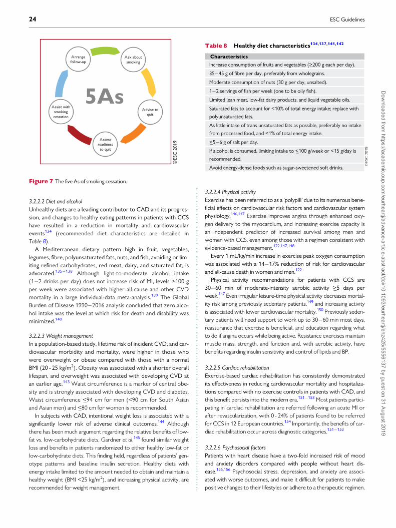
..
..
..
..
..
..
..
..
..
..
..
..
..
..
..
..
..
..
..
..
..
..
..
..
..
..
..
..
..
..
..
..
..
..
..
..
..
..
..
..
..
..
..
..
..
..
..
..
..
..
..
..
..
..
..
..
..
..
3.2.2.2 Diet and alcoholUnhealthy diets are a leading contributor to CAD and its progres-sion, and changes to healthy eating patterns in patients with CCShave resulted in a reduction in mortality and cardiovascularevents134 (recommended diet characteristics are detailed inTable 8).
A Mediterranean dietary pattern high in fruit, vegetables,legumes, fibre, polyunsaturated fats, nuts, and fish, avoiding or lim-iting refined carbohydrates, red meat, dairy, and saturated fat, isadvocated.135�138 Although light-to-moderate alcohol intake(1�2 drinks per day) does not increase risk of MI, levels >100 gper week were associated with higher all-cause and other CVDmortality in a large individual-data meta-analysis.139 The GlobalBurden of Disease 1990�2016 analysis concluded that zero alco-hol intake was the level at which risk for death and disability wasminimized.140
3.2.2.3 Weight managementIn a population-based study, lifetime risk of incident CVD, and car-diovascular morbidity and mortality, were higher in those whowere overweight or obese compared with those with a normalBMI (20 - 25 kg/m2). Obesity was associated with a shorter overalllifespan, and overweight was associated with developing CVD atan earlier age.143 Waist circumference is a marker of central obe-sity and is strongly associated with developing CVD and diabetes.Waist circumference <_94 cm for men (<90 cm for South Asianand Asian men) and <_80 cm for women is recommended.
In subjects with CAD, intentional weight loss is associated with asignificantly lower risk of adverse clinical outcomes.144 Althoughthere has been much argument regarding the relative benefits of low-fat vs. low-carbohydrate diets, Gardner et al.145 found similar weightloss and benefits in patients randomized to either healthy low-fat orlow-carbohydrate diets. This finding held, regardless of patients’ gen-otype patterns and baseline insulin secretion. Healthy diets withenergy intake limited to the amount needed to obtain and maintain ahealthy weight (BMI <25 kg/m2), and increasing physical activity, arerecommended for weight management.
3.2.2.4 Physical activityExercise has been referred to as a ‘polypill’ due to its numerous bene-ficial effects on cardiovascular risk factors and cardiovascular systemphysiology.146,147 Exercise improves angina through enhanced oxy-gen delivery to the myocardium, and increasing exercise capacity isan independent predictor of increased survival among men andwomen with CCS, even among those with a regimen consistent withevidence-based management.122,147,148
Every 1 mL/kg/min increase in exercise peak oxygen consumptionwas associated with a 14�17% reduction of risk for cardiovascularand all-cause death in women and men.122
Physical activity recommendations for patients with CCS are30�60 min of moderate-intensity aerobic activity >_5 days perweek.147 Even irregular leisure-time physical activity decreases mortal-ity risk among previously sedentary patients,149 and increasing activityis associated with lower cardiovascular mortality.150 Previously seden-tary patients will need support to work up to 30�60 min most days,reassurance that exercise is beneficial, and education regarding whatto do if angina occurs while being active. Resistance exercises maintainmuscle mass, strength, and function and, with aerobic activity, havebenefits regarding insulin sensitivity and control of lipids and BP.
3.2.2.5 Cardiac rehabilitationExercise-based cardiac rehabilitation has consistently demonstratedits effectiveness in reducing cardiovascular mortality and hospitaliza-tions compared with no exercise controls in patients with CAD, andthis benefit persists into the modern era.151�153 Most patients partici-pating in cardiac rehabilitation are referred following an acute MI orafter revascularization, with 0 - 24% of patients found to be referredfor CCS in 12 European countries.154 Importantly, the benefits of car-diac rehabilitation occur across diagnostic categories.151�153
3.2.2.6 Psychosocial factorsPatients with heart disease have a two-fold increased risk of moodand anxiety disorders compared with people without heart dis-ease.155,156 Psychosocial stress, depression, and anxiety are associ-ated with worse outcomes, and make it difficult for patients to makepositive changes to their lifestyles or adhere to a therapeutic regimen.
©ES
C 2
019
Assist with smoking cessation
Arrange follow-up
Advise to quit
Assess readiness to quit
Ask about smoking
5As
Figure 7 The five As of smoking cessation.
Table 8 Healthy diet characteristics134,137,141,142
Characteristics
Increase consumption of fruits and vegetables (>_200 g each per day).
35�45 g of fibre per day, preferably from wholegrains.
Moderate consumption of nuts (30 g per day, unsalted).
1�2 servings of fish per week (one to be oily fish).
Limited lean meat, low-fat dairy products, and liquid vegetable oils.
Saturated fats to account for <10% of total energy intake; replace with
polyunsaturated fats.
As little intake of trans unsaturated fats as possible, preferably no intake
from processed food, and <1% of total energy intake.
<_5�6 g of salt per day.
If alcohol is consumed, limiting intake to <_100 g/week or <15 g/day is
recommended.
Avoid energy-dense foods such as sugar-sweetened soft drinks.
24 ESC GuidelinesD
ownloaded from
https://academic.oup.com
/eurheartj/advance-article-abstract/doi/10.1093/eurheartj/ehz425/5556137 by guest on 31 August 2019

..
..
..
..
..
..
..
..
..
..
..
..
..
..
..
..
..
..
..
..
..
..
..
..
..
..
..
..
..
..
..
..
..
..
..
..
..
..
..
..
..
..
.The ESC Prevention Guidelines recommend assessment for psycho-social risk factors.15 Clinical trials have shown that psychological (e.g.counselling and/or cognitive behavioural therapy) and pharmacologi-cal interventions have a beneficial effect on depression, anxiety, andstress, with some evidence of a reduction in cardiac mortality andevents compared with placebo.157�159
3.2.2.7 Environmental factorsAir pollutants are estimated to be one of the 10 leading risk factors forglobal mortality. Exposure to air pollution increases risk of MI, as wellas hospitalization and death from heart failure, stroke, and arrhyth-mia.160 Patients with CCS should avoid heavily traffic-congested areas.Air purifiers with high-efficiency particulate air (‘HEPA’) filters reduceindoor pollution, and wearing N95 respirator face masks in heavily pol-luted areas has been shown to be protective.160 Environmental noisealso increases the risk of CVD.161 Policies and regulations that reduceair pollution and environmental noise should be supported, andpatients should be advised about these risks.
3.2.2.8 Sexual activityPatients with CCS often worry about the cardiovascular risk associ-ated with sexual activity and/or experience sexual dysfunction.162
The risk of triggering sudden death or an acute MI is very low, espe-cially when sexual activity is with a stable partner in a familiar environ-ment without stress, or excessive intake of food or alcoholbeforehand.163,164 Although sexual activity transiently increases therisk of MI, it is the cause of <1% of acute MIs and <1�1.7% of suddendeaths occur during sexual activity.164 The energy expenditure duringsexual activity is generally low-to-moderate (3 - 5 metabolic equiva-lents) and climbing two flights of stairs is often used as an equivalentactivity in terms of energy expended.163,164 Regular physical activitydecreases the risk of adverse events during sexual activity.165 Sexualdysfunction in patients with CCS includes decreased libido and sexualactivity, and a high prevalence of erectile dysfunction. Sexual dysfunc-tion may be caused by underlying vascular conditions, psychosocialfactors, specific medications, number of medications, and changes inrelationships.166 Thiazide diuretics and beta-blockers (except nebivo-lol) may negatively influence erectile function, but studies publishedsince 2011 have not found a consistent relationship between mostcontemporary cardiovascular medications and erectile dysfunc-tion.162,164,165 Phosphodiesterase-5 inhibitors to treat erectile dys-function are generally safe in CCS patients, but should not be used inthose taking nitrates.164 Healthcare providers should ask patientsabout sexual activity, and offer advice and counselling.
3.2.2.9 Adherence and sustainabilityAdherence to lifestyle modifications and to medications is a chal-lenge. A systematic review of epidemiological studies indicated that asubstantial proportion of patients do not adhere to cardiovascularmedications, and that 9% of cardiovascular events in Europe wereattributable to poor adherence.167 In older men with ischaemic heartdisease, greater adherence to medication guidelines appears to bepositively associated with better clinical outcomes, independent ofother conditions.168 Polypharmacy plays a negative role in adherenceto treatment,169 and complexity of drug regimen is associated with
non-adherence and higher rates of hospitalizations.170 Drug prescrip-tions should prioritize medications that have proved their benefitwith the highest level of evidence and those for whom the amplitudeof benefit is largest. Simplifying medication regimens may help, andthere is some evidence of benefits of cognitive educational strategies,electronically monitored feedback, and support by nurse case manag-ers. Medication reviews by primary care providers may be helpful inpatients with multiple comorbidities to minimize the risk of adverseinteractions and to simplify medication regimens.117,171�173
Promoting behaviour change and medication adherence should bepart of each clinical encounter in primary care and specialist follow-up,emphasising its importance, referring for support when needed, and con-gratulating patients for their achievements. Long-term support (intensivein the first 6 months, then every 6 months for 3 years) in the GOSPEL(Global secondary prevention strategies to limit event recurrence aftermyocardial infarction) trial resulted in significant improvements in riskfactors, and decreases in several clinical mortality and morbidity end-points.121 The Multicenter Lifestyle Demonstration Project showed thatCCS patients could make intensive lifestyle changes and improve theirrisk factors and fitness, with changes sustained at 12 months.174
3.2.2.10 Influenza vaccinationAn annual influenza vaccination can improve prevention of acute MIin patients with CCS,175,176 change HF prognosis,177 and decreasecardiovascular mortality in adults aged >_65 years.178�180 Therefore,annual influenza vaccination is recommended for patients with CAD,especially in the elderly.
Recommendations on lifestyle management
Recommendations Classa Levelb
Improvement of lifestyle factors in addition
to appropriate pharmacological management is
recommended.119�122,124,148�153
I A
Cognitive behavioural interventions are rec-
ommended to help individuals achieve a
healthy lifestyle.181�183
I A
Exercise-based cardiac rehabilitation is recom-
mended as an effective means for patients
with CCS to achieve a healthy lifestyle and
manage risk factors.151�153
I A
Involvement of multidisciplinary healthcare pro-
fessionals (e.g. cardiologists, GPs, nurses, dieti-
cians, physiotherapists, psychologists, and
pharmacists) is recommended.121,123,181,184
I A
Psychological interventions are recommended
to improve symptoms of depression in
patients with CCS.126,157
I B
Annual influenza vaccination is recommended
for patients with CCS, especially in the
elderly.175,176,178,179,185�187
I B
CCS = chronic coronary syndrome; GPs = general practitioners.aClass of recommendation.bLevel of evidence.
ESC Guidelines 25D
ownloaded from
https://academic.oup.com
/eurheartj/advance-article-abstract/doi/10.1093/eurheartj/ehz425/5556137 by guest on 31 August 2019
..
..
..
..
..
..
..
..
..
..
..
..
..
..
..
..
..
..
..
..
..
..
..
..
..
..
..
..
..
..
..
..
..
..
..
..
..
..
..
..
..
..
..
..
..
..
..
..
..
..
..
..
..
..
..
..
..
..
..
..
..
..
..
..
..
..
..
..
..
..
..
..
..
..
..
..
..
..
..
..
..
..
..
..
..
..
.3.3 Pharmacological managementThe aims of pharmacological management of CCS patients are toreduce angina symptoms and exercise-induced ischaemia, and to pre-vent cardiovascular events.
Immediate relief of anginal symptoms, or the prevention of symp-toms under circumstances likely to elicit angina, is usually obtainedwith rapidly acting formulations of nitroglycerin. Anti-ischaemicdrugs—but also lifestyle changes, regular exercise training, patienteducation, and revascularization—all play a role in minimizing oreradicating symptoms over the long-term (long-term prevention).
Prevention of cardiovascular events targets MI and death associ-ated with CAD, and focuses primarily on reducing the incidence ofacute thrombotic events and the development of ventricular dysfunc-tion. Strategies include pharmacological and lifestyle interventions, asdetailed in the 2016 European Guidelines on CVD prevention in clini-cal practice.15
3.3.1 Anti-ischaemic drugs
3.3.1.1 General strategyOptimal treatment can be defined as the treatment that satisfactorilycontrols symptoms and prevents cardiac events associated with CCS,with maximal patient adherence and minimal adverse events.188�191
However, there is no universal definition of an optimal treatment inpatients with CCS, and drug therapies must be adapted to eachpatient’s characteristics and preferences.192 Initial drug therapy usuallyconsists of one or two antianginal drugs, as necessary, plus drugs forsecondary prevention of CVD.193 The initial choice of antianginaldrug(s) depends on the expected tolerance related to the individualpatient’s profile and comorbidities, potential drug interactions with co-administered therapies, the patient’s preferences after being informedof potential adverse effects, and drug availability. Whether combinationtherapy with two antianginal drugs [e.g. a beta-blocker and a calciumchannel blocker (CCB)] is superior to monotherapy with any class ofantianginal drug in reducing clinical events remains unclear.194�197
Beta-adrenergic blockers or CCBs are recommended as the firstchoice, although no RCT to date has compared this strategy to analternative strategy using initial prescription of other anti-ischaemicdrugs, or the combination of a beta-blocker and a CCB.191,195 Theresults of a network meta-analysis of 46 studies and 71 treatment com-parisons supported the initial combination of a beta-blocker and aCCB.198 The same meta-analysis suggested that several second-lineadd-on anti-ischaemic drugs (long-acting nitrates, ranolazine, trimetazi-dine, and, to a lesser extent, ivabradine) may prove beneficial in combi-nation with a beta-blocker or a CCB as first-line therapy, while no datawere available for nicorandil. However, it should be noted that thestudy pooled RCTs using endpoints of nitrate use, angina frequency,time to angina or to ST-segment depression, and total exercise time,and no study or meta-analysis has yet assessed with sufficient powerthe influence of combining a beta-blocker or a CCB with a second-lineanti-ischaemic drug on morbidity or mortality events.198 Regardless ofthe initial strategy, response to initial antianginal therapy should bereassessed after 2�4 weeks of treatment initiation.
3.3.1.2 Available drugsAnti-ischaemic drugs have proved benefits regarding symptoms asso-ciated with myocardial ischaemia but do not prevent cardiovascular
events in most patients with CCS. Supplementary Table 3 in theSupplementary Data summarizes the principal major side effects,contraindications, drug�drug interactions, and precautions relatingto anti-ischaemic drugs. Supplementary Table 2 summarizes the mainmechanisms of action of anti-ischaemic drugs.
3.3.1.2.1 Nitrates.
Short-acting nitrates for acute effort anginaSublingual and spray nitroglycerin formulations provide immediate
relief of effort angina. Spray nitroglycerin acts more rapidly than sub-lingual nitroglycerin.199 At the onset of angina symptoms, the patientshould rest in a sitting position (standing promotes syncope, and lyingdown enhances venous return and preload) and take nitroglycerin(0.3�0.6 mg tablet sublingually and not swallowed, or 0.4 mg sprayto the tongue and not swallowed or inhaled) every 5 min until thepain disappears, or a maximum of 1.2 mg has been taken within 15min. During this time frame, if angina persists, immediate medicalattention is needed. Nitroglycerin can be administered for prophy-laxis before physical activities known to provoke angina. Isosorbidedinitrate (5 mg sublingually) has a slightly slower onset of action thannitroglycerin due to hepatic conversion to mononitrate. The effect ofisosorbide dinitrate may last <_1 h if the drug is taken sublingually orpersist for several hours if the drug is taken by oral ingestion.
Long-acting nitrates for angina prophylaxisLong-acting nitrate formulations (e.g. nitroglycerin, isosorbide dini-
trate, and isosorbide mononitrate) should be considered as second-line therapy for angina relief when initial therapy with a beta-blocker ornon-dihydropyridine (non-DHP) CCB is contraindicated, poorly toler-ated, or insufficient to control symptoms. In fact, there is a paucity ofdata comparing nitrates with beta-blockers or CCB from which todraw firm conclusions about their relative efficacies.200 When takenover a prolonged period, long-acting nitrates provoke tolerance withloss of efficacy, which requires prescription of a nitrate-free or nitrate-low interval of �10�14 h.201 Nitroglycerin can be administered orallyor transdermally through slow-release patch systems. Bioavailability ofisosorbide dinitrate depends on the interindividual variability in hepaticconversion and is generally lower than the bioavailability of isosorbidemononitrate (its active metabolite), which is 100% bioavailable.Titration of dose is essential with all formulations to obtain the maximalcontrol of symptoms at a tolerable dose. Discontinuation should betapered and not abrupt to avoid a rebound increase in angina.202 Themost common side effects are hypotension, headache, and flushing.Contraindications include hypertrophic obstructive cardiomyopathy,severe aortic valvular stenosis, and co-administration of phosphodies-terase inhibitors (e.g. sildenafil, tadalafil, or vardenafil) or riociguat.
3.3.1.2.2 Beta-blockers. The dose of beta-blockers should be adjustedto limit the heart rate to 55�60 b.p.m. (beats per minute) atrest.203,204 Discontinuation should be tapered and not abrupt. Beta-blockers can be combined with DHP CCBs to reduce DHP-inducedtachycardia, but with uncertain incremental clinical value.205�208
Caution is warranted when a beta-blocker is combined with verapa-mil or diltiazem due to the potential for developing worsening of HF,excessive bradycardia, and/or atrioventricular block. Combination ofa beta-blocker with a nitrate attenuates the reflex tachycardia ofthe latter. The principal side effects of beta-blockers are fatigue,depression, bradycardia, heart block, bronchospasm, peripheral
26 ESC GuidelinesD
ownloaded from
https://academic.oup.com
/eurheartj/advance-article-abstract/doi/10.1093/eurheartj/ehz425/5556137 by guest on 31 August 2019
..
..
..
..
..
..
..
..
..
..
..
..
..
..
..
..
..
..
..
..
..
..
..
..
..
..
..
..
..
..
..
..
..
..
..
..
..
..
..
..
..
..
..
..
..
..
..
..
..
..
..
..
..
..
..
..
..
..
..
..
..
..
..
..
..
..
..
..
..
..
..
..
..
..
..
..
..
..
..
..
..
..
..
..
..
..
.vasoconstriction, postural hypotension, impotence, and masking ofhypoglycaemia symptoms.
In certain patients with recent MI and those with chronic HF withreduced ejection fraction, beta-blockers have been associated with asignificant reduction in mortality and/or cardiovascular events,209�215
but the protective benefit in patients with CAD without prior MI orHF is less well established and lacks placebo-controlled trials.216 Aretrospective analysis of 21 860 matched patients from the REACH(REduction of Atherothrombosis for Continued Health) Registryshowed no reduction in cardiovascular mortality with beta-blockersin patients with either CAD with risk factors only, known prior MI, orknown CAD without MI.217 In a retrospective national registry of755 215 patients aged >_65 years with a history of CAD without priorMI or HF with reduced ejection fraction undergoing elective PCI,beta-blocker use at discharge was not associated with any reductionin cardiovascular morbidity or mortality at 30 day and 3 year follow-up.218 However, in patients with or without previous MI undergoingCABG, beta-blockers were associated with lower risk of long-termmortality and adverse cardiovascular events.219 Other observationalstudies and meta-analyses have questioned the benefit of long-term(>1 year) beta-blocker therapy in patients with a previousMI.216,220�224 This is still a matter for debate,225 and uncertaintiesremain on the comparative role of beta-blockers and angiotensin-converting enzyme (ACE) inhibitors.
3.3.1.2.3 Calcium channel blockers. While CCBs improve symptomsand myocardial ischaemia, they have not been shown to reducemajor morbidity endpoints or mortality in patients withCCS.192,226�228
NON-DIHYDROPYRIDINE AGENTS (HEART RATE-LOWERING CALCIUM CHANNEL
BLOCKERS)Verapamil. Verapamil has a large range of approved indications,including all varieties of angina (effort, vasospastic, and unstable),supraventricular tachycardias, and hypertension. Indirect evidencesuggests good safety but with risks of heart block, bradycardia, andHF. Compared with metoprolol, the antianginal activity was simi-lar.229 Compared with atenonol in hypertension with CAD, verapa-mil is associated with fewer cases of diabetes, fewer anginalattacks,230 and less psychological depression.231 Beta-blockade com-bined with verapamil is not advised (due to risk of heart block).
Diltiazem. Diltiazem, with its low-side effect profile, has advantagescompared with verapamil in the treatment of effort angina. Like vera-pamil, it acts by peripheral vasodilation, relief of exercise-inducedcoronary constriction, a modest negative inotropic effect, and sinusnode inhibition. There have been no outcome studies comparing dil-tiazem and verapamil.
In some selected patients, non-DHP agents may be combined withbeta-blockers for the treatment of angina. However, on such occa-sions they must be used under close monitoring of patients’ toleranceregarding excessive bradycardia or signs of HF. Use of non-DHPCCBs in patients with LV dysfunction is not advised.
DIHYDROPYRIDINE AGENTS
Long-acting nifedipine. This agent is a powerful arterial vasodilator withfew serious side effects. Long-acting nifedipine has been especiallywell tested in hypertensive anginal patients when added to beta-
blockade.232 In the large placebo-controlled ACTION (A Coronarydisease Trial Investigating Outcome with Nifedipine gastrointestinaltherapeutic system) trial, addition of long-acting nifedipine [60 mgo.d. (once a day)] to conventional treatment of angina had no effecton major cardiovascular event-free survival. Long-acting nifedipineproved to be safe, and reduced the need for coronary angiographyand cardiovascular interventions.232 Relative contraindications tonifedipine are few (severe aortic stenosis, hypertrophic obstructivecardiomyopathy, or HF), and careful combination with beta-blockadeis usually feasible and desirable. Vasodilatory side effects includeheadache and ankle oedema.
Amlodipine. The very long half-life of amlodipine and its good toler-ability make it an effective once-a-day antianginal and antihyperten-sive agent, setting it apart from drugs that are taken either twice orthree times daily. Side effects are few, mainly ankle oedema. Inpatients with CCS and normal BP (�75% receiving a beta-blocker),amlodipine 10 mg/day reduced coronary revascularizations and hos-pitalizations for angina in a 24 month trial.233 Exercise-induced ischae-mia is more effectively reduced by amlodipine, 5 mg titrated to10 mg/day, than by the beta-blocker atenolol, 50 mg/day, and theircombination is even better.234 However, the CCB�beta-blockercombination is often underused, even in some studies reporting ‘opti-mally treated’ stable effort angina.
3.3.1.2.4 Ivabradine. Ivabradine has been reported to be non-inferiorto atenolol or amlodipine in the treatment of angina and ischaemia inpatients with CCS.235,236 Adding ivabradine 7.5 mg b.i.d. [bis in die(twice a day)] to atenolol therapy gave better control of heart rateand anginal symptoms.237 In 10 917 patients with limiting previousangina enrolled in the morbidity�mortality evaluation of theBEAUTIFUL (If Inhibitor Ivabradine in Patients With Coronary ArteryDisease and Left Ventricular Dysfunction) trial, ivabradine did notreduce the composite primary endpoint of cardiovascular death, hos-pitalization with MI, or HF.238 Also, in the SIGNIFY (Study Assessingthe Morbidity�Mortality Benefits of the If Inhibitor Ivabradine inPatients with Coronary Artery Disease) study, consisting of 19 102patients with CAD without clinical HF and a heart rate >_70 b.p.m.,there was no significant difference between the ivabradine group andthe placebo group in the incidence of the primary composite end-point of death from cardiovascular causes or non-fatal MI.239
Ivabradine was associated with an increase in the incidence of the pri-mary endpoint among 12 049 patients with activity-limiting angina butnot among those without activity-limiting angina (P=0.02 for interac-tion). In 2014, the European Medicines Agency issued recommenda-tions to reduce the risk of bradycardia and placed ivabradine underadditional monitoring.240 In aggregate, these results support the useof ivabradine as a second-line drug in patients with CCS.
3.3.1.2.5 Nicorandil. Nicorandil is a nitrate derivative of nicotinamide,with antianginal effects similar to those of nitrates or beta-block-ers.241�244 Side effects include nausea, vomiting, and potentiallysevere oral, intestinal, and mucosal ulcerations.245
In the placebo-controlled IONA (Impact Of Nicorandil in Angina)trial (n = 5126), nicorandil significantly reduced the composite of cor-onary heart disease (CHD) death, non-fatal MI, or unplanned hospitaladmission for suspected anginal symptoms in patients with CCS, butthere was no effect on death from ischaemic heart disease or non-
ESC Guidelines 27D
ownloaded from
https://academic.oup.com
/eurheartj/advance-article-abstract/doi/10.1093/eurheartj/ehz425/5556137 by guest on 31 August 2019

..
..
..
..
..
..
..
..
..
..
..
..
..
..
..
..
..
..
..
..
..
..
..
..
..
..
..
..
..
..
..
..
.fatal MI.246 These results support the use of nicorandil as a second-line drug in patients with CCS.
3.3.1.2.6 Ranolazine. Ranolazine is a selective inhibitor of the lateinward sodium current. Side effects include dizziness, nausea, andconstipation. In addition, ranolazine increases QTc, and should there-fore be used carefully in patients with QT prolongation or on QT-prolonging drugs.
In a placebo-controlled trial of 6560 patients with non-ST-segmentelevation ACS, the addition of ranolazine to standard treatment didnot prove effective in reducing the primary efficacy endpoint of cardi-ovascular death, MI, or recurrent ischaemia.247 However, in the rela-tively large subgroup of patients with chronic angina (n = 3565),significant reductions in recurrent ischaemia, worsening angina, andintensification of antianginal therapy were observed.248 In anotherplacebo-controlled trial of patients with diabetes and CAD receivingone or two antianginal drugs, ranolazine reduced angina and sublin-gual nitroglycerin use with good tolerability.249 In the RIVER-PCI(Ranolazine for Incomplete Vessel Revascularization Post-Percutaneous Coronary Intervention) trial, ranolazine did not reducethe composite of ischaemia-driven revascularization or
hospitalization without revascularization in 2651 patients with a his-tory of chronic angina and incomplete revascularization after PCI,including those with and without PCI for a CAD indication, nor did itreduce angina symptoms at 1 year.250,251
These results support the use of ranolazine as a second-line drugin CCS patients with refractory angina despite commonly used anti-anginal agents such as beta-blockers, CCBs, and/or long-actingnitrates. Conversely, there is a lack of evidence to support the use ofranolazine in patients with CCS following PCI with incompleterevascularization.
3.3.1.2.7 Trimetazidine. Trimetazidine appears to have a haemodynami-cally neutral side effect profile.252 Trimetazidine (35 mg b.i.d.) added tobeta-blockade (atenolol) improved effort-induced myocardial ischae-mia, as reviewed by the European Medicines Agency in June2012.253,254 It remains contraindicated in Parkinson’s disease andmotion disorders, such as tremor (shaking), muscle rigidity, walking dis-orders, and restless leg syndrome. A 2014 meta-analysis of 13, mostlyChinese, studies consisting of 1628 patients showed that treatmentwith trimetazidine on top of other antianginal drugs was associatedwith a smaller weekly mean number of angina attacks, lower weekly
©ES
C 2
019
StandardTherapy
High heart rate(e.g. >80 b.p.m)
Low heart rate(e.g. <50 b.p.m)
LV dysfunctionor heart failure
Low bloodpressure
BB or CCBa1st stepBB or
non-DHP-CCB DHP-CCB BBLow-dose BB or Low-dose non-
DHP-CCB
BB + DHP-CCB2nd stepBB and
non-DHP-CCB LANAdd LAN orivabradine
Add low-dose LAN
Add 2nd line drug3rd step Add ivabradine DHP-CCB + LANAdd another 2nd line drug
Add ivabradine,ranolazine, ortrimetazidine
4th stepAdd nicorandil, ranolazine ortrimetazidine
Figure 8 Suggested stepwise strategy for long-term anti-ischaemic drug therapy in patients with chronic coronary syndromes and specific baseline char-acteristics. BB = beta-blocker; b.p.m. = beats per minute; CCB = [any class of] calcium channel blocker; DHP-CCB = dihydropyridine calcium channelblocker; HF = heart failure; LAN = long-acting nitrate; LV = left ventricular; NDHP-CCB: non-dihydropyridine calcium channel blocker. aCombination of aBB with a DHP-CCB should be considered as a first step; combination of a BB or a CCB with a second-line drug may be considered as a first step.
28 ESC GuidelinesD
ownloaded from
https://academic.oup.com
/eurheartj/advance-article-abstract/doi/10.1093/eurheartj/ehz425/5556137 by guest on 31 August 2019

..
..
..
..
..
..
..
..
..
..
..
..
..
..
..
..
..
..
..
..
..
..
..
..
..
..
..
..
..
..
..
.nitroglycerin use, longer time to 1 mm ST-segment depression,higher total work, and longer exercise duration at peak exercise thantreatment with the other antianginal drugs for stable angina pecto-ris.255 These results support the use of trimetazidine as a second-linedrug in patients with CCS whose symptoms are not adequately con-trolled by, or who are intolerant to, other medicines for anginapectoris.
3.3.1.2.8 Allopurinol. In 2010, a randomized crossover study of 65patients with CAD showed that allopurinol 600 mg/day increasedtimes to ST-segment depression and to angina.256 An observationalstudy of 29 298 episodes of incident allopurinol use found an associa-tion of allopurinol use with a reduction in the risk of incident MI inthe elderly, particularly when used for >2 years.257 However, therole of allopurinol in reducing clinical events in CVD remainsunclear.258
A stepwise strategy for anti-ischaemic drug therapy in CCS is pro-posed, depending on some baseline patient characteristics (Figure 8).Incomplete responses or poor tolerance at each step justify movingto the next step. The strategy must be adapted to each patient’s
characteristics and preferences, and does not necessarily follow thesteps indicated in the figure.
3.3.1.3 Patients with low blood pressureIn patients with low BP, it is recommended to start antianginal drugs atvery low doses, with preferential use of drugs with no or limited effectson BP. A low-dose beta-blocker or low-dose non-DHP-CCB can betested first under close monitoring of tolerance. Ivabradine (in patientswith sinus rhythm), ranolazine, or trimetazidine can also be used.
3.3.1.4 Patients with low heart rateIncreased heart rate correlates linearly with cardiovascularevents, and the benefit of heart-rate reduction as a treatmentgoal in subgroups of CCS patients has been demonstrated usingvarious drugs.203,259�261 However, in patients with baseline bra-dycardia (e.g. heart rate <60 b.p.m.) heart rate-lowering drugs(beta-blockers, ivabradine, and heart-rate lowering CCBs) shouldbe avoided or used with caution, and—if needed—started at verylow doses. Antianginal drugs without heart rate-lowering effectsshould preferably be given.
Recommendations on anti-ischaemic drugs in patients with chronic coronary syndromes
Recommendations Classa Levelb
General considerations
Medical treatment of symptomatic patients requires one or more drug(s) for angina/ischaemia relief in association with
drug(s) for event prevention.I C
It is recommended that patients are educated about the disease, risk factors, and treatment strategy. I C
Timely review of the patient’s response to medical therapies (e.g. 2�4 weeks after drug initiation) is recommended.262 I C
Angina/ischaemiac relief
Short-acting nitrates are recommended for immediate relief of effort angina.195,263 I B
First-line treatment is indicated with beta-blockers and/or CCBs to control heart rate and symptoms.205,264 I A
If angina symptoms are not successfully controlled on a beta-blocker or a CCB, the combination of a beta-blocker with a
DHP-CCB should be considered.IIa C
Initial first-line treatment with the combination of a beta-blocker and a DHP-CCB should be considered.194,198,264 IIa B
Long-acting nitrates should be considered as a second-line treatment option when initial therapy with a beta-blocker and/or a
non-DHP-CCB is contraindicated, poorly tolerated, or inadequate to control angina symptoms.200,201 IIa B
When long-acting nitrates are prescribed, a nitrate-free or low-nitrate interval should be considered to reduce tolerance.201 IIa B
Nicorandil,241�244,246 ranolazine,248,265 ivabradine,235�237 or trimetazidine252,255 should be considered as a second-line treat-
ment to reduce angina frequency and improve exercise tolerance in subjects who cannot tolerate, have contraindications to,
or whose symptoms are not adequately controlled by beta-blockers, CCBs, and long-acting nitrates.
IIa B
In subjects with baseline low heart rate and low BP, ranolazine or trimetazidine may be considered as a first-line drug to
reduce angina frequency and improve exercise tolerance.IIb C
In selected patients, the combination of a beta-blocker or a CCB with second-line drugs (ranolazine, nicorandil, ivabradine,
and trimetazidine) may be considered for first-line treatment according to heart rate, BP, and tolerance.198 IIb B
Nitrates are not recommended in patients with hypertrophic obstructive cardiomyopathy266 or co-administration of phos-
phodiesterase inhibitors.267 III B
BP = blood pressure; CCB = calcium channel blocker; CCS = chronic coronary syndromes; DHP-CCB = dihydropyridine calcium channel blocker.aClass of recommendation.bLevel of evidence.cNo demonstration of benefit on prognosis.
ESC Guidelines 29D
ownloaded from
https://academic.oup.com
/eurheartj/advance-article-abstract/doi/10.1093/eurheartj/ehz425/5556137 by guest on 31 August 2019
..
..
..
..
..
..
..
..
..
..
..
..
..
..
..
..
..
..
..
..
..
..
..
..
..
..
..
..
..
..
..
..
..
..
..
..
..
..
..
..
..
..
..
..
..
..
..
..
..
..
..
..
..
..
..
..
..
..
..
..
..
..
..
..
..
..
..
..
..
..
..
..
..
..
..
..
..
..
..
..
..
..
..
..
..
..
.3.3.2 Event prevention
3.3.2.1 Antiplatelet drugsPlatelet activation and aggregation is the driver for symptomatic cor-onary thrombosis, forming the basis for the use of antiplatelet drugsin patients with CCS in view of a favourable balance between the pre-vention of ischaemic events and increased risk of bleeding. Dual anti-platelet therapy (DAPT) with aspirin and an oral P2Y12 inhibitor isthe mainstay of antithrombotic therapy after MI and/or PCI.
3.3.2.1.1 Low-dose aspirin. Aspirin acts via irreversible inhibition of pla-telet cyclooxygenase-1 and thus thromboxane production, which isnormally complete with chronic dosing >_75 mg/day. The gastrointes-tinal side effects of aspirin increase at higher doses, and current evi-dence supports a daily dose of 75�100 mg for the prevention ofischaemic events in CAD patients with or without a history ofMI.268�270 As cyclooxygenase-1 inhibition by aspirin is consistent andpredictable in adherent patients, no platelet function testing isrequired to monitor individual response.271 Although other non-selective non-steroidal anti-inflammatory drugs, such as ibuprofen,reversibly inhibit cyclooxygenase-1, their adverse effects on cardio-vascular risk indicate that they cannot be recommended as an alter-native treatment in patients with aspirin intolerance.272
3.3.2.1.2 Oral P2Y12 inhibitors. P2Y12 inhibitors block the platelet P2Y12
receptor, which plays a key role in platelet activation and the amplifi-cation of arterial thrombus formation. Clopidogrel and prasugrel arethienopyridine prodrugs that irreversibly block P2Y12 via activemetabolites. Ticagrelor is a reversibly-binding P2Y12 inhibitor thatdoes not require metabolic activation.
The CAPRIE (Clopidogrel vs. Aspirin in Patients at Risk ofIschaemic Events) trial showed an overall slight benefit of clopidogrelcompared with aspirin, with a similar safety profile, in preventing car-diovascular events in patients with previous MI, previous stroke, orPAD.273 Subgroup analysis suggested greater benefit of clopidogrel inpatients with PAD. Despite its lesser antiplatelet efficacy, clopidogreldemonstrated equivalent efficacy to ticagrelor in patients withPAD.274 Clopidogrel is limited by variable pharmacodynamic effectsrelated to variable efficiency of conversion to its active metabolite,partly associated with loss-of-function variants in the CYP2C19 gene,leading to a lack of efficacy in some patients.271 Drugs that inhibitCYP2C19, such as omeprazole, may reduce the response toclopidogrel.275
Prasugrel has more rapid, more predictable, and, on average,greater antiplatelet effect compared with clopidogrel, and is not sus-ceptible to drug interactions or the effect of CYP2C19 loss-of-function variants. Prasugrel has greater efficacy than clopidogrel inaspirin-treated patients with ACS undergoing PCI, but not in medically-managed patients with ACS.276,277 Prasugrel has been associated withmore non-fatal and fatal bleeding events than clopidogrel in ACSpatients undergoing PCI, leading to apparent harm in those with a his-tory of ischaemic stroke, and a lack of apparent benefit in those aged>75 years or with body weight <60 kg.276
Ticagrelor has the most predictable and consistently high level ofP2Y12 inhibition during maintenance therapy in adherent patients,219
and also has more rapid onset, as well as more rapid and predictableoffset of action compared with clopidogrel.278�280 Ticagrelor asmonotherapy appears to have similar efficacy and safety to aspirin
monotherapy in patients with previous PCI.281 Ticagrelor, with a 180mg loading dose followed by 90 mg b.i.d., achieved greater reductionof ischaemic events compared with clopidogrel in aspirin-treatedACS patients, regardless of revascularization strategy, at the expenseof more non-fatal bleeding.282,283 Ticagrelor at doses of 90 or 60 mgb.i.d. reduced the 3 year combined incidence of MI, stroke, or cardio-vascular death compared with placebo in stable aspirin-treatedpatients with a history of MI 1�3 years previously.284 Both ticagrelordoses increased non-fatal but not fatal bleeding. The equivalent effica-cies and similar safeties of the two ticagrelor doses were explainedby similar levels of platelet inhibition.285 Ticagrelor may cause dysp-noea, which is often transient and most often mild and tolerable, butoccasionally necessitates switching to a thienopyridine.286,287
Ticagrelor is metabolized via CYP3A, and consequently should not beused with strong CYP3A inhibitors or inducers.
The optimal timing of initiation of P2Y12 inhibition before coronaryangiography and possible PCI in patients with CCS is uncertain, butincreasing use of a radial artery approach and clinical experience hasallowed consideration of clopidogrel pre-treatment in patients whohave a high chance of requiring PCI.284 Limited pharmacodynamicstudies support the unlicensed use of prasugrel or ticagrelor in stablepatients undergoing elective PCI who have a high risk of stent throm-bosis, but the safety/efficacy balance of this approach, compared withclopidogrel, has not been established.288
3.3.2.1.3 Duration of dual antiplatelet therapy. After PCI for stableangina, 6 months of DAPT achieves the optimum balance of efficacyand safety in most patients.284 Premature discontinuation of a P2Y12
inhibitor is associated with an increased risk of stent thrombosis andis discouraged.284 However, a shorter duration of DAPT may be con-sidered in those at high risk of life-threatening bleeding in view of thevery low risk of stent thrombosis after 1�3 months.284 On the basisof phase III trials, 12 months is the recommended default duration ofDAPT after ACS, but shorter duration may again be considered inthose at high bleeding risk.11,284 A DAPT study of patients undergoingPCI showed that extended therapy beyond 12 months with clopidog-rel or prasugrel reduced ischaemic events and stent thrombosis, butwithout mortality benefit and at the expense of increased bleeding.289
A greater benefit of extended clopidogrel or prasugrel was seen inpatients who were treated for MI.290
The PEGASUS-TIMI 54 (Prevention of Cardiovascular Events inPatients with Prior Heart Attack Using Ticagrelor Compared toPlacebo on a Background of Aspirin�Thrombolysis In MyocardialInfarction 54) trial demonstrated that long-term therapy with ticagre-lor 60 or 90 mg b.i.d., commenced in stable patients >_1 year after MI,reduced ischaemic events at the expense of more non-fatal bleed-ing.284 The 60 mg dose appeared better tolerated and is approved inmany countries for this indication. Subgroup analysis demonstratedgreater absolute reductions in ischaemic events with long-term tica-grelor (60 mg b.i.d.) in higher-risk post-MI patients with diabetes,PAD, or multivessel CAD.291�293
3.3.2.2 Anticoagulant drugs in sinus rhythmAnticoagulant drugs inhibit the action and/or formation of thrombin,which plays a pivotal role in both coagulation and platelet activation.Consequently, anticoagulants have been shown to reduce the risk ofarterial thrombotic events. The superior efficacy and safety of DAPT,
30 ESC GuidelinesD
ownloaded from
https://academic.oup.com
/eurheartj/advance-article-abstract/doi/10.1093/eurheartj/ehz425/5556137 by guest on 31 August 2019

..
..
..
..
..
..
..
..
..
..
..
..
..
..
..
..
..
..
..
..
..
..
..
..
..
..
..
..
..
..
..
..
..
..
..
..
..
..
..
..
..
..
..
..
..
..
..
..
..
..
..
..
..
..
..
..
..
..
..
..
..
..
..
..
..
..
..
..
..
..
..
..
..
..
..
..
..
..
..
..
..
..
..
..
..
..
.compared with aspirin and anticoagulation, in preventing stentthrombosis led to the latter strategy being abandoned in favour ofDAPT following PCI.284 Combination of antiplatelet therapy andstandard anticoagulant doses of warfarin or apixaban for secondaryprevention after ACS was associated with an unfavourable balance ofefficacy and bleeding.294,295 However, recently reported studies haverenewed interest in combining lower anticoagulant doses with anti-platelet therapy.
3.3.2.2.1 Low-dose rivaroxaban. Rivaroxaban is a factor Xa inhibitorthat has been studied at a low dose of 2.5 mg b.i.d. in several popula-tions of patients in sinus rhythm, this dose being one-quarter of thestandard dose used for anticoagulation in patients with AF.Rivaroxaban 2.5 mg b.i.d., compared with placebo, reduced the com-posite of MI, stroke, or cardiovascular death in stabilized patientstreated predominantly with aspirin and clopidogrel following ACS, atthe expense of increased bleeding but with evidence of a reduction incardiovascular death.296 Subsequently, in the COMPASS(Cardiovascular Outcomes for People Using AnticoagulationStrategies) trial, the same regimen in combination with aspirin wascompared with aspirin alone, as well as rivaroxaban 5 mg b.i.d. alone,in patients with CCS or PAD, and showed reduced ischaemic eventsat the expense of increased risk of predominantly non-fatal bleed-ing.297 Of note, the pre-specified significance thresholds for cardio-vascular mortality and all-cause mortality were not met. Greaterabsolute risk reductions were seen in higher-risk patients with diabe-tes, PAD, or moderate chronic kidney disease (CKD), as well as cur-rent smokers. In GEMINI-ACS (A Study to Compare the Safety ofRivaroxaban Versus Acetylsalicylic Acid in Addition to EitherClopidogrel or Ticagrelor Therapy in Participants With AcuteCoronary Syndrome), rivaroxaban 2.5 mg b.i.d. was compared withaspirin in patients treated with a P2Y12 inhibitor who were stable fol-lowing PCI. The results suggested similar safety of rivaroxaban toaspirin in this setting, but larger studies are required to substantiatethis finding.298 In addition, the safety of performing PCI withoutaspirin pre-treatment is unknown.
3.3.2.3 Anticoagulant drugs in atrial fibrillationAnticoagulant therapy is recommended in patients with AF and CCSfor reduction of ischaemic stroke and other ischaemic events.Anticoagulants in AF patients have demonstrated superiority overaspirin monotherapy or clopidogrel-based DAPT for stroke preven-tion, and are therefore recommended for this indication.299 Whenoral anticoagulation is initiated in a patient with AF who is eligible fora non-vitamin K antagonist oral anticoagulant (NOAC; apixaban,dabigatran, edoxaban, or rivaroxaban), a NOAC is recommended inpreference to a vitamin K antagonist (VKA).299
3.3.2.3.1 Combination anticoagulant and antiplatelet therapy following per-cutaneous coronary intervention for patients with atrial fibrillation oranother indication for oral anticoagulation. No studies to date have spe-cifically focused on CCS patients with AF undergoing PCI and clinicaldecisions must be based on clinical trials that have included a largeproportion of patients with ACS. For peri-procedural management,it is recommended that interruption of VKA is avoided, if feasible,whereas it is recommended that NOAC therapy is stopped for12�48 h before elective PCI, depending on renal function and the
particular NOAC regimen.300 Radial artery access is preferred alongwith intraprocedural unfractionated heparin either at a standard dose(70�100 U/kg) or, in those with uninterrupted VKA, at a lower doseof 30�50 U/kg.300 Pre-treatment with aspirin 75�100 mg daily is rec-ommended, and clopidogrel (300�600 mg loading dose if not onlong-term maintenance therapy) is recommended in preference toprasugrel or ticagrelor.300 VKA-treated patients receiving aspirin andclopidogrel post-PCI should have a target international normalizedratio in the range of 2.0�2.5, aiming for high time in therapeutic range(>70%).300 Subsequent to post-PCI trials of different antithromboticregimens considered in previous Guidelines,88,284 the AUGUSTUStrial (An Open-label, 2� 2 Factorial, Randomized Controlled,Clinical Trial to Evaluate the Safety of Apixaban vs. Vitamin KAntagonist and Aspirin vs. Aspirin Placebo in Patients With AtrialFibrillation and Acute Coronary Syndrome or PercutaneousCoronary Intervention) showed, firstly, that apixaban 5 mg b.i.d. (i.e.the licensed dose for thromboprophylaxis in AF) was associated withsignificantly less major or clinically relevant non-major bleeding thanVKA; and, secondly, that aspirin, compared with placebo, was associ-ated with significantly more bleeding, with the safest combinationbeing apixaban and placebo in addition to P2Y12 inhibitor (predomi-nantly clopidogrel).301 However, there were numerically, but notstatistically significantly, more stent thrombosis events with placebothan with aspirin, and the trial was not powered to assess differencesin these events between groups.301 Consequently, when concernsabout thrombotic risk prevail over concerns about bleeding risk, >_1month of triple therapy [oral anticoagulant (OAC), aspirin, and clopi-dogrel] is recommended to cover the period when the risk of stentthrombosis is presumed to exceed the risk of bleeding.300,301 Thereis currently limited evidence to support the use of OACs with tica-grelor or prasugrel as dual therapy after PCI as an alternative to tripletherapy.300,301
3.3.2.3.2 Long-term combination therapy in patients with atrial fibrillationor another indication for anticoagulation. OAC monotherapy is gener-ally recommended 6�12 months after PCI in patients with AF, asthere is a lack of specific data supporting long-term treatment with anOAC and a single antiplatelet agent; however, in highly selected caseswith high ischaemic risk, dual therapy with an OAC and aspirin or clo-pidogrel may be considered.300
3.3.2.4 Proton pump inhibitorsProton pump inhibitors reduce the risk of gastrointestinal bleeding inpatients treated with antiplatelet drugs and may be a useful adjunctivetreatment for improving safety.275 Long-term proton pump inhibitoruse is associated with hypomagnesaemia, but the role of monitoringserum magnesium levels is uncertain. Proton pump inhibitors thatinhibit CYP2C19, particularly omeprazole and esomeprazole, mayreduce the pharmacodynamic response to clopidogrel. Although thishas not been shown to affect the risk of ischaemic events or stentthrombosis, co-administration of omeprazole or esomeprazole withclopidogrel is generally not recommended.
3.3.2.5 Cardiac surgery and antithrombotic therapyAspirin should normally be continued in patients with CCS under-going elective cardiac surgery, and other antithrombotic drugsstopped at intervals according to their duration of action and
ESC Guidelines 31D
ownloaded from
https://academic.oup.com
/eurheartj/advance-article-abstract/doi/10.1093/eurheartj/ehz425/5556137 by guest on 31 August 2019

..
..
..
..
..
..
..
..
..
..
..
..
..
..
..
..
..
..
..
..
..
.indication (prasugrel stopped >_7 days before; clopidogrel >_5 daysbefore; ticagrelor >_3 days before; and rivaroxaban, apixaban, edoxa-ban, and dabigatran 1�2 days before depending on dose and renalfunction). Reloading of aspirin after CABG surgery may improve graftpatency.302 The role of DAPT or dual therapy with aspirin and rivar-oxaban after CABG surgery is uncertain as large prospective studiesare lacking. However, RCT results have suggested higher graftpatency rates with DAPT compared with aspirinmonotherapy.284,303,304
3.3.2.6 Non-cardiac surgery and antithrombotic therapyNon-cardiac surgery is associated with an increased risk of MI.Following PCI, whenever possible, it is recommended to postponeelective surgery until the recommended course of DAPT has been
completed. Usually, this will mean delaying surgery until 6 monthsafter PCI, but surgery between 3�6 months may be considered bya multidisciplinary team, including an interventional cardiologist, ifclinically indicated. In most types of surgery, aspirin should be con-tinued as the benefit outweighs the bleeding risk, but this may notbe appropriate for procedures associated with extremely highbleeding risk (intracranial procedures, transurethral prostatectomy,intraocular procedures, etc.).284 The COMPASS study includedCCS patients with a history of peripheral revascularization proce-dures, and demonstrated benefits of aspirin and rivaroxaban 2.5 mgb.i.d. compared with aspirin alone, including reductions in majoradverse limb events and mortality, suggesting the need to risk-stratify patients after non-cardiac vascular surgery for atheroscler-otic disease.305,306
Recommendations for event prevention I
Recommendations Classa Levelb
Antithrombotic therapy in patients with CCS and in sinus rhythm
Aspirin 75�100 mg daily is recommended in patients with a previous MI or revascularization.270 I A
Clopidogrel 75 mg daily is recommended as an alternative to aspirin in patients with aspirin intolerance.273 I B
Clopidogrel 75 mg daily may be considered in preference to aspirin in symptomatic or asymptomatic patients, with either
PAD or a history of ischaemic stroke or transient ischaemic attack.273 IIb B
Aspirin 75�100 mg daily may be considered in patients without a history of MI or revascularization, but with definitive evi-
dence of CAD on imaging.IIb C
Adding a second antithrombotic drug to aspirin for long-term secondary prevention should be considered in patients with
a high risk of ischaemic eventsc and without high bleeding riskd (see Table 9 for options).289,296,297,307 IIa A
Adding a second antithrombotic drug to aspirin for long-term secondary prevention may be considered in patients with at
least a moderately increased risk of ischaemic eventse and without high bleeding riskd (see Table 9 for
options).289,296,297,307
IIb A
Antithrombotic therapy post-PCI in patients with CCS and in sinus rhythm
Aspirin 75�100 mg daily is recommended following stenting.284 I A
Clopidogrel 75 mg daily following appropriate loading (e.g. 600 mg or >5 days of maintenance therapy) is recommended,
in addition to aspirin, for 6 months following coronary stenting, irrespective of stent type, unless a shorter duration (1�3
months) is indicated due to risk or the occurrence of life-threatening bleeding.284
I A
Clopidogrel 75 mg daily following appropriate loading (e.g. 600 mg or >5 days of maintenance therapy) should be consid-
ered for 3 months in patients with a higher risk of life-threatening bleeding.284 IIa A
Clopidogrel 75 mg daily following appropriate loading (e.g. 600 mg or >5 days of maintenance therapy) may be considered
for 1 month in patients with very high risk of life-threatening bleeding.284 IIb C
Prasugrel or ticagrelor may be considered, at least as initial therapy, in specific high-risk situations of elective stenting (e.g.
suboptimal stent deployment or other procedural characteristics associated with high risk of stent thrombosis, complex
left main stem, or multivessel stenting) or if DAPT cannot be used because of aspirin intolerance.
IIb C
Antithrombotic therapy in patients with CCS and AF
When oral anticoagulation is initiated in a patient with AF who is eligible for a NOAC,f a NOAC is recommended in pref-
erence to a VKA.299�301,308�311 I A
Long-term OAC therapy (NOAC or VKA with time in therapeutic range >70%) is recommended in patients with AF and
a CHA2DS2-VASc scoreg >_2 in males and >_3 in females.299 I A
Continued
32 ESC GuidelinesD
ownloaded from
https://academic.oup.com
/eurheartj/advance-article-abstract/doi/10.1093/eurheartj/ehz425/5556137 by guest on 31 August 2019

Long-term OAC therapy (NOAC or VKA with time in therapeutic range >70%) should be considered in patients with AF
and a CHA2DS2-VASc scoreg of 1 in males and 2 in females.299 IIa B
Aspirin 75�100 mg daily (or clopidogrel 75 mg daily) may be considered in addition to long-term OAC therapy in patients
with AF, history of MI, and at high risk of recurrent ischaemic eventsc who do not have a high bleeding risk.d 295,297,299 IIb B
Antithrombotic therapy in post-PCI patients with AF or another indication for an OAC
It is recommended that peri-procedural aspirin and clopidogrel are administered to patients undergoing coronary stent
implantation.I C
In patients who are eligible for a NOAC, it is recommended that a NOAC (apixaban 5 mg b.i.d., dabigatran 150 mg b.i.d.,
edoxaban 60 mg o.d., or rivaroxaban 20 mg o.d.)f is used in preference to a VKA in combination with antiplatelet
therapy.300,301,308,310,311
I A
When rivaroxaban is used and concerns about high bleeding riskd prevail over concerns about stent thrombosish or
ischaemic stroke,g rivaroxaban 15 mg o.d. should be considered in preference to rivaroxaban 20 mg o.d. for the duration
of concomitant single or dual antiplatelet therapy.300,301,308,310
IIa B
When dabigatran is used and concerns about high bleeding riskd prevail over concerns about stent thrombosish or ischae-
mic stroke,g dabigatran 110 mg b.i.d. should be considered in preference to dabigatran 150 mg b.i.d. for the duration of
concomitant single or dual antiplatelet therapy.300,301,308
IIa B
After uncomplicated PCI, early cessation (<_1 week) of aspirin and continuation of dual therapy with an OAC and clopi-
dogrel should be considered if the risk of stent thrombosish is low, or if concerns about bleeding risk prevail over con-
cerns about the risk of stent thrombosis,h irrespective of the type of stent used.301,308�310
IIa B
Triple therapy with aspirin, clopidogrel, and an OAC for >_1 month should be considered when the risk of stent throm-
bosish outweighs the bleeding risk, with the total duration (<_6 months) decided according to assessment of these risks
and clearly specified at hospital discharge.
IIa C
In patients with an indication for a VKA in combination with aspirin and/or clopidogrel, the dose intensity of the VKA
should be carefully regulated with a target international normalized ratio in the range of 2.0�2.5 and with time in thera-
peutic range >70%.300,301,308�310
IIa B
Dual therapy with an OAC and either ticagrelor or prasugrel may be considered as an alternative to triple therapy with
an OAC, aspirin, and clopidogrel in patients with a moderate or high risk of stent thrombosis,h irrespective of the type of
stent used.
IIb C
The use of ticagrelor or prasugrel is not recommended as part of triple antithrombotic therapy with aspirin and an OAC. III C
Use of proton pump inhibitors
Concomitant use of a proton pump inhibitor is recommended in patients receiving aspirin monotherapy, DAPT, or OAC
monotherapy who are at high risk of gastrointestinal bleeding.284 I A
AF = atrial fibrillation; b.i.d. = bis in die (twice a day); CAD = coronary artery disease; CCS = chronic coronary syndromes; CHA2DS2-VASc = Cardiac failure, Hypertension,Age >_75 [Doubled], Diabetes, Stroke [Doubled] � Vascular disease, Age 65�74 and Sex category [Female]; CKD = chronic kidney disease; DAPT = dual antiplatelet therapy;eGFR = estimated glomerular filtration rate; HF = heart failure; MI = myocardial infarction; NOAC = non-vitamin K antagonist oral anticoagulant; OAC = oral anticoagulant;o.d. = omni die (once a day); PAD = peripheral artery disease; PCI = percutaneous coronary intervention; VKA = vitamin K antagonist.aClass of recommendation.bLevel of evidence.cDiffuse multivessel CAD with at least one of the following: diabetes mellitus requiring medication, recurrent MI, PAD, or CKD with eGFR 15�59 mL/min/1.73 m2.dPrior history of intracerebral haemorrhage or ischaemic stroke, history of other intracranial pathology, recent gastrointestinal bleeding or anaemia due to possible gastrointes-tinal blood loss, other gastrointestinal pathology associated with increased bleeding risk, liver failure, bleeding diathesis or coagulopathy, extreme old age or frailty, or renal fail-ure requiring dialysis or with eGFR <15 mL/min/1.73 m2.eAt least one of the following: multivessel/diffuse CAD, diabetes mellitus requiring medication, recurrent MI, PAD, HF, or CKD with eGFR 15�59 mL/min/1.73 m2.fSee summary of product characteristics for reduced doses or contraindications for each NOAC in patients with CKD, body weight <60 kg, age >75�80 years, and/or druginteractions.gCongestive HF, hypertension, age >_75 years (2 points), diabetes, prior stroke/transient ischaemic attack/embolus (2 points), vascular disease (CAD on imaging or angiogra-phy,312 prior MI, PAD, or aortic plaque), age 65�74 years, and female sex.hRisk of stent thrombosis encompasses (i) the risk of thrombosis occurring and (ii) the risk of death should stent thrombosis occur, both of which relate to anatomical, proce-dural, and clinical characteristics. Risk factors for CCS patients include stenting of left main stem, proximal LAD, or last remaining patent artery; suboptimal stent deployment;stent length >60 mm; diabetes mellitus; CKD; bifurcation with two stents implanted; treatment of chronic total occlusion; and previous stent thrombosis on adequate antith-rombotic therapy.
ESC Guidelines 33D
ownloaded from
https://academic.oup.com
/eurheartj/advance-article-abstract/doi/10.1093/eurheartj/ehz425/5556137 by guest on 31 August 2019

..
..
..
..
..
..
..
..
..
..
..
..
..
..
..
..
..
..
..
..
..
..
..
..
..
..
..
..
..
..
..
..
..
..
..
..
..
..
..
..
..
..
..
..
..
..
..
..
..
..
..
..
..3.3.3 Statins and other lipid-lowering drugs
Dyslipidaemia should be managed according to lipid guidelines withpharmacological and lifestyle intervention.315 Patients with estab-lished CAD are regarded as being at very high risk for cardiovascularevents and statin treatment must be considered, irrespective of LDL-C levels. The goal of treatment is to lower LDL-C to <1.8 mmol/L(<70 mg/dL) or at least to reduce it by 50% if the baseline LDL-Clevel is 1.8�3.5 mmol/L (70�135 mg/dL). When this level cannot beachieved, the addition of ezetimibe has been demonstrated todecrease cholesterol and cardiovascular events in post-ACS patients,and in those with diabetes,316 with no further effect on mortality.317
In addition to exercise, diet, and weight control, which should be rec-ommended to all patients, dietary supplements including phytosterolsmay lower LDL-C to a lesser extent, but have not been shown toimprove clinical outcomes.318 These are also used in patients withintolerance to statins who constitute a group at higher risk for cardio-vascular events.319 Trials published since 2015 have demonstratedthat proprotein convertase subtilisin-kexin type 9 (PCSK9) inhibitors(evolocumab320 and alirocumab321�323) are very effective at reducingcholesterol, lowering LDL-C in a stable fashion to <_1.3 mmol/L (50mg/dL). In outcomes trials, these agents have demonstrated a reduc-tion of cardiovascular and mainly ischaemic events, with little or noimpact on mortality.324 Very low levels of cholesterol are well toler-ated and associated with fewer events,325 but the high cost of PCSK9inhibitors, unaffordable for many health systems,326 and theirunknown long-term safety have limited their use to date. Low-density lipoprotein apheresis and new therapies such as mipomersenand lomitapide need further research.
For patients undergoing PCI, high-dose atorvastatin has beenshown to reduce the frequency of peri-procedural events in both sta-tin-naıve patients and patients receiving chronic statin therapy.327
3.3.4 Reninangiotensinaldosterone system blockers
ACE inhibitors can reduce mortality, MI, stroke, and HFamong patients with LV dysfunction,328�330 previous vascular
disease,331�333 and high-risk diabetes.334 It is recommendedthat ACE inhibitors [or angiotensin receptor blockers (ARBs) incases of intolerance] be considered for the treatment ofpatients with CCS with coexisting hypertension, LVEF <_40%, dia-betes, or CKD, unless contraindicated (e.g. severe renal impair-ment, hyperkalaemia, etc.). However, not all trials havedemonstrated that ACE inhibitors reduce all-cause death, cardio-vascular death, non-fatal MI, stroke, or HF in patients with athero-sclerosis and without impaired LV function.331,332,335 A meta-analysis, including 24 trials and 61 961 patients, documented that,in CCS patients without HF, renin-angiotensin system (RAS) inhibi-tors reduced cardiovascular events and death only when comparedwith placebo, but not when compared with active controls.336
Hence, ACE inhibitor therapy in CCS patients without HF or highcardiovascular risk is not generally recommended, unless requiredto meet BP targets.
Neprilysin is an endogenous enzyme that degrades vasoactive pep-tides such as bradykinin and natriuretic peptides. Pharmacologicalinhibition of neprilysin raises the levels of these peptides, enhancingdiuresis, natriuresis, myocardial relaxation, and antiremodelling, andreducing renin and aldosterone secretion. The first in class isLCZ696, which combines valsartan and sacubitril (neprilysin inhibi-tor) in a single pill. In patients with HF (LVEF <_35%) who remainsymptomatic despite optimal treatment with an ACE inhibitor, abeta-blocker, and a mineralocorticoid receptor antagonist (MRA),sacubitril/valsartan is recommended as a replacement for an ACEinhibitor to further reduce the risk of HF hospitalization and death inambulatory patients.337
Aldosterone blockade with spironolactone or eplerenone is rec-ommended for use in post-MI patients who are already receivingtherapeutic doses of an ACE inhibitor and a beta-blocker, have anLVEF <_35%, and have either diabetes or HF.338,339 Caution should beexercised when MRAs are used in patients with impaired renal func-tion [estimated GFR (eGFR) <45 mL/min/1.73 m2] and in those withserum potassium levels >_5.0 mmol/L.340
Table 9 Treatment options for dual antithrombotic therapy in combination with aspirin 75 2 100 mg daily in patientswho have a higha or moderateb risk of ischaemic events, and do not have a high bleeding riskc
Drug option Dose Indication Additional cautions References
Clopidogrel 75 mg o.d. Post-MI in patients who have tolerated DAPT for 1 year 289,290
Prasugrel 10 mg o.d or 5 mg o.d.; if body
weight <60 kg or age >75 years
Post-PCI for MI in patients who have tolerated
DAPT for 1 year
Age >75 years 289,290,313
Rivaroxaban 2.5 mg b.i.d. Post-MI >1 year or multivessel CAD Creatinine clearance
15 - 29 mL/min
297
Ticagrelor 60 mg b.i.d. Post-MI in patients who have tolerated DAPT for 1 year 291�293,307,314
Treatment options are presented in alphabetical order.b.i.d. = bis in die (twice a day); CAD = coronary artery disease; CKD = chronic kidney disease; DAPT = dual antiplatelet therapy; eGFR = estimated glomerular filtration rate;HF = heart failure; MI = myocardial infarction; o.d. = omni die (once a day); PAD = peripheral artery disease; PCI = percutaneous coronary intervention.aHigh risk of ischaemic events is defined as diffuse multivessel CAD with at least one of the following: diabetes mellitus requiring medication, recurrent MI, PAD, or CKD witheGFR 15 - 59 mL/min/1.73 m2.bModerately increased risk of ischaemic events is defined as at least one of the following: multivessel/diffuse CAD, diabetes mellitus requiring medication, recurrent MI, PAD,HF, or CKD with eGFR 15 - 59 mL/min/1.73 m2.cHigh bleeding risk is defined as history of intracerebral haemorrhage or ischaemic stroke, history of other intracranial pathology, recent gastrointestinal bleeding or anaemiadue to possible gastrointestinal blood loss, other gastrointestinal pathology associated with increased bleeding risk, liver failure, bleeding diathesis or coagulopathy, extreme oldage or frailty, or renal failure requiring dialysis or with eGFR <15 mL/min/1.73 m2.
34 ESC GuidelinesD
ownloaded from
https://academic.oup.com
/eurheartj/advance-article-abstract/doi/10.1093/eurheartj/ehz425/5556137 by guest on 31 August 2019
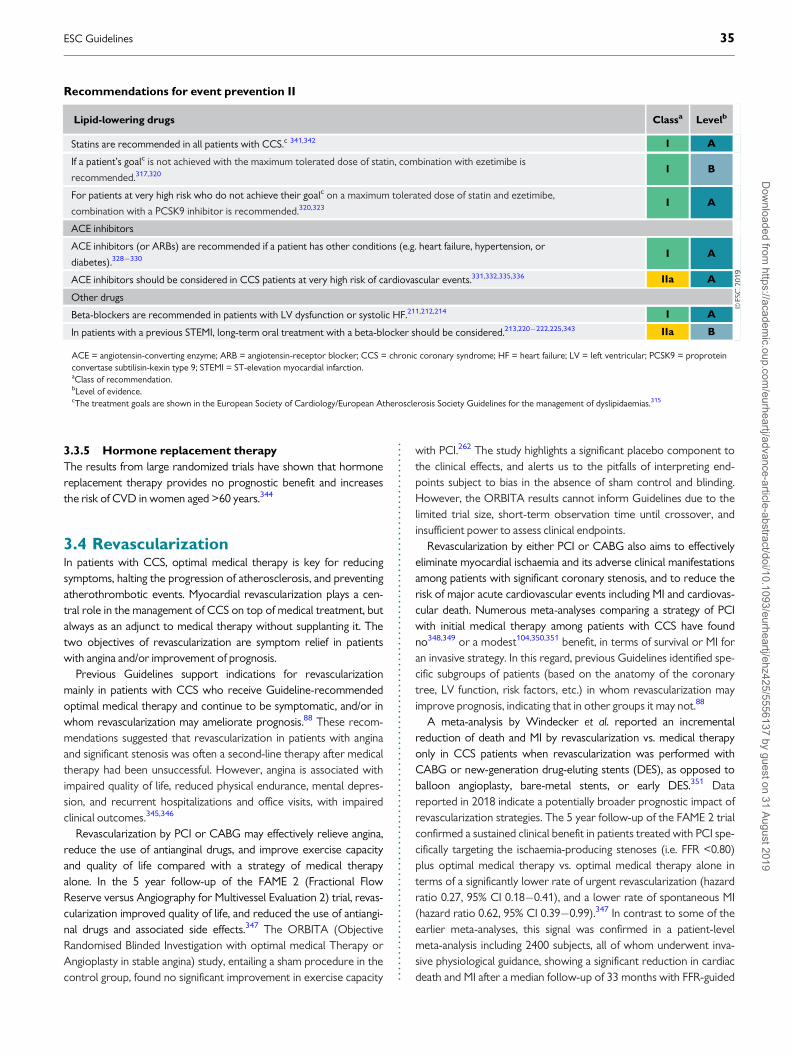
..
..
..
..
..
..
..
..
..
..
..
..
..
..
..
..
..
..
..
..
..
..
..
..
..
..
..
..
..
..
..
..
..
..
..
..
..
..
..
..
..
..
..
..
..
..
..
..
..
..
..
.3.3.5 Hormone replacement therapy
The results from large randomized trials have shown that hormonereplacement therapy provides no prognostic benefit and increasesthe risk of CVD in women aged >60 years.344
3.4 RevascularizationIn patients with CCS, optimal medical therapy is key for reducingsymptoms, halting the progression of atherosclerosis, and preventingatherothrombotic events. Myocardial revascularization plays a cen-tral role in the management of CCS on top of medical treatment, butalways as an adjunct to medical therapy without supplanting it. Thetwo objectives of revascularization are symptom relief in patientswith angina and/or improvement of prognosis.
Previous Guidelines support indications for revascularizationmainly in patients with CCS who receive Guideline-recommendedoptimal medical therapy and continue to be symptomatic, and/or inwhom revascularization may ameliorate prognosis.88 These recom-mendations suggested that revascularization in patients with anginaand significant stenosis was often a second-line therapy after medicaltherapy had been unsuccessful. However, angina is associated withimpaired quality of life, reduced physical endurance, mental depres-sion, and recurrent hospitalizations and office visits, with impairedclinical outcomes.345,346
Revascularization by PCI or CABG may effectively relieve angina,reduce the use of antianginal drugs, and improve exercise capacityand quality of life compared with a strategy of medical therapyalone. In the 5 year follow-up of the FAME 2 (Fractional FlowReserve versus Angiography for Multivessel Evaluation 2) trial, revas-cularization improved quality of life, and reduced the use of antiangi-nal drugs and associated side effects.347 The ORBITA (ObjectiveRandomised Blinded Investigation with optimal medical Therapy orAngioplasty in stable angina) study, entailing a sham procedure in thecontrol group, found no significant improvement in exercise capacity
with PCI.262 The study highlights a significant placebo component tothe clinical effects, and alerts us to the pitfalls of interpreting end-points subject to bias in the absence of sham control and blinding.However, the ORBITA results cannot inform Guidelines due to thelimited trial size, short-term observation time until crossover, andinsufficient power to assess clinical endpoints.
Revascularization by either PCI or CABG also aims to effectivelyeliminate myocardial ischaemia and its adverse clinical manifestationsamong patients with significant coronary stenosis, and to reduce therisk of major acute cardiovascular events including MI and cardiovas-cular death. Numerous meta-analyses comparing a strategy of PCIwith initial medical therapy among patients with CCS have foundno348,349 or a modest104,350,351 benefit, in terms of survival or MI foran invasive strategy. In this regard, previous Guidelines identified spe-cific subgroups of patients (based on the anatomy of the coronarytree, LV function, risk factors, etc.) in whom revascularization mayimprove prognosis, indicating that in other groups it may not.88
A meta-analysis by Windecker et al. reported an incrementalreduction of death and MI by revascularization vs. medical therapyonly in CCS patients when revascularization was performed withCABG or new-generation drug-eluting stents (DES), as opposed toballoon angioplasty, bare-metal stents, or early DES.351 Datareported in 2018 indicate a potentially broader prognostic impact ofrevascularization strategies. The 5 year follow-up of the FAME 2 trialconfirmed a sustained clinical benefit in patients treated with PCI spe-cifically targeting the ischaemia-producing stenoses (i.e. FFR <0.80)plus optimal medical therapy vs. optimal medical therapy alone interms of a significantly lower rate of urgent revascularization (hazardratio 0.27, 95% CI 0.18�0.41), and a lower rate of spontaneous MI(hazard ratio 0.62, 95% CI 0.39�0.99).347 In contrast to some of theearlier meta-analyses, this signal was confirmed in a patient-levelmeta-analysis including 2400 subjects, all of whom underwent inva-sive physiological guidance, showing a significant reduction in cardiacdeath and MI after a median follow-up of 33 months with FFR-guided
Recommendations for event prevention II
Lipid-lowering drugs Classa Levelb
Statins are recommended in all patients with CCS.c 341,342 I A
If a patient’s goalc is not achieved with the maximum tolerated dose of statin, combination with ezetimibe is
recommended.317,320 I B
For patients at very high risk who do not achieve their goalc on a maximum tolerated dose of statin and ezetimibe,
combination with a PCSK9 inhibitor is recommended.320,323 I A
ACE inhibitors
ACE inhibitors (or ARBs) are recommended if a patient has other conditions (e.g. heart failure, hypertension, or
diabetes).328�330 I A
ACE inhibitors should be considered in CCS patients at very high risk of cardiovascular events.331,332,335,336 IIa A
Other drugs
Beta-blockers are recommended in patients with LV dysfunction or systolic HF.211,212,214 I A
In patients with a previous STEMI, long-term oral treatment with a beta-blocker should be considered.213,220�222,225,343 IIa B
ACE = angiotensin-converting enzyme; ARB = angiotensin-receptor blocker; CCS = chronic coronary syndrome; HF = heart failure; LV = left ventricular; PCSK9 = proproteinconvertase subtilisin-kexin type 9; STEMI = ST-elevation myocardial infarction.aClass of recommendation.bLevel of evidence.cThe treatment goals are shown in the European Society of Cardiology/European Atherosclerosis Society Guidelines for the management of dyslipidaemias.315
ESC Guidelines 35D
ownloaded from
https://academic.oup.com
/eurheartj/advance-article-abstract/doi/10.1093/eurheartj/ehz425/5556137 by guest on 31 August 2019

..
..
..
..
..
..
..
..
..
..
..
..
..
..
..
..
..
..
..
.PCI vs. medical therapy (hazard ratio 0.74, 95% CI 0.56�0.989;P=0.041).352 Together, these new data support a less restrictive indi-cation for revascularization in CCS, in addition to specific anatomy[e.g. left main (LM)] or extended ischaemia (>10%), when PCI isrestricted to angiographic stenoses on large vessels causing a signifi-cant intracoronary pressure gradient. Figure 9 summarizes a practicalapproach to the indications of revascularization in CCS according tothe presence or absence of symptoms, and documentation of non-invasive ischaemia. However, the individual risk-benefit ratio shouldalways be evaluated and revascularization considered only if itsexpected benefit outweighs its potential risk. Also, the aspect ofshared decision-making is key, with full information given to thepatient about the anticipated advantages and disadvantages of the
two strategies, including the DAPT-related bleeding risks in cases ofrevascularization by PCI. For the discussion of the best choicebetween revascularization modalities�PCI or CABG�for individualpatients, we refer readers to the 2018 ESC myocardial revasculariza-tion Guidelines.88
4 Patients with new onset of heartfailure or reduced left ventricularfunction
CAD is the most common cause of HF in Europe, and most of thetrial evidence supporting management recommendations is based on
©ES
C 2
019
NoYes
NoYesNoYes
NoYes
Diameter stenosis >90%
FFR ≤0.80 or iwFR ≤0.89 in major vessel
LVEF ≤35% due to CAD
Large area of ischaemia (>10% of LV)
Diameter stenosis >90%
FFR ≤0.80 or iwFR ≤0.89 in major vessel
LVEF ≤35% due to CAD
Identify lesions with FFR ≤0.80 or iwFR ≤0.89
•
•
•
•
•
•
Consider revascularization on top of medical therapy
MVD
Documented ischaemia
Documented ischaemia
Angina symptoms
Figure 9 Decision tree for patients undergoing invasive coronary angiography. Decisions for revascularization by percutaneous coronary interventionor coronary artery bypass grafting are based on clinical presentation (symptoms present or absent), and prior documentation of ischaemia (present orabsent). In the absence of prior documentation of ischaemia, indications for revascularization depend on invasive evaluation of stenosis severity or prog-nostic indications. Patients with no symptoms and ischaemia include candidates for transcatheter aortic valve implantation, valve, and other surgery. CAD= coronary artery disease; FFR = fractional flow reserve; iwFR = instantaneous wave-free ratio; LV = left ventricle; LVEF = left ventricular ejection fraction;MVD = multivessel disease.
36 ESC GuidelinesD
ownloaded from
https://academic.oup.com
/eurheartj/advance-article-abstract/doi/10.1093/eurheartj/ehz425/5556137 by guest on 31 August 2019

..
..
..
..
..
..
..
..
..
..
..
..
..
..
..
..
..
..
..
..
..
..
..
..
..
..
..
..
..
..
..
..
..
..
..
..
..
..
..
..
..
..
..
..
..
..
..
..
..
..
..
..research conducted in patients with ischaemic cardiomyopathy. Thepathophysiology results in systolic dysfunction due to myocardialinjury and ischaemia, and most patients with symptomatic HF havereduced ejection fraction (<40%), although patients with CCSmay also have symptomatic HF and a preserved ejection fraction(>_50%). Patients with symptomatic HF should be managed clinicallyaccording to the 2016 ESC heart failure Guidelines.340
History should include the assessment of symptoms suggestive ofHF, especially exercise intolerance and dyspnoea on exertion. Allmajor past events related to CAD including MI and revascularizationprocedures are recorded, as well as all major cardiovascular comor-bidity requiring treatment such as AF, hypertension, or valvular dys-function, and non-cardiovascular comorbidity such as CKD, diabetes,anaemia, or cancer. Current medical therapy, adherence, and toler-ance should be reviewed.
Physical examination should assess the nutritional status of patients,and estimate biological age and cognitive ability. Recorded physicalsigns include heart rate, heart rhythm, supine BP, murmurs suggestiveof aortic stenosis or mitral insufficiency, signs of pulmonary conges-tion with basal rales or pleural effusion, signs of systemic congestionwith dependant oedema, hepatomegaly, and elevated jugular venouspressure.
A routine ECG provides information on heart rate and rhythm,extrasystole, signs of ischaemia, pathological Q waves, hypertrophy,conduction abnormalities, and bundle branch block.
Imaging should include an echocardiographic examination withDoppler to evaluate evidence of ischaemic cardiomyopathy with HFwith reduced ejection fraction, HF with mid-range ejection fraction,or HF with preserved ejection fraction, focal/diffuse LV or right ven-tricular systolic dysfunction, evidence of diastolic dysfunction, hyper-trophy, chamber volumes, valvular function, and evidence ofpulmonary hypertension. Chest X-ray can detect signs of pulmonarycongestion, interstitial oedema, infiltration, or pleural effusion. If notknown, coronary angiography (or coronary CTA) should be
performed to establish the presence and extent of CAD, and evalu-ate the potential for revascularization.52,53
Laboratory investigations should measure natriuretic peptide levels torule-out the diagnosis of suspected HF. When present, the severity ofHF can be assessed.25,49 Renal function along with serum electrolytesshould be measured routinely to detect the development of renalinsufficiency, hyponatraemia, or hyperkalaemia, especially at the initia-tion and during up-titration of pharmacological therapy.
The management of patients with symptomatic HF requiresadequate diuretic therapy, preferably with a loop diuretic, to relievesigns and symptoms of pulmonary and systemic congestion.Inhibitors of both the RAS system (ACE inhibitors, ARBs, angiotensinreceptor-neprilysin inhibitor) and the adrenergic nervous system(beta-blockers) are indicated in all symptomatic patients with HF.340
In patients with persistent symptoms, an MRA is also indicated. Up-titration of these drugs should be gradual to avoid symptomatic sys-tolic hypotension, renal insufficiency, or hyperkalaemia.
Patients who remain symptomatic, with LV systolic dysfunctionand evidence of ventricular dysrhythmia or bundle branch block, maybe eligible for a device [cardiac resynchronization therapy (CRT)/implantable cardioverter-defibrillator]. Such devices may providesymptomatic relief, reduce morbidity, and improve survival.353�356
Patients with HF may decompensate rapidly following the onset ofatrial or ventricular dysrhythmia, and should be treated according tocurrent Guidelines. Patients with HF, and haemodynamically signifi-cant aortic stenosis or mitral insufficiency, may require percutaneousor surgical intervention.
Myocardial revascularization should be considered in eligiblepatients with HF based on their symptoms, coronary anatomy, andrisk profile. Successful revascularization in patients with HF due toischaemic cardiomyopathy may improve LV dysfunction and progno-sis by reducing ischaemia to viable, hibernating myocardium. If avail-able, cooperation with a multidisciplinary HF team is stronglyadvised.348,357,358
General recommendations for the management of patients with chronic coronary syndromes and symptomatic heartfailure due to ischaemic cardiomyopathy and left ventricular systolic dysfunction
Recommendations for drug therapy Classa Levelb
Diuretic therapy is recommended in symptomatic patients with signs of pulmonary or systemic congestion to relieve HF
symptoms.359,360 I B
Beta-blockers are recommended as essential components of treatment due to their efficacy in both relieving angina, and
reducing morbidity and mortality in HF.214,361�367 I A
ACE inhibitor therapy is recommended in patients with symptomatic HF or asymptomatic LV dysfunction following MI, to
improve symptoms and reduce morbidity and mortality.333,368 I A
An ARB is recommended as an alternative in patients who do not tolerate ACE inhibition, or an angiotensin recep-
tor-neprilysin inhibitor in patients with persistent symptoms despite optimal medical therapy.337,369 I B
An MRA is recommended in patients who remain symptomatic despite adequate treatment with an ACE inhibitor and
beta-blocker, to reduce morbidity and mortality.360,370 I A
A short-acting oral or transcutaneous nitrate should be considered (effective antianginal treatment, safe in HF).371 IIa A
Ivabradine should be considered in patients with sinus rhythm, an LVEF <_35% and a resting heart rate >70 b.p.m. who
remain symptomatic despite adequate treatment with a beta-blocker, ACE inhibitor, and MRA, to reduce morbidity and
mortality.238,372,373
IIa B
Amlodipine may be considered for relief of angina in patients with HF who do not tolerate beta-blockers, and is consid-
ered safe in HF.374,375 IIb B
Continued
ESC Guidelines 37D
ownloaded from
https://academic.oup.com
/eurheartj/advance-article-abstract/doi/10.1093/eurheartj/ehz425/5556137 by guest on 31 August 2019
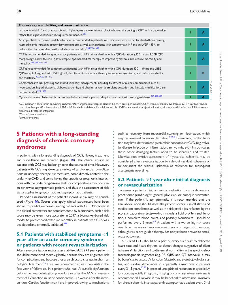
..
..
..
..
..
..
..
..
..
..
..
..
..
..
..
..
..
..
..
..
..
..
..
..
..
..
..
..
..
..
..
..
..
..
..
..
..
..
..
..
..
..
..
..
..
..
..
..
..
..
.5 Patients with a long-standingdiagnosis of chronic coronarysyndromes
In patients with a long-standing diagnosis of CCS, lifelong treatmentand surveillance are required (Figure 10). The clinical course ofpatients with CCS may be benign over the course of time. However,patients with CCS may develop a variety of cardiovascular complica-tions or undergo therapeutic measures, some directly related to theunderlying CAD, and some having therapeutic or prognostic interac-tions with the underlying disease. Risk for complications may occur inan otherwise asymptomatic patient, and thus the assessment of riskstatus applies to symptomatic and asymptomatic patients.
Periodic assessment of the patient’s individual risk may be consid-ered (Figure 10). Scores that apply clinical parameters have beenshown to predict outcomes among patients with CCS. Moreover, ifthe clinical parameters are complemented by biomarkers, such a riskscore may be even more accurate. In 2017, a biomarker-based riskmodel to predict cardiovascular mortality in patients with CCS wasdeveloped and externally validated.398
5.1 Patients with stabilized symptoms <1year after an acute coronary syndromeor patients with recent revascularizationAfter revascularization and/or after stabilized ACS (<1 year), patientsshould be monitored more vigilantly, because they are at greater riskfor complications and because they are subject to changes in pharma-cological treatment.45 Thus, we recommend at least two visits in thefirst year of follow-up. In a patient who had LV systolic dysfunctionbefore the revascularization procedure or after the ACS, a reassess-ment of LV function must be considered 8�12 weeks after the inter-vention. Cardiac function may have improved, owing to mechanisms
such as recovery from myocardial stunning or hibernation, whichmay be reversed by revascularization.52,53 Conversely, cardiac func-tion may have deteriorated given other concomitant CVD (e.g. valvu-lar disease, infection or inflammation, arrhythmia, etc.). In such cases,these other damaging factors need to be identified and treated.Likewise, non-invasive assessment of myocardial ischaemia may beconsidered after revascularization to rule-out residual ischaemia orto document the residual ischaemia as reference for subsequentassessments over time.
5.2 Patients >1 year after initial diagnosisor revascularizationTo assess a patient’s risk, an annual evaluation by a cardiovascularpractitioner (cardiologist, general physician, or nurse) is warranted,even if the patient is asymptomatic. It is recommended that theannual evaluation should assess the patient’s overall clinical status andmedication compliance, as well as the risk profile (as reflected by riskscores). Laboratory tests—which include a lipid profile, renal func-tion, a complete blood count, and possibly biomarkers—should beperformed every 2 years.45 A patient with a worsening risk scoreover time may warrant more intense therapy or diagnostic measures,although risk score-guided therapy has not yet been proved to ameli-orate outcomes.
A 12 lead ECG should be a part of every such visit to delineateheart rate and heart rhythm, to detect changes suggestive of silentischaemia/infarction, and to discern abnormalities in the specific elec-trocardiographic segments (e.g. PR, QRS, and QT intervals). It maybe beneficial to assess LV function (diastolic and systolic), valvular sta-tus, and cardiac dimensions in apparently asymptomatic patientsevery 3�5 years.52,53 In cases of unexplained reduction in systolic LVfunction, especially if regional, imaging of coronary artery anatomy isrecommended. Likewise, it may be beneficial to assess non-invasivelyfor silent ischaemia in an apparently asymptomatic patient every 3�5
For devices, comorbidities, and revascularization
In patients with HF and bradycardia with high-degree atrioventricular block who require pacing, a CRT with a pacemaker
rather than right ventricular pacing is recommended.353 I A
An implantable cardioverter-defibrillator is recommended in patients with documented ventricular dysrhythmia causing
haemodynamic instability (secondary prevention), as well as in patients with symptomatic HF and an LVEF <_35%, to
reduce the risk of sudden death and all-cause mortality.354,376�382
I A
CRT is recommended for symptomatic patients with HF in sinus rhythm with a QRS duration >_150 ms and LBBB QRS
morphology, and with LVEF <_35%, despite optimal medical therapy to improve symptoms, and reduce morbidity and
mortality.355,356,383�392
I A
CRT is recommended for symptomatic patients with HF in sinus rhythm with a QRS duration 130 - 149 ms and LBBB
QRS morphology, and with LVEF <_35%, despite optimal medical therapy to improve symptoms, and reduce morbidity
and mortality.355,356,383�392
I B
Comprehensive risk profiling and multidisciplinary management, including treatment of major comorbidities such as
hypertension, hyperlipidaemia, diabetes, anaemia, and obesity, as well as smoking cessation and lifestyle modification, are
recommended.393�396
I A
Myocardial revascularization is recommended when angina persists despite treatment with antianginal drugs.348,357,397 I A
ACE inhibitor = angiotensin-converting enzyme; ARB = angiotensin receptor blocker; b.p.m. = beats per minute; CCS = chronic coronary syndromes; CRT = cardiac resynch-ronization therapy; HF = heart failure; LBBB = left bundle branch block; LV = left ventricular; LVEF = left ventricular ejection fraction; MI = myocardial infarction; MRA = miner-alocorticoid receptor antagonist.aClass of recommendation.bLevel of evidence.
38 ESC GuidelinesD
ownloaded from
https://academic.oup.com
/eurheartj/advance-article-abstract/doi/10.1093/eurheartj/ehz425/5556137 by guest on 31 August 2019

..
..
..
..
..
..
..
..
..
..
..
..
..
.years, preferably applying stress imaging. Coronary CTA should notbe used for follow-up of patients with established CAD given itsstrength on morphological insight, but lack of functional informationrelated to ischaemia. However, coronary CTA may be used forunique cases, such as delineation of patency of coronary arterybypass grafts.
The lipid profile and glycaemia status should be reassessed periodi-cally to determine efficacy of treatment and, in patients without dia-betes, to detect new development of diabetes. There is no evidence
to support recommendations for the frequency of reassessment ofthese risk factors, but consensus suggests annual evaluation.
Elevated inflammatory markers, particularly of high-sensitivity C-reactive protein, have also been associated with an increased eventrisk in patients with and without CAD in multiple studies,25 althoughthe robustness of the association has been questioned because ofreporting and publication bias.399 In addition, von Willebrand factor,interleukin-6, and N-terminal pro-B-type natriuretic peptide (NT-proBNP) have identified as been predictors of outcome.25 Other
©ES
C 2
019
acardiologist, internist, general practitioner, or cardiovascular nurse
Invasive coronary angiography
As necessary, for patients at high risk based on noninvasive ischaemia testing, or severe angina symptoms (e.g. CCS class 3-4). Not recommended solely for risk stratification.
Stress test for inducible ischaemia
As necessary, to investigate changes in symptoms level, and/or early (e.g. 1-3 months) after revascularization to set as a reference and/or periodically (e.g. every 3-5 years) to reassess ischaemia.
Echocardiography at rest
Early (e.g. 1-3 months) after revascularization to set as a reference and/or periodically (e.g. at 1 year if previously abnormal and/or every 3-5 years) to evaluate LV function, valvular status and haemodynamic status.
Des
tabi
lizat
ion
Des
tabi
lizat
ion
Stab
iliza
tion
Baseline
3 months
6 months
12 months
Tim
e fr
om in
itial
eva
luat
ion
of p
ost-
AC
S C
CS
18 months
24 months
Yearly
Post-ACS CCS (e.g. >1 year after MI)
Yearly
Long-standing diagnosis of CCS (>1 year)
Baseline
3 months
6 months
12 months
Tim
e fr
om
initi
al e
valu
atio
nof
rec
ent
CC
S
Recent CCS diagnosis or revascularization
Legend Time for decision-making on DAPT continuation in PCI patients
Time for decision-making on optional dual antithrombotic therapy (see table 9)
Advisable timepoint
Optional timepoint
Cardiovascular caregivera visit
Risk score(s)stratification
RestingECG
ACS
���
Figure 10 Proposed algorithm according to patient types commonly observed at chronic coronary syndrome outpatient clinics. The frequency of fol-low-up may be subject to variation based on clinical judgement. ACS = acute coronary syndromes; CCS = chronic coronary syndromes; DAPT = dual anti-platelet therapy; ECG = electrocardiogram; LV = left ventricular; MI = myocardial infarction; PCI = percutaneous coronary intervention. aCardiologist,internist, general practitioner, or cardiovascular nurse.
ESC Guidelines 39D
ownloaded from
https://academic.oup.com
/eurheartj/advance-article-abstract/doi/10.1093/eurheartj/ehz425/5556137 by guest on 31 August 2019

..
..
..
..
..
..
..
..
..
..
..
..
..
..
..
..
..
..
..
..
..
..
..
..
..
..
..
..
..
..
..
..
..
..
..
..
..
..
..
..
..
..
..
..
..
..readily available biomarkers shown to predict prognosis in patientswith CCS include heart rate, haemoglobin, and white cell count.400
Scores based on aggregated biomarkers may have greater successthan individual biomarkers. A multiple biomarker score combininghigh-sensitivity C-reactive protein, heat shock protein 70, and fibrindegradation products significantly improved C-statistics and the netreclassification index compared with a basic model using clinicaldata.401 Similar results were reported for a combination of high-sensitivity cardiac troponin T, NT-proBNP, and LDL-C.398 In severalstudies, genetic risk scores have been shown to improve risk predic-tion above traditional risk factors in general population samples402,403
and to predict recurrent events in populations with knownCCS.404�407 Although there is additional prognostic value in usingseveral individual and aggregated biomarkers, there is currently noevidence that routine use leads to improved care. Nevertheless,these measurements may have a role in selected patients (e.g. whentesting for haemostatic abnormalities in those with previous MI with-out conventional risk factors or a strong family history of CAD).
Patients with unequivocal symptoms suggestive of ACS should beexpeditiously referred for evaluation, applying current Guidelines fordiagnosis and management. Among patients with more equivocalsymptoms, stress imaging is recommended408 and, if not available andthe ECG is amenable to identification of ischaemia, exercise stresselectrocardiography can be used as an alternative. In patients withsevere angina and a high-risk clinical profile, direct referral for ICA isrecommended, provided that ad hoc physiological assessment of hae-modynamic stenosis significance is readily available in the catheteriza-tion laboratory [e.g. instantaneous wave-free ratio (iwFR) or FFR].Likewise, ICA is recommended for patients with evidence of signifi-cant ischaemia obtained by non-invasive testing.
6 Angina without obstructivedisease in the epicardial coronaryarteries
In clinical practice, a marked discrepancy between findings regardingcoronary anatomy, the presence of symptoms, and the results ofnon-invasive tests frequently occurs.13 These patients deserve atten-tion, as angina and non-obstructive disease are associated with anincreased risk of adverse clinical events.14 Low diagnostic yield of ICAcan be explained by the presence of: (i) stenoses with mild or moder-ate angiographic severity, or diffuse coronary narrowing, with under-estimated functional significance identified by ICA; (ii) disordersaffecting the microcirculatory domain that escape the resolution ofangiographic techniques; and (iii) dynamic stenoses of epicardial ves-sels caused by coronary spasm or intramyocardial bridges that arenot evident during CTA or ICA. Intracoronary pressure measure-ments are useful in circumventing the first of these scenarios. Fordiagnostic workup, patients with angina and/or myocardial ischaemiashowing coronary stenoses with non-ischaemic FFR or iwFR valuesmay also be labelled as having non-obstructive epicardial disease.
The presence of clear-cut anginal symptoms and abnormal non-invasive tests in patients with non-obstructed epicardial vesselsshould lead to the suspicion of a non-obstructive cause of ischaemia.Quite often, and mainly as a result of persistence of symptoms,patients with angina and no obstructive disease undergo multiplediagnostic tests, including repeated coronary CTA or ICA, that con-tribute to increased healthcare costs.409 Because diagnostic pathwaysto investigate microcirculatory or vasomotor coronary disorders areoften not implemented, a final diagnosis supported by objective
Recommendations for patients with a long-standing diagnosis of chronic coronary syndromes
Recommendations for asymptomatic patients Classa Levelb
A periodic visit to a cardiovascular healthcare professional is recommended to reassess any potential change in the risk
status of patients, entailing clinical evaluation of lifestyle-modification measures, adherence to targets of cardiovascular
risk factors, and the development of comorbidities that may affect treatments and outcomes.
I C
In patients with mild or no symptoms receiving medical treatment in whom non-invasive risk stratification indicates a high
risk, and for whom revascularization is considered for improvement of prognosis, invasive coronary angiography (with
FFR when necessary) is recommended.
I C
Coronary CTA is not recommended as a routine follow-up test for patients with established CAD. III C
Invasive coronary angiography is not recommended solely for risk stratification. III C
Symptomatic patients
Reassessment of CAD status is recommended in patients with deteriorating LV systolic function that cannot be attributed
to a reversible cause (e.g. long-standing tachycardia or myocarditis).I C
Risk stratification is recommended in patients with new or worsening symptom levels, preferably using stress imaging or,
alternatively, exercise stress ECG.408 I B
It is recommended to expeditiously refer patients with significant worsening of symptoms for evaluation. I C
Invasive coronary angiography (with FFR/iwFR when necessary) is recommended for risk stratification in patients with
severe CAD, particularly if the symptoms are refractory to medical treatment or if they have a high-risk clinical profile.I C
CAD = coronary artery disease; CTA = computed tomography angiography; ECG = electrocardiogram; FFR = fractional flow reserve; iwFR = instantaneous wave-free ratio;LV = left ventricular.aClass of recommendation.bLevel of evidence.
40 ESC GuidelinesD
ownloaded from
https://academic.oup.com
/eurheartj/advance-article-abstract/doi/10.1093/eurheartj/ehz425/5556137 by guest on 31 August 2019

..
..
..
..
..
..
..
..
..
..
..
..
..
..
..
..
..
..
..
..
..
..
..
..
..
..
..
..
..
..
..
..
..
..
..
..
..
..
..
..
..
..
..
..
..
..
..
..
..
..
..
..
..
..
..
..
..
..
..
..
..
..
..
..
..
..
..
..
..
..
..
..
..
..
..
..
..
..
..
..evidence is seldom reached. Owing to this, patient dismay anddepression are not rare in this clinical population.410,411 Of note, theuse of a structured, systematic approach to explore microcirculatoryand vasomotor disorders in patients with non-obstructive CAD, asdelineated below, has been shown to increase diagnostic yield.412,413
Furthermore, an RCT, which reported in 2018, found that in patientswith non-obstructive coronary disease, tailored treatment guided bythe results of intracoronary testing [coronary flow reserve (CFR),microcirculatory resistance, and acetylcholine testing] resulted in asignificant reduction of anginal symptoms, compared with conven-tional, non-guided medical treatment.414
6.1 Microvascular anginaPatients with microvascular angina typically have exercise-relatedangina, evidence of ischaemia in non-invasive tests, and either nostenoses or mild-to-moderate stenoses (40�60%), revealed byICA or CTA, that are deemed functionally non-relevant.415 Giventhe similarity of angina symptoms, a microvascular origin of anginais typically suspected, after excluding obstructive epicardial coro-nary stenoses, during diagnostic workup of patients with suspectedmyocardial ischaemia. Regional LV wall motion abnormalities rarelydevelop during exercise or stress in patients with microvascularangina.412,416 Some patients may also have a mixed pattern ofangina, with occasional episodes at rest, particularly associated withexposure to cold.
Secondary microvascular angina, in the absence of epicardialobstruction, may result from cardiac or systemic conditions, includingthose that cause LV hypertrophy (such as hypertrophic cardiomyop-athy, aortic stenosis, and hypertensive heart disease) or inflammation(such as myocarditis or vasculitis).417
6.1.1 Risk stratification
The presence of microcirculatory dysfunction in patients with CCSentails a worse prognosis than originally thought, probably becausemost recent evidence has been based on follow-up of patients inwhom abnormalities in the microcirculation have been objectivelydocumented with invasive or non-invasive techniques.418�423
Microcirculatory dysfunction precedes the development of epicar-dial lesions, particularly in women,419 and is associated with impairedoutcomes. Among patients with diabetes undergoing diagnosticworkup, those without obstructive epicardial disease but with anabnormal CFR have similarly poor long-term prognosis as those withobstructive epicardial disease.421 In patients with non-significant cor-onary stenoses by FFR, the presence of abnormal CFR is associatedwith an excess of events in the long-term,418,422,423 particularly whenthe index of microcirculatory resistance (IMR) is also abnormal.422
6.1.2 Diagnosis
The possibility of a microcirculatory origin of angina should be con-sidered in patients with clear-cut angina, abnormal non-invasive func-tional tests, and coronary vessels that are either normal or have mildstenosis deemed functionally non-significant on ICA or CTA. One of
the challenges in performing a comprehensive assessment of micro-vascular function is testing the two main mechanisms of dysfunctionseparately: impaired microcirculatory conductance and arteriolardysregulation.424�426 Yet, outlining which of these two pathways isaffected is critically relevant in setting medical treatment to relievepatient symptoms.414
Impaired microcirculatory conductance can be diagnosed by measuringCFR or minimal microcirculatory resistance (the inverse of conduc-tance). CFR can be measured non-invasively with transthoracicDoppler echocardiography [by imaging left anterior descending(LAD) flow],427 magnetic resonance imaging (myocardial perfusionindex),428�430 or PET.431 Microcirculatory resistance can be measuredin the catheterization laboratory by combining intracoronary pressurewith thermodilution-based data (to calculate the IMR) or Dopplerflow velocity (to calculate hyperaemic microvascular resistance orHMR).432,433 Both intracoronary thermodilution and Doppler allowthe calculation of CFR. For decision-making purposes, values of IMR>_25 units or CFR <2.0 are indicative of abnormal microcirculatoryfunction.414 Both CFR and IMR are typically measured while usingintravenous vasodilators, such as adenosine or regadenoson.
In contrast, the diagnosis of arteriolar dysregulation requires theassessment of endothelial function in the coronary microcirculationby selective intracoronary acetylcholine infusion (see section 6.5). Inthe presence of dysfunctional vascular endothelium or abnormalfunction of smooth muscle cells, acetylcholine (an endothelium-dependent vasodilator that also acts directly on smooth muscle cells)triggers paradoxical arteriolar vasoconstriction.434 Thus, in patientswith microvascular angina and arteriolar dysregulation, acetylcholinechallenge is likely to trigger microvascular spasm. This arteriolarresponse to acetylcholine causes anginal symptoms with or withoutconcomitant ischaemic ECG changes, and a decrease in coronaryblood flow velocity if concomitant Doppler measurements are per-formed. Peripheral pulse tonometry during reactive hyperaemia mayalso reveal abnormal systemic endothelial function in patients withangina and non-obstructive CAD.435
6.1.3 Treatment
Treatment of microvascular angina should address the dominantmechanism of microcirculatory dysfunction. In patients with abnor-mal CFR <2.0 or IMR >_25 units, and a negative acetylcholine provo-cation test, beta-blockers, ACE inhibitors, and statins, along withlifestyle changes and weight loss, are indicated.436,437 Patients devel-oping ECG changes and angina in response to acetylcholine testingbut without severe epicardial vasoconstriction (all suggestive ofmicrovascular spasm) may be treated like vasospastic angina patients.The effectiveness of a tailored treatment strategy was investigated inthe CorMiCa trial, which randomized 151 patients to a stratifiedmedical treatment (based on the results of CFR, IMR, and acetylcho-line testing) vs. a standard-care group (including a sham interventionaldiagnostic procedure). At 1 year, there was a significant difference inangina scores favouring patients assigned to the stratified medicaltreatment arm.414
ESC Guidelines 41D
ownloaded from
https://academic.oup.com
/eurheartj/advance-article-abstract/doi/10.1093/eurheartj/ehz425/5556137 by guest on 31 August 2019
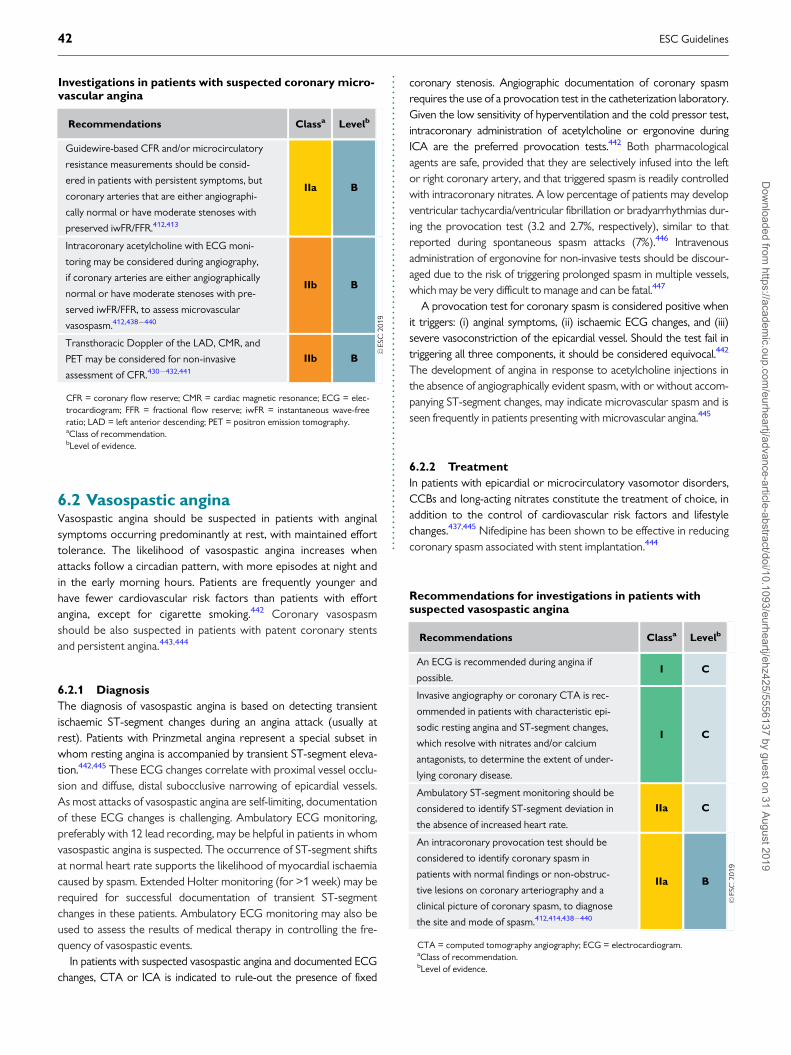
..
..
..
..
..
..
..
..
..
..
..
..
..
..
..
..
..
..
..
..
..
..
..
..
..
..
..
..
..
..
..
..
..
..
..
..
..
..
..
..
..
..
..
..
..
.
6.2 Vasospastic anginaVasospastic angina should be suspected in patients with anginalsymptoms occurring predominantly at rest, with maintained efforttolerance. The likelihood of vasospastic angina increases whenattacks follow a circadian pattern, with more episodes at night andin the early morning hours. Patients are frequently younger andhave fewer cardiovascular risk factors than patients with effortangina, except for cigarette smoking.442 Coronary vasospasmshould be also suspected in patients with patent coronary stentsand persistent angina.443,444
6.2.1 Diagnosis
The diagnosis of vasospastic angina is based on detecting transientischaemic ST-segment changes during an angina attack (usually atrest). Patients with Prinzmetal angina represent a special subset inwhom resting angina is accompanied by transient ST-segment eleva-tion.442,445 These ECG changes correlate with proximal vessel occlu-sion and diffuse, distal subocclusive narrowing of epicardial vessels.As most attacks of vasospastic angina are self-limiting, documentationof these ECG changes is challenging. Ambulatory ECG monitoring,preferably with 12 lead recording, may be helpful in patients in whomvasospastic angina is suspected. The occurrence of ST-segment shiftsat normal heart rate supports the likelihood of myocardial ischaemiacaused by spasm. Extended Holter monitoring (for >1 week) may berequired for successful documentation of transient ST-segmentchanges in these patients. Ambulatory ECG monitoring may also beused to assess the results of medical therapy in controlling the fre-quency of vasospastic events.
In patients with suspected vasospastic angina and documented ECGchanges, CTA or ICA is indicated to rule-out the presence of fixed
coronary stenosis. Angiographic documentation of coronary spasmrequires the use of a provocation test in the catheterization laboratory.Given the low sensitivity of hyperventilation and the cold pressor test,intracoronary administration of acetylcholine or ergonovine duringICA are the preferred provocation tests.442 Both pharmacologicalagents are safe, provided that they are selectively infused into the leftor right coronary artery, and that triggered spasm is readily controlledwith intracoronary nitrates. A low percentage of patients may developventricular tachycardia/ventricular fibrillation or bradyarrhythmias dur-ing the provocation test (3.2 and 2.7%, respectively), similar to thatreported during spontaneous spasm attacks (7%).446 Intravenousadministration of ergonovine for non-invasive tests should be discour-aged due to the risk of triggering prolonged spasm in multiple vessels,which may be very difficult to manage and can be fatal.447
A provocation test for coronary spasm is considered positive whenit triggers: (i) anginal symptoms, (ii) ischaemic ECG changes, and (iii)severe vasoconstriction of the epicardial vessel. Should the test fail intriggering all three components, it should be considered equivocal.442
The development of angina in response to acetylcholine injections inthe absence of angiographically evident spasm, with or without accom-panying ST-segment changes, may indicate microvascular spasm and isseen frequently in patients presenting with microvascular angina.445
6.2.2 Treatment
In patients with epicardial or microcirculatory vasomotor disorders,CCBs and long-acting nitrates constitute the treatment of choice, inaddition to the control of cardiovascular risk factors and lifestylechanges.437,445 Nifedipine has been shown to be effective in reducingcoronary spasm associated with stent implantation.444
Investigations in patients with suspected coronary micro-vascular angina
Recommendations Classa Levelb
Guidewire-based CFR and/or microcirculatory
resistance measurements should be consid-
ered in patients with persistent symptoms, but
coronary arteries that are either angiographi-
cally normal or have moderate stenoses with
preserved iwFR/FFR.412,413
IIa B
Intracoronary acetylcholine with ECG moni-
toring may be considered during angiography,
if coronary arteries are either angiographically
normal or have moderate stenoses with pre-
served iwFR/FFR, to assess microvascular
vasospasm.412,438�440
IIb B
Transthoracic Doppler of the LAD, CMR, and
PET may be considered for non-invasive
assessment of CFR.430�432,441
IIb B
CFR = coronary flow reserve; CMR = cardiac magnetic resonance; ECG = elec-trocardiogram; FFR = fractional flow reserve; iwFR = instantaneous wave-freeratio; LAD = left anterior descending; PET = positron emission tomography.aClass of recommendation.bLevel of evidence.
Recommendations for investigations in patients withsuspected vasospastic angina
Recommendations Classa Levelb
An ECG is recommended during angina if
possible.I C
Invasive angiography or coronary CTA is rec-
ommended in patients with characteristic epi-
sodic resting angina and ST-segment changes,
which resolve with nitrates and/or calcium
antagonists, to determine the extent of under-
lying coronary disease.
I C
Ambulatory ST-segment monitoring should be
considered to identify ST-segment deviation in
the absence of increased heart rate.
IIa C
An intracoronary provocation test should be
considered to identify coronary spasm in
patients with normal findings or non-obstruc-
tive lesions on coronary arteriography and a
clinical picture of coronary spasm, to diagnose
the site and mode of spasm.412,414,438�440
IIa B
CTA = computed tomography angiography; ECG = electrocardiogram.aClass of recommendation.bLevel of evidence.
42 ESC GuidelinesD
ownloaded from
https://academic.oup.com
/eurheartj/advance-article-abstract/doi/10.1093/eurheartj/ehz425/5556137 by guest on 31 August 2019

..
..
..
..
..
..
..
..
..
..
..
..
..
..
..
..
..
..
..
..
..
..
..
..
..
..
..
..
..
..
..
..
..
..
.7 Screening for coronary arterydisease in asymptomatic subjects
In an effort to lower the high burden of coronary deaths in asympto-matic adults, numerous measurements of risk factors and risk markers,as well as stress tests, are often performed as screening investigations.The 2016 European Guidelines on CVD prevention in clinical practicehave focused on these issues in detail.15 These recommendations havebeen adapted for the purpose of these Guidelines.
In general, the use of risk-estimation systems such as SCORE isrecommended (see also Figure 6). Subjects with a family history ofpremature CAD should be screened for familial hypercholesterolae-mia. Coronary calcium score, ankle-brachial index, and carotid ultra-sound for plaque detection may provide useful information about theatherosclerotic risk in selected patients, but routine use of bio-markers or other imaging tests for CAD are not recommended. Thenew biomarkers have incremental predictive value over classicalones,448 but the net reclassification improvement is still only modest(7�18%) compared, for example, with the coronary calcium score,which has a net reclassification improvement of 66%.449
Only subjects at high event risk should be considered for furthernon-invasive or invasive testing. There are no data on how to manage
asymptomatic subjects who receive testing and have a positive testresult beyond the recommendations listed in these Guidelines.However, the principles of risk stratification, as described above forsymptomatic patients, also apply to these individuals.450 It is impor-tant to remember that data demonstrating improved prognosis fol-lowing appropriate management based on new biomarkers are stilllacking.
It is important to note that patients with cancer and undergoingcancer treatment, or chronic inflammatory diseases such as inflam-matory bowel diseases, rheumatoid arthritis, and systemic lupuserythematosus, may deserve more intensive risk screening, counsel-ling, and management.451�454
Persons whose occupations involve public safety (e.g. airlinepilots, or lorry or bus drivers), or who are professional or high-profile athletes, commonly undergo periodic testing for the assess-ment of exercise capacity and evaluation of possible heart disease,including CAD. Although there are insufficient data to justify thisapproach, these evaluations may be done for medicolegal reasons.The threshold for performing an imaging test in such persons maybe lower than in the average patient. Otherwise, the same consid-erations as discussed above for other asymptomatic persons applyto these individuals.
Recommendations for screening for coronary artery disease in asymptomatic subjects
Recommendations Classa Levelb
Total risk estimation using a risk-estimation system such as SCORE is recommended for asymptomatic adults >40 years
of age without evidence of CVD, diabetes, CKD, or familial hypercholesterolaemia.I C
Assessment of family history of premature CVD (defined as a fatal or non-fatal CVD event, or/and established diagnosis of
CVD in first-degree male relatives before 55 years of age or female relatives before 65 years of age) is recommended as
part of cardiovascular risk assessment.
I C
It is recommended that all individuals aged <50 years with a family history of premature CVD in a first-degree relative
(<55 years of age in men or <65 years of age in women) or familial hypercholesterolaemia are screened using a validated
clinical score.455,456
I B
Assessment of coronary artery calcium score with computed tomography may be considered as a risk modifierc in the
cardiovascular risk assessment of asymptomatic subjects.449,457 IIb B
Atherosclerotic plaque detection by carotid artery ultrasound may be considered as a risk modifierc in the cardiovascular
risk assessment of asymptomatic subjects.458 IIb B
ABI may be considered as a risk modifierc in cardiovascular risk assessment.459 IIb B
In high-risk asymptomatic adults (with diabetes, a strong family history of CAD, or when previous risk-assessment tests
suggest a high risk of CAD), functional imaging or coronary CTA may be considered for cardiovascular risk assessment.IIb C
In asymptomatic adults (including sedentary adults considering starting a vigorous exercise programme), an exercise ECG
may be considered for cardiovascular risk assessment, particularly when attention is paid to non-ECG markers such as
exercise capacity.
IIb C
Carotid ultrasound IMT for cardiovascular risk assessment is not recommended.460 III A
In low-risk non-diabetic asymptomatic adults, coronary CTA or functional imaging for ischaemia are not indicated for fur-
ther diagnostic assessment.III C
Routine assessment of circulating biomarkers is not recommended for cardiovascular risk stratification.448,449,461,462 III B
ABI = ankle-brachial index; CAD = coronary artery disease; CKD = chronic kidney disease; CTA = computed tomography angiography; CVD = cardiovascular disease; ECG =electrocardiogram; IMT = intima-media thickness; SCORE = Systematic COronary Risk Evaluation.aClass of recommendation.bLevel of evidence.cReclassifies patients better into low- or high-risk groups.
ESC Guidelines 43D
ownloaded from
https://academic.oup.com
/eurheartj/advance-article-abstract/doi/10.1093/eurheartj/ehz425/5556137 by guest on 31 August 2019

..
..
..
..
..
..
..
..
..
..
..
..
..
..
..
..
..
..
..
..
..
..
..
..
..8 Chronic coronary syndromes inspecific circumstances
8.1 Cardiovascular comorbidities8.1.1 Hypertension
Hypertension is the most prevalent cardiovascular risk factor and isclosely associated with CCS. Thresholds for the definition of hyperten-sion are provided in Table 10. BP lowering can significantly reduce majorcardiovascular risk, including CHD. Meta-analysis suggests that for every10 mmHg reduction in systolic BP, CAD can be reduced by 17%.463
More intensive BP targets (office BP <130 mmHg) have been associatedwith favourable outcomes and are endorsed by the 2018 ESC/ESHGuidelines for the management of arterial hypertension.464 It is recom-mended treat hypertensive patients with CCS are treated to office tar-gets of 130/80 mmHg, because an increased systolic BP of >_140 mmHgand diastolic BP of >_80 mmHg, but also a systolic BP of <120 mmHgand diastolic BP of <70 mmHg, are associated with increased risk465,466
(Table 10). Whether the J-curve phenomenon exists in patients withrevascularized CAD remains uncertain. In hypertensive patients withCHD, beta-blockers and RAS blockers may improve post-MI out-comes.467 In patients with symptomatic angina, beta-blockers and cal-cium antagonists are the preferred components of the drug-treatmentstrategy. The combination of ACE inhibitors and ARBs is not recom-mended for the treatment of hypertension because of increased renaladverse events without beneficially influencing outcome.468,469
8.1.2 Valvular heart disease (including planned
transcatheter aortic valve implantation)
Coronary angiography for the assessment of CAD is recommendedbefore valve surgery or when percutaneous valvular intervention isplanned, to determine if revascularization is required. Coronary CTAmay be considered in patients with low risk for CAD, or in patients inwhom conventional ICA is technically not feasible or associated withincreased risk. The combination of PCI and transcatheter aortic valveimplantation appears feasible and safe, but more data are neededbefore definite recommendations can be provided.473,474 The routineuse of stress testing to detect CAD associated with severe sympto-matic valvular disease is not recommended because of low diagnosticvalue and potential risk. Symptom-limited stress testing in patientswith valvular heart disease appears safe, and may be useful to unmasksymptoms in asymptomatic patients or in patients with equivocalsymptoms.475
8.1.3 After heart transplantation
The follow-up and assessment of long-term heart transplant survi-vors requires specific know-how. Transplant CAD is largely an immu-nological phenomenon, and remains a significant cause of morbidityand mortality.477 ICA is recommended for the assessment of trans-plant CAD and should be performed annually for 5 years after trans-plantation. If there are no significant abnormalities, angiograms can beperformed biannually thereafter. Intravascular ultrasound
Recommendations for valvular disease in chronic coro-nary syndromes476
Recommendations Classa Levelb
ICA is recommended before valve surgery and
for any of the following: history of CVD, sus-
pected myocardial ischaemia, LV systolic dys-
function, in men >40 years of age and post-
menopausal women, or one or more cardio-
vascular risk factors.
I C
ICA is recommended in the evaluation of
moderate-to-severe functional mitral
regurgitation.
I C
Coronary CTA should be considered as an
alternative to coronary angiography before
valve intervention in patients with severe valv-
ular heart disease and low probability of CAD.
IIa C
PCI should be considered in patients under-
going transcatheter aortic valve implantation
and coronary artery diameter stenosis >70%
in proximal segments.
IIa C
In severe valvular heart disease, stress testing
should not be routinely used to detect CAD
because of the low diagnostic yield and poten-
tial risks.
III C
CAD = coronary artery disease; CTA = computed tomography angiography;CVD = cardiovascular disease; ICA = invasive coronary angiography; LV = leftventricular; PCI = percutaneous coronary intervention.aClass of recommendation.bLevel of evidence.
Recommendations for hypertension treatment in chroniccoronary syndromes
Recommendations Classa Levelb
It is recommended that office BP is controlled
to target values: systolic BP 120 - 130 mmHg in
general and systolic BP 130 - 140 mmHg in
older patients (aged >65 years).463�467,470�472
I A
In hypertensive patients with a recent MI, beta-
blockers and RAS blockers are recommended.467 I A
In patients with symptomatic angina, beta-
blockers and/or CCBs are recommended.467 I A
The combination of ACE inhibitors and ARBs is
not recommended.468,469 III A
ACE = angiotensin converting enzyme; ARB = angiotensin receptor blocker; BP =blood pressure; CCB = calcium channel blocker; RAS = renin-angiotensin system.aClass of recommendation.bLevel of evidence.
Table 10 Blood pressure thresholds for the definition ofhypertension with different types of blood pressuremeasurement470�472
Category Systolic BP
(mmHg)
Diastolic BP
(mmHg)
Office BP >_140 and/or >_90
>_80 years of age >_160 and/or >_90
Ambulatory BP
Daytime (or awake) >_135 and/or >_85
Night-time (or asleep) >_120 and/or >_70
24 h >_130 and/or >_80
Home BP >_135 and/or >_85
BP = blood pressure.
44 ESC GuidelinesD
ownloaded from
https://academic.oup.com
/eurheartj/advance-article-abstract/doi/10.1093/eurheartj/ehz425/5556137 by guest on 31 August 2019
..
..
..
..
..
..
..
..
..
..
..
..
..
..
..
..
..
..
..
..
..
..
..
..
..
..
..
..examinations may be useful in assessing cardiac allograft vasculopathyand plaque stability.478 Treatment options for CAD in transplantrecipients include pharmacotherapy and revascularization. PCI in thetransplanted heart has become an established therapy.479
8.2 Non-cardiovascular comorbidities8.2.1 Cancer
Occurrence of CAD in patients with active cancer is increasing451,452
as a side effect of cancer therapy (i.e. radiotherapy to the thorax/mediastinum, cardiotoxic chemotherapy, or immunotherapies) or aresult of extended cancer therapies in elderly individuals. CAD inpatients with active cancer is associated with challenges for cliniciansas treatment decisions should be the subject of individualized discus-sions based on life expectancy, additional comorbidities such asthrombocytopenia, increased thrombosis and bleeding propensity,and potential interactions between drugs used in CCS managementand antineoplastic drugs. In cancer patients with increased frailty, theleast invasive revascularization procedures are recommended. Forfurther information, see the ESC position paper on cancer treat-ments and cardiovascular toxicity.480
8.2.2 Diabetes mellitus
Diabetes mellitus confers about a two-fold increased risk for CAD481
and, consequently, control of risk factors is recommended for theprevention of CVD. Systolic BP in patients with diabetes should betargeted to <_130 mmHg, if tolerated, but not <120 mmHg, and dia-stolic BP to <80 mmHg, but not <70 mmHg.482 Initial antihyperten-sion treatment should consist of a combination of a RAS blocker witha CCB or thiazide/thiazide-like diuretic. ACE inhibitors reduce albu-minuria, and the appearance or progression of diabetic nephropathy,more effectively than other drug classes.482 Patients with diabetesand CAD are considered to be at very high risk; consequently, LDL-C should be lowered to <1.8 mmol/L (<70 mg/dL) or reduced by
>_50% if the baseline LCL-C is between 1.8 and 3.5 mmol/L (70 and135 mg/dL).15 For the majority of patients with diabetes and CAD, atarget glycated HbA1c level of <7% (<53 mmol/L) is recom-mended.483,484 Large safety studies on new glucose-lowering drugs,namely sodium-glucose co-transporter-2 and glucagon-like peptide-1receptor agonists, have demonstrated significant reductions in cardi-ovascular events. Indications for their clinical use are described in the2019 ESC/European Association for the Study of DiabetesGuidelines on diabetes mellitus, pre-diabetes, and cardiovasculardiseases.16
A 12 lead ECG is recommended as part of the routine assessmentfor screening for conduction abnormalities, LV hypertrophy, andarrhythmias. The high prevalence of significant CAD and prohibitivelyhigh cardiovascular mortality may suggest the usefulness of routinescreening for CAD (with functional imaging testing or coronaryCTA) in asymptomatic patients with diabetes, but no data haveshown an improvement in outcomes so far. Routine use of CTA inasymptomatic patients with diabetes is therefore not recommended.
Recommendations for diabetes mellitus in chronic coro-nary syndromes
Recommendations Classa Levelb
Risk factor (BP, LDL-C, and HbA1c) control
to targets is recommended in patients with
CAD and diabetes mellitus.482�484
I A
In asymptomatic patients with diabetes melli-
tus, a periodic resting ECG is recommended
for cardiovascular detection of conduction
abnormalities, AF, and silent MI.
I C
ACE inhibitor treatment is recommended in
CCS patients with diabetes for event
prevention.482
I B
The sodium-glucose co-transporter 2 inhibi-
tors empagliflozin, canagliflozin, or dapagliflo-
zin are recommended in patients with
diabetes and CVD.c 485�487
I A
A glucagon-like peptide-1 receptor agonist
(liraglutide or semaglutide) is recommended in
patients with diabetes and CVD.c 488�490
I A
In asymptomatic adults (age >40 years) with
diabetes, functional imaging or coronary CTA
may be considered for advanced cardiovascu-
lar risk assessment.491,492
IIb B
ACE = angiotensin-converting enzyme; AF = atrial fibrillation; BP = blood pres-sure; CAD = coronary artery disease; CCS = chronic coronary syndromes; CTA= computed tomography angiography; CVD = cardiovascular disease; ECG =electrocardiogram; HbA1c = glycated haemoglobin; LDL-C = low-density lipo-protein cholesterol; MI = myocardial infarction.aClass of recommendation.bLevel of evidence.cTreatment algorithm is available in the 2019 European Society of Cardiology/European Association for the Study of Diabetes Guidelines on diabetes mellitus,pre-diabetes, and cardiovascular diseases.16
Recommendations for active cancer in chronic coronarysyndromes
Recommendations Classa Levelb
Treatment decisions should be based on life
expectancy, additional comorbidities such as
thrombocytopenia, increased thrombosis pro-
pensity, and potential interactions between
drugs used in CCS management and antineo-
plastic agents.
I C
If revascularization is indicated in highly symp-
tomatic patients with active cancer and
increased frailty, the least invasive procedure
is recommended.
I C
CCS = chronic coronary syndromes.aClass of recommendation.bLevel of evidence.
ESC Guidelines 45D
ownloaded from
https://academic.oup.com
/eurheartj/advance-article-abstract/doi/10.1093/eurheartj/ehz425/5556137 by guest on 31 August 2019
..
..
..
..
..
..
..
..
.8.2.3 Chronic kidney disease
CAD is highly prevalent in patients with CKD and a growing numberof patients undergoing PCI have concomitant CKD.493 There is a lin-ear increase in the risk of cardiovascular mortality with decreasingGFR.494 Medical treatment for risk-factor control (lipids, BP, and glu-cose) can improve outcomes. Special attention during the workupfor CKD patients with suspected obstructive CAD should be paid tothe fact that angina is less common and silent ischaemia more com-mon.495 Additionally, non-invasive stress testing shows reduced accu-racy in patients with CKD.496 The use of an iodinated contrast agentshould be minimized to prevent further deterioration of renal func-tion. Decisions regarding diagnostic and treatment modalities shouldbe made accordingly. Interestingly, patients with CKD are less likelyto receive invasive management for treatment of CAD comparedwith those without, although benefits of invasive management havebeen reported.497 Revascularization options in patients with CKDinclude CABG and PCI. Meta-analyses suggest that CABG is associ-ated with higher short-term risk of death, stroke, and repeat revascu-larization, whereas PCI with a new-generation DES is associated witha higher long-term risk of repeat revascularization.498,499 Data onpatients on haemodialysis are very limited, making generalizabletreatment recommendations difficult.
8.2.4 Elderly
Ageing predisposes patients to a high incidence and prevalence ofCAD, in both men and women. Elderly patients (age >75 years) havethe greatest mortality and morbidity risk attributable to CCS, whichis enriched by the high prevalence of comorbidities (e.g. hyperten-sion, diabetes mellitus, CKD, etc.).505 Although the prevalence of eld-erly patients with CAD is increasing, this population is usuallyundertreated, underdiagnosed, and under-represented in clinical tri-als. Elderly patients often present with atypical symptoms, which maydelay proper diagnosis. The treatment of CCS in the elderly is com-plicated by a higher vulnerability to complications for both conserva-tive and invasive strategies, such as bleeding, renal failure, andneurological impairments, all of which require special attention. It isrecommended that radial access is used whenever possible to reduce
access-site complications, when choosing an invasive strategy forpatient management.506,507 The use of DES, compared with bare-metal stents, in combination with a short duration of DAPT is associ-ated with significant safety and efficacy benefits in elderlypatients.508,509
8.3 SexMaking up <_30% of study populations, women are widely under-represented in cardiovascular studies.510 This recruitment bias causesan evidence gap, as sex-based randomized controlled studies arelacking, and most data are extracted from meta-analyses and post hocanalyses of trials in ACS patients. Differences in symptom presenta-tion, the accuracy of diagnostic tests for CAD, and other factors thatlead to differential triage, evaluation, or early treatment of womenwith myocardial ischaemia compared with men could contribute totheir worse outcomes.511�514 Whether there are true sex-relateddifferences in mortality after myocardial ischaemia, or whether theyowing to older age or a higher prevalence of coexisting diseases inwomen, remains incompletely understood. It has become evidentthat sex-related mortality differences are particularly apparent inyounger patients, typically those aged <60 years.511,512,515 The rea-sons for this age-dependent disparity in mortality remain unclear.Women tend to be treated less aggressively than men.515 However,patient characteristics and treatments do not entirely account for sexdifferences in outcomes, even after PCI.512 It is therefore recom-mended that women who present with signs suggestive of cardiacischaemia undergo careful investigation, as clinical symptoms mightbe atypical. The diagnostic accuracy of the exercise ECG is evenlower in women than in men, which is in part related to functionalimpairment, precluding some women from achieving an adequateworkload. Stress echocardiography with exercise or dobutaminestress is an accurate, non-invasive technique for the detection ofobstructive CAD and risk among women with suspected CCS.516
Both women and men have experienced improvements in mortality
Recommendations for chronic kidney disease in chroniccoronary syndromes
Recommendations Classa Levelb
It is recommended that risk factors are
controlled to target values.500�502 I A
It is recommended that special attention is
paid to potential dose adjustments of renally
excreted drugs used in CCS.
I C
It is recommended that the use of iodinated
contrast agents is minimized in patients with
severe CKD and preserved urine production
to prevent further deterioration.503,504
I B
CKD = chronic coronary disease; CCS = chronic coronary syndromes.aClass of recommendation.bLevel of evidence.
Recommendations for elderly patients with chronic coro-nary syndromes
Recommendations Classa Levelb
It is recommended that particular attention is
paid to side effects of drugs, intolerance, and
overdosing in elderly patients.
I C
The use of DES is recommended in elderly
patients.508,509 I A
Radial access is recommended in elderly
patients to reduce access-site bleeding
complications.506,507
I B
It is recommended that diagnostic and revas-
cularization decisions are based on symptoms,
the extent of ischaemia, frailty, life expectancy,
and comorbidities.
I C
DES = drug-eluting stents.aClass of recommendation.bLevel of evidence.
46 ESC GuidelinesD
ownloaded from
https://academic.oup.com
/eurheartj/advance-article-abstract/doi/10.1093/eurheartj/ehz425/5556137 by guest on 31 August 2019

..
..
..
..
..
..
..
..
..
..
..
..
..
..
..
..
..
..
..
..
..
..
..
..
..
..
..
..
..
..
..
..
..
..
..
..
..
..
..
..
..
..
..
..
..
..
..
..
..
..
..
..
..
..
..when new-generation DES were used.517�519 The mortality reduc-tions were similar among women and men leaving sex disparities inoutcomes unchanged.512 Women have higher complication rates fol-lowing CABG and may also have higher mortality risk,520,521 espe-cially in elderly patients. Hormone replacement therapy in post-menopausal women does not reduce the risk of ischaemic myocar-dial disease (see section 3.3.5), and is therefore not recommended forprimary and secondary prevention.344,522,523
8.4 Patients with refractory anginaRefractory angina refers to long-lasting symptoms (for >_3 months)due to established reversible ischaemia in the presence of obstructiveCAD, which cannot be controlled by escalating medical therapy withthe use of second- and third-line pharmacological agents, bypassgrafting, or stenting including PCI of chronic total coronary occlusion.Incidence is growing with more advanced CAD, multiple comorbid-ities, and ageing of the population. The quality of life of patients withrefractory angina is poor, with frequent hospitalization and a highlevel of resource utilization. The number of potential treatmentoptions is increasing, but the level of evidence in support of theirsafety and efficacy varies from non-existent (in the case of transmyo-cardial laser application) to promising. RCTs with endpoints such asthe severity and frequency of angina, as well as quality of life, are
obviously needed, along with safety metrics. To confirm treatmentefficacy, trials with a sham-controlled design are desirable, a significantplacebo effect being part of the therapeutic effect. Patients withrefractory angina are best treated in dedicated ‘angina clinics’ by mul-tidisciplinary teams experienced in selecting the most suitable thera-peutic approach in the individual patient based on an accuratediagnosis of the mechanisms of the pain syndrome. Once conven-tional anti-ischaemic targets have been exhausted (through anincrease in nutrient blood flow delivery and/or reduction in oxygenconsumption), novel therapies can be ranked by mechanism ofaction: promotion of collateral growth, transmural redistribution ofblood flow, and neuromodulation of the cardiac pain syndrome(Table 11).
Both the STARTSTIM and RENEW (Efficacy and Safety ofTargeted Intramyocardial Delivery of Auto CD34þ Stem Cells forImproving Exercise Capacity in Subjects With Refractory Angina) tri-als were underpowered due to premature study termination. Ofnote, a patient-level pooled analysis of 304 patients included in threedouble-blind, cell therapy, placebo-controlled trials, among whichwas the RENEW trial, showed that active treatment with autologoushaematopoietic cells had significant effects on exercise time andangina frequency.528
Based on positive results from two RCTs in small groups ofpatients, both enhanced external counterpulsation and the coronarysinus reducer device represent alternative options in patients withrefractory angina, which is resistant after having exhausted all optionsfor medical therapy and mechanical revascularization. Controlledcoronary sinus narrowing with the implantation of a large stainless-steel device increases coronary sinus pressure, leading to improvedperfusion in the LAD territory.
Total reported experience with all novel therapeutic optionsremains limited, both regarding the number of treated patients andthe duration of follow-up. Larger RCTs are required to define therole of each treatment modality for specific subgroups, to decreasenon-responder rates and ascertain benefit beyond potential placeboeffects.
Recommendation for sex issues and chronic coronarysyndromes
Recommendation Classa Levelb
Hormone replacement therapy is not recom-
mended for risk reduction in post-menopausal
women.
III C
aClass of recommendation.bLevel of evidence.
Table 11 Potential treatment options for refractory angina and summary of trial data
Therapy Type of therapy RCT Type of control group Number of
patients enrolled
External counterpulsation Enhanced external counterpulsation MUST524 Sham 139
Extracorporeal shockwave Low-energy extracorporeal shockwave therapy Not available Not available —
Coronary sinus constriction Reducer device COSIRA525 Sham 104
Neuromodulation Spinal cord stimulation STARTSTIM526 Not available 68
Transcutaneous electrical neural stimulation Not available Not available —
Subcutaneous electrical neural stimulation Not available Not available —
Sympathectomy Denby et al.527 Placebo 65
Gene therapy Adenovirus fibroblast growth factor 5 Not available Not available —
Autologous cell therapy Mononuclear bone marrow-derived
haematopoietic progenitor cells
RENEW528 Placebo 112
RCT = randomized clinical trial.
ESC Guidelines 47D
ownloaded from
https://academic.oup.com
/eurheartj/advance-article-abstract/doi/10.1093/eurheartj/ehz425/5556137 by guest on 31 August 2019

..
..
..
..
..
..
..
..
..
..
..
..
..
..
..
..
..
..
..
..
..
..
..
..
..
..
..
..
..
..
..
..
..
..
..
..
..
..
..
..
..
..
..
..
..
..
..
..
..
..
..
..
..
..
..
..
..
..
.9 Key messages
(1) Careful evaluation of patient history, including the characterization
of anginal symptoms, and evaluation of risk factors and manifesta-
tions of CVD, as well as proper physical examination and basic
testing, are crucial for the diagnosis and management of CCS.
(2) Unless obstructive CAD can be excluded based on clinical evalua-
tion alone, either non-invasive functional imaging or anatomical
imaging using coronary CTA may be used as the initial test to rule-
out or establish the diagnosis of CCS.
(3) Selection of the initial non-invasive diagnostic test is based on the
PTP, the test’s performance in ruling-in or ruling-out obstructive
CAD, patient characteristics, local expertise, and the availability of
the test.
(4) For revascularization decisions, both anatomy and functional eval-
uation are to be considered. Either non-invasive or invasive func-
tional evaluation is required for the assessment of myocardial
ischaemia associated with angiographic stenosis, unless very high
grade (>90% diameter stenosis).
(5) Assessment of risk serves to identify CCS patients at high event risk
who are projected to derive prognostic benefit from revasculariza-
tion. Risk stratification includes the assessment of LV function.
(6) Patients at high event risk should undergo invasive investigation for
consideration of revascularization, even if they have mild or no
symptoms.
(7) Implementation of healthy lifestyle behaviours decreases the risk
of subsequent cardiovascular events and mortality, and is addi-
tional to appropriate secondary prevention therapy. Clinicians
should advise on and encourage necessary lifestyle changes in
every clinical encounter.
(8) Cognitive behavioural interventions such as supporting patients to
set realistic goals, self-monitor, plan how to implement changes
and deal with difficult situations, set environmental cues, and
engage social support are effective interventions for behaviour
change.
(9) Multidisciplinary teams can provide patients with support to make
healthy lifestyle changes, and address challenging aspects of behav-
iour and risk.
(10) Anti-ischaemic treatment must be adapted to the individual patient
based on comorbidities, co-administered therapies, expected tol-
erance and adherence, and patient preferences. The choice of
anti-ischaemic drugs to treat CCS should be adapted to the
patient’s heart rate, BP, and LV function.
(11) Beta-blockers and/or CCBs remain the first-line drugs in patients
with CCS. Beta-blockers are recommended in patients with LV
dysfunction or HF with reduced ejection fraction.
(12) Long-acting nitrates provoke tolerance with loss of efficacy. This
requires prescription of a daily nitrate-free or nitrate-low interval
of�10�14 h.
(13) Antithrombotic therapy is a key part of secondary prevention in
patients with CCS and warrants careful consideration. Patients
with a previous MI, who are at high risk of ischaemic events and
low risk of fatal bleeding, should be considered for long-term
DAPT with aspirin and either a P2Y12 inhibitor or very low-dose
rivaroxaban, unless they have an indication for an OAC such as
AF.
(14) Statins are recommended in all patients with CCS. ACE inhibitors
(or ARBs) are recommended in the presence of HF, diabetes, or
hypertension and should be considered in high-risk patients.
(15) Proton pump inhibitors are recommended in patients receiving
aspirin or combination antithrombotic therapy who are at high
risk of gastrointestinal bleeding.
(16) Efforts should be made to explain to patients the importance of
evidence-based prescriptions to increase adherence to treatment,
and repeated therapeutic education is essential in every clinical
encounter.
(17) Patients with a long-standing diagnosis of CCS should undergo
periodic visits to assess potential changes in risk status, adherence
to treatment targets, and the development of comorbidities.
Repeat stress imaging or ICA with functional testing is recom-
mended in the presence of worsening symptoms and/or increased
risk status.
(18) Assessment of myocardial and valvular function and dimensions, as
well as a functional test to rule-out significant myocardial silent
ischaemia, may be contemplated every 3�5 years in asymptomatic
patients with a long-standing diagnosis of CCS.
Recommendations for treatment options for refractory angina
Recommendations Classa Levelb
Enhanced external counterpulsation may be considered for symptom relief in patients with debilitating angina refractory
to optimal medical and revascularization strategies.524 IIb B
A reducer device for coronary sinus constriction may be considered to ameliorate symptoms of debilitating angina refrac-
tory to optimal medical and revascularization strategies.525 IIb B
Spinal cord stimulation may be considered to ameliorate symptoms and quality of life in patients with debilitating angina
refractory to optimal medical and revascularization strategies.526 IIb B
Transmyocardial revascularization is not recommended in patients with debilitating angina refractory to optimal medical
and revascularization strategies.529 III A
aClass of recommendation.bLevel of evidence.
48 ESC GuidelinesD
ownloaded from
https://academic.oup.com
/eurheartj/advance-article-abstract/doi/10.1093/eurheartj/ehz425/5556137 by guest on 31 August 2019

..
..
..
..
..
..
..
..
..
..
..
..
..
..
..
..
..
..
..
..
..
..
..
..
..
..
..
..
..
..
..
..
..
..
..
..
..
..
..
..
..
..
..
..
..
..
..
..
..
..
..
..
..
..
..
..
..
..
..
..
..
..
..
..
..
..
..
..
..
..
..
..
..
..
..
..
..
..
..
..
..
..
..
..
..
..
.(19) An assessment of coronary vasomotor function should be consid-
ered in patients with non-significant epicardial CAD and objective
evidence of ischaemia.
10 Gaps in the evidence
10.1 Diagnosis and assessmentMore information on the effects of various risk factors, biomarkers,and comorbidities on the PTP of obstructive CAD is needed.Adequately powered RCTs are needed to compare the effectivenessof different diagnostic strategies, and to evaluate how to best inte-grate diagnostic tests in patient care in terms of clinical outcomes andthe use of healthcare resources.
10.2 Assessment of riskStudies should address whether an initial invasive strategy, in additionto optimal medical therapy in patients with CCS and inducible ischae-mia by non-invasive testing, improves outcomes. Larger trials areneeded to verify the utility of systematic assessment of biomarkers inpatients with suspected obstructive CAD.
10.3 Lifestyle managementResearch regarding the most effective methods to support healthylifestyle behaviours in brief or very brief clinical encounters, and sus-tain medication and lifestyle behaviour adherence over time, isneeded. The cardiovascular effects of newer e-cigarettes over thelong-term remain unknown, as does their effectiveness in smokingcessation.
The relative benefits of high-intensity interval training vs.moderate-intensity exercise in patients with CCS should be furtherevaluated. The benefits of decreasing sedentary behaviour, and themost appropriate ‘dose’ and type of physical activity in patients withCCS, are unknown, as are the effectiveness and cost-effectiveness ofincreasing cardiac rehabilitation participation among patients withCCS.
10.4 Pharmacological managementThe need for and duration of beta-blocker therapy following MI tomaintain a protective effect on cardiac events in the absence of LVsystolic dysfunction are unknown.
In patients with CCS and without a previous MI, it remains to bedetermined whether current anti-ischaemic drugs improveprognosis.
Whether the initial use of second-line anti-ischaemic therapy (i.e.long-acting nitrates, ranolazine, nicorandil, ivabradine, or trimetazi-dine) alone or in combination with a first-line drug (i.e. beta-blockeror CCB) is superior to the combination of a beta-blocker with a CCBto control anginal symptoms and myocardial ischaemia in patientswith CCS remains to be proven.
The efficacy and safety of aspirin or an alternative antithrombotictherapy in patients with a mild extent of atherosclerotic disease, suchas that discovered by coronary CTA, requires further assessment,including the effect on cancer rates as well as cardiovascular events.The optimal long-term antithrombotic therapy, and strategies forindividualizing this, in patients at high risk of ischaemic events is uncer-tain. Consequently, clinical studies comparing the efficacy and safety
of aspirin þ P2Y12 inhibitor with aspirin þ factor Xa inhibitor arewarranted to determine which subgroups may be preferentiallytreated with one or other strategy. The potential clinical benefit ofticagrelor monotherapy, while stopping aspirin, remains unproved atpresent.
The role of biomarkers in stratifying patients’ risk of ischaemicevents and bleeding requires clarification, including the role of growthdifferentiation factor-15 in guiding the risk of bleeding with DAPT. Itis uncertain what effect novel lipid-lowering strategies will have onthe net clinical benefit of DAPT, with similar implications of otherstrategies such as intensive BP lowering and, potentially in the future,selective anti-inflammatory therapies.
10.5 RevascularizationFurther studies, including RCTs, are needed to assess the value offunctional vs. anatomical guidance for CABG. The concept of com-plete revascularization and its effect on prognosis needs to be re-evaluated by prospective comparisons of functional vs. anatomicalguidance for stenting on the one hand, and bypass on the other. Ofnote, none of the RCTs comparing PCI with CABG to date haveused combined anatomical and functional guidance for PCI, a strategythat is suggested to significantly improve outcomes of PCI (Syntax IIregistry).
10.6 Heart failure and left ventriculardysfunctionMost of the evidence from RCTs supporting the recommendationsfor the use of drugs and devices in patients with chronic heart failureis based on cohorts with stable ischaemic heart disease and reducedLV function. However, patients with CCS requiring acute or chronicmechanical support are largely excluded from clinical trials, and theoptimal management of such patients with drugs and devices duringepisodes of acute decompensation has not been adequatelyaddressed.
10.7 Patients with long-standing diagno-sis of chronic coronary syndromesThe incremental value of using risk scores to serially evaluatepatients’ risks, and more importantly to adjust the intensity of treat-ment, remains to be determined.
The optimal time intervals for serial visits remain to bedetermined.
10.8 Angina without obstructivecoronary artery diseaseDevelopment of safe and efficacious novel pharmacological agentsfor this indication remains an unmet need.
10.9 Screening in asymptomatic subjectsFurther studies on biomarkers and imaging tests for screening ofCAD in asymptomatic subjects are needed. Furthermore, there arelimited data on how to manage asymptomatic subjects who receivetesting and have a positive test result, as evidence demonstratingimproved prognosis following appropriate management is stilllacking.
ESC Guidelines 49D
ownloaded from
https://academic.oup.com
/eurheartj/advance-article-abstract/doi/10.1093/eurheartj/ehz425/5556137 by guest on 31 August 2019
..
..
..
..
..
..
..
..
..10.10 ComorbiditiesThe role of PCI in patients with aortic stenosis remains undeterminedwith respect to the indication for coronary revascularization and tim-ing vs. valve intervention. Further information is needed on how toadapt cardiovascular therapies in patients with chronic inflammatorydiseases.
10.11 Patients with refractory anginaLarger RCTs and registries are required to define the role of additionaltreatment modalities for specific subgroups, to decrease non-responder rates and ascertain benefit beyond potential placebo effects.
Recommendations: ‘what to do’ and ‘what not to do’ Classa Levelb
Basic biochemistry testing in the initial diagnostic management of patients with suspected CAD
If evaluation suggests clinical instability or ACS, repeated measurements of troponin, preferably using high-sensitivity or
ultrasensitive assays, are recommended to rule-out myocardial injury associated with ACS.I A
The following blood tests are recommended in all patients:
• Full blood count (including haemoglobin); I B
• Creatinine measurement and estimation of renal function; I A
• A lipid profile (including LDL-C). I A
It is recommended that screening for type 2 diabetes mellitus in patients with suspected and established CCS is imple-
mented with HbA1c and fasting plasma glucose measurements, and that an oral glucose tolerance test is added if HbA1c
and fasting plasma glucose results are inconclusive.
I B
Assessment of thyroid function is recommended in cases where there is clinical suspicion of thyroid disorders. I C
Resting ECG in the initial diagnostic management of patients with suspected CAD
A resting 12 lead ECG is recommended in all patients with chest pain without obvious non-cardiac cause. I C
A resting 12 lead ECG is recommended in all patients during or immediately after an episode of angina suspected to indi-
cate clinical instability of CAD.I C
ST-segment alterations recorded during supraventricular tachyarrhythmias should not be used as evidence of CAD. III C
Ambulatory ECG monitoring in the initial diagnostic management of patients with suspected CAD
Ambulatory ECG monitoring is recommended in patients with chest pain and suspected arrhythmias. I C
Ambulatory ECG monitoring should not be used as routine examination in patients with suspected CCS. III C
Resting echocardiography and CMR in the initial diagnostic management of patients with suspected CAD
A resting transthoracic echocardiogram is recommended in all patients for:
• Exclusion of alternative causes of angina;
• Identification of regional wall motion abnormalities suggestive of CAD;
• Measurement of LVEF for risk-stratification purposes;
• Evaluation of diastolic function.
I B
Chest X-ray in the initial diagnostic management of patients with suspected CAD
Chest X-ray is recommended for patients with an atypical presentation, signs and symptoms of heart failure, or suspicion
of pulmonary disease.I C
Use of diagnostic imaging tests in the initial diagnostic management of symptomatic patients with suspected CAD
Non-invasive functional imaging for myocardial ischaemia or coronary CTA is recommended as the initial test for diagnos-
ing CAD in symptomatic patients in whom obstructive CAD cannot be excluded by clinical assessment alone.I B
It is recommended that selection of the initial non-invasive diagnostic test is done based on the clinical likelihood of CAD
and other patient characteristics that influence test performance, local expertise, and the availability of tests.I C
Functional imaging for myocardial ischaemia is recommended if coronary CTA has shown CAD of uncertain functional sig-
nificance or is not diagnostic.I B
Invasive angiography is recommended as an alternative test to diagnose CAD in patients with a high clinical likelihood and
severe symptoms refractory to medical therapy, or typical angina at a low level of exercise and clinical evaluation that indi-
cates high event risk. Invasive functional assessment must be available and used to evaluate stenoses before revasculariza-
tion, unless very high grade (>90% diameter stenosis).
I B
Continued
11 ‘What to do’ and ‘what not to do’ messages from the Guidelines
50 ESC GuidelinesD
ownloaded from
https://academic.oup.com
/eurheartj/advance-article-abstract/doi/10.1093/eurheartj/ehz425/5556137 by guest on 31 August 2019

Coronary CTA is not recommended when extensive coronary calcification, irregular heart rate, significant obesity, inabil-
ity to cooperate with breath-hold commands, or any other conditions makes good image quality unlikely.III C
Coronary calcium detection by computed tomography is not recommended to identify individuals with obstructive CAD. III C
Performing exercise ECG in the initial diagnostic management of patients with suspected CAD
Exercise ECG is recommended for the assessment of exercise tolerance, symptoms, arrhythmias, BP response, and event
risk in selected patients.I C
Recommendations for risk assessment
Risk stratification is recommended based on clinical assessment and the result of the diagnostic test initially employed to
make a diagnosis of CAD.I B
Resting echocardiography is recommended to quantify LV function in all patients with suspected CAD. I C
Risk stratification, preferably using stress imaging or coronary CTA (if local expertise and availability permit), or alterna-
tively exercise stress ECG (if significant exercise can be performed and the ECG is amenable to the identification of
ischaemic changes), is recommended in patients with suspected or newly diagnosed CAD.
I B
In symptomatic patients with a high-risk clinical profile, ICA complemented by invasive physiological guidance (FFR) is rec-
ommended for cardiovascular risk stratification, particularly if the symptoms are inadequately responding to medical treat-
ment and revascularization is considered for improvement of prognosis.
I A
In patients with mild or no symptoms, ICA complemented by invasive physiological guidance (FFR/iwFR) is recommended
for patients undergoing medical treatment in whom non-invasive risk stratification indicates a high event risk and revascu-
larization is considered for the improvement of prognosis.
I A
ICA is not recommended solely for risk stratification. III C
Recommendations on lifestyle management
Improvement of lifestyle factors in addition to appropriate pharmacological management is recommended. I A
Cognitive behavioural interventions are recommended to help individuals achieve a healthy lifestyle. I A
Exercise-based cardiac rehabilitation is recommended as an effective means for patients with CCS to achieve a healthy
lifestyle and manage risk factors.I A
Involvement of multidisciplinary healthcare professionals (cardiologists, GPs, nurses, dieticians, physiotherapists, psycholo-
gists, and pharmacists) is recommended.I A
Psychological interventions are recommended to improve symptoms of depression in patients with CCS. I B
Annual influenza vaccination is recommended for patients with CCS, especially in the elderly. I B
Recommendations on anti-ischaemic drugs in patients with CCS
General considerations
Medical treatment of symptomatic patients requires one or more drug(s) for angina/ischaemia relief in association with
drug(s) for event prevention.I C
It is recommended that patients are educated about the disease, risk factors, and treatment strategy. I C
Timely review of the patient’s response to medical therapies (e.g. 2�4 weeks after drug initiation) is recommended. I C
Angina/ischaemia relief
Short-acting nitrates are recommended for immediate relief of effort angina. I B
First-line treatment is indicated with beta-blockers and/or CCBs to control heart rate and symptoms. I A
Nitrates are not recommended in patients with hypertrophic obstructive cardiomyopathy or co-administration of phos-
phodiesterase inhibitors.III B
Recommendations for event prevention
Antithrombotic therapy in patients with CCS and in sinus rhythm
Aspirin 75�100 mg daily is recommended in patients with a previous MI or revascularization. I A
Clopidogrel 75 mg daily is recommended as an alternative to aspirin in patients with aspirin intolerance. I B
Antithrombotic therapy post-PCI in patients with CCS and in sinus rhythm
Aspirin 75�100 mg daily is recommended following stenting. I A
Clopidogrel 75 mg daily following appropriate loading (e.g. 600 mg, >5 days, or maintenance therapy) is recommended, in
addition to aspirin, for 6 months following coronary stenting, irrespective of stent type, unless a shorter duration (1�3
months) is indicated due to the risk or occurrence of life-threatening bleeding.
I A
Antithrombotic therapy in patients with CCS and AF
When oral anticoagulation is initiated in a patient with AF who is eligible for a NOAC, a NOAC is recommended in pref-
erence to a VKA.I A
Long-term OAC therapy (a NOAC or VKA with time in therapeutic range >70%) is recommended in patients with AF
and a CHA2DS2-VASc score >_2 in males and >_3 in females.I A
Continued
ESC Guidelines 51D
ownloaded from
https://academic.oup.com
/eurheartj/advance-article-abstract/doi/10.1093/eurheartj/ehz425/5556137 by guest on 31 August 2019
Antithrombotic therapy in post-PCI patients with AF or another indication for an OAC
It is recommended that peri-procedural aspirin and clopidogrel are administered to patients undergoing coronary stent
implantation.I C
In patients who are eligible for a NOAC, it is recommended that a NOAC (apixaban 5 mg b.i.d., dabigatran 150 mg b.i.d.,
edoxaban 60 mg o.d., or rivaroxaban 20 mg o.d.) is used in preference to a VKA in combination with antiplatelet therapy.I A
The use of ticagrelor or prasugrel is not recommended as part of triple antithrombotic therapy with aspirin and an OAC. III C
Use of proton pump inhibitors
Concomitant use of a proton pump inhibitor is recommended in patients receiving aspirin monotherapy, DAPT, or OAC
monotherapy who are at high risk of gastrointestinal bleeding.I A
Lipid-lowering drugs
Statins are recommended in all patients with CCS. I A
If the goals are not achieved with the maximum tolerated dose of a statin, combination with ezetimibe is recommended. I B
For patients at very high risk who do not achieve their goal on a maximum tolerated dose of statin and ezetimibe, combi-
nation with a PCSK9 inhibitor is recommended.I A
ACE inhibitors
ACE inhibitors (or ARBs) are recommended in the presence of other conditions (e.g. HF, hypertension, or diabetes). I A
Other drugs
Beta-blockers are recommended in patients with LV dysfunction or systolic HF. I A
General recommendations for the management of patients with CCS and symptomatic HF due to ischaemic cardiomyopathy and LV
systolic dysfunction
Recommendations for drug therapy
Diuretic therapy is recommended in symptomatic patients with signs of pulmonary or systemic congestion to relieve HF
symptoms.I B
Beta-blockers are recommended as an essential component of treatment due to their efficacy in both relieving angina, and
reducing morbidity and mortality in HF.I A
ACE inhibitor therapy is recommended in patients with symptomatic HF or asymptomatic LV dysfunction following MI, to
improve symptoms and reduce morbidity and mortality.I A
An ARB is recommended as an alternative in patients who do not tolerate ACE inhibition or an angiotensin recep-
tor-neprilysin inhibitor in patients with persistent symptoms despite optimal medical therapy.I B
An MRA is recommended in patients who remain symptomatic despite adequate treatment with an ACE inhibitor and
beta-blocker to reduce morbidity and mortality.I A
For devices, comorbidities, and revascularization
In patients with HF and bradycardia with high-degree atrioventricular block who require pacing, a CRT with a pacemaker
rather than right ventricular pacing is recommended.I A
An implantable cardioverter-defibrillator is recommended in patients with documented ventricular dysrhythmia causing
haemodynamic instability (secondary prevention), as well as in patients with symptomatic HF and an LVEF <_35%, to
reduce the risk of sudden death and all-cause mortality.
I A
CRT is recommended for symptomatic patients with HF in sinus rhythm with a QRS duration >_150 ms and LBBB QRS
morphology, and with LVEF <_35% despite optimal medical therapy to improve symptoms and reduce morbidity and mor-
tality. 355,356,383�392,353,354,381�390
I A
CRT is recommended for symptomatic patients with HF in sinus rhythm with a QRS duration 130�149 ms and LBBB
QRS morphology, and with LVEF <_35% despite optimal medical therapy to improve symptoms and reduce morbidity and
mortality. 355,356,383�392,353,354,381�390
I B
Comprehensive risk profiling and multidisciplinary management, including treatment of major comorbidities such as
hypertension, hyperlipidaemia, diabetes, anaemia, and obesity, as well as smoking cessation and lifestyle modification, are
recommended.
I A
Myocardial revascularization is recommended when angina persists despite treatment with antianginal drugs. I A
Recommendations for patients with a long-standing diagnosis of CCS
Asymptomatic patients
A periodic visit to a cardiovascular healthcare professional is recommended to reassess potential changes in the risk status
of patients, entailing clinical evaluation of lifestyle-modification measures, adherence to targets of cardiovascular risk fac-
tors, and the development of comorbidities that may affect treatments and outcomes.
I C
Continued
52 ESC GuidelinesD
ownloaded from
https://academic.oup.com
/eurheartj/advance-article-abstract/doi/10.1093/eurheartj/ehz425/5556137 by guest on 31 August 2019

In patients with mild or no symptoms receiving medical treatment, in whom non-invasive risk stratification indicates a high
risk, and for whom revascularization is considered for improvement of prognosis, ICA (with FFR when necessary) is
recommended.
I C
Coronary CTA is not recommended as a routine follow-up test for patients with established CAD. III C
ICA is not recommended solely for risk stratification. III C
Symptomatic patients
Reassessment of CAD status is recommended in patients with deteriorating LV systolic function that cannot be attributed
to a reversible cause (e.g. long-standing tachycardia or myocarditis).I C
Risk stratification is recommended for patients with new or worsening symptom levels, preferably using stress imaging or,
alternatively, exercise stress ECG.I B
It is recommended that patients with significant worsening of symptoms be expeditiously referred for evaluation. I C
ICA (with FFR/iwFR when necessary) is recommended for risk stratification in patients with severe CAD, particularly if
the symptoms are refractory to medical treatment or if they have a high-risk clinical profile.I C
Investigations in patients with suspected vasospastic angina
An ECG is recommended during angina if possible. I C
Invasive angiography or coronary CTA is recommended in patients with characteristic episodic resting angina and ST-seg-
ment changes, which resolve with nitrates and/or calcium antagonists, to determine the extent of underlying coronary
disease.
I C
Screening for CAD in asymptomatic subjects
Total risk estimation using a risk-estimation system such as SCORE is recommended for asymptomatic adults aged >40
years without evidence of CVD, diabetes, CKD, or familial hypercholesterolaemia.I C
Assessment of family history of premature CVD (defined as a fatal or non-fatal CVD event, and/or established diagnosis of
CVD in first-degree male relatives before 55 years of age or female relatives before 65 years of age) is recommended as
part of cardiovascular risk assessment.
I C
It is recommended that all individuals aged <50 years with a family history of premature CVD in a first-degree relative
(<55 years of age in men, <65 years of age in women) are screened for familial hypercholesterolaemia using a validated
clinical score.
I B
Carotid ultrasound IMT for cardiovascular risk assessment is not recommended. III A
In low-risk non-diabetic asymptomatic adults, coronary CTA or functional imaging for ischaemia is not indicated for fur-
ther diagnostic assessment.III C
Routine assessment of circulating biomarkers is not recommendedfor cardiovascular risk stratification. III B
Recommendations for hypertension treatment in CCS
It is recommended that office BP be controlled to target values: systolic BP 120�130 mmHg in general and systolic BP
130�140 mmHg in older patients (aged >65 years).I A
In hypertensive patients with a recent MI, beta-blockers and RAS blockers are recommended. I A
In patients with symptomatic angina, beta-blockers and/or CCBs are recommended. I A
The combination of ACE inhibitors and an ARB is not recommended. III A
Recommendations for valvular disease in CCS
ICA is recommended before valve surgery and any of the following: history of CVD, suspected myocardial ischaemia, LV
systolic dysfunction, in men aged >40 years and post-menopausal women, or one or more cardiovascular risk factors.I C
ICA is recommended in the evaluation of moderate-to-severe functional mitral regurgitation. I C
In severe valvular heart disease, stress testing should not be routinely used to detect CAD because of the low diagnostic
yield and potential risks.III C
Recommendations for active cancer in CCS
Treatment decisions should be based on life expectancy, additional comorbidities such as thrombocytopenia, increased
thrombosis propensity, and potential interactions between drugs used in CCS management and antineoplastic agents.I C
If revascularization is indicated in highly symptomatic patients with active cancer and increased frailty, the least invasive
procedure is recommended.I C
Recommendations for diabetes mellitus in CCS
Risk factor (BP, LDL-C, and HbA1c) control to targets is recommended in patients with CAD and diabetes mellitus. I A
In asymptomatic patients with diabetes mellitus, a periodic resting ECG is recommended for cardiovascular detection of
conduction abnormalities, AF, and silent MI.I C
Continued
ESC Guidelines 53D
ownloaded from
https://academic.oup.com
/eurheartj/advance-article-abstract/doi/10.1093/eurheartj/ehz425/5556137 by guest on 31 August 2019

..
..
..
..
..
..
..
..
..
..
..
..
..
..
..
..
..
..
..
..
..
..
..
..
..
..
..
..
..
..
..
..
..
..
..12 Supplementary data
Supplementary Data with additional Supplementary Tables and Figurescomplementing the full text—as well as section 3 on patients with anginaand/or dyspnoea, and suspected coronary artery disease—are availableon the European Heart Journal website and via the ESC website at www.escardio.org/guidelines.
13 Appendix
Author/Task Force Member affiliations:
Stephan Achenbach, Department of Cardiology, Friedrich-Alexander-Universit€at Erlangen-Nurnberg, Erlangen, Germany; Stefan
Agewall, Department of Medicine, Clinical Science, Oslo, Norway;Emanuele Barbato, Advanced Biomedical Sciences, UniversityFederico II, Naples, Italy; Jeroen J. Bax, Cardiology, Leiden UniversityMedical Center, Leiden, Netherlands; Davide Capodanno,CardioThoracic-Vascular and Transplant Department, A.O.U.‘Policlinico-Vittorio Emanuele’, University of Catania, Catania, Italy;Thomas Cuisset, Cardiology, CHU Timone, Marseille, France;Christi Deaton, Public Health and Primary Care, University ofCambridge School of Clinical Medicine, Cambridge, United Kingdom;Kenneth Dickstein, Cardiology, Stavanger University Hospital,
University of Bergen, Stavanger, Norway; Thor Edvardsen,Cardiology, Oslo University Hospital, Oslo, Norway; Javier Escaned,Interventional Cardiology Unit, Hospital Clinico San Carlos, Madrid,Spain; Christian Funck-Brentano, Department of ClinicalPharmacology, Sorbonne Universite, AP-HP, ICAN and INSERM CICParis-Est, Paris, France; Bernard J. Gersh, Department ofCardiovascular Medicine, Mayo Clinic, Rochester, MN, United States ofAmerica; Martine Gilard, Cardiology, Brest University, Brest, France;David Hasdai, Cardiology, Rabin Medical Center Petah Tikva, Israel;Robert Hatala, Department of Cardiology and Angiology, SlovakCardiovascular Institute, Slovak Medical University, Bratislava, Slovakia;Felix Mahfoud, Internal Medicine III, Saarland University, Homburg,Germany; Josep Masip, Cardiology Department /Intensive CareDepartment, Hospital CIMA-Sanitas/Consorci Sanitari Integral/University of Barcelona, Barcelona, Spain; Claudio Muneretto,Cardiovascular Surgery, University of Brescia Medical School, Brescia,Italy; Eva Prescott, Department of Cardiology, Bispebjerg UniversityHospital, Copenhagen, Denmark; Antti Saraste, Heart Center, TurkuUniversity Hospital, Turku, Finland; Robert F. Storey, Department ofInfection, Immunity and Cardiovascular Disease, University of Sheffield,Sheffield, United Kingdom; Pavel Svitil, Cardiologic Practice, Practiceof General Cardiology, Jihlava, Czech Republic; Marco Valgimigli,Inselspital, University of Bern, Bern, Switzerland.
Treatment with ACE inhibitors is recommended in CCS patients with diabetes for event prevention. I B
The sodium-glucose co-transporter 2 inhibitors empagliflozin, canagliflozin, or dapagliflozin are recommended in patients
with diabetes and CVD.I A
A glucagon-like peptide-1 receptor agonist (liraglutide or semaglutide) is recommended in patients with diabetes and
CVD.I A
Recommendations for CKD in CCS
It is recommended that risk factors are controlled to target values. I A
It is recommended that special attention be paid to potential dose adjustments of renally excreted drugs used in CCS. I C
It is recommended that the use of iodinated contrast agents is minimized in patients with severe CKD and preserved urine
production to prevent further deterioration.I B
Recommendations for elderly patients with CCS
It is recommended that particular attention is paid to side effects of drugs, intolerance, and overdosing in elderly patients. I C
The use of DES is recommended in elderly patients. I A
Radial access is recommended in elderly patients to reduce access-site bleeding complications. I B
It is recommended that diagnostic and revascularization decisions are based on symptoms, the extent of ischaemia, frailty,
life expectancy, and comorbidities.I C
Recommendation for sex issues and CCS
Hormone replacement therapy is not recommended for risk reduction in post-menopausal women. III C
Treatment options in refractory angina
Transmyocardial revascularization is not recommended in patients with debilitating angina refractory to optimal medical
and revascularization strategies.III A
ACE = angiotensin-converting enzyme; ACS = acute coronary syndromes; AF = atrial fibrillation; ARB = angiotensin receptor blocker; b.i.d. = bis in die (twice a day); BP =blood pressure; CHA2DS2-VASc = Cardiac failure, Hypertension, Age >_75 [Doubled], Diabetes, Stroke [Doubled] � Vascular disease, Age 65�74 and Sex category [Female];CAD = coronary artery disease; CCB = calcium channel blocker; CCS = chronic coronary syndromes; CKD = chronic kidney disease; CMR = cardiac magnetic resonance;CRT = cardiac resynchronization therapy; CTA = computed tomography angiography; CVD = cardiovascular disease; DAPT = dual antiplatelet therapy; DES = drug-elutingstent; ECG = electrocardiogram; FFR = fractional flow reserve; GPs = general practitioners; HbA1C = glycated haemoglobin; HF = heart failure; ICA = invasive coronaryangiography; IMT = intima-media thickness; iwFR = instantaneous wave-free ratio (instant flow reserve); LBBB = left bundle branch block; LDL-C = low-density lipoprotein cho-lesterol; LV = left ventricular; LVEF = left ventricular ejection fraction; MI = myocardial infarction; MRA = mineralocorticoid receptor antagonist; NOAC = non-vitamin K antag-onist oral anticoagulant; OAC = oral anticoagulant; o.d. = omni die (once a day); PCI = percutaneous coronary intervention; PCSK9 = proprotein convertase subtilisin-kexintype 9; RAS = renin-angiotensin system; VKA = vitamin K antagonist.aClass of recommendation.bLevel of evidence.
54 ESC GuidelinesD
ownloaded from
https://academic.oup.com
/eurheartj/advance-article-abstract/doi/10.1093/eurheartj/ehz425/5556137 by guest on 31 August 2019

..
..
..
..
..
..
..
..
..
..
..
..
..
..
..
..
..
..
..
..
..
..
..
..
..
..
..
..
..
..
..
..
..
..
..
..
..
..
..
..
..
..
..
..
..
..
..
..
..
..
..
..
..
..
..
..
..
..
..
..
..
..
..
..
..
..
..
..
..
..
..
..
..
..
..
..
..
..
..
..
..
..
..
..
..
..
.ESC Committee for Practice Guidelines (CPG): StephanWindecker (Chairperson) (Switzerland), Victor Aboyans (France),Colin Baigent (United Kingdom), Jean-Philippe Collet (France),Veronica Dean (France), Victoria Delgado (Netherlands), DonnaFitzsimons (United Kingdom), Christopher P. Gale (UnitedKingdom), Diederick E. Grobbee (Netherlands), Sigrun Halvorsen(Norway), Gerhard Hindricks (Germany), Bernard Iung (France),Peter Juni (Canada), Hugo A. Katus (Germany), Ulf Landmesser(Germany), Christophe Leclercq (France), Maddalena Lettino (Italy),Basil S. Lewis (Israel), Bela Merkely (Hungary), Christian Mueller(Switzerland), Steffen Petersen (United Kingdom), Anna SoniaPetronio (Italy), Dimitrios J. Richter (Greece), Marco Roffi(Switzerland), Evgeny Shlyakhto (Russian Federation), Iain A.Simpson (United Kingdom), Miguel Sousa-Uva (Portugal), Rhian M.Touyz (United Kingdom).
ESC National Cardiac Societies actively involved in the reviewprocess of the 2019 ESC Guidelines for the diagnosis and manage-ment of chronic coronary syndromes.
Algeria: Algerian Society of Cardiology, Salim Benkhedda;Austria: Austrian Society of Cardiology, Bernhard Metzler;Belarus: Belorussian Scientific Society of Cardiologists, VolhaSujayeva; Belgium: Belgian Society of Cardiology, BernardCosyns; Bosnia and Herzegovina: Association of Cardiologistsof Bosnia and Herzegovina, Zumreta Kusljugic; Bulgaria: BulgarianSociety of Cardiology, Vasil Velchev; Cyprus: Cyprus Society ofCardiology, Georgios Panayi; Czech Republic: Czech Society ofCardiology, Petr Kala; Denmark: Danish Society of Cardiology,Sune Ammentorp Haahr-Pedersen; Egypt: Egyptian Society ofCardiology, Hamza Kabil; Estonia: Estonian Society of Cardiology,Tiia Ainla; Finland: Finnish Cardiac Society, Tomi Kaukonen;France: French Society of Cardiology, Guillaume Cayla; Georgia:
Georgian Society of Cardiology, Zurab Pagava; Germany:
German Cardiac Society, Jochen Woehrle; Greece: HellenicSociety of Cardiology, John Kanakakis; Hungary: HungarianSociety of Cardiology, K�alm�an T�oth; Iceland: Icelandic Society ofCardiology, Thorarinn Gudnason; Ireland: Irish Cardiac Society,Aaron Peace; Israel: Israel Heart Society, Doron Aronson; Italy:
Italian Federation of Cardiology, Carmine Riccio; Kosovo
(Republic of): Kosovo Society of Cardiology, Shpend Elezi;Kyrgyzstan: Kyrgyz Society of Cardiology, Erkin Mirrakhimov;Latvia: Latvian Society of Cardiology, Silvija Hansone; Lebanon:
Lebanese Society of Cardiology, Antoine Sarkis; Lithuania:
Lithuanian Society of Cardiology, Ruta Babarskiene;Luxembourg: Luxembourg Society of Cardiology, Jean Beissel;Malta: Maltese Cardiac Society, Andrew J. Cassar Maempel;Moldova (Republic of): Moldavian Society of Cardiology, ValeriuRevenco; Netherlands: Netherlands Society of Cardiology, G.J.de Grooth; North Macedonia: Macedonian FYR Society ofCardiology, Hristo Pejkov; Norway: Norwegian Society ofCardiology, Vibeke Juliebø; Poland: Polish Cardiac Society, PiotrLipiec; Portugal: Portuguese Society of Cardiology, Jose Santos;Romania: Romanian Society of Cardiology, Ovidiu Chioncel;Russian Federation: Russian Society of Cardiology, DmitryDuplyakov; San Marino: San Marino Society of Cardiology, LucaBertelli; Serbia: Cardiology Society of Serbia, Ana Djordjevic
Dikic; Slovakia: Slovak Society of Cardiology, Martin Studen�can;Slovenia: Slovenian Society of Cardiology, Matjaz Bunc; Spain:
Spanish Society of Cardiology, Fernando Alfonso; Sweden:
Swedish Society of Cardiology, Magnus B€ack; Switzerland: SwissSociety of Cardiology, Michael Zellweger; Tunisia: TunisianSociety of Cardiology and Cardio-Vascular Surgery, Faouzi Addad;Turkey: Turkish Society of Cardiology, Aylin Yildirir; Ukraine:
Ukrainian Association of Cardiology, Yuriy Sirenko; United
Kingdom of Great Britain and Northern Ireland: BritishCardiovascular Society, Brian Clapp.
14 References1. Williams RP, Manou-Stathopoulou V, Redwood SR, Marber MS. ‘Warm-up
Angina’: harnessing the benefits of exercise and myocardial ischaemia. Heart2014;100:106�114.
2. Diamond GA. A clinically relevant classification of chest discomfort. J Am CollCardiol 1983;1:574�575.
3. Genders TS, Steyerberg EW, Hunink MG, Nieman K, Galema TW, Mollet NR, deFeyter PJ, Krestin GP, Alkadhi H, Leschka S, Desbiolles L, Meijs MF, Cramer MJ,Knuuti J, Kajander S, Bogaert J, Goetschalckx K, Cademartiri F, Maffei E, Martini C,Seitun S, Aldrovandi A, Wildermuth S, Stinn B, Fornaro J, Feuchtner G, De ZordoT, Auer T, Plank F, Friedrich G, Pugliese F, Petersen SE, Davies LC, Schoepf UJ,Rowe GW, van Mieghem CA, van Driessche L, Sinitsyn V, Gopalan D, NikolaouK, Bamberg F, Cury RC, Battle J, Maurovich-Horvat P, Bartykowszki A, Merkely B,Becker D, Hadamitzky M, Hausleiter J, Dewey M, Zimmermann E, Laule M.Prediction model to estimate presence of coronary artery disease: retrospectivepooled analysis of existing cohorts. BMJ 2012;344:e3485.
4. Douglas PS, Hoffmann U, Patel MR, Mark DB, Al-Khalidi HR, Cavanaugh B, Cole J,Dolor RJ, Fordyce CB, Huang M, Khan MA, Kosinski AS, Krucoff MW, Malhotra V,Picard MH, Udelson JE, Velazquez EJ, Yow E, Cooper LS, Lee KL; PROMISEInvestigators. Outcomes of anatomical versus functional testing for coronaryartery disease. N Engl J Med 2015;372:1291�1300.
5. SCOT-HEART investigators. CT coronary angiography in patients with suspectedangina due to coronary heart disease (SCOT-HEART): an open-label, parallel-group, multicentre trial. Lancet 2015;385:2383�2391.
6. SCOT-HEART investigators, Newby DE, Adamson PD, Berry C, Boon NA,Dweck MR, Flather M, Forbes J, Hunter A, Lewis S, MacLean S, Mills NL, Norrie J,Roditi G, Shah ASV, Timmis AD, van Beek EJR, Williams MC. Coronary CTangiography and 5-year risk of myocardial infarction. N Engl J Med2018;379:924�933.
7. Reeh J, Therming CB, Heitmann M, Hojberg S, Sorum C, Bech J, Husum D,Dominguez H, Sehestedt T, Hermann T, Hansen KW, Simonsen L, Galatius S,Prescott E. Prediction of obstructive coronary artery disease and prognosis inpatients with suspected stable angina. Eur Heart J 2018;40:1426�1435.
8. Cheng VY, Berman DS, Rozanski A, Dunning AM, Achenbach S, Al-Mallah M,Budoff MJ, Cademartiri F, Callister TQ, Chang HJ, Chinnaiyan K, Chow BJ, DelagoA, Gomez M, Hadamitzky M, Hausleiter J, Karlsberg RP, Kaufmann P, Lin FY,Maffei E, Raff GL, Villines TC, Shaw LJ, Min JK. Performance of the traditional age,sex, and angina typicality-based approach for estimating pretest probability ofangiographically significant coronary artery disease in patients undergoing coro-nary computed tomographic angiography: results from the multinational coronaryCT angiography evaluation for clinical outcomes: an international multicenterregistry (CONFIRM). Circulation 2011;124:2423�2432, 2421�2428.
9. Campeau L. Letter: Grading of angina pectoris. Circulation 1976;54:522�523.10. Bosner S, Haasenritter J, Becker A, Karatolios K, Vaucher P, Gencer B, Herzig L,
Heinzel-Gutenbrunner M, Schaefer JR, Abu Hani M, Keller H, Sonnichsen AC, BaumE, Donner-Banzhoff N. Ruling out coronary artery disease in primary care: develop-ment and validation of a simple prediction rule. CMAJ 2010;182:1295�1300.
11. Roffi M, Patrono C, Collet JP, Mueller C, Valgimigli M, Andreotti F, Bax JJ, BorgerMA, Brotons C, Chew DP, Gencer B, Hasenfuss G, Kjeldsen K, Lancellotti P,Landmesser U, Mehilli J, Mukherjee D, Storey RF, Windecker S. 2015 ESCGuidelines for the management of acute coronary syndromes in patients pre-senting without persistent ST-segment elevation: Task Force for theManagement of Acute Coronary Syndromes in Patients Presenting withoutPersistent ST-Segment Elevation of the European Society of Cardiology (ESC). EurHeart J 2016;37:267�315.
12. Ibanez B, James S, Agewall S, Antunes MJ, Bucciarelli-Ducci C, Bueno H, CaforioALP, Crea F, Goudevenos JA, Halvorsen S, Hindricks G, Kastrati A, Lenzen MJ,Prescott E, Roffi M, Valgimigli M, Varenhorst C, Vranckx P, Widimsky P. 2017ESC Guidelines for the management of acute myocardial infarction in patientspresenting with ST-segment elevation: The Task Force for the management of
ESC Guidelines 55D
ownloaded from
https://academic.oup.com
/eurheartj/advance-article-abstract/doi/10.1093/eurheartj/ehz425/5556137 by guest on 31 August 2019

..
..
..
..
..
..
..
..
..
..
..
..
..
..
..
..
..
..
..
..
..
..
..
..
..
..
..
..
..
..
..
..
..
..
..
..
..
..
..
..
..
..
..
..
..
..
..
..
..
..
..
..
..
..
..
..
..
..
..
..
..
..
..
..
..
..
..
..
..
..
..
..
..
..
..
..
..
..
..
..
..
..
..
..
..
..
.acute myocardial infarction in patients presenting with ST-segment elevation ofthe European Society of Cardiology (ESC). Eur Heart J 2018;39:119�177.
13. Patel MR, Peterson ED, Dai D, Brennan JM, Redberg RF, Anderson HV, BrindisRG, Douglas PS. Low diagnostic yield of elective coronary angiography. N Engl JMed 2010;362:886�895.
14. Jespersen L, Hvelplund A, Abildstrom SZ, Pedersen F, Galatius S, Madsen JK,Jorgensen E, Kelbaek H, Prescott E. Stable angina pectoris with no obstructivecoronary artery disease is associated with increased risks of major adverse cardi-ovascular events. Eur Heart J 2012;33:734�744.
15. Piepoli MF, Hoes AW, Agewall S, Albus C, Brotons C, Catapano AL, CooneyMT, Corra U, Cosyns B, Deaton C, Graham I, Hall MS, Hobbs FDR, Lochen ML,Lollgen H, Marques-Vidal P, Perk J, Prescott E, Redon J, Richter DJ, Sattar N,Smulders Y, Tiberi M, van der Worp HB, van Dis I, Verschuren WMM, Binno S.2016 European Guidelines on cardiovascular disease prevention in clinical prac-tice: The Sixth Joint Task Force of the European Society of Cardiology andOther Societies on Cardiovascular Disease Prevention in Clinical Practice (con-stituted by representatives of 10 societies and by invited experts)Developedwith the special contribution of the European Association for CardiovascularPrevention & Rehabilitation (EACPR). Eur Heart J 2016;37:2315�2381.
16. 2019 ESC/EASD Guidelines on diabetes, pre-diabetes, and cardiovascular dis-eases. Eur Hear J 2019:In press.
17. Catapano AL, Graham I, De Backer G, Wiklund O, Chapman MJ, Drexel H,Hoes AW, Jennings CS, Landmesser U, Pedersen TR, Reiner Z, Riccardi G,Taskinen MR, Tokgozoglu L, Verschuren WMM, Vlachopoulos C, Wood DA,Zamorano JL, Cooney MT. 2016 ESC/EAS Guidelines for the management ofdyslipidaemias. Eur Heart J 2016;37:2999�3058.
18. Di Angelantonio E, Chowdhury R, Sarwar N, Aspelund T, Danesh J, GudnasonV. Chronic kidney disease and risk of major cardiovascular disease and non-vascular mortality: prospective population based cohort study. BMJ2010;341:c4986.
19. Nitsch D, Grams M, Sang Y, Black C, Cirillo M, Djurdjev O, Iseki K, Jassal SK,Kimm H, Kronenberg F, Oien CM, Levey AS, Levin A, Woodward M,Hemmelgarn BR; Chronic Kidney Disease Prognosis Consortium. Associations ofestimated glomerular filtration rate and albuminuria with mortality and renal fail-ure by sex: a meta-analysis. BMJ 2013;346:f324.
20. Lopes NH, da Silva Paulitsch F, Pereira A, Garzillo CL, Ferreira JF, Stolf N, HuebW. Mild chronic kidney dysfunction and treatment strategies for stable coronaryartery disease. J Thorac Cardiovasc Surg 2009;137:1443�1449.
21. Omland T, Pfeffer MA, Solomon SD, de Lemos JA, Røsjø H, �Saltyt _e Benth J,Maggioni A, Domanski MJ, Rouleau JL, Sabatine MS, Braunwald E; PEACEInvestigators. Prognostic value of cardiac troponin I measured with a highly sensi-tive assay in patients with stable coronary artery disease. J Am Coll Cardiol2013;61:1240�1249.
22. Omland T, de Lemos JA, Sabatine MS, Christophi CA, Rice MM, Jablonski KA,Tjora S, Domanski MJ, Gersh BJ, Rouleau JL, Pfeffer MA, Braunwald E, Preventionof Events with Angiotensin Converting Enzyme Inhibition (PEACE) TrialInvestigators. A sensitive cardiac troponin T assay in stable coronary artery dis-ease. N Engl J Med 2009;361:2538�2547.
23. Everett BM, Brooks MM, Vlachos HE, Chaitman BR, Frye RL, Bhatt DL; BARI 2DStudy Group. Troponin and cardiac events in stable ischemic heart disease anddiabetes. N Engl J Med 2015;373:610�620.
24. de Lemos JA, Drazner MH, Omland T, Ayers CR, Khera A, Rohatgi A, Hashim I,Berry JD, Das SR, Morrow DA, McGuire DK. Association of troponin T detectedwith a highly sensitive assay and cardiac structure and mortality risk in the gen-eral population. JAMA 2010;304:2503�2512.
25. van Holten TC, Waanders LF, de Groot PG, Vissers J, Hoefer IE, Pasterkamp G,Prins MW, Roest M. Circulating biomarkers for predicting cardiovascular diseaserisk; a systematic review and comprehensive overview of meta-analyses. PLoSOne 2013;8:e62080.
26. Madsen DM, Diederichsen ACP, Hosbond SE, Gerke O, Mickley H. Diagnosticand prognostic value of a careful symptom evaluation and high sensitive troponinin patients with suspected stable angina pectoris without prior cardiovasculardisease. Atherosclerosis 2017;258:131�137.
27. Laufer EM, Mingels AM, Winkens MH, Joosen IA, Schellings MW, Leiner T,Wildberger JE, Narula J, Van Dieijen-Visser MP, Hofstra L. The extent of coro-nary atherosclerosis is associated with increasing circulating levels of high sensi-tive cardiac troponin T. Arterioscler Thromb Vasc Biol 2010;30:1269�1275.
28. Reichlin T, Hochholzer W, Bassetti S, Steuer S, Stelzig C, Hartwiger S, Biedert S,Schaub N, Buerge C, Potocki M, Noveanu M, Breidthardt T, Twerenbold R,Winkler K, Bingisser R, Mueller C. Early diagnosis of myocardial infarction withsensitive cardiac troponin assays. N Engl J Med 2009;361:858�867.
29. Keller T, Zeller T, Peetz D, Tzikas S, Roth A, Czyz E, Bickel C, Baldus S,Warnholtz A, Frohlich M, Sinning CR, Eleftheriadis MS, Wild PS, Schnabel RB,Lubos E, Jachmann N, Genth-Zotz S, Post F, Nicaud V, Tiret L, Lackner KJ,Munzel TF, Blankenberg S. Sensitive troponin I assay in early diagnosis of acutemyocardial infarction. N Engl J Med 2009;361:868�877.
30. da Silveira AD, Ribeiro RA, Rossini AP, Stella SF, Ritta HA, Stein R, PolanczykCA. Association of anemia with clinical outcomes in stable coronary artery dis-ease. Coron Artery Dis 2008;19:21�26.
31. Di Angelantonio E, Danesh J, Eiriksdottir G, Gudnason V. Renal function and riskof coronary heart disease in general populations: new prospective study and sys-tematic review. PLoS Med 2007;4:e270.
32. Shlipak MG, Matsushita K, Arnlov J, Inker LA, Katz R, Polkinghorne KR,Rothenbacher D, Sarnak MJ, Astor BC, Coresh J, Levey AS, Gansevoort RT;Prognosis Consortium CKD. Cystatin C versus creatinine in determining riskbased on kidney function. N Engl J Med 2013;369:932�943.
33. Silverman MG, Ference BA, Im K, Wiviott SD, Giugliano RP, Grundy SM,Braunwald E, Sabatine MS. Association between lowering LDL-C and cardiovas-cular risk reduction among different therapeutic interventions: a systematicreview and meta-analysis. JAMA 2016;316:1289�1297.
34. Cholesterol Treatment Trialists’ (CTT) Collaboration, Fulcher J, O’Connell R,Voysey M, Emberson J, Blackwell L, Mihaylova B, Simes J, Collins R, Kirby A,Colhoun H, Braunwald E, La Rosa J, Pedersen TR, Tonkin A, Davis B, Sleight P,Franzosi MG, Baigent C, Keech A. Efficacy and safety of LDL-lowering therapyamong men and women: meta-analysis of individual data from 174,000 partici-pants in 27 randomised trials. Lancet 2015;385:1397�1405.
35. Bartnik M, Ryden L, Malmberg K, Ohrvik J, Pyorala K, Standl E, Ferrari R,Simoons M, Soler-Soler J; Euro Heart Survey Investigators. Oral glucose toler-ance test is needed for appropriate classification of glucose regulation in patientswith coronary artery disease: a report from the Euro Heart Survey on Diabetesand the Heart. Heart 2007;93:72�77.
36. Cohn PF, Fox KM, Daly C. Silent myocardial ischemia. Circulation2003;108:1263�1277.
37. Androulakis A, Aznaouridis KA, Aggeli CJ, Roussakis GN, Michaelides AP,Kartalis AN, Stougiannos PN, Dilaveris PE, Misovoulos PI, Stefanadis CI,Kallikazaros IE. Transient ST-segment depression during paroxysms of atrialfibrillation in otherwise normal individuals: relation with underlying coronaryartery disease. J Am Coll Cardiol 2007;50:1909�1911.
38. Guo Y, Zhang L, Wang C, Zhao Y, Chen W, Gao M, Zhu P, Yang T, Wang Y.Medical treatment and long-term outcome of chronic atrial fibrillation in theaged with chest distress: a retrospective analysis versus sinus rhythm. Clin IntervAging 2011;6:193�198.
39. Nucifora G, Schuijf JD, van Werkhoven JM, Trines SA, Kajander S, Tops LF,Turta O, Jukema JW, Schreur JH, Heijenbrok MW, Gaemperli O, Kaufmann PA,Knuuti J, van der Wall EE, Schalij MJ, Bax JJ. Relationship between obstructivecoronary artery disease and abnormal stress testing in patients with paroxysmalor persistent atrial fibrillation. Int J Cardiovasc Imaging 2011;27:777�785.
40. Pradhan R, Chaudhary A, Donato AA. Predictive accuracy of ST depression dur-ing rapid atrial fibrillation on the presence of obstructive coronary artery disease.Am J Emerg Med 2012;30:1042�1047.
41. Forslund L, Hjemdahl P, Held C, Bjorkander I, Eriksson SV, Rehnqvist N.Ischaemia during exercise and ambulatory monitoring in patients with stableangina pectoris and healthy controls. Gender differences and relationships to cat-echolamines. Eur Heart J 1998;19:578�587.
42. Davies RF, Goldberg AD, Forman S, Pepine CJ, Knatterud GL, Geller N, SopkoG, Pratt C, Deanfield J, Conti CR. Asymptomatic Cardiac Ischemia Pilot (ACIP)study two-year follow-up: outcomes of patients randomized to initial strategiesof medical therapy versus revascularization. Circulation 1997;95:2037�2043.
43. Stone PH, Chaitman BR, Forman S, Andrews TC, Bittner V, Bourassa MG,Davies RF, Deanfield JE, Frishman W, Goldberg AD, MacCallum G, Ouyang P,Pepine CJ, Pratt CM, Sharaf B, Steingart R, Knatterud GL, Sopko G, Conti CR.Prognostic significance of myocardial ischemia detected by ambulatory electro-cardiography, exercise treadmill testing, and electrocardiogram at rest to predictcardiac events by one year (the Asymptomatic Cardiac Ischemia Pilot [ACIP]study). Am J Cardiol 1997;80:1395�1401.
44. Daly C, Norrie J, Murdoch DL, Ford I, Dargie HJ, Fox K; TIBET (Total IschaemicBurden European Trial) study group. The value of routine non-invasive tests topredict clinical outcome in stable angina. Eur Heart J 2003;24:532�540.
45. Daly CA, De Stavola B, Sendon JL, Tavazzi L, Boersma E, Clemens F, Danchin N,Delahaye F, Gitt A, Julian D, Mulcahy D, Ruzyllo W, Thygesen K, Verheugt F,Fox KM; Euro Heart Survey Investigators. Predicting prognosis in stable angina–results from the Euro heart survey of stable angina: prospective observationalstudy. BMJ 2006;332:262�267.
46. Cerqueira MD, Weissman NJ, Dilsizian V, Jacobs AK, Kaul S, Laskey WK, PennellDJ, Rumberger JA, Ryan T, Verani MS; American Heart Association WritingGroup on Myocardial Segmentation and Registration for Cardiac Imaging.Standardized myocardial segmentation and nomenclature for tomographic imag-ing of the heart. A statement for healthcare professionals from the CardiacImaging Committee of the Council on Clinical Cardiology of the American HeartAssociation. Circulation 2002;105:539�542.
47. Eek C, Grenne B, Brunvand H, Aakhus S, Endresen K, Hol PK, Smith HJ, SmisethOA, Edvardsen T, Skulstad H. Strain echocardiography and wall motion score
56 ESC GuidelinesD
ownloaded from
https://academic.oup.com
/eurheartj/advance-article-abstract/doi/10.1093/eurheartj/ehz425/5556137 by guest on 31 August 2019

..
..
..
..
..
..
..
..
..
..
..
..
..
..
..
..
..
..
..
..
..
..
..
..
..
..
..
..
..
..
..
..
..
..
..
..
..
..
..
..
..
..
..
..
..
..
..
..
..
..
..
..
..
..
..
..
..
..
..
..
..
..
..
..
..
..
..
..
..
..
..
..
..
..
..
..
..
..
..
..
..
..
..
..
..
..
.index predicts final infarct size in patients with non-ST-segment-elevation myo-cardial infarction. Circ Cardiovasc Imaging 2010;3:187�194.
48. Smedsrud MK, Sarvari S, Haugaa KH, Gjesdal O, Orn S, Aaberge L, Smiseth OA,Edvardsen T. Duration of myocardial early systolic lengthening predicts the pres-ence of significant coronary artery disease. J Am Coll Cardiol 2012;60:1086�1093.
49. Smedsrud MK, Gravning J, Omland T, Eek C, Morkrid L, Skulstad H, Aaberge L,Bendz B, Kjekshus J, Edvardsen T. Sensitive cardiac troponins and N-terminalpro-B-type natriuretic peptide in stable coronary artery disease: correlation withleft ventricular function as assessed by myocardial strain. Int J Cardiovasc Imaging2015;31:967�973.
50. Biering-Sorensen T, Hoffmann S, Mogelvang R, Zeeberg Iversen A, Galatius S,Fritz-Hansen T, Bech J, Jensen JS. Myocardial strain analysis by 2-dimensionalspeckle tracking echocardiography improves diagnostics of coronary arterystenosis in stable angina pectoris. Circ Cardiovasc Imaging 2014;7:58�65.
51. Shaw LJ, Bugiardini R, Merz CN. Women and ischemic heart disease: evolvingknowledge. J Am Coll Cardiol 2009;54:1561�1575.
52. Nagueh SF, Smiseth OA, Appleton CP, Byrd BF III, Dokainish H, Edvardsen T,Flachskampf FA, Gillebert TC, Klein AL, Lancellotti P, Marino P, Oh JK,Alexandru Popescu B, Waggoner AD. Recommendations for the evaluation ofleft ventricular diastolic function by echocardiography: an update from theAmerican Society of Echocardiography and the European Association ofCardiovascular Imaging. Eur Heart J Cardiovasc Imaging 2016;17:1321�1360.
53. Steeds RP, Garbi M, Cardim N, Kasprzak JD, Sade E, Nihoyannopoulos P,Popescu BA, Stefanidis A, Cosyns B, Monaghan M, Aakhus S, Edvardsen T,Flachskampf F, Galiuto L, Athanassopoulos G, Lancellotti P; 2014�2016 EACVIScientific Documents Committee. EACVI appropriateness criteria for the use oftransthoracic echocardiography in adults: a report of literature and current prac-tice review. Eur Heart J Cardiovasc Imaging 2017;18:1191�1204.
54. Senior R, Becher H, Monaghan M, Agati L, Zamorano J, Vanoverschelde JL,Nihoyannopoulos P, Edvardsen T, Lancellotti P; EACVI Scientific DocumentsCommittee for 2014�16 and 2016�18. Clinical practice of contrastechocardiography: recommendation by the European Association ofCardiovascular Imaging (EACVI) 2017. Eur Heart J Cardiovasc Imaging 2017;18:1205�1205af.
55. Greenwood JP, Ripley DP, Berry C, McCann GP, Plein S, Bucciarelli-Ducci C,Dall’Armellina E, Prasad A, Bijsterveld P, Foley JR, Mangion K, Sculpher M, WalkerS, Everett CC, Cairns DA, Sharples LD, Brown JM; CE-MARC 2 Investigators.Effect of care guided by cardiovascular magnetic resonance, myocardial perfusionscintigraphy, or NICE guidelines on subsequent unnecessary angiography rates: theCE-MARC 2 randomized clinical trial. JAMA 2016;316:1051�1060.
56. Motwani M, Swoboda PP, Plein S, Greenwood JP. Role of cardiovascular mag-netic resonance in the management of patients with stable coronary artery dis-ease. Heart 2018;104:888�894.
57. Kim RJ, Wu E, Rafael A, Chen EL, Parker MA, Simonetti O, Klocke FJ, BonowRO, Judd RM. The use of contrast-enhanced magnetic resonance imaging toidentify reversible myocardial dysfunction. N Engl J Med 2000;343:1445�1453.
58. Vitarelli A, Tiukinhoy S, Di Luzio S, Zampino M, Gheorghiade M. The role ofechocardiography in the diagnosis and management of heart failure. Heart FailRev 2003;8:181�189.
59. Diamond GA, Forrester JS. Analysis of probability as an aid in the clinical diagno-sis of coronary-artery disease. N Engl J Med 1979;300:1350�1358.
60. Montalescot G, Sechtem U, Achenbach S, Andreotti F, Arden C, Budaj A,Bugiardini R, Crea F, Cuisset T, Di Mario C, Ferreira JR, Gersh BJ, Gitt AK, HulotJS, Marx N, Opie LH, Pfisterer M, Prescott E, Ruschitzka F, Sabate M, Senior R,Taggart DP, van der Wall EE, Vrints CJ. 2013 ESC guidelines on the managementof stable coronary artery disease: the Task Force on the management of stablecoronary artery disease of the European Society of Cardiology. Eur Heart J2013;34:2949�3003.
61. Genders TS, Steyerberg EW, Alkadhi H, Leschka S, Desbiolles L, Nieman K,Galema TW, Meijboom WB, Mollet NR, de Feyter PJ, Cademartiri F, Maffei E,Dewey M, Zimmermann E, Laule M, Pugliese F, Barbagallo R, Sinitsyn V, BogaertJ, Goetschalckx K, Schoepf UJ, Rowe GW, Schuijf JD, Bax JJ, de Graaf FR, KnuutiJ, Kajander S, van Mieghem CA, Meijs MF, Cramer MJ, Gopalan D, Feuchtner G,Friedrich G, Krestin GP, Hunink MG, CAD Consortium. A clinical predictionrule for the diagnosis of coronary artery disease: validation, updating, and exten-sion. Eur Heart J 2011;32:1316�1330.
62. Foldyna B, Udelson JE, Karady J, Banerji D, Lu MT, Mayrhofer T, Bittner DO,Meyersohn NM, Emami H, Genders TSS, Fordyce CB, Ferencik M, Douglas PS,Hoffmann U. Pretest probability for patients with suspected obstructive coro-nary artery disease: re-evaluating Diamond-Forrester for the contemporary eraand clinical implications: insights from the PROMISE trial. Eur Heart J CardiovascImaging 2018;20:574�581.
63. Adamson PD, Newby DE, Hill CL, Coles A, Douglas PS, Fordyce CB.Comparison of international guidelines for assessment of suspected stable angina:insights from the PROMISE and SCOT-HEART. JACC Cardiovasc Imaging2018;11:1301�1310.
64. Juarez-Orozco LE, Saraste A, Capodanno D, Prescott E, Ballo H, Bax JJ, WijnsW, Knuuti J. Impact of a decreasing pre-test probability on the performance ofdiagnostic tests for coronary artery disease. Eur Heart J Cardiovasc Imaging2019;doi: 10.1093/ehjci/jez054.
65. Versteylen MO, Joosen IA, Shaw LJ, Narula J, Hofstra L. Comparison ofFramingham, PROCAM, SCORE, and Diamond Forrester to predict coronaryatherosclerosis and cardiovascular events. J Nucl Cardiol 2011;18:904�911.
66. Fordyce CB, Douglas PS, Roberts RS, Hoffmann U, Al-Khalidi HR, Patel MR,Granger CB, Kostis J, Mark DB, Lee KL, Udelson JE; Prospective MulticenterImaging Study for Evaluation of Chest Pain (PROMISE) Investigators.Identification of patients with stable chest pain deriving minimal value from non-invasive testing: the PROMISE minimal-risk tool, a secondary analysis of arandomized clinical trial. JAMA Cardiol 2017;2:400�408.
67. Jensen JM, Voss M, Hansen VB, Andersen LK, Johansen PB, Munkholm H,Norgaard BL. Risk stratification of patients suspected of coronary artery disease:comparison of five different models. Atherosclerosis 2012;220:557�562.
68. Sharma A, Sekaran NK, Coles A, Pagidipati NJ, Hoffmann U, Mark DB, Lee KL,Al-Khalidi HR, Lu MT, Pellikka PA, Trong QA, Douglas PS. Impact of diabetesmellitus on the evaluation of stable chest pain patients: insights from thePROMISE (Prospective Multicenter Imaging Study for Evaluation of Chest Pain)trial. J Am Heart Assoc 2017;6:e007019.
69. Budoff MJ, Mayrhofer T, Ferencik M, Bittner D, Lee KL, Lu MT, Coles A, Jang J,Krishnam M, Douglas PS, Hoffmann U; PROMISE Investigators. Prognostic valueof coronary artery calcium in the PROMISE study (Prospective MulticenterImaging Study for Evaluation of Chest Pain). Circulation 2017;136:1993�2005.
70. Villines TC, Hulten EA, Shaw LJ, Goyal M, Dunning A, Achenbach S, Al-Mallah M,Berman DS, Budoff MJ, Cademartiri F, Callister TQ, Chang HJ, Cheng VY,Chinnaiyan K, Chow BJ, Delago A, Hadamitzky M, Hausleiter J, Kaufmann P, LinFY, Maffei E, Raff GL, Min JK; CONFIRM Registry Investigators. Prevalence andseverity of coronary artery disease and adverse events among symptomaticpatients with coronary artery calcification scores of zero undergoing coronarycomputed tomography angiography: results from the CONFIRM (Coronary CTAngiography Evaluation for Clinical Outcomes: An International Multicenter)registry. J Am Coll Cardiol 2011;58:2533�2540.
71. De Bruyne B, Pijls NH, Kalesan B, Barbato E, Tonino PA, Piroth Z, Jagic N,Mobius-Winkler S, Rioufol G, Witt N, Kala P, MacCarthy P, Engstrom T,Oldroyd KG, Mavromatis K, Manoharan G, Verlee P, Frobert O, Curzen N,Johnson JB, Juni P, Fearon WF; FAME 2 Trial Investigators. Fractional flowreserve-guided PCI versus medical therapy in stable coronary disease. N Engl JMed 2012;367:991�1001.
72. Tonino PA, De Bruyne B, Pijls NH, Siebert U, Ikeno F, van’ t Veer M, Klauss V,Manoharan G, Engstrom T, Oldroyd KG, Ver Lee PN, MacCarthy PA, FearonWF; FAME Study Investigators. Fractional flow reserve versus angiography forguiding percutaneous coronary intervention. N Engl J Med 2009;360:213�224.
73. Knuuti J, Ballo H, Juarez-Orozco LE, Saraste A, Kolh P, Rutjes AWS, Juni P,Windecker S, Bax JJ, Wijns W. The performance of non-invasive tests to rule-inand rule-out significant coronary artery stenosis in patients with stable angina: ameta-analysis focused on post-test disease probability. Eur Heart J2018;39:3322�3330.
74. Tonino PA, Fearon WF, De Bruyne B, Oldroyd KG, Leesar MA, Ver Lee PN,Maccarthy PA, Van’t Veer M, Pijls NH. Angiographic versus functional severity ofcoronary artery stenoses in the FAME study fractional flow reserve versusangiography in multivessel evaluation. J Am Coll Cardiol 2010;55:2816�2821.
75. Hoffmann U, Ferencik M, Udelson JE, Picard MH, Truong QA, Patel MR, HuangM, Pencina M, Mark DB, Heitner JF, Fordyce CB, Pellikka PA, Tardif JC, Budoff M,Nahhas G, Chow B, Kosinski AS, Lee KL, Douglas PS; PROMISE Investigators.Prognostic value of noninvasive cardiovascular testing in patients with stablechest pain: insights from the PROMISE trial (Prospective Multicenter ImagingStudy for Evaluation of Chest Pain). Circulation 2017;135:2320�2332.
76. Siontis GC, Mavridis D, Greenwood JP, Coles B, Nikolakopoulou A, Juni P,Salanti G, Windecker S. Outcomes of non-invasive diagnostic modalities for thedetection of coronary artery disease: network meta-analysis of diagnostic rando-mised controlled trials. BMJ 2018;360:k504.
77. Collet C, Onuma Y, Andreini D, Sonck J, Pompilio G, Mushtaq S, La Meir M,Miyazaki Y, de Mey J, Gaemperli O, Ouda A, Maureira JP, Mandry D, CamenzindE, Macron L, Doenst T, Teichgraber U, Sigusch H, Asano T, Katagiri Y, MorelMA, Lindeboom W, Pontone G, Luscher TF, Bartorelli AL, Serruys PW.Coronary computed tomography angiography for heart team decision-making inmultivessel coronary artery disease. Eur Heart J 2018;39:3689�3698.
78. Lubbers M, Dedic A, Coenen A, Galema T, Akkerhuis J, Bruning T, Krenning B,Musters P, Ouhlous M, Liem A, Niezen A, Hunink M, de Feijter P, Nieman K.Calcium imaging and selective computed tomography angiography in comparisonto functional testing for suspected coronary artery disease: the multicentre,randomized CRESCENT trial. Eur Heart J 2016;37:1232�1243.
79. Zacharias K, Ahmed A, Shah BN, Gurunathan S, Young G, Acosta D, Senior R.Relative clinical and economic impact of exercise echocardiography vs. exercise
ESC Guidelines 57D
ownloaded from
https://academic.oup.com
/eurheartj/advance-article-abstract/doi/10.1093/eurheartj/ehz425/5556137 by guest on 31 August 2019

..
..
..
..
..
..
..
..
..
..
..
..
..
..
..
..
..
..
..
..
..
..
..
..
..
..
..
..
..
..
..
..
..
..
..
..
..
..
..
..
..
..
..
..
..
..
..
..
..
..
..
..
..
..
..
..
..
..
..
..
..
..
..
..
..
..
..
..
..
..
..
..
..
..
..
..
..
..
..
..
..
..
..
..
..
..
.electrocardiography, as first line investigation in patients without known coro-nary artery disease and new stable angina: a randomized prospective study. EurHeart J Cardiovasc Imaging 2017;18:195�202.
80. Williams MC, Hunter A, Shah ASV, Assi V, Lewis S, Smith J, Berry C, Boon NA,Clark E, Flather M, Forbes J, McLean S, Roditi G, van Beek EJR, Timmis AD,Newby DE; SCOT-HEART Investigators. Use of coronary computed tomo-graphic angiography to guide management of patients with coronary disease.J Am Coll Cardiol 2016;67:1759�1768.
81. Jorgensen ME, Andersson C, Norgaard BL, Abdulla J, Shreibati JB, Torp-PedersenC, Gislason GH, Shaw RE, Hlatky MA. Functional testing or coronary computedtomography angiography in patients with stable coronary artery disease. J AmColl Cardiol 2017;69:1761�1770.
82. Roifman I, Wijeysundera HC, Austin PC, Rezai MR, Wright GA, Tu JV.Comparison of anatomic and clinical outcomes in patients undergoing alternativeinitial noninvasive testing strategies for the diagnosis of stable coronary arterydisease. J Am Heart Assoc 2017;6:e005462.
83. Shaw LJ, Mieres JH, Hendel RH, Boden WE, Gulati M, Veledar E, HachamovitchR, Arrighi JA, Merz CN, Gibbons RJ, Wenger NK, Heller GV; WOMEN TrialInvestigators. Comparative effectiveness of exercise electrocardiography with orwithout myocardial perfusion single photon emission computed tomography inwomen with suspected coronary artery disease: results from the What Is theOptimal Method for Ischemia Evaluation in Women (WOMEN) trial. Circulation2011;124:1239�1249.
84. Steurer J, Fischer JE, Bachmann LM, Koller M, ter Riet G. Communicating accu-racy of tests to general practitioners: a controlled study. BMJ2002;324:824�826.
85. Gueret P, Deux JF, Bonello L, Sarran A, Tron C, Christiaens L, Dacher JN,Bertrand D, Leborgne L, Renard C, Caussin C, Cluzel P, Helft G, Crochet D,Vernhet-Kovacsik H, Chabbert V, Ferrari E, Gilard M, Willoteaux S, Furber A,Barone-Rochette G, Jankowski A, Douek P, Mousseaux E, Sirol M, Niarra R,Chatellier G, Laissy JP. Diagnostic performance of computed tomography coro-nary angiography (from the prospective national multicenter multivendorEVASCAN study). Am J Cardiol 2013;111:471�478.
86. Karthikeyan G, Guzic Salobir B, Jug B, Devasenapathy N, Alexanderson E, VitolaJ, Kraft O, Ozkan E, Sharma S, Purohit G, Dolenc Novak M, Meave A, TrevethanS, Cerci R, Zier S, Gotthardtova L, Jonszta T, Altin T, Soydal C, Patel C, GulatiG, Paez D, Dondi M, Kashyap R. Functional compared to anatomical imaging inthe initial evaluation of patients with suspected coronary artery disease: an inter-national, multi-center, randomized controlled trial (IAEA-SPECT/CTA study).J Nucl Cardiol 2017;24:507�517.
87. Knuuti J, Bengel F, Bax JJ, Kaufmann PA, Le Guludec D, Perrone Filardi P,Marcassa C, Ajmone Marsan N, Achenbach S, Kitsiou A, Flotats A, Eeckhout E,Minn H, Hesse B. Risks and benefits of cardiac imaging: an analysis of risks relatedto imaging for coronary artery disease. Eur Heart J 2014;35:633�638.
88. Neumann FJ, Sousa-Uva M, Ahlsson A, Alfonso F, Banning AP, Benedetto U,Byrne RA, Collet JP, Falk V, Head SJ, Juni P, Kastrati A, Koller A, Kristensen SD,Niebauer J, Richter DJ, Seferovic PM, Sibbing D, Stefanini GG, Windecker S,Yadav R, Zembala MO. 2018 ESC/EACTS Guidelines on myocardial revasculari-zation. Eur Heart J 2019;40:87�165.
89. Escaned J, Echavarria-Pinto M, Garcia-Garcia HM, van de Hoef TP, de Vries T,Kaul P, Raveendran G, Altman JD, Kurz HI, Brechtken J, Tulli M, Von Birgelen C,Schneider JE, Khashaba AA, Jeremias A, Baucum J, Moreno R, Meuwissen M,Mishkel G, van Geuns RJ, Levite H, Lopez-Palop R, Mayhew M, Serruys PW,Samady H, Piek JJ, Lerman A; ADVISE II Study Group. Prospective assessment ofthe diagnostic accuracy of instantaneous wave-free ratio to assess coronarystenosis relevance: results of ADVISE II international, multicenter study(ADenosine Vasodilator Independent Stenosis Evaluation II). JACC CardiovascInterv 2015;8:824�833.
90. Toth G, Hamilos M, Pyxaras S, Mangiacapra F, Nelis O, De Vroey F, Di SerafinoL, Muller O, Van Mieghem C, Wyffels E, Heyndrickx GR, Bartunek J,Vanderheyden M, Barbato E, Wijns W, De Bruyne B. Evolving concepts of angio-gram: fractional flow reserve discordances in 4000 coronary stenoses. Eur Heart J2014;35:2831�2838.
91. Jeremias A, Maehara A, Genereux P, Asrress KN, Berry C, De Bruyne B, DaviesJE, Escaned J, Fearon WF, Gould KL, Johnson NP, Kirtane AJ, Koo BK, MarquesKM, Nijjer S, Oldroyd KG, Petraco R, Piek JJ, Pijls NH, Redwood S, Siebes M,Spaan JAE, van ’t Veer M, Mintz GS, Stone GW. Multicenter core laboratorycomparison of the instantaneous wave-free ratio and resting Pd/Pa with frac-tional flow reserve: the RESOLVE study. J Am Coll Cardiol 2014;63:1253�1261.
92. Van Belle E, Rioufol G, Pouillot C, Cuisset T, Bougrini K, Teiger E, Champagne S,Belle L, Barreau D, Hanssen M, Besnard C, Dauphin R, Dallongeville J, El Hahi Y,Sideris G, Bretelle C, Lhoest N, Barnay P, Leborgne L, Dupouy P; Investigators ofthe Registre Francais de la FFR�R3F. Outcome impact of coronary revasculariza-tion strategy reclassification with fractional flow reserve at time of diagnosticangiography: insights from a large French multicenter fractional flow reserveregistry. Circulation 2014;129:173�185.
93. Curzen N, Rana O, Nicholas Z, Golledge P, Zaman A, Oldroyd K, Hanratty C,Banning A, Wheatcroft S, Hobson A, Chitkara K, Hildick-Smith D, McKenzie D,Calver A, Dimitrov BD, Corbett S. Does routine pressure wire assessment influ-ence management strategy at coronary angiography for diagnosis of chest pain?:the RIPCORD study. Circ Cardiovasc Interv 2014;7:248�255.
94. Mason PJ, Shah B, Tamis-Holland JE, Bittl JA, Cohen MG, Safirstein J, DrachmanDE, Valle JA, Rhodes D, Gilchrist IC; American Heart Association InterventionalCardiovascular Care Committee of the Council on Clinical Cardiology; Councilon Cardiovascular and Stroke Nursing; Council on Peripheral Vascular Disease;Council on Genomic and Precision Medicine. An update on radial artery accessand best practices for transradial coronary angiography and intervention in acutecoronary syndrome: a scientific statement from the American Heart Association.Circ Cardiovasc Interv 2018;11:e000035.
95. Arora N, Matheny ME, Sepke C, Resnic FS. A propensity analysis of the risk ofvascular complications after cardiac catheterization procedures with the use ofvascular closure devices. Am Heart J 2007;153:606�611.
96. Noto TJ Jr, Johnson LW, Krone R, Weaver WF, Clark DA, Kramer JR Jr,Vetrovec GW. Cardiac catheterization 1990: a report of the Registry of theSociety for Cardiac Angiography and Interventions (SCA&I). Cathet CardiovascDiagn 1991;24:75�83.
97. Abidov A, Rozanski A, Hachamovitch R, Hayes SW, Aboul-Enein F, Cohen I,Friedman JD, Germano G, Berman DS. Prognostic significance ofdyspnea in patients referred for cardiac stress testing. N Engl J Med2005;353:1889�1898.
98. Gupta S, Rohatgi A, Ayers CR, Willis BL, Haskell WL, Khera A, Drazner MH, deLemos JA, Berry JD. Cardiorespiratory fitness and classification of risk of cardio-vascular disease mortality. Circulation 2011;123:1377�1383.
99. Gulati M, Black HR, Shaw LJ, Arnsdorf MF, Merz CN, Lauer MS, Marwick TH,Pandey DK, Wicklund RH, Thisted RA. The prognostic value of a nomogram forexercise capacity in women. N Engl J Med 2005;353:468�475.
100. Myers J, Prakash M, Froelicher V, Do D, Partington S, Atwood JE. Exercisecapacity and mortality among men referred for exercise testing. N Engl J Med2002;346:793�801.
101. Emond M, Mock MB, Davis KB, Fisher LD, Holmes DR Jr, Chaitman BR, KaiserGC, Alderman E, Killip T III. Long-term survival of medically treated patients inthe Coronary Artery Surgery Study (CASS) Registry. Circulation1994;90:2645�2657.
102. Mark DB, Shaw L, Harrell FE Jr, Hlatky MA, Lee KL, Bengtson JR, McCants CB,Califf RM, Pryor DB. Prognostic value of a treadmill exercise score inoutpatients with suspected coronary artery disease. N Engl J Med1991;325:849�853.
103. Shaw LJ, Berman DS, Picard MH, Friedrich MG, Kwong RY, Stone GW, SeniorR, Min JK, Hachamovitch R, Scherrer-Crosbie M, Mieres JH, Marwick TH,Phillips LM, Chaudhry FA, Pellikka PA, Slomka P, Arai AE, Iskandrian AE,Bateman TM, Heller GV, Miller TD, Nagel E, Goyal A, Borges-Neto S, BodenWE, Reynolds HR, Hochman JS, Maron DJ, Douglas PS; National Institutes ofHealth/National Heart, Lung, and Blood Institute-Sponsored ISCHEMIA TrialInvestigators. Comparative definitions for moderate-severe ischemia in stressnuclear, echocardiography, and magnetic resonance imaging. JACC CardiovascImaging 2014;7:593�604.
104. Johnson NP, Toth GG, Lai D, Zhu H, Acar G, Agostoni P, Appelman Y, ArslanF, Barbato E, Chen SL, Di Serafino L, Dominguez-Franco AJ, Dupouy P, EsenAM, Esen OB, Hamilos M, Iwasaki K, Jensen LO, Jimenez-Navarro MF, KatritsisDG, Kocaman SA, Koo BK, Lopez-Palop R, Lorin JD, Miller LH, Muller O, NamCW, Oud N, Puymirat E, Rieber J, Rioufol G, Rodes-Cabau J, Sedlis SP, TakeishiY, Tonino PA, Van Belle E, Verna E, Werner GS, Fearon WF, Pijls NH, DeBruyne B, Gould KL. Prognostic value of fractional flow reserve: linking physio-logic severity to clinical outcomes. J Am Coll Cardiol 2014;64:1641�1654.
105. Smulders MW, Jaarsma C, Nelemans PJ, Bekkers S, Bucerius J, Leiner T, CrijnsH, Wildberger JE, Schalla S. Comparison of the prognostic value of negativenon-invasive cardiac investigations in patients with suspected or known coro-nary artery disease-a meta-analysis. Eur Heart J Cardiovasc Imaging2017;18:980�987.
106. Hachamovitch R, Rozanski A, Shaw LJ, Stone GW, Thomson LE, Friedman JD,Hayes SW, Cohen I, Germano G, Berman DS. Impact of ischaemia and scar onthe therapeutic benefit derived from myocardial revascularization vs. medicaltherapy among patients undergoing stress-rest myocardial perfusion scintigra-phy. Eur Heart J 2011;32:1012�1024.
107. Barbato E, Toth GG, Johnson NP, Pijls NH, Fearon WF, Tonino PA, Curzen N,Piroth Z, Rioufol G, Juni P, De Bruyne B. A prospective natural history study ofcoronary atherosclerosis using fractional flow reserve. J Am Coll Cardiol2016;68:2247�2255.
108. Maaniitty T, Stenstrom I, Bax JJ, Uusitalo V, Ukkonen H, Kajander S, Maki M,Saraste A, Knuuti J. Prognostic value of coronary CT angiography with selectivePET perfusion imaging in coronary artery disease. JACC Cardiovasc Imaging2017;10:1361�1370.
58 ESC GuidelinesD
ownloaded from
https://academic.oup.com
/eurheartj/advance-article-abstract/doi/10.1093/eurheartj/ehz425/5556137 by guest on 31 August 2019

..
..
..
..
..
..
..
..
..
..
..
..
..
..
..
..
..
..
..
..
..
..
..
..
..
..
..
..
..
..
..
..
..
..
..
..
..
..
..
..
..
..
..
..
..
..
..
..
..
..
..
..
..
..
..
..
..
..
..
..
..
..
..
..
..
..
..
..
..
..
..
..
..
..
..
..
..
..
..
..
..
..
..
..
..
..
.109. Pazhenkottil AP, Nkoulou RN, Ghadri JR, Herzog BA, Buechel RR, Kuest SM,
Wolfrum M, Fiechter M, Husmann L, Gaemperli O, Kaufmann PA. Prognosticvalue of cardiac hybrid imaging integrating single-photon emission computedtomography with coronary computed tomography angiography. Eur Heart J2011;32:1465�1471.
110. Stanton T, Leano R, Marwick TH. Prediction of all-cause mortality from globallongitudinal speckle strain: comparison with ejection fraction and wall motionscoring. Circ Cardiovasc Imaging 2009;2:356�364.
111. Haugaa KH, Grenne BL, Eek CH, Ersboll M, Valeur N, Svendsen JH, Florian A,Sjoli B, Brunvand H, Kober L, Voigt JU, Desmet W, Smiseth OA, Edvardsen T.Strain echocardiography improves risk prediction of ventricular arrhythmiasafter myocardial infarction. JACC Cardiovasc Imaging 2013;6:841�850.
112. Ng ACT, Prihadi EA, Antoni ML, Bertini M, Ewe SH, Ajmone Marsan N, LeungDY, Delgado V, Bax JJ. Left ventricular global longitudinal strain is predictive ofall-cause mortality independent of aortic stenosis severity and ejection fraction.Eur Heart J Cardiovasc Imaging 2018;19:859�867.
113. Kalam K, Otahal P, Marwick TH. Prognostic implications of global LV dysfunc-tion: a systematic review and meta-analysis of global longitudinal strain and ejec-tion fraction. Heart 2014;100:1673�1680.
114. Scharrenbroich J, Hamada S, Keszei A, Schroder J, Napp A, Almalla M, BeckerM, Altiok E. Use of two-dimensional speckle tracking echocardiography to pre-dict cardiac events: comparison of patients with acute myocardial infarction andchronic coronary artery disease. Clin Cardiol 2018;41:111�118.
115. Park SJ, Ahn JM, Kang SJ, Yoon SH, Koo BK, Lee JY, Kim WJ, Park DW, LeeSW, Kim YH, Lee CW, Park SW. Intravascular ultrasound-derived minimallumen area criteria for functionally significant left main coronary artery stenosis.JACC Cardiovasc Interv 2014;7:868�874.
116. D’Ascenzo F, Barbero U, Cerrato E, Lipinski MJ, Omede P, Montefusco A, TahaS, Naganuma T, Reith S, Voros S, Latib A, Gonzalo N, Quadri G, Colombo A,Biondi-Zoccai G, Escaned J, Moretti C, Gaita F. Accuracy of intravascular ultra-sound and optical coherence tomography in identifying functionally significantcoronary stenosis according to vessel diameter: a meta-analysis of 2,581patients and 2,807 lesions. Am Heart J 2015;169:663�673.
117. Maron DJ, Boden WE, O’Rourke RA, Hartigan PM, Calfas KJ, Mancini GB,Spertus JA, Dada M, Kostuk WJ, Knudtson M, Harris CL, Sedlis SP, Zoble RG,Title LM, Gosselin G, Nawaz S, Gau GT, Blaustein AS, Bates ER, Shaw LJ,Berman DS, Chaitman BR, Weintraub WS, Teo KK; COURAGE Trial ResearchGroup. Intensive multifactorial intervention for stable coronary artery disease:optimal medical therapy in the COURAGE (Clinical Outcomes UtilizingRevascularization and Aggressive Drug Evaluation) trial. J Am Coll Cardiol2010;55:1348�1358.
118. Rotenstein LS, Huckman RS, Wagle NW. Making patients and doctors happier -the potential of patient-reported outcomes. N Engl J Med2017;377:1309�1312.
119. Chow CK, Jolly S, Rao-Melacini P, Fox KA, Anand SS, Yusuf S. Association ofdiet, exercise, and smoking modification with risk of early cardiovascular eventsafter acute coronary syndromes. Circulation 2010;121:750�758.
120. Booth JN III, Levitan EB, Brown TM, Farkouh ME, Safford MM, Muntner P.Effect of sustaining lifestyle modifications (nonsmoking, weight reduction, physi-cal activity, and mediterranean diet) after healing of myocardial infarction, per-cutaneous intervention, or coronary bypass (from the REasons for Geographicand Racial Differences in Stroke Study). Am J Cardiol 2014;113:1933�1940.
121. Giannuzzi P, Temporelli PL, Marchioli R, Maggioni AP, Balestroni G, Ceci V,Chieffo C, Gattone M, Griffo R, Schweiger C, Tavazzi L, Urbinati S, Valagussa F,Vanuzzo D; GOSPEL Investigators. Global secondary prevention strategies tolimit event recurrence after myocardial infarction: results of the GOSPEL study,a multicenter, randomized controlled trial from the Italian CardiacRehabilitation Network. Arch Intern Med 2008;168:2194�2204.
122. Keteyian SJ, Brawner CA, Savage PD, Ehrman JK, Schairer J, Divine G, Aldred H,Ophaug K, Ades PA. Peak aerobic capacity predicts prognosis in patients withcoronary heart disease. Am Heart J 2008;156:292�300.
123. Wood DA, Kotseva K, Connolly S, Jennings C, Mead A, Jones J, Holden A, DeBacquer D, Collier T, De Backer G, Faergeman O; EUROACTION StudyGroup. Nurse-coordinated multidisciplinary, family-based cardiovascular diseaseprevention programme (EUROACTION) for patients with coronary heart dis-ease and asymptomatic individuals at high risk of cardiovascular disease: apaired, cluster-randomised controlled trial. Lancet 2008;371:1999�2012.
124. Critchley JA, Capewell S. Mortality risk reduction associated with smoking ces-sation in patients with coronary heart disease: a systematic review. JAMA2003;290:86�97.
125. Prochaska JJ, Benowitz NL. The past, present, and future of nicotine addictiontherapy. Annu Rev Med 2016;67:467�486.
126. Barth J, Jacob T, Daha I, Critchley JA. Psychosocial interventions for smokingcessation in patients with coronary heart disease. Cochrane Database Syst Rev2015;7:CD006886.
127. Mills EJ, Thorlund K, Eapen S, Wu P, Prochaska JJ. Cardiovascular events associ-ated with smoking cessation pharmacotherapies: a network meta-analysis.Circulation 2014;129:28�41.
128. Qasim H, Karim ZA, Rivera JO, Khasawneh FT, Alshbool FZ. Impact of elec-tronic cigarettes on the cardiovascular system. J Am Heart Assoc2017;6:e006353.
129. El Dib R, Suzumura EA, Akl EA, Gomaa H, Agarwal A, Chang Y, Prasad M,Ashoorion V, Heels-Ansdell D, Maziak W, Guyatt G. Electronic nicotine deliv-ery systems and/or electronic non-nicotine delivery systems for tobacco smok-ing cessation or reduction: a systematic review and meta-analysis. BMJ Open2017;7:e012680.
130. Hartmann-Boyce J, McRobbie H, Bullen C, Begh R, Stead LF, Hajek P. Electroniccigarettes for smoking cessation. Cochrane Database Syst Rev 2016;9:CD010216.
131. Kalkhoran S, Glantz SA. E-cigarettes and smoking cessation in real-world andclinical settings: a systematic review and meta-analysis. Lancet Respir Med2016;4:116�128.
132. Malas M, van der Tempel J, Schwartz R, Minichiello A, Lightfoot C,Noormohamed A, Andrews J, Zawertailo L, Ferrence R. Electronic cigarettesfor smoking cessation: a systematic review. Nicotine Tob Res2016;18:1926�1936.
133. Hajek P, Phillips-Waller A, Przulj D, Pesola F, Myers Smith K, Bisal N, Li J,Parrott S, Sasieni P, Dawkins L, Ross L, Goniewicz M, Wu Q, McRobbie HJ. Arandomized trial of E-cigarettes versus nicotine-replacement therapy. N Engl JMed 2019;380:629�637.
134. Freeman AM, Morris PB, Barnard N, Esselstyn CB, Ros E, Agatston A, DevriesS, O’Keefe J, Miller M, Ornish D, Williams K, Kris-Etherton P. Trending cardio-vascular nutrition controversies. J Am Coll Cardiol 2017;69:1172�1187.
135. Wang X, Ouyang Y, Liu J, Zhu M, Zhao G, Bao W, Hu FB. Fruit and vegetableconsumption and mortality from all causes, cardiovascular disease, and cancer:systematic review and dose-response meta-analysis of prospective cohort stud-ies. BMJ 2014;349:g4490.
136. Sofi F, Abbate R, Gensini GF, Casini A. Accruing evidence on benefits of adher-ence to the Mediterranean diet on health: an updated systematic review andmeta-analysis. Am J Clin Nutr 2010;92:1189�1196.
137. Miller V, Mente A, Dehghan M, Rangarajan S, Zhang X, Swaminathan S,Dagenais G, Gupta R, Mohan V, Lear S, Bangdiwala SI, Schutte AE, Wentzel-Viljoen E, Avezum A, Altuntas Y, Yusoff K, Ismail N, Peer N, Chifamba J, Diaz R,Rahman O, Mohammadifard N, Lana F, Zatonska K, Wielgosz A, Yusufali A,Iqbal R, Lopez-Jaramillo P, Khatib R, Rosengren A, Kutty VR, Li W, Liu J, Liu X,Yin L, Teo K, Anand S, Yusuf S; Prospective Urban Rural Epidemiology (PURE)study investigators. Fruit, vegetable, and legume intake, and cardiovascular dis-ease and deaths in 18 countries (PURE): a prospective cohort study. Lancet2017;390:2037�2049.
138. Estruch R, Ros E, Salas-Salvad�o J, Covas MI, Corella D, Ar�os F, G�omez-GraciaE, Ruiz-Gutierrez V, Fiol M, Lapetra J, Lamuela-Raventos RM, Serra-Majem L,Pint�o X, Basora J, Mu~noz MA, Sorl�ı JV, Mart�ınez JA, Fit�o M, Gea A, Hern�an MA,Mart�ınez-Gonz�alez MA; PREDIMED Study Investigators. Primary prevention ofcardiovascular disease with a Mediterranean diet supplemented with extra-virgin olive oil or nuts. N Engl J Med 2018;378:e34.
139. Wood AM, Kaptoge S, Butterworth AS, Willeit P, Warnakula S, Bolton T, PaigeE, Paul DS, Sweeting M, Burgess S, Bell S, Astle W, Stevens D, Koulman A,Selmer RM, Verschuren WMM, Sato S, Njolstad I, Woodward M, Salomaa V,Nordestgaard BG, Yeap BB, Fletcher A, Melander O, Kuller LH, Balkau B,Marmot M, Koenig W, Casiglia E, Cooper C, Arndt V, Franco OH, WennbergP, Gallacher J, de la Camara AG, Volzke H, Dahm CC, Dale CE, Bergmann MM,Crespo CJ, van der Schouw YT, Kaaks R, Simons LA, Lagiou P, Schoufour JD,Boer JMA, Key TJ, Rodriguez B, Moreno-Iribas C, Davidson KW, Taylor JO,Sacerdote C, Wallace RB, Quiros JR, Tumino R, Blazer DG II, Linneberg A,Daimon M, Panico S, Howard B, Skeie G, Strandberg T, Weiderpass E, NietertPJ, Psaty BM, Kromhout D, Salamanca-Fernandez E, Kiechl S, Krumholz HM,Grioni S, Palli D, Huerta JM, Price J, Sundstrom J, Arriola L, Arima H, Travis RC,Panagiotakos DB, Karakatsani A, Trichopoulou A, Kuhn T, Grobbee DE,Barrett-Connor E, van Schoor N, Boeing H, Overvad K, Kauhanen J, WarehamN, Langenberg C, Forouhi N, Wennberg M, Despres JP, Cushman M, CooperJA, Rodriguez CJ, Sakurai M, Shaw JE, Knuiman M, Voortman T, Meisinger C,Tjonneland A, Brenner H, Palmieri L, Dallongeville J, Brunner EJ, Assmann G,Trevisan M, Gillum RF, Ford I, Sattar N, Lazo M, Thompson SG, Ferrari P, LeonDA, Smith GD, Peto R, Jackson R, Banks E, Di Angelantonio E, Danesh J;Emerging Risk Factors Collaboration/EPIC-CVD/UK Biobank Alcohol StudyGroup. Risk thresholds for alcohol consumption: combined analysis ofindividual-participant data for 599 912 current drinkers in 83 prospective stud-ies. Lancet 2018;391:1513�1523.
140. GBD 2016 Alcohol Collaborators. Alcohol use and burden for 195 countriesand territories, 1990-2016: a systematic analysis for the Global Burden ofDisease Study 2016. Lancet 2018;392:1015�1035.
ESC Guidelines 59D
ownloaded from
https://academic.oup.com
/eurheartj/advance-article-abstract/doi/10.1093/eurheartj/ehz425/5556137 by guest on 31 August 2019
..
..
..
..
..
..
..
..
..
..
..
..
..
..
..
..
..
..
..
..
..
..
..
..
..
..
..
..
..
..
..
..
..
..
..
..
..
..
..
..
..
..
..
..
..
..
..
..
..
..
..
..
..
..
..
..
..
..
..
..
..
..
..
..
..
..
..
..
..
..
..
..
..
..
..
..
..
..
..
..
..
..
..
..
..
..
.141. Yusuf S, Hawken S, Ounpuu S, Dans T, Avezum A, Lanas F, McQueen M, Budaj
A, Pais P, Varigos J, Lisheng L; INTERHEART Study Investigators. Effect ofpotentially modifiable risk factors associated with myocardial infarction in 52countries (the INTERHEART study): case-control study. Lancet2004;364:937�952.
142. Piano MR. Alcohol’s effects on the cardiovascular system. Alcohol Res2017;38:219�241.
143. Khan SS, Ning H, Wilkins JT, Allen N, Carnethon M, Berry JD, Sweis RN, Lloyd-Jones DM. Association of body mass index with lifetime risk of cardiovasculardisease and compression of morbidity. JAMA Cardiol 2018;3:280�287.
144. Pack QR, Rodriguez-Escudero JP, Thomas RJ, Ades PA, West CP, Somers VK,Lopez-Jimenez F. The prognostic importance of weight loss in coronary arterydisease: a systematic review and meta-analysis. Mayo Clin Proc2014;89:1368�1377.
145. Gardner CD, Trepanowski JF, Del Gobbo LC, Hauser ME, Rigdon J, IoannidisJPA, Desai M, King AC. Effect of low-fat vs low-carbohydrate diet on 12-monthweight loss in overweight adults and the association with genotype pattern orinsulin secretion: the DIETFITS randomized clinical trial. JAMA2018;319:667�679.
146. Fiuza-Luces C, Garatachea N, Berger NA, Lucia A. Exercise is the real polypill.Physiology (Bethesda) 2013;28:330�358.
147. Bruning RS, Sturek M. Benefits of exercise training on coronary blood flow incoronary artery disease patients. Prog Cardiovasc Dis 2015;57:443�453.
148. Cheng W, Zhang Z, Cheng W, Yang C, Diao L, Liu W. Associations of leisure-time physical activity with cardiovascular mortality: a systematic review andmeta-analysis of 44 prospective cohort studies. Eur J Prev Cardiol2018;25:1864�1872.
149. Lahtinen M, Toukola T, Junttila MJ, Piira OP, Lepojarvi S, Kaariainen M, Huikuri HV,Tulppo MP, Kiviniemi AM. Effect of changes in physical activity on risk for cardiacdeath in patients with coronary artery disease. Am J Cardiol 2018;121:143�148.
150. Stewart RAH, Held C, Hadziosmanovic N, Armstrong PW, Cannon CP,Granger CB, Hagstrom E, Hochman JS, Koenig W, Lonn E, Nicolau JC, Steg PG,Vedin O, Wallentin L, White HD; STABILITY Investigators. Physical activity andmortality in patients with stable coronary heart disease. J Am Coll Cardiol2017;70:1689�1700.
151. Anderson L, Thompson DR, Oldridge N, Zwisler AD, Rees K, Martin N, TaylorRS. Exercise-based cardiac rehabilitation for coronary heart disease. CochraneDatabase Syst Rev 2016;1:CD001800.
152. Rauch B, Davos CH, Doherty P, Saure D, Metzendorf MI, Salzwedel A, VollerH, Jensen K, Schmid JP; ‘Cardiac Rehabilitation Section’, European Associationof Preventive Cardiology (EAPC), in cooperation with the Institute of MedicalBiometry and Informatics (IMBI), Department of Medical Biometry, Universityof Heidelberg, Cochrane Metabolic and Endocrine Disorders Group, Instituteof General Practice, Heinrich-Heine University, Dusseldorf, Germany. Theprognostic effect of cardiac rehabilitation in the era of acute revascularisationand statin therapy: A systematic review and meta-analysis of randomized andnon-randomized studies - The Cardiac Rehabilitation Outcome Study (CROS).Eur J Prev Cardiol 2016;23:1914�1939.
153. de Vries H, Kemps HM, van Engen-Verheul MM, Kraaijenhagen RA, Peek N.Cardiac rehabilitation and survival in a large representative community cohortof Dutch patients. Eur Heart J 2015;36:1519�1528.
154. Benzer W, Rauch B, Schmid JP, Zwisler AD, Dendale P, Davos CH, Kouidi E,Simon A, Abreu A, Pogosova N, Gaita D, Miletic B, Bonner G, Ouarrak T,McGee H; EuroCaReD study group. Exercise-based cardiac rehabilitation intwelve European countries results of the European cardiac rehabilitation regis-try. Int J Cardiol 2017;228:58�67.
155. Ormel J, Von Korff M, Burger H, Scott K, Demyttenaere K, Huang YQ, Posada-Villa J, Pierre Lepine J, Angermeyer MC, Levinson D, de Girolamo G, KawakamiN, Karam E, Medina-Mora ME, Gureje O, Williams D, Haro JM, Bromet EJ,Alonso J, Kessler R. Mental disorders among persons with heart disease -results from World Mental Health surveys. Gen Hosp Psychiatry2007;29:325�334.
156. Rudisch B, Nemeroff CB. Epidemiology of comorbid coronary artery diseaseand depression. Biol Psychiatry 2003;54:227�240.
157. Baumeister H, Hutter N, Bengel J. Psychological and pharmacological interven-tions for depression in patients with coronary artery disease. Cochrane DatabaseSyst Rev 2011;9:CD008012.
158. Richards SH, Anderson L, Jenkinson CE, Whalley B, Rees K, Davies P, BennettP, Liu Z, West R, Thompson DR, Taylor RS. Psychological interventions forcoronary heart disease: Cochrane systematic review and meta-analysis. Eur JPrev Cardiol 2018;25:247�259.
159. Rutledge T, Redwine LS, Linke SE, Mills PJ. A meta-analysis of mental health treat-ments and cardiac rehabilitation for improving clinical outcomes and depressionamong patients with coronary heart disease. Psychosom Med 2013;75:335�349.
160. Brook RD, Newby DE, Rajagopalan S. Air pollution and cardiometabolic dis-ease: an update and call for clinical trials. Am J Hypertens 2017;31:1�10.
161. Munzel T, Schmidt FP, Steven S, Herzog J, Daiber A, Sorensen M.Environmental noise and the cardiovascular system. J Am Coll Cardiol2018;71:688�697.
162. Steinke EE, Jaarsma T, Barnason SA, Byrne M, Doherty S, Dougherty CM,Fridlund B, Kautz DD, Martensson J, Mosack V, Moser DK; Council onCardiovascular and Stroke Nursing of the American Heart Association and theESC Council on Cardiovascular Nursing and Allied Professions (CCNAP).Sexual counselling for individuals with cardiovascular disease and their partners:a consensus document from the American Heart Association and the ESCCouncil on Cardiovascular Nursing and Allied Professions (CCNAP). Eur HeartJ 2013;34:3217�3235.
163. Bispo GS, de Lima Lopes J, de Barros AL. Cardiovascular changes resultingfrom sexual activity and sexual dysfunction after myocardial infarction: integra-tive review. J Clin Nurs 2013;22:3522�3531.
164. Levine GN, Steinke EE, Bakaeen FG, Bozkurt B, Cheitlin MD, Conti JB, Foster E,Jaarsma T, Kloner RA, Lange RA, Lindau ST, Maron BJ, Moser DK, Ohman EM,Seftel AD, Stewart WJ; American Heart Association Council on ClinicalCardiology; Council on Cardiovascular Nursing; Council on CardiovascularSurgery and Anesthesia; Council on Quality of Care and Outcomes Research.Sexual activity and cardiovascular disease: a scientific statement from theAmerican Heart Association. Circulation 2012;125:1058�1072.
165. Stein R, Sardinha A, Araujo CG. Sexual activity and heart patients: a contempo-rary perspective. Can J Cardiol 2016;32:410�420.
166. Steinke EE, Mosack V, Hill TJ. Change in sexual activity after a cardiac event: therole of medications, comorbidity, and psychosocial factors. Appl Nurs Res2015;28:244�250.
167. Chowdhury R, Khan H, Heydon E, Shroufi A, Fahimi S, Moore C, Stricker B,Mendis S, Hofman A, Mant J, Franco OH. Adherence to cardiovascular therapy:a meta-analysis of prevalence and clinical consequences. Eur Heart J2013;34:2940�2948.
168. Gnjidic D, Bennett A, Le Couteur DG, Blyth FM, Cumming RG, Waite L,Handelsman D, Naganathan V, Matthews S, Hilmer SN. Ischemic heart disease,prescription of optimal medical therapy and geriatric syndromes in community-dwelling older men: a population-based study. Int J Cardiol 2015;192:49�55.
169. Mohammed S, Arabi A, El-Menyar A, Abdulkarim S, AlJundi A, Alqahtani A,Arafa S, Al Suwaidi J. Impact of polypharmacy on adherence to evidence-basedmedication in patients who underwent percutaneous coronary intervention.Curr Vasc Pharmacol 2016;14:388�393.
170. Wimmer BC, Cross AJ, Jokanovic N, Wiese MD, George J, Johnell K, Diug B,Bell JS. Clinical outcomes associated with medication regimen complexity inolder people: a systematic review. J Am Geriatr Soc 2017;65:747�753.
171. Nieuwlaat R, Wilczynski N, Navarro T, Hobson N, Jeffery R, Keepanasseril A,Agoritsas T, Mistry N, Iorio A, Jack S, Sivaramalingam B, Iserman E, Mustafa RA,Jedraszewski D, Cotoi C, Haynes RB. Interventions for enhancing medicationadherence. Cochrane Database Syst Rev 2014;11:CD000011.
172. Demonceau J, Ruppar T, Kristanto P, Hughes DA, Fargher E, Kardas P, DeGeest S, Dobbels F, Lewek P, Urquhart J, Vrijens B; ABC project team.Identification and assessment of adherence-enhancing interventions in studiesassessing medication adherence through electronically compiled drug dosinghistories: a systematic literature review and meta-analysis. Drugs2013;73:545�562.
173. Acharjee S, Teo KK, Jacobs AK, Hartigan PM, Barn K, Gosselin G, Tanguay JF,Maron DJ, Kostuk WJ, Chaitman BR, Mancini GB, Spertus JA, Dada MR, BatesER, Booth DC, Weintraub WS, O’Rourke RA, Boden WE; COURAGE TrialResearch Group. Optimal medical therapy with or without percutaneous coro-nary intervention in women with stable coronary disease: a pre-specified subsetanalysis of the Clinical Outcomes Utilizing Revascularization and AggressivedruG Evaluation (COURAGE) trial. Am Heart J 2016;173:108�117.
174. Koertge J, Weidner G, Elliott-Eller M, Scherwitz L, Merritt-Worden TA, MarlinR, Lipsenthal L, Guarneri M, Finkel R, Saunders DE Jr, McCormac P, Scheer JM,Collins RE, Ornish D. Improvement in medical risk factors and quality of life inwomen and men with coronary artery disease in the Multicenter LifestyleDemonstration Project. Am J Cardiol 2003;91:1316�1322.
175. MacIntyre CR, Mahimbo A, Moa AM, Barnes M. Influenza vaccine as a coronaryintervention for prevention of myocardial infarction. Heart2016;102:1953�1956.
176. Hebsur S, Vakil E, Oetgen WJ, Kumar PN, Lazarous DF. Influenza and coronaryartery disease: exploring a clinical association with myocardial infarction andanalyzing the utility of vaccination in prevention of myocardial infarction. RevCardiovasc Med 2014;15:168�175.
177. Kadoglou NPE, Bracke F, Simmers T, Tsiodras S, Parissis J. Influenza infectionand heart failure-vaccination may change heart failure prognosis? Heart Fail Rev2017;22:329�336.
178. Clar C, Oseni Z, Flowers N, Keshtkar-Jahromi M, Rees K. Influenza vaccines forpreventing cardiovascular disease. Cochrane Database Syst Rev2015;5:CD005050.
60 ESC GuidelinesD
ownloaded from
https://academic.oup.com
/eurheartj/advance-article-abstract/doi/10.1093/eurheartj/ehz425/5556137 by guest on 31 August 2019

..
..
..
..
..
..
..
..
..
..
..
..
..
..
..
..
..
..
..
..
..
..
..
..
..
..
..
..
..
..
..
..
..
..
..
..
..
..
..
..
..
..
..
..
..
..
..
..
..
..
..
..
..
..
..
..
..
..
..
..
..
..
..
..
..
..
..
..
..
..
..
..
..
..
..
..
..
..
..
..
..
..
..
..
..
..
.179. Paules CI, Subbarao K. Influenza vaccination and prevention of cardiovascular
disease mortality - Authors’ reply. Lancet 2018;391:427�428.180. Mohseni H, Kiran A, Khorshidi R, Rahimi K. Influenza vaccination and risk of
hospitalization in patients with heart failure: a self-controlled case series study.Eur Heart J 2017;38:326�333.
181. Artinian NT, Fletcher GF, Mozaffarian D, Kris-Etherton P, Van Horn L,Lichtenstein AH, Kumanyika S, Kraus WE, Fleg JL, Redeker NS, Meininger JC,Banks J, Stuart-Shor EM, Fletcher BJ, Miller TD, Hughes S, Braun LT, Kopin LA,Berra K, Hayman LL, Ewing LJ, Ades PA, Durstine JL, Houston-Miller N, BurkeLE; American Heart Association Prevention Committee of the Council onCardiovascular Nursing. Interventions to promote physical activity and dietarylifestyle changes for cardiovascular risk factor reduction in adults: a scientificstatement from the American Heart Association. Circulation2010;122:406�441.
182. Janssen V, De Gucht V, Dusseldorp E, Maes S. Lifestyle modification programmesfor patients with coronary heart disease: a systematic review and meta-analysis ofrandomized controlled trials. Eur J Prev Cardiol 2013;20:620�640.
183. Aldcroft SA, Taylor NF, Blackstock FC, O’Halloran PD. Psychoeducationalrehabilitation for health behavior change in coronary artery disease: a systematicreview of controlled trials. J Cardiopulm Rehabil Prev 2011;31:273�281.
184. Voogdt-Pruis HR, Beusmans GH, Gorgels AP, Kester AD, Van Ree JW.Effectiveness of nurse-delivered cardiovascular risk management in primarycare: a randomised trial. Br J Gen Pract 2010;60:40�46.
185. Caldeira D, Ferreira JJ, Costa J. Influenza vaccination and prevention of cardio-vascular disease mortality. Lancet 2018;391:426�427.
186. Caldeira D, Costa J, Vaz-Carneiro A. [Analysis of the Cochrane Review:Influenza Vaccines for Preventing Cardiovascular Disease. Cochrane DatabaseSyst Rev. 2015;5:CD005050]. Acta Med Port 2015;28:424�426.
187. Udell JA, Farkouh ME, Solomon SD, Vardeny O. Does influenza vaccination influencecardiovascular complications? Expert Rev Cardiovasc Ther 2015;13:593�596.
188. Camm AJ, Manolis A, Ambrosio G, Daly C, Komajda M, Lopez de Sa E, Lopez-Sendon JL, Mugelli A, Muggli F, Tamargo J. Unresolved issues in the manage-ment of chronic stable angina. Int J Cardiol 2015;201:200�207.
189. Ferrari R, Camici PG, Crea F, Danchin N, Fox K, Maggioni AP, Manolis AJ, MarzilliM, Rosano GMC, Lopez-Sendon JL. Expert consensus document: A ‘diamond’approach to personalized treatment of angina. Nat Rev Cardiol 2018;15:120�132.
190. Ambrosio G, Mugelli A, Lopez-Sendon J, Tamargo J, Camm J. Management ofstable angina: A commentary on the European Society of Cardiology guidelines.Eur J Prev Cardiol 2016;23:1401�1412.
191. Thadani U. Management of stable angina - current guidelines: a critical appraisal.Cardiovasc Drugs Ther 2016;30:419�426.
192. Husted SE, Ohman EM. Pharmacological and emerging therapies in the treat-ment of chronic angina. Lancet 2015;386:691�701.
193. National Institute for Health and Care Excellence (NICE). Stable angina: man-agement. Clinical guideline [CG126]. https://www.nice.org.uk/guidance/cg126(28 March 2019).
194. Klein WW, Jackson G, Tavazzi L. Efficacy of monotherapy compared with com-bined antianginal drugs in the treatment of chronic stable angina pectoris: ameta-analysis. Coron Artery Dis 2002;13:427�436.
195. Rousan TA, Mathew ST, Thadani U. Drug therapy for stable angina pectoris.Drugs 2017;77:265�284.
196. Pehrsson SK, Ringqvist I, Ekdahl S, Karlson BW, Ulvenstam G, Persson S.Monotherapy with amlodipine or atenolol versus their combination in stableangina pectoris. Clin Cardiol 2000;23:763�770.
197. Emanuelsson H, Egstrup K, Nikus K, Ellstrom J, Glud T, Pater C, Scheibel M,Tisell A, Totterman KJ, Forsby M. Antianginal efficacy of the combination offelodipine-metoprolol 10/100 mg compared with each drug alone in patientswith stable effort-induced angina pectoris: a multicenter parallel group study.The TRAFFIC Study Group. Am Heart J 1999;137:854�862.
198. Belsey J, Savelieva I, Mugelli A, Camm AJ. Relative efficacy of antianginal drugsused as add-on therapy in patients with stable angina: a systematic review andmeta-analysis. Eur J Prev Cardiol 2015;22:837�848.
199. Wight LJ, VandenBurg MJ, Potter CE, Freeth CJ. A large scale comparative studyin general practice with nitroglycerin spray and tablet formulations in elderlypatients with angina pectoris. Eur J Clin Pharmacol 1992;42:341�342.
200. Heidenreich PA, McDonald KM, Hastie T, Fadel B, Hagan V, Lee BK, HlatkyMA. Meta-analysis of trials comparing beta-blockers, calcium antagonists, andnitrates for stable angina. JAMA 1999;281:1927�1936.
201. Wei J, Wu T, Yang Q, Chen M, Ni J, Huang D. Nitrates for stable angina: a sys-tematic review and meta-analysis of randomized clinical trials. Int J Cardiol2011;146:4�12.
202. Ferratini M. Risk of rebound phenomenon during nitrate withdrawal. Int JCardiol 1994;45:89�96.
203. Diaz A, Bourassa MG, Guertin MC, Tardif JC. Long-term prognostic value ofresting heart rate in patients with suspected or proven coronary artery disease.Eur Heart J 2005;26:967�974.
204. Jouven X, Empana JP, Schwartz PJ, Desnos M, Courbon D, Ducimetiere P.Heart-rate profile during exercise as a predictor of sudden death. N Engl J Med2005;352:1951�1958.
205. Shu de F, Dong BR, Lin XF, Wu TX, Liu GJ. Long-term beta blockers for stableangina: systematic review and meta-analysis. Eur J Prev Cardiol 2012;19:330�341.
206. Fox KM, Mulcahy D, Findlay I, Ford I, Dargie HJ. The Total Ischaemic BurdenEuropean Trial (TIBET). Effects of atenolol, nifedipine SR and their combinationon the exercise test and the total ischaemic burden in 608 patients with stableangina. The TIBET Study Group. Eur Heart J 1996;17:96�103.
207. Wallace WA, Wellington KL, Chess MA, Liang CS. Comparison of nifedipinegastrointestinal therapeutic system and atenolol on antianginal efficacies andexercise hemodynamic responses in stable angina pectoris. Am J Cardiol1994;73:23�28.
208. Kawanishi DT, Reid CL, Morrison EC, Rahimtoola SH. Response of angina andischemia to long-term treatment in patients with chronic stable angina: adouble-blind randomized individualized dosing trial of nifedipine, propranololand their combination. J Am Coll Cardiol 1992;19:409�417.
209. Bangalore S, Bhatt DL, Steg PG, Weber MA, Boden WE, Hamm CW,Montalescot G, Hsu A, Fox KA, Lincoff AM. beta-blockers and cardiovascularevents in patients with and without myocardial infarction: post hoc analysisfrom the CHARISMA trial. Circ Cardiovasc Qual Outcomes 2014;7:872�881.
210. Andersson C, Shilane D, Go AS, Chang TI, Kazi D, Solomon MD, BoothroydDB, Hlatky MA. b-blocker therapy and cardiac events among patientswith newly diagnosed coronary heart disease. J Am Coll Cardiol 2014;64:247�252.
211. Poole-Wilson PA, Swedberg K, Cleland JG, Di Lenarda A, Hanrath P, KomajdaM, Lubsen J, Lutiger B, Metra M, Remme WJ, Torp-Pedersen C, Scherhag A,Skene A; Carvedilol Or Metoprolol European Trial Investigators. Comparisonof carvedilol and metoprolol on clinical outcomes in patients with chronic heartfailure in the Carvedilol Or Metoprolol European Trial (COMET): randomisedcontrolled trial. Lancet 2003;362:7�13.
212. Leizorovicz A, Lechat P, Cucherat M, Bugnard F. Bisoprolol for the treatmentof chronic heart failure: a meta-analysis on individual data of two placebo-controlled studies–CIBIS and CIBIS II. Cardiac Insufficiency Bisoprolol Study. AmHeart J 2002;143:301�307.
213. Freemantle N, Cleland J, Young P, Mason J, Harrison J. beta Blockade after myo-cardial infarction: systematic review and meta regression analysis. BMJ1999;318:1730�1737.
214. Packer M, Bristow MR, Cohn JN, Colucci WS, Fowler MB, Gilbert EM,Shusterman NH. The effect of carvedilol on morbidity and mortality in patientswith chronic heart failure. U.S. Carvedilol Heart Failure Study Group. N Engl JMed 1996;334:1349�1355.
215. Hwang D, Lee JM, Kim HK, Choi KH, Rhee TM, Park J, Park TK, Yang JH, SongYB, Choi JH, Hahn JY, Choi SH, Koo BK, Kim YJ, Chae SC, Cho MC, Kim CJ,Gwon HC, Jeong MH, Kim HS; KAMIR Investigators. Prognostic impact of beta-blocker dose after acute myocardial infarction. Circ J 2019;83:410�417.
216. Dahl Aarvik M, Sandven I, Dondo TB, Gale CP, Ruddox V, Munkhaugen J, AtarD, Otterstad JE. Effect of oral beta-blocker treatment on mortality in contem-porary post-myocardial infarction patients: a systematic review and meta-analy-sis. Eur Heart J Cardiovasc Pharmacother 2019;5:12�20.
217. Bangalore S, Steg G, Deedwania P, Crowley K, Eagle KA, Goto S, Ohman EM,Cannon CP, Smith SC, Zeymer U, Hoffman EB, Messerli FH, Bhatt DL; REACHRegistry Investigators. b-Blocker use and clinical outcomes in stable outpatientswith and without coronary artery disease. JAMA 2012;308:1340�1349.
218. Motivala AA, Parikh V, Roe M, Dai D, Abbott JD, Prasad A, Mukherjee D.Predictors, trends, and outcomes (among older patients >_65 years of age) asso-ciated with beta-blocker use in patients with stable angina undergoing electivepercutaneous coronary intervention: insights From the NCDR registry. JACCCardiovasc Interv 2016;9:1639�1648.
219. Zhang H, Yuan X, Zhang H, Chen S, Zhao Y, Hua K, Rao C, Wang W, Sun H,Hu S, Zheng Z. Efficacy of long-term beta-blocker therapy for secondary pre-vention of long-term outcomes after coronary artery bypass grafting surgery.Circulation 2015;131:2194�2201.
220. Puymirat E, Riant E, Aissaoui N, Soria A, Ducrocq G, Coste P, Cottin Y,Aupetit JF, Bonnefoy E, Blanchard D, Cattan S, Steg G, Schiele F, Ferrieres J,Juilliere Y, Simon T, Danchin N. b blockers and mortality after myocardialinfarction in patients without heart failure: multicentre prospective cohortstudy. BMJ 2016;354:i4801.
221. Bangalore S, Makani H, Radford M, Thakur K, Toklu B, Katz SD, DiNicolantonioJJ, Devereaux PJ, Alexander KP, Wetterslev J, Messerli FH. Clinical outcomeswith beta-blockers for myocardial infarction: a meta-analysis of randomized tri-als. Am J Med 2014;127:939�953.
222. Hong J, Barry AR. Long-term beta-blocker therapy after myocardial infarctionin the reperfusion era: a systematic review. Pharmacotherapy 2018;38:546�554.
223. Tsujimoto T, Kajio H, Shapiro MF, Sugiyama T. Risk of all-cause mortality in dia-betic patients taking beta-blockers. Mayo Clin Proc 2018;93:409�418.
ESC Guidelines 61D
ownloaded from
https://academic.oup.com
/eurheartj/advance-article-abstract/doi/10.1093/eurheartj/ehz425/5556137 by guest on 31 August 2019

..
..
..
..
..
..
..
..
..
..
..
..
..
..
..
..
..
..
..
..
..
..
..
..
..
..
..
..
..
..
..
..
..
..
..
..
..
..
..
..
..
..
..
..
..
..
..
..
..
..
..
..
..
..
..
..
..
..
..
..
..
..
..
..
..
..
..
..
..
..
..
..
..
..
..
..
..
..
..
..
..
..
..
..
..
..
.224. Sorbets E, Steg PG, Young R, Danchin N, Greenlaw N, Ford I, Tendera M,
Ferrari R, Merkely B, Parkhomenko A, Reid C, Tardif JC, Fox KM. b-blockers,calcium antagonists, and mortality in stable coronary artery disease: an interna-tional cohort study. Eur Heart J 2018:40;1399�1407.
225. Neumann A, Maura G, Weill A, Alla F, Danchin N. Clinical events after discon-tinuation of beta-blockers in patients without heart failure optimally treatedafter acute myocardial infarction: a cohort study on the French healthcare data-bases. Circ Cardiovasc Qual Outcomes 2018;11:e004356.
226. Pascual I, Moris C, Avanzas P. Beta-blockers and calcium channel blockers: firstline agents. Cardiovasc Drugs Ther 2016;30:357�365.
227. Cooper-DeHoff RM, Chang SW, Pepine CJ. Calcium antagonists in the treat-ment of coronary artery disease. Curr Opin Pharmacol 2013;13:301�308.
228. Padala SK, Lavelle MP, Sidhu MS, Cabral KP, Morrone D, Boden WE, Toth PP.Antianginal therapy for stable ischemic heart disease: a contemporary review.J Cardiovasc Pharmacol Ther 2017;22:499�510.
229. Rehnqvist N, Hjemdahl P, Billing E, Bjorkander I, Eriksson SV, Forslund L, HeldC, Nasman P, Wallen NH. Effects of metoprolol vs verapamil in patients withstable angina pectoris. The Angina Prognosis Study in Stockholm (APSIS). EurHeart J 1996;17:76�81.
230. Pepine CJ, Handberg EM, Cooper-DeHoff RM, Marks RG, Kowey P, MesserliFH, Mancia G, Cangiano JL, Garcia-Barreto D, Keltai M, Erdine S, Bristol HA,Kolb HR, Bakris GL, Cohen JD, Parmley WW; INVEST Investigators. A calciumantagonist vs a non-calcium antagonist hypertension treatment strategy forpatients with coronary artery disease. The International Verapamil-TrandolaprilStudy (INVEST): a randomized controlled trial. JAMA 2003;290:2805�2816.
231. Ried LD, Tueth MJ, Handberg E, Kupfer S, Pepine CJ; INVEST Study Group. AStudy of Antihypertensive Drugs and Depressive Symptoms (SADD-Sx) inpatients treated with a calcium antagonist versus an atenolol hypertensiontreatment strategy in the International Verapamil SR-Trandolapril Study(INVEST). Psychosom Med 2005;67:398�406.
232. Poole-Wilson PA, Lubsen J, Kirwan BA, van Dalen FJ, Wagener G, Danchin N,Just H, Fox KA, Pocock SJ, Clayton TC, Motro M, Parker JD, Bourassa MG,Dart AM, Hildebrandt P, Hjalmarson A, Kragten JA, Molhoek GP, Otterstad JE,Seabra-Gomes R, Soler-Soler J, Weber S, Coronary disease Trial InvestigatingOutcome with Nifedipine gastrointestinal therapeutic system investigators.Effect of long-acting nifedipine on mortality and cardiovascular morbidity inpatients with stable angina requiring treatment (ACTION trial): randomisedcontrolled trial. Lancet 2004;364:849�857.
233. Nissen SE, Tuzcu EM, Libby P, Thompson PD, Ghali M, Garza D, Berman L, ShiH, Buebendorf E, Topol EJ; CAMELOT Investigators. Effect of antihypertensiveagents on cardiovascular events in patients with coronary disease and normalblood pressure: the CAMELOT study: a randomized controlled trial. JAMA2004;292:2217�2225.
234. Frishman WH, Glasser S, Stone P, Deedwania PC, Johnson M, Fakouhi TD.Comparison of controlled-onset, extended-release verapamil with amlodipineand amlodipine plus atenolol on exercise performance and ambulatory ischemiain patients with chronic stable angina pectoris. Am J Cardiol 1999;83:507�514.
235. Tardif JC, Ford I, Tendera M, Bourassa MG, Fox K; INITIATIVE Investigators.Efficacy of ivabradine, a new selective I(f) inhibitor, compared with atenolol inpatients with chronic stable angina. Eur Heart J 2005;26:2529�2536.
236. Ruzyllo W, Tendera M, Ford I, Fox KM. Antianginal efficacy and safety of ivabra-dine compared with amlodipine in patients with stable effort angina pectoris: a3-month randomised, double-blind, multicentre, noninferiority trial. Drugs2007;67:393�405.
237. Tardif JC, Ponikowski P, Kahan T; ASSOCIATE Study Investigators. Efficacy ofthe I(f) current inhibitor ivabradine in patients with chronic stable angina receiv-ing beta-blocker therapy: a 4-month, randomized, placebo-controlled trial. EurHeart J 2009;30:540�548.
238. Fox K, Ford I, Steg PG, Tendera M, Ferrari R; BEAUTIFUL Investigators.Ivabradine for patients with stable coronary artery disease and left-ventricularsystolic dysfunction (BEAUTIFUL): a randomised, double-blind, placebo-controlled trial. Lancet 2008;372:807�816.
239. Fox K, Ford I, Steg PG, Tardif JC, Tendera M, Ferrari R; SIGNIFY Investigators.Ivabradine in stable coronary artery disease without clinical heart failure. N EnglJ Med 2014;371:1091�1099.
240. European Medicines Agency. European Medicines Agency recommends meas-ures to reduce risk of heart problems with Corlentor/Procoralan (ivabradine).https://www.ema.europa.eu/en/news/european-medicines-agency-recommends-measures-reduce-risk-heart-problems-corlentorprocoralan (28 March 2019).
241. Doring G. Antianginal and anti-ischemic efficacy of nicorandil in comparisonwith isosorbide-5-mononitrate and isosorbide dinitrate: results from two multi-center, double-blind, randomized studies with stable coronary heart diseasepatients. J Cardiovasc Pharmacol 1992;20:S74�S81.
242. Di Somma S, Liguori V, Petitto M, Carotenuto A, Bokor D, de Divitiis O, deDivitiis M. A double-blind comparison of nicorandil and metoprolol in stableeffort angina pectoris. Cardiovasc Drugs Ther 1993;7:119�123.
243. Zhu WL, Shan YD, Guo JX, Wei JP, Yang XC, Li TD, Jia SQ, He Q, Chen JZ,Wu ZG, Li ZQ, You K. Double-blind, multicenter, active-controlled, random-ized clinical trial to assess the safety and efficacy of orally administered nicoran-dil in patients with stable angina pectoris in China. Circ J 2007;71:826�833.
244. Jiang J, Li Y, Zhou Y, Li X, Li H, Tang B, Dai X, Ma T, Li L, Huo Y. Oral nicoran-dil reduces ischemic attacks in patients with stable angina: a prospective, multi-center, open-label, randomized, controlled study. Int J Cardiol2016;224:183�187.
245. Medicines & Healthcare products Regulatory Agency (MHRA). PublicAssessment Report. UKPAR. Nicorandil 10 mg Tablets. Nicorandil 20 mgTablets. http://www.mhra.gov.uk/home/groups/par/documents/websiteresources/con786861.pdf (28 March 2019).
246. IONA Study Group. Effect of nicorandil on coronary events in patients withstable angina: the Impact Of Nicorandil in Angina (IONA) randomised trial.Lancet 2002;359:1269�1275.
247. Morrow DA, Scirica BM, Karwatowska-Prokopczuk E, Murphy SA, Budaj A,Varshavsky S, Wolff AA, Skene A, McCabe CH, Braunwald E; MERLIN-TIMI 36Trial Investigators. Effects of ranolazine on recurrent cardiovascular events inpatients with non-ST-elevation acute coronary syndromes: the MERLIN-TIMI36 randomized trial. JAMA 2007;297:1775�1783.
248. Wilson SR, Scirica BM, Braunwald E, Murphy SA, Karwatowska-Prokopczuk E,Buros JL, Chaitman BR, Morrow DA. Efficacy of ranolazine in patients withchronic angina observations from the randomized, double-blind, placebo-con-trolled MERLIN-TIMI (Metabolic Efficiency With Ranolazine for Less Ischemia inNon-ST-Segment Elevation Acute Coronary Syndromes) 36 Trial. J Am CollCardiol 2009;53:1510�1516.
249. Kosiborod M, Arnold SV, Spertus JA, McGuire DK, Li Y, Yue P, Ben-Yehuda O,Katz A, Jones PG, Olmsted A, Belardinelli L, Chaitman BR. Evaluation of ranola-zine in patients with type 2 diabetes mellitus and chronic stable angina: resultsfrom the TERISA randomized clinical trial (Type 2 Diabetes Evaluation ofRanolazine in Subjects With Chronic Stable Angina). J Am Coll Cardiol2013;61:2038�2045.
250. Alexander KP, Weisz G, Prather K, James S, Mark DB, Anstrom KJ, Davidson-Ray L, Witkowski A, Mulkay AJ, Osmukhina A, Farzaneh-Far R, Ben-Yehuda O,Stone GW, Ohman EM. Effects of ranolazine on angina and quality of life afterpercutaneous coronary intervention with incomplete revascularization: resultsfrom the Ranolazine for Incomplete Vessel Revascularization (RIVER-PCI) Trial.Circulation 2016;133:39�47.
251. Weisz G, Genereux P, Iniguez A, Zurakowski A, Shechter M, Alexander KP,Dressler O, Osmukhina A, James S, Ohman EM, Ben-Yehuda O, Farzaneh-FarR, Stone GW; RIVER-PCI investigators. Ranolazine in patients with incompleterevascularisation after percutaneous coronary intervention (RIVER-PCI): a mul-ticentre, randomised, double-blind, placebo-controlled trial. Lancet2016;387:136�145.
252. McCarthy CP, Mullins KV, Kerins DM. The role of trimetazidine in cardiovascu-lar disease: beyond an anti-anginal agent. Eur Heart J Cardiovasc Pharmacother2016;2:266�272.
253. European Medicines Agency. Questions and answers on the review of medi-cines containing trimetazidine (20 mg tablets, 35 mg modified release tablet and20 mg/ml oral solution). https://www.ema.europa.eu/en/documents/referral/questions-answers-review-medicines-containing-trimetazidine-20-mg-tablets-35-mg-modified-release/ml-oral-solution_en.pdf (28 March 2019).
254. European Medicines Agency. Assessment Report for trimetazidine containingmedicinal products. https://www.ema.europa.eu/en/documents/referral/trimetazidine-article-31-referral-assessment-report_en.pdf (28 March 2019).
255. Peng S, Zhao M, Wan J, Fang Q, Fang D, Li K. The efficacy of trimetazidine onstable angina pectoris: a meta-analysis of randomized clinical trials. Int J Cardiol2014;177:780�785.
256. Noman A, Ang DS, Ogston S, Lang CC, Struthers AD. Effect of high-dose allo-purinol on exercise in patients with chronic stable angina: a randomised, pla-cebo controlled crossover trial. Lancet 2010;375:2161�2167.
257. Singh JA, Yu S. Allopurinol reduces the risk of myocardial infarction (MI) in theelderly: a study of Medicare claims. Arthritis Res Ther 2016;18:209.
258. Okafor ON, Farrington K, Gorog DA. Allopurinol as a therapeutic option incardiovascular disease. Pharmacol Ther 2017;172:139�150.
259. Kolloch R, Legler UF, Champion A, Cooper-Dehoff RM, Handberg E, Zhou Q,Pepine CJ. Impact of resting heart rate on outcomes in hypertensive patientswith coronary artery disease: findings from the INternational VErapamil-SR/trandolapril STudy (INVEST). Eur Heart J 2008;29:1327�1334.
260. Tuunanen H, Engblom E, Naum A, Nagren K, Scheinin M, Hesse B, JuhaniAiraksinen KE, Nuutila P, Iozzo P, Ukkonen H, Opie LH, Knuuti J.Trimetazidine, a metabolic modulator, has cardiac and extracardiac benefits inidiopathic dilated cardiomyopathy. Circulation 2008;118:1250�1258.
261. Ho JE, Bittner V, Demicco DA, Breazna A, Deedwania PC, Waters DD.Usefulness of heart rate at rest as a predictor of mortality, hospitalization forheart failure, myocardial infarction, and stroke in patients with stable coronary
62 ESC GuidelinesD
ownloaded from
https://academic.oup.com
/eurheartj/advance-article-abstract/doi/10.1093/eurheartj/ehz425/5556137 by guest on 31 August 2019

..
..
..
..
..
..
..
..
..
..
..
..
..
..
..
..
..
..
..
..
..
..
..
..
..
..
..
..
..
..
..
..
..
..
..
..
..
..
..
..
..
..
..
..
..
..
..
..
..
..
..
..
..
..
..
..
..
..
..
..
..
..
..
..
..
..
..
..
..
..
..
..
..
..
..
..
..
..
..
..
..
..
..
..
..
..
.heart disease (Data from the Treating to New Targets [TNT] trial). Am J Cardiol2010;105:905�911.
262. Al-Lamee R, Thompson D, Dehbi HM, Sen S, Tang K, Davies J, Keeble T,Mielewczik M, Kaprielian R, Malik IS, Nijjer SS, Petraco R, Cook C, Ahmad Y,Howard J, Baker C, Sharp A, Gerber R, Talwar S, Assomull R, Mayet J, WenselR, Collier D, Shun-Shin M, Thom SA, Davies JE, Francis DP; ORBITA investiga-tors. Percutaneous coronary intervention in stable angina (ORBITA): a double-blind, randomised controlled trial. Lancet 2018;391:31�40.
263. Nossaman VE, Nossaman BD, Kadowitz PJ. Nitrates and nitrites in the treat-ment of ischemic cardiac disease. Cardiol Rev 2010;18:190�197.
264. Davies RF, Habibi H, Klinke WP, Dessain P, Nadeau C, Phaneuf DC, Lepage S,Raman S, Herbert M, Foris K, Linden W, Buttars JA; Canadian Amlodipine/ate-nolol In Silent Ischemia Study (CASIS). Effect of amlodipine, atenolol and theircombination on myocardial ischemia during treadmill exercise and ambulatorymonitoring. Canadian Amlodipine/Atenolol in Silent Ischemia Study (CASIS)Investigators. J Am Coll Cardiol 1995;25:619�625.
265. Salazar CA, Basilio Flores JE, Veramendi Espinoza LE, Mejia Dolores JW, ReyRodriguez DE, Loza Munarriz C. Ranolazine for stable angina pectoris. CochraneDatabase Syst Rev 2017;2:CD011747.
266. Badran HM, Ibrahim WA, Faheem N, Yassin R, Alashkar T, Yacoub M.Provocation of left ventricular outflow tract obstruction using nitrate inhalationin hypertrophic cardiomyopathy: relation to electromechanical delay. GlobCardiol Sci Pract 2015;2015:15.
267. Webb DJ, Muirhead GJ, Wulff M, Sutton JA, Levi R, Dinsmore WW. Sildenafilcitrate potentiates the hypotensive effects of nitric oxide donor drugs in malepatients with stable angina. J Am Coll Cardiol 2000;36:25�31.
268. CURRENT-OASIS 7 Investigators, Mehta SR, Bassand JP, Chrolavicius S, Diaz R,Eikelboom JW, Fox KA, Granger CB, Jolly S, Joyner CD, Rupprecht HJ,Widimsky P, Afzal R, Pogue J, Yusuf S. Dose comparisons of clopidogrel andaspirin in acute coronary syndromes. N Engl J Med 2010;363:930�942.
269. Antithrombotic Trialists’ Collaboration. Collaborative meta-analysis of rando-mised trials of antiplatelet therapy for prevention of death, myocardial infarc-tion, and stroke in high risk patients. BMJ 2002;324:71�86.
270. Antithrombotic Trialists’ (ATT) Collaboration, Baigent C, Blackwell L, CollinsR, Emberson J, Godwin J, Peto R, Buring J, Hennekens C, Kearney P, Meade T,Patrono C, Roncaglioni MC, Zanchetti A. Aspirin in the primary and secondaryprevention of vascular disease: collaborative meta-analysis of individual partici-pant data from randomised trials. Lancet 2009;373:1849�1860.
271. Aradi D, Storey RF, Kom�ocsi A, Trenk D, Gulba D, Kiss RG, Husted S, BonelloL, Sibbing D, Collet JP, Huber K; Working Group on Thrombosis of theEuropean Society of Cardiology. Expert position paper on the role of plateletfunction testing in patients undergoing percutaneous coronary intervention. EurHeart J 2014;35:209�215.
272. Coxib and traditional NSAID Trialists’ (CNT) Collaboration, Bhala N,Emberson J, Merhi A, Abramson S, Arber N, Baron JA, Bombardier C, CannonC, Farkouh ME, FitzGerald GA, Goss P, Halls H, Hawk E, Hawkey C,Hennekens C, Hochberg M, Holland LE, Kearney PM, Laine L, Lanas A, Lance P,Laupacis A, Oates J, Patrono C, Schnitzer TJ, Solomon S, Tugwell P, Wilson K,Wittes J, Baigent C. Vascular and upper gastrointestinal effects of non-steroidalanti-inflammatory drugs: meta-analyses of individual participant data from rand-omised trials. Lancet 2013;382:769�779.
273. CAPRIE Steering Committee. A randomised, blinded, trial of clopidogrel versusaspirin in patients at risk of ischaemic events (CAPRIE). Lancet1996;348:1329�1339.
274. Hiatt WR, Fowkes FG, Heizer G, Berger JS, Baumgartner I, Held P, Katona BG,Mahaffey KW, Norgren L, Jones WS, Blomster J, Millegard M, Reist C, PatelMR; EUCLID Trial Steering Committee and Investigators. Ticagrelor versus clo-pidogrel in symptomatic peripheral artery disease. N Engl J Med2017;376:32�40.
275. Agewall S, Cattaneo M, Collet JP, Andreotti F, Lip GY, Verheugt FW, Huber K,Grove EL, Morais J, Husted S, Wassmann S, Rosano G, Atar D, Pathak A,Kjeldsen K, Storey RF; ESC Working Group on Cardiovascular Pharmacologyand Drug Therapy and ESC Working Group on Thrombosis. Expert positionpaper on the use of proton pump inhibitors in patients with cardiovascular dis-ease and antithrombotic therapy. Eur Heart J 2013;34:1708�1713,1713a�1713b.
276. Wiviott SD, Braunwald E, McCabe CH, Montalescot G, Ruzyllo W, Gottlieb S,Neumann FJ, Ardissino D, De Servi S, Murphy SA, Riesmeyer J, Weerakkody G,Gibson CM, Antman EM; TRITON-TIMI 38 Investigators. Prasugrel versus clopi-dogrel in patients with acute coronary syndromes. N Engl J Med2007;357:2001�2015.
277. Roe MT, Armstrong PW, Fox KA, White HD, Prabhakaran D, Goodman SG,Cornel JH, Bhatt DL, Clemmensen P, Martinez F, Ardissino D, Nicolau JC,Boden WE, Gurbel PA, Ruzyllo W, Dalby AJ, McGuire DK, Leiva-Pons JL,Parkhomenko A, Gottlieb S, Topacio GO, Hamm C, Pavlides G, Goudev AR,Oto A, Tseng CD, Merkely B, Gasparovic V, Corbalan R, Cinteza M, McLendon
RC, Winters KJ, Brown EB, Lokhnygina Y, Aylward PE, Huber K, Hochman JS,Ohman EM; TRILOGY ACS Investigators. Prasugrel versus clopidogrel for acutecoronary syndromes without revascularization. N Engl J Med2012;367:1297�1309.
278. Gurbel PA, Bliden KP, Butler K, Tantry US, Gesheff T, Wei C, Teng R,Antonino MJ, Patil SB, Karunakaran A, Kereiakes DJ, Parris C, Purdy D, WilsonV, Ledley GS, Storey RF. Randomized double-blind assessment of the ONSETand OFFSET of the antiplatelet effects of ticagrelor versus clopidogrel inpatients with stable coronary artery disease: the ONSET/OFFSET study.Circulation 2009;120:2577�2585.
279. Storey RF, Angiolillo DJ, Patil SB, Desai B, Ecob R, Husted S, Emanuelsson H,Cannon CP, Becker RC, Wallentin L. Inhibitory effects of ticagrelor comparedwith clopidogrel on platelet function in patients with acute coronary syn-dromes: the PLATO (PLATelet inhibition and patient Outcomes) PLATELETsubstudy. J Am Coll Cardiol 2010;56:1456�1462.
280. Storey RF, Bliden KP, Ecob R, Karunakaran A, Butler K, Wei C, Tantry U,Gurbel PA. Earlier recovery of platelet function after discontinuation of treat-ment with ticagrelor compared with clopidogrel in patients with high antiplate-let responses. J Thromb Haemost 2011;9:1730�1737.
281. Vranckx P, Valgimigli M, Windecker S, Steg PG, Hamm C, Juni P, Garcia-GarciaHM, van Es GA, Serruys PW. Long-term ticagrelor monotherapy versus stand-ard dual antiplatelet therapy followed by aspirin monotherapy in patientsundergoing biolimus-eluting stent implantation: rationale and design of theGLOBAL LEADERS trial. EuroIntervention 2016;12:1239�1245.
282. Wallentin L, Becker RC, Budaj A, Cannon CP, Emanuelsson H, Held C, HorrowJ, Husted S, James S, Katus H, Mahaffey KW, Scirica BM, Skene A, Steg PG,Storey RF, Harrington RA; PLATO Investigators, Freij A, Thorsen M. Ticagrelorversus clopidogrel in patients with acute coronary syndromes. N Engl J Med2009;361:1045�1057.
283. Wallentin L, Lindholm D, Siegbahn A, Wernroth L, Becker RC, Cannon CP,Cornel JH, Himmelmann A, Giannitsis E, Harrington RA, Held C, Husted S,Katus HA, Mahaffey KW, Steg PG, Storey RF, James SK; PLATO study group.Biomarkers in relation to the effects of ticagrelor in comparison with clopidog-rel in non-ST-elevation acute coronary syndrome patients managed with orwithout in-hospital revascularization: a substudy from the ProspectiveRandomized Platelet Inhibition and Patient Outcomes (PLATO) trial. Circulation2014;129:293�303.
284. Valgimigli M, Bueno H, Byrne RA, Collet JP, Costa F, Jeppsson A, Juni P, KastratiA, Kolh P, Mauri L, Montalescot G, Neumann FJ, Petricevic M, Roffi M, Steg PG,Windecker S, Zamorano JL, Levine GN. 2017 ESC focused update on dual anti-platelet therapy in coronary artery disease developed in collaboration withEACTS: The Task Force for dual antiplatelet therapy in coronary artery diseaseof the European Society of Cardiology (ESC) and of the European Associationfor Cardio-Thoracic Surgery (EACTS). Eur Heart J 2018;39:213�260.
285. Storey RF, Angiolillo DJ, Bonaca MP, Thomas MR, Judge HM, Rollini F,Franchi F, Ahsan AJ, Bhatt DL, Kuder JF, Steg PG, Cohen M, Muthusamy R,Braunwald E, Sabatine MS. Platelet inhibition with ticagrelor 60 mg versus 90mg twice daily in the PEGASUS-TIMI 54 trial. J Am Coll Cardiol 2016;67:1145�1154.
286. Storey RF, Bliden KP, Patil SB, Karunakaran A, Ecob R, Butler K, Teng R, Wei C,Tantry US, Gurbel PA; ONSET/OFFSET Investigators. Incidence of dyspnea andassessment of cardiac and pulmonary function in patients with stable coronaryartery disease receiving ticagrelor, clopidogrel, or placebo in the ONSET/OFFSET study. J Am Coll Cardiol 2010;56:185�193.
287. Storey RF, Becker RC, Harrington RA, Husted S, James SK, Cools F, Steg PG,Khurmi NS, Emanuelsson H, Cooper A, Cairns R, Cannon CP, Wallentin L.Characterization of dyspnoea in PLATO study patients treated with ticagreloror clopidogrel and its association with clinical outcomes. Eur Heart J2011;32:2945�2953.
288. Orme RC, Parker WAE, Thomas MR, Judge HM, Baster K, Sumaya W, MorganKP, McMellon HC, Richardson JD, Grech ED, Wheeldon NM, Hall IR, Iqbal J,Barmby D, Gunn JP, Storey RF. Study of two dose regimens of ticagrelor com-pared with clopidogrel in patients undergoing percutaneous coronary interven-tion for stable coronary artery disease (STEEL-PCI). Circulation2018;138:1290�1300.
289. Mauri L, Kereiakes DJ, Yeh RW, Driscoll-Shempp P, Cutlip DE, Steg PG,Normand SL, Braunwald E, Wiviott SD, Cohen DJ, Holmes DR Jr, Krucoff MW,Hermiller J, Dauerman HL, Simon DI, Kandzari DE, Garratt KN, Lee DP, PowTK, Ver Lee P, Rinaldi MJ, Massaro JM; DAPT Study Investigators. Twelve or 30months of dual antiplatelet therapy after drug-eluting stents. N Engl J Med2014;371:2155�2166.
290. Yeh RW, Kereiakes DJ, Steg PG, Windecker S, Rinaldi MJ, Gershlick AH, CutlipDE, Cohen DJ, Tanguay JF, Jacobs A, Wiviott SD, Massaro JM, Iancu AC, MauriL; DAPT Study Investigators. Benefits and risks of extended duration dual anti-platelet therapy after PCI in patients with and without acute myocardial infarc-tion. J Am Coll Cardiol 2015;65:2211�2221.
ESC Guidelines 63D
ownloaded from
https://academic.oup.com
/eurheartj/advance-article-abstract/doi/10.1093/eurheartj/ehz425/5556137 by guest on 31 August 2019

..
..
..
..
..
..
..
..
..
..
..
..
..
..
..
..
..
..
..
..
..
..
..
..
..
..
..
..
..
..
..
..
..
..
..
..
..
..
..
..
..
..
..
..
..
..
..
..
..
..
..
..
..
..
..
..
..
..
..
..
..
..
..
..
..
..
..
..
..
..
..
..
..
..
..
..
..
..
..
..
..
..
..
..
..
..
.291. Bhatt DL, Bonaca MP, Bansilal S, Angiolillo DJ, Cohen M, Storey RF, Im K,
Murphy SA, Held P, Braunwald E, Sabatine MS, Steg PG. Reduction in ischemicevents with ticagrelor in diabetic patients with prior myocardial infarction inPEGASUS-TIMI 54. J Am Coll Cardiol 2016;67:2732�2740.
292. Bansilal S, Bonaca MP, Cornel JH, Storey RF, Bhatt DL, Steg PG, Im K, MurphySA, Angiolillo DJ, Kiss RG, Parkhomenko AN, Lopez-Sendon J, Isaza D, GoudevA, Kontny F, Held P, Jensen EC, Braunwald E, Sabatine MS, Oude Ophuis AJ.Ticagrelor for secondary prevention of atherothrombotic events in patientswith multivessel coronary disease. J Am Coll Cardiol 2018;71:489�496.
293. Bonaca MP, Bhatt DL, Storey RF, Steg PG, Cohen M, Kuder J, Goodrich E,Nicolau JC, Parkhomenko A, Lopez-Sendon J, Dellborg M, Dalby A, Spinar J,Aylward P, Corbalan R, Abola MTB, Jensen EC, Held P, Braunwald E, SabatineMS. Ticagrelor for prevention of ischemic events after myocardial infarction inpatients with peripheral artery disease. J Am Coll Cardiol 2016;67:2719�2728.
294. Alexander JH, Lopes RD, James S, Kilaru R, He Y, Mohan P, Bhatt DL,Goodman S, Verheugt FW, Flather M, Huber K, Liaw D, Husted SE, Lopez-Sendon J, De Caterina R, Jansky P, Darius H, Vinereanu D, Cornel JH, Cools F,Atar D, Leiva-Pons JL, Keltai M, Ogawa H, Pais P, Parkhomenko A, Ruzyllo W,Diaz R, White H, Ruda M, Geraldes M, Lawrence J, Harrington RA, Wallentin L;APPRAISE-2 Investigators. Apixaban with antiplatelet therapy after acute coro-nary syndrome. N Engl J Med 2011;365:699�708.
295. Hurlen M, Abdelnoor M, Smith P, Erikssen J, Arnesen H. Warfarin, aspirin, orboth after myocardial infarction. N Engl J Med 2002;347:969�974.
296. Mega JL, Braunwald E, Wiviott SD, Bassand JP, Bhatt DL, Bode C, Burton P,Cohen M, Cook-Bruns N, Fox KA, Goto S, Murphy SA, Plotnikov AN,Schneider D, Sun X, Verheugt FW, Gibson CM; ATLAS ACS 2�TIMI 51Investigators. Rivaroxaban in patients with a recent acute coronary syndrome.N Engl J Med 2012;366:9�19.
297. Eikelboom JW, Connolly SJ, Bosch J, Dagenais GR, Hart RG, Shestakovska O,Diaz R, Alings M, Lonn EM, Anand SS, Widimsky P, Hori M, Avezum A, PiegasLS, Branch KRH, Probstfield J, Bhatt DL, Zhu J, Liang Y, Maggioni AP, Lopez-Jaramillo P, O’Donnell M, Kakkar AK, Fox KAA, Parkhomenko AN, Ertl G,Stork S, Keltai M, Ryden L, Pogosova N, Dans AL, Lanas F, Commerford PJ,Torp-Pedersen C, Guzik TJ, Verhamme PB, Vinereanu D, Kim JH, Tonkin AM,Lewis BS, Felix C, Yusoff K, Steg PG, Metsarinne KP, Cook Bruns N, MisselwitzF, Chen E, Leong D, Yusuf S; COMPASS Investigators. Rivaroxaban with orwithout aspirin in stable cardiovascular disease. N Engl J Med2017;377:1319�1330.
298. Ohman EM, Roe MT, Steg PG, James SK, Povsic TJ, White J, Rockhold F,Plotnikov A, Mundl H, Strony J, Sun X, Husted S, Tendera M, Montalescot G,Bahit MC, Ardissino D, Bueno H, Claeys MJ, Nicolau JC, Cornel JH, Goto S,Kiss RG, Guray U, Park DW, Bode C, Welsh RC, Gibson CM. Clinically signifi-cant bleeding with low-dose rivaroxaban versus aspirin, in addition to P2Y12inhibition, in acute coronary syndromes (GEMINI-ACS-1): a double-blind, multi-centre, randomised trial. Lancet 2017;389:1799�1808.
299. Kirchhof P, Benussi S, Kotecha D, Ahlsson A, Atar D, Casadei B, Castella M,Diener HC, Heidbuchel H, Hendriks J, Hindricks G, Manolis AS, Oldgren J,Popescu BA, Schotten U, Van Putte B, Vardas P. 2016 ESC Guidelines for themanagement of atrial fibrillation developed in collaboration with EACTS. EurHeart J 2016;37:2893�2962.
300. Lip GYH, Collet JP, Haude M, Byrne R, Chung EH, Fauchier L, Halvorsen S, LauD, Lopez-Cabanillas N, Lettino M, Marin F, Obel I, Rubboli A, Storey RF,Valgimigli M, Huber K. 2018 Joint European consensus document on the man-agement of antithrombotic therapy in atrial fibrillation patients presenting withacute coronary syndrome and/or undergoing percutaneous cardiovascularinterventions: a joint consensus document of the European Heart RhythmAssociation (EHRA), European Society of Cardiology Working Group onThrombosis, European Association of Percutaneous CardiovascularInterventions (EAPCI), and European Association of Acute Cardiac Care(ACCA) endorsed by the Heart Rhythm Society (HRS), Asia-Pacific HeartRhythm Society (APHRS), Latin America Heart Rhythm Society (LAHRS), andCardiac Arrhythmia Society of Southern Africa (CASSA). Europace2019;21:192�193.
301. Lopes RD, Heizer G, Aronson R, Vora AN, Massaro T, Mehran R, GoodmanSG, Windecker S, Darius H, Li J, Averkov O, Bahit MC, Berwanger O, Budaj A,Hijazi Z, Parkhomenko A, Sinnaeve P, Storey RF, Thiele H, Vinereanu D,Granger CB, Alexander JH; AUGUSTUS Investigators. Antithrombotic therapyafter acute coronary syndrome or PCI in atrial fibrillation. N Engl J Med2019;380:1509�1524.
302. Sousa-Uva M, Storey R, Huber K, Falk V, Leite-Moreira AF, Amour J, Al-AttarN, Ascione R, Taggart D, Collet JP; ESC Working Group on CardiovascularSurgery and ESC Working Group on Thrombosis. Expert position paper on themanagement of antiplatelet therapy in patients undergoing coronary arterybypass graft surgery. Eur Heart J 2014;35:1510�1514.
303. Saw J, Wong GC, Mayo J, Bernstein V, Mancini GB, Ye J, Skarsgard P,Starovoytov A, Cairns J. Ticagrelor and aspirin for the prevention of
cardiovascular events after coronary artery bypass graft surgery. Heart2016;102:763�769.
304. Zhao Q, Zhu Y, Xu Z, Cheng Z, Mei J, Chen X, Wang X. Effect of ticagrelorplus aspirin, ticagrelor alone, or aspirin alone on saphenous vein graft patency 1year after coronary artery bypass grafting: a randomized clinical trial. JAMA2018;319:1677�1686.
305. Connolly SJ, Eikelboom JW, Bosch J, Dagenais G, Dyal L, Lanas F, Metsarinne K,O’Donnell M, Dans AL, Ha JW, Parkhomenko AN, Avezum AA, Lonn E,Lisheng L, Torp-Pedersen C, Widimsky P, Maggioni AP, Felix C, Keltai K, HoriM, Yusoff K, Guzik TJ, Bhatt DL, Branch KRH, Cook Bruns N, Berkowitz SD,Anand SS, Varigos JD, Fox KAA, Yusuf S; COMPASS investigators. Rivaroxabanwith or without aspirin in patients with stable coronary artery disease: an inter-national, randomised, double-blind, placebo-controlled trial. Lancet2018;391:205�218.
306. Anand SS, Caron F, Eikelboom JW, Bosch J, Dyal L, Aboyans V, Abola MT,Branch KRH, Keltai K, Bhatt DL, Verhamme P, Fox KAA, Cook-Bruns N, LaniusV, Connolly SJ, Yusuf S. Major adverse limb events and mortality in patientswith peripheral artery disease: the COMPASS trial. J Am Coll Cardiol2018;71:2306�2315.
307. Bonaca MP, Bhatt DL, Cohen M, Steg PG, Storey RF, Jensen EC, Magnani G,Bansilal S, Fish MP, Im K, Bengtsson O, Oude Ophuis T, Budaj A, Theroux P,Ruda M, Hamm C, Goto S, Spinar J, Nicolau JC, Kiss RG, Murphy SA, WiviottSD, Held P, Braunwald E, Sabatine MS; PEGASUS-TIMI 54 Steering Committeeand Investigators. Long-term use of ticagrelor in patients with prior myocardialinfarction. N Engl J Med 2015;372:1791�1800.
308. Cannon CP, Bhatt DL, Oldgren J, Lip GYH, Ellis SG, Kimura T, Maeng M,Merkely B, Zeymer U, Gropper S, Nordaby M, Kleine E, Harper R, Manassie J,Januzzi JL, Ten Berg JM, Steg PG, Hohnloser SH; RE-DUAL PCI SteeringCommittee and Investigators. Dual antithrombotic therapy with dabigatran afterPCI in atrial fibrillation. N Engl J Med 2017;377:1513�1524.
309. Dewilde WJ, Oirbans T, Verheugt FW, Kelder JC, De Smet BJ, Herrman JP,Adriaenssens T, Vrolix M, Heestermans AA, Vis MM, Tijsen JG, van ’t Hof AW,ten Berg JM; WOEST study investigators. Use of clopidogrel with or withoutaspirin in patients taking oral anticoagulant therapy and undergoing percutane-ous coronary intervention: an open-label, randomised, controlled trial. Lancet2013;381:1107�1115.
310. Gibson CM, Mehran R, Bode C, Halperin J, Verheugt FW, Wildgoose P,Birmingham M, Ianus J, Burton P, van Eickels M, Korjian S, Daaboul Y, Lip GY,Cohen M, Husted S, Peterson ED, Fox KA. Prevention of bleeding in patientswith atrial fibrillation undergoing PCI. N Engl J Med 2016;375:2423�2434.
311. Giugliano RP, Ruff CT, Braunwald E, Murphy SA, Wiviott SD, Halperin JL,Waldo AL, Ezekowitz MD, Weitz JI, Spinar J, Ruzyllo W, Ruda M, Koretsune Y,Betcher J, Shi M, Grip LT, Patel SP, Patel I, Hanyok JJ, Mercuri M, Antman EM;ENGAGE AF-TIMI 48 Investigators. Edoxaban versus warfarin in patients withatrial fibrillation. N Engl J Med 2013;369:2093�2104.
312. Steensig K, Olesen KKW, Thim T, Nielsen JC, Jensen SE, Jensen LO, KristensenSD, Botker HE, Lip GYH, Maeng M. Should the presence or extent of coronaryartery disease be quantified in the CHA2DS2-VASc score in atrial fibrillation? Areport from the Western Denmark Heart Registry. Thromb Haemost2018;118:2162�2170.
313. Garratt KN, Weaver WD, Jenkins RG, Pow TK, Mauri L, Kereiakes DJ, WintersKJ, Christen T, Allocco DJ, Lee DP. Prasugrel plus aspirin beyond 12 months isassociated with improved outcomes after TAXUS Liberte paclitaxel-eluting cor-onary stent placement. Circulation 2015;131:62�73.
314. Bonaca MP, Bhatt DL, Steg PG, Storey RF, Cohen M, Im K, Oude Ophuis T,Budaj A, Goto S, Lopez-Sendon J, Diaz R, Dalby A, Van de Werf F, ArdissinoD, Montalescot G, Aylward P, Magnani G, Jensen EC, Held P, Braunwald E,Sabatine MS. Ischaemic risk and efficacy of ticagrelor in relation to time fromP2Y12 inhibitor withdrawal in patients with prior myocardial infarction: insightsfrom PEGASUS-TIMI 54. Eur Heart J 2016;37:1133�1142.
315. Mach F, Baigent C, Catapano AL, Koskinas KC, Casula M, Badimon L, ChapmanMJ, De Backer GG, Delgado V, Ference BA, Graham IM, Halliday A,Landmesser U, Mihaylova B, Pedersen TR, Riccardi G, Richter DJ, Sabatine MS,Taskinen M-R, Tokgozoglu L, Wiklund O. 2019 ESC/EAS Guidelines for themanagement of dyslipidaemias: lipid modification to reduce cardiovascular risk.Eur Heart J 2019; doi:10.1093/eurheartj/ehz455.
316. Giugliano RP, Cannon CP, Blazing MA, Nicolau JC, Corbalan R, Spinar J, ParkJG, White JA, Bohula EA, Braunwald E; IMPROVE-IT (Improved Reduction ofOutcomes: Vytorin Efficacy International Trial) Investigators. Benefit of addingezetimibe to statin therapy on cardiovascular outcomes and safety in patientswith versus without diabetes mellitus: results from IMPROVE-IT (ImprovedReduction of Outcomes: Vytorin Efficacy International Trial). Circulation2018;137:1571�1582.
317. Cannon CP, Blazing MA, Giugliano RP, McCagg A, White JA, Theroux P, DariusH, Lewis BS, Ophuis TO, Jukema JW, De Ferrari GM, Ruzyllo W, De Lucca P, ImK, Bohula EA, Reist C, Wiviott SD, Tershakovec AM, Musliner TA, Braunwald E,
64 ESC GuidelinesD
ownloaded from
https://academic.oup.com
/eurheartj/advance-article-abstract/doi/10.1093/eurheartj/ehz425/5556137 by guest on 31 August 2019

..
..
..
..
..
..
..
..
..
..
..
..
..
..
..
..
..
..
..
..
..
..
..
..
..
..
..
..
..
..
..
..
..
..
..
..
..
..
..
..
..
..
..
..
..
..
..
..
..
..
..
..
..
..
..
..
..
..
..
..
..
..
..
..
..
..
..
..
..
..
..
..
..
..
..
..
..
..
..
..
..
..
..
..
..
..
.Califf RM; IMPROVE-IT Investigators. Ezetimibe added to statin therapy after acutecoronary syndromes. N Engl J Med 2015;372:2387�2397.
318. Lloyd-Jones DM, Morris PB, Ballantyne CM, Birtcher KK, Daly DD Jr, DePalmaSM, Minissian MB, Orringer CE, Smith SC Jr. 2017 focused update of the 2016ACC expert consensus decision pathway on the role of non-statin therapiesfor LDL-cholesterol lowering in the management of atherosclerotic cardiovas-cular disease risk: a report of the American College of Cardiology Task Forceon Expert Consensus Decision Pathways. J Am Coll Cardiol 2017;70:1785�1822.
319. Rosenson RS, Baker S, Banach M, Borow KM, Braun LT, Bruckert E, BrunhamLR, Catapano AL, Elam MB, Mancini GBJ, Moriarty PM, Morris PB, Muntner P,Ray KK, Stroes ES, Taylor BA, Taylor VH, Watts GF, Thompson PD.Optimizing cholesterol treatment in patients with muscle complaints. J Am CollCardiol 2017;70:1290�1301.
320. Sabatine MS, Giugliano RP, Keech AC, Honarpour N, Wiviott SD, Murphy SA,Kuder JF, Wang H, Liu T, Wasserman SM, Sever PS, Pedersen TR; FOURIERSteering Committee and Investigators. Evolocumab and clinical outcomes inpatients with cardiovascular disease. N Engl J Med 2017;376:1713�1722.
321. Ray KK, Ginsberg HN, Davidson MH, Pordy R, Bessac L, Minini P, Eckel RH,Cannon CP. Reductions in atherogenic lipids and major cardiovascular events: apooled analysis of 10 ODYSSEY trials comparing alirocumab with control.Circulation 2016;134:1931�1943.
322. Robinson JG, Farnier M, Krempf M, Bergeron J, Luc G, Averna M, Stroes ES,Langslet G, Raal FJ, El Shahawy M, Koren MJ, Lepor NE, Lorenzato C, Pordy R,Chaudhari U, Kastelein JJ; ODYSSEY LONG TERM Investigators. Efficacy andsafety of alirocumab in reducing lipids and cardiovascular events. N Engl J Med2015;372:1489�1499.
323. Schwartz GG, Steg PG, Szarek M, Bhatt DL, Bittner VA, Diaz R, Edelberg JM,Goodman SG, Hanotin C, Harrington RA, Jukema JW, Lecorps G, MahaffeyKW, Moryusef A, Pordy R, Quintero K, Roe MT, Sasiela WJ, Tamby JF, TricociP, White HD, Zeiher AM; ODYSSEY OUTCOMES Committees andInvestigators. Alirocumab and cardiovascular outcomes after acute coronarysyndrome. N Engl J Med 2018;379:2097�2107.
324. Schmidt AF, Pearce LS, Wilkins JT, Overington JP, Hingorani AD, Casas JP.PCSK9 monoclonal antibodies for the primary and secondary prevention of car-diovascular disease. Cochrane Database Syst Rev 2017;4:CD011748.
325. Robinson JG, Rosenson RS, Farnier M, Chaudhari U, Sasiela WJ, Merlet L, MillerK, Kastelein JJ. Safety of very low low-density lipoprotein cholesterol levels withalirocumab: pooled data from randomized trials. J Am Coll Cardiol2017;69:471�482.
326. Arbel R, Hammerman A, Triki N, Greenberg D. PCSK9 inhibitors may improvecardiovascular outcomes-Can we afford them? Int J Cardiol 2016;220:242�245.
327. Zhai C, Cong H, Liu Y, Zhang Y, Liu X, Zhang H, Ren Z. Effect of high-dose sta-tin pretreatment on the incidence of periprocedural myocardial infarction inpatients undergoing percutaneous coronary intervention: grading the evidencethrough a cumulative meta-analysis. Clin Cardiol 2015;38:668�678.
328. SOLVD Investigators, Yusuf S, Pitt B, Davis CE, Hood WB, Cohn JN. Effect ofenalapril on survival in patients with reduced left ventricular ejection fractionsand congestive heart failure. N Engl J Med 1991;325:293�302.
329. Pfeffer MA, Braunwald E, Moye LA, Basta L, Brown EJ Jr, Cuddy TE, Davis BR,Geltman EM, Goldman S, Flaker GC, Klein M, Lamas GA, Packer M, Rouleau J,Rouleau JL, Rutherford J, Wertheimer JH, Hawkins M; SAVE Investigators.Effect of captopril on mortality and morbidity in patients with left ventriculardysfunction after myocardial infarction. Results of the survival and ventricularenlargement trial. The SAVE Investigators. N Engl J Med 1992;327:669�677.
330. Flather MD, Yusuf S, Kober L, Pfeffer M, Hall A, Murray G, Torp-Pedersen C,Ball S, Pogue J, Moye L, Braunwald E. Long-term ACE-inhibitor therapy inpatients with heart failure or left-ventricular dysfunction: a systematic overviewof data from individual patients. ACE-Inhibitor Myocardial InfarctionCollaborative Group. Lancet 2000;355:1575�1581.
331. Heart Outcomes Prevention Evaluation Study Investigators, Yusuf S, Sleight P,Pogue J, Bosch J, Davies R, Dagenais G. Effects of an angiotensin-converting-enzyme inhibitor, ramipril, on cardiovascular events in high-risk patients. N EnglJ Med 2000;342:145�153.
332. Fox KM; EURopean trial On reduction of cardiac events with Perindopril in sta-ble coronary Artery disease Investigators. Efficacy of perindopril in reduction ofcardiovascular events among patients with stable coronary artery disease: rand-omised, double-blind, placebo-controlled, multicentre trial (the EUROPAstudy). Lancet 2003;362:782�788.
333. Dagenais GR, Pogue J, Fox K, Simoons ML, Yusuf S. Angiotensin-converting-enzyme inhibitors in stable vascular disease without left ventricular systolic dys-function or heart failure: a combined analysis of three trials. Lancet2006;368:581�588.
334. Patel A, ADVANCE Collaborative Group, MacMahon S, Chalmers J, Neal B,Woodward M, Billot L, Harrap S, Poulter N, Marre M, Cooper M, Glasziou P,Grobbee DE, Hamet P, Heller S, Liu LS, Mancia G, Mogensen CE, Pan CY,Rodgers A, Williams B. Effects of a fixed combination of perindopril and
indapamide on macrovascular and microvascular outcomes in patients withtype 2 diabetes mellitus (the ADVANCE trial): a randomised controlled trial.Lancet 2007;370:829�840.
335. Braunwald E, Domanski MJ, Fowler SE, Geller NL, Gersh BJ, Hsia J, Pfeffer MA,Rice MM, Rosenberg YD, Rouleau JL; PEACE Trial Investigators. Angiotensin-converting-enzyme inhibition in stable coronary artery disease. N Engl J Med2004;351:2058�2068.
336. Bangalore S, Fakheri R, Wandel S, Toklu B, Wandel J, Messerli FH. Renin angio-tensin system inhibitors for patients with stable coronary artery disease withoutheart failure: systematic review and meta-analysis of randomized trials. BMJ2017;356:j4.
337. McMurray JJ, Packer M, Desai AS, Gong J, Lefkowitz MP, Rizkala AR, Rouleau JL,Shi VC, Solomon SD, Swedberg K, Zile MR; PARADIGM-HF Investigators andCommittees. Angiotensin-neprilysin inhibition versus enalapril in heart failure. NEngl J Med 2014;371:993�1004.
338. Pitt B, Zannad F, Remme WJ, Cody R, Castaigne A, Perez A, Palensky J, WittesJ. The effect of spironolactone on morbidity and mortality in patients withsevere heart failure. Randomized Aldactone Evaluation Study Investigators.N Engl J Med 1999;341:709�717.
339. Rossignol P, Girerd N, Bakris G, Vardeny O, Claggett B, McMurray JJV,Swedberg K, Krum H, van Veldhuisen DJ, Shi H, Spanyers S, Vincent J, Fay R,Lamiral Z, Solomon SD, Zannad F, Pitt B. Impact of eplerenone on cardiovascu-lar outcomes in heart failure patients with hypokalaemia. Eur J Heart Fail2017;19:792�799.
340. Ponikowski P, Voors AA, Anker SD, Bueno H, Cleland JGF, Coats AJS, Falk V,Gonzalez-Juanatey JR, Harjola VP, Jankowska EA, Jessup M, Linde C,Nihoyannopoulos P, Parissis JT, Pieske B, Riley JP, Rosano GMC, Ruilope LM,Ruschitzka F, Rutten FH, van der Meer P. 2016 ESC Guidelines for the diagnosisand treatment of acute and chronic heart failure: The Task Force for the diag-nosis and treatment of acute and chronic heart failure of the European Societyof Cardiology (ESC)Developed with the special contribution of the HeartFailure Association (HFA) of the ESC. Eur Heart J 2016;37:2129�2200.
341. Baigent C, Keech A, Kearney PM, Blackwell L, Buck G, Pollicino C, Kirby A,Sourjina T, Peto R, Collins R, Simes R, Cholesterol Treatment Trialists’ (CTT)Collaborators. Efficacy and safety of cholesterol-lowering treatment: prospec-tive meta-analysis of data from 90,056 participants in 14 randomised trials ofstatins. Lancet 2005;366:1267�1278.
342. Cholesterol Treatment Trialists’ (CTT) Collaboration, Baigent C, Blackwell L,Emberson J, Holland LE, Reith C, Bhala N, Peto R, Barnes EH, Keech A, Simes J,Collins R. Efficacy and safety of more intensive lowering of LDL cholesterol: ameta-analysis of data from 170,000 participants in 26 randomised trials. Lancet2010;376:1670�1681.
343. Kernis SJ, Harjai KJ, Stone GW, Grines LL, Boura JA, O’Neill WW, Grines CL.Does beta-blocker therapy improve clinical outcomes of acute myocardial infarc-tion after successful primary angioplasty? J Am Coll Cardiol 2004;43:1773�1779.
344. Manson JE, Hsia J, Johnson KC, Rossouw JE, Assaf AR, Lasser NL, Trevisan M,Black HR, Heckbert SR, Detrano R, Strickland OL, Wong ND, Crouse JR, SteinE, Cushman M; Women’s Health Initiative Investigators. Estrogen plus progestinand the risk of coronary heart disease. N Engl J Med 2003;349:523�534.
345. Spertus JA, Salisbury AC, Jones PG, Conaway DG, Thompson RC. Predictors ofquality-of-life benefit after percutaneous coronary intervention. Circulation2004;110:3789�3794.
346. Steg PG, Greenlaw N, Tendera M, Tardif JC, Ferrari R, Al-Zaibag M, Dorian P,Hu D, Shalnova S, Sokn FJ, Ford I, Fox KM; Prospective ObservationalLongitudinal Registry of Patients With Stable Coronary Artery Disease(CLARIFY) Investigators. Prevalence of anginal symptoms and myocardial ische-mia and their effect on clinical outcomes in outpatients with stable coronaryartery disease: data from the International Observational CLARIFY Registry.JAMA Intern Med 2014;174:1651�1659.
347. Xaplanteris P, Fournier S, Pijls NHJ, Fearon WF, Barbato E, Tonino PAL,Engstrom T, Kaab S, Dambrink JH, Rioufol G, Toth GG, Piroth Z, Witt N,Frobert O, Kala P, Linke A, Jagic N, Mates M, Mavromatis K, Samady H, IrimpenA, Oldroyd K, Campo G, Rothenbuhler M, Juni P, De Bruyne B; FAME 2Investigators. Five-year outcomes with PCI guided by fractional flow reserve. NEngl J Med 2018;379:250�259.
348. Trikalinos TA, Alsheikh-Ali AA, Tatsioni A, Nallamothu BK, Kent DM.Percutaneous coronary interventions for non-acute coronary artery disease: aquantitative 20-year synopsis and a network meta-analysis. Lancet2009;373:911�918.
349. Stergiopoulos K, Brown DL. Initial coronary stent implantation withmedical therapy vs medical therapy alone for stable coronary artery disease:meta-analysis of randomized controlled trials. Arch Intern Med 2012;172:312�319.
350. Bangalore S, Pursnani S, Kumar S, Bagos PG. Percutaneous coronary interven-tion versus optimal medical therapy for prevention of spontaneous myocardialinfarction in subjects with stable ischemic heart disease. Circulation2013;127:769�781.
ESC Guidelines 65D
ownloaded from
https://academic.oup.com
/eurheartj/advance-article-abstract/doi/10.1093/eurheartj/ehz425/5556137 by guest on 31 August 2019
..
..
..
..
..
..
..
..
..
..
..
..
..
..
..
..
..
..
..
..
..
..
..
..
..
..
..
..
..
..
..
..
..
..
..
..
..
..
..
..
..
..
..
..
..
..
..
..
..
..
..
..
..
..
..
..
..
..
..
..
..
..
..
..
..
..
..
..
..
..
..
..
..
..
..
..
..
..
..
..
..
..
..
..
..
..
.351. Windecker S, Stortecky S, Stefanini GG, da Costa BR, Rutjes AW, Di Nisio M,
Silletta MG, Maione A, Alfonso F, Clemmensen PM, Collet JP, Cremer J, Falk V,Filippatos G, Hamm C, Head S, Kappetein AP, Kastrati A, Knuuti J, LandmesserU, Laufer G, Neumann FJ, Richter D, Schauerte P, Sousa Uva M, Taggart DP,Torracca L, Valgimigli M, Wijns W, Witkowski A, Kolh P, Juni P.Revascularisation versus medical treatment in patients with stable coronaryartery disease: network meta-analysis. BMJ 2014;348:g3859.
352. Zimmermann FM, Omerovic E, Fournier S, Kelbaek H, Johnson NP,Rothenbuhler M, Xaplanteris P, Abdel-Wahab M, Barbato E, Hofsten DE,Tonino PAL, Boxma-de Klerk BM, Fearon WF, Kober L, Smits PC, De BruyneB, Pijls NHJ, Juni P, Engstrom T. Fractional flow reserve-guided percutaneouscoronary intervention vs. medical therapy for patients with stable coronarylesions: meta-analysis of individual patient data. Eur Heart J 2019;40:180�186.
353. Gage RM, Burns KV, Bank AJ. Echocardiographic and clinical response to car-diac resynchronization therapy in heart failure patients with and without pre-vious right ventricular pacing. Eur J Heart Fail 2014;16:1199�1205.
354. Moss AJ, Zareba W, Hall WJ, Klein H, Wilber DJ, Cannom DS, Daubert JP,Higgins SL, Brown MW, Andrews ML; Multicenter Automatic DefibrillatorImplantation Trial II Investigators. Prophylactic implantation of a defibrillator inpatients with myocardial infarction and reduced ejection fraction. N Engl J Med2002;346:877�883.
355. Cleland JG, Daubert JC, Erdmann E, Freemantle N, Gras D, Kappenberger L,Tavazzi L; Cardiac Resynchronization-Heart Failure (CARE-HF) StudyInvestigators. The effect of cardiac resynchronization on morbidity and mortal-ity in heart failure. N Engl J Med 2005;352:1539�1549.
356. Daubert C, Gold MR, Abraham WT, Ghio S, Hassager C, Goode G, Szili-TorokT, Linde C; REVERSE Study Group. Prevention of disease progression by car-diac resynchronization therapy in patients with asymptomatic or mildly sympto-matic left ventricular dysfunction: insights from the European cohort of theREVERSE (Resynchronization Reverses Remodeling in Systolic Left VentricularDysfunction) trial. J Am Coll Cardiol 2009;54:1837�1846.
357. Velazquez EJ, Lee KL, Deja MA, Jain A, Sopko G, Marchenko A, Ali IS, PohostG, Gradinac S, Abraham WT, Yii M, Prabhakaran D, Szwed H, Ferrazzi P, PetrieMC, O’Connor CM, Panchavinnin P, She L, Bonow RO, Rankin GR, Jones RH,Rouleau JL; STICH Investigators. Coronary-artery bypass surgery in patientswith left ventricular dysfunction. N Engl J Med 2011;364:1607�1616.
358. Petrie MC, Jhund PS, She L, Adlbrecht C, Doenst T, Panza JA, Hill JA, Lee KL,Rouleau JL, Prior DL, Ali IS, Maddury J, Golba KS, White HD, Carson P,Chrzanowski L, Romanov A, Miller AB, Velazquez EJ; STICH Trial Investigators.Ten-year outcomes after coronary artery bypass grafting according to age inpatients with heart failure and left ventricular systolic dysfunction: an analysis ofthe extended follow-up of the STICH trial (Surgical Treatment for IschemicHeart Failure). Circulation 2016;134:1314�1324.
359. Faris RF, Flather M, Purcell H, Poole-Wilson PA, Coats AJ. Diuretics for heartfailure. Cochrane Database Syst Rev 2012;1:CD003838.
360. Faris R, Flather M, Purcell H, Henein M, Poole-Wilson P, Coats A. Current evi-dence supporting the role of diuretics in heart failure: a meta analysis of rando-mised controlled trials. Int J Cardiol 2002;82:149�158.
361. Hjalmarson A, Goldstein S, Fagerberg B, Wedel H, Waagstein F, Kjekshus J,Wikstrand J, El Allaf D, Vitovec J, Aldershvile J, Halinen M, Dietz R, NeuhausKL, Janosi A, Thorgeirsson G, Dunselman PH, Gullestad L, Kuch J, Herlitz J,Rickenbacher P, Ball S, Gottlieb S, Deedwania P. Effects of controlled-releasemetoprolol on total mortality, hospitalizations, and well-being in patients withheart failure: the Metoprolol CR/XL Randomized Intervention Trial in conges-tive heart failure (MERIT-HF). MERIT-HF Study Group. JAMA2000;283:1295�1302.
362. Packer M, Coats AJ, Fowler MB, Katus HA, Krum H, Mohacsi P, Rouleau JL,Tendera M, Castaigne A, Roecker EB, Schultz MK, DeMets DL; CarvedilolProspective Randomized Cumulative Survival Study Group. Effect of carvedilolon survival in severe chronic heart failure. N Engl J Med 2001;344:1651�1658.
363. MERIT-HF Study Group. Effect of metoprolol CR/XL in chronic heart failure:Metoprolol CR/XL Randomised Intervention Trial in Congestive Heart Failure(MERIT-HF). Lancet 1999;353:2001�2007.
364. Packer M, Fowler MB, Roecker EB, Coats AJ, Katus HA, Krum H, Mohacsi P,Rouleau JL, Tendera M, Staiger C, Holcslaw TL, Amann-Zalan I, DeMets DL;Carvedilol Prospective Randomized Cumulative Survival (COPERNICUS) StudyGroup. Effect of carvedilol on the morbidity of patients with severe chronicheart failure: results of the carvedilol prospective randomized cumulative sur-vival (COPERNICUS) study. Circulation 2002;106:2194�2199.
365. CIBIS-II Investigators and Committees. The Cardiac Insufficiency BisoprololStudy II (CIBIS-II): a randomised trial. Lancet 1999;353:9�13.
366. Flather MD, Shibata MC, Coats AJ, Van Veldhuisen DJ, Parkhomenko A,Borbola J, Cohen-Solal A, Dumitrascu D, Ferrari R, Lechat P, Soler-Soler J,Tavazzi L, Spinarova L, Toman J, Bohm M, Anker SD, Thompson SG, Poole-Wilson PA; SENIORS Investigators. Randomized trial to determine the effect of
nebivolol on mortality and cardiovascular hospital admission in elderly patientswith heart failure (SENIORS). Eur Heart J 2005;26:215�225.
367. Dargie HJ. Effect of carvedilol on outcome after myocardial infarction inpatients with left-ventricular dysfunction: the CAPRICORN randomised trial.Lancet 2001;357:1385�1390.
368. SOLVD Investigators, Yusuf S, Pitt B, Davis CE, Hood WB Jr, Cohn JN. Effect ofenalapril on mortality and the development of heart failure in asymptomaticpatients with reduced left ventricular ejection fractions. N Engl J Med1992;327:685�691.
369. Granger CB, McMurray JJ, Yusuf S, Held P, Michelson EL, Olofsson B,Ostergren J, Pfeffer MA, Swedberg K; CHARM Investigators and Committees.Effects of candesartan in patients with chronic heart failure and reduced left-ventricular systolic function intolerant to angiotensin-converting-enzyme inhibi-tors: the CHARM-Alternative trial. Lancet 2003;362:772�776.
370. Zannad F, McMurray JJ, Krum H, van Veldhuisen DJ, Swedberg K, Shi H, VincentJ, Pocock SJ, Pitt B; EMPHASIS-HF Study Group. Eplerenone in patients withsystolic heart failure and mild symptoms. N Engl J Med 2011;364:11�21.
371. Taylor AL, Ziesche S, Yancy C, Carson P, D’Agostino R Jr, Ferdinand K, TaylorM, Adams K, Sabolinski M, Worcel M, Cohn JN; African-American Heart FailureTrial Investigators. Combination of isosorbide dinitrate and hydralazine in blackswith heart failure. N Engl J Med 2004;351:2049�2057.
372. Swedberg K, Komajda M, Bohm M, Borer JS, Ford I, Dubost-Brama A,Lerebours G, Tavazzi L; SHIFT Investigators. Ivabradine and outcomes inchronic heart failure (SHIFT): a randomised placebo-controlled study. Lancet2010;376:875�885.
373. Fox K, Ford I, Steg PG, Tendera M, Robertson M, Ferrari R; BEAUTIFULInvestigators. Relationship between ivabradine treatment and cardiovascularoutcomes in patients with stable coronary artery disease and left ventricularsystolic dysfunction with limiting angina: a subgroup analysis of the randomized,controlled BEAUTIFUL trial. Eur Heart J 2009;30:2337�2345.
374. Packer M, O’Connor CM, Ghali JK, Pressler ML, Carson PE, Belkin RN, MillerAB, Neuberg GW, Frid D, Wertheimer JH, Cropp AB, DeMets DL. Effect ofamlodipine on morbidity and mortality in severe chronic heart failure.Prospective Randomized Amlodipine Survival Evaluation Study Group. N Engl JMed 1996;335:1107�1114.
375. Wijeysundera HC, Hansen MS, Stanton E, Cropp AS, Hall C, Dhalla NS, Ghali J,Rouleau JL; PRAISE II Investigators. Neurohormones and oxidative stress innonischemic cardiomyopathy: relationship to survival and the effect of treat-ment with amlodipine. Am Heart J 2003;146:291�297.
376. Desai AS, Fang JC, Maisel WH, Baughman KL. Implantable defibrillators for theprevention of mortality in patients with nonischemic cardiomyopathy: a meta-analysis of randomized controlled trials. JAMA 2004;292:2874�2879.
377. Kadish A, Dyer A, Daubert JP, Quigg R, Estes NA, Anderson KP, Calkins H,Hoch D, Goldberger J, Shalaby A, Sanders WE, Schaechter A, Levine JH;Defibrillators in Non-Ischemic Cardiomyopathy Treatment Evaluation(DEFINITE) Investigators. Prophylactic defibrillator implantation in patients withnonischemic dilated cardiomyopathy. N Engl J Med 2004;350:2151�2158.
378. Hohnloser SH, Kuck KH, Dorian P, Roberts RS, Hampton JR, Hatala R, Fain E,Gent M, Connolly SJ; DINAMIT Investigators. Prophylactic use of an implant-able cardioverter-defibrillator after acute myocardial infarction. N Engl J Med2004;351:2481�2488.
379. Antiarrhythmics versus Implantable Defibrillators (AVID) Investigators. A com-parison of antiarrhythmic-drug therapy with implantable defibrillators inpatients resuscitated from near-fatal ventricular arrhythmias. N Engl J Med1997;337:1576�1583.
380. Connolly SJ, Hallstrom AP, Cappato R, Schron EB, Kuck KH, Zipes DP, GreeneHL, Boczor S, Domanski M, Follmann D, Gent M, Roberts RS. Meta-analysis ofthe implantable cardioverter defibrillator secondary prevention trials. AVID,CASH and CIDS studies. Antiarrhythmics vs Implantable Defibrillator study.Cardiac Arrest Study Hamburg . Canadian Implantable Defibrillator Study. EurHeart J 2000;21:2071�2078.
381. Connolly SJ, Gent M, Roberts RS, Dorian P, Roy D, Sheldon RS, Mitchell LB,Green MS, Klein GJ, O’Brien B. Canadian implantable defibrillator study (CIDS): a randomized trial of the implantable cardioverter defibrillator against amio-darone. Circulation 2000;101:1297�1302.
382. Kuck KH, Cappato R, Siebels J, Ruppel R. Randomized comparison of antiar-rhythmic drug therapy with implantable defibrillators in patients resuscitatedfrom cardiac arrest : the Cardiac Arrest Study Hamburg (CASH). Circulation2000;102:748�754.
383. Linde C, Abraham WT, Gold MR, St John Sutton M, Ghio S, Daubert C;REVERSE (REsynchronization reVErses Remodeling in Systolic left vEntriculardysfunction) Study Group. Randomized trial of cardiac resynchronization inmildly symptomatic heart failure patients and in asymptomatic patients with leftventricular dysfunction and previous heart failure symptoms. J Am Coll Cardiol2008;52:1834�1843.
66 ESC GuidelinesD
ownloaded from
https://academic.oup.com
/eurheartj/advance-article-abstract/doi/10.1093/eurheartj/ehz425/5556137 by guest on 31 August 2019

..
..
..
..
..
..
..
..
..
..
..
..
..
..
..
..
..
..
..
..
..
..
..
..
..
..
..
..
..
..
..
..
..
..
..
..
..
..
..
..
..
..
..
..
..
..
..
..
..
..
..
..
..
..
..
..
..
..
..
..
..
..
..
..
..
..
..
..
..
..
..
..
..
..
..
..
..
..
..
..
..
..
..
..
..
..
.384. Linde C, Gold MR, Abraham WT, St John Sutton M, Ghio S, Cerkvenik J,
Daubert C; REsynchronization reVErses Remodeling in Systolic left vEntriculardysfunction Study Group. Long-term impact of cardiac resynchronization ther-apy in mild heart failure: 5-year results from the REsynchronization reVErsesRemodeling in Systolic left vEntricular dysfunction (REVERSE) study. Eur Heart J2013;34:2592�2599.
385. Cazeau S, Leclercq C, Lavergne T, Walker S, Varma C, Linde C, Garrigue S,Kappenberger L, Haywood GA, Santini M, Bailleul C, Daubert JC; MultisiteStimulation in Cardiomyopathies (MUSTIC) Study Investigators. Effects of multi-site biventricular pacing in patients with heart failure and intraventricular con-duction delay. N Engl J Med 2001;344:873�880.
386. Cleland JG, Daubert JC, Erdmann E, Freemantle N, Gras D, Kappenberger L,Tavazzi L. Longer-term effects of cardiac resynchronization therapy on mortal-ity in heart failure [the CArdiac REsynchronization-Heart Failure (CARE-HF)trial extension phase]. Eur Heart J 2006;27:1928�1932.
387. Cleland JG, Freemantle N, Erdmann E, Gras D, Kappenberger L, Tavazzi L,Daubert JC. Long-term mortality with cardiac resynchronization therapy in theCardiac Resynchronization-Heart Failure (CARE-HF) trial. Eur J Heart Fail2012;14:628�634.
388. Bristow MR, Saxon LA, Boehmer J, Krueger S, Kass DA, De Marco T, Carson P,DiCarlo L, DeMets D, White BG, DeVries DW, Feldman AM; Comparison ofMedical Therapy, Pacing, and Defibrillation in Heart Failure (COMPANION)Investigators. Cardiac-resynchronization therapy with or without an implantabledefibrillator in advanced chronic heart failure. N Engl J Med2004;350:2140�2150.
389. Cleland JG, Abraham WT, Linde C, Gold MR, Young JB, Claude Daubert J,Sherfesee L, Wells GA, Tang AS. An individual patient meta-analysis of fiverandomized trials assessing the effects of cardiac resynchronization therapy onmorbidity and mortality in patients with symptomatic heart failure. Eur Heart J2013;34:3547�3556.
390. Tang AS, Wells GA, Talajic M, Arnold MO, Sheldon R, Connolly S, HohnloserSH, Nichol G, Birnie DH, Sapp JL, Yee R, Healey JS, Rouleau JL;Resynchronization-Defibrillation for Ambulatory Heart Failure TrialInvestigators. Cardiac-resynchronization therapy for mild-to-moderate heartfailure. N Engl J Med 2010;363:2385�2395.
391. Moss AJ, Hall WJ, Cannom DS, Klein H, Brown MW, Daubert JP, Estes NA III,Foster E, Greenberg H, Higgins SL, Pfeffer MA, Solomon SD, Wilber D, ZarebaW; MADIT-CRT Trial Investigators. Cardiac-resynchronization therapy for theprevention of heart-failure events. N Engl J Med 2009;361:1329�1338.
392. Goldenberg I, Kutyifa V, Klein HU, Cannom DS, Brown MW, Dan A, DaubertJP, Estes NA III, Foster E, Greenberg H, Kautzner J, Klempfner R, Kuniss M,Merkely B, Pfeffer MA, Quesada A, Viskin S, McNitt S, Polonsky B, Ghanem A,Solomon SD, Wilber D, Zareba W, Moss AJ. Survival with cardiac-resynchronization therapy in mild heart failure. N Engl J Med 2014;370:1694�1701.
393. Phillips CO, Wright SM, Kern DE, Singa RM, Shepperd S, Rubin HR.Comprehensive discharge planning with postdischarge support for olderpatients with congestive heart failure: a meta-analysis. JAMA 2004;291:1358�1367.
394. Stewart S, Vandenbroek AJ, Pearson S, Horowitz JD. Prolonged beneficial effectsof a home-based intervention on unplanned readmissions and mortality amongpatients with congestive heart failure. Arch Intern Med 1999;159:257�261.
395. McAlister FA, Stewart S, Ferrua S, McMurray JJ. Multidisciplinary strategies forthe management of heart failure patients at high risk for admission: a systematicreview of randomized trials. J Am Coll Cardiol 2004;44:810�819.
396. Feltner C, Jones CD, Cene CW, Zheng ZJ, Sueta CA, Coker-Schwimmer EJ,Arvanitis M, Lohr KN, Middleton JC, Jonas DE. Transitional care interventionsto prevent readmissions for persons with heart failure: a systematic review andmeta-analysis. Ann Intern Med 2014;160:774�784.
397. Pursnani S, Korley F, Gopaul R, Kanade P, Chandra N, Shaw RE, Bangalore S.Percutaneous coronary intervention versus optimal medical therapy in stablecoronary artery disease: a systematic review and meta-analysis of randomizedclinical trials. Circ Cardiovasc Interv 2012;5:476�490.
398. Lindholm D, Lindback J, Armstrong PW, Budaj A, Cannon CP, Granger CB,Hagstrom E, Held C, Koenig W, Ostlund O, Stewart RAH, Soffer J, White HD,de Winter RJ, Steg PG, Siegbahn A, Kleber ME, Dressel A, Grammer TB, MarzW, Wallentin L. Biomarker-based risk model to predict cardiovascularmortality in patients with stable coronary disease. J Am Coll Cardiol 2017;70:813�826.
399. Hemingway H, Philipson P, Chen R, Fitzpatrick NK, Damant J, Shipley M,Abrams KR, Moreno S, McAllister KS, Palmer S, Kaski JC, Timmis AD,Hingorani AD. Evaluating the quality of research into a single prognostic bio-marker: a systematic review and meta-analysis of 83 studies of C-reactive pro-tein in stable coronary artery disease. PLoS Med 2010;7:e1000286.
400. Rapsomaniki E, Shah A, Perel P, Denaxas S, George J, Nicholas O, Udumyan R,Feder GS, Hingorani AD, Timmis A, Smeeth L, Hemingway H. Prognostic
models for stable coronary artery disease based on electronic health recordcohort of 102 023 patients. Eur Heart J 2014;35:844�852.
401. Eapen DJ, Manocha P, Patel RS, Hammadah M, Veledar E, Wassel C,Nanjundappa RA, Sikora S, Malayter D, Wilson PW, Sperling L, Quyyumi AA,Epstein SE. Aggregate risk score based on markers of inflammation, cell stress,and coagulation is an independent predictor of adverse cardiovascular out-comes. J Am Coll Cardiol 2013;62:329�337.
402. Inouye M, Abraham G, Nelson CP, Wood AM, Sweeting MJ, Dudbridge F, LaiFY, Kaptoge S, Brozynska M, Wang T, Ye S, Webb TR, Rutter MK, Tzoulaki I,Patel RS, Loos RJF, Keavney B, Hemingway H, Thompson J, Watkins H,Deloukas P, Di Angelantonio E, Butterworth AS, Danesh J, Samani NJ; UKBiobank CardioMetabolic Consortium CHD Working Group. Genomic riskprediction of coronary artery disease in 480,000 adults: implications for primaryprevention. J Am Coll Cardiol 2018;72:1883�1893.
403. Tada H, Melander O, Louie JZ, Catanese JJ, Rowland CM, Devlin JJ, KathiresanS, Shiffman D. Risk prediction by genetic risk scores for coronary heart diseaseis independent of self-reported family history. Eur Heart J 2016;37:561�567.
404. Pereira A, Mendonca MI, Sousa AC, Borges S, Freitas S, Henriques E, RodriguesM, Freitas AI, Guerra G, Ornelas I, Pereira D, Brehm A, Palma Dos Reis R.Genetic risk score and cardiovascular mortality in a southern european popula-tion with coronary artery disease. Int J Clin Pract 2017;71:e12956.
405. Christiansen MK, Nyegaard M, Larsen SB, Grove EL, Wurtz M, Neergaard-Petersen S, Hvas AM, Jensen HK, Kristensen SD. A genetic risk score predictscardiovascular events in patients with stable coronary artery disease. Int JCardiol 2017;241:411�416.
406. Vaara S, Tikkanen E, Parkkonen O, Lokki ML, Ripatti S, Perola M, Nieminen MS,Sinisalo J. Genetic risk scores predict recurrence of acute coronary syndrome.Circ Cardiovasc Genet 2016;9:172�178.
407. Mega JL, Stitziel NO, Smith JG, Chasman DI, Caulfield M, Devlin JJ, Nordio F,Hyde C, Cannon CP, Sacks F, Poulter N, Sever P, Ridker PM, Braunwald E,Melander O, Kathiresan S, Sabatine MS. Genetic risk, coronary heart diseaseevents, and the clinical benefit of statin therapy: an analysis of primary and sec-ondary prevention trials. Lancet 2015;385:2264�2271.
408. Harb SC, Marwick TH. Prognostic value of stress imaging after revasculariza-tion: a systematic review of stress echocardiography and stress nuclear imaging.Am Heart J 2014;167:77�85.
409. Bugiardini R, Bairey Merz CN. Angina with "normal" coronary arteries: a chang-ing philosophy. JAMA 2005;293:477�484.
410. Vermeltfoort IA, Raijmakers PG, Odekerken DA, Kuijper AF, Zwijnenburg A,Teule GJ. Association between anxiety disorder and the extent of ischemiaobserved in cardiac syndrome X. J Nucl Cardiol 2009;16:405�410.
411. Asbury EA, Creed F, Collins P. Distinct psychosocial differences betweenwomen with coronary heart disease and cardiac syndrome X. Eur Heart J2004;25:1695�1701.
412. Sara JD, Widmer RJ, Matsuzawa Y, Lennon RJ, Lerman LO, Lerman A.Prevalence of coronary microvascular dysfunction among patients with chestpain and nonobstructive coronary artery disease. JACC Cardiovasc Interv2015;8:1445�1453.
413. Lee BK, Lim HS, Fearon WF, Yong AS, Yamada R, Tanaka S, Lee DP, YeungAC, Tremmel JA. Invasive evaluation of patients with angina in the absence ofobstructive coronary artery disease. Circulation 2015;131:1054�1060.
414. Ford TJ, Stanley B, Good R, Rocchiccioli P, McEntegart M, Watkins S, Eteiba H,Shaukat A, Lindsay M, Robertson K, Hood S, McGeoch R, McDade R, Yii E,Sidik N, McCartney P, Corcoran D, Collison D, Rush C, McConnachie A,Touyz RM, Oldroyd KG, Berry C. Stratified medical therapy using invasive coro-nary function testing in angina: the CorMicA trial. J Am Coll Cardiol2018;72:2841�2855.
415. Ong P, Camici PG, Beltrame JF, Crea F, Shimokawa H, Sechtem U, Kaski JC,Bairey Merz CN; Coronary Vasomotion Disorders International Study Group(COVADIS). International standardization of diagnostic criteria for microvascu-lar angina. Int J Cardiol 2018;250:16�20.
416. Mygind ND, Michelsen MM, Pena A, Frestad D, Dose N, Aziz A, Faber R, HostN, Gustafsson I, Hansen PR, Hansen HS, Bairey Merz CN, Kastrup J, Prescott E.Coronary microvascular function and cardiovascular risk factors in women withangina pectoris and no obstructive coronary artery disease: the iPOWER study.J Am Heart Assoc 2016;5:e003064.
417. Crea F, Camici PG, Bairey Merz CN. Coronary microvascular dysfunction: anupdate. Eur Heart J 2014;35:1101�1111.
418. van de Hoef TP, van Lavieren MA, Damman P, Delewi R, Piek MA, ChamuleauSA, Voskuil M, Henriques JP, Koch KT, de Winter RJ, Spaan JA, Siebes M,Tijssen JG, Meuwissen M, Piek JJ. Physiological basis and long-term clinical out-come of discordance between fractional flow reserve and coronary flow veloc-ity reserve in coronary stenoses of intermediate severity. Circ Cardiovasc Interv2014;7:301�311.
419. Pepine CJ, Anderson RD, Sharaf BL, Reis SE, Smith KM, Handberg EM, JohnsonBD, Sopko G, Bairey Merz CN. Coronary microvascular reactivity to adenosine
ESC Guidelines 67D
ownloaded from
https://academic.oup.com
/eurheartj/advance-article-abstract/doi/10.1093/eurheartj/ehz425/5556137 by guest on 31 August 2019

..
..
..
..
..
..
..
..
..
..
..
..
..
..
..
..
..
..
..
..
..
..
..
..
..
..
..
..
..
..
..
..
..
..
..
..
..
..
..
..
..
..
..
..
..
..
..
..
..
..
..
..
..
..
..
..
..
..
..
..
..
..
..
..
..
..
..
..
..
..
..
..
..
..
..
..
..
..
..
..
..
..
..
..
..
..
.predicts adverse outcome in women evaluated for suspected ischemia resultsfrom the National Heart, Lung and Blood Institute WISE (Women’s IschemiaSyndrome Evaluation) study. J Am Coll Cardiol 2010;55:2825�2832.
420. Taqueti VR, Everett BM, Murthy VL, Gaber M, Foster CR, Hainer J, BlanksteinR, Dorbala S, Di Carli MF. Interaction of impaired coronary flow reserve andcardiomyocyte injury on adverse cardiovascular outcomes in patients withoutovert coronary artery disease. Circulation 2015;131:528�535.
421. Murthy VL, Naya M, Foster CR, Gaber M, Hainer J, Klein J, Dorbala S,Blankstein R, Di Carli MF. Association between coronary vascular dysfunctionand cardiac mortality in patients with and without diabetes mellitus. Circulation2012;126:1858�1868.
422. Lee JM, Jung JH, Hwang D, Park J, Fan Y, Na SH, Doh JH, Nam CW, Shin ES,Koo BK. Coronary flow reserve and microcirculatory resistance in patientswith intermediate coronary stenosis. J Am Coll Cardiol 2016;67:1158�1169.
423. Lee JM, Choi KH, Hwang D, Park J, Jung JH, Kim HY, Jung HW, Cho YK, YoonHJ, Song YB, Hahn JY, Doh JH, Nam CW, Shin ES, Hur SH, Koo BK. Prognosticimplication of thermodilution coronary flow reserve in patients undergoingfractional flow reserve measurement. JACC Cardiovasc Interv2018;11:1423�1433.
424. Radico F, Cicchitti V, Zimarino M, De Caterina R. Angina pectoris and myocar-dial ischemia in the absence of obstructive coronary artery disease: practicalconsiderations for diagnostic tests. JACC Cardiovasc Interv 2014;7:453�463.
425. Mejia-Renteria H, van der Hoeven N, van de Hoef TP, Heemelaar J, Ryan N,Lerman A, van Royen N, Escaned J. Targeting the dominant mechanism of coro-nary microvascular dysfunction with intracoronary physiology tests. Int JCardiovasc Imaging 2017;33:1041�1059.
426. Leung M, Juergens CP, Lo ST, Leung DY. Evaluation of coronary microvascularfunction by left ventricular contractile reserve with low-dose dobutamine echo-cardiography. EuroIntervention 2014;9:1202�1209.
427. Sicari R, Rigo F, Cortigiani L, Gherardi S, Galderisi M, Picano E. Additive prog-nostic value of coronary flow reserve in patients with chest pain syndrome andnormal or near-normal coronary arteries. Am J Cardiol 2009;103:626�631.
428. Liu A, Wijesurendra RS, Liu JM, Forfar JC, Channon KM, Jerosch-Herold M,Piechnik SK, Neubauer S, Kharbanda RK, Ferreira VM. Diagnosis of microvascu-lar angina using cardiac magnetic resonance. J Am Coll Cardiol 2018;71:969�979.
429. Vermeltfoort IA, Bondarenko O, Raijmakers PG, Odekerken DA, Kuijper AF,Zwijnenburg A, van der Vis-Melsen MJ, Twisk JW, Beek AM, Teule GJ, vanRossum AC. Is subendocardial ischaemia present in patients with chest pain andnormal coronary angiograms? A cardiovascular MR study. Eur Heart J2007;28:1554�1558.
430. Shufelt CL, Thomson LE, Goykhman P, Agarwal M, Mehta PK, Sedlak T, Li N,Gill E, Samuels B, Azabal B, Kar S, Kothawade K, Minissian M, Slomka P, BermanDS, Bairey Merz CN. Cardiac magnetic resonance imaging myocardial perfusionreserve index assessment in women with microvascular coronary dysfunctionand reference controls. Cardiovasc Diagn Ther 2013;3:153�160.
431. Taqueti VR, Hachamovitch R, Murthy VL, Naya M, Foster CR, Hainer J, DorbalaS, Blankstein R, Carli MF Di. Global coronary flow reserve is associated withadverse cardiovascular events independently of luminal angiographic severityand modifies the effect of early revascularization. Circulation 2015;131:19�27.
432. Echavarria-Pinto M, Escaned J, Macias E, Medina M, Gonzalo N, Petraco R, SenS, Jimenez-Quevedo P, Hernandez R, Mila R, Ibanez B, Nunez-Gil IJ, FernandezC, Alfonso F, Banuelos C, Garcia E, Davies J, Fernandez-Ortiz A, Macaya C.Disturbed coronary hemodynamics in vessels with intermediate stenoses eval-uated with fractional flow reserve: a combined analysis of epicardial and micro-circulatory involvement in ischemic heart disease. Circulation2013;128:2557�2566.
433. Nolte F, van de Hoef TP, Meuwissen M, Voskuil M, Chamuleau SA, HenriquesJP, Verberne HJ, van Eck-Smit BL, Koch KT, de Winter RJ, Spaan JA, Tijssen JG,Siebes M, Piek JJ. Increased hyperaemic coronary microvascular resistanceadds to the presence of myocardial ischaemia. EuroIntervention 2014;9:1423�1431.
434. Gutierrez E, Flammer AJ, Lerman LO, Elizaga J, Lerman A, Fernandez-Aviles F.Endothelial dysfunction over the course of coronary artery disease. Eur Heart J2013;34:3175�3181.
435. Matsuzawa Y, Sugiyama S, Sugamura K, Nozaki T, Ohba K, Konishi M,Matsubara J, Sumida H, Kaikita K, Kojima S, Nagayoshi Y, Yamamuro M, IzumiyaY, Iwashita S, Matsui K, Jinnouchi H, Kimura K, Umemura S, Ogawa H. Digitalassessment of endothelial function and ischemic heart disease in women. J AmColl Cardiol 2010;55:1688�1696.
436. Pauly DF, Johnson BD, Anderson RD, Handberg EM, Smith KM, Cooper-DeHoff RM, Sopko G, Sharaf BM, Kelsey SF, Merz CN, Pepine CJ. In womenwith symptoms of cardiac ischemia, nonobstructive coronary arteries, andmicrovascular dysfunction, angiotensin-converting enzyme inhibition is associ-ated with improved microvascular function: a double-blind randomized studyfrom the National Heart, Lung and Blood Institute Women’s IschemiaSyndrome Evaluation (WISE). Am Heart J 2011;162:678�684.
437. Ong P, Athanasiadis A, Sechtem U. Pharmacotherapy for coronary microvascu-lar dysfunction. Eur Heart J Cardiovasc Pharmacother 2015;1:65�71.
438. Ong P, Athanasiadis A, Borgulya G, Vokshi I, Bastiaenen R, Kubik S, Hill S,Schaufele T, Mahrholdt H, Kaski JC, Sechtem U. Clinical usefulness, angiographiccharacteristics, and safety evaluation of intracoronary acetylcholine provocationtesting among 921 consecutive white patients with unobstructed coronaryarteries. Circulation 2014;129:1723�1730.
439. Schoenenberger AW, Adler E, Gujer S, Jamshidi P, Kobza R, Stuck AE, ResinkTJ, Erne P. Prognostic value of an abnormal response to acetylcholine inpatients with angina and non-obstructive coronary artery disease: long-term fol-low-up of the Heart Quest cohort. Int J Cardiol 2016;221:539�545.
440. Aziz A, Hansen HS, Sechtem U, Prescott E, Ong P. Sex-related differences invasomotor function in patients with angina and unobstructed coronary arteries.J Am Coll Cardiol 2017;70:2349�2358.
441. Brainin P, Frestad D, Prescott E. The prognostic value of coronary endothelialand microvascular dysfunction in subjects with normal or non-obstructive coro-nary artery disease: a systematic review and meta-analysis. Int J Cardiol2018;254:1�9.
442. Beltrame JF, Crea F, Kaski JC, Ogawa H, Ong P, Sechtem U, Shimokawa H,Bairey Merz CN; Coronary Vasomotion Disorders International Study Group(COVADIS). International standardization of diagnostic criteria for vasospasticangina. Eur Heart J 2017;38:2565�2568.
443. Ong P, Athanasiadis A, Perne A, Mahrholdt H, Schaufele T, Hill S, Sechtem U.Coronary vasomotor abnormalities in patients with stable angina after success-ful stent implantation but without in-stent restenosis. Clin Res Cardiol2014;103:11�19.
444. Tsuburaya R, Takahashi J, Nakamura A, Nozaki E, Sugi M, Yamamoto Y,Hiramoto T, Horiguchi S, Inoue K, Goto T, Kato A, Shinozaki T, Ishida E, MiyataS, Yasuda S, Shimokawa H; NOVEL Investigators. Beneficial effects of long-acting nifedipine on coronary vasomotion abnormalities after drug-eluting stentimplantation: the NOVEL study. Eur Heart J 2016;37:2713�2721.
445. JCS Joint Working Group. Guidelines for diagnosis and treatment of patientswith vasospastic angina (Coronary Spastic Angina) (JCS 2013). Circ J2014;78:2779�2801.
446. Takagi Y, Yasuda S, Takahashi J, Tsunoda R, Ogata Y, Seki A, Sumiyoshi T,Matsui M, Goto T, Tanabe Y, Sueda S, Sato T, Ogawa S, Kubo N, Momomura S,Ogawa H, Shimokawa H; Japanese Coronary Spasm Association. Clinical impli-cations of provocation tests for coronary artery spasm: safety, arrhythmic com-plications, and prognostic impact: multicentre registry study of the JapaneseCoronary Spasm Association. Eur Heart J 2013;34:258�267.
447. Buxton A, Goldberg S, Hirshfeld JW, Wilson J, Mann T, Williams DO, OverlieP, Oliva P. Refractory ergonovine-induced coronary vasospasm: importance ofintracoronary nitroglycerin. Am J Cardiol 1980;46:329�334.
448. Laaksonen R, Ekroos K, Sysi-Aho M, Hilvo M, Vihervaara T, Kauhanen D,Suoniemi M, Hurme R, Marz W, Scharnagl H, Stojakovic T, Vlachopoulou E,Lokki ML, Nieminen MS, Klingenberg R, Matter CM, Hornemann T, Juni P,Rodondi N, Raber L, Windecker S, Gencer B, Pedersen ER, Tell GS, Nygard O,Mach F, Sinisalo J, Luscher TF. Plasma ceramides predict cardiovascular death inpatients with stable coronary artery disease and acute coronary syndromesbeyond LDL-cholesterol. Eur Heart J 2016;37:1967�1976.
449. Yeboah J, McClelland RL, Polonsky TS, Burke GL, Sibley CT, O’Leary D, Carr JJ,Goff DC, Greenland P, Herrington DM. Comparison of novel risk markers forimprovement in cardiovascular risk assessment in intermediate-risk individuals.JAMA 2012;308:788�795.
450. Zellweger MJ, Hachamovitch R, Kang X, Hayes SW, Friedman JD, Germano G,Berman DS. Threshold, incidence, and predictors of prognostically high-risksilent ischemia in asymptomatic patients without prior diagnosis of coronaryartery disease. J Nucl Cardiol 2009;16:193�200.
451. Dahlen T, Edgren G, Lambe M, Hoglund M, Bjorkholm M, Sandin F, Sj€alander A,Richter J, Olsson-Stromberg U, Ohm L, B€ack M, Stenke L; Swedish CML Groupand the Swedish CML Register Group. Cardiovascular events associated withuse of tyrosine kinase inhibitors in chronic myeloid leukemia: a population-based cohort study. Ann Intern Med 2016;165:161�166.
452. Darby S, McGale P, Peto R, Granath F, Hall P, Ekbom A. Mortality fromcardiovascular disease more than 10 years after radiotherapy for breastcancer: nationwide cohort study of 90 000 Swedish women. BMJ 2003;326:256�257.
453. Hooning MJ, Botma A, Aleman BM, Baaijens MH, Bartelink H, Klijn JG, TaylorCW, van Leeuwen FE. Long-term risk of cardiovascular disease in 10-year survi-vors of breast cancer. J Natl Cancer Inst 2007;99:365�375.
454. Peters MJ, Symmons DP, McCarey D, Dijkmans BA, Nicola P, Kvien TK,McInnes IB, Haentzschel H, Gonzalez-Gay MA, Provan S, Semb A, SidiropoulosP, Kitas G, Smulders YM, Soubrier M, Szekanecz Z, Sattar N, NurmohamedMT. EULAR evidence-based recommendations for cardiovascular risk manage-ment in patients with rheumatoid arthritis and other forms of inflammatoryarthritis. Ann Rheum Dis 2010;69:325�331.
68 ESC GuidelinesD
ownloaded from
https://academic.oup.com
/eurheartj/advance-article-abstract/doi/10.1093/eurheartj/ehz425/5556137 by guest on 31 August 2019
..
..
..
..
..
..
..
..
..
..
..
..
..
..
..
..
..
..
..
..
..
..
..
..
..
..
..
..
..
..
..
..
..
..
..
..
..
..
..
..
..
..
..
..
..
..
..
..
..
..
..
..
..
..
..
..
..
..
..
..
..
..
..
..
..
..
..
..
..
..
..
..
..
..
..
..
..
..
..
..
..
..
..
..
..
..
.455. Scientific Steering Committee on behalf of the Simon Broome Register Group.
Risk of fatal coronary heart disease in familial hypercholesterolaemia. BMJ1991;303:893�896.
456. Nordestgaard BG, Chapman MJ, Humphries SE, Ginsberg HN, Masana L,Descamps OS, Wiklund O, Hegele RA, Raal FJ, Defesche JC, Wiegman A,Santos RD, Watts GF, Parhofer KG, Hovingh GK, Kovanen PT, Boileau C,Averna M, Boren J, Bruckert E, Catapano AL, Kuivenhoven JA, Pajukanta P, RayK, Stalenhoef AF, Stroes E, Taskinen MR, Tybjaerg-Hansen A; EuropeanAtherosclerosis Society Consensus Panel. Familial hypercholesterolaemia isunderdiagnosed and undertreated in the general population: guidance for clini-cians to prevent coronary heart disease: consensus statement of the EuropeanAtherosclerosis Society. Eur Heart J 2013;34:3478�3490a.
457. Zeb I, Budoff M. Coronary artery calcium screening: does it perform betterthan other cardiovascular risk stratification tools? Int J Mol Sci2015;16:6606�6620.
458. Gupta A, Kesavabhotla K, Baradaran H, Kamel H, Pandya A, Giambrone AE,Wright D, Pain KJ, Mtui EE, Suri JS, Sanelli PC, Mushlin AI. Plaque echolucencyand stroke risk in asymptomatic carotid stenosis: a systematic review and meta-analysis. Stroke 2015;46:91�97.
459. Ankle Brachial Index Collaboration, Fowkes FG, Murray GD, Butcher I, HealdCL, Lee RJ, Chambless LE, Folsom AR, Hirsch AT, Dramaix M, deBacker G,Wautrecht JC, Kornitzer M, Newman AB, Cushman M, Sutton-Tyrrell K,Fowkes FG, Lee AJ, Price JF, d’Agostino RB, Murabito JM, Norman PE, JamrozikK, Curb JD, Masaki KH, Rodriguez BL, Dekker JM, Bouter LM, Heine RJ, NijpelsG, Stehouwer CD, Ferrucci L, McDermott MM, Stoffers HE, Hooi JD,Knottnerus JA, Ogren M, Hedblad B, Witteman JC, Breteler MM, Hunink MG,Hofman A, Criqui MH, Langer RD, Fronek A, Hiatt WR, Hamman R, ResnickHE, Guralnik J, McDermott MM. Ankle brachial index combined withFramingham Risk Score to predict cardiovascular events and mortality: a meta-analysis. JAMA 2008;300:197�208.
460. Den Ruijter HM, Peters SA, Anderson TJ, Britton AR, Dekker JM, Eijkemans MJ,Engstrom G, Evans GW, de Graaf J, Grobbee DE, Hedblad B, Hofman A,Holewijn S, Ikeda A, Kavousi M, Kitagawa K, Kitamura A, Koffijberg H, Lonn EM,Lorenz MW, Mathiesen EB, Nijpels G, Okazaki S, O’Leary DH, Polak JF, PriceJF, Robertson C, Rembold CM, Rosvall M, Rundek T, Salonen JT, Sitzer M,Stehouwer CD, Witteman JC, Moons KG, Bots ML. Common carotid intima-media thickness measurements in cardiovascular risk prediction: a meta-analysis.JAMA 2012;308:796�803.
461. Ioannidis JP, Tzoulaki I. Minimal and null predictive effects for the most popularblood biomarkers of cardiovascular disease. Circ Res 2012;110:658�662.
462. Wurtz P, Havulinna AS, Soininen P, Tynkkynen T, Prieto-Merino D, Tillin T,Ghorbani A, Artati A, Wang Q, Tiainen M, Kangas AJ, Kettunen J, Kaikkonen J,Mikkila V, Jula A, Kahonen M, Lehtimaki T, Lawlor DA, Gaunt TR, Hughes AD,Sattar N, Illig T, Adamski J, Wang TJ, Perola M, Ripatti S, Vasan RS, RaitakariOT, Gerszten RE, Casas JP, Chaturvedi N, Ala-Korpela M, Salomaa V.Metabolite profiling and cardiovascular event risk: a prospective study of 3population-based cohorts. Circulation 2015;131:774�785.
463. Ettehad D, Emdin CA, Kiran A, Anderson SG, Callender T, Emberson J,Chalmers J, Rodgers A, Rahimi K. Blood pressure lowering for prevention ofcardiovascular disease and death: a systematic review and meta-analysis. Lancet2016;387:957�967.
464. Williams B, Mancia G, Spiering W, Agabiti Rosei E, Azizi M, Burnier M, ClementDL, Coca A, de Simone G, Dominiczak A, Kahan T, Mahfoud F, Redon J,Ruilope L, Zanchetti A, Kerins M, Kjeldsen SE, Kreutz R, Laurent S, Lip GYH,McManus R, Narkiewicz K, Ruschitzka F, Schmieder RE, Shlyakhto E, Tsioufis C,Aboyans V, Desormais I. 2018 ESC/ESH Guidelines for the management ofarterial hypertension. Eur Heart J 2018;39:3021�3104.
465. Vidal-Petiot E, Ford I, Greenlaw N, Ferrari R, Fox KM, Tardif JC, Tendera M,Tavazzi L, Bhatt DL, Steg PG; CLARIFY Investigators. Cardiovascular eventrates and mortality according to achieved systolic and diastolic blood pressurein patients with stable coronary artery disease: an international cohort study.Lancet 2016;388:2142�2152.
466. Bohm M, Schumacher H, Teo KK, Lonn EM, Mahfoud F, Mann JFE, Mancia G,Redon J, Schmieder RE, Sliwa K, Weber MA, Williams B, Yusuf S. Achievedblood pressure and cardiovascular outcomes in high-risk patients: results fromONTARGET and TRANSCEND trials. Lancet 2017;389:2226�2237.
467. Law MR, Morris JK, Wald NJ. Use of blood pressure lowering drugs in the preven-tion of cardiovascular disease: meta-analysis of 147 randomised trials in the contextof expectations from prospective epidemiological studies. BMJ 2009;338:b1665.
468. Fried LF, Emanuele N, Zhang JH, Brophy M, Conner TA, Duckworth W,Leehey DJ, McCullough PA, O’Connor T, Palevsky PM, Reilly RF, Seliger SL,Warren SR, Watnick S, Peduzzi P, Guarino P, VA NEPHRON-D Investigators.Combined angiotensin inhibition for the treatment of diabetic nephropathy. NEngl J Med 2013;369:1892�1903.
469. ONTARGET Investigators, Yusuf S, Teo KK, Pogue J, Dyal L, Copland I,Schumacher H, Dagenais G, Sleight P, Anderson C. Telmisartan, ramipril, or
both in patients at high risk for vascular events. N Engl J Med2008;358:1547�1559.
470. Thomopoulos C, Parati G, Zanchetti A. Effects of blood pressure lowering onoutcome incidence in hypertension. 1. Overview, meta-analyses, and meta-regression analyses of randomized trials. J Hypertens 2014;32:2285�2295.
471. Williamson JD, Supiano MA, Applegate WB, Berlowitz DR, Campbell RC,Chertow GM, Fine LJ, Haley WE, Hawfield AT, Ix JH, Kitzman DW, Kostis JB,Krousel-Wood MA, Launer LJ, Oparil S, Rodriguez CJ, Roumie CL, Shorr RI,Sink KM, Wadley VG, Whelton PK, Whittle J, Woolard NF, Wright JT Jr,Pajewski NM; SPRINT Research Group. Intensive vs standard blood pressurecontrol and cardiovascular disease outcomes in adults aged >_75 years: arandomized clinical trial. JAMA 2016;315:2673�2682.
472. Odden MC, McClure LA, Sawaya BP, White CL, Peralta CA, Field TS, Hart RG,Benavente OR, Pergola PE. Achieved blood pressure and outcomes in the sec-ondary prevention of small subcortical strokes trial. Hypertension2016;67:63�69.
473. Witberg G, Regev E, Chen S, Assali A, Barbash IM, Planer D, Vaknin-Assa H,Guetta V, Vukasinovic V, Orvin K, Danenberg HD, Segev A, Kornowski R. Theprognostic effects of coronary disease severity and completeness of revasculari-zation on mortality in patients undergoing transcatheter aortic valve replace-ment. JACC Cardiovasc Interv 2017;10:1428�1435.
474. Chakravarty T, Sharma R, Abramowitz Y, Kapadia S, Latib A, Jilaihawi H, PoddarKL, Giustino G, Ribeiro HB, Tchetche D, Monteil B, Testa L, Tarantini G,Facchin M, Lefevre T, Lindman BR, Hariri B, Patel J, Takahashi N, Matar G,Mirocha J, Cheng W, Tuzcu ME, Sievert H, Rodes-Cabau J, Colombo A,Finkelstein A, Fajadet J, Makkar RR. Outcomes in patients with transcatheteraortic valve replacement and left main stenting: the TAVR-LM registry. J Am CollCardiol 2016;67:951�960.
475. Rafique AM, Biner S, Ray I, Forrester JS, Tolstrup K, Siegel RJ. Meta-analysis ofprognostic value of stress testing in patients with asymptomatic severe aorticstenosis. Am J Cardiol 2009;104:972�977.
476. Baumgartner H, Falk V, Bax JJ, De Bonis M, Hamm C, Holm PJ, Iung B,Lancellotti P, Lansac E, Rodriguez Munoz D, Rosenhek R, Sjogren J, Tornos MasP, Vahanian A, Walther T, Wendler O, Windecker S, Zamorano JL. 2017 ESC/EACTS Guidelines for the management of valvular heart disease. Eur Heart J2017;38:2739�2791.
477. Zimmer RJ, Lee MS. Transplant coronary artery disease. JACC Cardiovasc Interv2010;3:367�377.
478. Okada K, Fearon WF, Luikart H, Kitahara H, Otagiri K, Tanaka S, Kimura T,Yock PG, Fitzgerald PJ, Yeung AC, Valantine HA, Khush KK, Honda Y.Attenuated-signal plaque progression predicts long-term mortality after hearttransplantation: IVUS assessment of cardiac allograft vasculopathy. J Am CollCardiol 2016;68:382�392.
479. Luc JGY, Choi JH, Rizvi SA, Phan K, Moncho Escriva E, Patel S, Reeves GR,Boyle AJ, Entwistle JW, Morris RJ, Massey HT, Tchantchaleishvili V.Percutaneous coronary intervention versus coronary artery bypass graftingin heart transplant recipients with coronary allograft vasculopathy: asystematic review and meta-analysis of 1,520 patients. Ann Cardiothorac Surg2018;7:19�30.
480. Zamorano JL, Lancellotti P, Rodriguez Munoz D, Aboyans V, Asteggiano R,Galderisi M, Habib G, Lenihan DJ, Lip GYH, Lyon AR, Lopez Fernandez T,Mohty D, Piepoli MF, Tamargo J, Torbicki A, Suter TM. 2016 ESC PositionPaper on cancer treatments and cardiovascular toxicity developed under theauspices of the ESC Committee for Practice Guidelines: The Task Force forcancer treatments and cardiovascular toxicity of the European Society ofCardiology (ESC). Eur Heart J 2016;37:2768�2801.
481. Emerging Risk Factors Collaboration, Sarwar N, Gao P, Seshasai SR, Gobin R,Kaptoge S, Di Angelantonio E, Ingelsson E, Lawlor DA, Selvin E, Stampfer M,Stehouwer CD, Lewington S, Pennells L, Thompson A, Sattar N, White IR, RayKK, Danesh J. Diabetes mellitus, fasting blood glucose concentration, and risk ofvascular disease: a collaborative meta-analysis of 102 prospective studies. Lancet2010;375:2215�2222.
482. Thomopoulos C, Parati G, Zanchetti A. Effects of blood-pressure-loweringtreatment on outcome incidence in hypertension: 10 - Should blood pressuremanagement differ in hypertensive patients with and without diabetes mellitus?Overview and meta-analyses of randomized trials. J Hypertens2017;35:922�944.
483. Shepherd J, Barter P, Carmena R, Deedwania P, Fruchart JC, Haffner S, Hsia J,Breazna A, LaRosa J, Grundy S, Waters D. Effect of lowering LDL cholesterolsubstantially below currently recommended levels in patients with coronaryheart disease and diabetes: the Treating to New Targets (TNT) study. DiabetesCare 2006;29:1220�1226.
484. ADVANCE Collaborative Group, Patel A, MacMahon S, Chalmers J, Neal B,Billot L, Woodward M, Marre M, Cooper M, Glasziou P, Grobbee D, Hamet P,Harrap S, Heller S, Liu L, Mancia G, Mogensen CE, Pan C, Poulter N, RodgersA, Williams B, Bompoint S, de Galan BE, Joshi R, Travert F. Intensive blood
ESC Guidelines 69D
ownloaded from
https://academic.oup.com
/eurheartj/advance-article-abstract/doi/10.1093/eurheartj/ehz425/5556137 by guest on 31 August 2019
..
..
..
..
..
..
..
..
..
..
..
..
..
..
..
..
..
..
..
..
..
..
..
..
..
..
..
..
..
..
..
..
..
..
..
..
..
..
..
..
..
..
..
..
..
..
..
..
..
..
..
..
..
..
..
..
..
..
..
..
..
..
..
..
..
..
..
..
..
..
..
..
..
..
..
..
..
..
..
..
..
..
..
..
..
..
.glucose control and vascular outcomes in patients with type 2 diabetes. N Engl JMed 2008;358:2560�2572.
485. Zinman B, Wanner C, Lachin JM, Fitchett D, Bluhmki E, Hantel S, Mattheus M,Devins T, Johansen OE, Woerle HJ, Broedl UC, Inzucchi SE; EMPA-REGOUTCOME Investigators. Empagliflozin, cardiovascular outcomes, and mortal-ity in type 2 diabetes. N Engl J Med 2015;373:2117�2128.
486. Neal B, Perkovic V, Matthews DR, Mahaffey KW, Fulcher G, Meininger G,Erondu N, Desai M, Shaw W, Vercruysse F, Yee J, Deng H, de Zeeuw D;CANVAS-R Trial Collaborative Group. Rationale, design and baseline charac-teristics of the CANagliflozin cardioVascular Assessment Study-Renal(CANVAS-R): a randomized, placebo-controlled trial. Diabetes Obes Metab2017;19:387�393.
487. Neal B, Perkovic V, Mahaffey KW, de Zeeuw D, Fulcher G, Erondu N, ShawW, Law G, Desai M, Matthews DR; CANVAS Program Collaborative Group.Canagliflozin and cardiovascular and renal events in type 2 diabetes. N Engl JMed 2017;377:644�657.
488. Marso SP, Daniels GH, Brown-Frandsen K, Kristensen P, Mann JF, Nauck MA,Nissen SE, Pocock S, Poulter NR, Ravn LS, Steinberg WM, Stockner M, ZinmanB, Bergenstal RM, Buse JB; LEADER Steering Committee; LEADER TrialInvestigators. Liraglutide and cardiovascular outcomes in type 2 diabetes. N EnglJ Med 2016;375:311�322.
489. Marso SP, Bain SC, Consoli A, Eliaschewitz FG, Jodar E, Leiter LA, Lingvay I,Rosenstock J, Seufert J, Warren ML, Woo V, Hansen O, Holst AG, PetterssonJ, Vilsboll T; SUSTAIN-6 Investigators. Semaglutide and cardiovascular out-comes in patients with type 2 diabetes. N Engl J Med 2016;375:1834�1844.
490. Hernandez AF, Green JB, Janmohamed S, D’Agostino RB, Sr., Granger CB,Jones NP, Leiter LA, Rosenberg AE, Sigmon KN, Somerville MC, Thorpe KM,McMurray JJV, Del Prato S; Harmony Outcomes committees and investigators.Albiglutide and cardiovascular outcomes in patients with type 2 diabetes andcardiovascular disease (Harmony Outcomes): a double-blind, randomisedplacebo-controlled trial. Lancet 2018;392:1519�1529.
491. Kang SH, Park GM, Lee SW, Yun SC, Kim YH, Cho YR, Park HW, Suh J, YangDH, Kang JW, Lim TH, Jung CH, Koh EH, Lee WJ, Kim MS, Lee KU, Park JY.Long-term prognostic value of coronary CT angiography in asymptomatic type2 diabetes mellitus. JACC Cardiovasc Imaging 2016;9:1292�1300.
492. Clerc OF, Fuchs TA, Stehli J, Benz DC, Grani C, Messerli M, Giannopoulos AA,Buechel RR, Luscher TF, Pazhenkottil AP, Kaufmann PA, Gaemperli O. Non-invasive screening for coronary artery disease in asymptomatic diabetic patients:a systematic review and meta-analysis of randomised controlled trials. Eur HeartJ Cardiovasc Imaging 2018;19:838�846.
493. Lee JM, Kang J, Lee E, Hwang D, Rhee TM, Park J, Kim HL, Lee SE, Han JK, YangHM, Park KW, Na SH, Kang HJ, Koo BK, Kim HS. Chronic kidney disease in thesecond-generation drug-eluting stent era: pooled analysis of the KoreanMulticenter Drug-Eluting Stent Registry. JACC Cardiovasc Interv2016;9:2097�2109.
494. Fox CS, Matsushita K, Woodward M, Bilo HJ, Chalmers J, Heerspink HJ, Lee BJ,Perkins RM, Rossing P, Sairenchi T, Tonelli M, Vassalotti JA, Yamagishi K,Coresh J, de Jong PE, Wen CP, Nelson RG; Chronic Kidney Disease PrognosisConsortium. Associations of kidney disease measures with mortality and end-stage renal disease in individuals with and without diabetes: a meta-analysis.Lancet 2012;380:1662�1673.
495. Schmidt A, Stefenelli T, Schuster E, Mayer G. Informational contribution of non-invasive screening tests for coronary artery disease in patients on chronic renalreplacement therapy. Am J Kidney Dis 2001;37:56�63.
496. Bangalore S. Stress testing in patients with chronic kidney disease: the need forancillary markers for effective risk stratification and prognosis. J Nucl Cardiol2016;23:570�574.
497. Smilowitz NR, Gupta N, Guo Y, Mauricio R, Bangalore S. Management and out-comes of acute myocardial infarction in patients with chronic kidney disease. IntJ Cardiol 2017;227:1�7.
498. Bangalore S, Guo Y, Samadashvili Z, Blecker S, Xu J, Hannan EL.Revascularization in patients with multivessel coronary artery disease andchronic kidney disease: everolimus-eluting stents versus coronary artery bypassgraft surgery. J Am Coll Cardiol 2015;66:1209�1220.
499. Wang Y, Zhu S, Gao P, Zhang Q. Comparison of coronary artery bypass graft-ing and drug-eluting stents in patients with chronic kidney disease and multives-sel disease: a meta-analysis. Eur J Intern Med 2017;43:28�35.
500. Palmer SC, Navaneethan SD, Craig JC, Johnson DW, Perkovic V, Hegbrant J,Strippoli GF. HMG CoA reductase inhibitors (statins) for people with chronickidney disease not requiring dialysis. Cochrane Database Syst Rev2014;5:CD007784.
501. Lv J, Ehteshami P, Sarnak MJ, Tighiouart H, Jun M, Ninomiya T, Foote C,Rodgers A, Zhang H, Wang H, Strippoli GF, Perkovic V. Effects of intensiveblood pressure lowering on the progression of chronic kidney disease: a sys-tematic review and meta-analysis. CMAJ 2013;185:949�957.
502. Klag MJ, Whelton PK, Randall BL, Neaton JD, Brancati FL, Ford CE, ShulmanNB, Stamler J. Blood pressure and end-stage renal disease in men. N Engl J Med1996;334:13�18.
503. Marenzi G, Assanelli E, Campodonico J, Lauri G, Marana I, De Metrio M,Moltrasio M, Grazi M, Rubino M, Veglia F, Fabbiocchi F, Bartorelli AL. Contrastvolume during primary percutaneous coronary intervention and subsequentcontrast-induced nephropathy and mortality. Ann Intern Med2009;150:170�177.
504. Laskey WK, Jenkins C, Selzer F, Marroquin OC, Wilensky RL, Glaser R, CohenHA, Holmes DR Jr; NHLBI Dynamic Registry Investigators. Volume-to-creati-nine clearance ratio: a pharmacokinetically based risk factor for prediction ofearly creatinine increase after percutaneous coronary intervention. J Am CollCardiol 2007;50:584�590.
505. Malkin CJ, Prakash R, Chew DP. The impact of increased age on outcome froma strategy of early invasive management and revascularisation in patients withacute coronary syndromes: retrospective analysis study from the ACACIAregistry. BMJ Open 2012;2:e000540.
506. Vranckx P, Frigoli E, Rothenbuhler M, Tomassini F, Garducci S, Ando G, PicchiA, Sganzerla P, Paggi A, Ugo F, Ausiello A, Sardella G, Franco N, Nazzaro M, deCesare N, Tosi P, Falcone C, Vigna C, Mazzarotto P, Di Lorenzo E, Moretti C,Campo G, Penzo C, Pasquetto G, Heg D, Juni P, Windecker S, Valgimigli M;MATRIX Investigators. Radial versus femoral access in patients with acute coro-nary syndromes with or without ST-segment elevation. Eur Heart J2017;38:1069�1080.
507. Cantor WJ, Mehta SR, Yuan F, Dzavik V, Worthley M, Niemela K, Valentin V,Fung A, Cheema AN, Widimsky P, Natarajan M, Jedrzejowski B, Jolly SS. Radialversus femoral access for elderly patients with acute coronary syndromeundergoing coronary angiography and intervention: insights from the RIVALtrial. Am Heart J 2015;170:880�886.
508. Varenne O, Cook S, Sideris G, Kedev S, Cuisset T, Carrie D, Hovasse T, GarotP, El Mahmoud R, Spaulding C, Helft G, Diaz Fernandez JF, Brugaletta S, Pinar-Bermudez E, Mauri Ferre J, Commeau P, Teiger E, Bogaerts K, Sabate M,Morice MC, Sinnaeve PR; SENIOR investigators. Drug-eluting stents in elderlypatients with coronary artery disease (SENIOR): a randomised single-blind trial.Lancet 2018;391:41�50.
509. Urban P, Meredith IT, Abizaid A, Pocock SJ, Carrie D, Naber C, Lipiecki J,Richardt G, Iniguez A, Brunel P, Valdes-Chavarri M, Garot P, Talwar S, BerlandJ, Abdellaoui M, Eberli F, Oldroyd K, Zambahari R, Gregson J, Greene S, StollHP, Morice MC; LEADERS FREE Investigators. Polymer-free drug-coated coro-nary stents in patients at high bleeding risk. N Engl J Med 2015;373:2038�2047.
510. Kim ES, Carrigan TP, Menon V. Enrollment of women in National Heart, Lung,and Blood Institute-funded cardiovascular randomized controlled trials fails tomeet current federal mandates for inclusion. J Am Coll Cardiol2008;52:672�673.
511. Ricci B, Cenko E, Vasiljevic Z, Stankovic G, Kedev S, Kalpak O, Vavlukis M,Zdravkovic M, Hinic S, Milicic D, Manfrini O, Badimon L, Bugiardini R. Acutecoronary syndrome: the risk to young women. J Am Heart Assoc2017;6:e007519.
512. Cenko E, Yoon J, Kedev S, Stankovic G, Vasiljevic Z, Krljanac G, Kalpak O, RicciB, Milicic D, Manfrini O, van der Schaar M, Badimon L, Bugiardini R. Sex differ-ences in outcomes after STEMI: effect modification by treatment strategy andage. JAMA Intern Med 2018;178:632�639.
513. Khan NA, Daskalopoulou SS, Karp I, Eisenberg MJ, Pelletier R, Tsadok MA,Dasgupta K, Norris CM, Pilote L; GENESIS PRAXY Team. Sex differences inacute coronary syndrome symptom presentation in young patients. JAMA InternMed 2013;173:1863�1871.
514. Oertelt-Prigione S, Seeland U, Kendel F, Rucke M, Floel A, Gaissmaier W, HeimC, Schnabel R, Stangl V, Regitz-Zagrosek V. Cardiovascular risk factor distribu-tion and subjective risk estimation in urban women–the BEFRI study: a random-ized cross-sectional study. BMC Med 2015;13:52.
515. Davis M, Diamond J, Montgomery D, Krishnan S, Eagle K, Jackson E. Acute cor-onary syndrome in young women under 55 years of age: clinical characteristics,treatment, and outcomes. Clin Res Cardiol 2015;104:648�655.
516. Aggeli C, Polytarchou K, Felekos I, Zisimos K, Venieri E, Verveniotis A,Varvarousis D, Toutouzas K, Tsiamis E, Tousoulis D. Effect of gender on theprognostic value of dobutamine stress myocardial contrast echocardiography.Hellenic J Cardiol 2017;58:419�424.
517. Morice MC, Mikhail GW, Mauri i Ferre F, Modena MG, Strasser RH, Grinfeld L,Sudhir K, Stuteville M, Papeleu P, Li D, Rutledge D, Windecker S. SPIRITWomen, evaluation of the safety and efficacy of the XIENCE V everolimus-eluting stent system in female patients: referral time for coronary interventionand 2-year clinical outcomes. EuroIntervention 2012;8:325�335.
518. Duvernoy CS, Smith DE, Manohar P, Schaefer A, Kline-Rogers E, Share D,McNamara R, Gurm HS, Moscucci M. Gender differences in adverse outcomesafter contemporary percutaneous coronary intervention: an analysis from the
70 ESC GuidelinesD
ownloaded from
https://academic.oup.com
/eurheartj/advance-article-abstract/doi/10.1093/eurheartj/ehz425/5556137 by guest on 31 August 2019
..
..
..
..
..
..
..
..
..
..
..
..
..
..
..
..
..
..
..
..
..
..
..
..
..
..
..
..
.Blue Cross Blue Shield of Michigan Cardiovascular Consortium (BMC2) percu-taneous coronary intervention registry. Am Heart J 2010;159:677�683 e671.
519. Giustino G, Baber U, Salianski O, Sartori S, Stone GW, Leon MB, Aquino M,Stefanini GG, Steg PG, Windecker S, M OD, Wijns W, Serruys PW, ValgimigliM, Morice MC, Camenzind E, Weisz G, Smits PC, Kandzari D, Von Birgelen C,Dangas GD, Cha JY, Galatius S, Jeger RV, Kimura T, Mikhail GW, Itchhaporia D,Mehta L, Ortega R, Kim HS, Kastrati A, Genereux P, Chieffo A, Mehran R.Safety and efficacy of new-generation drug-eluting stents in women at high riskfor atherothrombosis: from the Women in Innovation and Drug-Eluting Stentscollaborative patient-level pooled analysis. Circ Cardiovasc Interv 2016;9:e002995.
520. Filardo G, Hamman BL, Pollock BD, da Graca B, Sass DM, Phan TK, Edgerton J,Prince SL, Ring WS. Excess short-term mortality in women after isolated coro-nary artery bypass graft surgery. Open Heart 2016;3:e000386.
521. Arif R, Farag M, Gertner V, Szabo G, Weymann A, Veres G, Ruhparwar A,Bekeredjian R, Bruckner T, Karck M, Kallenbach K, Beller CJ. Female genderand differences in outcome after isolated coronary artery bypass graft surgery:does age play a role? PLoS One 2016;11:e0145371.
522. Banks E, Canfell K. Invited commentary: hormone therapy risks and benefits–the Women’s Health Initiative findings and the postmenopausal estrogen timinghypothesis. Am J Epidemiol 2009;170:24�28.
523. Clarkson TB, Melendez GC, Appt SE. Timing hypothesis for postmenopausalhormone therapy: its origin, current status, and future. Menopause2013;20:342�353.
524. Arora RR, Chou TM, Jain D, Fleishman B, Crawford L, McKiernan T, Nesto R,Ferrans CE, Keller S. Effects of enhanced external counterpulsation on Health-
Related Quality of Life continue 12 months after treatment: a substudy of theMulticenter Study of Enhanced External Counterpulsation. J Investig Med2002;50:25�32.
525. Verheye S, Jolicoeur EM, Behan MW, Pettersson T, Sainsbury P, Hill J, Vrolix M,Agostoni P, Engstrom T, Labinaz M, de Silva R, Schwartz M, Meyten N, UrenNG, Doucet S, Tanguay JF, Lindsay S, Henry TD, White CJ, Edelman ER, BanaiS. Efficacy of a device to narrow the coronary sinus in refractory angina. N EnglJ Med 2015;372:519�527.
526. Zipes DP, Svorkdal N, Berman D, Boortz-Marx R, Henry T, Lerman A, Ross E,Turner M, Irwin C. Spinal cord stimulation therapy for patients with refractoryangina who are not candidates for revascularization. Neuromodulation2012;15:550�558; discussion 558�559.
527. Denby C, Groves DG, Eleuteri A, Tsang HK, Leach A, Hammond C, BridsonJD, Fisher M, Elt M, Laflin R, Fisher AC. Temporary sympathectomy in chronicrefractory angina: a randomised, double-blind, placebo-controlled trial. Br J Pain2015;9:142�148.
528. Henry TD, Losordo DW, Traverse JH, Schatz RA, Jolicoeur EM, Schaer GL,Clare R, Chiswell K, White CJ, Fortuin FD, Kereiakes DJ, Zeiher AM, ShermanW, Hunt AS, Povsic TJ. Autologous CD34þ cell therapy improves exercisecapacity, angina frequency and reduces mortality in no-option refractory angina:a patient-level pooled analysis of randomized double-blinded trials. Eur Heart J2018;39:2208�2216.
529. Briones E, Lacalle JR, Marin-Leon I, Rueda JR. Transmyocardial laser revasculari-zation versus medical therapy for refractory angina. Cochrane Database Syst Rev2015;2:CD003712.
ESC Guidelines 71D
ownloaded from
https://academic.oup.com
/eurheartj/advance-article-abstract/doi/10.1093/eurheartj/ehz425/5556137 by guest on 31 August 2019


![[Guidelines for the diagnosis, treatment and management of mastocytosis]](https://img.dokumen.tips/doc/110x75/635edba5dcf4a1629e036655/guidelines-for-the-diagnosis-treatment-and-management-of-mastocytosis.jpg)
![[I Latin American guidelines for the diagnosis and treatment of Chagas cardiomyopathy]](https://img.dokumen.tips/doc/110x75/633492e07a687b71aa08beac/i-latin-american-guidelines-for-the-diagnosis-and-treatment-of-chagas-cardiomyopathy-1682332312.jpg)


