Embed Size (px)
Citation preview
14
The Arthropods:
Blueprint for Success
Arthropods are among the most misunderstood of alt the animals. Studying chapters 14 and 15, free of blemished preconceptions, will help you understand
how studying members of this phylum has enthralled professional and amateur zoologists for hundreds of years.
14.1 EVOLUTIONARY PERSPECTIVE
LEARNING OUTCOMES
1. Describe characteristics of members of the phylum Arthropoda.2. Explain the phylogenetic relationships of arthropods to other phyla.
To be misrepresented and misunderstood-what an awful way to live one's life. The image of a spider conjures up fears that we have learned, not from encounters
with spiders, but from relatives and friends who learned their fears in a similar fashion. Virtually no one became fearful of spiders from being bitten-spider bites are rare and seldom serious. While spiders do have fangs, they are adapted for preying on insects and other arthropods, and most spider fangs are unable to pierce human skin. The rare spider bite usually occurs by unknowingly placing a hand into some spider abode. Then, if the spider can, it may bite to defend itself or its egg sac. Bites, sores, and rashes may be diagnosed as "spider bites," but often mistakenly so. Other arthropods-like fleas, ticks, and mosquitoes-can bite and leave sores and
rashes that are misdiagnosed. Bacterial infections and reactions to plant toxins may also be diagnosed as "spider bites."
To misunderstand and misrepresent-that's also an unfortunate way to live one's life. There are a few spiders around the world that are venomous to humans, and two of these will be discussed later in this chapter. There are over 30,000 species of spiders that provide the service of insect control. They are among the most numerous of all predators of insects, although their value in this regard has been very difficult to quantify. They are a valuable food resource of other predators in terrestrial food webs. Very importantly, they are teaching us novel ways to use, and even to produce, the silk that makes up their very impressive webs. Finally, once we set aside our preconceived and falsely placed fears, we can begin to appreciate the intricate structure and beauty of these remarkable creatures (figure 14.1).
Spiders are one of the many groups of animals belonging to the phylum Arthropoda (ar"thrah-po' dab) (Gr. arthro, joint + podos, foot). Crayfish, lobsters,
mites, scorpions, and insects are also arthropods. Zoologists have described about 1 million species of arthropods and estimate that millions more are undescribed. In this chapter and chapter 15, you will discover the many ways in which some arthro
pods are considered among the most successful of all animals. Characteristics of the phylum Arthropoda include:
1. Metamerism modified by the specialization of body regions for specificfunctions (tagmatization)
Chapter Outline 14.1 Evolutionary Perspective
Classification and Relationships to Other Animals
14.2 Metamerism and Tagmatization 14.3 The Exoskeleton 14.4 The Hemocoel 14.5 Metamorphosis 14.6 Subphylum Trilobitomorpha 14.7 Subphylum Chelicerata
Class Merostomata Class Arachnida Class Pycnogonida (Subphylum
Cheliceriformes?) 14.8 Subphylum Myriapoda
Class Diplopoda Class Chilopoda Classes Pauropoda and Symphy/a
14.9 Further Phylogenetic Considerations
256 CHAPTER POURTEEN
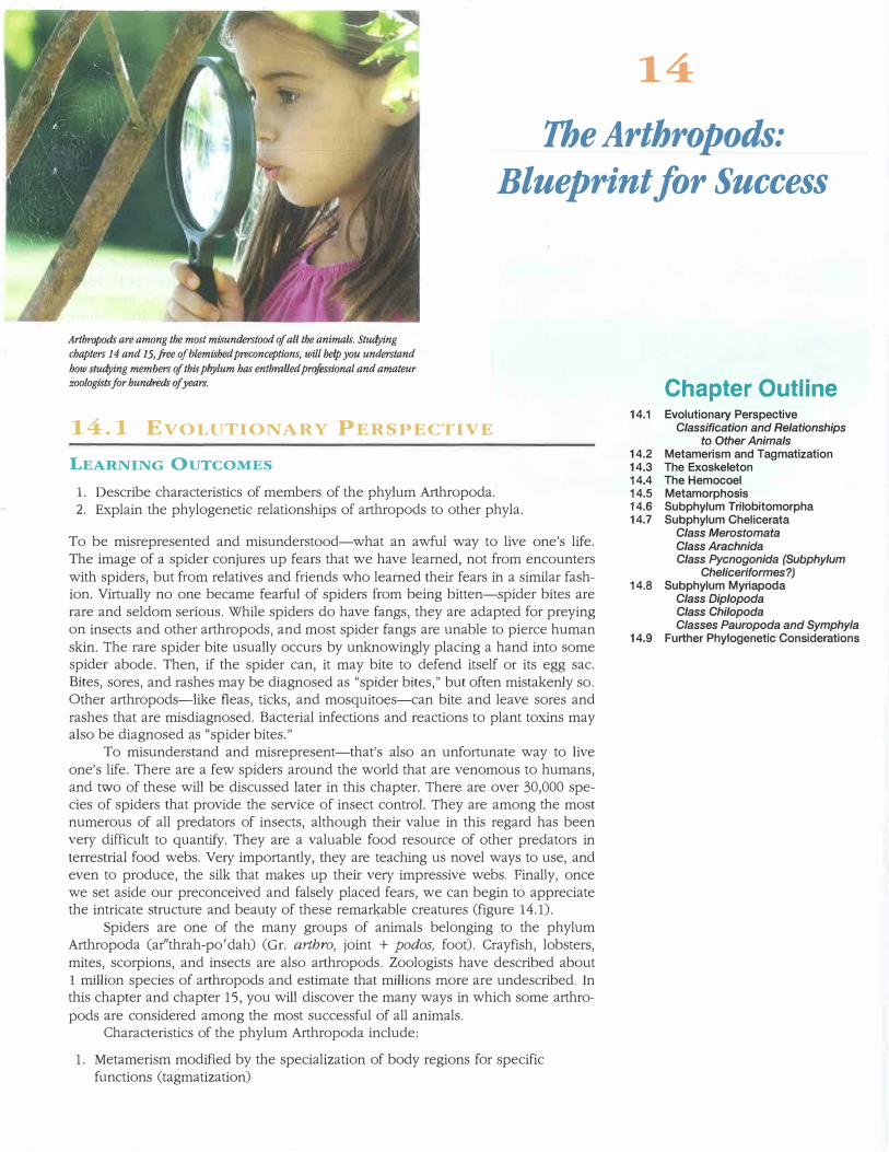
FIGURE 14.1
Class Arachnida, Order Araneae. Members of the family Araneidae, the orb weavers, produce some of the most beautiful and intricate spider webs. Many species are relatively large, like this garden spider-Aigiope. A web is not a permanent construction. When webs become wet with rain or dew, or when they age, they lose their stickiness. The entire web, or at least the spiraled portion, is then eaten and replaced.
2. Chitinous exoskeleton that provides support andprotection and is modified to form sensory structures
3. Paired, jointed appendages4. Growth accompanied by ccdysis or molting5. Ventral ne1vous system6. Coelom reduced to cavities surrounding gonads and
sometimes excretory organs7. Open circulatory system in which blood is released into
tissue spaces (hemocoel) derived from the blastocoel8. Complete digestive tract9. Metamorphosis often present; reduces competition
between immature and adult stages
Classification and Relationships to Other Animals
Arthropoda is a monophyletic taxon that is a part of the protosome clade Ecdysozoa. Arthropods are thus related to the Nematoda, Nematomorpha, Kinorhyncha, and others (figure 14.2 and see chapter 13). Synapomorphies for this clade include a cuticle, loss of epidermal cilia, and shedding the cuticle in a process called ecdysis. Two smaller phyla within the Ecdysozoa, Onychophora, and Tardigrada, are sister groups with the Atthropoda.
These three phyla form the ecdysozoan clade Panarthropoda (see figure 13.17.). The onychophorans and tardigrades, and their position within the Panarthropoda, will be discussed at the end of chapter 15.
There has been an explosion of new information regarding the evolutionary relationships within the phylum Arthropoda that is causing zoologists to reexamine current and older hypotheses regarding arthropod phylogeny. Living arthropods are divided into four subphyla: Chelicerata, Myriapoda, Hexapoda, and Crustacea. All members of a fifth subphylum, Trilobitomorpha, are extinct (table 14.1). Ideas regarding the evolutiona1y relationships among these subphyla are discussed at the end of chapter 15. This chapter examines Trilobitomorpha, Chelicerata, and Myriapoda, and chapter 15 covers Crustacea and Hexapoda.
SECTION REVIEW 14.1
The phylum A1thropoda includes crayfish, lobsters, spiders, insects, and others. Arthropods are characterized by metamerism with tagmatization, a chitinous exoskeleton, and paired jointed appendages. As ecdysozoans, arthropods are most closely related to the nematodes, nematomorphs, and other animals that shed a cuticle during growth. Five subphyla of arthropods have been described.
Why is metamerism, which is common to the Annelida
and Arthropoda, no longer considered to be evidence
of close evolutionary ties between the two phyla?
E'J'Al\-lEHISM
LEARNING OUTCOME
1. Compare arthropod metamerism with annelidmetamerism.
Four aspects of arthropod biology have contributed to their success. One of these is metamerism. Metamerism of arthropods is most evident externally because the arthropod body is often composed of a series of similar segments, each bearing a pair of appendages. Internally, however, septa do not divide the body cavity of an arthropod, and most organ systems are not metamerically arranged. The reason for the loss of internal metamerism is speculative; however, the presence of metamerically arranged hydrostatic compartments would be of little value in the support or locomotion of animals enclosed by an external skeleton (discussed under "The Exoskeleton").
As discussed in chapter 12, metamerism permits the specialization of regions of the body for specific functions. This regional specialization is called tagmatization. In arthropods, body regions, called tagmata (sing., tagma), are specialized for feeding and sensory perception, locomotion, and visceral functions.
Protists.
FIGURE 14.2
Basal Phyla
Lophotrochozoa
The Arthropods: Blueprint for Success 257
Ecdysozoa
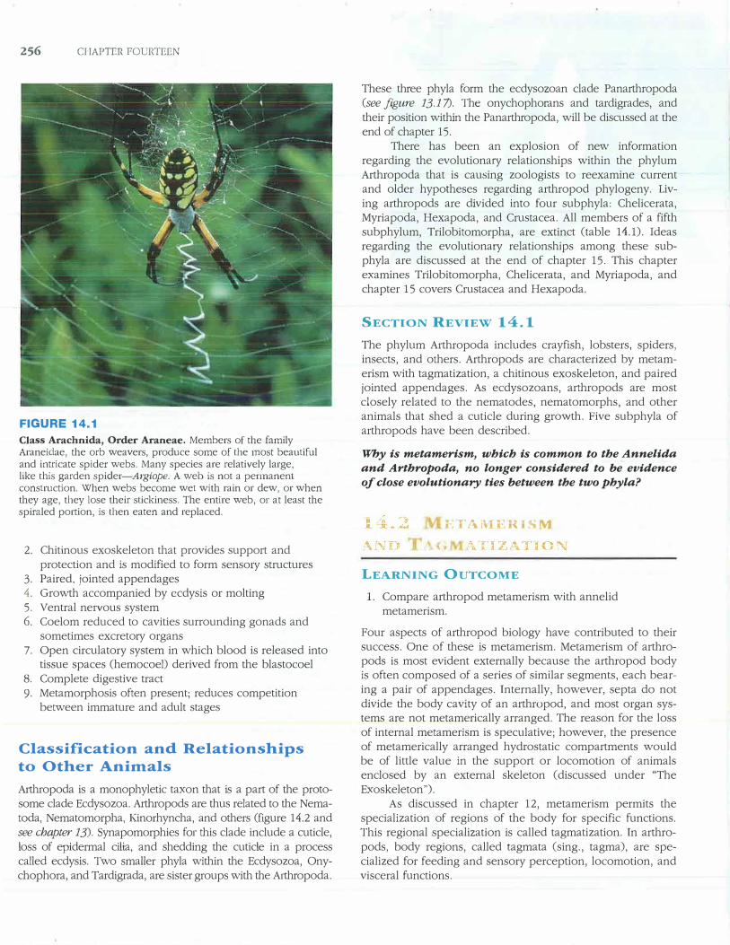
E olutlonacy Relationships of the Arthropods to Other Animals. This figure shows on' inLerprelation nr lh,e relationsl1ips or the ArLhropocl,1 rn oth··r mc::mhers ol' th �1nimal I illgd m. Th rel.1Lltmslllps tkpi ·te<l here ar-' I ased ou evidence rwm develop1m;ntal ancl mole ·uh1r biology. Arthrop id� are pl�1 ·et! wiLhi11 th·' Il:l.;dysozoa along wi1h rhe Nematoda. Nematornorph;t. ny h • pit rn, and others (see pa�es .n 1i-.,.:um. The r,li)'lum includes over L mill.ion spe iei:,. This sc<)rpion (B1tlf.111s sp.> bdongs to the �uhphylum Chelicern1a--ol1e of 1hc four arthropod subphyla containing living species. This photo was taken in the Sarah desert.
SECTION REVIEW 14.2
Metamerism of arthropods is most evident externally where body segmentation is most obvious and has been modified through tagmatization. Internal metamerism has been reduced.
How is the metamerism of arthropods different from
that observed in the Annelida (see chapter 12)?
l I )
LEARNING OUTCOMES
1. Describe the structure of the atthropod exoskeleton orcuticle.
2. Assess the influence the exoskeleton has had on the evolution of the arthropods.
An external, jointed skeleton, called an exoskeleton or cuticle, encloses arthropods. The exoskeleton is often cited as the major reason for arthropod success. It provides structural support, protection, impermeable surfaces for the prevention of water loss, and a system of levers for muscle attachment and movement.
The exoskeleton covers all body surfaces and invaginations of the body wall, such as the anterior and posterior
portions of the gut tract. It is nonliving and is secreted by a single layer of epidermal cells (figure 14.3). The epidermal layer is sometimes called the hypodermis because, unlike other epidermal tissues, it is covered on the outside by an exoskeleton, rather than being directly exposed to air or water.
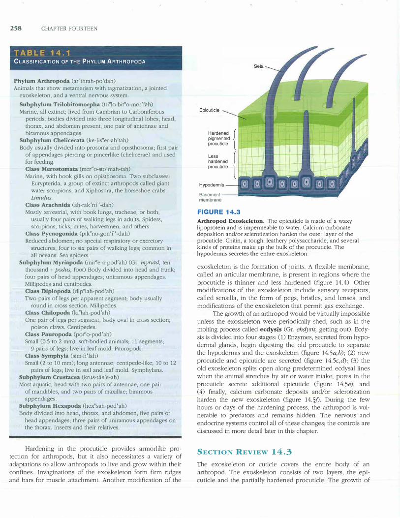
The exoskeleton has two layers. The epicuticle is the outermost layer. Made of a waxy lipoprotein, it is impermeable to water and a barrier to microorganisms and pesticides. The bulk of the exoskeleton is below the epicuticle and is called the procuticle. (In crustaceans, the procuticle is sometimes called the endocuticle.) The procuticle is composed of chitin, a tough, leathe1y polysaccharide, and several kinds of proteins. The procuticle hardens through a process called sclerotization and sometimes by impregnation with calcium carbonate. Sclerotization is a tanning process in which layers of protein are chemically cross-linked with one anotherhardening and darkening the exoskeleton. In insects and most other arthropods, this bonding occurs in the outer portion of the procuticle. The exoskeleton of crustaceans hardens by sclerotization and by the deposition of calcium carbonate in the middle regions of the procuticle. Some proteins give the exoskeleton resiliency. Distortion of the exoskeleton stores energy for such activities as flapping wings and jumping. The inner portion of the procuticle does not harden.
258 CHAPTER FOURTEEN
TABLE 14.1
CLASSIFICATION OF THE PHYLUM ARTHROPODA
Phylum Arthropoda (ar''thrah-po'dah) Animals that show metamerism with tagmatization, a jointed
exoskeleton, and a ventral nervous system.
Subphylum Trilobitomorpha (tri"lo-bit"o-mor'fah) Marine, all extinct; lived from Cambrian to Carboniferous
periods; bodies divided into three longitudinal lobes; head, thorax, and abdomen present; one pair of antennae and biramous appendages.
Subphylum Chelicerata (ke-lis"er-ah'tah) Body usually divided into prosoma and opisthosoma; first pair
of appendages piercing or pincerlike (chelicerae) and used for feeding. Class Merostomata (mer''o-sto'mah-tah) Marine, with book gills on opisthosoma. Two subclasses:
Eurypterida, a group of extinct arthropods called giant water scorpions, and Xiphosura, the horseshoe crabs. Limulus.
Class Arachnida (ah-rak'n1 '-dah) Mostly terrestrial, with book lungs, tracheae, or both;
usually four pairs of walking legs in adults. Spiders, scorpions, ticks, mites, harvestmen, and others.
Class Pycnogonida (pik" no-gon' 1 '-dah) Reduced abdomen; no special respiratory or excretory
structures; four to six pairs of walking legs; common in all oceans. Sea spiders.
Subphylum Myriapoda (mir''e-a-pod'ah) (Gr. myriad, ten thousand + podus, foot) Body divided into head and trunk; four pairs of head appendages; uniramous appendages. Millipedes and centipedes. Class Diplopoda (dip"lah-pod'ah) Two pairs of legs per apparent segment; body usually
round in cross section. Millipedes. Class Chilopoda (ki"lah-pod'ah) One pair of legs per segment; body oval in cw:;:; :;ecliun;
poison claws. Centipedes. Class Pauropoda (por''o-pod'ah) Small (0.5 to 2 mm), soft-bodied animals; 11 segments;
9 pairs of legs; live in leaf mold. Pauropods. Class Symphyla (sim-fi'lah) Small (2 to 10 mm); long antennae; centipede-like; 10 to 12
pairs of legs; live in soil and leaf mold. Symphylans. Subphylum Crustacea (krus-tas'e-ah) Most aquatic, head with two pairs of antennae, one pair
of mandibles, and two pairs of maxillae; biramous appendages.
Subphylum Hexapoda (hex''sah-pod' ah) Body divided into head, thorax, and abdomen; five pairs of
head appendages; three pairs of uniramous appendages on
the thorax. Insects and their relatives.
Hardening in the procuticle provides armorlike protection for arthropods, but it also necessitates a variety of adaptations to allow arthropods to live and grow within their
confines. Invaginations of the exoskeleton form firm ridges and bars for muscle attachment. Another modification of the
Epicuticle
Hardened pigmented procuticle
Less hardened procuticle
Hypodermls
Basement membrane
FIGURE 14.3
Arthropod Exoskeleton. The epicuticle is made of a waxy lipoprotein and is impermeable to water. Calcium carbonate deposition and/or sclerotization harden the outer layer of the procuticle. Chitin, a tough, leathery polysaccharide, and several kinds of proteins make up the bulk of the procuticle. The hypodermis secretes the entire exoskeleton.
exoskeleton is the formation of joints. A flexible membrane, called an articular membrane, is present in regions where the
procuticle is thinner and less hardened (figure 14.4). Other modifications of the exoskeleton include sensory receptors, called sensilla, in the form of pegs, bristles, and lenses, and
modifications of the exoskeleton that permit gas exchange. The growth of an arthropod would be virtually impossible
unless the exoskeleton were periodically shed, such as in the molting process called ecdysis (Gr. ekdysis, getting out). Ecdysis is divided into four stages: (1) Enzymes, secreted from hypodermal glands, begin digesting the old procuticle to separate the hypodermis and the exoskeleton (figure 14.Sa,h); (2) new procuticle and epicuticle are secreted (figure 14.Sc,d); (3) the old exoskeleton splits open along predetermined ecdysal lines when the animal stretches by air or water intake; pores in the
procuticle secrete additional epicuticle (figure 14.Se); and (4) finally, calcium carbonate deposits and/or sclerotizationharden the new exoskeleton (figure 14.5.f). During the fewhours or days of the hardening process, the arthropod is vulnerable to predators and remains hidden. The nervous andendocrine systems control all of these changes; the controls are
discussed in more detail later in this chapter.
SECTION REVIEW 14.3
The exoskeleton or cuticle covers the entire body of an
arthropod. The exoskeleton consists of two layers, the epi
cuticle and the partially hardened procuticle. The growth of
(a)
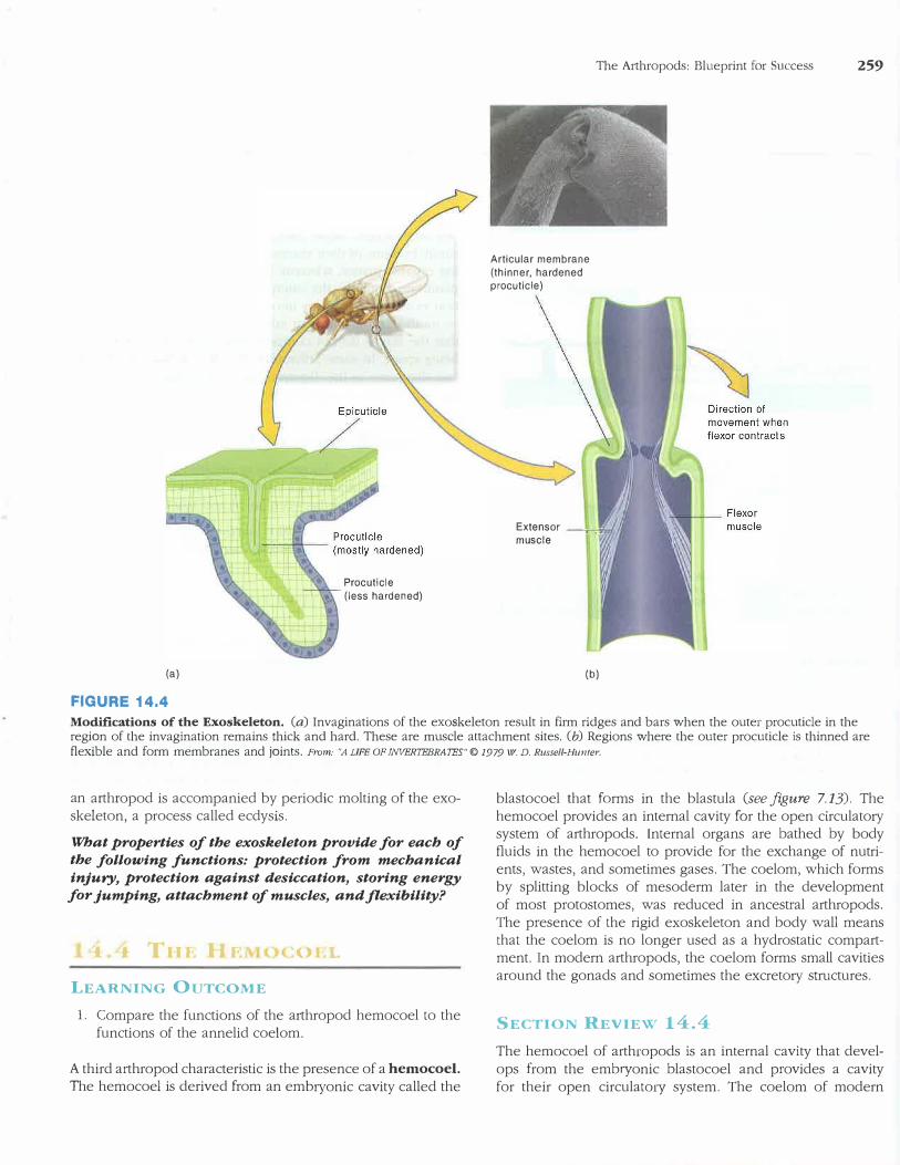
FIGURE 14.4
Procuticle (less hardened)
Articular membrane (thinner, hardened procuticle)
The Arthropods: Blueprint for Success 259
Extensor ---:,----:,;m muscle
(b)
Modifications of the Exoskeleton. (a) Invaginations of the exoskeleton result in firm ridges and bars when the outer procuticle in the region of the invagination remains thick and hard. These are muscle attachment sites. (b) Regions where the outer procuticle is thinned are flexible and form membranes and joints. From: ''.A LIFE OF INVERTEBRATES"© 1979 w. D. Russell-Hunter.
an arthropod is accompanied by periodic molting of the exoskeleton, a process called ecdysis.
What properties of the exoskeleton provide for each of the following functions: protection from mechanical
injury, protection against desiccation, storing energy for jumping, attachment of muscles, and flexibility?
1 1 Tur. llr Of.L
LEARNING OUTCOME
1. Compare the functions of the a1thropod hemocoel to thefunctions of the annelid coelom.
A third arthropod characteristic is the presence of a hemocoel.
The hemocoel is derived from an embryonic cavity called the
blastocoel that forms in the blastula (see figure 7.13). The hemocoel provides an internal cavity for the open circulato1y system of arthropods. Internal organs are bathed by body fluids in the hemocoel to provide for the exchange of nutrients, wastes, and sometimes gases. The coelom, which forms by splitting blocks of mesoderm later in the development of most protostomes, was reduced in ancestral arthropods. The presence of the rigid exoskeleton and body wall means that the coelom is no longer used as a hydrostatic compartment. In modern arthropods, the coelom forms small cavities around the gonads and sometimes the excreto1y structures.
SECTION REVIEW 14.4
The hemocoel of arthropods is an internal cavity that develops from the embryonic blastocoel and provides a cavity for their open circula to1y system. The coelom of modern
260 CHAPTER FOURTEEN
(a)
Molting gel
Lew epicuticle and procuticle
(b)
Hypodermis -
(c)
Shed epicuticle and procuticle
----,- Secretory
(e)
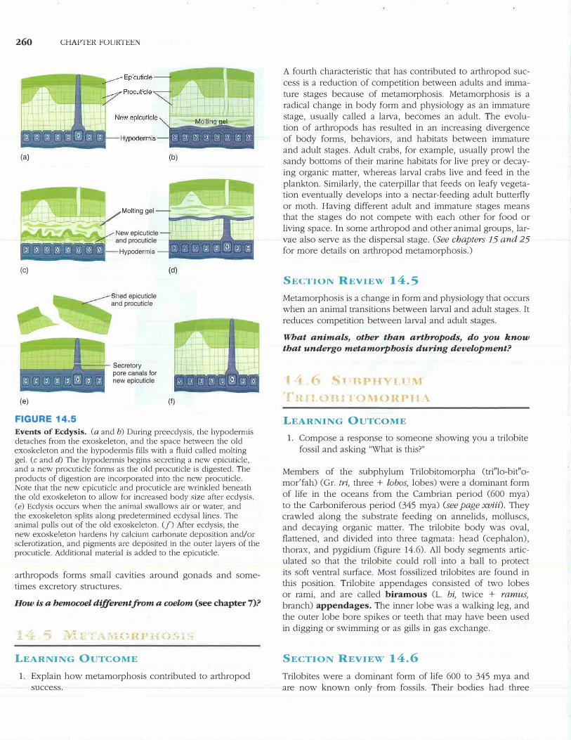
FIGURE 14.5
pore canals for new epicuticle
(d)
(f)
Events of Ecdysis. (a and b) During preecdysis, the hypodermis detaches from the exoskeleton, and the space between the old exoskeleton and the hypodermis fills with a fluid called molting gel. (c and d) The hypodermis begins secreting a new epicuticle, and a new procuticle forms as the old procuticle is digested. The products of digestion are incorporated into the new procuticle. Note that the new epicuticle and procuticle are wrinkled beneath the old exoskeleton to allow for increased body size after ecdysis. (e) Ecdysis occurs when the animal swallows air or water, andthe exoskeleton splits along predetermined ecdysal lines. Theanimal pulls out of the old exoskeleton. Cf) After ecdysis, thenew exoskeleton hardens hy calcium carbonate deposition and/orsclerotization, and pigments are deposited in the outer layers of theprocuticle. Additional material is added to the epicuticle.
arthropods forms small cavities around gonads and sometimes excretory structures.
How is a hemocoel different from a coelom (see chapter 7)?
LEARNING OUTCOME
1. Explain how metamorphosis contributed to arthropodsuccess.
A fourth characteristic that has contributed to arthropod success is a reduction of competition between adults and immature stages because of metamorphosis. Metamorphosis is a radical change in body form and physiology as an immature stage, usually called a larva, becomes an adult. The evolution of arthropods has resulted in an increasing divergence of body forms, behaviors, and habitats between immature and adult stages. Adult crabs, for example, usually prowl the sandy bottoms of their marine habitats for live prey or decaying organic matter, whereas larval crabs live and feed in the plankton. Similarly, the caterpillar that feeds on leafy vegetation eventually develops into a nectar-feeding adult butterfly or moth. Having different adult and immature stages means that the stages do not compete with each other for food or living space. In some arthropod and other animal groups, larvae also serve as the dispersal stage. (See chapters 15 and 25 for more details on arthropod metamorphosis.)
SEl.:TlON REVIEW 14.5
Metamorphosis is a change in form and physiology that occurs when an animal transitions between larval and adult stages. It reduces competition between larval and adult stages.
What animals, other than arthropods, do you know
that undergo metamorphosis during development?
14 -6 Sl!lJ.PHYI lTM
'fn_a OBITOMOUPlIA
LEARNING OUTCOME
1. Compose a response to someone showing you a trilobitefossil and asking "What is this?"
Members of the subphylum Trilobitomorpha (tri"lo-bit"omor'fah) (Gr. tri, three + !obos, lobes) were a dominant form of life in the oceans from the Cambrian period (600 mya) to the Carboniferous period (345 mya) (see page xviii). They crawled along the substrate feeding on annelids, molluscs, and decaying organic matter. The trilobite body was oval, flattened, and divided into three tagmata: head (cephalon), thorax, and pygidium (figure 14.6). All body segments articulated so that the trilobite could roll into a ball to protect its soft ventral surface. Most fossilized trilobites are found in this position. Trilobite appendages consisted of two lobes or rami, and are called biramous (L. bi, twice + ramus,
branch) appendages. The inner lobe was a walking leg, and the outer lobe bore spikes or teeth that may have been used in digging or swimming or as gills in gas exchange.
SECTION REVIEW 14.6
Trilobites were a dominant form of life 600 to 345 mya and are now known only from fossils. Their bodies had three
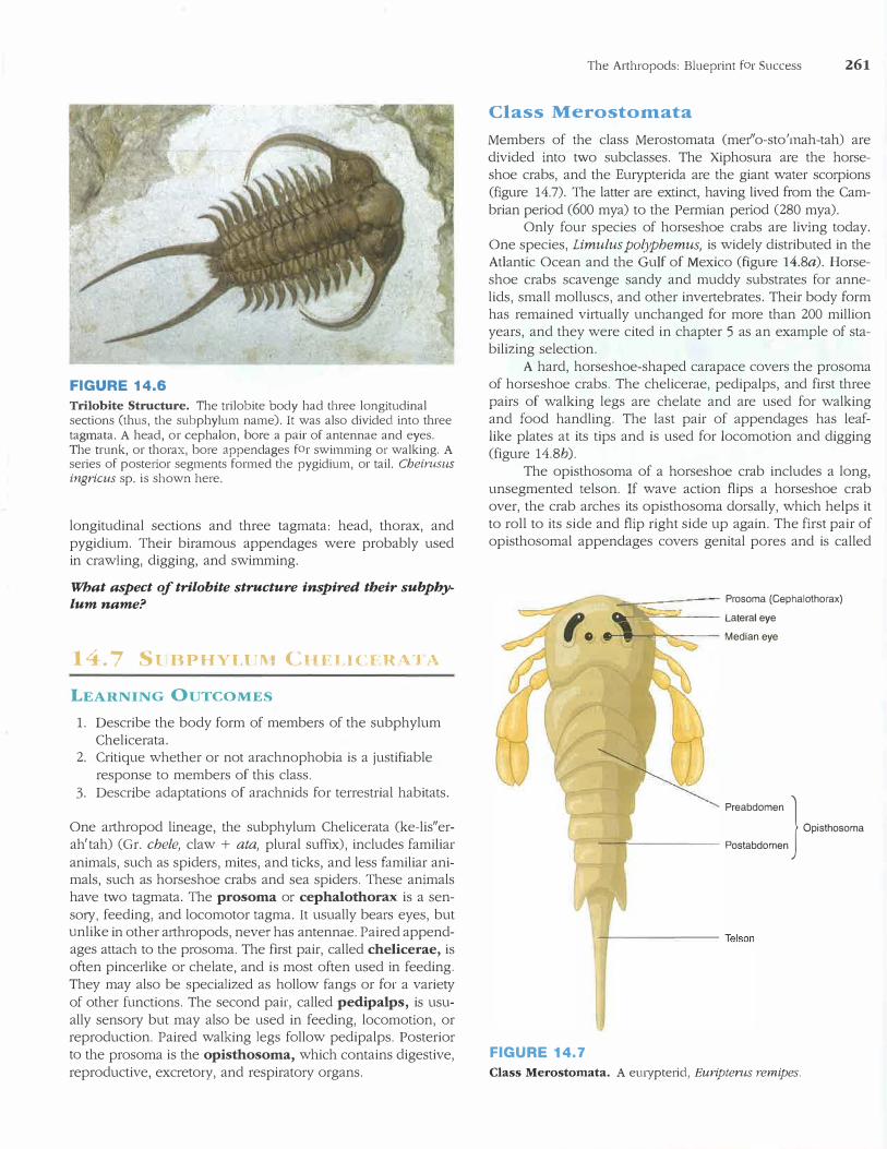
FIGURE 14.6
Trilobite Structure. The trilobite body had three longitudinal sections (thus, the subphylum name). It was also divided into three tagmata. A head, or cephalon, bore a pair of antennae and eyes. The trunk, or thorax, bore appendages for swimming or walking. A series of posterior segments formed the pygidium, or tail. Cheimsus ingricus sp. is shown here.
longitudinal sections and three tagmata: head, thorax, and pygidium. Their biramous appendages were probably used in crawling, digging, and swimming.
What aspect of trilobite structure inspired their subpby
lum name?
14.7 St BPHYLl"\I CHFLJCfR \.'I A
LEARNING OUTCOMES
1. Describe the body form of members of the subphylumChelicerata.
2. Critique whether or not arachnophobia is a justifiableresponse to members of this class.
3. Describe adaptations of arachnids for terrestrial habitats.
One a1thropod lineage, the subphylum Chelicerata (ke-lis"erah'tah) (Gr. chele, claw + ata, plural suffix), includes familiar animals, such as spiders, mites, and ticks, and less familiar animals, such as horseshoe crabs and sea spiders. These animals have two tagmata. The prosoma or cephalothorax is a sens01y, feeding, and locomotor tagma. It usually bears eyes, but unlike in other arthropods, never has antennae. Paired appendages attach to the prosoma. The first pair, called chelicerae, is often pincerlike or chelate, and is most often used in feeding. They may also be specialized as hollow fangs or for a variety of other functions. The second pair, called pedipalps, is usually sensory but may also be used in feeding, locomotion, or reproduction. Paired walking legs follow pedipalps. Posterior to the prosoma is the opisthosoma, which contains digestive, reproductive, excretory, and respiratory organs.
The Arthropods: Blueprint for Success 261
Class Merostomata
Members of the class Merostomata (mer'' o-sto' mah-tah) are divided into two subclasses. The Xiphosura are the horseshoe crabs, and the Eurypterida are the giant water scorpions (figure 14.7). The latter are extinct, having lived from the Cambrian period (600 mya) to the Permian period (280 mya).
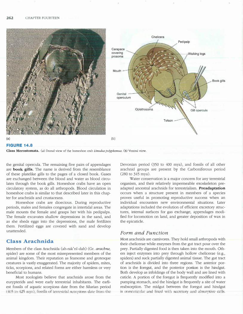
Only four species of horseshoe crabs are living today. One species, Limulus polyphemus, is widely distributed in the Atlantic Ocean and the Gulf of Mexico (figure 14.8a). Horseshoe crabs scavenge sandy and muddy substrates for annelids, small molluscs, and other invertebrates. Their body form has remained virtually unchanged for more than 200 million years, and they were cited in chapter 5 as an example of stabilizing selection.
A hard, horseshoe-shaped carapace covers the prosoma of horseshoe crabs. The chelicerae, pedipalps, and first three pairs of walking legs are chelate and are used for walking and food handling. The last pair of appendages has leaflike plates at its tips and is used for locomotion and digging (figure 14.8b).
The opisthosoma of a horseshoe crab includes a long, unsegmented telson. If wave action flips a horseshoe crab over, the crab arches its opisthosoma dorsally, which helps it to roll to its side and flip right side up again. The first pair of opisthosomal appendages covers genital pores and is called
FIGURE 14.7
?.._�:-�f�-;-;-�-,__-_ -_-_- Prosoma (Cephalothorax)
Ill Lateral eye .---,----:-=;�--- Median eye
Preabdomen
} Opisthosoma ------- - Postabdomen
Class Merostomata. A ernypterid, Eu1·ipterus remipes.
262 CHAPTER FOURTEEN
(a)
FIGURE 14.8
Carapace covering prosoma
Mouth
(b)
Genital operculum
Chelicera
Book gills
Class Merostomata. (a) Dorsal view of the horseshoe crab Limulus polyphemus. (b) Ventral view.
the genital opercula. The remaining five pairs of appendages are book gills. The name is derived from the resemblance of these platelike gills to the pages of a closed book. Gases are exchanged between the blood and water as blood circulates through the book gills. Horseshoe crabs have an open circulatory system, as do all arthropods. Blood circulation in horseshoe crabs is similar to that described later in this chapter for arachnids and crustaceans.
Horseshoe crabs are dioecious. During reproductive periods, males and females congregate in intertidal areas. The male mounts the female and grasps her with his pedipalps. The female excavates shallow depressions in the sand, and as she sheds eggs into the depressions, the male fertilizes them. Fertilized eggs are covered with sand and develop unattended.
Class Arachnida
Members of the class Arachnida (ah-rak'n1-dah) (Gr. arachne,
spider) are some of the most misrepresented members of the animal kingdom. Their reputation as fearsome and grotesque creatures is vastly exaggerated. The majority of spiders, mites, ticks, scorpions, and related forms are either harmless or very beneficial to humans.
Most zoologists believe that arachnids arose from the eurypterids and were early terrestrial inhabitants. The earliest fossils of aquatic scorpions date from the Silurian period ( 05 m 25 my�1) fossils or 1:errestriaJ scorpions date from t.he
Devonian period (350 to 400 mya), and fossils of all other arachnid groups are present by the Carboniferous period (280 to 345 mya).
Water conservation is a major concern for any terrestrial organism, and their relatively impermeable exoskeleton preadapted ancestral arachnids for terrestrialism. Preadaptation
occurs when a structure present in members of a species proves useful in promoting reproductive success when an individual encounters new environmental situations. Later adaptations included the evolution of efficient excretory strnctures, internal surfaces for gas exchange, appendages modified for locomotion on land, and greater deposition of wax in the epicuticle.
Form and Function
Most arachnids are carnivores. They hold small arthropods with their chelicerae while enzymes from the gut tract pour over the prey. Partially digested food is then taken into the mouth. Others inject enzymes into prey through hollow chelicerae (e.g., spiders) and suck partially digested animal tissue. The gut tract of arachnids is divided into three regions. The anterior portion is the foregut, and the posterior portion is the hindgut. Both develop as infoldings of the body wall and are lined with cuticle. A portion of the foregut is frequently modified into a pumping stomach, and the hindgut is frequently a site of water reabsorption. The midgut between the foregut and hindgut is t1()nculi ular ·md lined witb sco"to1y a_nd abso,,plive c !Is.
Lateral dive1ticula increase the area available for absorption and storage.
Arachnids use coxal glands and/or Malpighian tubules for excreting nitrogenous wastes. Coxal glands are paired, thin-walled, spherical sacs bathed in the blood of body sinuses. Coxal glands are probably homologous to nephridia. Nitrogenous wastes are absorbed across the wall of the sacs, transported in a long, convoluted tubule, and excreted through excretory pores at the base of the posterior appendages. Arachnids that are adapted to dry environments possess blind-ending diverticula of the gut tract that arise at the juncture of the midgut and hindgut. These tubules, called Malpighian tubules, absorb waste materials from the blood and empty them into the gut tract. Excretory wastes are then eliminated with digestive wastes. The major excretoty product of arachnids is uric acid. As discussed in chapter 28, uric acid excretion is advantageous for terrestrial animals because uric acid is excreted as a semisolid with little water loss.
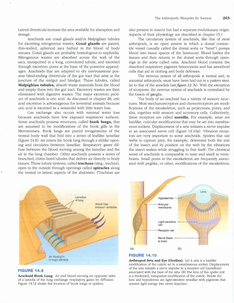
Gas exchange also occurs with minimal water loss because arachnids have few exposed respiratory surfaces. Some arachnids possess stmctures, called book lungs, that are assumed to be modifications of the book gills in the Merostomata. Book lungs are paired invaginations of the ventral body wall that fold into a series of leaflike lamellae (figure 14.9). Air enters the book lung through a slitlike opening and circulates between lamellae. Respiratory gases diffuse between the blood moving among the lamellae and the air in the lung chamber. Other arachnids possess a series of branched, chitin-lined tubules that deliver air directly to body tissues. These tubule systems, called tracheae (sing., trachea), open to the outside through openings called spiracles along the ventral or lateral aspects of the abdomen. (Tracheae are
FIGURE 14.9
Air tlowl11g 11'1 thro_ugh splraole
Bloodllow between lameUae
Arachnid Book Lung. Air and blood moving on opposite sides of a lamella of the lung exchange respiratory gases by diffusion. Figure 14.12 shows the location of book lungs in spiders.
The Arthropods: Blueprint for Success 263
also present in insects but had a separate evolutiona1y origin. Aspects of their physiology are described in chapter 15.)
The circulatory system of arachnids, like that of most a1thropods, is an open system in which a dorsal contractile vessel (usually called the dorsal aorta or "heart") pumps blood into tissue spaces of the hemocoel. Blood bathes the tissues and then returns to the dorsal aorta through openings in the aorta called ostia. Arachnid blood contains the dissolved respiratory pigment hemocyanin and has amoeboid cells that aid in clotting and body defenses.
The nervous system of all arthropods is ventral and, in ancestral aithropods, must have been laid out in a pattern similar to that of the annelids (see figure 12. 7a). With the exception of scorpions, the nervous system of arachnids is centralized by the fusion of ganglia.
The body of an arachnid has a variety of sensory structures. Most mechanoreceptors and chemoreceptors are modifications of the exoskeleton, such as projections, pores, and slits, together with sensory and accesso1y cells. Collectively, these receptors are called sensilla. For example, setae are hairlike, cuticular modifications that may be set into membranous sockets. Displacement of a seta initiates a nerve impulse in an associated nerve cell (figure 14.lOa). Vibration receptors are ve1y important to some arachnids. Spiders that use webs to capture prey, for example, determine both the size of the insect and its position on the web by the vibrations the insect makes while stmggling to free itself. The chemical sense of arachnids is comparable to taste and smell in ve1tebrates. Small pores in the exoskeleton are frequently associated with peglike, or other, modifications of the exoskeleton,
---Articular membrane
Light-sensitive ----,,-cell
..---------Receptor cell
Nerve fibers ------+to brain
(a) (b)
FIGURE 14.10
Arthropod Seta and Eye (Ocellus). (a) A seta is a hairlike modification of the cuticle set in a membranous socket. Displacement of the seta initiates a nerve impulse in a receptor cell (sensillum) associated with the base of the seta. (b) The lens of this spider eye is a thickened, transparent modification of the cuticle. Below the lens and hypodermis are light-sensitive sensillae with pigments that convert light energy into nerve impulses.
264 CHAPTER FOURTEEN
and they allow chemicals to stimulate nerve cells. Arachnids possess one or more pairs of eyes, which they use primarily for detecting movement and changes in light intensity (figure 14.l0b). The eyes of some hunting spiders probably form images.
Arachnids are dioecious. Paired genital openings are on the ventral side of the second abdominal segment. Sperm transfer is usually indirect. The male often packages sperm in a spermatophore, which is then transferred to the female. Courtship rituals confirm that individuals are of the same species, attract a female to the spermatophore, and position the female to receive the spermatophore. In some taxa (e.g., spiders), copulation occurs, and sperm is transferred via a modified pedipalp of the male. Development is direct, and the young hatch from eggs as miniature adults. Many arachnids tend their developing eggs and young during and after development.
Order Scorpionida
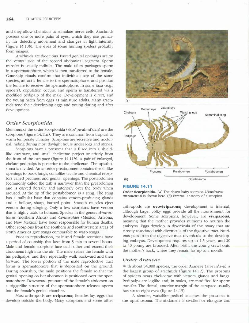
Members of the order Scorpionida (skor''pe-ah-ni' dah) are the scorpions (figure 14.lla). They are common from tropical to warm temperate climates. Scorpions are secretive and nocturnal, hiding during most daylight hours under logs and stones.
Scorpions have a prosoma that is fused into a shieldlike carapace, and small chelicerae project anteriorly from the front of the carapace (figure 14.llb). A pair of enlarged, chelate pedipalps is posterior to the chelicerae. The opisthosoma is divided. An anterior preabdomen contains the slitlike openings to book lungs, comblike tactile and chemical receptors called pectines, and genital openings. The postabdomen (commonly called the tail) is narrower than the preabdomen and is curved dorsally and anteriorly over the body when aroused. At the tip of the postabdomen is a sting. The sting has a hulhular hase that contain.<; w�nom-producing glands and a hollow, sharp, barbed point. Smooth muscles eject venom during stinging. Only a few scorpions have venom that is highly toxic to humans. Species in the genera Androc
tonus (northern Africa) and Centuroides (Mexico, Arizona, and New Mexico) have been responsible for human deaths. Other scorpions from the southern and southwestern areas of Notth America give stings comparable to wasp stings.
Prior to reproduction, male and female scorpions have a period of courtship that lasts from 5 min to several hours. Male and female scorpions face each other and extend their abdomens high into the air. The male seizes the female with his pedipalps, and they repeatedly walk backward and then forward. The lower portion of the male reproductive tract forms a spermatophore that is deposited on the ground. During courtship, the male positions the female so that the genital opening on her abdomen is positioned over the spermatophore. Downward pressure of the female's abdomen on a triggerlike structure of the spermatophore releases sperm into the female's genital chamber.
Most arthropods are oviparous; females lay eggs that c.levelop utsl I th ho )y. !Vhuly :, ·tirpion:;. anc.l .mme t ther
Median eye Lateral eye
Chelicera
Prosoma Preabdomen Postabdomen
(b) Opisthosoma
FIGURE 14.11
Order Scorpionida. (a) The dese1t haity scorpion (Hardrurus
arizonensis) is shown here. (b) External anatomy of a scorpion.
arthropods are ovoviviparous; development is internal, although large, yolky eggs provide all the nourishment for development. Some scorpions, however, are viviparous,
meaning that the mother provides nutrients to nourish the emb1yos. Eggs develop in diverticula of the ova1y that are
closely associated with diverticula of the digestive tract. Nutrients pass from the digestive tract diverticula to the developing embryos. Development requires up to 1.5 years, and 20 to 40 young are brooded. After birth, the young crawl onto the mother's back, where they remain for up to a month.
Order Araneae
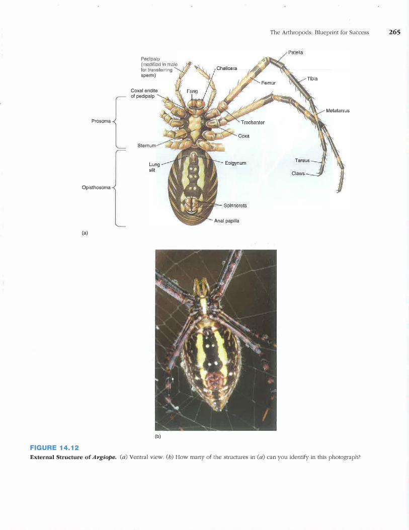
With about 34,000 species, the order Araneae (ah-ran'a-e) is the largest group of arachnids (figure 14.12). The prosoma of spiders bears chelicerae with venom glands and fangs. Pedipalps are leglike and, in males, are modified for sperm transfer. The dorsal, anterior margin of the carapace usually has six to eight eyes (figure 14.13).
A slender, waistlike pedicel attaches the prosoma to the-opisthosoma-:- -The---abdomen-is--swollen- or-elongate-and
Prosoma
Opisthosoma
(a)
Ped)palp (modnied in male for transferring sperm)
Coxal endite of pedipalp
Sternum
(b)
FIGURE 14.12
Anal papilla
The Arthropods: Blueprint for Success 265
External Structure of Argiope. (a) Ventral view. (b) How many of the structures in (a) can you identify in this photograph?
266 CHAPTER FOURTEEN
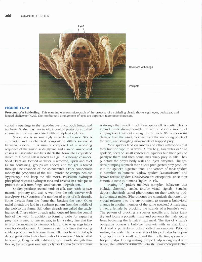
FIGURE 14.13
Eyes
Chelicera with fangs
Pedipalp
Prosoma of a Spiderling. This scanning electron micrograph of the prosoma of a spiderling clearly shows eight eyes, pedipalps, and fanged chelicerae (X20). The number and arrangement of eyes are important taxonomic characters.
contains openings to the reproductive tract, book lungs, and tracheae. It also has two to eight conical projections, called spinnerets, that are associated with multiple silk glands.
Spider silk is an amazingly versatile substance. Silk is a protein, and its chemical composition differs somewhat between species. It is usually composed of a repeating sequence of the amino acids glycine and alanine. Amino acid chains self-assemble into beta sheets that form into a crystalline structure. Unspun silk is stored as a gel in a storage chamber. Solid fibers are formed as water is removed, lipids and thiol (sulfur containing) groups are added, and the gel is forced through fine channels of the spinnerettes. Other compounds modify the properties of the silk. Pyrrolidine compounds are hygroscopic and keep the silk moist. Potassium hydrogen phosphate releases hydrogen ions and creates an acidic pH to protect the silk from fungal and bacterial degradation.
Spiders produce several kinds of silk, each with its own material property and use. A web like the orb weaver web (see.figure 14.1) consists of a number of types of silk threads. Some threads form the frame that borders the web. Other radial threads are laid in a sunburst pattern from the middle of the web to the frame. Still other threads are called the catching spiral. These sticky threads spiral outward from the central hub of the web. In addition to forming webs for capturing prey, silk is used to line retreats, to lay a safety line that fastens to the substrate to interrupt a fall, and to wrap eggs into a case for development. Air currents catch silk lines that young spiders produce and disperse them. Silk lines have carried spiders at great altitudes for hundreds of kilometers. This is called ballooning. Dragline silk exhibits greater tensile strength than Kevlaf;-the 'lrong s1 synih "lie polymer known (wnicnin turn
is stronger than steel). In addition, spider silk is elastic. Elasticity and tensile strength enable the web to stop the motion of a flying insect without damage to the web. Webs also resist damage from the wind, movements of the anchoring points of the web, and struggling movements of trapped prey.
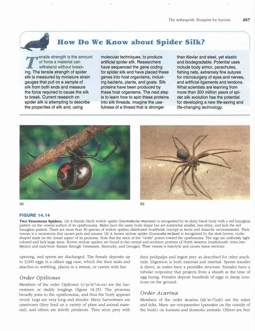
Most spiders feed on insects and other arthropods that they hunt or capture in webs. A few (e.g., tarantulas or "bird spiders") feed on small vertebrates. Spiders bite their prey to paralyze them and then sometimes wrap prey in silk. They puncture the prey's body wall and inject enzymes. The spider's pumping stomach then sucks predigested prey products into the spider's digestive tract. The venom of most spiders is harmless to humans. Widow spiders (Lactrodectus) and brown recluse spiders (Loxosceles) are exceptions, since their venom is toxic to humans (figure 14.14).
Mating of spiders involves complex behaviors that include chemical, tactile, and/or visual signals. Females deposit chemicals called pheromones on their webs or bodies to attract males. (Pheromones are chemicals that one indi
vidual releases into the environment to create a behavioral change in another member of the same species.) A male may attract a female by plucking the strands of a female's web. The pattern of plucking is species specific and helps identify and locate a potential mate and prevents the male spider from becoming the female's next meal. The tips of a male's pedipalps possess a bulblike reservoir with an ejaculatory duct and a penislike structure called an embolus. Prior to mating, the male fills the reservoir of his pedipalps by depositing sperm on a small web and then collecting sperm with his pedipalps. During mating, the pedipalp is engorged with blood, Lh� emoolus1s1nsertecl i.nto rhe fomale's-repToductive-
The Arthropods: Blueprint for Success 267
How Do We Know about Spider Silk? I
(a)
Tensile strength is the amount of force a material can withstand without break-
ing. The tensile strength of spider silk is measured by miniature strain gauges that pull on a sample of silk from both ends and measure the force required to cause the silk to break. Current research on spider silk is attempting to describe the properties of silk and, using
FIGURE 14.14
molecular techniques, to produce artificial spider silk. Researchers have sequenced the gene coding for spider silk and have placed these genes into host organisms, including bacteria, plants, and goats. Silk proteins have been produced by these host organisms. The next step is to learn how to spin these proteins into silk threads. Imagine the usefulness of a thread that is stronger
(b)
than Kevlar and steel, yet elastic and biodegradable. Potential uses include body armor, parachutes, fishing nets, extremely fine sutures for microsurgery of eyes and nerves, and artificial ligaments and tendons. What scientists are learning from more than 300 million years of spider silk evolution has the potential for developing a new life-saving and life-changing technology.
Two Venomous Spiders. (a) A female black widow spider (Lactrodectus mactans) is recognized by its shiny black body with a red hourglass pattern on the ventral surface of its opisthosoma. Males have the same body shape but are somewhat smaller, less shiny, and lack the red hourglass pattern. There are more than 30 species of widow spiders distributed worldwide (except in Arctic and Antarctic environments). Their venom is a neurotoxin that causes pain and nausea. (b) A brown recluse spider (Loxosceles reclusa) is recognized by the dark brown, violinshaped mark on the dorsal aspect of its prosoma. Note that the neck of the "violin" points toward the opsthosoma. The legs are uniformly light colored and lack large setae. Brown recluse spiders are found in the central and southern portions of North America (north/south: Iowa into Mexico and east/west: Kansas through Tennessee, Kentucky, and Georgia). Their venom is histolytic and causes tissue necrosis.
opening, and sperm are discharged. The female deposits up to 3,000 eggs in a silken egg case, which she then seals and attaches to webbing, places in a retreat, or carries with her.
Order Opiliones
Members of the order Opiliones (o'pi-le"on-es) are the harvestmen or daddy longlegs (figure 14.15). The prosoma broadly joins to the opisthosoma, and thus the body appears ovoid. Legs are very long and slender. Many harvestmen are omnivores (they feed on a variety of plant and animal material), and others are strictly predators. They seize prey with
their pedipalps and ingest prey as described for other arachnids. Digestion is both external and internal. Sperm transfer is direct, as males have a penislike structure. Females have a tubular ovipositor that projects from a sheath at the time of egg laying. Females deposit hundreds of eggs in damp locations on the ground.
Order Acarina
Members of the order Acarina (ak' ar-i"nah) are the mites and ticks. Many are ectoparasites (parasites on the outside of the body) on humans and domestic animals. Others are free
268 CHAPTER FOURTEEN
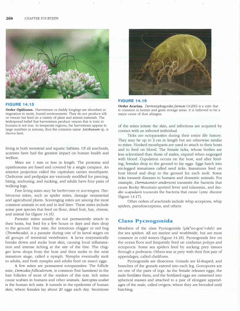
FIGURE 14.15
Order Opiliones. Harvestmen or daddy longlegs are abundant in vegetation in moist, humid environments. They do not produce silk or venom but feed on a variety of plant and animal materials. The widespread belief that harvestmen produce venom that is toxic to humans is not tme. In temperate regions, the haivestmen appear in large numbers in autumn, thus the common name. Leiobunum sp. is shown here.
living in both terrestrial and aquatic habitats. Of all arachnids, acarines have had the greatest impact on human health and welfare.
Mites are 1 mm or less in length. The prosoma and opisthosoma are fused and covered by a single carapace. An anterior projection called the capitulum carries mouthparts. Chelicerae and pedipalps are variously modified for piercing, biting, anchoring, and sucking, and adults have four pairs of walking legs.
Free living mites may be herbivores or scavengers. Herbivorous mites, such as spider mites, damage ornamental and agricultural plants. Scavenging mites are among the most common animals in soil and in leaf litter. These mites include some pest species that feed on flour, dried fruit, hay, cheese, and animal fur (figure 14.16).
Parasitic mites usually do not permanently attach to their hosts, but feed for a few hours or days and then drop to the ground. One mite, the notorious chigger or red bug (Trombicula), is a parasite during one of its larval stages on all groups of terrestrial vertebrates. A larva enzymatically breaks down and sucks host skin, causing local inflammation and intense itching at the site of the bite. The chigger larva drops from the host and then molts to the next immature stage, called a nymph. Nymphs eventually molt to adults, and both nymphs and adults feed on insect eggs.
A few mites are permanent ectoparasites. The follicle mite, Demodexfolliculorum, is common (but harmless) in the hair follicles of most of the readers of this text. Itch mites cause scabies in humans and other animals. Sarcoptes scabei
is the human itch mite. It tunnels in the epidermis of human skin, where females lay about 20 eggs each day. Secretions
FIGURE 14.16
Order Acarina. Dermatophagoidesfarinae (X200) is a mite that is common in homes and grain storage areas. It is believed to be a major cause of dust allergies.
of the mites irritate the skin, and infections are acquired by contact with an infected individual.
Ticks are ectoparasites during their entire life history. They may be up to 3 cm in length but are otherwise similar to mites. Hooked mouthparts are used to attach to their hosts and to feed on blood. The female ticks, whose bodies are less sclerotized than those of males, expand when engorged with blood. Copulation occurs on the host, and after feeding, females drop to the ground to lay eggs. Eggs hatch into six-legged immatures called seed ticks. Immatures feed on host blood and drop to the ground for each molt. Some ticks transmit diseases to humans and domestic animals. For example, Dermacentor andersoni transmits the bacteria that cause Rocky Mountain spotted fever and tularemia, and Ixodes scapularis tia11sinits the bacteria that cause Lyme disease (figure 14.17).
Other orders of arachnids include whip scorpions, whip spiders, pseudoscorpions, and others.
Class Pycnogonida
Members of the class Pycnogonida (pik"no-gon' i-dah) are the sea spiders. All are marine and worldwide, but are most common in cold waters (figure 14.18). Pycnogonids live on the ocean floor and frequently feed on cnidarian polyps and ectoprocts. Some sea spiders feed by sucking prey tissues through a proboscis. Others tear at prey with their first pair of appendages, called chelifores.
Pycnogonids are dioecious. Gonads are LI-shaped, and branches of the gonads extend into each leg. Gonopores are on one of the pairs of legs. As the female releases eggs, the male fertilizes them, and the fertilized eggs are cemented into spherical masses and attached to a pair of elongate appendages of the male, called ovigers, where they are brooded until hatching.
(a)
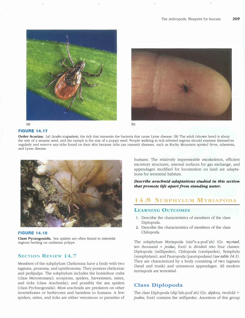
FIGURE 14.17
(b)
The Arthropods: Blueprint for Success 269
Order Acarina. (a) Ixodes scapularis, the tick that transmits the bacteria that cause Lyme disease. (b) The adult (shown here) is about the size of a sesame seed, and the nymph is the size of a poppy seed. People walking in tick-infested regions should examine themselves regularly and remove any ticks found on their skin because ticks can transmit diseases, such as Rocky Mountain spotted fever, tularemia, and Lyme disease.
FIGURE 14.18
Class Pycnogonida. Sea spiders are often found in intertidal regions feeding on cnidarian polyps.
SECTION REVIEW 14.7
Members of the subphylum Chelicerata have a body with two
tagmata, prosoma, and opisthosoma. They possess chelicerae
and pedipalps. The subphylum includes the horseshoe crabs
(class Merostomata); scorpions, spiders, harvestmen, mites,
and ticks (class Arachnida); and possibly the sea spiders
(class Pychnogonida). Most arachnids are predators on other
invertebrates or herbivores and harmless to humans. A few
spiders, mites, and ticks are either venomous or parasites of
humans. The relatively impermeable exoskeleton, efficient
excretory structures, internal surfaces for gas exchange, and
appendages modified for locomotion on land are adapta
tions for terrestrial habitats.
Describe arachnid adaptations studied in this section
that promote life apart from standing water.
14.8 SUBPHYLUM MYRIA >ODA
LEARNING OUTCOMES
1. Describe the characteristics of members of the classDiplopoda.
2. Describe the characteristics of members of the classChilopoda.
The subphylum Myriapoda (mir"e-a-pod'ah) (Gr. myriad,
ten thousand + podus, foot) is divided into four classes:
Diplopoda (millipedes), Chilopoda (centipedes), Symphyla
(symphylans), and Pauropoda (pauropodans) (see table 14.1).
They are characterized by a body consisting of two tagmata
(head and trunk) and uniramous appendages. All modern
myriapods are terrestrial.
Class Diplopoda
The class Diplopoda (dip'lah-pod'ah) (Gr. diploos, twofold+
podus, foot) contains the millipedes. Ancestors of this group
270 CHAPTER FOURTEEN
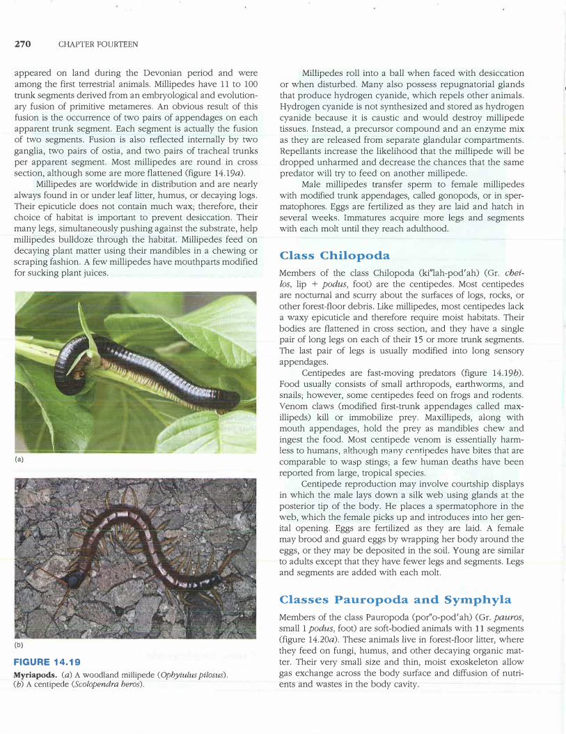
appeared on land during the Devonian period and were among the first terrestrial animals. Millipedes have 11 to 100 trunk segments derived from an emb1yological and evolutionary fusion of primitive metameres. An obvious result of this fusion is the occurrence of two pairs of appendages on each apparent trunk segment. Each segment is actually the fusion of two segments. Fusion is also reflected internally by two ganglia, two pairs of ostia, and two pairs of tracheal trunks per apparent segment. Most millipedes are round in cross section, although some are more flattened (figure 14.19a).
Millipedes are worldwide in distribution and are nearly always found in or under leaf litter, humus, or decaying logs. Their epicuticle does not contain much wax; therefore, their choice of habitat is important to prevent desiccation. Their many legs, simultaneously pushing against the substrate, help millipedes bulldoze through the habitat. Millipedes feed on decaying plant matter using their mandibles in a chewing or scraping fashion. A few millipedes have mouthparts modified for sucking plant juices.
(a)
(b)
FIGURE 14.19
Myriapods. (a) A woodland millipede (Ophyiulus pilosus). (b) A centipede (Scolopendra heros).
Millipedes roll into a ball when faced with desiccation or when disturbed. Many also possess repugnatorial glands that produce hydrogen cyanide, which repels other animals. Hydrogen cyanide is not synthesized and stored as hydrogen cyanide because it is caustic and would destroy millipede tissues. Instead, a precursor compound and an enzyme mix as they are released from separate glandular compartments. Repellants increase the likelihood that the millipede will be dropped unharmed and decrease the chances that the same predator will try to feed on another millipede.
Male millipedes transfer sperm to female millipedes with modified trunk appendages, called gonopods, or in spermatophores. Eggs are fertilized as they are laid and hatch in several weeks. Immatures acquire more legs and segments with each molt until they reach adulthood.
Class Chilopoda
Members of the class Chilopoda (ki"lah-pod'ah) (Gr. chei
los, lip + podus, foot) are the centipedes. Most centipedes are nocturnal and scurry about the surfaces of logs, rocks, or other forest-floor debris. Like millipedes, most centipedes lack a waxy epicuticle and therefore require moist habitats. Their bodies are flattened in cross section, and they have a single pair of long legs on each of their 15 or more trnnk segments. The last pair of legs is usually modified into long sensory appendages.
Centipedes are fast-moving predators (figure 14.19b). Food usually consists of small arthropods, earthworms, and snails; however, some centipedes feed on frogs and rodents. Venom claws (modified first-trunk appendages called maxillipeds) kill or immobilize prey. Maxillipeds, along with mouth appendages, hold the prey as mandibles chew and ingest the food. Most centipede venom is essentially harmless to humans, although many centipedes have bites that are comparable to wasp stings; a few human deaths have been reported from large, tropical species.
Centipede reproduction may involve courtship displays in which the male lays down a silk web using glands at the posterior tip of the body. He places a spermatophore in the web, which the female picks up and introduces into her genital opening. Eggs are fertilized as they are laid. A female may brood and guard eggs by wrapping her body around the eggs, or they may be deposited in the soil. Young are similar to adults except that they have fewer legs and segments. Legs and segments are added with each molt.
Classes Pauropoda and Sytnphyla
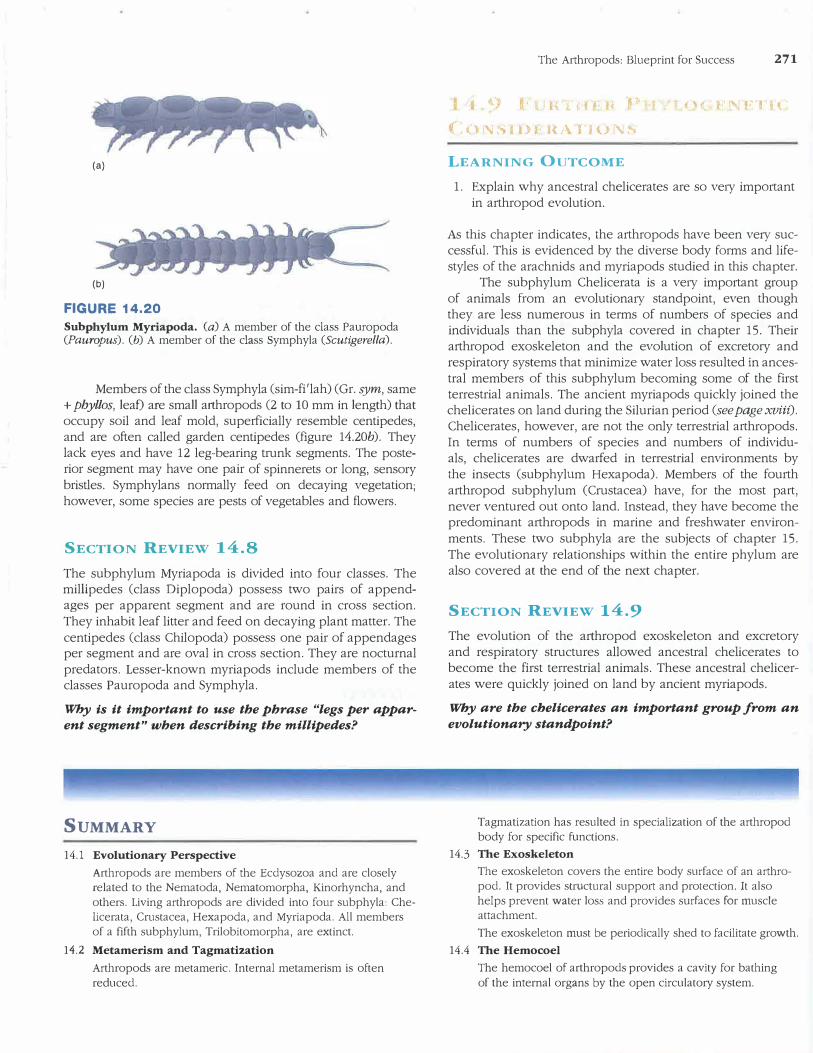
Members of the class Pauropoda (por"o-pod'ah) (Gr. pauros,
small 1 podus, foot) are soft-bodied animals with 11 segments (figure 14.20a). These animals live in forest-floor litter, where they feed on fungi, humus, and other decaying organic matter. Their very small size and thin, moist exoskeleton allow gas exchange across the body surface and diffusion of nutrients and wastes in the body cavity.
(a)
(b)
FIGURE 14.20 Subphylum Myriapoda. (a) A member of the class Pauropoda (Pauropus). (b) A member of the class Symphyla (Scutigerella).
Members of the class Symphyla (sim-fi'lah) (Gr. sym, same + phyllos, leaf) are small a1thropods (2 to 10 mm in length) thatoccupy soil and leaf mold, superficially resemble centipedes,and are often called garden centipedes (figure 14.20b). Theylack eyes and have 12 leg-bearing trunk segments. The posterior segment may have one pair of spinnerets or long, sensorybristles. Symphylans normally feed on decaying vegetation;however, some species are pests of vegetables and flowers.
SECTION REVIEW 14.8
The subphylum Myriapoda is divided into four classes. The millipedes (class Diplopoda) possess two pairs of appendages per apparent segment and are round in cross section. They inhabit leaf litter and feed on decaying plant matter. The
centipedes (class Chilopoda) possess one pair of appendages per segment and are oval in cross section. They are nocturnal
predators. Lesser-known myriapods include members of the classes Pauropoda and Symphyla.
Why is it important to use the phrase "legs per apparent segment" when describing the millipedes?
SUMMARY
14.1 Evolutionary Perspective
Arthropods are members of the Ecdysozoa and are closely
related to the Nematoda, Nematomorpha, Kinorhyncha, and
others. Living arthropods are divided into four subphyla: Che
licerata, Crustacea, Hexapoda, and Myriapoda. All members
of a fifth subphylum, Trilobitomorpha, are extinct.
14.2 Metamerism and Tagmatization
Arthropods are metameric. Internal metamerism is often
reduced.
The Arthropods: Blueprint for Success 271
l t 9 i, r h I d t· h i O
1 , « �- F : F'J H.
(:o'\ISIIH Hi\Tioi\!�
LEARNING OUTCOME
1. Explain why ancestral chelicerates are so ve1y imp01tant
in arthropod evolution.
As this chapter indicates, the arthropods have been ve1y successful. This is evidenced by the diverse body forms and lifestyles of the arachnids and myriapods studied in this chapter.
The subphylum Chelicerata is a ve1y important group
of animals from an evolutiona1y standpoint, even though they are less numerous in terms of numbers of species and individuals than the subphyla covered in chapter 15. Their arthropod exoskeleton and the evolution of excretory and respiratory systems that minimize water loss resulted in ancestral members of this subphylum becoming some of the first terrestrial animals. The ancient myriapods quickly joined the chelicerates on land during the Silurian period (see page xviii).
Chelicerates, however, are not the only terrestrial arthropods. In terms of numbers of species and numbers of individuals, chelicerates are dwarfed in terrestrial environments by the insects (subphylum Hexapoda). Members of the fourth arthropod subphylum (Crustacea) have, for the most part, never ventured out onto land. Instead, they have become the predominant arthropods in marine and freshwater environ
ments. These two subphyla are the subjects of chapter 15.
The evolutiona1y relationships within the entire phylum are also covered at the end of the next chapter.
SECTION REVIEW 14.9
The evolution of the arthropod exoskeleton and excretory and respiratory structures allowed ancestral chelicerates to become the first terrestrial animals. These ancestral chelicerates were quickly joined on land by ancient myriapods.
Why are the cbelicerates an important group from an
evolutionary standpoint?
Tagmatization has resulted in specialization of the arthropod
body for specific functions.
14.3 The Exoskeleton
The exoskeleton covers the entire body surface of an arthro
pod. It provides structural support and protection. It also
helps prevent water loss and provides surfaces for muscle
attachment.
The exoskeleton must be periodically shed to facilitate growth.
14.4 The Hemocoel
The hemocoel of arthropods provides a cavity for bathing
of the internal organs by the open circulatory system.
272 CHAPTER FOURTEEN
14.5 Metamorphosis
Metamorphosis has contributed to arthropod success by reducing competition between immature and adult
arthropods.
14.6 Subphylum Trilobitomorpha
Members of the extinct subphylum Trilobitomorpha had oval,
flattened bodies that consisted of three tagmata and three
longitudinal lobes. Appendages were biramous.
14.7 Subphylum Chelicerata
The subphylum Chelicerata has members whose bodies are
divided into a prosoma and an opisthosoma. They also pos
sess a pair of feeding appendages called chelicerae.
The horseshoe crabs and the giant water scorpions belong to Lhe class Meroslomala.
The class Arachnida includes spiders, mites, ticks, scorpions,
and others. Their exoskeleton partially preadapted the arach
nids for their terrestrial habitats.
The sea spiders are the only members of the class
Pycnogonida.
14.8 Subphylum Myriapoda
The subphylum Myriapoda includes four classes of
arthropods.
Members of the class Diplopoda (the millipedes) are charac
terized by apparent segments bearing two pairs of legs.
Members of the class Chilopoda (the centipedes) are charac
terized by a single pair of legs on each of their 15 segments
and a body that is flattened in cross section.
The class Pauropoda contains soft-bodied animals that feed
on fungi and decaying organic matter in forest-floor litter.
Members of the class Symphyla are centipede-like arthropods
that live in soil and leaf mold, where they feed on decaying
vegetation.
14.9 Further Phylogenetic Considerations
The exoskeleton, and efficient respiratory and excretory sys
tems, preadapted ancient members of the subphylum Chelic
erata for life on land. Ancient myriapods invaded terrestrial
environments shortly after the chelicerates became terrestrial.
CONCEPT REVIEW QUESTIONS
1. All of the following are grouped with the Arthropods in theEcdysozoa, except one. Select the exception.
a. Nematoda
b. Mollusca
c. Nematomorpha
d. Kinorhyncha
2. The inner layer of the exoskeleton of an arthropod is called
the _ __ _ __ . It contains chitin, and its outer region is
hardened. The outermost layer of the exoskeleton is waxy and
impermeable to water. Which one of the following terms cor
rectly fills the blank in the first sentence?
a. epicuticle
b. procuticle
c. hypodermis
d. basement membrane
3. Members of the class ______ include the spiders, scor-
pions, ticks, and mites.
a. Merostomata
b. Arachnida
C. Pycnogonida
d. Branchiopoda
e. Malacostraca
4. Centipedes are members of the subphylum _ __ __ and
the class ____ _
a. Myriapoda; Diplopoda
b. Hexapoda; Chilopoda
c. Myriapoda; Chilopoda
d. Chelicerata; Diplopoda
5. Members of this class appear to have two pairs of legs on
each body segment. This condition is misleading because eachapparent segment results from a fusion of two segments.
a. Chilopoda
b. Arachnida
c. Diplopoda
d. Pycnogonida
ANALYSIS AND APPLICATION
QUESTIONS
1. What is tagmatization, and why is it advantageous for metameric animals?
2. Explain how, in spite of being an armorlike covering, theexoskeleton permits movement and growth.
3. Why is the arthropod exoskeleton often cited as the major rea
son for arthropod success?
4. Explain why excretory and respiratory systems of ancestralarachnids probably preadapted these organisms for terrestrial
fo•hi!ats.
connect 110<>1 ..
Enhance your study of this chapter with study tools and practice
tests. Also ask your instructor about the resources available through
Connect, including a media-rich eBook, interactive learning tools,
and animations.