Embed Size (px)
Citation preview

J. Steroid Biochem. Molec. BioL Vol. 41, No. 3-8, pp. 231-240, 1992 0960-0760/92 $5.00 + 0.00 Printed in Great Britain. All fights reserved Copyright © 1992 Pergamon Press plc
1 , 2 5 ( O H ) 2 - V I T A M I N D3, A S T E R O I D H O R M O N E
T H A T P R O D U C E S B I O L O G I C E F F E C T S V I A B O T H
G E N O M I C A N D N O N G E N O M I C P A T H W A Y S
ANTHONY W. NORMAN, l* ILKA NEMERE, l LI-XIN ZHOU, l JUNE E. BISHOP, l KAREN E. LOWE, l ANITA C. MAIYAR, j ELAINE D. COLLINS, l TERUHISA TAOKA, 1 IGOR SERGEEV 1
and MARY C. FARACH-CARSON 2
Division of Biomedical Sciences and Department of Biochemistry, University of California Riverside, CA 92521 and 2Department of Biological Chemistry, University of Texas Dental Branch, Houston,
TX 77030, U.S.A.
Summary--The hormonally active form of vitamin D is 1,25(OH)2-vitamin D 3 [1,25(OH)2 D 3 ]. This seco-steroid is the key mediator of the vitamin D endocrine system which produces biological effects in over 28 target tissues. In these target tissues, the biological responses may be generated both by a signal transduction mechanism which involves a nuclear receptor for 1,25(OH)2D 3 that modulates gene transcription or a signal transduction pathway which involves rapid opening of Ca 2+ channels which are externally located on the plasma membrane. This paper reviews the evidence in support of the pleiotropic effects of this steroid hormone and presents evidence that the receptor of the genomic effects is likely to be separate from the receptor/membrane recognition element which initiates the rapid nongenomic biological effects.
INTRODUCTION
Over the past 25 years it has been established that the fat soluble vitamin, vitamin D, is in reality the precursor of a steroid hormone [1-3]. Vitamin D is subject to a complex series of metabolic conversions which generate some 37 separate metabolites; most of these new daughter compounds differ from the parent vitamin D by the presence of 1-3 hydroxyls, oxo, or lactone functionalities [4, 5]. Currently it is generally ac- cepted that 1,25(OH)2-vitamin D3 [1,25(OH)2 D3] is the hormonally active form of vitamin D [2-7]; this metabolite is generated first by hydroxyl- ation on carbon-25 in the liver and then by hydroxylation on carbon-l , a process carried out principally in the kidney [8], but which under some physiological circumstances can occur in activated macrophages, particularly in the bone marrow[9]. The structure of 1,25(OH)2D 3 is presented later in Fig. 8.
Much evidence has been accumulated documenting that one of the major pathways employed by 1,25(OH)2 D3 to generate biological responses is through the interaction of
Proceedings of the lOth International Symposium of the Journal o f Steroid Biochemistry and Molecular Biology, Recent Advances in Steroid Biochemistry and Molecular Biology, Paris, France, 26-29 May 1991.
*To whom correspondence should be addressed.
1,25(OH)2D3 with a cytosolic/nuclear receptor [3, 6, 10, 11] that mediates changes in gene tran- scription of specific proteins. However, over the past 6-8 years it has become apparent that 1,25(OH)2D3 also has the potential to generate biological responses via signal transduction pathways not dependent upon the immediate regulation of gene transcription [12-15].
Figure 1 illustrates the two pathways of signal transduction which are available to the hormonally active 1,25(OH)2D 3. One pathway involves the interaction of the steroid hormone with its nuclear receptor. After formation of a steroid-receptor complex this moiety interacts with selected regions of the promoters of genes, designated as hormone response elements, HREs, whose transcription is regulated by 1,25(OH)2D 3. As reviewed by Minghetti and Norman [6] there are now over 60 genes whose transcription has been shown to be modulated by 1,25(OH)2D 3 . These include the well-studied calbindin-D2si< [16], a vitamin D-induced calcium binding protein, and osteocalcin, an extracellular bone matrix protein[17]. In this model the steroid hormone is delivered to the target organ bound to a specific plasma transport protein (in the case of 1,25(OH)2D3 this is the D-binding protein [18]). The target cell possesses a nuclear receptor which confers upon that cell the ability to take up that particular steroid hormone.
231

232 ANTHONY W. NORMAN et al.
The spectrum of biological responses produced by the aeco-aterold 1,25(OH)2D 3 may be mediated by two different mechanisms.
~ c
;> (A) > Receptor > Genome ) . Selected new
(nucleus/cytoaol) Activation proteins (Celblndln O tubulln)
1.25(OH)2D 3
> (B)--~Membrane "receptor"-->-Actlvate voltage
interaction -dependent Ca 2+
channels
Biological Responses )
~. Increase
Intrecellular Ca ̀?.+
N ONG_ I~NOMIC
Fig. 1. Pathways for generation of biological responses by 1,25(OH)2D3.
The lower portion of Fig. 1 illustrates how 1,25(OH)2D3 may produce biological responses utilizing a signal transduction pathway which is
independent of the regulation of gene transcrip- tion. Here the hormone interacts with a putative membrane recognition element (MRE) which
SUMMARY OF 1,2S(OH)2D 3 NUCLEAR ACTIONS
:JF
GENE REPRESSION:
CELL
5' ONA 1 REPLIC
BLOCK \ I
PRE - mRNA 3' I PROCESSING I
TRANSCRIPTION BLOCK
SHUT-DOWN PRODUCTION OF GENE PROOUCTS
PROTEINS
NEW ~ ' - ~ m RNA )2D3- DEPENDENT ~TRANSLAT ION pR OTE i N( S),,P------"
• BIOLOGICAL CONSEQUENCES : ' f ~1NTESTINAL CELLS
¢)aT~Wa, q ~ .)BONE CELLS P / ' M / A W / V A DIFFERENTIATION . . . . . . . . ~ IMMUNE CELLS
198R ~.SKIN CELLS, ETC B PROLIFERATION PATHWAY b CANCER CELLS-ONA REPLICATION
BLOCK; Co 2+ FLUX C, DEVELOPMENTAL PATHWAY ~ TEMPORAL EXPRESSION
OF GENES: EMBRYOGENESIS 0 CALCIUM, PHOSPHORUS
TRANSPORT/HOMEOSTASIS
Fig. 2. Model for the interaction of 1,25(OH):D 3 with its nuclear receptor and generation of biological responses via gene activation and gene repression. DBP, serum vitamin D binding protein; R, 1,25(OH)~ D3 receptor; S, the 1,25(OH)2 D3 steroid; P, phosphorus; POL II, RNA polymerase II; F, transcription factor;
DNA, deoxyribonucleic acid; mRNA, messenger ribonucleic acid.
1,25(OH)2-vitamin D3, a steroid hormone 233
then in some fashion is coupled to the opening of Ca 2+ channels such that a rapid biological response is initiated. Two important rapid bio- logical responses are transcaltachia, defined as the "very rapid stimulation of intestinal Ca 2+ transport" as studied in the chick intestine [15, 19], and the opening of voltage-gated Ca 2÷ channels in rat-derived ROS 17/2.8 osteosarcoma cells [20]. In both systems the biological response appears within seconds to minutes of application of the initiating 1,25(OH)2D3, which contrasts with the 60 min to hours required for genomic responses to be detectable. This paper reviews the current status of our understanding of the ability of 1,25(OH)2D3 to stimulate both genomic and nongenomic biological responses. Other reviews have recently appeared describing other aspects of the vitamin D endocrine system [21, 22].
G E N O M I C A C T I O N S O F 1,25(OH)2D s
Figure 2 presents a general model to describe the regulation of gene transcription by 1,25(OH)2D3. In this model the 1,25(OH)2D3 receptor is a DNA-binding protein capable of up-regulating or down-regulating the transcrip- tion of specific RNA polymerase If-transcribed genes. Furthermore, DNA-binding loops or zinc-fingers have been proposed for the avian [23] and human [24] 1,25(OH)2D3 receptor and other related steroid receptors[25] that are similar to the zinc-fingers proposed for the 5S transcription factor TFIIIA [26]. However, the structure of the zinc-finger in the steroid receptor involves the coordination of four cysteines to one zinc atom, in contrast to TFIIIA, which involves histidines. The 1,25(OH)2D 3 receptor is proposed to regulate four classes of genes: those associated with (a) a differentiation pathway, (b) a proliferation pathway, (c) a developmental pathway, and (d) calcium and phosphorus homeostasis [6].
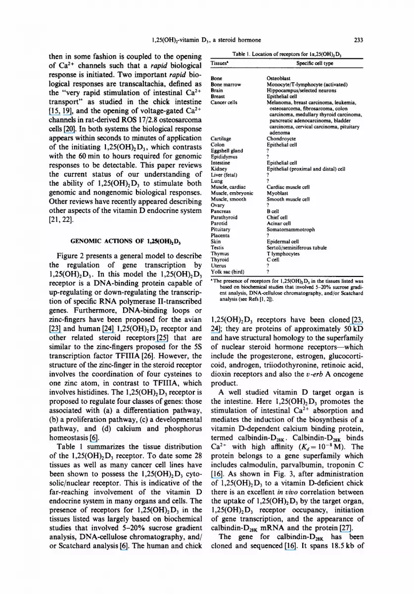
Table 1 summarizes the tissue distribution of the 1,25(OH)2D3 receptor. To date some 28 tissues as well as many cancer cell lines have been shown to possess the 1,25(OH)2D 3 cyto- solic/nuclear receptor. This is indicative of the far-reaching involvement of the vitamin D endocrine system in many organs and cells. The presence of receptors for 1,25(OH)2D3 in the tissues listed was largely based on biochemical studies that involved 5-20% sucrose gradient analysis, DNA-cellulose chromatography, and/ or Scatchard analysis [6]. The human and chick
Table 1. Location of receptors for I~t,25(OH) 2 D 3
Tissues' Specific cell type
Bone Osteoblast Bone marrow Monocyte/T-lymphocyte (activated) Brain Hippocampus/selected neurons Breast Epithelial cell Cancer cells Melanoma, breast carcinoma, leukemia,
osteosarcoma, fibrosarcoma, colon carcinoma, medullary thyroid carcinoma, pancreatic adenocarcinoma, bladder carcinoma, cervical carcinoma, pituitary adenoma
Cartilage Chondroycte Colon Epithelial cell Eggshell gland ? Epididymus ? Intestine Epithelial cell Kidney Epithelial (proximal and distal) cell Liver (fetal) ? Lung ? Muscle, cardiac Cardiac muscle cell Muscle, embryonic Myoblast Muscle, smooth Smooth muscle cell Ovary ? Pancreas B cell Parathyroid Chief cell Parotid Acinar cell Pituitary Somatomammotroph Placenta ? Skin Epidermal cell Testis Sertoli/seminiferous tubule Thymus T lymphocytes Thyroid C cell Uterus ? Yolk sac (bird) ?
IThe presence of receptors for 1,25(OH)~D~ in the tissues listed was based on biochemical studies that involved 5-20% sucrose gradi- ent analysis, DNA-cellulose chromatography, and/or Scatchard analysis (see Refs [1, 21).
1,25(OH)2D3 receptors have been cloned[23, 24]; they are proteins of approximately 50 kD and have structural homology to the superfamily of nuclear steroid hormone receptors--which include the progesterone, estrogen, glucocorti- coid, androgen, triiodothyronine, retinoic acid, dioxin receptors and also the v-erb A oncogene product.
A well studied vitamin D target organ is the intestine. Here 1,25(OH)2Da promotes the stimulation of intestinal Ca 2÷ absorption and mediates the induction of the biosynthesis of a vitamin D-dependent calcium binding protein, termed calbindin-D2s K. Calbindin-D28~: binds Ca 2÷ with high affinity (Kd= 10-aM). The protein belongs to a gene superfamily which includes calmodulin, parvalbumin, troponin C [16]. As shown in Fig. 3, after administration of 1,25(OH)2D3 to a vitamin D-deficient chick there is an excellent in vivo correlation between the uptake of 1,25(OH)2 D3 by the target organ, 1,25(OH)2D 3 receptor occupancy, initiation of gene transcription, and the appearance of calbindin-D28~ mRNA and the protein [27].
The gene for calbindin-D28~: has been cloned and sequenced [16]. It spans 18.5 kb of

234 ANTHONY W. NORMAN et al.
chromosomal DNA and is split into 11 coding exons by 10 intervening sequences. The gene has a markedly G + C-rich (60-80%) region extending from -225 to +400. Within this region there are 70 CpG dinucleotides, four G-C boxes, and numerous other promoter regu- latory signals including a TATA box at -30 , a CAT box (CCAAT) at -326, a glucocorticoid- like hormone responsive element (GRE) at -410 (TCTACACACTGTTCC) and a puta- tive 1,25(OH)2D3-response element (DRE) at -313 to -329 [28].
In order to examine the cis-acting elements of the calbindin-D28 K promoter and the trans- activation of calbindin-D28K by 1,25(OH)2D3, chimeric plasmids containing 2.1 kb of 5'-flank- ing region linked to the reporter gene chlor- amphenicol acetyl transferase (CAT) were
transfected by lipofection into primary cultures of chick kidney cells [29]. Some of our recent results are summarized in Fig. 5. Panel 5A demonstrates that extracts from cells transfected with plasmid containing the calbindin-D28~ pro- moter fragment in the 5'-3' orientation showed a significant basal promoter activity which was stimulated 2-fold by 10-7M 1,25(OH)2D 3. In contrast, the plasmid containing the same gene fragment, but in the 3'-5' orientation, showed no detectable CAT activity either in the presence or absence of 1,25(OH)2D3. Panel 5B presents the transcriptional activity of different deletion mutants which were gener- ated from pCaBP2.1CAT. The -2100/+44, -924/+44 , - 449 /+44 and - 3 6 5 / + 4 4 frag- ments all displayed a reproducible stimulation by 1,25(OH)2D 3 while fragment -679 /+44
,4
12
~,o
!
6
- i ~ 3 •
T I M E A F T E R 1 , 2 5 ( O H ) z D $ ( h )
A
E S 0 0 u n-
O 6 0 0 G. t~J o
4 0 0
i 2 0 0
O
i u i i i i i i ! l I i u i i i
D r : O . 8 7
. j , . ~ • j ; i I I I I I I 4 I
2 4 6 8 IO 12 I
CoBP GENE TRANSCRIPTION (ppm)
i , , , i , , , ,
o n d t i a i a I I I o I ! $ 4
T I M E A F T E R 1 , 2 5 ( O H ) I D 3 ( h )
i , , i , , , -') i
,.! o i oQ ii o
I
I
4
o i i i i 1 l i
T I M E A F T E R I , / S ( O H I I D 3 ( h i
iO0, 'v" * * , * , , , , , , ~l#~-#---r -~.i ,
.--I St
T I M E A F T E R 1 , 2 5 ( O H l t O 3 l h )
o,o,,,,,, o i m . _ ~ F - - - ! x
0 I 2 3 4 5 /" 7 8 9 I 0 I I 12 2 4 4 8 7 2
T I M E A F T E R 1 , 2 5 ( O H ) I D 3 (h )
Fig. 3. Chronology of events in the intestine of a vitamin D-deficient chick given a physiological dose of 1,25(OH)2D3: (A) Nuclear uptake of tritiated 1,25(OH)2D3; (B) 1,25(OH)2D 3 receptor occupancy; (C) calbindin-D2s t gene transcription: (D) calbindin-D2s K gene transcription vs 1,25(OH)2D 3 receptor
occupancy; (E) accumulation of calbindin-D2s K mRNA; (F) calbindin-D28 K accumulation.

1,25(OH)2-vitamin D3, a steroid hormone
-326 -354 1-318 -299 -30 +1
CACCC CAT Cond CACCC TATA 80X BOX DRE BOX BOX
Fig. 4. Promoter region of chick calbindin-D28K gene (see Ref. [16] for details).
235
(A) ,n 4
o 3
o,I
~ 2
1
10 ,o
0 J
- 2 1 0 0 / + 4 4 5' - 3'
Colbindin -D2s k
-2100/+44 3'- 5'
Promoter Constructs
(B)
2
g -~, o ID ~ I---
\ 1
~ 2 -o
lm -.-°-° :.:.:. :1::11 °%°°° ...*.°
.:.:.1 .°%%
iiiiii ,,°%°
| |
- 2 1 0 0 + 4 4 - 9 2 4 / + 4 4 - 6 7 9 1 + 4 4 - 4 4 9 / + 4 4 - 3 6 8 / + 4 4
Colbindin - D2ek Promoter Constructs
800 c
-g
o- 600 Oa
E
>-
>_- 200
<~
u o
(C)
l:ll KIDNEY MCF-7 T-47D
CONSTRUCT - 2 1 0 0 / + 4 4
ROS17/2.B
Fig. 5. Transient transfection of portions of the avian Y-flanking calbindin-D2s K promoter into primary chick kidney cells: (A) transcriptional activity of calbindin-D2sK promoter pCaBP2.1CAT construct as assessed via CAT assay in normal orientation ( - 2 1 0 0 / + 4 4 5'-3') and negative orientation; (B) evaluation by CAT assay of progressive deletions of Y-flanking DNA; (C) transient transfection in primary kidney cells, MCF-7, T-47D, and ROS 17/2.8 cell lines transfected with the -2100 /+44 , 5'-3' calbindin-D2s K
promoter fragment.
Table 2. Hormone response elements response to the 1,25(OH) 2 D 3 receptor
Protein Hormone response elements Species Reference
Osteocalcin 5'-GGTGACTCA CCGGG TFAACG-3' (Human) Kerner et aL [30] GGTGACTCA CCGGG TGAAC (Human) Morrison et aL [32]
TCTCCCCA CTGGA TGAGCGG (Rat) Markose et aL [31] Calbindin D2s K 5'-AGCCCA ATGGC TGAACA-Y (Chick) Minghetti et aL [28]
(putative) CCA ATCTC TGAGCA (Human) Minghetti et aL [28]
(putative)

236 ANTHONY W. NORMAN et al.
showed a 2.2-fold repression in the presence of 1 ,25(OH)2D 3. Panel 5C reports the results of transfection of the pCaBP2.1 CAT fusion gene constructs into three mammalian cell lines: MCF-7 cells, T-47D cells and ROS 17/2.8 cells. The CAT reporter gene was only weakly expressed in the ROS 17/2.8 and T-47D cell lines, while slightly higher levels of CAT activity were observed in the MCF-7 cell line, although expression was not responsive to the addition of 1,25(OH)2D3. However, the avian calbindin- D28 K promoter was not responsive to the addition of 1,25(OH)2D3 in any of these three mammalian cell lines.
Table 2 summarizes the current status of known hormone response elements (DREs) for 1,25(OH)2D3 in the promoter of osteocalcin [30-32] and calbindin-D2sK[28]. The DRE regions for calbindin-D2sK are candidates in that they have only been proposed on the basis of computer searching of the nucleotide sequence. The osteocalcin half-site palindromic DRE shows only partial sequence homology to the calbindin-D2sK putative DRE (-329 AGCC- CAATGGCTGAACA -313). However, DNase "footprinting" has demonstrated a protected site near the CCAAT region and the putative 1,25(OH)2D3 DRE of the calbindin-D2sK gene promoter[33]. Clearly further work will be necessary to understand the precise calbindin- D28~ DRE and its relationship to other DREs.
NONGENOMIC ACTIONS OF 1,25(OH)2D 3
The 1,25(OH)2D3-stimulated transport of calcium across the intestinal epithelium has been extensively studied in our laboratory[15, 34- 37]. We reported in 1984 that 1,25(OH)2Ds can stimulate the rapid (within 2-4 min) transport of 45Ca2+ in vascularly perfused chick duodenal loops[15]. This rapid 1,25(OH)2D3-mediated transport of calcium has been termed trans-
caltachia and is clearly independent of gene transcription effects of 1,25(OH)2D3. Figure 6 presents typical results for a dose-relationship between 1,25(OH)2D 3 and stimulation of trans- caltachia. Panel 6A presents the actual per- fusion data over 40 min of perfusion. The results on the ordinate are expressed as the ratio of [treated]/[control] where "control" represents the base-line of 45 C a 2 + observed from - 2 0 min to "zero time" when the 1,25(OH)2D3 was intro- duced. The response to 1,25(OH)2D3 is clearly biphasic [see Fig. 6(B)] with the maximal stimu- lation occurring at a physiological concentra-
tion of 1,25(OH)2D3, i.e. 130 pM. In data not shown here, we have demonstrated that trans- caltachia exhibits a polarity in that transport of calcium occurs only when 1 ,25(OH)2D 3 is presented to the basal lateral membrane of the intestinal epithelial cell but not when it is presented to the brush border side of the cell.
Table 3 summarizes the results of inhibitor studies of transcaltachia. Transcaltachia can be inhibited by the anti-microtubule agent colchi- cine, an antagonist of lysosomal cathepsin B, leupeptin, and the Ca 2+ channel blockers verapamil and nifedipine.
The inhibitory results with verapamil and nifedipine suggested the involvement of C a 2+
channels in the process of transcaltachia [34, 35]. Figure 7(A) shows that the Ca 2+ channel agonist BAY K8644, like 1,25(OH)2D3, can
(A) I I I I I I I I I ] I
u:, z= 3
8~ Z ~
z~2
O:
r-i CONTROL T / ~ 0 1 3 0 ( p M ) T T
-- " 6 ~ 0 It T I ~ l r ~ T A t30Otp, M) T T A~ r - 7- T
F I l I I I I I I I l I 0 4 8 12 16 20 24 28 32 36 4 0
T I M E ( ra i n )
(B) 5
c
E 4-- 0 ,¢
~ 3
/Z
0 130 65O I 3 0 0 6 3 0 0
1 , 2 5 ( O H ) z D 3 ( p M )
Fig. 6. Stimulation of transcaltachia by 1,25(OH)2 D3 in the pcrfused chick intestine; (A) 1,25(OH)2D s dose-response studies with the data for 0-40 min presented. The perfusion labeled "control" was exposed only to vehicle and not exposed to 1,25(OH)2D3; (B) the ratio of treated/basal data
obtained at 40 min for panel A is replotted.

1,25(0H),-vitamin D,, a steroid hormone 231
Table 3. Evaluation of transcaltachia: identification of inhibitors (A)
Compound
Actinomycin D Leupeptin Pepstatin Monensin Colchicine Cytochalasin B Verapamil Nifedipine Staurosporine
Inhibition of Inhibits transcaltachia
Nuclear transcription No Cathepsin B Yes Cathepsin D, pepsin No Golgi No Microtubules Yes Microfilaments No Ca2+ channel antagonist Yes Ca’+ channel antagonist Yes Protein kinase C Yes
initiate transcaltachia. The potential involvement of protein kinases on the 1,25(OH),D,-depen- dent rapid stimulation of intestinal Ca*+ trans- port was also evaluated by measuring the effect of 10 p M forskolin and 100 nM TPA; vascular perfusion with either the protein kinase A or C agonist induced transcaltachia suggesting the involvement of phosphorylation events in this process [37]. In other studies we have also shown that staurosporine, a protein kinase C inhibitor totally abolished the transcaltachic response mediated by 1,25(OH)* D, . Collectively these results have suggested that protein kinase C and CAMP-dependent protein kinase mediate 1,25(OH)*D3 activation of basal lateral mem- brane Ca*+ channels as an early effect in the transcaltachic response.
4 OOlI”“““““~“~ 0 4 6 I2 16 20 24 26
TIME (mln )
(B) 4.0
, , I , 1 I I I I
I- 3.5 -
E
s-so- e::
w:: 2.,-
93 0 0 2.0 - z.
"j'= $! 1.5 -
5::
5 c LO-
s- B 0.5 -
001 ’ I 1 I 1 1 , 1 I 0 4 6 It 16 20 24 26
TIME (min)
In a separate system, the rat-derived osteo- blast-like ROS 17/2.8 cells, we have also studied the ability of 1,25(OH),D, and its analogs to open Ca*+ channels. Caffrey and FarachCarson have previously reported that 1,25(OH)* D3 can open a voltage-sensitive Ca*+ channel within 30 s of application to ROS 17/2.8 cells. Table 4 summarizes our results of a comparison of the ability of analogs and metabolites of 1,L?5(0H)2 D, to stimulate transmembrane 45 Ca*+ influx versus their ability to bind to the classical nuclear receptors present in cellular extracts of osteoblast-like cells [37] in comparison to their
Fig. 7. Studies on transcaltachia: (A) effect of 1,25(OH),D, (130 PM), the phorbol ester TPA (100 nM), and forskolin (10 FM) on the appearance of ‘%a*+ in the venous effluent of perfused duodena from normal chicks (see Ref. [35]); (B) Effects of 1,25(OH),D, (130 PM) and BAY K8644 on the appearance of Wa*+ in the venous effluent of perfused duodena from vitamin D-replete chick (see Ref. [37]).
ability to stimulate transcaltachia and bind to the classical chick intestinal nuclear receptor. The relative channel opening ability (RCO) is also tabulated. It is clear that some analogs of 1 ,25(OH)*DS are more effective at opening Ca*+ channels relative to receptor binding while other analogs bind quite effectively to the classic 1,25(OH),D3 receptor but are poor channel
Table 4. Relative competitive index (chick intestine and ROS 17/2.8 cell) and stimulation of transcaltachia and Ca2+ channel opening
Chick Analog intestinal ROS 17/2.8 Transcaltachia’
Analog name code name RCI RCI (cone) (% max) RCOb
I ,25(01-1), D, C I00 100 650 100 loo I ,24S(OH),-22ene-
26,27-dehydrovitamin D, BT Ill 115 1300 24 0.01 1,25(OH),-16ene-23yne-D, V 75 30 1300 24 20 25(OH)-I6ene-23yne-D, AT 0.43 0.08 650 59 36 25(OH)-23yne-D, Y 0.03 0.06 6500 59 0.3 I ,25R,26(OH),D, DN 77 - 6500 12 -
‘The entries reported under the heading of Tramcaltachia were obtained in the perfused chick intestine. The [cone] is the concentration of analog (in PM) required to achieve a maximum response while (% mar.) is the percent of maximum response above control (unstimulated) as measured after 40 min of perfusion.
bRCO = Relative Channel Opening = [l,25(OH),D,],,~i,/[Analoplr~~i~, x 100 as determined in the ROS 17/2.8 cells.

238 ANTHOI~ W. NORMAN et al.
HO" . ~ O H HO ~ . ~ O H HO"
C* V* 1,25-(OH)2-D3 1,25-(OH)2-16--ene-23-yne-D 3
Y 25-(OH)-23-yne-D 3
HO" HO" . ~ O H HO ~
OH OH
AT BT* DN 25-(OH)-16-ene-23-yne-D 3 1,24-(OH )2-22--en-24-eyclopropyl-D 3 1 ~t,25R,26-(OH)3-D 3
Fig. 8. Structure of analogs of 1,25(OH)2D3: C = 1,25(OH)2D3; V = 1,25(OH)2-16ene-23yne-D3; BT = 1,24(OH)2-24-cyclopropyl-D 3; AT = 25(OH)- 16ene-23yne-D3; Y = 25(OH)-23yne-D 3 ; and
DN = 1,25R,26(OH)3D 3.
agonists. Thus the results obtained in the ROS 17/2.8 cells extend to a new species, the rat, and a new target organ, bone, the existence of rapid nongenomic responses mediated by 1,25(OH)2D3.
As a consequence of these results, a challeng- ing problem has arisen--namely, to identify the signal transduction factor element which initiates transcaltachia. Is it the same or different from the classical nuclear receptor for 1,25(OH)2D3?
E
g ¢D
8
1 .25 (OH)~3 AT Y BT V BO HN DN t - - - - - 1 [ f i I ~ 1 r - - 1 r - - I f - - 1 i i
ANALOGS (pM)
Fig. 9. Summary of dose-response studies of analogs of 1,25(OH)2 D 3 in stimulating transcaltachia, e x vivo, in the chick intestine.

1,25(OH)2-vitamin D 3, a steroid hormone 239
~2o z .jlO0
~ 80
z ~ , o
# o
Fig.
~ TRAN$- r / /A CALTACHIA ~ RECEPTOR
C BT V AT Y
10. Structure-function evaluation of stimulation of transcaltachia, in vivo, in the chick and binding to the classic
chick intestinal nuclear 1,25(OH)2D 3 receptor, in vitro.
To study this problem we have carried out structure function studies of transcaltachia using seven analogs of 1,25(OH)2D3 (see Fig. 8) which have differing affinities for the classic nuclear 1,25(OH)2D 3 receptor as measured by a determination in a steroid competition assay of the relative competitive index (RCI)[39]. The relative competitive index of 1,25(OH)2D3 is by definition 100. As shown in Fig. 9, dose- dependent stimulation of Ca 2+ transport in the perfused chick intestine indicates that different structural features are essential for initiating the transcaltachic response. Vascular perfusion with either analog AT [25(OH)2-16ene-23yne-D3] or Y [25(OH)E-23yne-D3] stimulated Ca 2+ trans- port to a maximum of 3-fold at 650 pM of AT or 6500 pM of Y. The analogs BO [25(OH)D3] and HN [25(OH)16ene-D3] stimulated Ca 2+ transport to 3.8-fold and 2.9-fold, respectively, at a concentration of 65 nM analog. In contrast, analogs BT [la,24S(OH)2-22ene-26,27-dehydro- vitamin D3], V [1,25(OH)2-16ene-23yne-D3] and DN [1,25R,26(OH)3 D3] were far less efficient in stimulating transcaltachia.
Figure l0 summarizes these structure-func- tion studies in terms of the RCI and stimulation of transcaltachia. These results clearly indicate that there are some analogs of 1,25(OH)2D3 which are highly effective at stimulating trans- caltachia(analogs AT, Y) but which bind only poorly to the classic nuclear 1,25(OH):D 3 receptor. In contrast there are other analogs (BT and V) which are poor initiators of transcaltachia but which bind effectively to the classical chick intestinal nuclear receptor for 1,25(OH)2D3. It is important to emphasize that 1,25(OH)2D3 is the optimal agonist for both receptor binding and stimulation of transcaltachia and opening of Ca 2+ channels in the ROS 17/2.8 system.
A complete picture o f the molecular signal- - '120 X
_~ ling pathways involved in the regulation of ROS ,o0,7, 17/2.8 cells and the chick intestine will require
> an understanding of both the long term genomic v- 8O 7- and rapid nongenomic effects of 1,25(OH)2D3.
6o ~ o
40 ~ R E F E R E N C E S
I. Norman A. W.: The mode of action of vitamin D. Biol. 20 Rev. 43 (1968) 97-137.
2. Norman A. W.: Vitamin D: The Calcium Homeostatic o Steroid Hormone. Academic Press, New York (1979).
3. Norman A. W., Roth J. and Orci L.: The vitamin D endocrine system: Steroid metabolism, hormone recep- tors and biological response (calcium binding proteins). Endocrine Rev. 3 (1982) 331-366.
4. Henry H. L. and Norman A. W.: Vitamin D: Metab- olism and biological actions. A. Rev. Nutr. 4 (1984) 493-520.
5. Henry H. L. and Norman A. W.: Vitamin D: Metab- olism and Mechanism of Action. Primer on Metabolic Bone Diseases and Disorders o f Mineral Metabolism, 1st edn (1990) pp. 47-52.
6. Minghetti P. P. and Norman A. W.: 1,25(OH)2-vitamin D 3 receptors: Gene regulation and genetic circuitry. FASEB Jl 2 (1988) 3043-3053.
7. Haussler M. R.: Vitamin D receptors: nature and function. A. Rev. Nutr. 6 (1986) 527-562.
8. Henry H. L., Dutta C., Cunningham N., Blanchard R., Penny R., Tang C., Marchetto G. and Chou S-Y.: The cellular and molecular regulation of 1,25(OH)2D 3 pro- duction. J. Steroid Biochem. Molec. Biol. 41 (1992) 401-407.
9. Reichel H. and Norman A. W.: Systemic effects of vitamin D. A. Rev. Med. 40 (1989) 71-78.
10. Haussler M. R. and Norman A. W.: Chromosomal receptor for a vitamin D metabolite. Proc. Natn. Acad. Sci. U.S.A. 62 (1969) 155-162.
l 1. Norman A. W., Schaefer K., Grigoleit H. G. and Herrath D. V.: Vitamin D: Molecular, Cellular and Clinical Endocrinology. Walter de Gruyter, Berlin (1988).
12. Nemere I. and Norman A. W.: The rapid, hormonally stimulated transport of calcium (transcaltachia). J. Bone Min. Res. 2 (1987) 167-169.
13. Nemere I. and Norman A. W.: Steroid hormone actions at the plasma membrane: induced calcium uptake and exocytotic events. Molec. Cell. Endocr. 80 (1991) C16542169.
14. Lieberherr M., Grosse B., Duchambon P. and Driieke T.: A functional cell surface type receptor is required for the early action of 1,25-dihydroxyvitamin D 3 on the phosphoinositide metabolism in rat enterocytes. J. Biol. Chem. 264 (1989) 20403-20406.
15. Nemere I., Yoshimoto Y. and Norman A. W.: Studies on the mode of action of calciferol. LIV. Calcium transport in perfused duodena from normal chicks: Enhancement within 14 minutes of exposure to 1,25- dihydroxyvitamin D 3 . Endocrinology 115 (1984) 1476-1483.
16. Minghetti P. P., Cancela L., Fujisawa Y., Theofan G. and Norman A. W.: Molecular structure of the chicken vitamin D-induced calbindin-D28 K gene reveals eleven exons, six Ca2+-binding domains, and numerous promoter regulatory elements. Molec. Endocr. 2 (1988) 355-367.
17. Poser J. W. and Price P. A.: A method for decarboxyl- ation of gamma-carboxyglutamic acid in proteins. Properties of the decarboxylated gamma-carboxyglut- amic acid protein from calf bone. J. Biol. Chem. 254 (1979) 431-436.

240 ANTHONY W. NORMAN et al.
18. Baelen H. V., Allewaert K. and Bouillon R.: New aspects of the plasma carrier protein for 25-hydroxy- cholecalciferol in vertebrates. Ann. N.Y. Acad. Sci. 538 (1988) (,0-68.
19. Nemere I. and Norman A. W.: Transcaltachia, vesicular calcium transport, and microtubule-associated calbindin- D~K: emerging views of 1,25-dihydroxyvitamin D s- mediated intestinal calcium absorption. Min. Elec. Metab. 16 (1990) 109-114.
20. Caffrey J. M. and Farach-Carson M. C.: Vitamin D s metabolites modulate dihydropyridine-sensitive calcium currents in clonal rat osteosarcoma cells. J. Biol. Chem. 264 (1989) 20265-20274.
21. Nemere I. and Norman A. W.: Transport of calcium. In Handbook of Physiology (Edited by M. Field and R. A. Frizzell). Am. Physiol. Soc., Bethesda, MD, Section 6, Vol. IV, 13 (1991) 337-360.
22. Reichel H., Koeffler H. P. and Norman A. W.: The role of the vitamin D endocrine system in health and disease. New Engl. J. Med. 320 (1989) 980-991.
23. McDonnell D. P., Mangelsdorf D. J., Pike J. W., Haussler R. M. and O'Malley B. W.: Molecular cloning of complementary DNA encoding the avian receptor for vitamin D. Science, N.Y. 235 (1987) 1214-1217.
24. Baker A. R., McDonnell D. P., Hughes M., Crisp T. M., Mangelsdorf D. J., Haussler R. M., Pike J. W., Shine J. and O'Malley B. W.: Cloning and expression of full-length cDNA encoding human vitamin D receptor. Proc. Natn. Acad. Sci. U.S.A. 85 (1988) 3294-3298.
25. Petkovich M., Brand N. J., Krust A. and Chambon P.: A human retinoic acid receptor which belongs to the family of nuclear receptors. Nature, Lond. 330 (1987) 444-450.
26. Miller J., McLachlan A. D. and Klug A.: Repetitive zinc-binding domains in the protein transcription factor Il iA from Xenopus oocytes. EMBO JI 4 (1985) 1609-1614.
27. Theofan G., Nguyen A. P. and Norman A. W.: Regulation of calbindin-D28K gene expression by 1,25- dihydroxyvitamin D 3 is correlated to receptor occupancy. J. Biol. Chem. 261 (1986) 16943-16947.
28. Minghetti P. P., Gibbs P. E. M. and Norman A. W.: Computer analysis of 1,25-dihydroxyvitamin D3-receptor regulated promoters: Identification of a candidate D3- response element. Biochem. Biophys. Res. Commun. 162 (1989) 869-875.
29. Maiyar A. C., Minghetti P. P. and Norman A. W.: Transfection of avian vitamin D-dependent calbindin- Desk gene in primary chick kidney cells. Molec. Cell. Endocr. 78 (1991) 127-135.
30. Kerner S. A., Scott R. A. and Pike J. W.: Sequence elements in the human osteocalcin gene confer basal activation and inducible response to hormonal vitamin D 3 . Proc. Natn. Acad. Sci. U.S.A. 86 (1989) 4455- 4459.
31. Markose E. R., Stein J. L., Stein G. S. and Lian J. B.: Vitamin D-mediated modifications in protein-DNA interactions at two promoter elements of the osteocalcin gene. Proc. Natn. Acad. Sci. U.S.A. 87 (1990) 1701-1705.
32. Morrison N. A., Shine J., Fragonas J.-C., Verkest V., McMenemy M. L. and Eisman J. A.: 1,25-Dihydroxy- vitamin D-responsive element and glucocorticoid repression in the osteocalcin gene. Science, N.Y. 246 (1989) 1158-1161.
33. Boland R., Minghetti P. P., Lowe K. E. and Norman A. W.: Sequences near the CCAAT resio~and putative 1,25-dihydroxyvitamin D3-response element and further upstream novel regulatory sequences of calbindin-D2sK promoter show DNase I footprinting protection. Molec. Cell. Endocr. 75/1 (1991) 57-63.
34. Nemere I. and Norman A. W.: The rapid, hormonally stimulated transport of calcium (transcaltachia). J. Bone Min. Res. 2 (1987) 167-169.
35. de Boland A. R., Nemere I. and Norman A. W.: Ca2+-channel agonist bay K8644 mimics 1,25(OH)2- vitamin D s rapid enhancement of Ca 2+ transport in chick perfused duodenum. Biochem. Biophys. Res. Commun. 166 (1990) 217-222.
36. de Boland A. R. and Norman A. W.: Influx of extra- cellular calcium mediates 1,25-dihydroxyvitamin D r dependent transcaltachia (the rapid stimulation of duodenal Ca 2+ transport). Endocrinology 127/5 (1990) 2475-2480.
37. de Boland A. R. and Norman A. W.: Evidence for involvement of protein kinase C and cyclic Adenosine 3',5' Monophosphate-dependent protein kinase in the 1,25 Dihydroxy-vitamin D3-mediated rapid stimulation of intestinal calcium transport (transcaltachia). Endocrinology 127 (1990) 39-45.
38. Farach-Carson M. C., Sergeev I. and Norman A. W.: Non-genomic actions of 1,25(OH)2D 3 in rat osteo- sarcoma cells: structure-function studies using ligand agonist analogs. Endocrinology 129 (1991) 1876-1884.
39. Siebert P. D., Ohnuma N. and Norman A. W.: Studies on the mode of action of calciferol XXII. A 24R- hydroxyl-group can replace the 25-hydroxyl-group of la,25-dihydroxyvitamin D 3 for optimal binding to the chick intestinal receptor. Biochem. Biophys. Res. Commun. 91 (1979) 827-834.


