Embed Size (px)
Citation preview

Int. J. Mol. Sci. 2014, 15, 7639-7650; doi:10.3390/ijms15057639
International Journal of
Molecular Sciences ISSN 1422-0067
www.mdpi.com/journal/ijms
Article
Optimization of Cell Adhesion on Mg Based Implant Materials by Pre-Incubation under Cell Culture Conditions
Regine Willumeit *, Anneke Möhring and Frank Feyerabend
Helmholtz-Zentrum Geesthacht, Institute of Materials Research, Max-Planck-Str. 1, Geesthacht 21502,
Germany; E-Mails: [email protected] (A.M.); [email protected] (F.F.)
* Author to whom correspondence should be addressed; E-Mail: [email protected];
Tel.: +49-4152-87-1291; Fax: +49-4152-87-2666.
Received: 27 February 2014; in revised form: 20 March 2014 / Accepted: 16 April 2014 /
Published: 5 May 2014
Abstract: Magnesium based implants could revolutionize applications where orthopedic
implants such as nails, screws or bone plates are used because they are load bearing and
degrade over time. This prevents a second surgery to remove conventional implants.
To improve the biocompatibility we studied here if and for how long a pre-incubation of
the material under cell culture conditions is favorable for cell attachment and proliferation.
For two materials, Mg and Mg10Gd1Nd, we could show that 6 h pre-incubation are
already enough to form a natural protective layer suitable for cell culture.
Keywords: magnesium implants; natural corrosion protection; pre-incubation;
biocompatibility
1. Introduction
To make use of magnesium alloys as orthopedic biomaterial is an idea which has developed already
in the first half of the 20th century [1]. Mg is an essential element in life and about half of the 30 g
which can be found in the body is stored in bones [2]. The material is load bearing and the mechanical
properties tensile yield strength, fracture toughness or Young’s modulus [3] are more similar to those
of natural bone than polymers, ceramics implant materials or titanium (Ti) which remains the most
common and preferred implant material. Last but not least the metal is degrading which should have
several advantages over permanent implant materials [4,5]. However, due to their high degradation
rates accompanied by hydrogen (H2) release up to now only few implant prototypes are about to come
OPEN ACCESS
Int. J. Mol. Sci. 2014, 15 7640
for orthopedic [6] or cardiovascular [7,8] applications. The major problem remains the decrease of the
degradation rate to keep the stability of the implant and to reduce the ion release and H2 production to
a value the body can deal with [9].
There are several approaches to tailor the degradation rate. The first step is the use of alloying
elements (e.g., rare earth elements (REE [10,11], Ca, Y, Ag etc. [12])) accompanied by heat
treatments [13] or for example extrusion processes which change the microstructure and thus the
mechanical and corrosive properties. If this is still not enough coatings come into play. The principle
idea is to reduce the immediate corrosion to a level the body can cope with directly after implantation.
Over time also the coating should degrade offering the possibility for the underlying Mg material to
have access to the surrounding liquid which will then start the degradation process of the metal.
In the literature several approaches can be found (for an overview see [9,14–16]). The most often
used coating is based on calcium phosphates (for review see [17]). This is relative straight forward
because a material which is also found in the anorganic matrix of the bone is used as coating.
In addition under certain circumstances it is already a corrosion product of the degrading Mg
material [18–20]. The application of CaP coatings not only changes the surface chemistry and can be
influenced by the presence of chloride salts and proteins [21]. In addition, the micro- [22,23] or
nanostructure of the HA crystallites deposited on the surface can improve cell adhesion and reduce the
corrosion rate [24]. To improve the performance of the CaP coatings also composites with degradable
polymers were prepared [25,26].
Degradable (bio) polymers as such are also a useful approach to increase the resistance of the Mg
material against corrosion. They do not only hinder the immediate corrosion but can also influence
the long term behavior of the specimens [27]. In addition these layers can be chemically modified to
introduce specific properties or can be used as drug delivery systems [28].
A promising alternative to relatively thick polymer coatings can be the covalent binding of
proteins [29,30] or for example 3-amino-propyltrimethoxysilane [31] and the lipid head group
phosphatidylcholine (PC) [32] to activated Mg-alloy surfaces.
Protection layers can also be formed by a fluoride coating [33,34] or micro-arc oxidation
(MAO) [35,36]. Doping the surface with Nd by diffusion coating [37] or treating it with NaOH [38,39]
also reduces the corrosion rate.
The most straight forward approach however is to make use of the naturally formed protective layer
which is found after some immersion time (for example [40]) and also in vivo [41,42]. The formation
of this biomimetic coating was studied in several experiments. Mg specimens were either soaked at
37 °C in SBF for 5 days [43], in growth medium DMEM (without and with FBS) or pure FBS for 24 h
under cell culture conditions [44]. The results showed that in all cases the cell attachment and
proliferation rate was improved with respect to the untreated and thus faster degrading specimens.
The best results were obtained for the incubation in DMEM.
In this study, we systematically investigated which pre-incubation time gives the optimal results
with respect to cell adhesion, proliferation, corrosion rate and composition of the corrosion layer.
We used extruded material and elucidated pure Mg and the alloy Mg10Gd1Nd which have different
corrosion rates. All pre-incubation tests were performed in DMEM and under cell culture conditions
varying the exposure time between 30 min and three days.

Int. J. Mol. Sci. 2014, 15 7641
2. Results and Discussion
The aim of this study was to elucidate if a time dependent improvement of cell interaction
depending on varying pre-incubation times can be observed. Therefore specimens were pre-incubated
to form a natural corrosion and probably protective layer. Then the samples were washed, dried and
stored until the cell experiments were started. All specimens were incubated with Saos-2 cells for
30 min to achieve cell adhesion. Subsequently all specimens were then incubated for three further days
to be examined for the parameters corrosion rate, pH, osmolality, cell viability and shape as well as
element distribution on the surface. Thus the total immersion time varied from 72.5 h for untreated
specimen (non pre-incubation, only cell adhesion time + cell culture incubation) to 144.5 h after 3 days
of pre-incubation.
2.1. Determination of the Corrosion Rate, pH and Ssmolality
The corrosion rate of the two materials without pre-incubation and cells was determined to
0.81 ± 0.04 mm/year for Mg. For the alloy Mg10Gd1Nd this value is two third = 0.53 ± 0.08 mm/year.
This was decreased upon pre-incubation to values of 0.33 ± 0.02 and 0.35 ± 0.03 mm/year for Mg and
Mg10Gd1Nd (Figure 1A).
Figure 1. (A) Corrosion rate; (B) pH values and (C) osmolality determined after the
indicated pre-incubation time.
To determine the pH values the samples were first pre-incubated in cell culture medium for the
indicated duration. Then the specimens were washed, dried and then incubated with cells for 3 days
and the pH value at the end of this period was determined (Figure 1B). As it can be seen the pH is
increased above the physiological level to 8.6 and nearly the same for both materials when the
specimens were not pre-incubated. However, for pre-incubated specimens clear differences can be
seen for the materials. In the case of pure extruded Mg the pre-incubation has nearly no influence on
the pH observed. It is in all cases around 8.6 which is significantly higher than the pH-value of fresh,
pure Mc Coy’s medium which is between 6.9 and 7.1. For the Mg10Gd1Nd alloy a clear decrease in
pH depending on the pre-incubation time can be seen. This indicates the formation of a protective layer
which does not completely hinder the corrosion process.
The osmolality was measured in the supernatant which was also used for pH determination.
The results of this measurement are shown in Figure 1C. The measured osmolality of the supernatants
Int. J. Mol. Sci. 2014, 15 7642
is equal or lower than the cell culture medium which equals to 0.283 (±0.001, n = 6). Two trends can
be observed: the osmolality of the medium from the extruded Mg10Gd1Nd decreases with longer
pre-incubation time while the osmolality of the medium from pure magnesium increases with longer
pre-incubation times. During degradation of the material in the absence of cells normally an increase
of osmolality can be observed [45]. A drop in osmolality could indicates that the ion release from
the degrading material is lower than the consumption of ions by metabolically active cells and could be
used as an indirect measure of cell viability.
2.2. Determination of Cell Viability
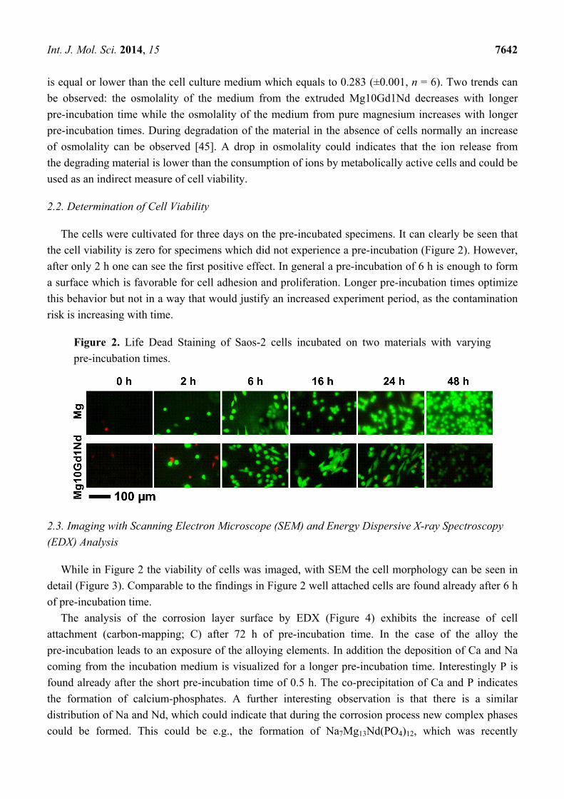
The cells were cultivated for three days on the pre-incubated specimens. It can clearly be seen that
the cell viability is zero for specimens which did not experience a pre-incubation (Figure 2). However,
after only 2 h one can see the first positive effect. In general a pre-incubation of 6 h is enough to form
a surface which is favorable for cell adhesion and proliferation. Longer pre-incubation times optimize
this behavior but not in a way that would justify an increased experiment period, as the contamination
risk is increasing with time.
Figure 2. Life Dead Staining of Saos-2 cells incubated on two materials with varying
pre-incubation times.
2.3. Imaging with Scanning Electron Microscope (SEM) and Energy Dispersive X-ray Spectroscopy
(EDX) Analysis
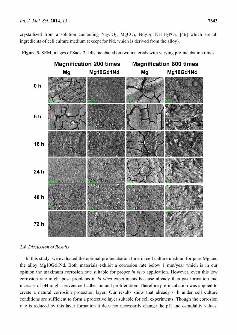
While in Figure 2 the viability of cells was imaged, with SEM the cell morphology can be seen in
detail (Figure 3). Comparable to the findings in Figure 2 well attached cells are found already after 6 h
of pre-incubation time.
The analysis of the corrosion layer surface by EDX (Figure 4) exhibits the increase of cell
attachment (carbon-mapping; C) after 72 h of pre-incubation time. In the case of the alloy the
pre-incubation leads to an exposure of the alloying elements. In addition the deposition of Ca and Na
coming from the incubation medium is visualized for a longer pre-incubation time. Interestingly P is
found already after the short pre-incubation time of 0.5 h. The co-precipitation of Ca and P indicates
the formation of calcium-phosphates. A further interesting observation is that there is a similar
distribution of Na and Nd, which could indicate that during the corrosion process new complex phases
could be formed. This could be e.g., the formation of Na7Mg13Nd(PO4)12, which was recently
Int. J. Mol. Sci. 2014, 15 7643
crystallized from a solution containing Na2CO3, MgCO3, Nd2O3, NH4H2PO4, [46] which are all
ingredients of cell culture medium (except for Nd, which is derived from the alloy).
Figure 3. SEM images of Saos-2 cells incubated on two materials with varying pre-incubation times.
2.4. Discussion of Results
In this study, we evaluated the optimal pre-incubation time in cell culture medium for pure Mg and
the alloy Mg10Gd1Nd. Both materials exhibit a corrosion rate below 1 mm/year which is in our
opinion the maximum corrosion rate suitable for proper in vivo application. However, even this low
corrosion rate might pose problems in in vitro experiments because already then gas formation and
increase of pH might prevent cell adhesion and proliferation. Therefore pre-incubation was applied to
create a natural corrosion protection layer. Our results show that already 6 h under cell culture
conditions are sufficient to form a protective layer suitable for cell experiments. Though the corrosion
rate is reduced by this layer formation it does not necessarily change the pH and osmolality values.

Int. J. Mol. Sci. 2014, 15 7644
This indicates that not the mere decrease in corrosion rate facilitates cell adhesion but that probably the
combination of reduced gas formation plus a change in surface chemistry and morphology supports
cell growth on the material. The results—in terms of cell adhesion and proliferation—are comparable
for both materials. This indicates that influence of alloying elements on the cells can be neglected at
least in the first few days of experiments.
A detailed study of the composition of the corrosion layer could shed light on the mechanisms of
corrosion in this very complex environment and might bridge the gap toward in vivo corrosion studies.
Figure 4. EDX element mapping of specimens with Saos-2 cells incubated on two
materials with varying pre-incubation times.
3. Experimental Section
3.1. Materials
Pure magnesium and Mg10Gd1Nd were cast in the Magnesium Innovation Center (MagIC) of the
Helmholtz-Zentrum Geesthacht by direct-chill casting. High-purity magnesium (99.94%) was obtained
from Magnesium Elektron (Manchester, UK). Pure Gd and Nd (99.5%) were obtained from Grirem
Int. J. Mol. Sci. 2014, 15 7645
(Beijing, China). The nominal amounts were added to the melt (750 °C) under a protective atmosphere
(Ar + 2% SF6, Linde, Hamburg, Germany), which was stirred at 200 rpm and then poured in
a preheated mold (550 °C). The cleanliness of the ingots was assured by using a filter (Foseco SIVEX
FC, Foseco, London, UK). Cylindrical ingots with a diameter of 12 cm and a length of 20 cm were
obtained. The materials were afterwards extruded in the Strangpresszentrum Berlin to rods with 12 mm
diameter and machined to round discs with dimensions of 10 × 1.5 mm. The average weight of the
specimens was approximately 0.2 g. They were used without further surface treatment.
Samples were disinfected in 70% ethanol (VWR International, Darmstadt, Germany) in an ultrasonic
bath for 20 min. Afterwards they were processed in parallel using standard 24 well plates (Cellstar,
Greiner Bio-one GmbH, Frickenhausen, Germany), with one sample per well. Pure Mg and
Mg10Gd1Nd discs were directly used (0 h) or immersed in 2 mL Mc Coy’s cell culture medium
(Mc Coy’s 5A Medium, Life Technologies, Darmstadt, Germany) supplemented with 10% FBS
(FBS Gold, PAA Laboratories, Linz, Austria) for 0.5, 2, 6, 16, 24, 48 h and under cell culture
conditions (37 °C, 21% O2, 5% CO2, 95% relative humidity). In each case n = 4 was chosen per alloy
and per pre-incubation time.
3.2. Determination of Corrosion Rate, pH and Osmolality
The corrosion rate was determined by weight loss. Each sample was weighed (Scaltec SBA52,
Scaltec Instruments GmbH, Göttingen, Germany), numbered and sterilized (again 20 min in ultrasonic
bath with ethanol) before pre-incubation (n = 3 per alloy and pre-incubation time). After pre-incubation
the samples were placed into a new agarose coated 12-well-plate and treated as described below with
cells. After three days of incubation under cell culture conditions the samples were taken from the
corrosion medium. To remove all corrosion residues from the samples they were immersed in chromic
acid (180 g/L in distilled water, VWR International, Darmstadt, Germany) for 10 min, turned and left
for another 10 min. Afterwards the samples were cleaned with distilled water and then with 100%
ethanol. When the samples were dried their weight was measured again and the corrosion rate (CR)
was calculated as follows [47]:
48.76 10 WCR
A t
A = surface area [cm2], t = time [h], ρ = density (Mg = 1.74; Mg10Gd1Nd = 1.90 [g/cm3]),
ΔW = mass loss [g].
After the final three days of incubation with cells on the pre-incubated specimens the cell culture
medium was removed from the wells and used for analysis of the pH-value and osmolality.
For measuring the pH-value the electrode was calibrated each time before using. Then the electrode was
dipped into the solution and left there until the value on the display of the measurement device was stable.
The osmolality of the supernatants was analyzed by cryoscopic osmometer (Osmomat 030-D,
Gonotec GmbH, Berlin, Germany). 60 µL of the supernatant were pipetted into a small cup (1 mL) and
put into the osmometer. The device is a freezing point osmometer which determines the osmolality by
measuring the freezing point. This goes down by 1.86 °C when 1 mole of a non ionic solute is added to
one kilogram of solvent [48].
Int. J. Mol. Sci. 2014, 15 7646
3.3. Determination of Cell Viability
The human osteosarcoma cell line Saos-2 was purchased from the European Collection of Cell
Cultures (ECACC, Salisbury, UK) and cultured under cell culture conditions.
After pre-incubation, the Mg samples were transferred into agarose pre-coated 12-well-plate
(one sample per well). The agarose-coating was applied by heating pre-sterilized agarose (1% w/v in a dest;
VWR International, Darmstadt, Germany) to about 80 °C and rapid filling and decanting of the wells.
This leads to a thin film of agarose throughout the well. For cell adhesion, Saos-2 cells were
trypsinized, centrifuged and afterwards resuspended in Mc Coy’s medium + 10% FBS to a concentration
of 1 × 10⁶ cells per mL. Then 50 µL of the cell suspension were homogeneously applied onto each
sample. Samples were incubated for 30 min to allow the study of initial and fast adhesion. Then 2 mL
Mc Coy’s medium + 10% FBS was added in each well of the 12-well-plate followed by a further
incubation for three days under cell culture conditions.
Cell viability was assessed via a two-color fluorescence assay (Live/Dead Viability/Cytotoxicity
Kit, Life Technologies, Darmstadt, Germany). One sample per pre-incubation time was used for
live/dead staining. After removing the cell culture medium the sample was washed with phosphate
buffer solution (PBS, 137 mM NaCl, 2.7 mM KCl, 4.3 mM Na2HPO4, 1.4 mM KH2PO4, all chemicals
from VWR), then transferred to a new 12-well-plate and covered with the staining solution (2 µL
Calcein AM and 5 µL ethidiumhomidimer-1 in 5 mL PBS) and incubated for 20 min. Afterwards the
staining solution was replaced by PBS and samples were directly examined with a fluorescent
microscope (Eclipse Ti-S, Nikon, Düsseldorf, Germany). Living cells can be detected by using the
FITC-filter (Ex: 465–495 nm, Em: 515–555 nm, Mirror at 505 nm) as green areas, dead cells can be
observed by using the Texas Red-filter (Ex: 540–580 nm, Em: 600–660 nm, Mirror at 595 nm) as red
areas. The overlaying image was merged by the microscope software.
3.4. Imaging with Low-Voltage Scanning Electron Microscope (LVSEM) and Energy Dispersive X-ray
Spectroscopy (EDX) Analysis
To visualize magnesium samples covered with cells without the application of additional coatings
a special sample preparation was needed. After removing the cell culture medium the samples were
washed with PBS and put on a new 12-well-plate (one sample per well). All chemicals were supplied
by VWR International. 2.5% glutaraldehyde solution was loaded in each well and the plate was stored
overnight under the fume hood. On the next day samples were placed in a new plate and the
glutaraldehyde was replaced by 1% osmiumtetroxide for 30 min for lipid counterstaining [49].
Samples were again put into a new well to start the alcoholic row. At first 20% isopropanol was added
in each well so that the samples were completely covered. After one to two hours it was replaced by
40% isopropanol for another one to two hours. The same procedure was repeated with 60%, 80%, and
finally with 100% isopropanol followed by critical point drying (EM CPD030, Leica Mikrosysteme
Vertrieb GmbH, Wetzlar, Germany). During the procedure of critical point drying the isopropanol
contained in the cells was slowly and totally replaced by CO2 at 8 °C. Afterwards, temperature and
pressure were raised to the critical point of CO2, leading to water-free preparations. After drying
samples were transferred into a new 12-well-plate and stored in a drying cabinet. Cells on carriers were

Int. J. Mol. Sci. 2014, 15 7647
then visualised by low voltage mode in charge contrast at 1 keV, using the SEM in lens detector
(SEM Auriga, Carl Zeiss, Jena, Germany).
Additionally to the SEM an analysis with energy dispersive X-ray spectroscopy (EDX) was
performed (EDS; Apollo XP, EDAX, Ametek GmbH, Wiesbaden, Germany). The preparation of the
samples was done in the same way as for SEM. EDX-mapping of various elements (C, Na, Mg, Nd, Gd,
Ca, P) was done at an accelerating voltage of 8 keV with 120 µm aperture size at 200-fold magnification.
4. Conclusions
The natural protection layer which is formed already after 6 h of pre-incubation under cell culture
conditions is enough to improve cell adhesion and viability. This is—at least for Mg and Mg10Gd1Nd
studied here—independent of the material and its corrosion rate.
Acknowledgments
We wish to thank the colleagues from the MagIC, especially Norbert Hort and Karl-Ulrich Kainer,
for the provision of the materials and Bernhard Eltzschig for the preparation of the samples. We deeply
acknowledge the help of Bérengère Luthringer, Daniela Lange and Gabriele Salamon in the cell
experiments. Finally we are very grateful to Daniel Laipple who technically supported the SEM/EDX
analysis and Christine Selhuber-Unkel from the CAU Kiel for the intense discussion.
Author Contributions
A. M. performed the experiments as part of her BSc thesis, F. F. supervised her work in the
laboratory and gave advice for the experiments. R. W. was the main supervisor of the BSc thesis and
wrote the manuscript.
Conflicts of Interest
The authors declare no conflict of interest.
References
1. Witte, F. The history of biodegradable magnesium implants: A review. Acta Biomater. 2010, 6,
1680–1692.
2. Vormann, J. Magnesium: Nutrition and metabolism. Mol. Asp. Med. 2003, 24, 27–37.
3. Staiger, M.P.; Pietak, A.M.; Huadmai, J.; Dias, G. Magnesium and its alloys as orthopedic
biomaterials: A review. Biomaterials 2006, 27, 1728–1734.
4. Atrens, A.; Liu, M.; Zainal Abidin, N.I. Corrosion mechanism applicable to biodegradable
magnesium implants. Mater. Sci. Eng. B 2011, 176, 1609–1636.
5. Song, G.L.; Atrens, A. Corrosion mechanisms of magnesium alloys. Adv. Eng. Mater. 1999, 1, 11–33.
6. Windhagen, H.; Radtke, K.; Weizbauer, A.; Diekmann, J.; Noll, Y.; Kreimeyer, U.; Schavan, R.;
Stukenborg-Colsman, C.; Waizy, H. Biodegradable magnesium-based screw clinically equivalent
to titanium screw in hallux valgus surgery: Short term results of the first prospective, randomized,
controlled clinical pilot study. BioMed. Eng. OnLine 2013, 12, 10.

Int. J. Mol. Sci. 2014, 15 7648
7. Haude, M.; Erbel, R.; Erne, P.; Verheye, S.; Degen, H.; Böse, D.; Vermeersch, P.; Wijnbergen, I.;
Weissman, N.; Prati, F.; et al. Safety and performance of the drug-eluting absorbable metal
scaffold (DREAMS) in patients with de-novo coronary lesions: 12 month results of the
prospective, multicenter, first-in-man BIOSOLVE-I trial. Lancet 2013, 381, 836–844.
8. Erbel, R.; Böse, D.; Haude, M.; Kordish, I.; Churzidze, S.; Malyar, N.; Konorza, T.; Sack, S.
Absorbierbare Stents. Herz 2007, 32, 308–319.
9. Virtanen, S. Biodegradable Mg and Mg alloys: Corrosion and biocompatibility. Mater. Sci. Eng. B
2011, 176, 1600–1608.
10. Feyerabend, F.; Fischer, J.; Holtz, J.; Witte, F.; Willumeit, R.; Drücker, H.; Vogt, C.; Hort, N.
Evaluation of short-term effects of rare earth and other elements used in magnesium alloys on
primary cells and cell lines. Acta Biomater. 2010, 6, 1834–1842.
11. Witte, F.; Hort, N.; Vogt, C.; Cohen, S.; Kainer, K.U.; Willumeit, R.; Feyerabend, F. Degradable
biomaterials based on magnesium corrosion. Curr. Opin. Solid State Mater. Sci. 2008, 12, 63–72.
12. Kainer, K.U.; Bala Srinivasan, P.; Blawert, C.; Dietzel, W. 3.09-Corrosion of magnesium and its
alloys. In Shreir’s Corrosion; Cottis, B., Graham, M., Lindsay, R., Lyon, S., Richardson, T.,
Scantlebury, D., Stott, H., Eds.; Elsevier: Oxford, UK, 2010; pp. 2011–2041.
13. Yang, L.; Huang, Y.; Feyerabend, F.; Willumeit, R.; Kainer, K.U.; Hort, N. Influence of ageing
treatment on microstructure, mechanical and bio-corrosion properties of Mg-Dy alloys. J. Mech.
Behav. Biomed. Mater. 2012, 13, 36–44.
14. Gray-Munro, J.E.; Seguin, C.; Strong, M. Influence of surface modification on the in vitro
corrosion rate of magnesium alloy AZ31. J. Biomed. Mater. Res. A 2009, 91A, 221–230.
15. Hornberger, H.; Virtanen, S.; Boccaccini, A.R. Biomedical coatings on magnesium alloys—A
review. Acta Biomater. 2012, 8, 2442–2455.
16. Yang, J.; Cui, F.; Lee, I. Surface modifications of magnesium alloys for biomedical applications.
Ann. Biomed. Eng. 2011, 39, 1857–1871.
17. Shadanbaz, S.; Dias, G.J. Calcium phosphate coatings on magnesium alloys for biomedical
applications: A review. Acta Biomater. 2012, 8, 20–30.
18. Badar, M.; Lünsdorf, H.; Evertz, F.; Rahim, M.I.; Glasmacher, B.; Hauser, H.; Mueller, P.P.
The formation of an organic coat and the release of corrosion microparticles from metallic
magnesium implants. Acta Biomater. 2013, 9, 7580–7589.
19. Bornapour, M.; Muja, N.; Shum-Tim, D.; Cerruti, M.; Pekguleryuz, M. Biocompatibility and
biodegradability of Mg-Sr alloys: The formation of Sr-substituted hydroxyapatite. Acta Biomater.
2013, 9, 5319–5330.
20. Fan, J.; Qiu, X.; Niu, X.; Tian, Z.; Sun, W.; Liu, X.; Li, Y.; Li, W.; Meng, J. Microstructure,
mechanical properties, in vitro degradation and cytotoxicity evaluations of Mg-1.5Y-1.2Zn-
0.44Zr alloys for biodegradable metallic implants. Mater. Sci. Eng. C 2013, 33, 2345–2352.
21. Gray-Munro, J.E.; Strong, M. A study on the interfacial chemistry of magnesium hydroxide
surfaces in aqueous phosphate solutions: Influence of Ca2+, Cl− and protein. J. Colloid Interface Sci.
2013, 393, 421–428.
22. Kannan, M.B. Enhancing the performance of calcium phosphate coating on a magnesium alloy for
bioimplant applications. Mater. Lett. 2012, 76, 109–112.

Int. J. Mol. Sci. 2014, 15 7649
23. Kim, S.-M.; Jo, J.-H.; Lee, S.-M.; Kang, M.-H.; Kim, H.-E.; Estrin, Y.; Lee, J.-H.; Lee, J.-W.;
Koh, Y.-H. Hydroxyapatite-coated magnesium implants with improved in vitro and in vivo
biocorrosion, biocompatibility, and bone response. J. Biomed. Mater. Res. A 2014, 102, 429–441.
24. Iskandar, M.E.; Aslani, A.; Liu, H. The effects of nanostructured hydroxyapatite coating on the
biodegradation and cytocompatibility of magnesium implants. J. Biomed. Mater. Res. A 2013,
101A, 2340–2354.
25. Johnson, I.; Akari, K.; Liu, H. Nanostructured hydroxyapatite/poly(lactic-co-glycolic acid)
composite coating for controlling magnesium degradation in simulated body fluid.
Nanotechnology 2013, 24, 375103.
26. Zomorodian, A.; Garcia, M.P.; Moura e Silva, T.; Fernandes, J.C.; Fernandes, M.H.;
Montemor, M.F. Corrosion resistance of a composite polymeric coating applied on biodegradable
AZ31 magnesium alloy. Acta Biomater. 2013, 9, 8660–8670.
27. Ostrowski, N.; Lee, B.; Enick, N.; Carlson, B.; Kunjukunju, S.; Roy, A.; Kumta, P.N. Corrosion
protection and improved cytocompatibility of biodegradable polymeric layer-by-layer coatings on
AZ31 magnesium alloys. Acta Biomater. 2013, 9, 8704–8713.
28. Kunjukunju, S.; Roy, A.; Ramanathan, M.; Lee, B.; Candiello, J.E.; Kumta, P.N. A layer-by-layer
approach to natural polymer-derived bioactive coatings on magnesium alloys. Acta Biomater.
2013, 9, 8690–8703.
29. Killian, M.S.; Wagener, V.; Schmuki, P.; Virtanen, S. Functionalization of metallic magnesium
with protein layers via linker molecules. Langmuir 2010, 26, 12044–12048.
30. Wagener, V.; Killian, M.S.; Turhan, C.M.; Virtanen, S. Albumin coating on magnesium via linker
molecules—Comparing different coating mechanisms. Colloids Surf. B 2013, 103, 586–594.
31. Liu, X.; Yue, Z.; Romeo, T.; Weber, J.; Scheuermann, T.; Moulton, S.; Wallace, G.
Biofunctionalized anti-corrosive silane coatings for magnesium alloys. Acta Biomater. 2013, 9,
8671–8677.
32. Ye, S.-H.; Jang, Y.-S.; Yun, Y.-H.; Shankarraman, V.; Woolley, J.R.; Hong, Y.; Gamble, L.J.;
Ishihara, K.; Wagner, W.R. Surface modification of a biodegradable magnesium alloy with
phosphorylcholine (PC) and sulfobetaine (SB) functional macromolecules for reduced
thrombogenicity and acute corrosion resistance. Langmuir 2013, 29, 8320–8327.
33. Drynda, A.; Hassel, T.; Hoehn, R.; Perz, A.; Bach, F.-W.; Peuster, M. Development and
biocompatibility of a novel corrodible fluoride-coated magnesium-calcium alloy with improved
degradation kinetics and adequate mechanical properties for cardiovascular applications.
J. Biomed. Mater. Res. A 2010, 93A, 763–775.
34. Lozano, R.M.; Pérez-Maceda, B.T.; Carboneras, M.; Onofre-Bustamante, E.; García-Alonso, M.C.;
Escudero, M.L. Response of MC3T3-E1 osteoblasts, L929 fibroblasts, and J774 macrophages
to fluoride surface-modified AZ31 magnesium alloy. J. Biomed. Mater. Res. A 2013, 101,
2753–2762.
35. Lin, X.; Tan, L.; Zhang, Q.; Yang, K.; Hu, Z.; Qiu, J.; Cai, Y. The in vitro degradation process
and biocompatibility of a ZK60 magnesium alloy with a forsterite-containing micro-arc oxidation
coating. Acta Biomater. 2013, 9, 8631–8642.

Int. J. Mol. Sci. 2014, 15 7650
36. Wang, Y.M.; Guo, J.W.; Shao, Z.K.; Zhuang, J.P.; Jin, M.S.; Wu, C.J.; Wei, D.Q.; Zhou, Y.
A metasilicate-based ceramic coating formed on magnesium alloy by microarc oxidation and its
corrosion in simulated body fluid. Surf. Coat. Technol. 2013, 219, 8–14.
37. Levy, G.; Aghion, E. Effect of diffusion coating of Nd on the corrosion resistance of biodegradable
Mg implants in simulated physiological electrolyte. Acta Biomater. 2013, 9, 8624–8630.
38. Weng, L.; Webster, T.J. Nanostructured magnesium increases bone cell density. Nanotechnology
2012, 23, doi:10.1088/0957-4484/23/48/485105.
39. Weng, L.; Webster, T.J. Nanostructured magnesium has fewer detrimental effects on osteoblast
function. Int. J. Nanomed. 2013, 8, 1773–1781.
40. Willumeit, R.; Fischer, J.; Feyerabend, F.; Hort, N.; Bismayer, U.; Heidrich, S.; Mihailova, B.
Chemical surface alteration of biodegradable magnesium exposed to corrosion media. Acta Biomater.
2011, 7, 2704–2715.
41. Witte, F.; Kaese, V.; Haferkamp, H.; Switzer, E.; Meyer-Lindenberg, A.; Wirth, C.J.; Windhagen, H.
In vivo corrosion of four magnesium alloys and the associated bone response. Biomaterials 2005,
26, 3557–3563.
42. Bowen, P.K.; Drelich, J.; Goldman, J. Magnesium in the murine artery: Probing the products
of corrosion. Acta Biomater. 2014, 10, 1475–1483.
43. Lorenz, C.; Brunner, J.G.; Kollmannsberger, P.; Jaafar, L.; Fabry, B.; Virtanen, S. Effect of
surface pre-treatments on biocompatibility of magnesium. Acta Biomater. 2009, 5, 2783–2789.
44. Keim, S.; Brunner, J.G.; Fabry, B.; Virtanen, S. Control of magnesium corrosion and
biocompatibility with biomimetic coatings. J. Biomed. Mater. Res. B 2011, 96B, 84–90.
45. Feyerabend, F.; Drücker, H.; Laipple, D.; Vogt, C.; Stekker, M.; Hort, N.; Willumeit, R. Ion
release from magnesium materials in physiological solutions under different oxygen tensions.
J. Mater. Sci.: Mater. Med. 2012, 23, 9–24.
46. Jerbi, H.; Hidouri, M.; Mongi, B.A. Na7Mg13Nd(PO4)12. Acta Crystallogr. Sect. E 2012, 68, i44.
47. International, A. Standard guide for laboratory immersion corrosion testing of metals. In ASTM
Standard NACE/ASTMG31-12a; ASTM International: West Conshohocken, PA, USA, 2012.
48. Abele, J.E. The physical background to freezing point osmometry and its medical-biological
applications. Am. J. Med. Electron. 1963, 2, 32–41.
49. Angermüller, S.; Fahimi, H.D. Imidazole-buffered osmium tetroxide: An excellent stain for
visualization of lipids in transmission electron microscopy. Histochem. J. 1982, 14, 823–835.
© 2014 by the authors; licensee MDPI, Basel, Switzerland. This article is an open access article
distributed under the terms and conditions of the Creative Commons Attribution license
(http://creativecommons.org/licenses/by/3.0/).


![2010 Ebenso Ijms Paper Published[1]](https://img.dokumen.tips/doc/110x75/5420c2907bef0ab6128b45fb/2010-ebenso-ijms-paper-published1.jpg)