Embed Size (px)
Citation preview
7 OCTOBER 2015
Scientific Background on the Nobel Prize in Chemistry 2015
M E C H A N I S T I C S T U D I E S O F D N A R E PA I R
compiled by the Class for Chemistry of the Royal Swedish Academy of Sciences
THE ROYAL SWEDISH ACADEMY OF SCIENCES, founded in 1739, is an independent organisation whose overall objective is to promote the sciences and strengthen their influence in society. The Academy takes special responsibility for the natural sciences and mathematics, but endeavours to promote the exchange of ideas between various disciplines.
BOX 50005 (LILLA FRESCATIVÄGEN 4 A), SE-104 05 STOCKHOLM, SWEDEN TEL +46 8 673 95 00, [email protected] HTTP://KVA.SE

1 (15)
The Royal Swedish Academy of Sciences has decided to award Tomas Lindahl, Paul Modrich
and Aziz Sancar the Nobel Prize in Chemistry 2015 for their “Mechanistic studies of DNA repair”
Damage to the genetic material poses a threat to all organisms. To counteract this threat, cells
have evolved a series of intricate DNA repair pathways that correct DNA lesions affecting base
pairing or structure of DNA. Today we understand the molecular mechanisms of these pathways
in great detail, in large part due to the pioneering studies by Lindahl, Modrich and Sancar that
opened up the field.
Background
The human genome encodes the information needed to create a complete human being. During
every cell division, more than three billion DNA base pairs are replicated and copies of the
genome are transferred to the daughter cells. Although very efficient, the DNA replication
machinery responsible for this task still makes occasional mistakes. Given the size of the human
genome and the large number of cells in a human body (about 3.7 × 10¹³) mistakes will
inevitably accumulate during the lifetime of an individual. Most of these errors will remain
silent, but they can also cause serious diseases.
Despite its essential role in storing genetic information, the DNA molecule has limited chemical
stability and is subject to spontaneous decay [1]. Processes such as hydrolysis and oxidation
occur at significant levels in vivo, in part due to reactive metabolites continuously generated in
various physiological processes. In addition, external factors like radiation and genotoxic
chemicals will further stimulate of DNA damage formation.
The inherent instability of DNA constitutes both an opportunity and a threat. DNA lesions can
block important cellular processes such as DNA replication and transcription, cause genome
instability and impair gene expression. Lesions can also be mutagenic and change the coding
capacity of the genome, which can lead to devastating diseases and conditions associated with
genome instability, including cancer, neurodegenerative disorders and biological ageing. At the
same time, without mutations Darwinian evolution would not be possible. Furthermore,
mutagenic chemicals and radiation can also have a healing effect; they can for instance be used
to treat cancer, by introducing DNA lesions that halt cell proliferation and stimulate
programmed cell death.
2 (15)
The cell has developed ways to counteract DNA lesions and to keep DNA mutations at a
tolerable level. A number of different DNA repair mechanisms correct lesions and safeguard the
integrity of the genome. Four fundamental DNA repair pathways delineated by this year’s Nobel
Prize laureates will be discussed here.
Photoreactivation – the first DNA repair mechanism
In the 1920s, the American geneticist Hermann Muller (Nobel Prize in Physiology or Medicine,
1946) found that X-rays could mutate and kill cells [2]. Later on, other types of agents, including
ultraviolet (UV) light, were also shown to affect cell viability and mutation levels. The cellular
target for the lethal effects of X-rays and UV-light was unknown at this time, and there were no
identified cellular mechanisms that could repair the lesions once they occurred. A breakthrough
came in the late 1940s, when Albert Kelner studied bacteria and their recovery in response to
damage caused by UV-light. Kelner found that visible light could dramatically stimulate growth
recovery after a growth arrest caused by UV exposure [3], [4]. The phenomenon was termed
photoreactivation and it pointed to the existence of a light-dependent cellular mechanism that
could correct UV-induced cellular damage.
Oswald Avery and co-workers had demonstrated in 1944 that DNA is the material of heredity [5]
and in the 1950s it became clear that UV-induced damage most likely was targeted to DNA. At
this point, Renato Dulbecco (Nobel Prize in Physiology or Medicine, 1975) suggested that
photoreactivation was an enzymatic reaction dependent on visible light [6]. The correctness of
this assumption was demonstrated by Stanley Rupert, who in a series of reports showed that
DNA could be reactivated by visible light in the presence of a cell-free extract from Escherichia
coli or Saccharomyces cerevisiae [7, 8]. The observation of this enzymatic activity, known as the
photolyase, was of profound importance, since it demonstrated for the first time the existence of
DNA repair enzymes that could rescue UV-irradiated DNA. At first, the photolyase was just an
activity in an extract, but in 1978 Aziz Sancar could clone the E. coli photolyase gene and amplify
the gene product in vivo [9]. Sancar was at the time a PhD student of Rupert’s, but instead of
continuing to characterise the photolyase, he wrote his PhD dissertation and graduated. It
would take another six years, before Sancar returned to photolyase research.
Dark repair – the discovery of nucleotide excision repair
In addition to photoreactivation, UV damage can also be repaired in a light-independent process
(known as “dark repair”). That UV-irradiation of DNA introduced thymine dimers in vitro was
reported in 1960, but the in vivo relevance of this finding was unclear [10]. A couple of years
later, Jane Setlow and Richard Setlow demonstrated that thymine dimers inactivated
transforming DNA in the bacterium Hemophilus influenzae and that this type of lesion was
responsible for the biological effect of UV-radiation [11, 12]. This realization made it possible to
study the precise molecular consequences of thymine dimers and investigate how cells deal with
3 (15)
them. In 1963, Richard Setlow reported that thymine dimers inhibit DNA synthesis and that
these lesions are excised from DNA in wild-type UV-irradiated bacteria, but not in an UV-
sensitive, mutant E. coli strain [13]. In 1964, he made the seminal discovery that thymine dimers
disappeared from the irradiated, high molecular weight genomic DNA shortly after exposure to
UV and instead appeared in low molecular weight fractions. Richard Setlow and his colleague
William Carrier correctly interpreted this result as thymine dimers being excised (removed)
from the DNA, hence the name excision repair [14]. Around this time, Richard Boyce and Paul
Howard-Flanders also published observations with conclusions similar to those reached by
Richard Setlow [15]. The emerging mechanisms of what later became known as nucleotide
excision repair (NER) was further elucidated in crucial work by Philip Hanawalt and David
Pettijohn, who found that UV-irradiation stimulated DNA repair synthesis even outside the S-
phase, i.e. independent of genome replication [16]. The groundbreaking contributions of these
early pioneers clearly indicated the existence of repair mechanisms that could correct UV-
induced lesions, but the precise molecular mechanisms underlying NER remained obscure.
The molecular mechanisms of NER
The identification of the enzymes responsible for NER was greatly assisted by genetics analyses.
Earlier bacterial work had identified uvrA, uvrB, and uvrC genes in a search for mutations that
impaired NER and hindered growth resumption after UV irradiation [17]. Work in vivo [18-20]
and in E. coli extracts [21] by Erling Seeberg and others indicated that the uvr gene products
functioned by endonucleolytic cleavage of irradiated DNA, but this could not be examined in
detail due to the lack of purified proteins.
In the 1970s, identification of proteins was a major challenge, since DNA sequencing techniques
were limited and a given gene locus could encode more than one protein. Aziz Sancar, now
working with W. Dean Rupp at Yale School of Medicine, developed the elegant Maxicell
technique, which relies on a UV-repair deficient bacterial strain [22]. After transformation of a
plasmid DNA of interest, the hypersensitive bacteria can be UV-irradiated, which causes
breakdown of the larger chromosomal DNA, whereas the plasmid molecules that have not been
hit by UV can continue to replicate and express proteins. In combination with radioactive amino
acid incorporation, the technique allows for labelling and detection of plasmid-encoded proteins
in the absence of a chromosomal background. The Maxicell technique was soon applied to a
wide variety of protein identification projects and allowed Sancar to rapidly identify the proteins
encoded by the uvrA, uvrB and uvrC genes [23-25].
In a groundbreaking work published in 1983 [26], Sancar used the purified UvrA, UvrB, and
UvrC proteins to reconstitute essential steps in the NER pathway. The three proteins acted
specifically on damaged DNA. With UV-irradiated DNA as a substrate, the proteins hydrolysed
two phosphodiester-bonds on the damaged DNA strand. The incisions were performed at
4 (15)
precise locations relative the UV adduct, one at the 8th phosphodiester bond 5' to the lesion and
a second at the 4th or 5th bond 3' to the same lesion, thus generating a 12-13 nt long fragment.
Later, Sancar could show that the rate of the reaction is stimulated by UvrD (DNA helicase II)
and DNA polymerase I (Pol I), which catalyses the removal of the incised strand and synthesis of
the new DNA strand, respectively [27]. Finally, DNA ligase catalyses the formation of two new
phosphodiester bonds and thus seals the sugar-phosphate backbone. In the 1983 paper, Sancar
described the UvrA, B, and C proteins as working together in a single complex (an excinuclease),
but later findings by him and also others have modified this view. We know today that Uvr
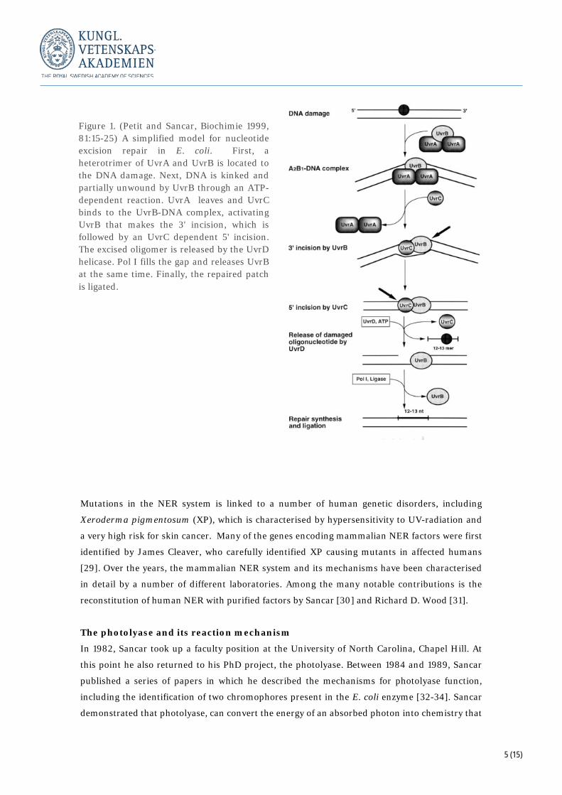
proteins associate with DNA lesions in a step-wise fashion (Figure 1) [28]. First, a complex of
two UvrA and one UvrB subunit (UvrA2B) tracks along the DNA. The UvrA subunits are
responsible for the initial damage recognition on double-stranded DNA, which causes the
UvrA2B complex to halt. At this point, the UvrB helicase activity is activated, leading to local
unwinding of DNA around the lesion (about 5 bp), kinking of the template and further
recognition of the damaged strand by UvrB. As a consequence, the UvrA proteins dissociate
from UvrB and a single UvrC subunit binds to the remaining UvrB-DNA complex. UvrC
activates UvrB, which makes the 3' incision, which is followed by UvrC catalysed incision at the
5' side of the lesion. The UvrD helicase displaces the damaged DNA strand, after which only
UvrB remains bound to the gapped DNA. At this point, Pol I associates with DNA, fills the gap
and UvrB is released. Finally, the repaired patch is ligated.
We know today that thymine dimers are just one of numerous types of lesions that interfere with
normal base pairing and thus distort the helical structure of DNA. NER can recognise these
problems and correct them by its cut-and-patch mechanism. The overall mechanisms of NER in
mammalian cells are closely related to those characterised in bacteria. The mechanisms of
damage recognition, incision on both sides of the lesion, removal of the damaged oligomer and
resynthesis of the gap are very similar between the two systems. However, even if the overall
strategy of repair is the same, the proteins responsible are distinct. Whereas damage recognition
and dual incision is carried out by only three proteins in E. coli more than fifteen proteins are
required to carry out the same function in human cells.
5 (15)
Mutations in the NER system is linked to a number of human genetic disorders, including
Xeroderma pigmentosum (XP), which is characterised by hypersensitivity to UV-radiation and
a very high risk for skin cancer. Many of the genes encoding mammalian NER factors were first
identified by James Cleaver, who carefully identified XP causing mutants in affected humans
[29]. Over the years, the mammalian NER system and its mechanisms have been characterised
in detail by a number of different laboratories. Among the many notable contributions is the
reconstitution of human NER with purified factors by Sancar [30] and Richard D. Wood [31].
The photolyase and its reaction mechanism
In 1982, Sancar took up a faculty position at the University of North Carolina, Chapel Hill. At
this point he also returned to his PhD project, the photolyase. Between 1984 and 1989, Sancar
published a series of papers in which he described the mechanisms for photolyase function,
including the identification of two chromophores present in the E. coli enzyme [32-34]. Sancar
demonstrated that photolyase, can convert the energy of an absorbed photon into chemistry that
Figure 1. (Petit and Sancar, Biochimie 1999, 81:15-25) A simplified model for nucleotide excision repair in E. coli. First, a heterotrimer of UvrA and UvrB is located to the DNA damage. Next, DNA is kinked and partially unwound by UvrB through an ATP-dependent reaction. UvrA leaves and UvrC binds to the UvrB-DNA complex, activating UvrB that makes the 3' incision, which is followed by an UvrC dependent 5' incision. The excised oligomer is released by the UvrD helicase. Pol I fills the gap and releases UvrB at the same time. Finally, the repaired patch is ligated.
6 (15)
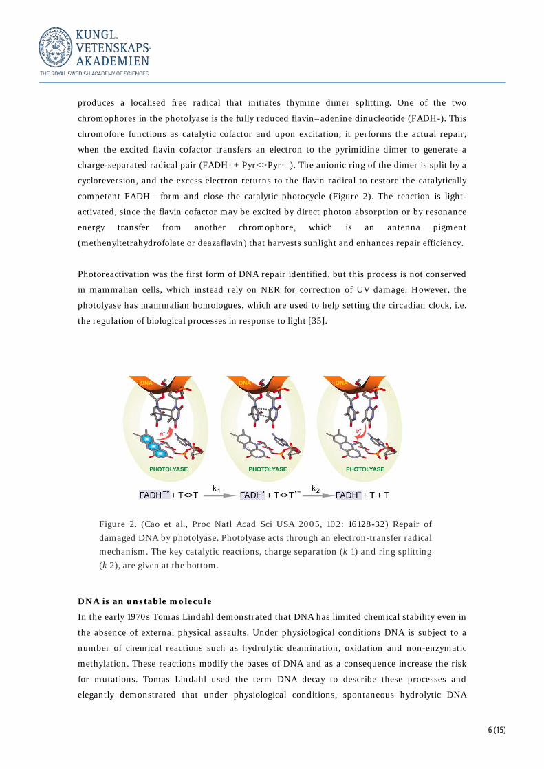
produces a localised free radical that initiates thymine dimer splitting. One of the two
chromophores in the photolyase is the fully reduced flavin–adenine dinucleotide (FADH-). This
chromofore functions as catalytic cofactor and upon excitation, it performs the actual repair,
when the excited flavin cofactor transfers an electron to the pyrimidine dimer to generate a
charge-separated radical pair (FADH· + Pyr<>Pyr·–). The anionic ring of the dimer is split by a
cycloreversion, and the excess electron returns to the flavin radical to restore the catalytically
competent FADH– form and close the catalytic photocycle (Figure 2). The reaction is light-
activated, since the flavin cofactor may be excited by direct photon absorption or by resonance
energy transfer from another chromophore, which is an antenna pigment
(methenyltetrahydrofolate or deazaflavin) that harvests sunlight and enhances repair efficiency.
Photoreactivation was the first form of DNA repair identified, but this process is not conserved
in mammalian cells, which instead rely on NER for correction of UV damage. However, the
photolyase has mammalian homologues, which are used to help setting the circadian clock, i.e.
the regulation of biological processes in response to light [35].
DNA is an unstable molecule
In the early 1970s Tomas Lindahl demonstrated that DNA has limited chemical stability even in
the absence of external physical assaults. Under physiological conditions DNA is subject to a
number of chemical reactions such as hydrolytic deamination, oxidation and non-enzymatic
methylation. These reactions modify the bases of DNA and as a consequence increase the risk
for mutations. Tomas Lindahl used the term DNA decay to describe these processes and
elegantly demonstrated that under physiological conditions, spontaneous hydrolytic DNA
Figure 2. (Cao et al., Proc Natl Acad Sci USA 2005, 102: 16128-32) Repair of damaged DNA by photolyase. Photolyase acts through an electron-transfer radical mechanism. The key catalytic reactions, charge separation (k 1) and ring splitting (k 2), are given at the bottom.
7 (15)
depurination occur at significant levels [36] and stimulate cleavage of DNA chains [37]. Perhaps
the most fascinating discovery was the demonstration of high levels of spontaneous cytosine
deamination under physiological conditions, which leads to the formation of uracil. Since uracil
forms base pairs with adenine, cytosine deaminiation is a highly mutagenic process, with
important long-term consequences. High levels of cytosine deamination pose a risk of depleting the
genetic material from cytosine-guanine base pairs and replacing them with thymine-adenine [38].
Discovery of base excision repair
Based on his observation that uracil is frequently formed in DNA, Tomas Lindahl came to the
conclusion that there must exist an enzymatic pathway that can handle this and other types of
base lesions. In a now classic study, he single-handedly identified the E. coli uracil-DNA
glycosylase (UNG) as the first repair protein [39] and two years later a second glycosylase,
specific for 3-methyladenine DNA [40]. We know today that UNG is the founding member of a
large family of proteins that orchestrate base excision repair (BER). The identification of UNG
relied on careful analysis of enzymatic release of uracil as a free base from DNA in vitro. Lindahl
demonstrated that the enzyme was specific to DNA and did not act on deoxymononucleotides or
any form of RNA. He also showed that the DNA backbone remained intact in the process, which
immediately suggested the involvement of another category of enzymes, the
apurinc/apyrimidinic endonucleases. An E. coli activity specific for apurinic sites had been
identified just two years earlier by Walter Verly [41, 42]. Lindahl could outline the basic
concepts for BER already in the 1974 paper and in a series of papers published in the years to
follow he and others could verify the proposed model. Through continued studies, Lindahl could
reconstitute the entire BER with purified enzyme, both from E. coli [43] and human cells [44].
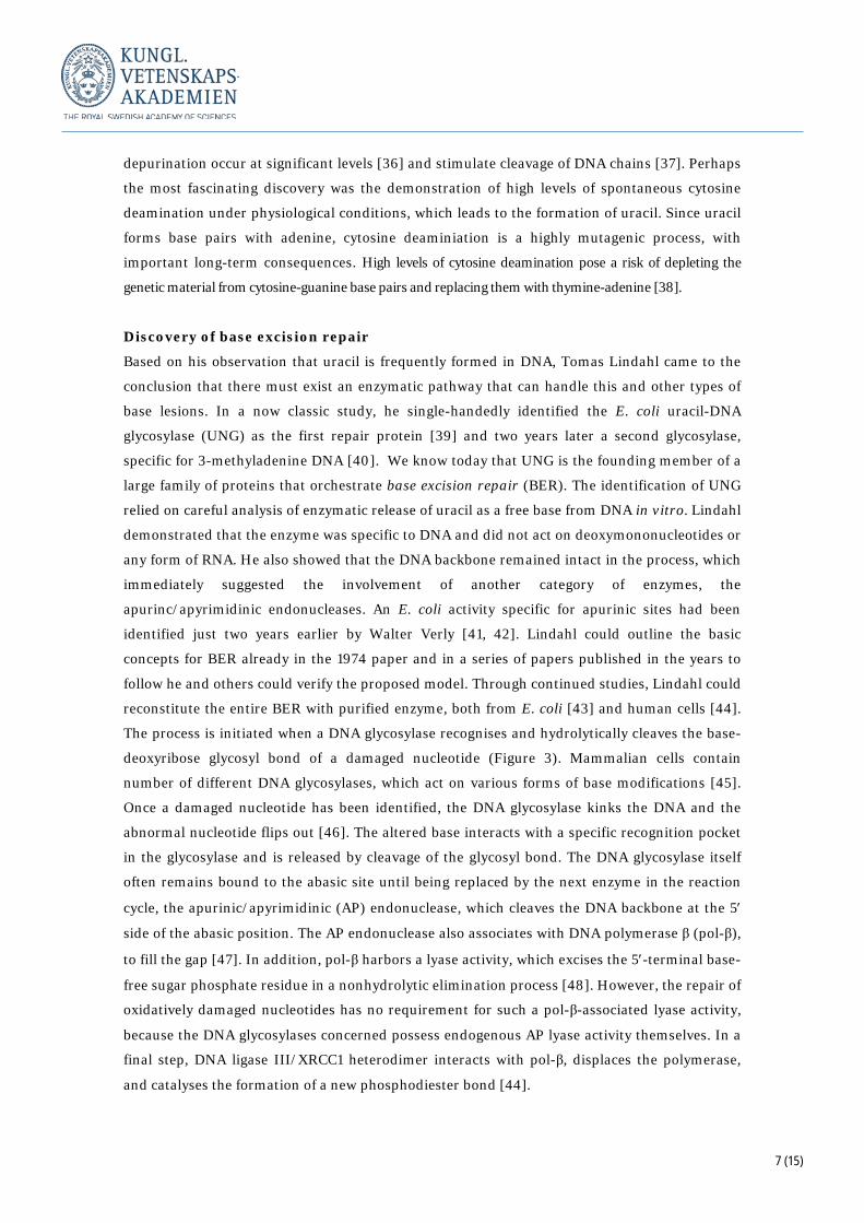
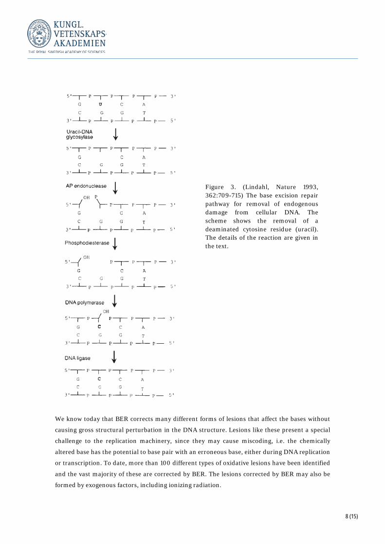
The process is initiated when a DNA glycosylase recognises and hydrolytically cleaves the base-
deoxyribose glycosyl bond of a damaged nucleotide (Figure 3). Mammalian cells contain
number of different DNA glycosylases, which act on various forms of base modifications [45].
Once a damaged nucleotide has been identified, the DNA glycosylase kinks the DNA and the
abnormal nucleotide flips out [46]. The altered base interacts with a specific recognition pocket
in the glycosylase and is released by cleavage of the glycosyl bond. The DNA glycosylase itself
often remains bound to the abasic site until being replaced by the next enzyme in the reaction
cycle, the apurinic/apyrimidinic (AP) endonuclease, which cleaves the DNA backbone at the 5′
side of the abasic position. The AP endonuclease also associates with DNA polymerase β (pol-β),
to fill the gap [47]. In addition, pol-β harbors a lyase activity, which excises the 5′-terminal base-
free sugar phosphate residue in a nonhydrolytic elimination process [48]. However, the repair of
oxidatively damaged nucleotides has no requirement for such a pol-β-associated lyase activity,
because the DNA glycosylases concerned possess endogenous AP lyase activity themselves. In a
final step, DNA ligase III/XRCC1 heterodimer interacts with pol-β, displaces the polymerase,
and catalyses the formation of a new phosphodiester bond [44].
8 (15)
We know today that BER corrects many different forms of lesions that affect the bases without
causing gross structural perturbation in the DNA structure. Lesions like these present a special
challenge to the replication machinery, since they may cause miscoding, i.e. the chemically
altered base has the potential to base pair with an erroneous base, either during DNA replication
or transcription. To date, more than 100 different types of oxidative lesions have been identified
and the vast majority of these are corrected by BER. The lesions corrected by BER may also be
formed by exogenous factors, including ionizing radiation.
Figure 3. (Lindahl, Nature 1993, 362:709-715) The base excision repair pathway for removal of endogenous damage from cellular DNA. The scheme shows the removal of a deaminated cytosine residue (uracil). The details of the reaction are given in the text.
9 (15)
Mismatch repair
As noted above, the DNA replication machinery is not error-free. There is always the possibility
that an incorrect nucleotide is introduced during synthesis of a new DNA strand. As a result, a
non-Watson-Crick base pair is formed, which distorts the double-stranded DNA helix. These
types of errors are known as mismatches and they have the capacity to change the sequence of
DNA, i.e. to introduce mutations. As a first line of defence against mismatches, replicating DNA
polymerases contain a 3′ to 5′ exonuclease activity that allows them to proofread the newly
synthesized DNA strand [49]. The exonuclease activity can correct mistakes during DNA
replication by reversing the direction of the polymerase and excising incorrectly introduced
nucleotides. Even if proofreading efficiently corrects most mistakes made during DNA synthesis,
some non-Watson-Crick base pairs still remain. To correct these errors, cells use mismatch
repair. It is estimated that that replicative DNA polymerases with proofreading in vitro have
error frequencies of about 5 × 10⁻⁵. Mismatch repair lowers this frequency significantly and the
mutation rate in for example human germ cells is estimated to be close to 1 × 10⁻⁸ per base pair
per generation [50].
The early studies of mismatch repair were mainly carried out by geneticists interested in
recombination. Robin Holliday had hypothesised that heteroduplexes between different DNA
strands would be formed during recombination [51]. The mismatches formed in this way had to
be corrected by some sort of cellular system, leading to gene conversion. Indeed, transfection of
heteroduplex molecules of lambda phage DNA into E. coli led to repair of the mismatches, but
the enzymatic system required for this effect was unknown. An important step was taken in
1976, when Robert Wagner and Matthew Meselson reported that repair of two or more close
sites on the same heteroduplex DNA molecule occur more often on the same strand than on the
opposite strand [52]. This observation led them to propose that mismatches are repaired by a
strand-specific mechanism directed towards one strand, which allows for repair of long DNA
tracts. They also speculated that strand-specific mismatch repair could be used to correct non-
canonical base pairs formed during DNA synthesis. The repair machinery could be guided to the
newly replicated strand either via a special relation to the replication machinery or directed by
under-methylation of the newly synthesised strand. In E. coli DNA is normally methylated on
both strands at GATC-sites, but during DNA synthesis the nascent strand is unmethylated for a
period of time [53], which could allow the mismatch repair machinery to distinguish newly
replicated DNA from the template DNA strand. In support of this notion, loss of cellular DNA
methylase, the enzyme that adds a methyl group to the adenine of the sequence GATC, was
already in 1975 shown to cause higher mutation rate in E. coli [54]. The genetics of mismatch
repair was further clarified when Barry W. Glickman and Miroslav Radman could demonstrate
10 (15)
that mutH, mutL, mutS and uvrD genes were all required for the methylation-instructed DNA
mismatch correction [55].
Direct evidence for methyl-directed repair of mismatches came in 1983. First, Paul Modrich and
Matthew Meselson used heteroduplex constructs with defined states of DNA methylation to
demonstrate that DNA methylation indeed directed strand-specific elimination of mismatches
in E. coli [56]. Furthermore, Modrich developed an assay that allowed analysis of DNA
mismatch repair in cell-free E. coli extracts [57]. In his assay, he used the classical bacteriophage
heteroduplexes, but introduced mismatches within overlapping sequences recognised by two
different restriction endonucleases. About 1,000 bp from the mismatch was a GATC methylation
site, which could be methylated in a controlled way and thus used to monitor strand-specific
correction. Using the assay, Modrich could demonstrate that the repair activity was dependent
on ATP, the methylation state of the heteroduplex, and that mutations affecting mutH, mutL,
mutS, and uvrD all impaired mismatch repair in cell-free E. coli extracts. With the help of this
elegant assay, Modrich could in a series of papers isolate the products of the different repair
genes to near homogeneity, identify the proteins and investigate their properties in vitro in
great detail [58-60].
Modrich’s work culminated in a groundbreaking paper published in 1989 [61], in which he could
finally reconstitute DNA mismatch correction in a defined in vitro system. In the paper,
Modrich demonstrated the requirement of DNA polymerase III, exonuclease I, and DNA ligase
for mismatch repair. He then combined these factors with purified MutH, MutL, MutS, UvrD,
and single-stranded DNA-binding protein. Together these factors could process mismatches in
vivo in a strand-specific manner directed by the single, GATC sequence methylated on only one
strand (hemimethylated) and located distant from the mismatch. In this and other studies,
Modrich demonstrated that MutS, which recognises and binds to non-Watson-Crick base pairs,
performs a core function in mismatch repair system. To confer strand-specificity, MutH binds at
hemimethylated GATC sites on the nascent strand. MutL acts as a mediator, which interacts
with both MutH and MutS. MutL transduces signals from MutS, which leads to activation of the
latent MutH endonuclease activity causing a nick in the nascent DNA strand near the
hemimethylated GATC-site. The machinery now interacts with a helicase (UvrD), which
together with the MutS, MutL, and MutH proteins separates the two DNA strands towards the
location of the mismatch. Displacement of the mutant strand continues past, and halts just
downstream of, the mismatch. The nascent strand is then replaced by a gap-filling reaction, in
which DNA polymerase III uses the parental strand as a template.
Mismatch repair in mammalian cells.
Later studies by Modrich and others have demonstrated the conservation of mismatch repair in
eukaryotic cells and in 2004, Modrich managed to reconstitute human mismatch repair with
only purified factors [62]. In contrast to the situation in E. coli, DNA methylation does not direct
11 (15)
strand specific DNA repair in eukaryotic cells. One possibility is that the strand specific nicks
formed during DNA replication can direct strand-specific error correction. In support of this
notion, mismatch repair is more efficient on the lagging strand at the replication fork [63], and a
single nick is sufficient to direct strand specific repair in in vitro [62, 64]. Alternatively, the
mismatch repair machinery may be directed by ribonucleotides transiently present in DNA after
replication [65]. In other aspects, eukaryotic mismatch repair is closely related to the E. coli
system with conserved homologues to key factors such as MutS and MutL. The importance of
the mammalian mismatch repair system is underscored by the finding that defects in this
pathway cause hereditary nonpolyposis colon cancer [66, 67].
Summary
Tomas Lindahl, Paul Modrich, and Aziz Sancar have made fundamental and groundbreaking
discoveries on the enzymatic mechanisms of DNA repair. Lindahl demonstrated that DNA is an
inherently unstable molecule, subject to decay even under physiological conditions. Guided by
thia observation, Lindahl identified a completely new group of DNA glycosylases and described
their role in base excision repair. Modrich transformed the field of mismatch repair from genetic
observations to a detailed biochemical understanding, first in bacteria, and later in eukaryotic
cells. Sancar has transformed the field of nucleotide excision repair, from genetics and
phenomena in cell extracts, to a detailed molecular description of the mechanisms involved, first
in bacteria, and later also in eukaryotic cells. Sancar also explained the molecular mechanisms
underlying photoreactivation, the first form of DNA repair described.
Claes M. Gustafsson
Professor of Medical Chemistry and Cell Biology, University of Gothenburg
Member of the Nobel Committee for Chemistry
12 (15)
Referenser 1. Lindahl, T., Instability and decay of the primary structure of DNA. Nature, 1993.
362(6422): p. 709-15. 2. Muller, H.J., Artificial Transmutation of the Gene. Science, 1927. 66(1699): p. 84-7. 3. Kelner, A., Effect of Visible Light on the Recovery of Streptomyces Griseus Conidia
from Ultra-violet Irradiation Injury. Proc Natl Acad Sci U S A, 1949. 35(2): p. 73-9. 4. Kelner, A., Photoreactivation of Ultraviolet-Irradiated Escherichia Coli, with Special
Reference to the Dose-Reduction Principle and to Ultraviolet-Induced Mutation. J Bacteriol, 1949. 58(4): p. 511-22.
5. Avery, O.T., C.M. Macleod, and M. McCarty, Studies on the Chemical Nature of the Substance Inducing Transformation of Pneumococcal Types : Induction of Transformation by a Desoxyribonucleic Acid Fraction Isolated from Pneumococcus Type Iii. J Exp Med, 1944. 79(2): p. 137-58.
6. Dulbecco, R., Experiments on photoreactivation of bacteriophages inactivated with ultraviolet radiation. J Bacteriol, 1950. 59(3): p. 329-47.
7. Rupert, C.S., S.H. Goodgal, and R.M. Herriott, Photoreactivation in vitro of ultraviolet-inactivated Hemophilus influenzae transforming factor. J Gen Physiol, 1958. 41(3): p. 451-71.
8. Rupert, C.S., Photoreactivation of transforming DNA by an enzyme from bakers' yeast. J Gen Physiol, 1960. 43: p. 573-95.
9. Sancar, A. and C.S. Rupert, Cloning of the phr gene and amplification of photolyase in Escherichia coli. Gene, 1978. 4(4): p. 295-308.
10. Beukers, R. and W. Berends, Isolation and identification of the irradiation product of thymine. Biochim Biophys Acta, 1960. 41: p. 550-1.
11. Setlow, R.B. and J.K. Setlow, Evidence that ultraviolet-induced thymine dimers in DNA cause biological damage. Proc Natl Acad Sci U S A, 1962. 48: p. 1250-7.
12. Setlow, J.K. and R.B. Setlow, Nature of Photoreactivable Ultra-Violet Lesion in Deoxyribonucleic Acid. Nature, 1963. 197(486): p. 560-&.
13. Setlow, R.B., P.A. Swenson, and W.L. Carrier, Thymine Dimers and Inhibition of DNA Synthesis by Ultraviolet Irradiation of Cells. Science, 1963. 142(3598): p. 1464-6.
14. Setlow, R.B. and W.L. Carrier, The Disappearance of Thymine Dimers from DNA: An Error-Correcting Mechanism. Proc Natl Acad Sci U S A, 1964. 51: p. 226-31.
15. Boyce, R.P. and P. Howard-Flanders, Release of Ultraviolet Light-Induced Thymine Dimers from DNA in E. Coli K-12. Proc Natl Acad Sci U S A, 1964. 51: p. 293-300.
16. Pettijohn, D. and P. Hanawalt, Evidence for Repair-Replication of Ultraviolet Damaged DNA in Bacteria. J Mol Biol, 1964. 9: p. 395-410.
17. Howard-Flanders, P., R.P. Boyce, and L. Theriot, Three loci in Escherichia coli K-12 that control the excision of pyrimidine dimers and certain other mutagen products from DNA. Genetics, 1966. 53(6): p. 1119-36.
18. Rupp, W.D. and P. Howard-Flanders, Discontinuities in the DNA synthesized in an excision-defective strain of Escherichia coli following ultraviolet irradiation. 1968. DNA Repair (Amst), 2005. 4(5): p. 620-33.
19. Shimada, K., H. Ogawa, and J. Tomizawa, Studies on radiation-sensitive mutants of E. coli. II. Breakage and repair of ultraviolet irradiated intracellular DNA of phage lambda. Mol Gen Genet, 1968. 101(3): p. 245-56.
20. Seeberg, E., W.D. Rupp, and P. Strike, Impaired incision of ultraviolet-irradiated deoxyribonucleic acid in uvrC mutants of Escherichia coli. J Bacteriol, 1980. 144(1): p. 97-104.
13 (15)
21. Seeberg, E., J. Nissen-Meyer, and P. Strike, Incision of ultraviolet-irradiated DNA by extracts of E. coli requires three different gene products. Nature, 1976. 263(5577): p. 524-6.
22. Sancar, A., A.M. Hack, and W.D. Rupp, Simple method for identification of plasmid-coded proteins. J Bacteriol, 1979. 137(1): p. 692-3.
23. Sancar, A., et al., Identification of the uvrB gene product. J Mol Biol, 1981. 148(1): p. 63-76.
24. Sancar, A., et al., Identification of the uvrA gene product. J Mol Biol, 1981. 148(1): p. 45-62.
25. Sancar, A., et al., Identification of the uvrC gene product. Proc Natl Acad Sci U S A, 1981. 78(9): p. 5450-4.
26. Sancar, A. and W.D. Rupp, A novel repair enzyme: UVRABC excision nuclease of Escherichia coli cuts a DNA strand on both sides of the damaged region. Cell, 1983. 33(1): p. 249-60.
27. Husain, I., et al., Effect of DNA polymerase I and DNA helicase II on the turnover rate of UvrABC excision nuclease. Proc Natl Acad Sci U S A, 1985. 82(20): p. 6774-8.
28. Petit, C. and A. Sancar, Nucleotide excision repair: from E. coli to man. Biochimie, 1999. 81(1-2): p. 15-25.
29. Cleaver, J.E., Defective repair replication of DNA in xeroderma pigmentosum. Nature, 1968. 218(5142): p. 652-6.
30. Mu, D., et al., Reconstitution of human DNA repair excision nuclease in a highly defined system. J Biol Chem, 1995. 270(6): p. 2415-8.
31. Aboussekhra, A., et al., Mammalian DNA nucleotide excision repair reconstituted with purified protein components. Cell, 1995. 80(6): p. 859-68.
32. Sancar, G.B., et al., Action mechanism of Escherichia coli DNA photolyase. I. Formation of the enzyme-substrate complex. J Biol Chem, 1987. 262(1): p. 478-85.
33. Jorns, M.S., et al., Action mechanism of Escherichia coli DNA photolyase. II. Role of the chromophores in catalysis. J Biol Chem, 1987. 262(1): p. 486-91.
34. Sancar, G.B., et al., Action mechanism of Escherichia coli DNA photolyase. III. Photolysis of the enzyme-substrate complex and the absolute action spectrum. J Biol Chem, 1987. 262(1): p. 492-8.
35. Sancar, A., Regulation of the mammalian circadian clock by cryptochrome. J Biol Chem, 2004. 279(33): p. 34079-82.
36. Lindahl, T. and B. Nyberg, Rate of depurination of native deoxyribonucleic acid. Biochemistry, 1972. 11(19): p. 3610-8.
37. Lindahl, T. and A. Andersson, Rate of chain breakage at apurinic sites in double-stranded deoxyribonucleic acid. Biochemistry, 1972. 11(19): p. 3618-23.
38. Lindahl, T. and B. Nyberg, Heat-induced deamination of cytosine residues in deoxyribonucleic acid. Biochemistry, 1974. 13(16): p. 3405-10.
39. Lindahl, T., An N-glycosidase from Escherichia coli that releases free uracil from DNA containing deaminated cytosine residues. Proc Natl Acad Sci U S A, 1974. 71(9): p. 3649-53.
40. Lindahl, T., New class of enzymes acting on damaged DNA. Nature, 1976. 259(5538): p. 64-6.
41. Verly, W.G. and Y. Paquette, An endonuclease for depurinated DNA in Escherichia coli B. Can J Biochem, 1972. 50(2): p. 217-24.
42. Verly, W.G., Y. Paquette, and L. Thibodeau, Nuclease for DNA apurinic sites may be involved in the maintenance of DNA in normal cells. Nat New Biol, 1973. 244(133): p. 67-9.
43. Dianov, G. and T. Lindahl, Reconstitution of the DNA base excision-repair pathway. Curr Biol, 1994. 4(12): p. 1069-76.
14 (15)
44. Kubota, Y., et al., Reconstitution of DNA base excision-repair with purified human proteins: interaction between DNA polymerase beta and the XRCC1 protein. EMBO J, 1996. 15(23): p. 6662-70.
45. Lindahl, T. and R.D. Wood, Quality control by DNA repair. Science, 1999. 286(5446): p. 1897-905.
46. Mol, C.D., et al., Crystal structure and mutational analysis of human uracil-DNA glycosylase: structural basis for specificity and catalysis. Cell, 1995. 80(6): p. 869-78.
47. Sobol, R.W., et al., Requirement of mammalian DNA polymerase-beta in base-excision repair. Nature, 1996. 379(6561): p. 183-6.
48. Matsumoto, Y. and K. Kim, Excision of deoxyribose phosphate residues by DNA polymerase beta during DNA repair. Science, 1995. 269(5224): p. 699-702.
49. Brutlag, D. and A. Kornberg, Enzymatic synthesis of deoxyribonucleic acid. 36. A proofreading function for the 3' leads to 5' exonuclease activity in deoxyribonucleic acid polymerases. J Biol Chem, 1972. 247(1): p. 241-8.
50. Segurel, L., M.J. Wyman, and M. Przeworski, Determinants of mutation rate variation in the human germline. Annu Rev Genomics Hum Genet, 2014. 15: p. 47-70.
51. Holliday, R., Mechanism for Gene Conversion in Fungi. Genetical Research, 1964. 5(2): p. 282-&.
52. Wagner, R., Jr. and M. Meselson, Repair tracts in mismatched DNA heteroduplexes. Proc Natl Acad Sci U S A, 1976. 73(11): p. 4135-9.
53. Marinus, M.G., Adenine methylation of Okazaki fragments in Escherichia coli. J Bacteriol, 1976. 128(3): p. 853-4.
54. Marinus, M.G. and N.R. Morris, Pleiotropic effects of a DNA adenine methylation mutation (dam-3) in Escherichia coli K12. Mutat Res, 1975. 28(1): p. 15-26.
55. Glickman, B.W. and M. Radman, Escherichia coli mutator mutants deficient in methylation-instructed DNA mismatch correction. Proc Natl Acad Sci U S A, 1980. 77(2): p. 1063-7.
56. Pukkila, P.J., et al., Effects of high levels of DNA adenine methylation on methyl-directed mismatch repair in Escherichia coli. Genetics, 1983. 104(4): p. 571-82.
57. Lu, A.L., S. Clark, and P. Modrich, Methyl-directed repair of DNA base-pair mismatches in vitro. Proc Natl Acad Sci U S A, 1983. 80(15): p. 4639-43.
58. Su, S.S. and P. Modrich, Escherichia coli mutS-encoded protein binds to mismatched DNA base pairs. Proc Natl Acad Sci U S A, 1986. 83(14): p. 5057-61.
59. Grilley, M., et al., Isolation and characterization of the Escherichia coli mutL gene product. J Biol Chem, 1989. 264(2): p. 1000-4.
60. Welsh, K.M., et al., Isolation and characterization of the Escherichia coli mutH gene product. J Biol Chem, 1987. 262(32): p. 15624-9.
61. Lahue, R.S., K.G. Au, and P. Modrich, DNA mismatch correction in a defined system. Science, 1989. 245(4914): p. 160-4.
62. Dzantiev, L., et al., A defined human system that supports bidirectional mismatch-provoked excision. Mol Cell, 2004. 15(1): p. 31-41.
63. Pavlov, Y.I., I.M. Mian, and T.A. Kunkel, Evidence for preferential mismatch repair of lagging strand DNA replication errors in yeast. Curr Biol, 2003. 13(9): p. 744-8.
64. Holmes, J., Jr., S. Clark, and P. Modrich, Strand-specific mismatch correction in nuclear extracts of human and Drosophila melanogaster cell lines. Proc Natl Acad Sci U S A, 1990. 87(15): p. 5837-41.
65. Lujan, S.A., et al., Ribonucleotides are signals for mismatch repair of leading-strand replication errors. Mol Cell, 2013. 50(3): p. 437-43.
15 (15)
66. Fishel, R., et al., The human mutator gene homolog MSH2 and its association with hereditary nonpolyposis colon cancer. Cell, 1993. 75(5): p. 1027-38.
67. Parsons, R., et al., Hypermutability and mismatch repair deficiency in RER+ tumor cells. Cell, 1993. 75(6): p. 1227-36.