Embed Size (px)
DESCRIPTION
this document includes the detailed information about respirators and ventilators
Citation preview
Qwertyuiopasdfghjklzxcvbnmqwertyuiopasdfghjklzxcvbnmqwertyuiopasdfghjklzxcvbnmqwertyuiopasdfghjklzxcvbnmq we rtyuiopasdfghjklzxcvbnmqwertyuiopasdfghjklzxcvbnmqwertyuiopasdfghjklzxcvbnmqwertyuiopasdfghjklzxcvbnmqwertyuiopasdfghjklzxcvbnmqwertyuiopasdfghjklzxcvbnmqwertyuiopasdfghjklzxcvbnmqwertyuiopasdfghjklzxcvbnmqwertyuiopasdfghjklzxcvbnmqwertyuiopasdfghjklzxcvbnmqwertyuiopasdfghjklzxcvbnmqwertyuiopasdfghjklzxcvbnmqwertyuiopasdfghjklzxcvbnmqwertyuiopasdfghjklzxcvbnmrtyuiopasdfghjklzxcvbnmqwertyuiopasdfghjklzxcvbnmqwertyuiopasdfghjklzxcvbnmqw
CLINICAL TEACHINGVENTILATORS AND RESPIRATORS
VENTILATORS AND RESPIRATORS
VENTILATORS
INTRODUCTION:- Mechanical ventilation is often a life-saving intervention to mechanically assist or replace spontaneous breathing .This may involve a machine called a ventilator. It can be used as a short term measure, for example during an operation or critical illness (often in the setting of an intensive care unit). It may be used at home or in a nursing or rehabilitation institution if patients have chronic illnesses that require long-term ventilatory assistance.
DEFINITION:- A mechanical ventilator is a positive or negative pressure breathing device that can maintain ventilation and oxygen delivery for a prolonged time.
Or
A mechanical ventilator is a machine that generates a controlled flow of gas into patient’s airway. Oxygen and air are received from cylinders or wall outlets and gas pressure is reduced and blended according to the prescribed inspired oxygen tension accumulated in receptacle in the machine and delivered to the patient using one of available modes for ventilation.
INDICATIONS :-
1) Decrease in oxygenation
2) Increase in arterial carbon dioxide levels
3) Persistent acidosis
4) Thoracic and abdominal surgery
5) Drug overdose
6) Neuromuscular disorders
7) Inhalation injury
8) COPD
9) Multiple trauma
10) Multiple system failure
11) Shock and coma
CLASSIFICATION OF VENTILATORS:-
Mechanical ventilators are classified according to the method by which they support ventilation.The two general categories are:-
A) Negative pressure ventilators
B) Positive pressure ventilators
The most common category in use today is the positive pressure ventilators.
A) Negative pressure ventilators:- Negative pressure ventilators exert a negative pressure on external chest. Decreasing the intrathoracic pressure during inspiration allows the air to flow into the lungs, filling its volume. Physiologically, this type of assisted ventilation is similar to spontaneous ventilation.It is used mainly in chronic respiratory failure associated with neuromuscular conditions such as poliomyelitis, muscular dystrophy, amyotropic lateral sclerosis and myasthenia gravis. It is inappropriatefor he patients whose condition is unstable or complex or who require frequent ventilator changes. Negative pressure ventilators are simple to use and do not require intubation of the airway; consequently they are especially adaptable for home use.
There are several types of negative pressure ventilators:
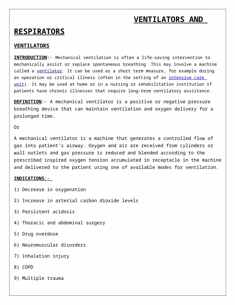
1.Iron lung (Drinker respirator tank):- The iron lung is a negative pressure chamber used for ventilation. It was used extensively during polio epidemics in the past and currently is used by few polio survivors and patients with neuromuscular disorders.
IRON LUNG (INVENTED IN 1929 BY PHILIP DRINKER)
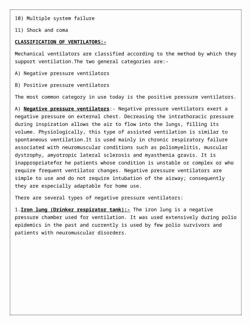
2. Body wrap (pneumo wrap) and chest cuirass (tortoise shell):- Both of these portable devices require a rigid cage or shell to create a negative pressure chamber around thorax and abdomen. Because of problems with proper fit and system leaks, these types of ventilators are used only with carefully selected patients.
CHEST CUIRASS
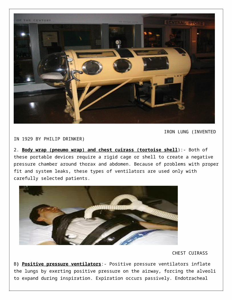
B) Positive pressure ventilators:- Positive pressure ventilators inflate the lungs by exerting positive pressure on the airway, forcing the alveoli to expand during inspiration. Expiration occurs passively. Endotracheal intubation or tracheostomy is necessary in most cases. These ventilators are mostly used in hospital setting and are increasingly used at home for the patients with primary lung disease. The various types of positive pressure ventilators are as follows:-
1. Pressure cycled ventilators:- When pressure cycled ventilators are on, it delivers a flow of air (inspiration) until it reaches a preset pressure and cycle off and expiration occurs passively. Its major limitation is that volume of oxygen or air may vary as patient’s airway resistance or compliances changes.As a result, the tidal volume delivered may be inconsistent, possibly compromising ventilation. Consequently, in adults pressure cycled ventilators are intended only for short term use.
2. Time cycled ventilators:- Time cycled ventilators terminate or control inspiration after a preset time. The volume of air the patient receives is regulated by the length of inspiration and flow rate of air. Most ventilators have a rate control that determines the respiratory rate but pure time cycling is rarely usedfor adults. These ventilators are used in newborns and infants.
3. Volume cycled vebtilators:- Volume cycled ventilators are by far the most commonly used ventilators today. With this type of ventilator, the volume of air delivered with each inspiration is preset. Once this prest volume is delivered to the patient, the ventilator cycles off and exhalation occurs passively. From breath to breath, the volume of air delivered by the ventilator is relatively constant, ensuring consistent, adequate breaths despite varying airway pressures.
Noninvasive positive pressure ventilation:- Positive pressure ventilation can be given via face masks that cover the nose and mouth, nasal masks or other oral or nasal pillow. NIPPV eliminates the need for endotracheal intubation or tracheostomy and decreases the risk of nosocomial infections such as pneumonia. The most comfortable mode for patient is pressure controlled ventilation with pressure support. This eases the work of breathing and enhances gas exchange. The ventilator can be set with a minimum back up rates for the patients with apnea.
Patients are considered candidates for noninvasive ventilation if they have:-
-acute or chronic respiratory failure
- acute pulmonary edema
-COPD
-chronic heart failure
-sleep related breathing disorders.
Contraindications:-
-Respiratory arrest
-serious dysrhythmias
-cognitive impairment
-head or facial trauma
Continuous positive airway pressure (CPAP):- CPAP provides positive pressure to the airways throughout respiratory cycle. Although, it can be used as an adjunct to mechanical ventilation with a cuffed endotracheal tube or tracheostomy tube to open the alveoli, it is also used with a leakproof mask to keep alveoli open, thereby preventing respiratory failure. CPAP is the most effective treatment for obstructive sleep apnea, because positive pressure acts as a splint, keeping the upper airway and trachea and airway open during sleep. To use CPAP, the patient must be breathing independently.
Bilevel positive pressure airway (bi- PAP):- bi-PAP ventilation offers independent control of inspiratory and expiratory pressures while providing pressure support ventilation. It delivers two levels of positive airway pressure provided via a nasal or oral mask or mouthpiece with tight seal and portable ventilator. Each inspiration can be initiated either by the patient or by the machine if it is programmed with a back up rate. The back up rate ensures that the patient will receive a set number of breaths per minute. Bi-PAP is most often is used for patients who require ventilator assistance at night such as those with severe COPD or sleep apnea.
VENTILATOR MODES
Ventilatory modes refers to how breaths are delivered to the patient. The most commonly used modes are assist control, Intermittent mandatory ventilation, Synchronized intermittent mandatory ventilation, pressure support ventilation and airway pressure release ventilation.
1.Assist-control ventilation:- It provides full ventilator support by delivering a preset tidal volume and respiratory rate. If the patient initiates a breath between between the machine’s breath, the ventilator delivers at the preset volume. The cycle does not adapt to the patient’s spontaneous efforts; every breath is preset volume
2. Intermittent mandatory ventilation:- It provides a combination of mechanically assisted breaths and spontaneous breaths. Therefore, the patient can increase the respiratory rate but each spontaneous breath is limited to the tidal volume the patient generates. Mechanical breaths are delivered at preset intervals and a preselected tidal volume regardless of patients efforts. IMV allows the patient to use their own muscles of ventilation to help prevent muscle atrophy. It lowers mean airway pressure which can assist in preventing barotraumas.
3. Synchronised intermittent mandatory ventilation (SIMV):- It delivers a preset tidal volume and no. of breaths per minute. Between ventilator delivered breaths, the patient can breath spontaneously with no assistance from ventilator on those extra breaths. As a patient’s ability to breath spontaneously
increases, the preset number of ventilator breaths is decreased and patient does more of the work of breathing. IMV and SIMV can be used to provide full or partial ventilator support. Nursing interventions for patient receiving IMV or SIMV include monitoring progress by recording respiratory rate, minute volume, spontaneous and machine generated tidal volume, FiO2 and arterial blood gas levels.
4. Pressure support ventilation (PSV):- It assists SIMV by applying pressure plateau to the airway throughout the patient triggered inspiration to decrease resistance within the tracheal tube and ventilator tubings. Pressure support is decreased gradually as patient’s strength increases. An SIMV back up rate may be added for extra support. The nurse must closely observe the patient’s respiratory rate and tidal volumes on initiation of PSV. It may be necessary to adjust the pressure support to avoid tachypnea or large tidal volume.
5. Airway pressure release ventilation (ARPV):- It produces tidal ventilation by release of airway pressure from an elevated baseline airway pressure to stimulate expiration. ARPV is a time triggered , pressure limited, time cycled mode of mechanical ventilation that allows unrestricted, spontaneous breathing throughout the ventilator cycle. It also allows alveolar gas to be expelled through lungs natural recoil.
6. Proportional assist ventilation (PAV):- It is a relatively new mode of support providing partial ventilatory support in which the ventilator generates pressure in proportion to patient’s inspiratory efforts. With every breath, the ventilator synchronises with the patient’s ventilator efforts. The more inspiratory efforts patient generates, the more pressure ventilator generates, amplifying the patient’s effort; the depth and frequency of breaths are controlled by the patient.
ADJUSTING THE VENTILATOR
The ventilator is adjusted so that the patient is comfortable and breaths “in sync” with machine. Minimal alteration of the normal cardiovascular and pulmonary dynamics is desired. If volume ventilator is adjusted appropriately the patients arterial blood gas values will be satisfactory and there will little or no cardiovascular compromise.
VENTRICULAR CONTROLS AND SETTING
1.TIDAL VOLUME:- The volume of air that the client receives with each breath. (10-15ml/kg)
2. RATE:- Number of ventilator breaths delivered per minue.
3. FRACTION OF INSPIRED OXYGEN:- The oxygen concentration delivered to the client, which is determined by the client’s condition and ABGs. (80-100mmHg)
4. SIGHS:-
a) sighs are volumes of air that are 1.5 to 2 times the set tidal volume, delivered 6 to 10 times per hour.
b) sighs may be used to prevent atelectasis.
5. PEAK AIRWAY INSPIRATORY PRESSURE:-
a) peak inspiratory airway pressure is the pressure needed by the ventilator to deliver a ste tidalvolume at a given temperature.
b) Monitoring peak airway inspiratory pressure reflects changes in compliance of the lungs and resistance in the ventilator or client.
6. CONTINUOUS POSITIVE AIRWAY PRESSURE
a) Continuous positive airway pressure is application of positive airway pressure throughout the entire respiratory cycle for spontaneously breathing clients.
b) Continuous positive airway pressure keeps the alveoli open during inspiration and prevents alveolar collapse.
c) Continuous positive airway pressure is used primarily as a weaning modality.
d) During continuous positive airway pressure, no ventilator breaths are delivered, but the ventilator delivers oxygen and provides monitoring and alarm system.
e) The respiratory pattern is determined by client’s efforts.
7. POSITIVE END EXPIRATORY PRESSURE [PEEP]
a) positive pressure is exerted during the expiratory phase of ventilation.
b) Positive end expiratory pressure improves oxygenation by enhancing gas exchange and preventing atelectasis.
c) The need for PEEP indicates a severe gas exchange disturbance.
d) higher amounts of PEEP (more than 15) increase the chance of complications such as barotraumas tension pneumothorax.
8. PRESSURE SUPPORT
a) Pressure support is application of positive pressure on inspiration.
b) It eases the workload of breathing.
c) It may be used in combination with PEEP as a weaning method.
d) As the warning process ensures, the amount of pressure applied to inspiration is gradually decreased.
ASSESSING THE EQUIPMENT
Ventilator needs to be assessed to make sure that it is functioning properly and that the settings are appropriate. Even though the nurse may not be primarily may not be primarily responsible for adjusting the settings of ventilator or measuring ventilator parameters, the nurse is responsible for the patient and therefore needs to evaluate how the ventilator affects the patient’s overall status.
When monitoring the ventilator, the nurse notes the following:-
- Type of ventilator- Controlling mode- Tidal volume and rate settings- FiO2 setting- Inspiratory pressure reached and pressure limit. ( normal is 15-20cm H2O; this increases if there is
increased airway resistance or decreased compliance.)- Sensitivity ( a 2cm H2O Inspiratory force should trigger the ventilator)- Inspiratory to expiratory ratio ( 1:3)- Minute volume ( tidal volume × respiratory rate, usually 6-8l/min)- Sigh settings ( usually set at 1.5 times the tidal volume and ranging from 1-3 hours)- Water in the tubing, disconnection or kinking of the tubing.- Humidification and temperature- Alarms - PEEP and pressure support level, if applicable ( PEEP is usually set at 5-15cm H2O)
CAUSES OF ALARMS
High pressure alarm:-
1. Increased secretions are in the airway.
2. Wheezing or bronchospasm causes decreased airway size.
3. The endotracheal tube is displaced.
4. The endotracheal tube is obstructed as a result of water or kink in the tubing.
5. Client coughs, gags, or bites on the endotracheal tube.
6. Client is anxious or fights the ventilator.
Low pressure alarm:-
1. Disconnection or leak in the ventilator or in the client’s airway cuff occurs.
2. The client stops spontaneous breathing.
PROBLEMS WITH MECHANICAL VENTILATION
Because of the seriousness of the patient’s condition and highly complex and technical nature of mechanical, a number of problems or complications can occur. Ventilators problems are as follows:-
1. Increase in peak airway pressure
Cause solution
- Coughing or plugged airway tube - suction airway for secretions, empty
condensation fluid from circuit.
- patient “bucking” ventilator - Adjust sensitivity
- decreasing lung compliance - Manually ventilate the patient, assess for
Hypoxia or bronchospasm, check arterial
Blood gas values, sedate if necessary.
- Tubing kinking - check tubings; reposition client; insert oral
Airway if necessary.
- Pneumothorax - Manually ventilate the client, notify physician.
- Atelectasis or bronchospasm - clear secretions
2. Decrease in pressure or loss of volume
Cause solution
- Increase in compliance - none
- leak in ventilator or tubing; cuff on tube/ - Check entire ventilation circuit for patency.
Humidifier not tight correct leak.
COMPLICATIONS
1. Hypotension caused by application of positive pressure which increases intrathoracic pressure and inhibits blood return to the heart.
2. Respiratory complications such as pnemothorax or subcutaneous emphysema as a result of positive pressure.
3. Gastrointestinal alterations such as stress ulcers.
4. Malnutrition if nutrition is not maintained.
5. Infections.
6. Muscular deconditioning.
7. Ventilator dependence or inability to wean.
WEANING
The process of going from ventilator dependence to spontaneous breathing.
1. SIMV:-
a. The client breathes between the preset breaths per minute rate of the ventilator.
b. The SIMV rate is decreased gradually until the client is breathing on his or her own without the use of the ventilator.
2. T- piece
a) The client is taken off the ventilator and the ventilator is replaced with a Tpiece or continuous positive airway pressure which delivers humidified oxygen.
b) The client is taken off the ventilator for short periods initially and allowed to breathe spontaneously.
c) Weaning progresses as the client is able to tolerate progressively longer periods off the ventilator.
3. Pressure support
a) Pressure support is a predetermined pressure on the ventilator to assist the client in respiratory effort.
b) As weaning continues the amount of pressure is decreased gradually.
c) With pressure support, pressure may be maintained while the preset breaths per minute of the ventilator gradually are decreased.
NURSING RESPONSIBILITIES
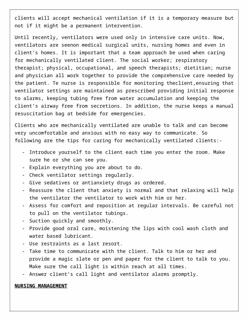
Before initiating mechanical ventilation, it is important that the health care team be aware of advance directives and consult with client and family because many clients do not wish to be mechanically ventilated. Some clients will accept mechanical ventilation if it is a temporary measure but not if it might be a permanent intervention.
Until recently, ventilators were used only in intensive care units. Now, ventilators are seenon medical surgical units, nursing homes and even in client’s homes. It is important that a team approach be used when caring for
mechanically ventilated client. The social worker; respiratory therapist; physical, occupational, and speech therapists; dietitian; nurse and physician all work together to provide the comprehensive care needed by the patient. Te nurse is responsible for monitoring theclient,ensuring that ventilator settings are maintained as prescribed providing initial response to alarms, keeping tubing free from water accumulation and keeping the client’s airway free from secretions. In addition, the nurse keeps a manual resuscitation bag at bedside for emergencies.
Clients who are mechanically ventilated are unable to talk and can become very uncomfortable and anxious with no easy way to communicate. So following are the tips for caring for mechanically ventilated clients:-
- Introduce yourself to the client each time you enter the room. Make sure he or she can see you.- Explain everything you are about to do.- Check ventilator settings regularly.- Give sedatives or antianxiety drugs as ordered.- Reassure the client that anxiety is normal and that relaxing will help the ventilator the ventilator to
work with him or her.- Assess for comfort and reposition at regular intervals. Be careful not to pull on the ventilator tubings.- Suction quickly and smoothly.- Provide good oral care, moistening the lips with cool wash cloth and water based lubricant.- Use restraints as a last resort.- Take time to communicate with the client. Talk to him or her and provide a magic slate or pen and
paper for the client to talk to you. Make sure the call light is within reach at all times.- Answer client’s call light and ventilator alarms promptly.
NURSING MANAGEMENT
Nursing care of the mechanically ventilated patient requires expert technical and interpersonal skill. The specific interventions used by the nurse are determined by the underlying disease process and patient’s response.
Two general nursing interventions that are important in the care of the mechanically ventilated patient are pulmonary auscultation and interpretation of arterial blood gas measurements. The nurse is often first to note changes in the physical assessment findings or significant trends in blood gases that signal the development of a serious problem. The various nursing interventions are given below:-
Enhancing gas exchange:-
Nursing interventions to promote optimal gas exchange include judicious administration of analgesic agents to relieve pain without suppressing the respiratory drive and frequent repositioning to diminish the pulmonary effects of immobility. The nurse also monitors for adequate fluid balance by assessing for the presence of peripheral edema, calculating daily intake and output and monitoring daily weights.
Promoting effective airway clearance:-
Continuous positive pressure ventilation increases the production of secretions regardless of the patient’s underlying condition. The nurse assesses for the presence of secretions by lung auscultation at least 2-4 hours.
Measures to clear airway include suctioning, CPT, frequent position changes and increased motility as soon as possible. Humidification of the airway via ventilator is maintained to help liquefy secretions so that they are easily removed. Bronchodilators are administered to dilate the bronchioles.
Preventing trauma and infection:-
Maintaining the endotracheal or tracheostomy tube is an essential part of airway management. The nurse positions the ventilator so that there is minimal pulling or distortion of the tube in the trachea, reducing the risk of trauma to the trachea. Cuff pressure is monitored every 6-8hours maintain the pressure at less than 25mm Hg. Tracheostomy care is performed at least 8 hours and more frequently if needed. The nurse also administers oral hygiene frequently because oral cavity is a primary source of contamination of the lungs in intubated patient. The nurse positions the patient with the head elevated above stomach to prevent aspiration.
Promoting optimal level of mobility:-
Being connected to a ventilator limits the patient’s mobility. The nurse helps the patient whose condition has become stable to get out of the bed and move to a chair as soon as possible. If the patient is not able to get out of bed the nurse encourages performance of active range of motion exercises him to perform active range of motion exercises every 6-8hours. If the patient can not perform these exercises, the nurse performs passive range of motion exercises every 8 hours to prevent contracture and venous stasis.
Promoting optimal communication:-
It is important to develop alternative methods communication for the patient who is receiving mechanical ventilation. The nurse assesses the patient’s communication abilities to evaluate for limitations.
Promoting coping ability:-
Dependence on a ventilator is frightening to both the patient and family. Encouraging the family to verbalize their feelings about ventilator, the patient’s condition and environment in general is beneficial. Explaining procedures every time they are performed helps to reduce anxiety.
Promoting home and community based care:-
Increasingly, patients are being cared for in extended care facilities or at home while receiving mechanical ventilation with a tracheostomy tube or receiving oxygen therapy. Patients receiving home ventilator care usually have a chronic neuromuscular condition or COPD. Providing the opportunity for ventilator dependent patients to return home to live with their families in familiar surroundings can be a positive experience. The ultimate goal of home ventilator therapy is to enhance the patient’s quality of life not simply to support or prolong life.
RESPIRATORS
1.NASAL CANNULA:-
a) A nasal cannula is used at flow rate of 1 to 6l/min providing approximate oxygen concentrations of 24% to 44%.
b) Flow rate higher than 6l/min do not significantly increase oxygenation because the anatomical reserve or dead space is full.
c) A nasal cannula is used for the client with chronic airflow limitation and for long term oxygen use.
d) A client who is hypoxemic and also has chronic hypercarbia requires low levels of oxygen delivery at 1 to 2l/min; a low arterial oxygen level is the client’s primary drive for breathing.
e) Effective oxygen concentration can be delivered to nose breathers and mouth breathers with the use of cannula.
Interventions:-
a) Place the nasal prongs into the nostrils with the opening facing the client.
b) Add humidification as prescribed when a flow rate is higher than 2l/min as prescribed.
c) Check the water level and change the humidifier as needed.
d) Assess the client for changes in respiratory rate or depth.
e) Assess the mucosa because high flow rates have a drying effect and increase mucosal irritation.
f) Assess skin integrity because the oxygen tubing can irritate the skin.
g) Provide water soluble jelly to the nares as needed.
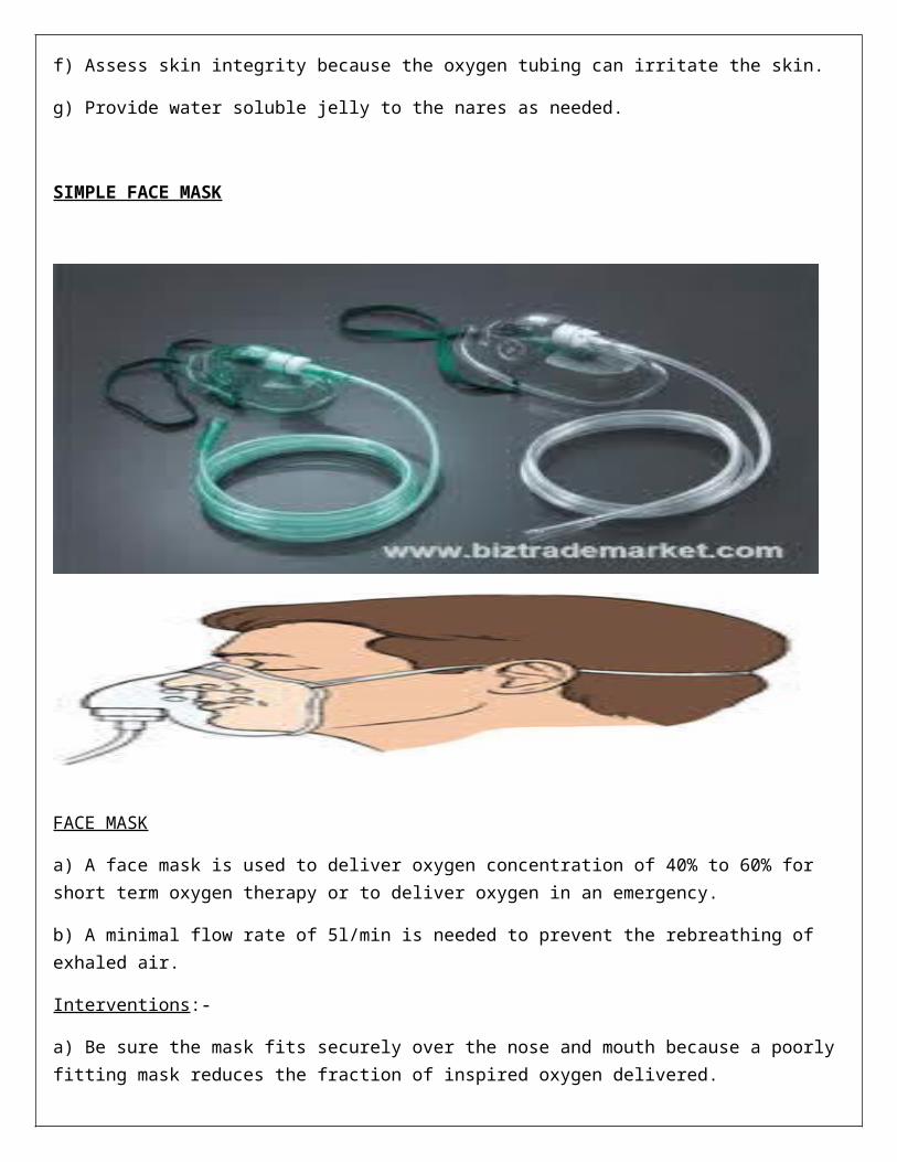
SIMPLE FACE MASK
FACE MASK
a) A face mask is used to deliver oxygen concentration of 40% to 60% for short term oxygen therapy or to deliver oxygen in an emergency.
b) A minimal flow rate of 5l/min is needed to prevent the rebreathing of exhaled air.
Interventions:-
a) Be sure the mask fits securely over the nose and mouth because a poorly fitting mask reduces the fraction of inspired oxygen delivered.
b) Assess skin and provide skin care to the area covered by mask because pressure and moisture under the mask may cause skin breakdown.
c) Monitor the client closely for risk of aspiration because the mask limits the client’s ability to clear the mouth especially when vomiting occurs.
d) Provide emotional support to derease anxiety in the client who feels claustrophobic.
e) Consult with the physician regarding switching the client from mask to a nasal cannula during eating.
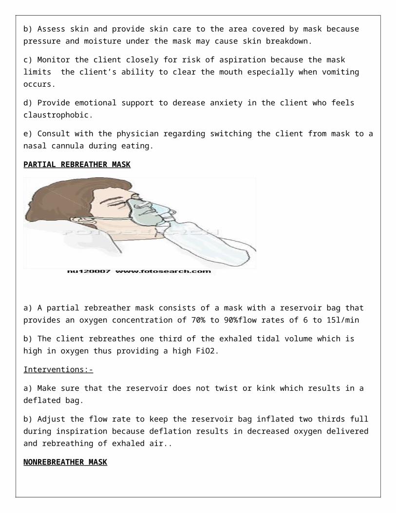
PARTIAL REBREATHER MASK
a) A partial rebreather mask consists of a mask with a reservoir bag that provides an oxygen concentration of 70% to 90%flow rates of 6 to 15l/min
b) The client rebreathes one third of the exhaled tidal volume which is high in oxygen thus providing a high FiO2.
Interventions:-
a) Make sure that the reservoir does not twist or kink which results in a deflated bag.
b) Adjust the flow rate to keep the reservoir bag inflated two thirds full during inspiration because deflation results in decreased oxygen delivered and rebreathing of exhaled air..
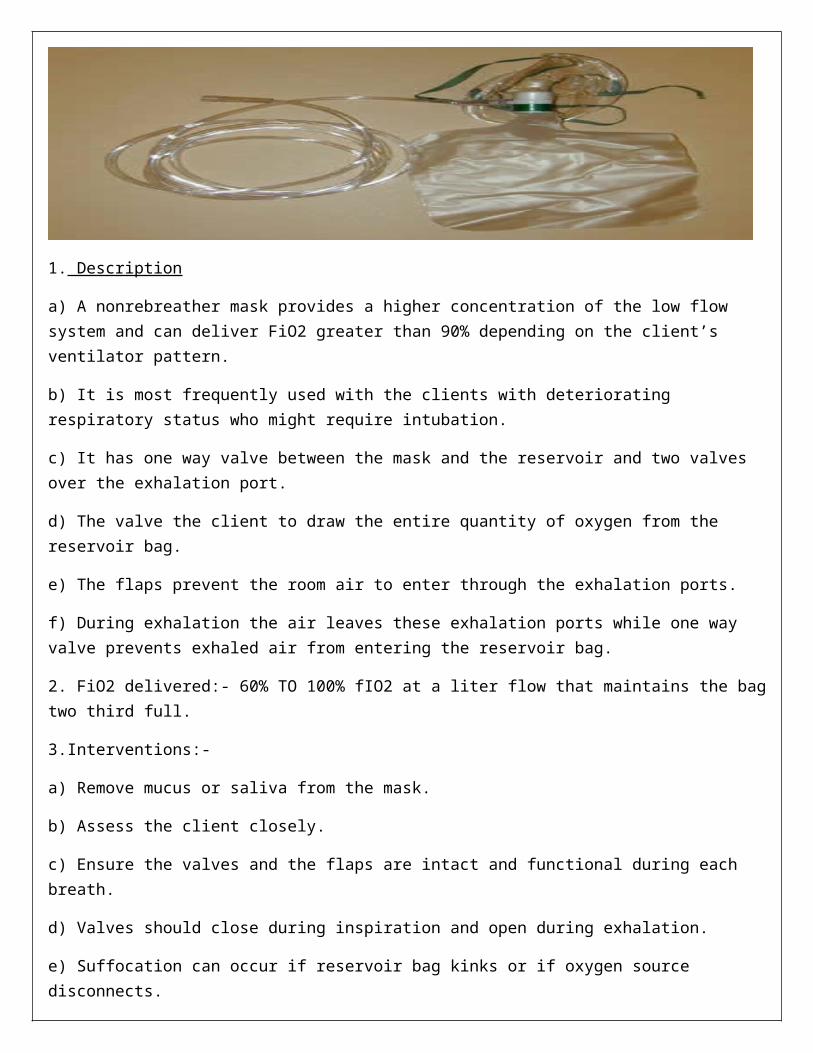
NONREBREATHER MASK
1. Description
a) A nonrebreather mask provides a higher concentration of the low flow system and can deliver FiO2 greater than 90% depending on the client’s ventilator pattern.
b) It is most frequently used with the clients with deteriorating respiratory status who might require intubation.
c) It has one way valve between the mask and the reservoir and two valves over the exhalation port.
d) The valve the client to draw the entire quantity of oxygen from the reservoir bag.
e) The flaps prevent the room air to enter through the exhalation ports.
f) During exhalation the air leaves these exhalation ports while one way valve prevents exhaled air from entering the reservoir bag.
2. FiO2 delivered:- 60% TO 100% fIO2 at a liter flow that maintains the bag two third full.
3.Interventions:-
a) Remove mucus or saliva from the mask.
b) Assess the client closely.
c) Ensure the valves and the flaps are intact and functional during each breath.
d) Valves should close during inspiration and open during exhalation.
e) Suffocation can occur if reservoir bag kinks or if oxygen source disconnects.
FACE TENT
a) A face tent fits over the client’s chin with top extending half way across the face.
b) The oxygen concentration varies; but the face tent is useful instead of tight fitting mask for the clients with facial burns or trauma.
AEROSOL MASK

It is used for the clients who need high humidity after extubation or upper airway surgery or clients who have thick secretions.
TRECHEOSTOMY COLLAR OR T – PIECE
T-PIECE
a) A tracheostomy collar can be used to deliver high humidity and desired oxygen to the clients with a tracheostomy.
b) A special adapter called T piece can be used to deliver any desired Fio2 to the client with tracheostomy laryngectomy or endotracheal tube.
Interventions:-
1. Change delivery system to a nasal cannula during mealtime
2. Assess that aerosol mist escapes from the vents of the delivery system during inspiration or expiration
3. Empty condensation from the tubing to prevent the client from being lavaged with water and to promote adequate floe rate
4. Ensure that sufficient water is in the canister and change the aerosol water container as needed.
5. Keep the exhalation port on the T piece open and uncovered.
6. Position the T piece so that it does not pull on the tracheostomy or endotracheal tube and cause erosion of skin at tracheostomy insertion site
7. Make sure the humidifier creates enough mist; mist should be seen during inspiration and expiration.
VENTURI MASK
a) A venturi mask provides high flow oxygen delivery sysyem.b) B) Operation of the venture mask based on a mechanism that pulls in a specific proportional amount of
room air for each liter of oxygen.c) An adpter is located between the bottom of the mask and oxygen source the adapter contains holes of
different sizes that allows specific amounys of air mix with the oxygend) The adapter allows selection of the amount ot oxygen desired.
2. Fio2 delivered: 24% to 55% with flow rates of 4- 10l/min
Interventions:-
a) Momitor client closely to ensure an accurate flow for specific FiO2b) Keep the orifice for venture adapter open and uncovered to ensure adequate oxygen deliveryc) Ensure that mask fits snugly and that tubings is free of kinks because FiO2 is altered if kinking occurs or
mask fits poorlyd) Assess the client for dry mucus membranes;humidity or aerosol can be added to the system.
Health care team roles
Oxygen is considered a drug and in this context is administered by a licensed nurse or respiratory therapist in the medical setting. Once oxygen therapy has been initiated, non-professional staff may assist in caring for the
patient using oxygen therapy, including removing and replacing the mask or cannula for skin care, meals or brief ambulation to the bathroom. Nonprofessional staff must be instructed to remember to turn the oxygen flow back on after removal. They may also be trained to check oxygen apparatus such as, checking tubing connections, replacing the oximeter probe, filling the humidity bottles and cleaning or wiping humidity out of the oxygen mask. This should be performed under the direct supervision of the licensed nurse and the nurse or respiratory therapist will continually assess the patient's respiratory status and oxygen levels. When a patient is going home with oxygen, the licensed nurse or respiratory therapist will educate the patient and the patient's caregivers about the safe use of oxygen in the home. Patients using oxygen in the home should have initial and follow-up visits by a home care nurse or respiratory technician to check the patient's status and equipment function in the home. Patients receiving oxygen in the home should be scheduled for regular visits to the physician for follow-up assessment, including pulse oximetry and other therapy.
SUMMARY
Today we have discussed about :-
- mechanical ventilation
-its definition and types
- modes of ventilation
- ventilator settings and assessment
- problems with mechanical ventilation
- complications
- Nursing management of the patient with mechanical ventilation
- Various types of respirators such as nasal cannula, simple face mask, partial rebreather mask, non rebreather mask, venturi mask, aerosol mask, face tent tracheostomy collar or T- piece.
BIBLIOGRAPHY
1. Suzanne C. Smeltzer, Brenda G. Bare, Janice L. hinkle, Kerry H. Cheever; Brunner and suddath’s Textbook of medical surgical nursing; 11th edition; Lippincott Williams and wilkins, Philadelphia 2008; pg. no. 739-751.
2. Lewis; Medical surgical nursing; 3rd edition; Mosby year book, United states of America; 1959. Pg. no. 632-647.
3. Williams S. linda, Hopper D. Paula; Medical surgical nursing; 2nd edition; F.A Davis company; Philadelphia 1954; 501-504.
4. Joyce M. Black, Hawks Hokanson Jane, Keene M. Annabelle; Medical surgical nursing; 6th edition; W.B Sauders company; Philadelphia 2001; 1752-1762.
5. Finkelmeier A. Betsy; Cardiothoracic surgical nursing; second edition; Lippincott Williams and wilkins; Philadelphia 2000; pg. no. 361.
6. Silvestri Anne Linda; Saunders comprehensive review for the NCLEX-RN Examination; third edition; saunders Elsevier; Saint Louis 2006; pg. no.729-732.
7. www.medicalimages.com
8. www.wikipedia.com 9. www.savvycafe.com 10. www.righthealth.com