Embed Size (px)
Citation preview
A Natural Product Telomerase Activator As Partof a Health Maintenance Program
Calvin B. Harley,1,6 Weimin Liu,2 Maria Blasco,3 Elsa Vera,3 William H. Andrews,4
Laura A. Briggs,4 and Joseph M. Raffaele5
Abstract
Most human cells lack sufficient telomerase to maintain telomeres, hence these genetic elements shorten withtime and stress, contributing to aging and disease. In January, 2007, a commercial health maintenance program,PattonProtocol-1, was launched that included a natural product-derived telomerase activator (TA-65�, 10–50 mgdaily), a comprehensive dietary supplement pack, and physician counseling/laboratory tests at baseline andevery 3–6 months thereafter. We report here analysis of the first year of data focusing on the immune system.Low nanomolar levels of TA-65� moderately activated telomerase in human keratinocytes, fibroblasts, andimmune cells in culture; similar plasma levels of TA-65� were achieved in pilot human pharmacokinetic studieswith single 10- to 50-mg doses. The most striking in vivo effects were declines in the percent senescent cytotoxic(CD8þ/CD28�) T cells (1.5, 4.4, 8.6, and 7.5% at 3, 6, 9, and 12 months, respectively; p¼not significant [N.S.],0.018, 0.0024, 0.0062) and natural killer cells at 6 and 12 months ( p¼ 0.028 and 0.00013, respectively). Most ofthese decreases were seen in cytomegalovirus (CMV) seropositive subjects. In a subset of subjects, the distri-bution of telomere lengths in leukocytes at baseline and 12 months was measured. Although mean telomerelength did not increase, there was a significant reduction in the percent short (<4 kbp) telomeres ( p¼ 0.037). Noadverse events were attributed to PattonProtocol-1. We conclude that the protocol lengthens critically shorttelomeres and remodels the relative proportions of circulating leukocytes of CMVþ subjects toward the more‘‘youthful’’ profile of CMV� subjects. Controlled randomized trials are planned to assess TA-65�-specific effectsin humans.
Introduction
People take dietary supplements with the intent topreserve mental, physical, and emotional health and
vigor into old age. Although drugs and surgical proceduresthat target diseases of the elderly will hopefully arrest orpartially reverse tissue damage caused by aging and chronicstress, measures to maintain health are arguably a betterapproach to lengthening our healthy life span. Most dietarysupplement programs include combinations of vitamins,antioxidants, and other constituents, some of which havebeen shown to have significant health benefits in controlledclinical studies, whereas others may show adverse effects,1–6
underscoring the need to assess functional effects of combi-nation products. This paper presents initial data from an
ongoing observational study of a novel dietary supplementprogram, PattonProtocol-1, which includes a natural prod-uct-derived telomerase activator targeting a fundamentalaspect of cellular aging.
Telomerase is an enzyme that synthesizes the specificDNA sequence at telomeres, i.e., the terminal DNA at theends of all chromosomes.7,8 Telomeres are essential geneticelements responsible for protecting chromosome ends frombeing recognized as ‘‘broken DNA.’’ Because telomericDNA cannot be fully replicated by conventional DNApolymerases, and because telomeres undergo degradativeprocessing and are a ‘‘hotspot’’ for oxidative damage,9
telomeres will gradually shorten with time and cell divisionunless there is sufficient telomerase activity to maintaintelomere length.
1Geron Corporation, Menlo Park, California.2TA Sciences, New York, New York.3Spanish National Cancer Center, Madrid, Spain.4Sierra Sciences, Reno, Nevada.5PhysioAge Systems, New York, New York.6Present address: Telome Health Inc., Menlo Park, California.
REJUVENATION RESEARCHVolume 14, Number 1, 2011ª Mary Ann Liebert, Inc.DOI: 10.1089/rej.2010.1085
45

Telomerase is activated in fetal development, thus pro-tecting telomeres from significant loss during this period ofdramatic cell expansion.10,11 However, telomerase is re-pressed before birth in most somatic tissue, and, as a con-sequence, birth marks the beginning of telomere erosion inmost tissues throughout life. Tissues with continual cellturnover or periods of rapid proliferation are ‘‘telomerasecompetent’’ in that they upregulate telomerase during earlyphases of progenitor expansion.12,13 All adult somatic stemcells appear to be capable of activating telomerase duringtissue regeneration. However, these periods of activation areinsufficient to prevent telomere loss, and this is compoundedby a decreased ability to activate telomerase during agingand stress.14–16 In addition, stress can accelerate telomere lossby increasing cell turnover and the amount of telomericDNA lost per cell division.17,18
In cross-sectional studies, humans lose telomeric DNA ata very modest rate of about 15–60 bp per year, likely re-flecting the small numbers of stem cells that are activelydividing in proliferative tissues compared to the total stemcell reserve, and the quiescent state of cells in other tissues.Telomere shortening has been investigated in human cells inculture, in human genetic diseases with mutated telomerase,and in animal models of telomerase deficiency.13,19–26 Thesestudies point to a causal relationship between telomere loss,cell aging, reduced tissue regeneration, and loss of tissuestructure and function. In support of this causal relationship,epidemiological studies show that short telomeres in hu-mans are a risk factor for atherosclerosis, hypertension,cardiovascular disease, Alzheimer disease, infections, dia-betes, fibrosis, metabolic syndrome, cancer, and overallmortality.18,24,25,27–30
Chronic viral infections such as cytomegalovirus (CMV)and human immunodeficiency virus (HIV) accelerate telo-mere loss and premature aging of the immune system, es-pecially the virus-specific cytotoxic T cells31–36 responsiblefor killing infected cells. In addition to telomere loss, thesecells often lack expression of the co-stimulatory receptorCD28 and have reduced proliferative capacity, reducedability to secrete antiviral cytokines and chemokines, in-creased resistance to apoptosis, and compromised ability tolyse infected cells. About 50% of the U.S. population is in-fected with CMV as judged by circulating CMV-specificantibodies, but after an initial 30% seropositivity rate by age&10, there is &1% annual seroconversion rate throughoutlife leading to &90% seropositivity by the ninth decade. Thislinear increase has made it difficult to distinguish the effectsof pure immunosenescence from those that can be attributedto this extremely common virus.37,38
Here we report initial findings from a dietary supple-ment program which includes TA-65�, a purified small-molecule telomerase activator derived from an extract of aplant commonly used in traditional Chinese medicine.Telomerase activation and functional studies on a relatedmolecule (TAT2) from the same plant have been previouslyreported for human skin keratinocytes and immune cells inculture.36 Effects of TAT2 in tissue culture studies withCD8þ T cells from HIV/acquired immunodeficiency syn-drome (AIDS) subjects included increased replicative ca-pacity, improved cytokine and chemokine responses toantigens, and increased killing of autologous HIV-infectedCD4þ cells.
Methods
PattonProtocol-1
PattonProtocol-1 was launched in January, 2007, by TASciences (New York) as a commercial age-managementproduct composed of a natural product–derived telomeraseactivator (TA-65�, described below), a dietary supplementpack (online material S1), laboratory testing (Table 1), andphysician counseling. All subjects signed a comprehensiveCustomer Acknowledgement Form. Baseline assays (Tables1 and 2) indicated that most individuals were within thenormal ranges for the majority of tests. In a small number ofcases described in the Results section, the consulting physi-cian prescribed medications for subjects based upon clinicaltests. There was no qualitative change in the overall con-clusions whether these subjects were included or censoredfrom the analysis. We report results for all evaluable subjectswho completed 12 months of the protocol by June, 2009. Thenumber of subjects at 3, 6, 9, and 12 months for most testswas 43, 59, 27, and 37, respectively. The age and genderfrequencies of the subset at each time point were similar tothose of the total baseline population (n¼ 114; 63� 12 years,72% male).
TA-65�
TA-65�, exclusively licensed to TA Sciences from GeronCorporation, is a >95% pure single chemical entity isolatedfrom a proprietary extract of the dried root of Astragalusmembranaceus and formulated into 5- to 10-mg capsules withinert excipients. Starting doses of 5–10 mg/day were con-sidered safe on the basis of historical usage of extracts. Somesubjects increased their dosage after several months on theproduct to 25–50 mg/day. Cumulative dose consumed dur-ing the year was recorded for each subject and used forpreliminary dose–response analysis.
Clinical laboratory assays
At baseline and each time, point blood samples weredrawn and shipped the same day at ambient temperature toanalytical laboratories. Assays for standard blood counts,blood chemistry, specialized immune subsets, CMV anti-body titer, and inflammation markers were conducted atQuest Diagnostics or Bio-Reference Laboratory. Specializedimmune subset analyses were conducted at UCLA ClinicalLaboratories and Pathology Services.
Telomerase activity assay in cultured humankeratinocytes and fibroblasts
Telomerase activity was measured in human neonatalkeratinocytes (Cascade Biologics, Portland, OR) and inMRC5 fetal human fibroblasts (ATCC# CCL-171) pre-andposttreatment in culture with TA-65� using the telomererepeat amplification protocol (TRAP) assay, essentially asdescribed elsewhere.39 In brief, telomerase activity wasmeasured in actively growing cells incubated for 24–48 hwith TA-65� in the vehicle (dimethylsulfoxide [DMSO] at 1%vol/vol [keratinocyte culture] or 0.5% [fibroblasts]) versusvehicle alone. Measurements were typically made at 5–10population doublings (PD) (keratinocytes) or 30–40 PD (fi-broblasts). Telomerase reaction products were resolved by
46 HARLEY ET AL.

Ta
bl
e1.
Assa
ys
an
dB
io
ma
rk
er
s
Pot
enti
alsa
fety
and
info
rmat
ion
alas
say
sE
xp
lora
tory
biom
arke
ras
say
s
Blo
od
cou
nt
Blo
od
sug
ar
an
din
suli
nB
aso
ph
ils
(%,
abs)
,eo
sin
op
hil
s(%
,ab
s),
hem
ato
crit
,h
emo
glo
bin
,ly
mp
ho
cyte
s(%
,ab
s),
MC
H,
MC
HC
,M
CV
,m
on
ocy
tes
(%,a
bs)
,M
PV
,n
eutr
op
hil
s(%
,ab
s),
pla
tele
tco
un
t,R
BC
,R
DW
,W
BC
Glu
cose
,h
emo
glo
bin
A1C
,se
rum
insu
lin
Vir
al
an
tib
od
yIn
fla
mm
ati
on
bio
ma
rke
rC
MV
IgG
Car
dio
CR
P,
ho
mo
cyst
ein
e
Kid
ne
yfu
nct
ion
Imm
un
eb
iom
ark
ers
BU
N/
crea
tin
ine
rati
o,
calc
ium
,ch
lori
de,
CO
2,
crea
tin
ine,
ph
osp
ho
rus,
po
tass
ium
,so
diu
m,
ure
an
itro
gen
,u
ric
acid
CD
8þ/
CD
28�
gat
edo
nC
D3
(%,
abs)
;C
D8þ
/C
D28�
gat
edo
nC
D8
(%,
abs)
;C
D8þ
/C
D95þ
gat
edo
nC
D3
(%,
abs)
;C
D8þ
/C
D95þ
gat
edo
nC
D8
(%,
abs)
Liv
er
Fu
nct
ion
Ca
rdio
va
scu
lar
Bio
ma
rke
rsA
/G
rati
o,
alb
um
in,
alk
alin
ep
ho
sph
atas
es,
AL
T,
AS
T,
bil
iru
bin
dir
ect,
bil
iru
bin
tota
l,G
GT
,ca
lcu
late
dg
lob
uli
n,
LD
H(L
D),
tota
lp
rote
inS
ph
yg
mo
Co
ras
say
s,sy
sto
lic
blo
od
pre
ssu
re,
dia
sto
lic
blo
od
pre
ssu
re
En
do
crin
efu
nct
ion
(als
oa
se
xp
loa
rato
ryb
iom
ark
ers
)P
ulm
on
ary
bio
ma
rke
rsD
HE
Asu
lfat
e,es
trad
iol,
FS
H,
IGF
-BP
3,IG
F-1
,L
H,
pro
ges
tero
ne,
PS
Ato
tal,
SH
BG
,T
3(f
ree
&to
tal)
,T
4to
tal,
test
ost
ero
ne
(fre
e,%
free
,an
dto
tal)
,T
SH
3rd
gen
erat
ion
Sp
iro
met
ryF
EV
1,sp
iro
met
ryF
VC
Ch
ole
ste
rol
(als
oa
se
xp
lora
tory
bio
ma
rke
rs)
Sk
inb
iom
ark
ers
Ch
ole
ster
ol/
HD
Lra
tio
,ch
ole
ster
ol
tota
l,H
DL
cho
lest
ero
l,ca
lcu
late
dL
DL
cho
lest
ero
l,tr
igly
ceri
des
Fo
rear
mcu
tom
eter
mea
sure
men
ts
Iro
nV
isio
nb
iom
ark
ers
Fer
riti
n,
iro
n(t
ota
lan
dsa
tura
ted
),T
IBC
Co
ntr
ast
sen
siti
vit
y(l
eft/
rig
ht
eye)
Vit
am
ins
(als
oa
se
xp
lora
tory
bio
ma
rke
rs)
Co
gn
itiv
eb
iom
ark
ers
Ser
um
fola
te,
seru
mv
itam
inB
12,
vit
amin
D(2
5-O
HD
2,25
-OH
D3,
25-O
Hto
tal)
Cen
tral
ner
vo
us
syst
emv
ital
sig
ns
pan
el
Oth
er
Te
lom
ere
bio
ma
rke
rsH
eig
ht,
wei
gh
tL
ym
ph
ocy
tes
(Flo
wF
ISH
med
ian
),g
ran
ulo
cyte
s(F
low
FIS
Hm
edia
n),
WB
Cg
eno
mic
DN
A(q
PC
Rm
ean
),W
BC
(qF
ISH
)B
on
em
ine
ral
de
nsi
tyD
ual
-en
erg
yX
-ray
abso
rpti
om
etry
abs,
;M
CH
,m
ean
corp
usc
ula
rh
emo
glo
bin
;M
CH
C,
mea
nco
rpu
scu
lar
hem
og
lob
inco
nce
ntr
atio
n;
MC
V,
mea
nco
rpu
scu
lar
vo
lum
e;M
PV
,m
ean
pla
tele
tv
olu
me;
RB
C,
red
blo
od
cell
s;R
DW
,R
edce
lld
istr
ibu
tio
nw
idth
;C
MV
IgG
,cy
tom
egal
ov
iru
sim
mu
no
glo
bu
lin
G;
CR
P,
C-r
eact
ive
pro
tein
;B
UN
,b
loo
du
rea
nit
rog
en;
A/
Gra
tio
,al
bu
min
/g
lob
uli
nra
tio
;A
LT
,al
anin
eam
ino
tran
sfer
ase;
AS
T,
asp
arta
team
ino
tran
sfer
ase;
GG
T,
gam
ma
glu
tam
yl
tran
spep
tid
ase;
LD
H,
lact
ate
deh
yd
rog
enas
e;L
D,
;D
HE
A,
deh
yd
roep
ian
dro
ster
on
e;F
SH
,fo
llic
le-s
tim
ula
tin
gh
orm
on
e;IG
F-B
P3,
insu
lin
-lik
eg
row
thfa
cto
rb
ind
ing
pro
tein
3;IG
F-1
,in
suli
n-l
ike
gro
wth
fact
or-
1;L
H,
lute
niz
ing
ho
rmo
ne;
PS
A,
pro
stat
e-sp
ecifi
can
tig
en;
SH
BG
,se
xh
orm
on
e-b
ind
ing
glo
bu
lin
;T
3,tr
iio
do
thy
ron
ine;
T4,
thy
rox
ine;
TS
H,
thy
roid
-sti
mu
lati
ng
ho
rmo
ne;
FE
V1,
forc
edex
pir
ato
ryv
olu
me
du
rin
g1st
seco
nd
;F
VC
,fo
rced
vit
alca
pac
ity
;H
DL
,h
igh
-den
sity
lip
op
rote
in;
LD
L,
low
-den
sity
lip
op
rote
in;
TIB
C,
tota
lir
on
-bin
din
gca
pac
ity
;25
-OH
D2,
25-d
ihy
dro
xyv
itam
inD
2;25
-OH
D3,
25-d
ihy
dro
xy
vit
amin
D3;
25-O
Hto
tal,
tota
l25
-hy
dro
xyv
itam
inD
;F
low
FIS
H,
Flo
w-fl
uo
resc
ent
insi
tuh
yb
rid
izat
ion
;W
BC
,w
hit
eb
loo
dce
lls;
qP
CR
,q
uan
tita
tiv
ep
oly
mer
ase
chai
nre
acti
on
.
47

Ta
bl
e2.
Ba
se
lin
eV
al
ue
sfo
rS
el
ec
te
dB
io
ma
rk
er
s,
Re
le
va
nt
Bl
oo
d/
Im
mu
ne
Va
ria
bl
es,
an
dR
el
at
io
nsh
ip
to
Ag
e
Age
Lym
phoc
yte
telo
mer
ele
ngth
a
Gra
nu
locy
tete
lom
ere
len
gth
CM
VIg
GA
nti
body
bW
BC
c
(UC
LA
)#L
ym
ph
(UC
LA
)%
CD
3(U
CL
A)
#C
D3
(UC
LA
)%
CD
8(U
CL
A)
#C
D8
(UC
LA
)%
CD
8þ
CD
28�
(UC
LA
)#
CD
8þC
D28�
(UC
LA
)
Mea
n62
.55.
73k
b6.
711.
8356
9018
5070
.113
2022
.843
534
.717
5S
D12
.51.
05k
b0.
881.
7715
2056
58.
5747
99.
1425
019
.918
0n
114
114
114
8811
311
311
311
311
311
311
311
3m
dN
A�
55b
p�
34b
p0.
024
22�
6.6
�0.
14�
6.4
�0.
08�
3.0
0.37
1.6
Rsq
uar
edN
A0.
420.
240.
030.
032
0.02
10.
044
0.02
90.
012
0.02
30.
055
0.19
0p
val
ue
NA
4.8
E-1
53.1
E-8
0.11
0.05
70.
120.
026
0.07
30.
240.
110.0
12
0.25
#C
D28þ
(cal
c)e
%C
D28þ
(cal
c)#
CD
95�
(UC
LA
)%
CD
95�
(UC
LA
)#
Lym
phoc
yte
s%
Lym
phoc
yte
s#
NK
%N
K%
CD
19
#C
D19
%C
D3
#C
D3
Mea
n26
065
6318
1500
2822
115
.011
.718
671
.711
50S
D13
220
5312
524
8.70
100
7.2
5.04
100
8.16
414
n11
311
357
5754
103
103
103
103
103
103
103
m�
4.6
�0.
37�
2.7
�0.
51�
2.5
�0.
212.
20.
21�
0.13
�2.
6�
0.09
1�
3.8
Rsq
uar
ed0.
190.
055
0.47
0.44
0.00
40.
090.
070.
130.
040.
10.
014
0.00
pv
alu
e1.4
E-0
60.0
12
6.7
E-9
6.4
E-6
0.72
0.0
02
0.0
06
0.0
0015
0.0
0067
0.0
011
0.17
0.25
CD
4/C
D8
rati
o%
CD
4þ
%C
D8þ
#C
D8þ
#W
BC
#N
eutr
ophil
s%
Neu
trop
hil
sG
luco
seS
eru
min
suli
nD
HE
Asu
lfat
eT
esto
ster
one
(fre
e)T
esto
ster
one
(tot
al)
Mea
n2.
849
2237
057
0036
0061
977.
114
833
400
SD
2.7
8.9
8.3
200
1600
1300
9.3
157.
013
142
340
n10
310
310
310
310
610
210
310
710
710
610
610
6m
0.06
40.
045
-0.1
0-2
.123
031
0.17
0.33
0.02
5�
2.6
0.23
1.1
Rsq
uar
ed0.
007
0.00
40.
023
0.01
70.
080.
050.
080.
002
0.06
0.00
50.
002
pv
alu
e0.0
024
0.53
0.13
0.19
0.05
10.
039
0.0
20
0.0
0460
0.64
0.0
11
0.47
0.31
Chol
este
rol
(tot
al)
Chol
este
rol
(LD
L)
SB
PD
BP
Hom
ocyst
ein
eS
eru
mfo
late
Vit
amin
D25-O
Hto
tal
Vit
amin
B12
Mea
n19
011
013
077
1117
4079
0S
D38
3419
103.
65.
816
410
n10
510
497
9710
710
310
710
5m
�0.
29�
0.37
0.51
�0.
018
0.06
6�
0.12
0.11
6.1
Rsq
uar
ed0.
010.
020.
108
0.00
00.
050.
020.
007
0.03
6p
val
ue
0.32
0.16
0.0
010
0.83
0.0
16
0.0
086
0.38
0.05
3
aL
ym
ph
ocy
tean
dg
ran
ulo
cyte
telo
mer
ele
ng
ths
are
fro
mF
low
FIS
Han
aly
ses.
bF
or
som
esu
bje
cts,
CM
Van
tib
od
yti
ters
wer
efi
rst
tak
enat
ap
ost
bas
elin
eti
me
po
int.
c Rep
lica
teb
loo
dsa
mp
les
wer
eco
llec
ted
and
sen
tto
UC
LA
Cli
nic
alL
abs
for
anal
yse
sw
hic
hfo
cuse
do
nas
sess
men
to
fse
nes
cen
tC
D28�
/C
D8þ
Tce
lls,
and
toQ
ues
tD
iag
no
stic
sfo
rro
uti
ne
blo
od
cou
nts
.D
iscr
epan
cies
inn
um
ber
sfo
rth
esa
me
lab
anal
ysi
sli
kel
yre
flec
tv
aria
tio
ns
bet
wee
nla
bo
rato
ryte
chn
iqu
es.
Tab
leen
trie
sre
pre
sen
td
ata
fro
mQ
ues
tex
cep
tw
her
eU
CL
Ais
ind
icat
ed.
dS
lop
eo
fth
eli
nea
rre
gre
ssio
nli
ne
of
var
iab
lein
dic
ated
ver
sus
age
of
do
no
r.eN
um
ber
and
per
cen
tC
D8þ
CD
28þ
cell
sw
ere
der
ived
fro
mth
em
easu
red
nu
mb
ero
fC
D8þ
CD
28�
and
tota
lC
D8þ
cell
s.C
MV
,C
yto
meg
alo
vir
us;
IgG
,im
mu
no
glo
bu
lin
G;
SD
,st
and
ard
dev
iati
on
;N
K,
nat
ura
lk
ille
r;W
BC
,w
hit
eb
loo
dce
lls;
DH
EA
,5-
deh
yd
roep
ian
dro
ster
on
e;L
DL
,lo
w-d
ensi
tyli
po
pro
tein
;S
BP
,sy
sto
lic
blo
od
pre
ssu
re;
DB
P,
dia
sto
lic
blo
od
pre
ssu
re;
Flo
wF
ISH
,F
low
flu
ore
scen
cein
situ
hy
bri
diz
atio
n.
48
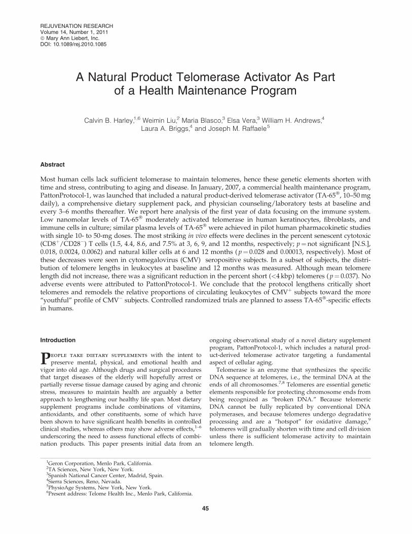
electrophoresis on nondenaturing polyacrylamide gels andquantified by exposure to Phosphor Screens and imaging onPhosphoImager SL (Molecular Dynamics).
Telomere length assays
Median telomere length in peripheral lymphocytes andgranulocytes was determined by FlowFISH at Repeat Diag-nostics (Vancouver, Canada) essentially as described else-where.40 Mean telomere length by qPCR41 was performed inthe laboratory of Dr. Richard Cawthon (University of Utah,Salt Lake City, UT). High-throughput quantitative fluores-cence in situ hybridization (HT qFISH)42 was performed atCNIO, Madrid, for inter- and intranuclear telomere lengthdistributions.
Statistics
Data from this study were collected primarily as a hy-pothesis-generating exercise because subjects were not par-ticipating in a controlled prospective study, and statisticalanalyses were not formally defined a priori. Baseline datawere analyzed for cross-sectional age effects. Student t-testswere used for comparison of means and the F-distributionfor significance of linear regression against subject age. Ex-cept where indicated, two-tailed paired t-tests were con-ducted at each time point for comparisons to the baselinevalues. For percentage of short telomeres analyzed by HTqFISH, individual differences between baseline and post-product data were analyzed by chi-squared analysis.
Results
Mechanism of action
TA-65� activates telomerase in human neonatal kerati-nocytes and fetal fibroblasts in culture. TA-65� upregulatestelomerase activity in low- and mid-passage human neonatalkeratinocytes two- to three-fold in a dose-responsive manner(Fig. 1A). In these studies, activation was two- to three-foldat the lowest concentrations tested (30–100 nM), and activa-tion was not as great at higher concentrations. This pattern issimilar to that seen for TAT2, a related molecule tested inhuman immune cells.36 The ability of TA-65� to upregulatetelomerase activity was also tested in human fetal fibroblasts(MRC5) over a broad concentration range (Fig. 1B). Un-treated and vehicle (DMSO)-treated MRC5 cultures showedextremely weak telomerase activity; there was essentially notelomerase extension products with a size greater than thatof T1, the minimum size needed to detect a product in theTRAP assay. Results from three independent experimentsindicated that TA-65� at concentrations as low as 1 nM in-duced processive telomerase activity in MRC5 cells. Telo-merase activation by TA-65� in the 1–30 nM range inmultiple cell types is important, because plasma levels of TA-65� are typically in the 1–20 nM range 4–8 h postoral inges-tion of 5–100 mg TA-65� (unpublished data).
Baseline observations
The relationships between age and various biomarkersincluding telomere length have been reported in a number ofcross-sectional studies. Table 2 shows mean values, standarddeviations, count, slope, and R2 from linear regression on
subject age, and the statistical significance of the slope for thebaseline tests investigated in this report. As expected, thispopulation showed a highly significant decline as a functionof client age in both lymphocyte and granulocyte telomerelength by FlowFISH analysis, and the slopes of the decline(55 and 34 bp/year; p¼ 10�15 and 10�8, respectively) arecomparable to those reported previously.43–45 Age-dependentincreases are seen in the percent senescent (CD8þCD28�)cytotoxic T cells, percent natural killer (NK) cells, andpercent and absolute number of neutrophils. Significant
FIG. 1. Telomerase activation by TA-65� in neonatal fore-skin keratinocytes and fetal lung MRC-5 fibroblasts. (A)Keratinocytes in triplicate wells were exposed for 48–72 hin different experiments to the dimethylsulfoxide (DMSO)vehicle control, epidermal growth factor (EGF) (positivecontrol, typically 10 ng/mL), or TA-65� at indicated con-centrations, and products were analyzed as described inMethods. Results from analysis of telomere repeat amplifi-cation protocol (TRAP) ladders resolved by gel electropho-resis and quantified by ImageQuant on a PhosphoImager areshown for a typical experiment. (B) MRC-5 cells were ex-posed to TA-65� at concentrations shown for 48 h. Eachreplicate represents an independent lysate (a replicate culturedish within one experiment). ‘‘Chaps’’ represents the lysisbuffer control (no cell extract). HeLa cells are used as positivecontrol cells as described in Methods. T1 is the first telo-merase extension product capable of amplification by PCR.IC is the internal control PCR product. Shown is a repre-sentative gel from three independent experiments.
TELOMERASE ACTIVATOR TA-65 FOR HEALTH MAINTENANCE 49

FIG. 2. Baseline telomere length and immune subset data as a function of subject age segregated by cytomegalovirus (CMV)status. Overall baseline data without segregation by CMV status is provided in Table 2. Correlations with age for lymphocyteand granulocute telomere length (A,B) and immune subsets (C–J) at baseline were determined by linear regression in theCMVþ and CMV� subjects. Telomere length and immune parameters were analyzed by flow cytometry as described inMethods.
50 HARLEY ET AL.

age-dependent decreases are seen in naı̈ve (CD8þCD95�)cytotoxic T cells, B cells, and lymphocytes.
Because CMV infection can have a significant impact onimmune markers,37,46 we analyzed the age-dependency ofbaseline immune subsets by CMV status (Fig. 2 and Table 3).Lymphocyte, but not granulocyte, telomere length was longerin CMV� subjects than that in CMVþ subjects, suggesting thatCMV infection drives increased turnover (and hence telomereshortening) in lymphocytes, but has relatively little effect onhematopoietic stem cells. Telomere length in granulocytes isconsidered a surrogate of telomere length in hematopoieticstem cells due to their short transit time to peripheral blood,and short half-life in circulation.42 The ages of the CMVþ andCMV� subjects (65� 12 and 62� 13, respectively) were notsignificantly different, but the mean lymphocyte telomerelength in CMVþ individuals was 680 bp less than thatobserved in the CMV� group ( p¼ 0.003), suggesting an ac-celeration of aging by about 10 years in the CMVþ groupbased on �55bp per year for lymphocytes.
As expected, baseline numbers and the rate of increase insenescent cytotoxic T cells (CD8þ/CD28�) in percent andabsolute counts was highest in the CMVþ population (Table 3and Fig. 2C,D). The slight increase in per cent of senescentcytotoxic T cells as a function of age in CMV� subjects (Fig. 2C)despite declining absolute numbers of these cells (Fig. 2D) is aconsequence of a significant decline in total CD8þ cells as afunction of donor age in CMV� subjects (Fig. 2 E,F). The dif-ference in the mean number of CD8þ T cells between CMVþ
and CMV� subjects (208 cells/mL) is essentially accounted forby the difference in mean number of senescent CD8þ T cells
between these two subpopulations (200 cells/mL) (Table 3).Although the %CD8þCD28þ cells at baseline was significantlyhigher in CMV� subjects ( p< 0.0001), this was due primarily tothe elevated absolute number of CD8þCD28� cells in theCMVþ subset (i.e., an increased denominator for the CMVþ
group). The absolute number of nonsenescent cytotoxic T cells(CD8þCD28þ) was not significantly different between CMVþ
and CMV� subjects (Table 3).CMVþ subjects had significantly fewer absolute and per-
cent naı̈ve cytotoxic T cells (CD8þCD95�) at baseline thandid CMV� subjects ( p< 0.0005 and 0.07, respectively) (Table3), but in both populations there was dramatic reduction inthe absolute and relative abundance of these cells as afunction of subject age (Fig. 2 G,H), consistent with previousstudies.47,48 In 80- to 90-year-old subjects, there were <50naı̈ve CD8þ cells/mL.
A novel finding in the baseline dataset is an apparent ef-fect of CMV infection on neutrophils: CMV� subjects showan increase in neutrophil number and percentage as a func-tion of subject age compared to an essentially flat profilewith age for the CMVþ subjects (Fig. 2I,J). At baseline, themean number of neutrophils in CMV� subjects was about20% higher than that in CMVþ subjects ( p¼ 0.02 by absolutecounts, p¼ 0.003 by %) (Table 3). There was also highlysignificant increase in baseline percent ( p¼ 0.00015) andabsolute numbers of NK cells ( p¼ 0.006) in the total popu-lation (Table 2), but there was no significant difference be-tween CMV� and CMVþ subjects.
Changes from baseline
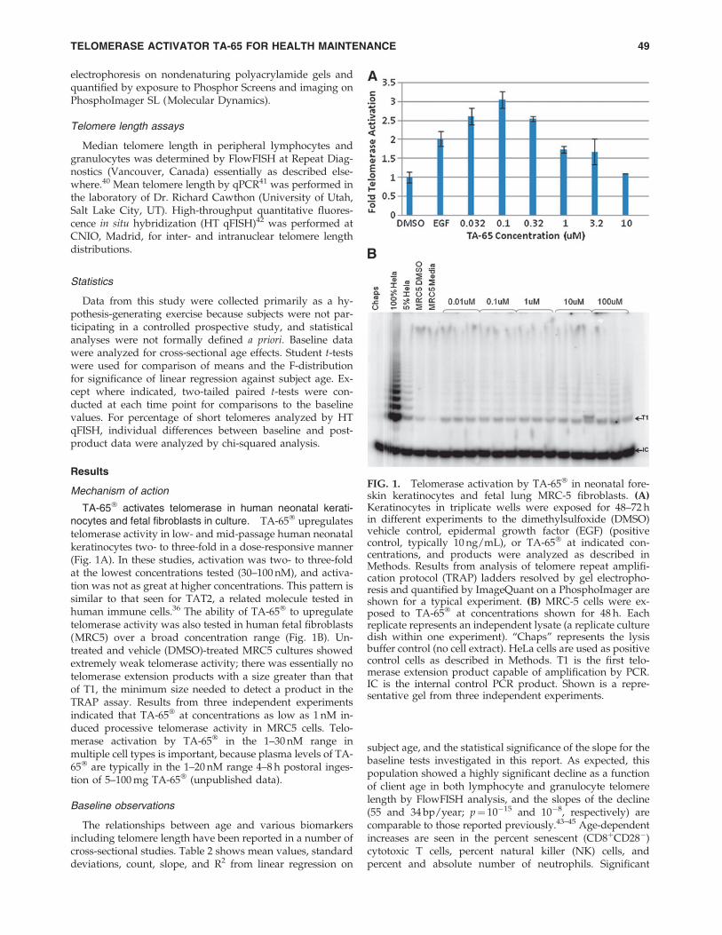
Reduction in percent cells with short telomeres. Twoindependent measures of median or mean telomere length(by FlowFISH and qPCR) showed no consistent change withtime on PattonProtocol-1 (data not shown). However, wealso analyzed the distribution of individual telomere lengthsusing automated high-throughput confocal microscopy (HTqFISH42). Telomere signals within the nuclei of white bloodcells were analyzed from 13 subjects at baseline and a follow-up time point between 12 or 18 months. Mean telomerelength by HT qFISH correlates relatively well with mediantelomere length by FlowFISH (supplemental data S2) andand although some individuals showed a significant increaseor decrease (Fig. 3A), overall there was no significant declinein mean telomere length by HT qFISH ( p¼ 0.29). However,HT qFISH revealed a decline in the percentage of nuclei withshort telomeres (<4kbp) in 10 of the 13 individuals ( p< 0.05for 7 of those 10) at 12–18 months compared to baseline,while only one of the remaining 3 individuals had a signif-icant increase in percent short telomeres (Fig. 3B). Giventhese data, we used a one-tailed paired t-test to determinethe probability that the overall mean reduction across all 13subjects was due to chance ( p¼ 0.038). In separate studies inmurine cells in culture and in vivo we have shown that TA-65� alone will reduce the percentage of cells with shorttelomeres with minimal effects on mean telomere length(M.B., manuscript in preparation).
Positive remodeling of the immune system. There were anumber of striking changes from baseline in the adaptiveand innate immune system of subjects on PattonProtocol-1.We saw statistically significant ‘‘age-reversal’’ effects in the
Table 3. Baseline Immune Subsets
by Cytomegalovirus status
Immune parameter CMVþ CMV� t-test
White blood cells # 5464 6037 0.104
Neutrophils % 58 65 0.003# 3259 4025 0.022
Monocytes % 7.8 7.4 0.453# 429 449 0.557
Lymphocytes % 35.3 29.9 0.025# 1927 1830 0.292
CD19þ
B cell% 10.8 12.9 0.007# 192 189 0.334
CD56þ,CD16þ,CD3�
Natural killer cell% 14.1 16 0.433# 220 221 0.953
CD3þ
T cell% 72 68 0.001# 1416 1202 0.03
CD3þ,CD4þ
Helper T% 47.8 49.9 0.336
# 814 734 0.332CD3þ,CD8þ
Cytotoxic T% 27 18 <0.0001
# 530 322 <0.0001CD4/CD8 ratio 2.12 3.88 0.008
CD8þ,CD28þ
Normal CD8% 53 78 0.0001# 260 250 0.976
CD8þ,CD28�
Senescent CD8% 47.2 22.3 <0.0001# 272 72 <0.0001
CD95�,CD8þ
Naı̈ve CD8% 12.000 23.000 0.0005# 48.000 72.000 0.07
p values less than 0.002 are highlighted.
TELOMERASE ACTIVATOR TA-65 FOR HEALTH MAINTENANCE 51
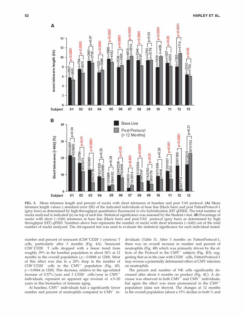
number and percent of senescent (CD8þCD28�) cytotoxic Tcells, particularly after 3 months (Fig. 4A). SenescentCD8þCD28� T cells dropped with a linear trend fromroughly 39% in the baseline population to about 36% at 12months in the overall population ( p¼ 0.0068 at 12M). Mostof this effect was due to a 20% drop in the number ofCD8þCD28� cells in the CMVþ population (Fig. 4D,p¼ 0.0044 at 12M). This decrease, relative to the age-relatedincrease of 0.57%/year and 3 CD28� cells/year in CMVþ
individuals, represent an apparent age reversal of &5–20years in this biomarker of immune aging.
At baseline, CMVþ individuals had a significantly lowernumber and percent of neutrophils compared to CMV� in-
dividuals (Table 3). After 3 months on PattonProtocol-1,there was an overall increase in number and percent ofneutrophils (Fig. 4B) which was primarily driven by the ef-fects of the Protocol in the CMVþ subjects (Fig. 4D), sug-gesting that as in the case with CD28� cells, PattonProtocol-1may reverse a potentially detrimental effect of CMV infectionon neutrophils.
The percent and number of NK cells significantly de-creased after about 6 months on product (Fig. 4C). A de-crease was observed in both CMVþ and CMV� individuals,but again the effect was more pronounced in the CMVþ
population (data not shown). The changes at 12 monthsin the overall population (about a 15% decline in both % and
FIG. 3. Mean telomere length and percent of nuclei with short telomeres at baseline and post TAS protocol. (A) Meantelomere length values� standard error (SE) of the indicated individuals at base line (black bars) and post PattonProtocol-1(grey bars) as determined by high-throughput quantitative fluorescent in situ hybridization (HT qFISH). The total number ofnuclei analyzed is indicated (n) on top of each bar. Statistical significance was assessed by the Student t-test. (B) Percentage ofnuclei with short (<4 kb) telomeres at base line (black bars) and post-TAS protocol (grey bars) as determined by highthroughput (HT) qFISH. Numbers above bars represents the number of nuclei with short telomeres (<4 kb) out of the totalnumber of nuclei analyzed. The chi-squared test was used to evaluate the statistical significance for each individual tested.
52 HARLEY ET AL.

number of NK cells) was highly significant ( p¼ 0.0035and 0.0021 for number and per cent cells, respectively), anddramatic compared to the baseline increases per year in NKcells for the CMVþ group (þ2.4 cells per year and þ0.3% peryear).
Discussion and Conclusions
The inability to maintain telomeres with age and chronicstress has been linked to declining health and the increasedrisk of disease and death from many causes, including can-cer.16,25,49–52 In this study, we report that a 1-year healthmaintenance program consisting of a dietary supplementpack combined with a natural product–derived telomeraseactivator results in a decreased percentage of short leukocytetelomeres and remodeling of the relative proportions of thecirculating leukocytes of CMVþ subjects toward the more‘‘youthful’’ profile of CMV� subjects.
One of the strengths of our study is the low CMV-positivity rate (54%) in a relatively older population, whichallows us to separate the effects of age and CMV status onimmunosenescence. It also serves to mitigate one of ourstudy’s weaknesses—the lack of a control group—as thesubjects were initially unaware of their CMV status and
their subsequent knowledge is unlikely to have caused thesegregation of many of the effects of the protocol by CMVstatus.
Our description of the decrease in CD8þCD28� T cells as apositive remodeling of the immune system is supported bythe increased morbidity and mortality associated with whatis known as the immune risk profile (IRP). This profile hasbeen defined as a CD4/CD8 less than 1 in association withCMV seropositivity by longitudinal studies of individuals intheir eighties and nineties in the OCTO/NONA Swedishcohort.53 As in our cohort, the major driving force for thedecreased CD4/CD8 in CMVþ subjects in this population islikely the accumulation of virus-specific CD8þCD28� T cells.These studies have reported 6-year follow-up data, and noindividuals who have survived to 100 years old exhibit theIRP, even if they are CMVþ. The authors conclude thatsuccessful immune aging entails being able to control CMVinfection without accumulating senescent cytotoxic T cells.Thus, we conclude that the 20% reduction in CD8þCD28� Tcells is a salutary effect, even though we have yet to seeincreases in the number of CD8þCD28þ T cells. Telomereshortening associated with replicative senescence is theprobable cause of loss of CD28 expression and apoptosisresistance of CD8þ T cells.54 The decrease in the percentage
FIG. 4. Relative changes from baseline as a function of time (months) for immune subsets. The mean of the absolute changefrom baseline for each parameter across all subjects for which data at that time point was calculated and then expressed as apercent change from the mean value at baseline for those subjects. In A–C, data for both cytomegalovirus-positive (CMVþ)and CMV� subjects are combined. (D) Relative mean change is shown for the CMVþ population subpopulation only.Asterisks next to a data point signifies p< 0.01 (***), p< 0.05 (**), and p< 0.1 (*) for a two-tailed paired (within-subject) t-testanalysis comparing baseline to time point values.
TELOMERASE ACTIVATOR TA-65 FOR HEALTH MAINTENANCE 53

of short telomeres we found makes upregulation of telo-merase by TA-65 the most likely mechanism for this salutaryeffect.
Age-related changes in the innate immune system havenot been as well characterized as those of the adaptive im-mune system, although the importance of changes in theformer is increasingly being recognized.55 It is generallyagreed that the per-cell activity of neutrophils as measuredby oxidative burst, phagocytosis, and chemotaxis decreaseswith age.56 There is less agreement on the effect of aging onneutrophil number which has been variously reported to bepreserved,57 decreased,58 or increased53 with age. CMV sta-tus is not reported in the first two studies, but in the laststudy from the above-mentioned NONA cohort, the increasein neutrophil number is based on a comparison between 18middle-aged (55-year-old) subjects with a 55% CMVþ prev-alence rate and 120 very old subjects (92–100-year-old) with a87% CMVþprevalence rate. Most of the cross-sectional in-crease of 960 neutrophils occurs within the 92 to 100 yearolds, with only a 52-cell increase between 55 and 92. There isalso a significant longitudinal increase over a 6-year intervalin the very old group. This suggests that there is selection forthose very old subjects able to increase their circulatingneutrophils in the face of deteriorating tissues, increasedinflammation, and increased exposure to infectious agents.Our novel finding that by age 62 the neutrophil number is20% higher and continues to increase with age only in CMV�
subjects can be interpreted as a compensatory increase in theface of declining per cell activity and barrier function, as wellas increased antigenic load. The absence of a cross-sectionalincrease with age in neutrophil number in CMVþ subjectssuggests that this compensation is blocked in the CMVþ
subjects perhaps due to inhibitory cytokine production bythe senescent T cells. The effect of the protocol to increaseneutrophil count in CMVþ subjects can be interpreted as asalutary removal of this block in part through reduction inthe number of senescent T cells.
There is a broad consensus that NK cell number increaseswith age to compensate for decreased per-cell activity, whichresults from impaired signal transduction,59 but othermechanisms such as decreased barrier function and in-creased antigenic/pathogenic load may also contribute toincreased NK cells with age.60 The decrease in NK cellnumber induced by the protocol is an ‘‘age reversal,’’ butbecause we did not measure NK activity we cannot saywhether it is from improved barrier function or improvedsignal transduction. Unlike other cells of the innate immunesystem, NK cells proliferate after activation and experiencefurther telomere shortening once they are released from thebone marrow.61 The decrease in the percentage of shorttelomeres we found could result in improved signal trans-duction as a mechanism for the reduction in NK cell number.Taken together, these three changes in leukocyte numberinduced by the protocol represent a remodeling of the im-mune systems of CMVþ subjects to look more like those ofCMV� subjects and successfully aging CMVþ centenarians.
Physicians who monitored the health of the current studysubjects through 1 year on the product reported no adverseevents that were likely related to the protocol. However, 2subjects who recently escalated their daily dose reportedfeeling ‘‘anxious’’ on 100 mg/day but not when they swit-ched back to 50 mg/day. A placebo-controlled study will be
needed to determine if this potential adverse effect is real. Nonew cases of cancer or cardiovascular disease were reportedduring the overall 260 person-years of dosing with Patton-Protocol-1 through June, 2010, and this is statistically sig-nificant ( p< 0.05, cancer; p< 0.02, CVD ) assuming baselineage-specific risks in our population were similar to those ofthe U.S. population.
TA-65� activated telomerase in cultured human cells atconcentrations seen in the plasma of subjects on the protocol.Paradoxically, although &40% of subjects showed an in-crease in mean telomere length over time, on average acrossall subjects there was a nonsignificant decline in mean telo-mere length. However, we speculate this effect is explainedby cell dynamics and the fact that telomerase preferentiallylengths the shortest telomeres.62–64 Rescue and selective ex-pansion of near-senescent cells with short telomeres couldlead to a reduction in the population mean telomere lengthdespite some lengthening of telomeres in all cells. Becausedetrimental effects of telomere loss are primarily driven byshort, dysfunctional telomeres, and loss of tissue function anddisease onset in proliferative tissues have been associatedwith telomere lengths <4 kbp,25,65 we believe that our ob-served reduction in telomeres <4 kbp in subjects on Patton-Protocol-1 is a significant, positive response, and that TA-65�
contributes to the apparent benefit of the dietary supplement.In support of this, studies with TA-65� given orally in oldmice showed similar reductions in percent cells with shorttelomeres and positive functional effects on tissues (Blascoet al., submitted) and preliminary dose–response analysesshowed an increase in salutary effects with TA-65� doses upto 20–30 mg per day average compared to the initial 5- to 10-mg per day dose (data not shown). Finally, analysis of ad-ditional biomarkers of aging in subjects on PattonProtocol-1suggest improvements in the cardiovascular system, metab-olism, and bone mineral density, which will be furtherstudied and reported elsewhere. Independent randomizedcontrolled studies with TA-65� alone are planned.
Acknowledgments
We thank Drs. Tom Okarma, Spencer Brown, and RitaEffros for critical review of the manuscript, and Noel Pattonfor patience, encouragement, and financial support of thestudies. We also thank the early telomerase activation teamat Geron Corporation for generating data in human kerati-nocytes, Marissa Chunisingh for acting as a key interfacebetween clients and TA Sciences staff, and Drs. NathanWong and Sherman Xi for critical feedback and help withstatistical analyses.
Author Disclosure Statement
Calvin Harley is one of the inventors of TA-65. He consultsfor TA Sciences and is personally taking TA-65 and is one ofthe subjects studied to generate data for this article. He ownsstock and stock options in Geron Corporation, a company thatis developing telomerase activators for therapeutic purposesand the company that licensed TA-65 to TA Sciences. He is co-founder, President, and CEO, and holds stock in TelomeHealth, Inc., a diagnostics company that will provide telo-mere- and telomerase-related assay services to the healthcareindustry. Joseph Raffaele consults for TA Sciences; he is CEOof PhysioAge Systems, LLC, a company that provides bio-
54 HARLEY ET AL.

markers of aging analysis, including telomere lengths, tomedical practices, and he is co-founder of PhysioAge MedicalGroup which offers TA-65 through the Patton Protocol. Wil-liam H. Andrews owns stock or membership options in GeronCorporation and Sierra Sciences; he consults for TA Science, isa client of TA Sciences who is taking TA-65, and is one of thesubjects studied to generate data for this manuscript. He is thefounder, President, and CEO of Sierra Sciences, a companythat develops therapeutics for inducing telomerase expressionand has provided financial support for some of the studiesdescribed in this manuscript. Weimin Liu is an employee ofTA Sciences. Elsa Vera and Maria Blasco have no competingfinancial interests.
References
1. Marik PE, Varon J. Omega-3 dietary supplements and therisk of cardiovascular events: A systematic review. ClinCardiol 2009;32:365–372.
2. Khandanpour N, Armon MP, Jennings B, Finglas PM, WillisG, Clark A, Meyer FJ. Randomized clinical trial of folatesupplementation in patients with peripheral arterial disease.Br J Surg 2009;96:990–998.
3. Bemis DL, Katz AE, Buttyan R. Clinical trials of naturalproducts as chemopreventive agents for prostate cancer.Expert Opin Investig Drugs 2006;15:1191–1200.
4. Iannitti T, Palmieri B. Antioxidant therapy effectiveness: anup to date. Eur Rev Med Pharmacol Sci 2009;13:245–278.
5. Judd SE, Tangpricha V. Vitamin D deficiency and risk forcardiovascular disease. Am J Med Sci 2009;338:40–44.
6. Nigwekar SU, Kandula P. N-acetylcysteine in cardiovascu-lar-surgery-associated renal failure: A meta-analysis. AnnThorac Surg 2009;87:139–147.
7. Blackburn EH, Greider CW, Szostak JW. Telomeresand telomerase: the path from maize, Tetrahymena and yeastto human cancer and aging. Nat Med 2006;12:1133–1138.
8. Harley CB. Telomerase therapeutics for degenerative dis-eases. Curr Mol Med 2005;5:205–211.
9. Ayouaz A, Raynaud C, Heride C, Revaud D, Sabatier L.Telomeres: Hallmarks of radiosensitivity. Biochimie 2008;90:60–72.
10. Wright DL, Jones E, Mayer J, Oehninger S, Gibbons W,Lanzendorf SE. Characterization of telomerase activity in thehuman oocyte and preimplantation embryo. Mol Hum Re-prod 2001;7:947–955.
11. Ulaner GA, Giudice LC. Developmental regulation of telo-merase activity in human fetal tissues during gestation. MolHum Reprod 1997;3:769–773.
12. Harley CB. Telomerase is not an oncogene. Oncogene2002;21:494–502.
13. Flores I, Benetti R, Blasco MA. Telomerase regulation andstem cell behaviour. Curr Opin Cell Biol 2006;18:254–260.
14. Effros RB. Telomerase induction in T cells: A cure for agingand disease? Gerontol 2007;42:416–420.
15. Effros RB. Genetic alterations in the ageing immune system:Impact on infection and cancer. Mech Ageing Dev 2003;124:71–77.
16. Epel ES. Psychological and metabolic stress: a recipe foraccelerated cellular aging? Hormones (Athens) 2009;8:7–22.
17. von Zglinicki T. Oxidative stress shortens telomeres. TrendsBiochem Sci 2002;27:339–344.
18. Epel ES, Blackburn EH, Lin J, Dhabhar FS, Adler NE, Mor-row JD, Cawthon RM. Accelerated telomere shortening in
response to life stress. Proc Natl Acad Sci USA 2004;101:17312–17315.
19. Harley CB, Futcher AB, Greider CW. Telomeres shortenduring ageing of human fibroblasts. Nature 1990;345:458–460.
20. Hastie ND, Dempster M, Dunlop MG, Thompson AM, GreenDK, Allshire RC. Telomere reduction in human colorectalcarcinoma and with ageing. Nature 1990;346:866–868.
21. Garcia CK, Wright WE, Shay JW. Human diseases of telo-merase dysfunction: insights into Tissue aging. NucleicAcids Res 2007;35:7406–7416.
22. Armanios MY, Chenn JJ, Cogan JD, Alder JK, Ingersoll RG,Markin C, Lawson WE, Xie M, Vulto I, Phillips JAr, Lans-dorp PM, Greider CW, Loyd JE. Telomerase mutations infamilies with idiopathic pulmonary fibrosis. N Engl J Med2007;356:1370–1372.
23. Sebastian C, Herrero C, Serra M, Lloberas J, Blasco MA,Celada A. Telomere shortening and oxidative stress in agedmacrophages results in impaired STAT5a phosphorylation. JImmunol 2009;183:2356–2364.
24. Calado RT. Telomeres and marrow failure. Hematology AmSoc Hematol Educ Program 2009:338–343.
25. Calado RT, Young NS. Telomere diseases. N Engl J Med2009;361:2353–2365.
26. Rajaraman S, Choi J, Cheung P, Beaudry V, Moore H, Ar-tandi SE. Telomere uncapping in progenitor cells withcritical telomere shortening is coupled to S-phase pro-gression in vivo. Proc Natl Acad Sci USA 2007;104:17747–17752.
27. Cawthon RM, Smith KR, O’Brien E, Sivatchenko A, KerberRA. Association between telomere length in blood andmortality in people aged 60 years or older. Lancet2003;361:359–395.
28. Savage SA, Alter BP. The role of telomere biology in bonemarrow failure and other disorders. Mech Ageing Dev2008;129:35–47.
29. Aviv A, Valdes A, Gardner JP, Swaminathan R, Kimura M,Spector TD. Menopause modifies the association ofleukocyte telomere length with insulin resistance and in-flammation. J Clin Endocrinol Metab 2006;91:635–640.
30. Lukens JN, Van Deerlin V, Clark CM, Xie SX, Johnson FB.Comparisons of telomere lengths in peripheral blood andcerebellum in Alzheimer’s disease. Alzheimers Dement2009;5:463–469.
31. Weyand CM, Fujii H, Shao L, Goronzy JJ. Rejuvenating theimmune system in rheumatoid arthritis. Nat Rev Rheumatol2009;5:583–588.
32. Spyridopoulos I, Hoffmann J, Aicher A, Brummendorf TH,Doerr HW, Zeiher AM, Dimmeler S. Accelerated telomereshortening in leukocyte subpopulations of patients withcoronary heart disease: role of cytomegalovirus seroposi-tivity. Circulation 2009;120:1364–1372.
33. Effros RB. Telomerase induction in T cells: A cure for agingand disease? Exp Gerontol 2007;42:416–420.
34. Lichterfeld M, Mou D, Cung TD, Williams KL, Waring MT,Huang J, Pereyra F, Trocha A, Freeman GJ, Rosenberg ES,Walker BD, Yu XG. Telomerase activity of HIV-1-specificCD8þ T cells: constitutive up-regulation in controllers andselective increase by blockade of PD ligand 1 in progressors.Blood 2008;112:3679–3687.
35. Dagarag M, Evazyan T, Rao N, Effros RB. Genetic manip-ulation of telomerase in HIV-specific CD8 T cells: enhancedanti-viral functions accompany the increased proliferativepotential and telomere stabilization. J Immunol 2004;173:6303–6311.
TELOMERASE ACTIVATOR TA-65 FOR HEALTH MAINTENANCE 55

36. Fauce SR, Jamieson BD, Chin AC, Mitsuyasu RT, Parish ST,Ng HL, Kitchen CM, Yang OO, Harley CB, Effros RB. Tel-omerase-based pharmacologic enhancement of antiviralfunction of human CD8þ T lymphocytes. J Immunol2008;181:7400–7406.
37. Pawelec G, Derhovanessian E, Larbi A, Strindhall J, WikbyA. Cytomegalovirus and human immunosenescence. RevMed Virol 2009;19:47–56.
38. Dowd JB, Zajacova A, Aiello A. Early origins of health dis-parities: burden of infection, health, and socioeconomicstatus in U.S. children. Soc Sci Med 2009;68:699–707.
39. Kim NW, Wu F. Advances in quantification and character-ization of telomerase activity by the telomeric repeat amplifi-cation protocol (TRAP). Nucleic Acids Res 1997;25:2595–2597.
40. Baerlocher GM, Vulto I, de Jong G, Lansdorp PM. Flowcytometry and FISH to measure the average length of telo-meres (flow FISH). Nat Protoc 2006;1:2365–2376.
41. Cawthon RM. Telomere length measurement by a novelmonochrome multiplex quantitative PCR method. NucleicAcids Res 2009;37:e21.
42. Canela A, Vera E, Klatt P, Blasco MA. High-throughputtelomere length quantification by FISH and its application tohuman population studies. Proc Natl Acad Sci USA2007;104:5300–5305.
43. Vaziri H, Dragowska W, Allsopp RC, Thomas TE, HarleyCB, Lansdorp PM. Evidence for a mitotic clock in humanhematopoietic stem cells: Loss of telomeric DNA with age.Proc Natl Acad Sci USA 1994;91:9857–9860.
44. Rufer N, Brummendorf TH, Kolvraa S, Bischoff C, Chris-tensen K, Wadsworth L, Schulzer M, Lansdorp PM. Telo-mere fluorescence measurements in granulocytes and Tlymphocyte subsets point to a high turnover of hematopoi-etic stem cells and memory T cells in early childhood. J ExpMed 1999;190:157–167.
45. Frenck RW, Blackburn EH, Shannon KM. The rate of telo-mere sequence loss in human leukocytes varies with age.Proc Natl Acad Sci USA 1998;95:5607–5610.
46. Chidrawar S, Khan N, Wei W, McLarnon A, Smith N, NayakL, Moss P. Cytomegalovirus-seropositivity has a pro-found influence on the magnitude of major lymphoid sub-sets within healthy individuals. Clin Exp Immunol2009;155:423–432.
47. Hadrup SR, Strindhall J, Kollgaard T, Seremet T, JohanssonB, Pawelec G, thor Straten P, Wikby A. Longitudinal studiesof clonally expanded CD8 T cells reveal a repertoireshrinkage predicting mortality and an increased number ofdysfunctional cytomegalovirus-specific T cells in the veryelderly. J Immunol 2006;176:2645–2653.
48. Effros RB, Pawelec G. Replicative senescence of T cells: Doesthe hayflick limit lead to immune exhaustion? ImmunolToday 1997;18:450–454.
49. Cawthon RM. Telomere measurement by quantitative PCR.Nucleic Acids Res 2002;30.
50. Farzaneh-Far R, Lin J, Epel E, Lapham K, Blackburn E,Whooley MA. Telomere length trajectory and its determinantsin persons with coronary artery disease: longitudinal findingsfrom the heart and soul study. PLoS One 2010;5:e8612.
51. Brouilette SW, Whittaker A, Stevens SE, van der Harst P,Goodall AH, Samani NJ. Telomere length is shorter inhealthy offspring of subjects with coronary artery disease:Support for the telomere hypothesis. Heart 2008;94:422–425.
52. van der Harst P, van der Steege G, de Boer RA, Voors AA,Hall AS, Mulder MJ, van Gilst WH, van Veldhuisen DJ.
Telomere length of circulating leukocytes is decreased inpatients with chronic heart failure. J Am Coll Cardiol2007;49:1459–1464.
53. Strindhall J, Nilsson BO, Lofgren S, Ernerudh J, Pawelec G,Johansson B, Wikby A. No Immune Risk Profile among in-dividuals who reach 100 years of age: Findings from theSwedish NONA immune longitudinal study. Exp Gerontol2007;42:753–761.
54. Effros RB, Dagarag M, Spaulding C, Man J. The role ofCD8þ T-cell replicative senescence in human aging. Im-munol Rev 2005;205:147–157.
55. Gomez CR, Nomellini V, Faunce DE, Kovacs EJ. Innateimmunity and aging. Exp Gerontol 2008;43:718–728.
56. Wenisch C, Patruta S, Daxbock F, Krause R, Horl W. Effectof age on human neutrophil function. J Leukoc Biol2000;67:40–45.
57. Chatta GS, Andrews RG, Rodger E, Schrag M, HammondWP, Dale DC. Hematopoietic progenitors and aging: alter-ations in granulocytic precursors and responsiveness to re-combinant human G-CSF, GM-CSF, and IL-3. J Gerontol1993;48:M207–M212.
58. De Martinis M, Modesti M, Ginaldi L. Phenotypic andfunctional changes of circulating monocytes and polymor-phonuclear leucocytes from elderly persons. Immunol CellBiol 2004;82:415–420.
59. Panda A, Arjona A, Sapey E, Bai F, Fikrig E, MontgomeryRR, Lord JM, Shaw AC. Human innate immunosenescence:causes and consequences for immunity in old age. TrendsImmunol 2009;30:325–333.
60. Solana R, Alonso MC, Pena J. Natural killer cells in healthyaging. Exp Gerontol 1999;34:435–443.
61. Mariani E, Meneghetti A, Formentini I, Neri S, Cattini L,Ravaglia G, Forti P, Facchini A. Different rates of telomereshortening and telomerase activity reduction in CD8 T andCD16 NK lymphocytes with ageing. Exp Gerontol 2003;38:653–659.
62. Hemann M, Strong M, Hao L, Greider C. The shortest telo-mere, not average telomere length, is critical for cell viabilityand chromosome stability. Cell 2001;107:67–77.
63. Samper E, Flores J, Blasco M. Restoration of telomerase ac-tivity rescues chromosomal instability and premature aging inTerc mice with short telomeres. EMBO reports 2001;2:800–807.
64. Teixeira MT, Arneric M, Sperisen P, Lingner J. Telomerelength homeostasis is achieved via a switch between telo-merase- extendible and -nonextendible states. Cell 2004;117:323–335.
65. Alter BP, Baerlocher GM, Savage SA, Chanock SJ, WekslerBB, Willner JP, Peters JA, Giri N, Lansdorp PM. Very shorttelomere length by flow fluorescence in situ hybridizationidentifies patients with dyskeratosis congenita. Blood2007;110:1439–1447.
Address correspondence to:Calvin B. Harley
Telome Health, Inc.1455 Adams Drive
Menlo Park, CA 94025
E-mail: [email protected]
Received: June 25, 2010Accepted: August 9, 2010
56 HARLEY ET AL.