Embed Size (px)
Citation preview

Spinal CordBY SHAWETA KHOSA

Spinal Cord • Cylindrical in shape • Runs through the vertebral canal• Extends from foramen magnum to second
lumbar vertebra L1/L2 (adult), L3 (NEWBORN)
• Occupies upper 2/3 of vertebral canal .• Regions
• Cervical • Thoracic • Lumbar• Sacral• Coccygeal
• Gives rise to 31 pairs of spinal nerves• All are mixed nerves
• Not uniform in diameter• Cervical enlargement: supplies upper
limbs• Lumbar enlargement: supplies lower
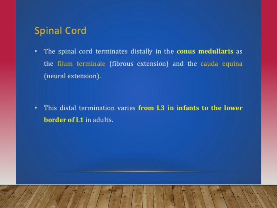
limbs• Conus medullaris- tapered inferior end
• Ends between L1 and L2• Cauda equina - origin of spinal nerves
extending inferiorly from conus medullaris.


Cross Section of Spinal Cord
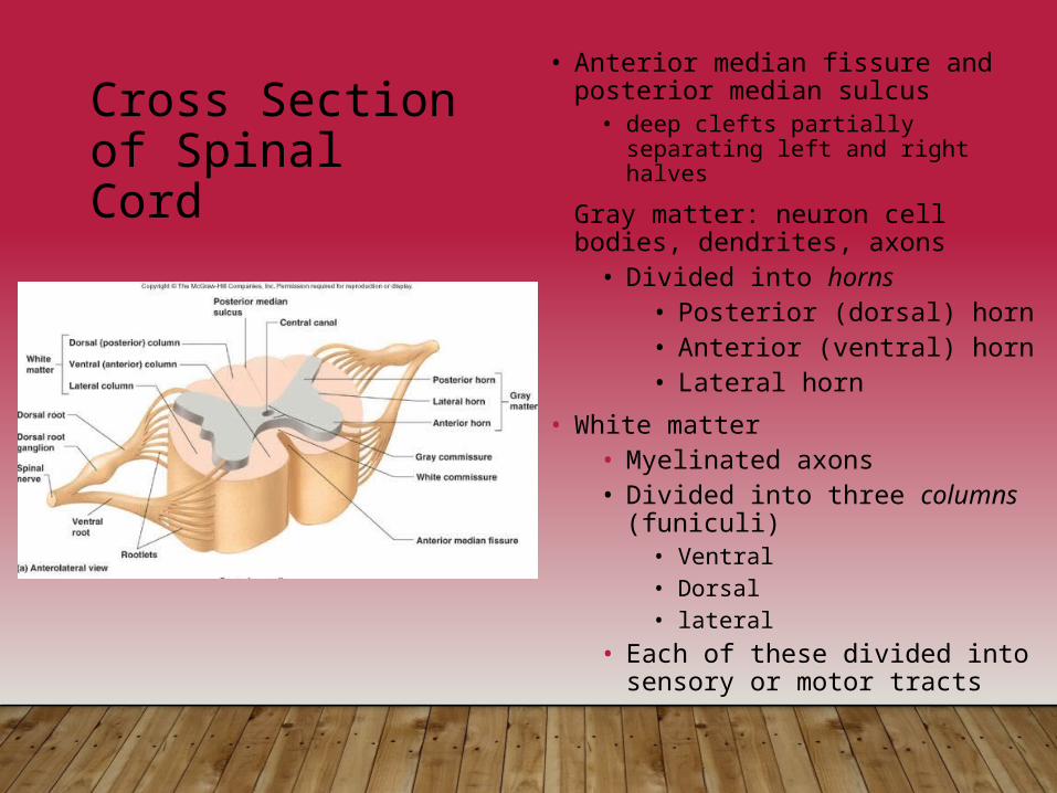
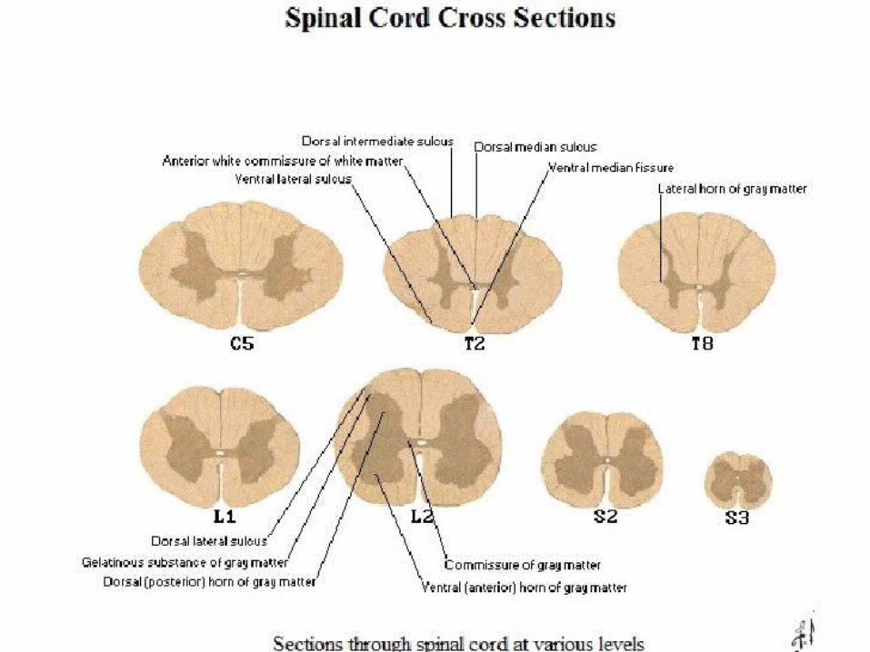
• Anterior median fissure and posterior median sulcus
• deep clefts partially separating left and right halves
• Gray matter: neuron cell bodies, dendrites, axons
• Divided into horns• Posterior (dorsal) horn• Anterior (ventral) horn• Lateral horn
• White matter• Myelinated axons• Divided into three columns
(funiculi)• Ventral• Dorsal• lateral
• Each of these divided into sensory or motor tracts
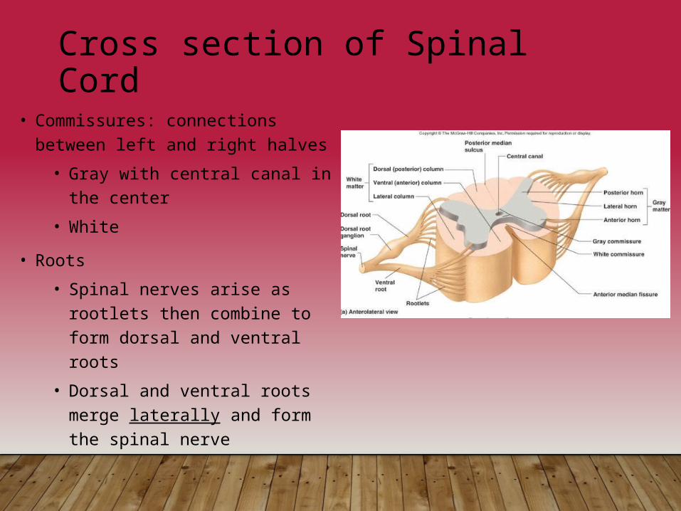
Cross section of Spinal Cord• Commissures: connections
between left and right halves• Gray with central canal in the
center• White
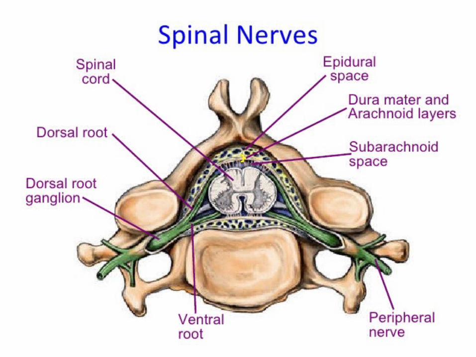
• Roots• Spinal nerves arise as rootlets
then combine to form dorsal and ventral roots
• Dorsal and ventral roots merge laterally and form the spinal nerve
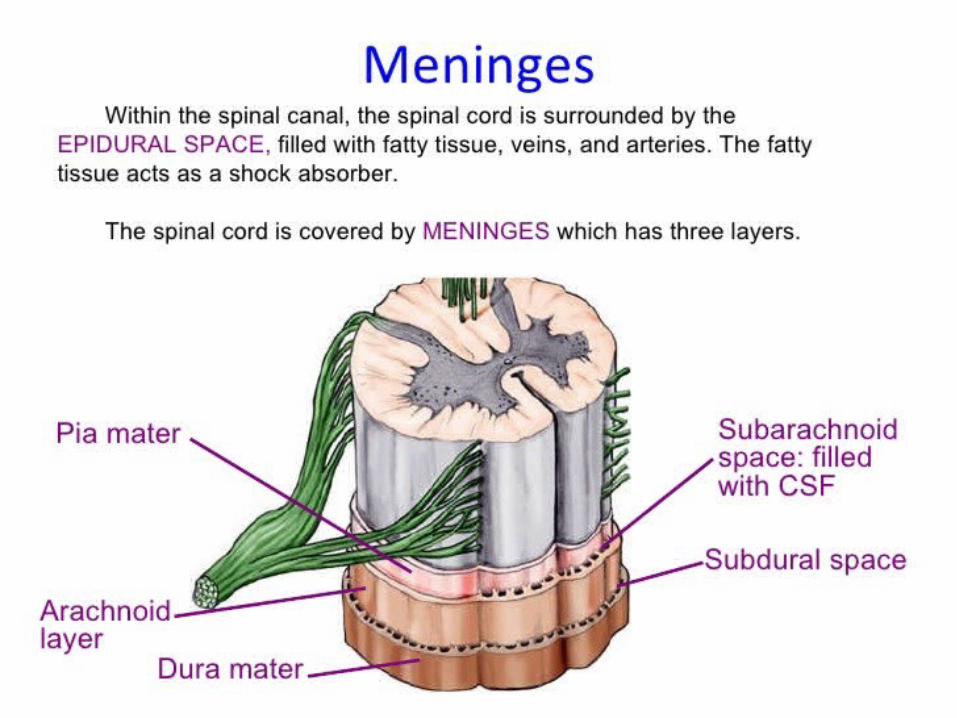
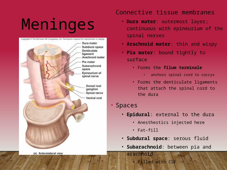
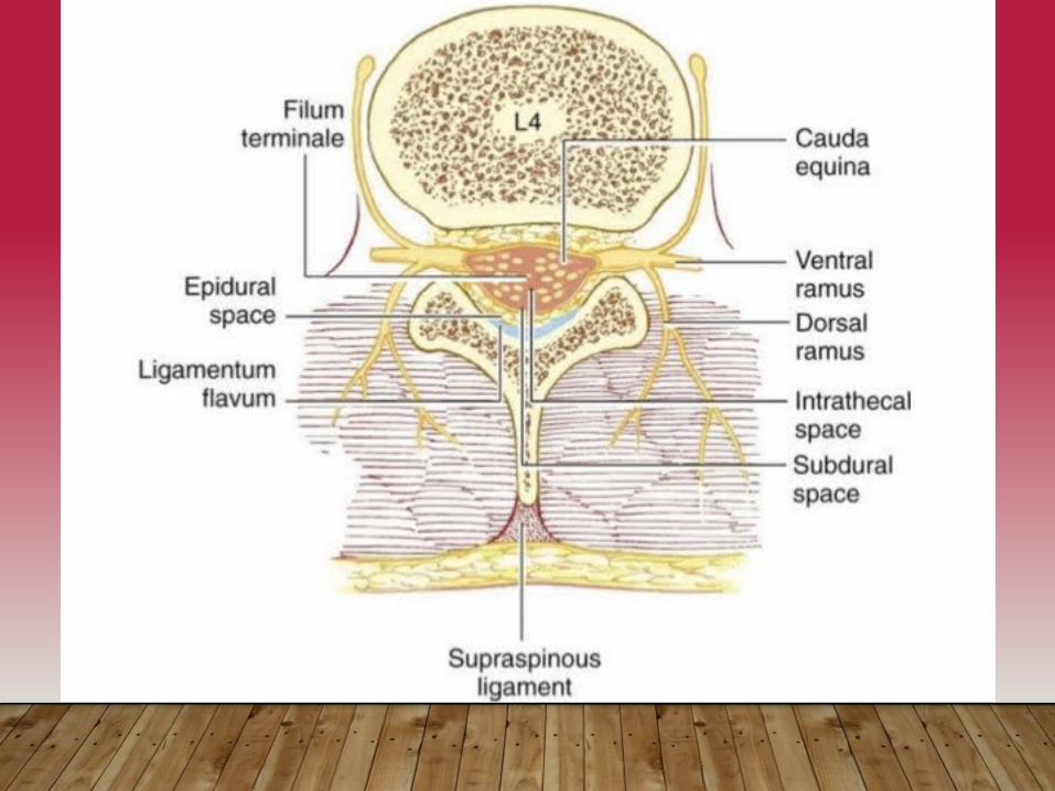
Meninges• Connective tissue membranes
• Dura mater: outermost layer; continuous with epineurium of the spinal nerves
• Arachnoid mater: thin and wispy• Pia mater: bound tightly to surface
• Forms the filum terminale• anchors spinal cord to coccyx
• Forms the denticulate ligaments that attach the spinal cord to the dura
• Spaces• Epidural: external to the dura
• Anesthestics injected here • Fat-fill
• Subdural space: serous fluid• Subarachnoid: between pia and
arachnoid• Filled with CSF


Organization of Spinal Cord Gray Matter
• Recall, it is divided into horns• H- shaped pillar with anterior and posterior gray horns • United by gray commissure containing the central canal.• Lateral gray horns (only in thoracic region and upper lumbar segments)• Dorsal half – sensory roots and ganglia• Ventral half – motor roots• Based on the type of neurons/cell bodies located in each horn, it is specialized further into 4 regions
• Somatic sensory (SS) - axons of somatic sensory neurons• Visceral sensory (VS) - neurons of visceral sensory neur.• Visceral motor (VM) - cell bodies of visceral motor neurons• Somatic motor (SM) - cell bodies of somatic motor neurons

Gray matter
THE AMOUNT OF GRAY MATTER RELATED TO THE AMOUNT OF MUSCLE INNERVATED.
CONSISTS OF NERVE CELLS, NEUROLAGIA, BLOOD VESSELS.

Gray Matter: Organization
NERVE CELLS IN THE ANTERIOE GRAY COLUMNS :
• LARGE AND MULTIPOLAR• AXONS PASS OUT IN THE ANTERIOR NERVE ROOTS AS THE alpha efferents.
• SMALLER NERVE CELLS ARE MULTIPOLAR• AXONS PASS OUT IN ANTERIOR ROOTS AS THE GAMMA EFFERENTS.
Nerve cells in the posterior gray columns
•4 nerve cell groups•SUBSTANIA GELATINOSA:Situated at the apexThroughout the length of spinal cordComposed mainly of Golgi type 2 neuronsReceives afferent fibers concerning with pain, temperature and touch from Posterior root

White Matter in the Spinal Cord
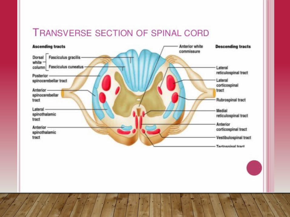
• Divided into three funiculi (columns) – posterior, lateral, and anterior
• Columns contain 3 different types of fibers (Ascend., Descend., Trans.)
• Fibers run in three directions• Ascending fibers - compose the sensory tracts• Descending fibers - compose the motor tracts• Commissural (transverse) fibers - connect opposite sides
of cord

White Matter Fiber Tract Generalizations
• Pathways decussate (most)• Most consist of a chain of two or three neurons• Most exhibit somatotopy (precise spatial
relationships)• All pathways are paired
• one on each side of the spinal cord

White Matter: Pathway Generalizations
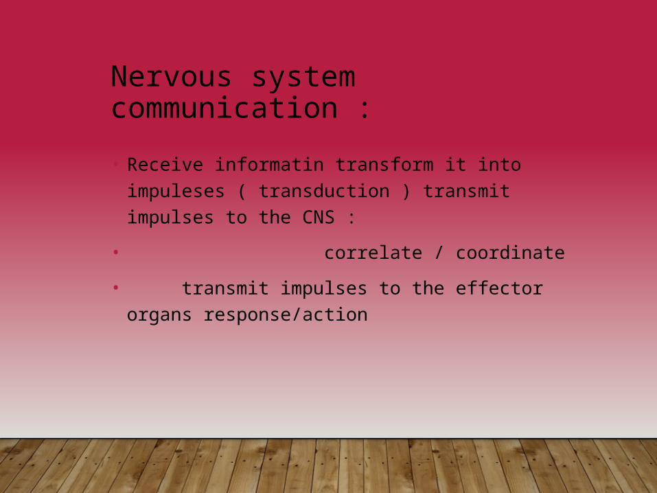
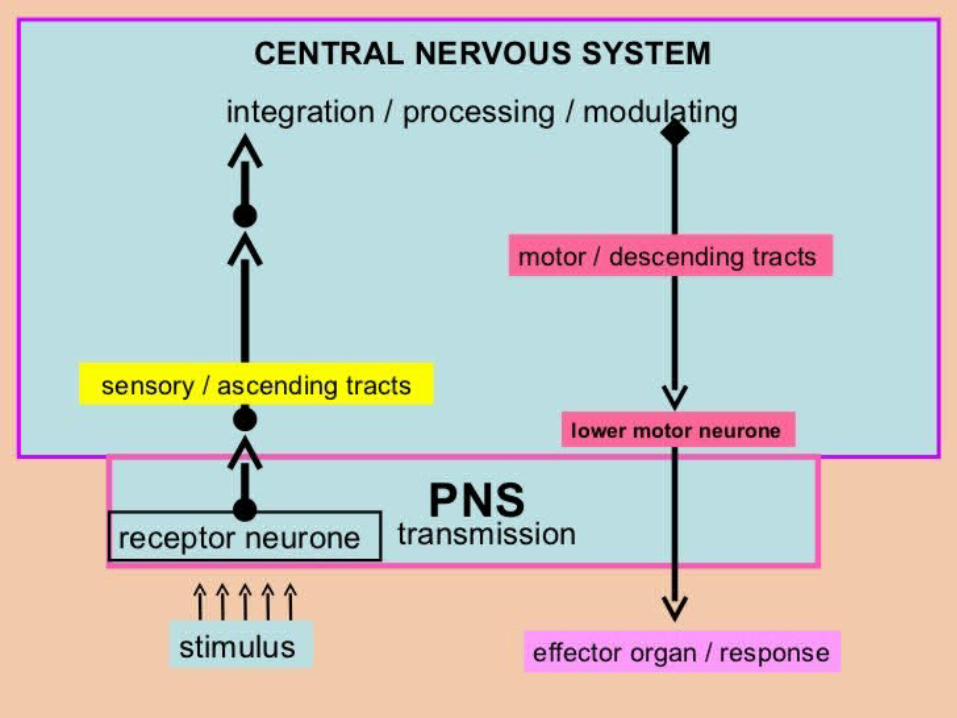
Nervous system communication :• Receive informatin transform it into impuleses (
transduction ) transmit impulses to the CNS : • correlate / coordinate• transmit impulses to the effector organs
response/action
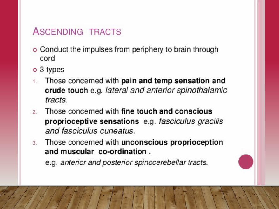
Tracts of spinal cord

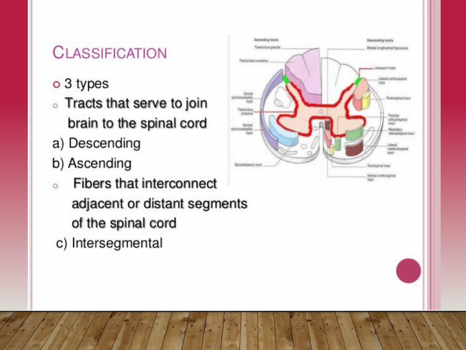
Nomenclature of tracts
• Tracts are named after the names of masses of grey matter connected by them.
• Name consist of two components • First term- origin , second term – termination• i. e. corticospinal tract , spinothalamic tract.


Descending (Motor) Pathways
• Descending tracts deliver motor instructions from the brain to the spinal cord
• Divided into two groups• Pyramidal, or corticospinal, tracts• Indirect pathways, essentially all others
• Motor pathways involve two neurons • Upper motor neuron (UMN)• Lower motor neuron (LMN)
• aka ‘anterior horn motor neuron” (also, final common pathway)
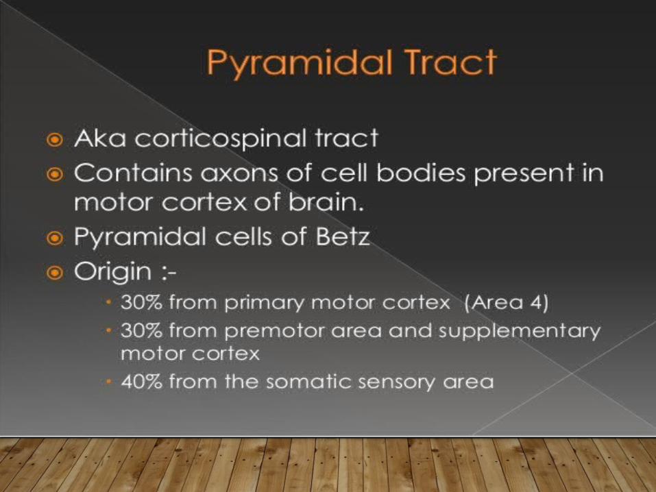
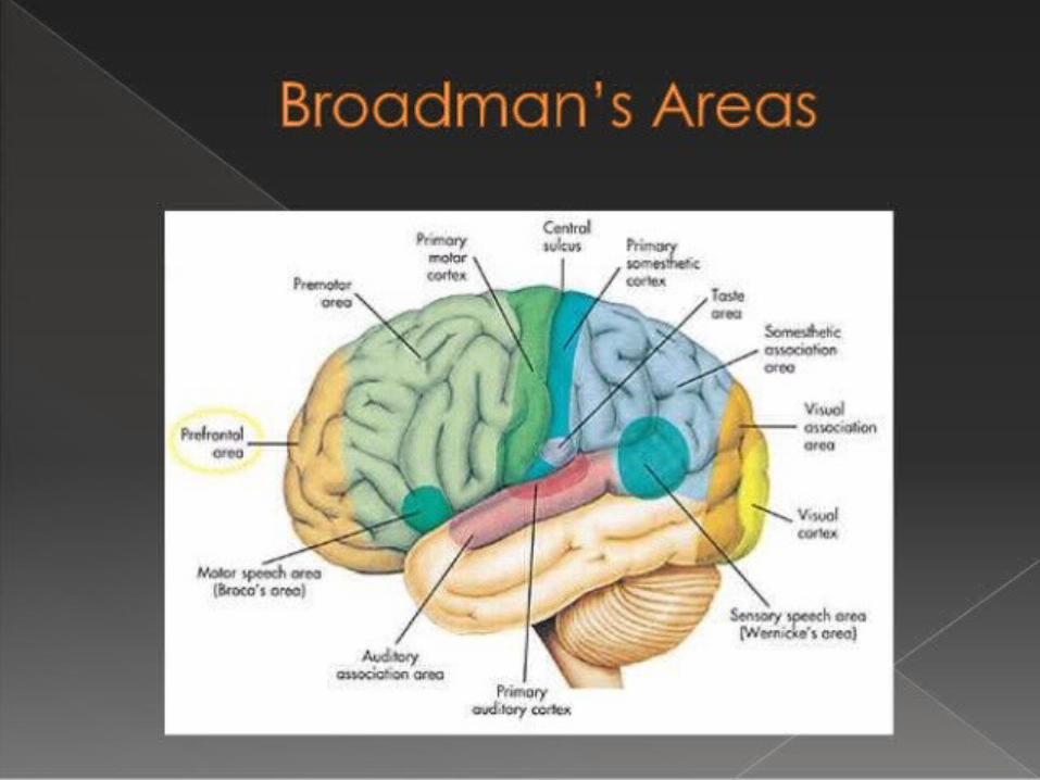
Pyramidal (Corticospinal) Tracts• Originate in the precentral gyrus of brain (aka, primary motor area)
• I.e., cell body of the UMN located in precentral gyrus• Pyramidal neuron is the UMN
• Its axon forms the corticospinal tract
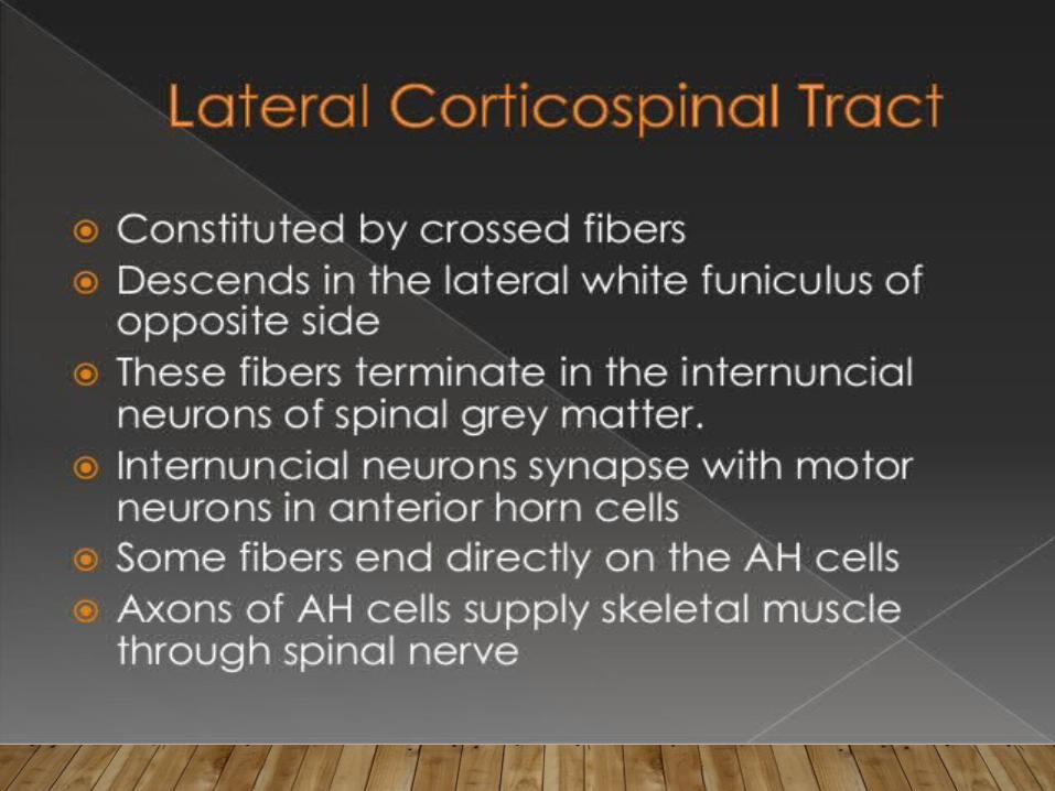
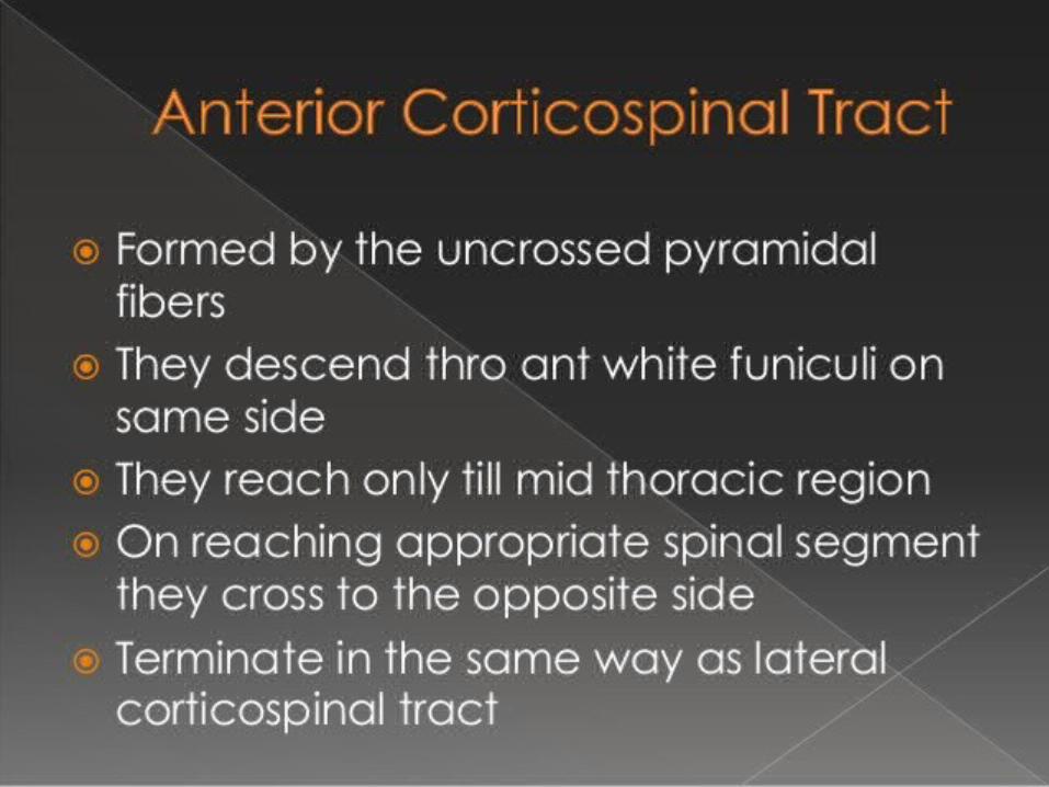
• UMN synapses in the anterior horn with LMN• Some UMN decussate in pyramids = Lateral corticospinal tracts• Others decussate at other levels of s.c. = Anterior corticospinal tracts-
• LMN (anterior horn motor neurons)• Exits spinal cord via anterior root • Activates skeletal muscles
• Regulates fast and fine (skilled) movements

Corticospinal tracts
1. Location of UMN cell body in cerebral cortex
2. Decussation of UMN axon in pyramids or at level of exit of LMN
3. Synapse of UMN and LMN occurs in anterior horn of s.c.
4. LMN axon exits via anterior root

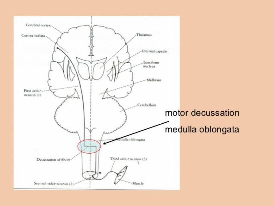
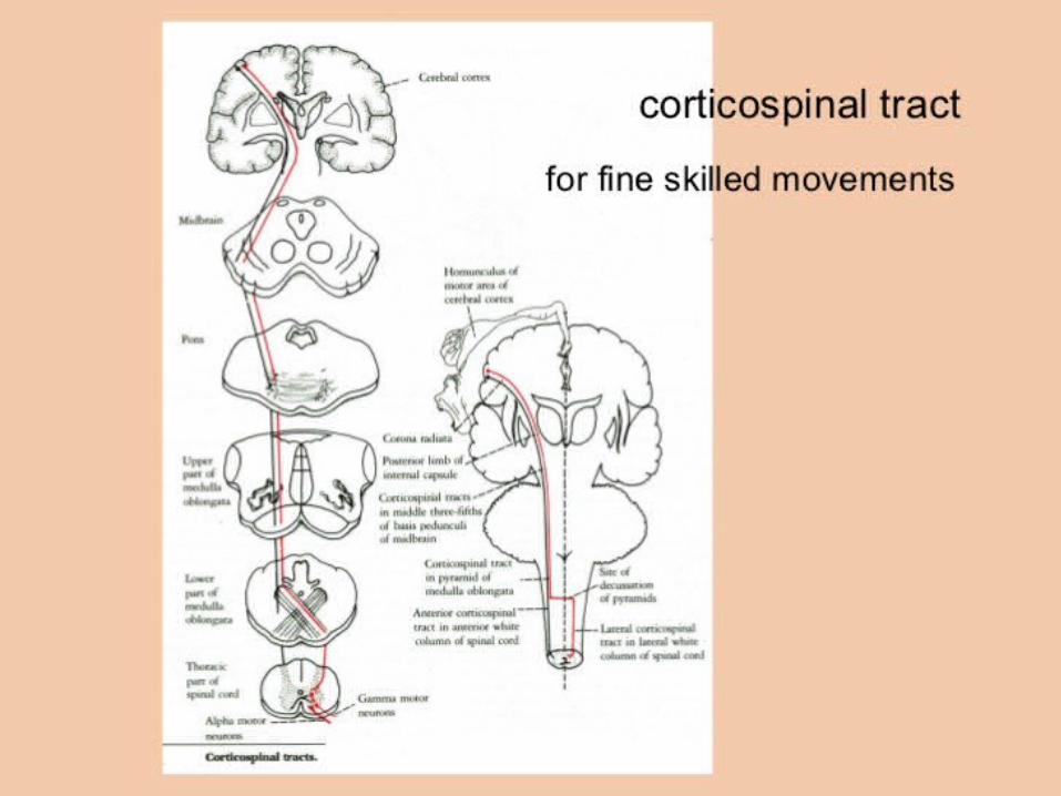
Corticospinal tract fibers travel through :
•Corona radiata•Posterior limb of the internal capsule•Cerebral peduncle (middle 3/5th)•Pons•Medulla oblongata (passes through the pyramids)


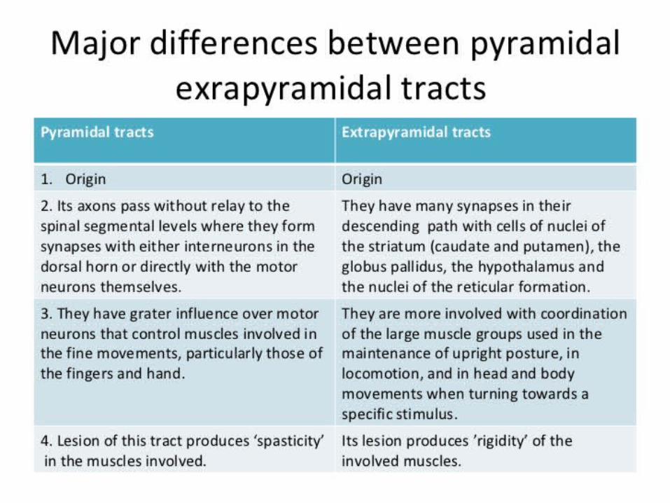


Extrapyramidal Motor Tracts
• Includes all motor pathways not part of the pyramidal system• Upper motor neuron (UMN) originates in nuclei deep in
cerebrum (not in cerebral cortex)• UMN does not pass through the pyramids!• LMN is an anterior horn motor neuron• This system includes
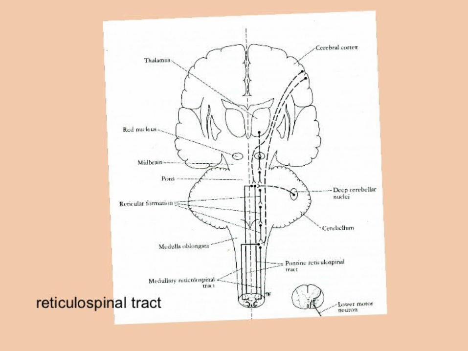
• Rubrospinal• Vestibulospinal• Reticulospinal• Tectospinal tracts
• Regulate:• Axial muscles that maintain balance and posture• Muscles controlling coarse movements of the proximal portions of limbs• Head, neck, and eye movement
Extrapyramidal Tract
Note:1. UMN cell body location2. UMN axon decussates in pons3. Synapse between UMN and LMN occurs in anterior horn of sc3. LMN exits via ventral root4. LMN axon stimulates skeletal muscle
Extrapyramidal (Multineuronal) Pathways
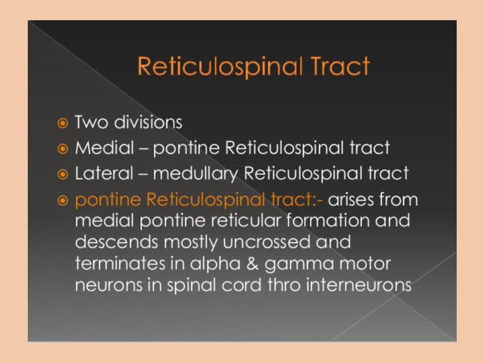
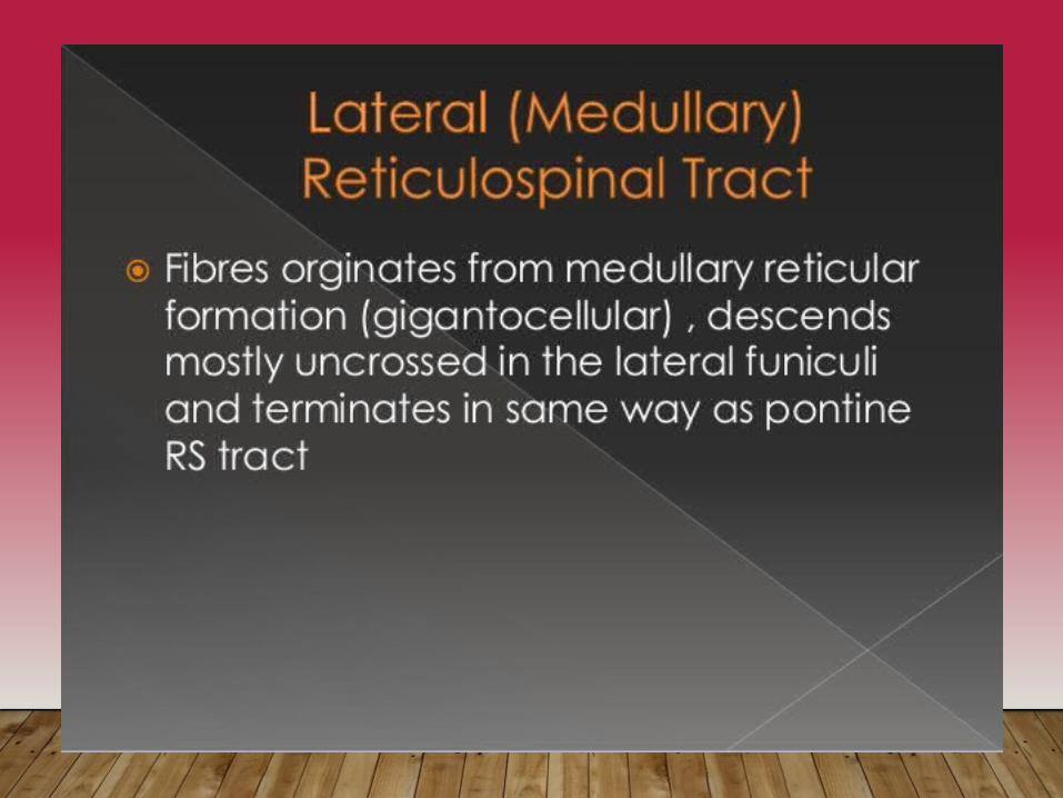
• Reticulospinal tracts – originates at reticular formation of brain; maintain balance
• Rubrospinal tracts – originate in ‘red nucleus’ of midbrain; control flexor muscles
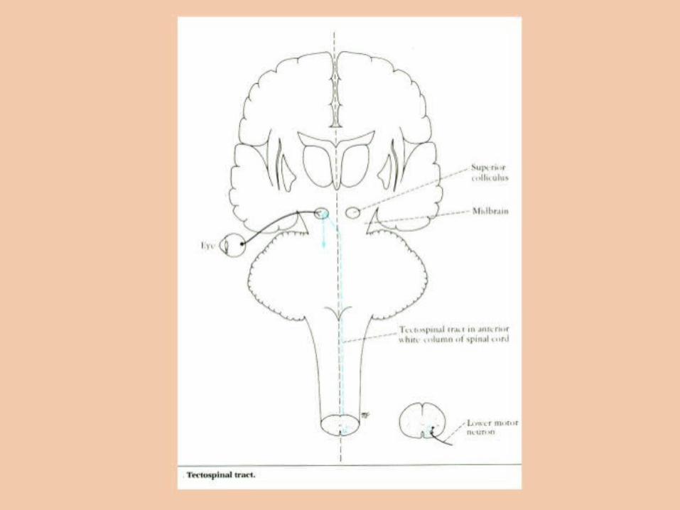
• Tectospinal tracts - originate in superior colliculi and mediate head and eye movements towards visual targets (flash of light)

Spinal Cord Trauma and Disorders
• Severe damage to ventral root results in flaccid paralysis (limp and unresponsive)
• Skeletal muscles cannot move either voluntarily or involuntarily• Without stimulation, muscles atrophy.
• When only UMN of primary motor cortex is damaged• spastic paralysis occurs - muscles affected by persistent spasms and exaggerated tendon reflexes• Muscles remain healthy longer but their movements are no longer subject to voluntary control.• Muscles commonly become permanently shortened.
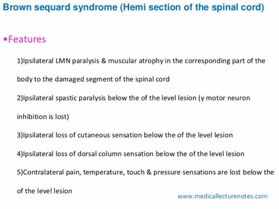
• Transection (cross sectioning) at any level results in total motor and sensory loss in body regions inferior to site of damage.
• If injury in cervical region, all four limbs affected (quadriplegia)• If injury between T1 and L1, only lower limbs affected (paraplegia)

Spinal Cord Trauma and Disorders• Spinal shock - transient period of functional loss that follows the injury
• Results in immediate depression of all reflex activity caudal to lesion.• Bowel and bladder reflexes stop, blood pressure falls, and all muscles (somatic and visceral) below the injury are paralyzed and insensitive.• Neural function usually returns within a few hours following injury• If function does not resume within 48 hrs, paralysis is permanent.
• Amyotrophic Lateral Sclerosis (aka, Lou Gehrig’s disease)• Progressive destruction of anterior horn motor neurons and fibers of the pyramidal tracts• Lose ability to speak, swallow, breathe.• Death within 5 yrs• Cause unknown (90%); others have high glutamate levels
• Poliomyelitis• Virus destroys anterior horn motor neurons• Victims die from paralysis of respiratory muscles• Virus enters body in feces-contaminated water (public swimming pools)

2 Primary SystemsDorsal column-Medial Lemniscal System Corticospinal Tract
Dorsal Column (SC) -Medial Lemniscal (brain stem) System
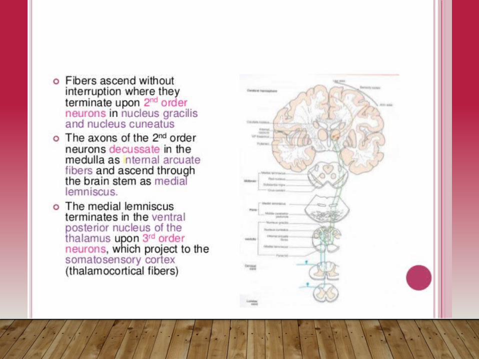
• 1° sensory function – brings info from sensory receptors in the periphery all the way to the 1° somatic sensory cortex.
• Refer to the postcentral gyrus of parietal lobe! • Via the SC, brainstem, and thalamus.• A 3-neuron-circuit (sites of synaptic contact):
Dorsal Column (SC) -Medial Lemniscal (brain
stem) System
i. DRG cells – bipolar (pseudounipolar) neurons receive info from peripheral sensory receptors and bring it to SC and bs, where the info is 1st processed (through dorsal column). *synapse at relay nucleus in medulla: dorsal column nucleus.
ii. Axons of these neurons from the dorsal column nucleus cross over (decussate) here at the medulla and continue as the medial lemniscus thalamus.
iii. These next thalamic neurons send their axons into the internal capsule (white mattter underlying the cortex) synapse at 1° somatic sensory cortex.
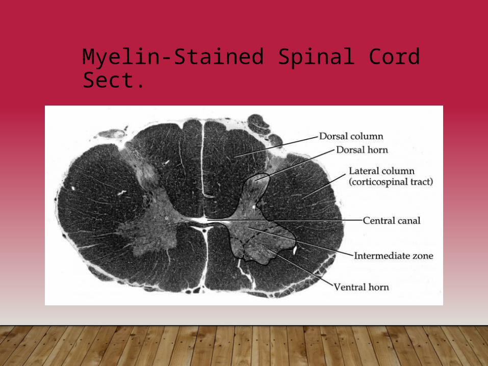
Myelin-Stained Spinal Cord Sect.


