Embed Size (px)
Citation preview
SMALL CELL LUNG CANCER (SCLC)
G. Giaccone
Chief Medical Oncology Branch
National Cancer Institute
Bethesda, Maryland
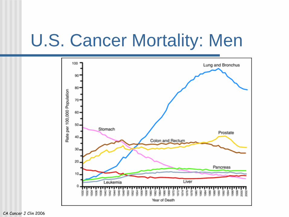
U.S. Cancer Mortality: Men
CA Cancer J Clin 2006
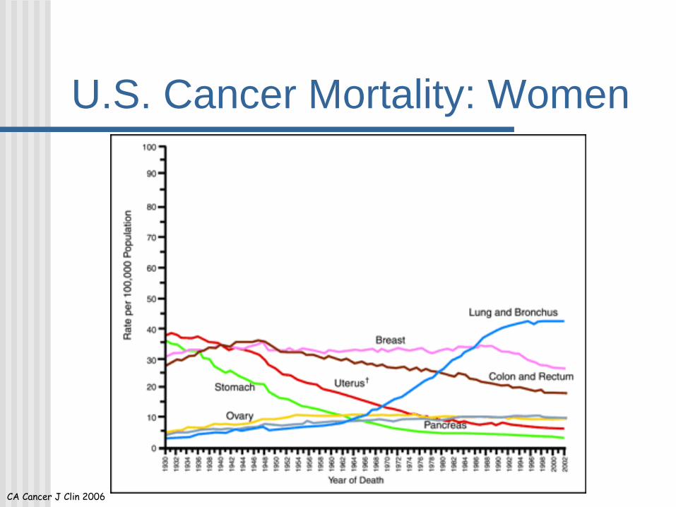
U.S. Cancer Mortality: Women
CA Cancer J Clin 2006
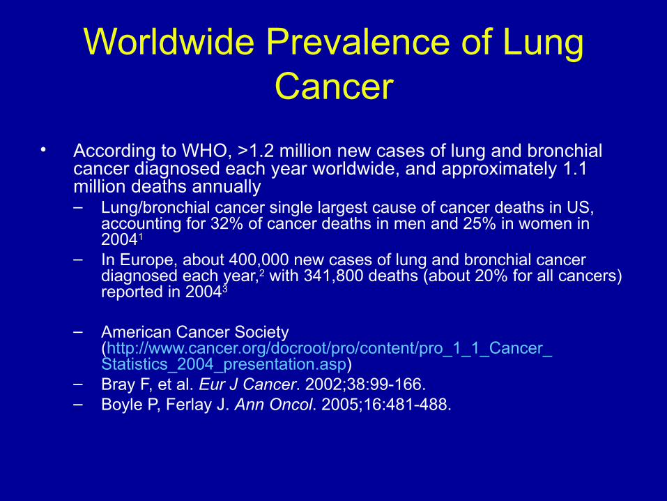
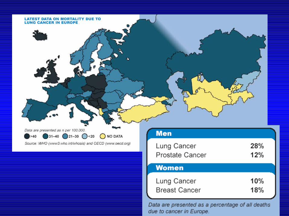
Worldwide Prevalence of Lung Cancer
• According to WHO, >1.2 million new cases of lung and bronchial cancer diagnosed each year worldwide, and approximately 1.1 million deaths annually – Lung/bronchial cancer single largest cause of cancer deaths in US,
accounting for 32% of cancer deaths in men and 25% in women in 20041
– In Europe, about 400,000 new cases of lung and bronchial cancer diagnosed each year,2 with 341,800 deaths (about 20% for all cancers) reported in 20043
– American Cancer Society(http://www.cancer.org/docroot/pro/content/pro_1_1_Cancer_Statistics_2004_presentation.asp)
– Bray F, et al. Eur J Cancer. 2002;38:99-166.– Boyle P, Ferlay J. Ann Oncol. 2005;16:481-488.
Lung Cancer Demographics• Second most frequently diagnosed cancer in the
United States– ~12% of all new diagnoses– ~173,770 individual cases in 2004– Median age at diagnosis is approximately 70 years– Over 1/3 of all diagnoses are made in patients over
75 years of age• Leading cause of cancer deaths in the
United States– ~160,440 patients will die in 2004– 32% and 25% of all cancer deaths in American men and
women, respectively
Jemal et al. CA Cancer J Clin. 2004;54:8.SEER Cancer Statistics Review, 1975-2001. At: http://seer.cancer.gov/csr/1975_2001/. Accessed October 22, 2004.
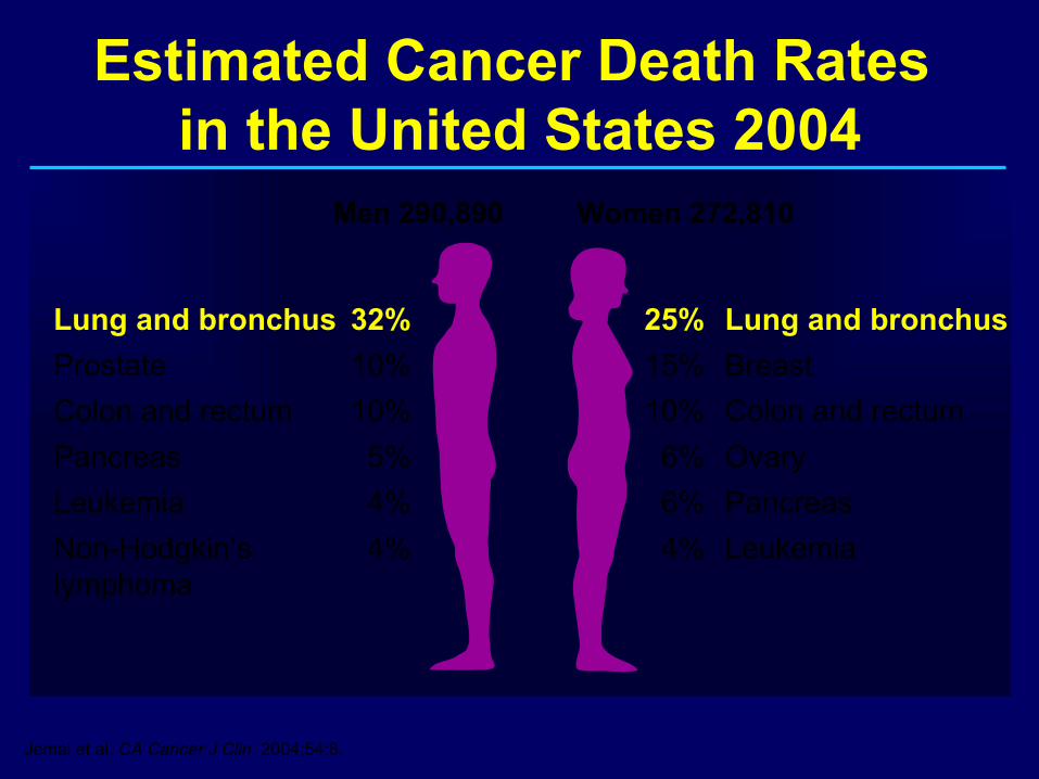
Estimated Cancer Death Rates in the United States 2004
Men 290,890 Women 272,810
25% Lung and bronchus
15% Breast
10% Colon and rectum
6% Ovary
6% Pancreas
4% Leukemia
Lung and bronchus 32%
Prostate 10%
Colon and rectum 10%
Pancreas 5%
Leukemia 4%
Non-Hodgkin’s 4%lymphoma
Jemal et al. CA Cancer J Clin. 2004;54:8.
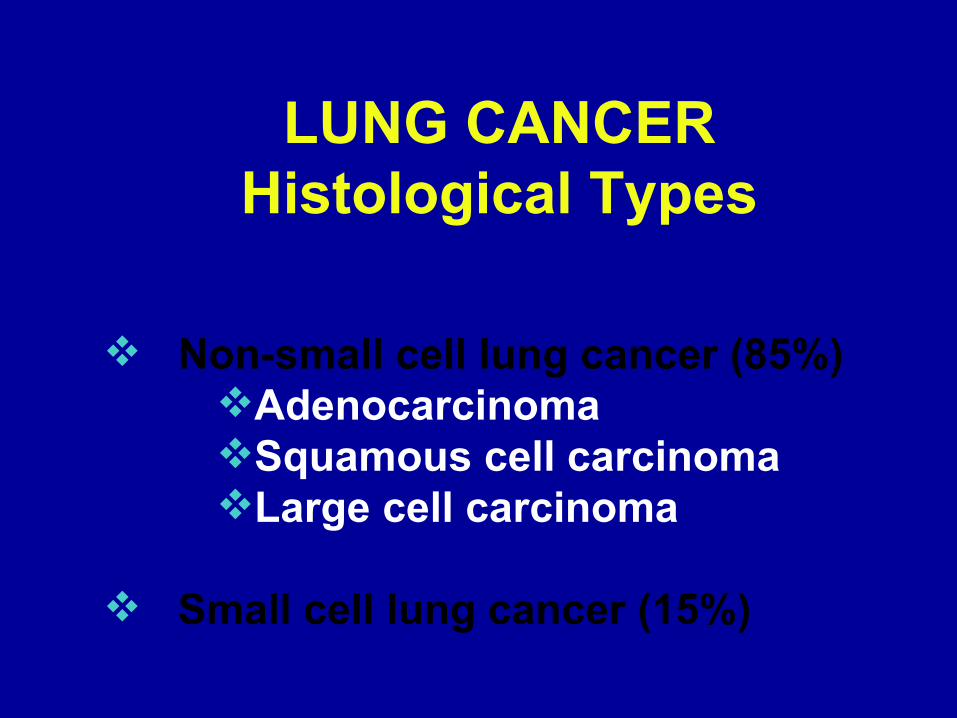
LUNG CANCERHistological Types
Non-small cell lung cancer (85%) AdenocarcinomaSquamous cell carcinomaLarge cell carcinoma
Small cell lung cancer (15%)
SCLC
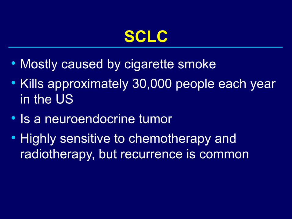
• Mostly caused by cigarette smoke
• Kills approximately 30,000 people each year in the US
• Is a neuroendocrine tumor
• Highly sensitive to chemotherapy and radiotherapy, but recurrence is common
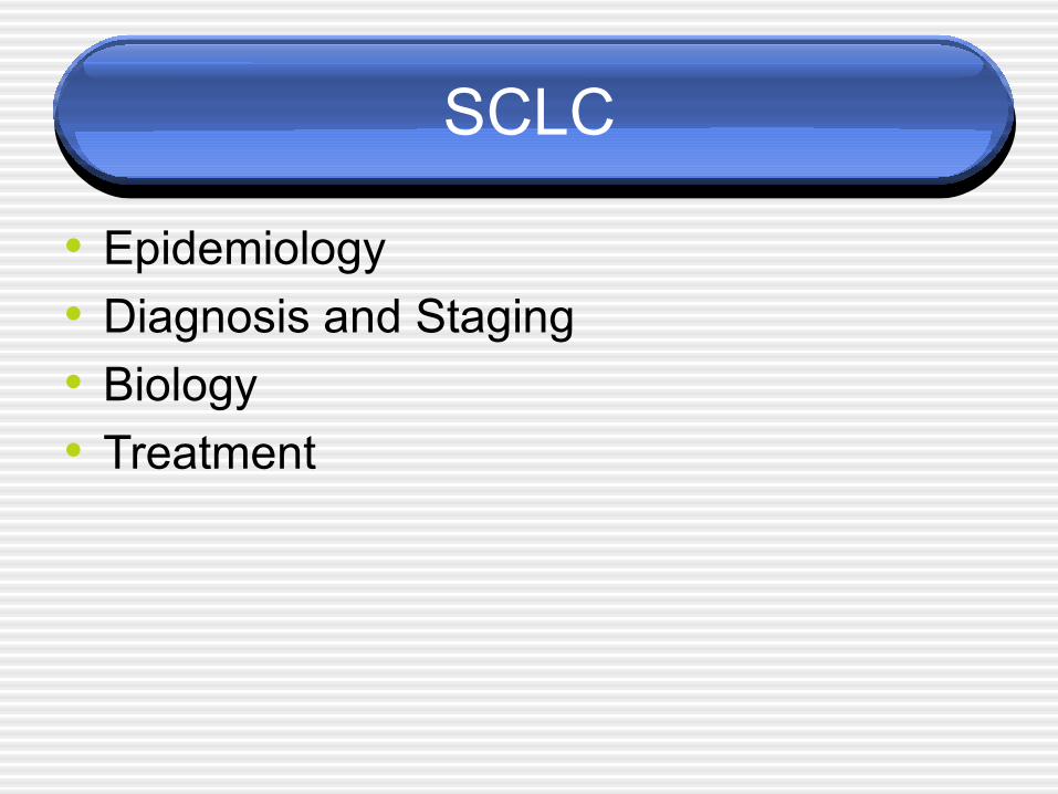
SCLC
• Epidemiology
• Diagnosis and Staging
• Biology
• Treatment
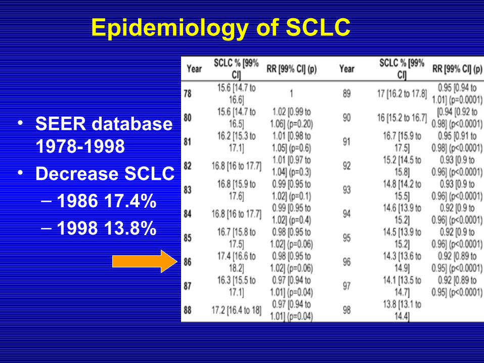
Epidemiology of SCLC
• SEER database 1978-1998
• Decrease SCLC– 1986 17.4%
– 1998 13.8%
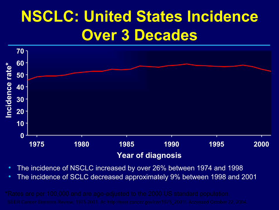
NSCLC: United States Incidence Over 3 Decades
*Rates are per 100,000 and are age-adjusted to the 2000 US standard population.SEER Cancer Statistics Review, 1975-2001. At: http://seer.cancer.gov/csr/1975_2001/. Accessed October 22, 2004.
• The incidence of NSCLC increased by over 26% between 1974 and 1998• The incidence of SCLC decreased approximately 9% between 1998 and 2001
0
10
20
30
40
50
60
70
1975 1980 1985 1990 1995 2000
Year of diagnosis
Inci
de
nc
e ra
te*
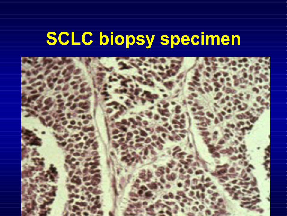
SCLC biopsy specimen
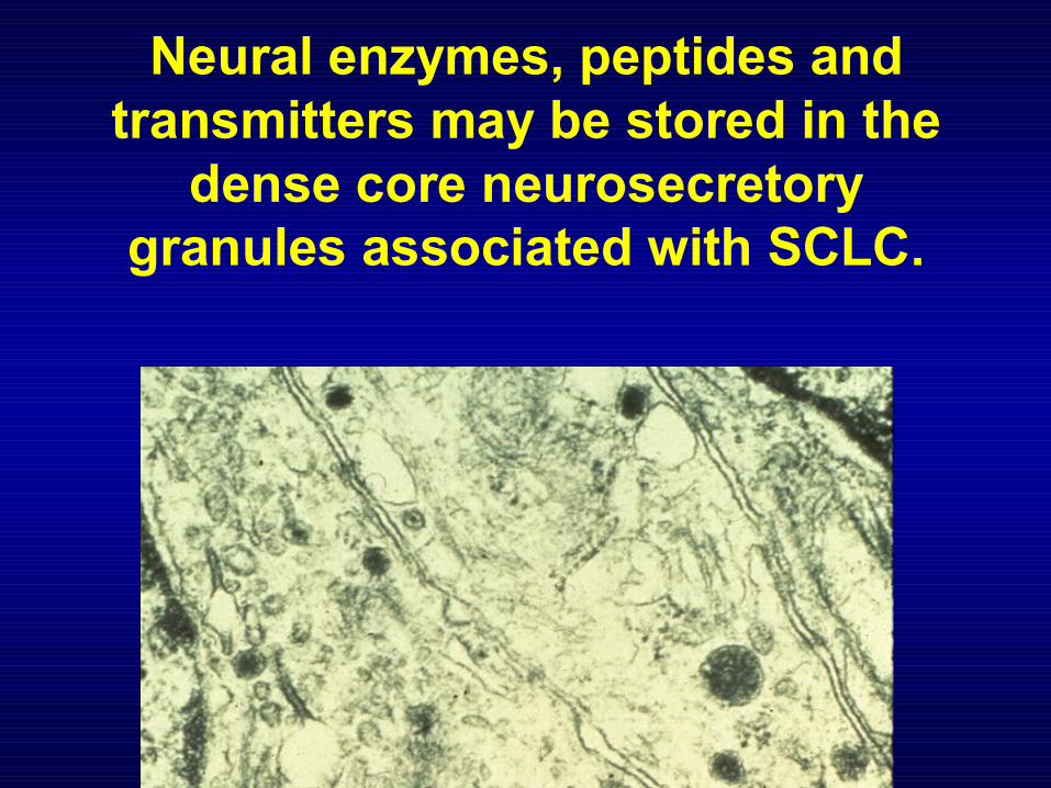
Neural enzymes, peptides and transmitters may be stored in the
dense core neurosecretory granules associated with SCLC.
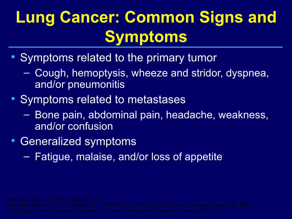
Lung Cancer: Common Signs and Symptoms
• Symptoms related to the primary tumor– Cough, hemoptysis, wheeze and stridor, dyspnea,
and/or pneumonitis• Symptoms related to metastases
– Bone pain, abdominal pain, headache, weakness, and/or confusion
• Generalized symptoms– Fatigue, malaise, and/or loss of appetite
American Society of Clinical Oncology. At: http://asco.org/ac/1,1003,_12-002611-00_18-0026183-00_19-00-00_20-001,00.asp. Accessed October 26, 2004.Ginsberg et al. Non–small cell lung cancer. In: Cancer: Principles & Practice of Oncology. 2001:925.
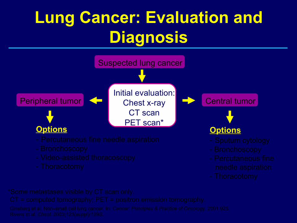
Initial evaluation:Chest x-ray
CT scanPET scan*
Peripheral tumor Central tumor
Options- Percutaneous fine needle aspiration- Bronchoscopy- Video-assisted thoracoscopy- Thoracotomy
Options- Sputum cytology- Bronchoscopy- Percutaneous fine
needle aspiration- Thoracotomy
*Some metastases visible by CT scan only.CT = computed tomography; PET = positron emission tomography.Ginsberg et al. Non–small cell lung cancer. In: Cancer: Principles & Practice of Oncology. 2001:925.Rivera et al. Chest. 2003;123(suppl):129S.
Lung Cancer: Evaluation and Diagnosis
Suspected lung cancer
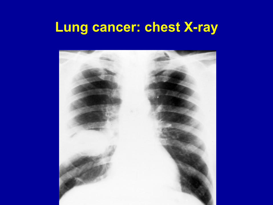
Lung cancer: chest X-ray
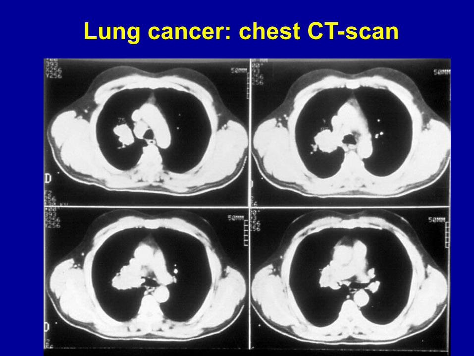
Lung cancer: chest CT-scan
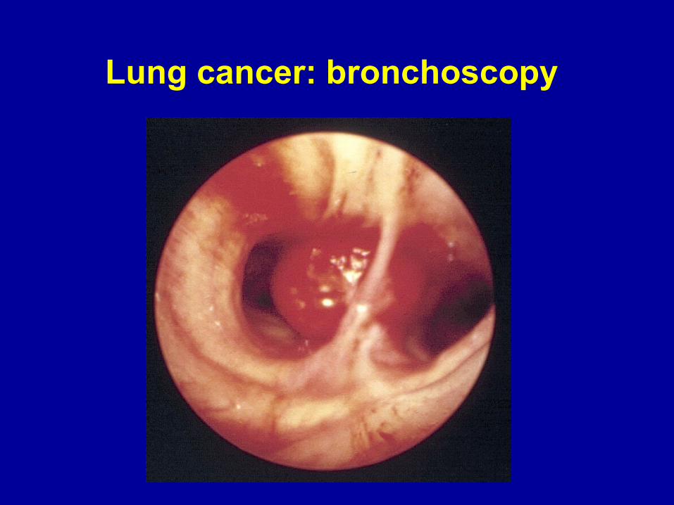
Lung cancer: bronchoscopy
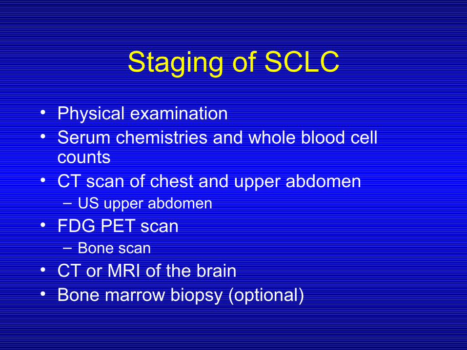
Staging of SCLC
• Physical examination• Serum chemistries and whole blood cell
counts• CT scan of chest and upper abdomen
– US upper abdomen
• FDG PET scan– Bone scan
• CT or MRI of the brain• Bone marrow biopsy (optional)
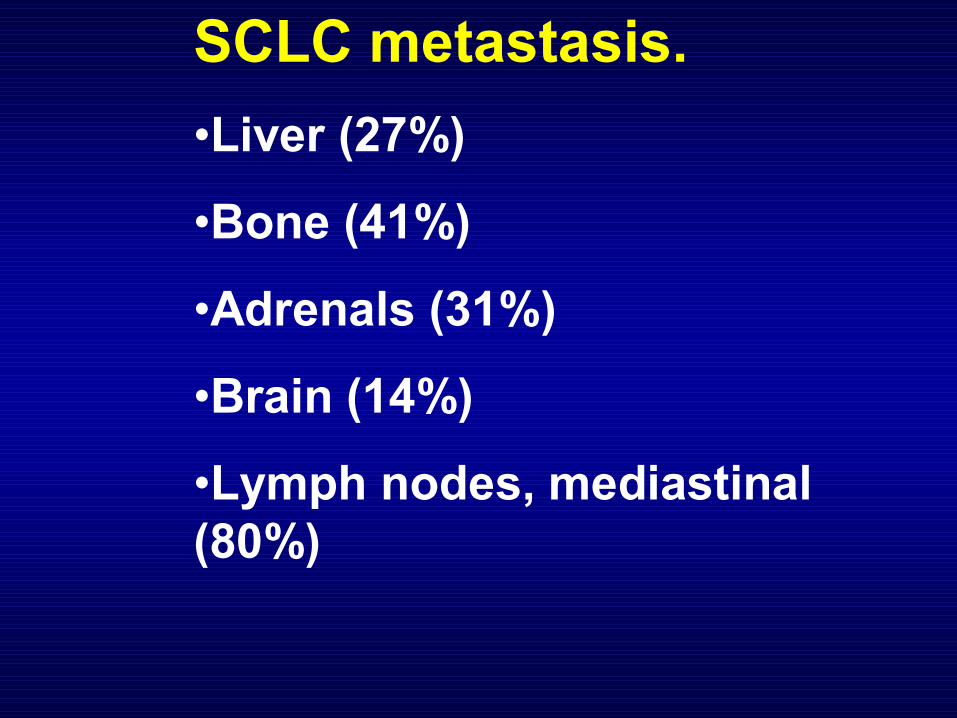
SCLC metastasis.
•Liver (27%)
•Bone (41%)
•Adrenals (31%)
•Brain (14%)
•Lymph nodes, mediastinal(80%)
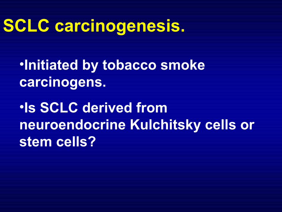
SCLC carcinogenesis.
•Initiated by tobacco smoke carcinogens.
•Is SCLC derived from neuroendocrine Kulchitsky cells or stem cells?
SCLC cell lines.• Bone marrow aspirates were obtained from patients and mononuclear cells collected.• Lymph node aspirates and other solid tumors were mechanically dissociated and cell suspensions obtained by mincing and passing through 60 gauge steel mesh.• The cells were cultured in a serum free medium containing selenium, IGF-I andtransferrin. SCLC cells grew as suspension cultures.
SCLC cell lines.
•Over a 20 year period, NCI established 113 SCLC and 110 NSCLC continuous human cell lines.
• A subset of SCLC is variant SCLC, which has low levels of DDC, BB and NSE.
Phelps et al., J. Cell Bioc. Supp. 24:32(1996).
SCLC molecular abnormalities.
•Allelic loss (3p, 4p, 4q, 5q, 8p, 9p, 10q, 13q, 17p, 22q)•Microsatellite instabilities (35%)• MYC overexpression (30%)•Stem cell factor, c-kit overexpression (30%)•Bombesin/ Gastrin releasing peptide (BB/GRP), GRP receptor, IGF-I receptor
Chromosome losses in SCLC include:
•3p loss is an early event and
•5q, 13q and 17p loss occurs later.
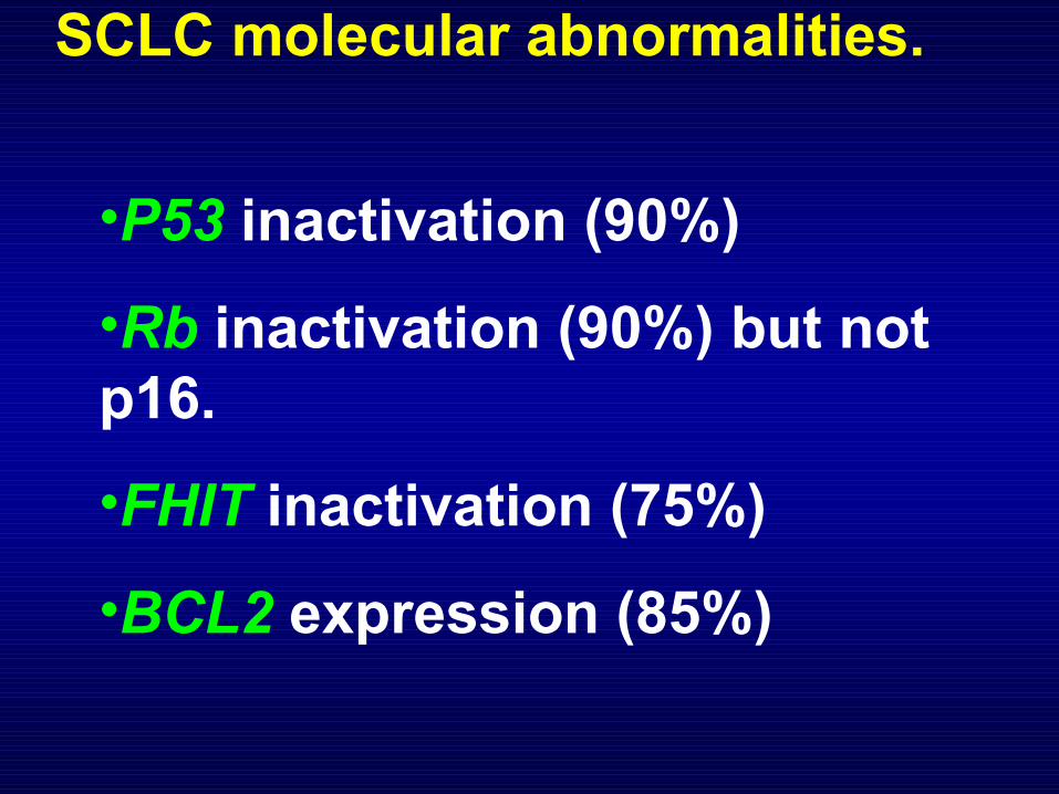
SCLC molecular abnormalities.
•P53 inactivation (90%)
•Rb inactivation (90%) but not p16.
•FHIT inactivation (75%)
•BCL2 expression (85%)
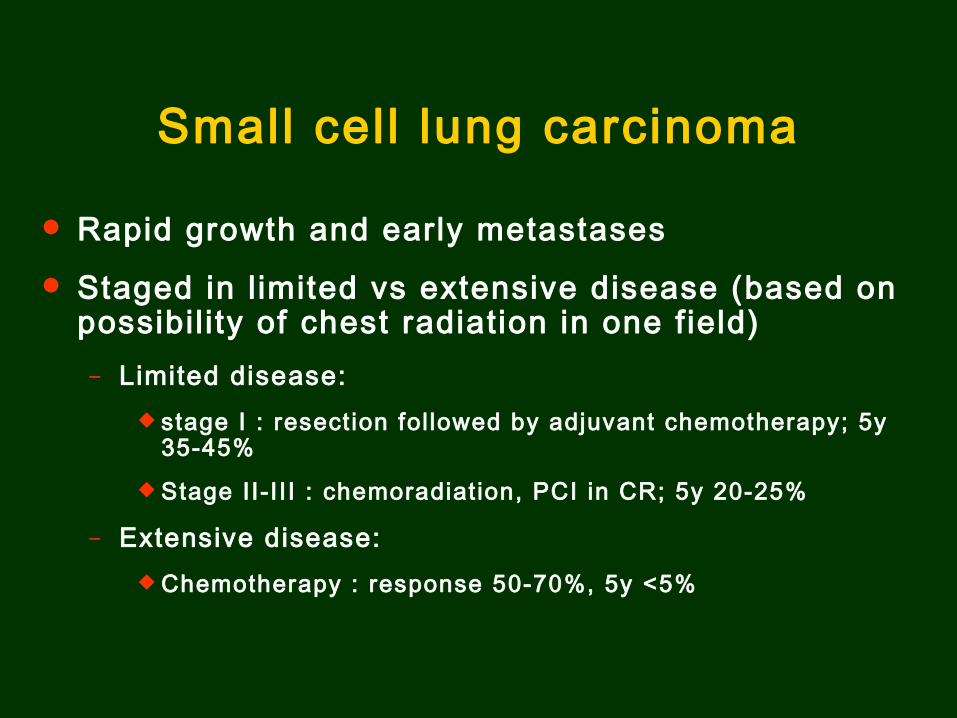
Small cel l lung carcinoma
Rapid growth and early metastases
Staged in l imited vs extensive disease (based on possibil i ty of chest radiation in one f ield)
– Limited disease: stage I : resection fol lowed by adjuvant chemotherapy; 5y
35-45%
Stage II-I I I : chemoradiat ion, PCI in CR; 5y 20-25%
– Extensive disease: Chemotherapy : response 50-70%, 5y <5%
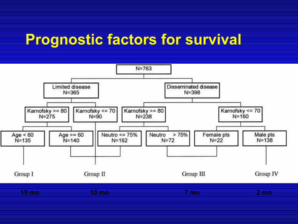
Prognostic factors for survival
19 mo 10 mo 7 mo 2 mo
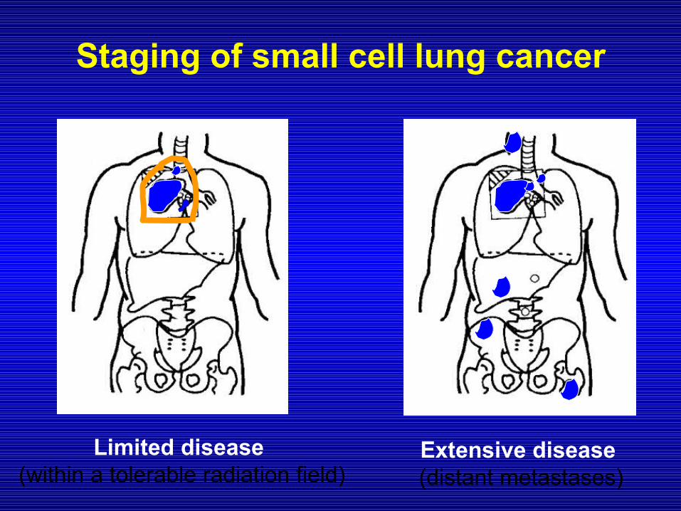
Staging of small cell lung cancer
Limited disease (within a tolerable radiation field)
Extensive disease (distant metastases)
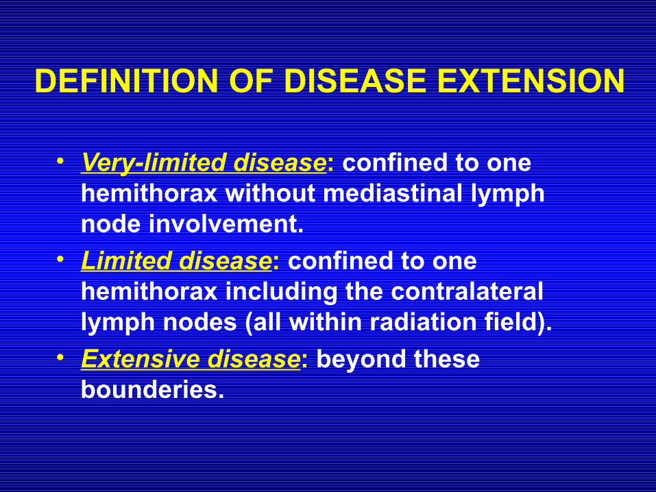
DEFINITION OF DISEASE EXTENSION
• Very-limited disease: confined to one hemithorax without mediastinal lymph node involvement.
• Limited disease: confined to one hemithorax including the contralateral lymph nodes (all within radiation field).
• Extensive disease: beyond these bounderies.
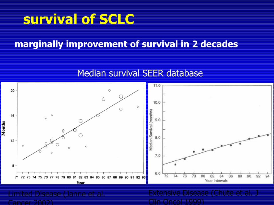
survival of SCLC
marginally improvement of survival in 2 decades
Limited Disease (Janne et al. Cancer 2002)
Median survival SEER database
Extensive Disease (Chute et al. J Clin Oncol 1999)
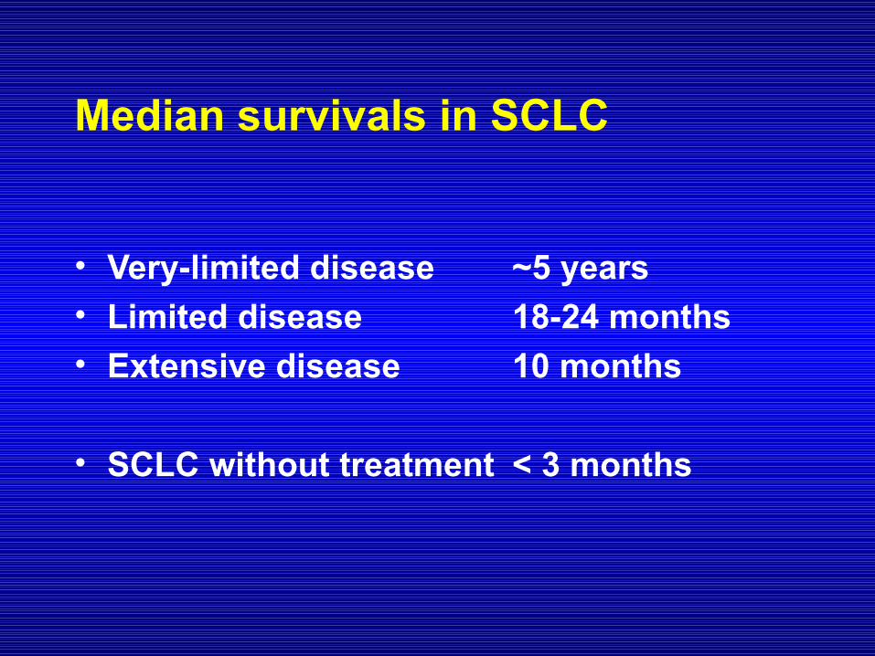
Median survivals in SCLC
• Very-limited disease ~5 years• Limited disease 18-24 months• Extensive disease 10 months
• SCLC without treatment < 3 months
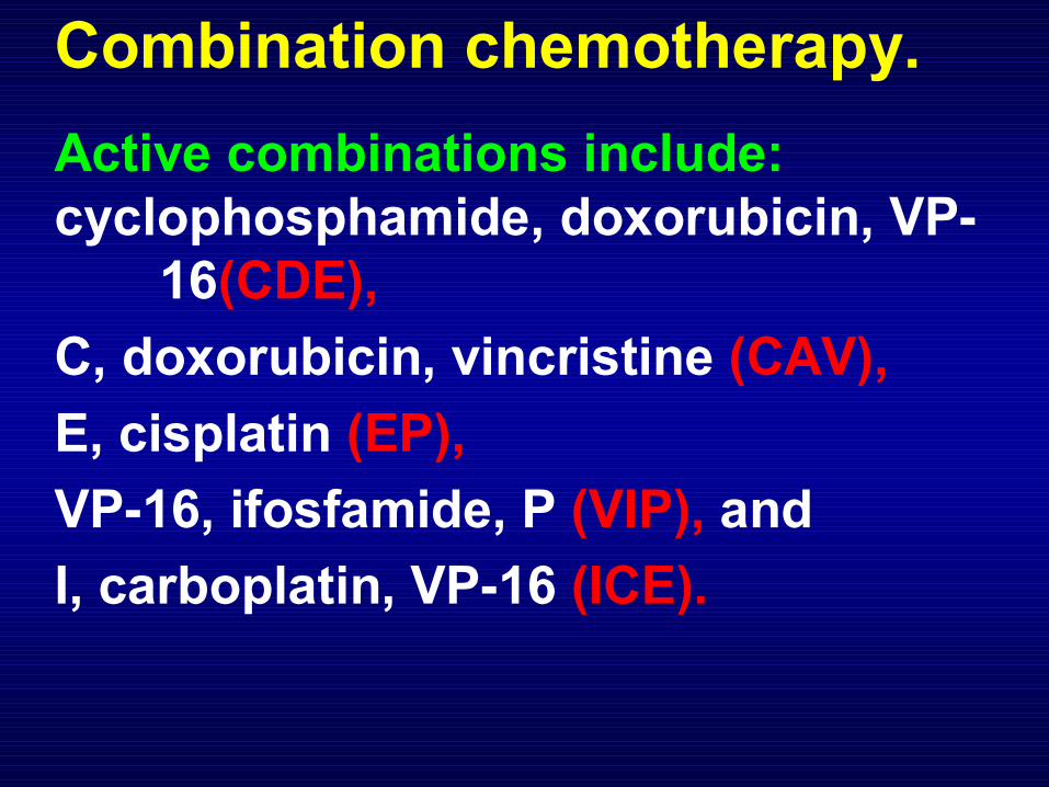
Combination chemotherapy.
Active combinations include:cyclophosphamide, doxorubicin, VP-
16(CDE),
C, doxorubicin, vincristine (CAV),
E, cisplatin (EP),
VP-16, ifosfamide, P (VIP), and
I, carboplatin, VP-16 (ICE).
Approach to very-limited disease
Surgery followed by chemotherapy
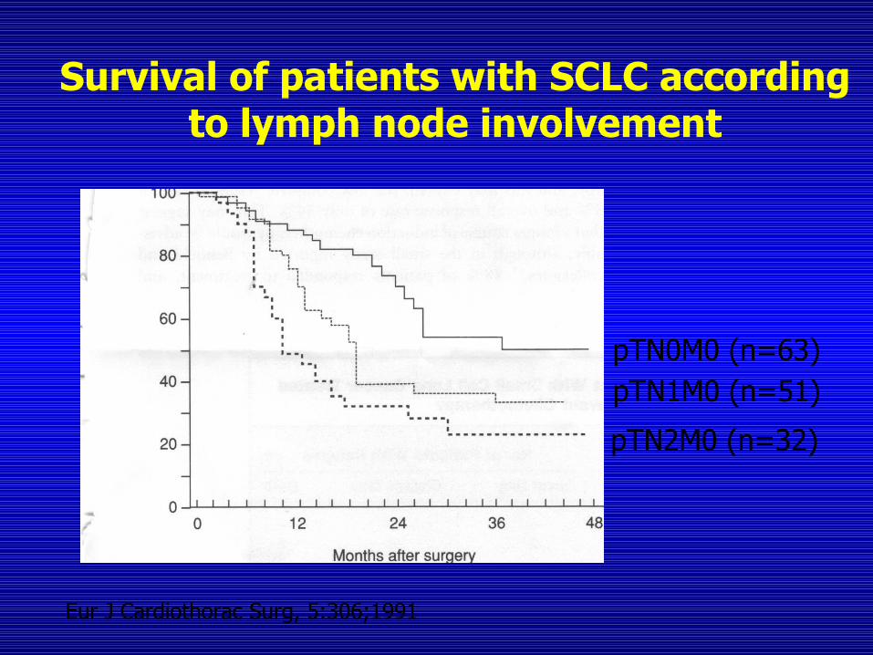
Survival of patients with SCLC according to lymph node involvement
pTN1M0 (n=51)
pTN2M0 (n=32)
Eur J Cardiothorac Surg, 5:306;1991
pTN0M0 (n=63)
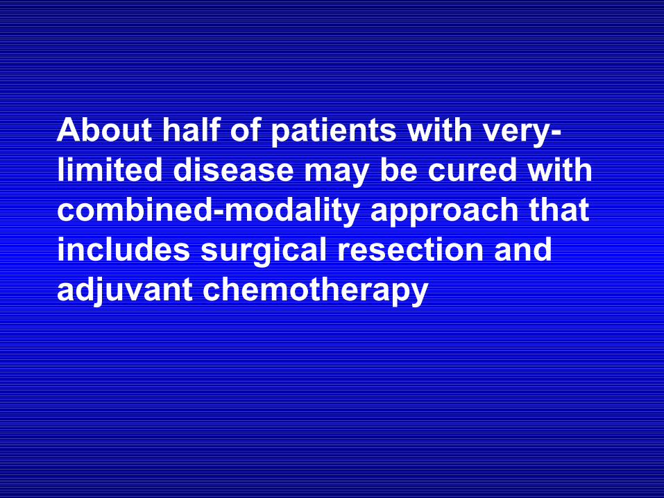
About half of patients with very-limited disease may be cured with combined-modality approach that includes surgical resection and adjuvant chemotherapy
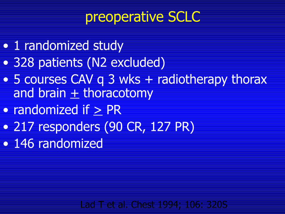
preoperative SCLC
• 1 randomized study• 328 patients (N2 excluded)• 5 courses CAV q 3 wks + radiotherapy thorax
and brain + thoracotomy• randomized if > PR• 217 responders (90 CR, 127 PR)• 146 randomized
Lad T et al. Chest 1994; 106: 320S
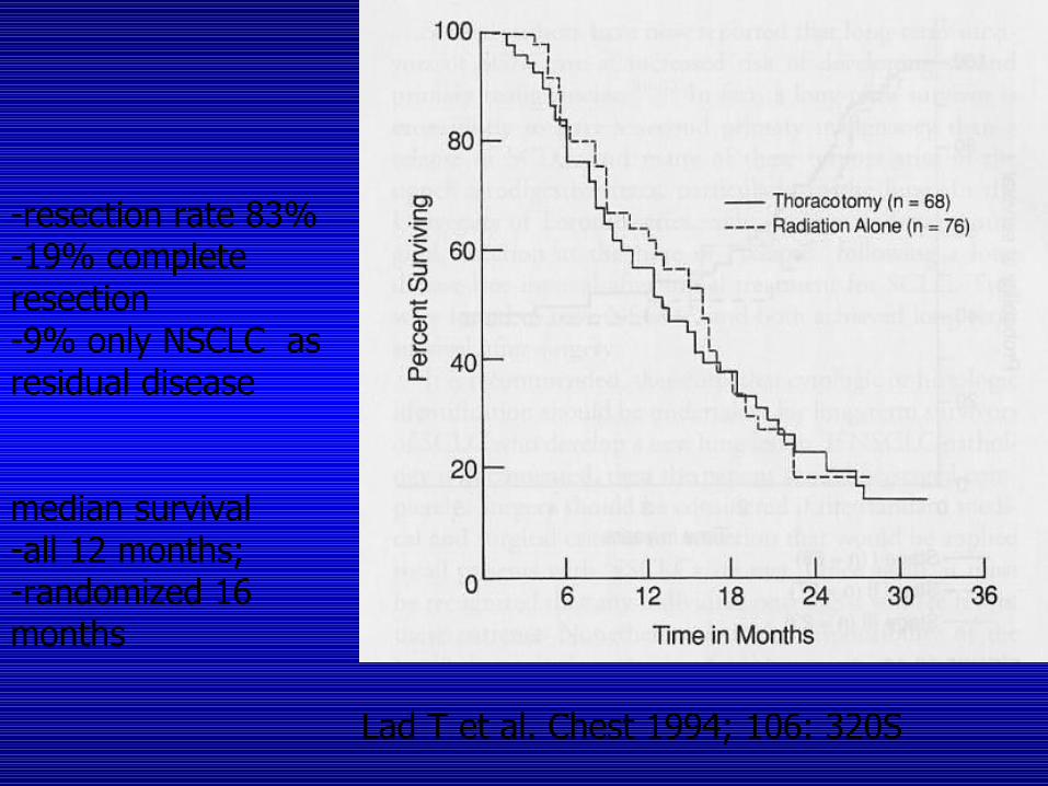
-resection rate 83%-19% complete resection -9% only NSCLC as residual disease
median survival-all 12 months; -randomized 16 months
Lad T et al. Chest 1994; 106: 320S

Approach to limited disease
Limited Disease - SCLC
• treatment has a small but definitively curative intent ( 5y survival: 10 – 25 % )
• combination chemotherapy is the backbone of treat-ment
• thoracic radiotherapy significantly improves long term survival
• early thoracic radiotherapy gives better results than late radiotherapy
limited disease - SCLC
• cisplatin and etoposide are most easily combined within concurrent chemoradiation protocols (Turrisi et al )
• BID radiotherapy gives better local control and better long term survival than QD (5y survival %: 26% Turrisi et al, NEJM 99 )
• PCI significantly improves survival by 4-5 % at 5 years when given to complete responders (Auperin et al )
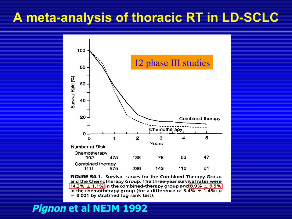
A meta-analysis of thoracic RT in LD-SCLC
12 phase III studies
Pignon et al NEJM 1992
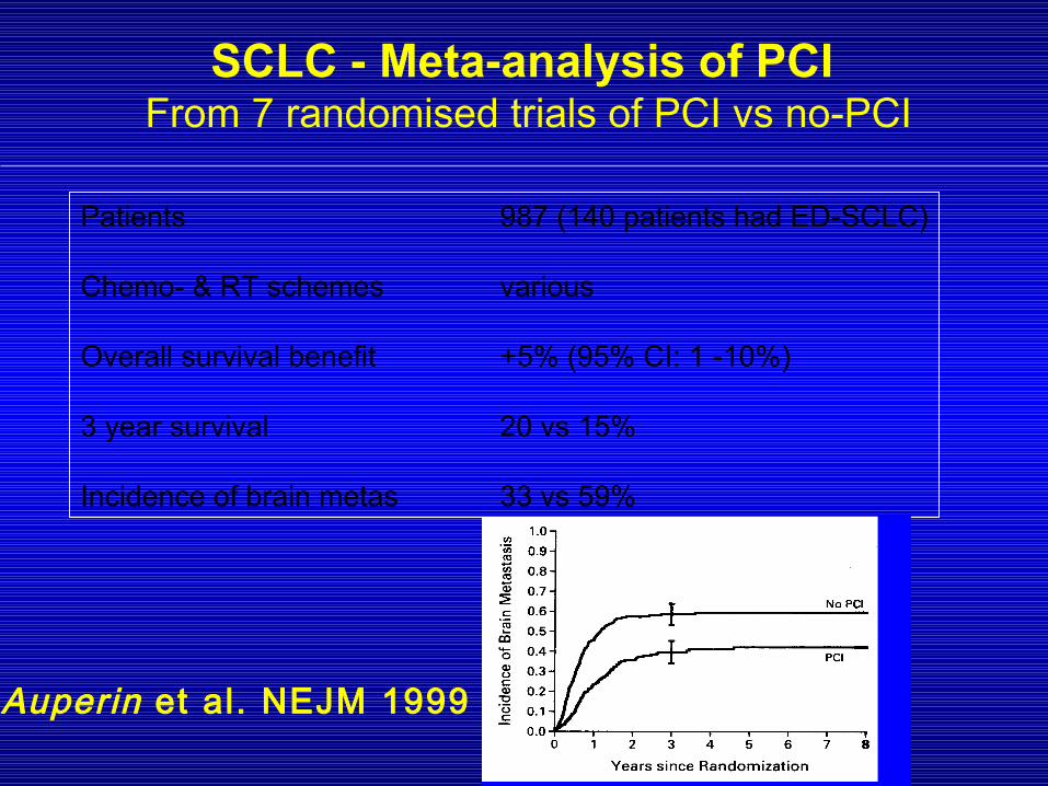
SCLC - Meta-analysis of PCI From 7 randomised trials of PCI vs no-PCI
Patients 987 (140 patients had ED-SCLC)
Chemo- & RT schemes various
Overall survival benefit +5% (95% CI: 1 -10%)
3 year survival 20 vs 15%
Incidence of brain metas 33 vs 59%
Auperin et al. NEJM 1999
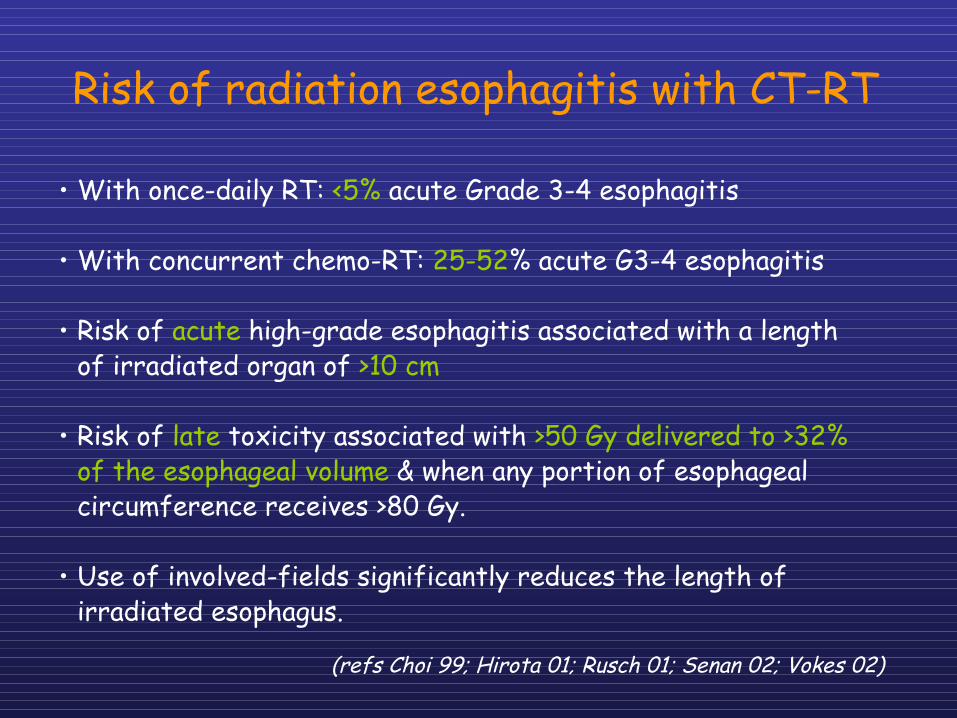
• With once-daily RT: <5% acute Grade 3-4 esophagitis
• With concurrent chemo-RT: 25-52% acute G3-4 esophagitis
• Risk of acute high-grade esophagitis associated with a length of irradiated organ of >10 cm
• Risk of late toxicity associated with >50 Gy delivered to >32% of the esophageal volume & when any portion of esophageal circumference receives >80 Gy.
• Use of involved-fields significantly reduces the length of irradiated esophagus.
Risk of radiation esophagitis with CT-RT
(refs Choi 99; Hirota 01; Rusch 01; Senan 02; Vokes 02)
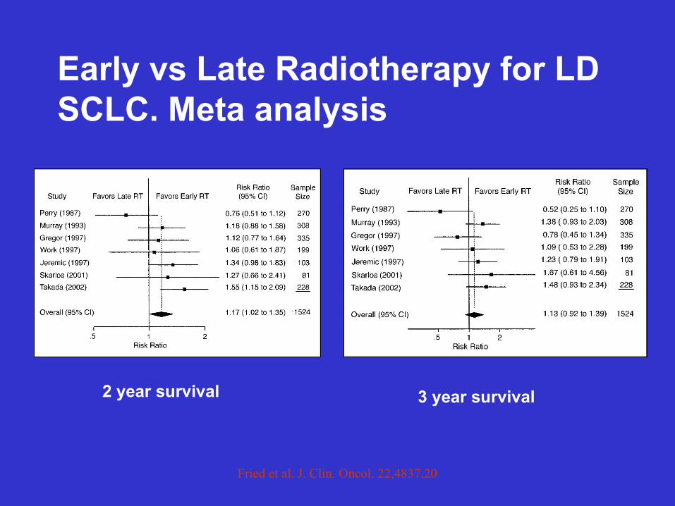
Fried et al. J. Clin. Oncol. 22,4837,2004
Early vs Late Radiotherapy for LD SCLC. Meta analysis
2 year survival 3 year survival
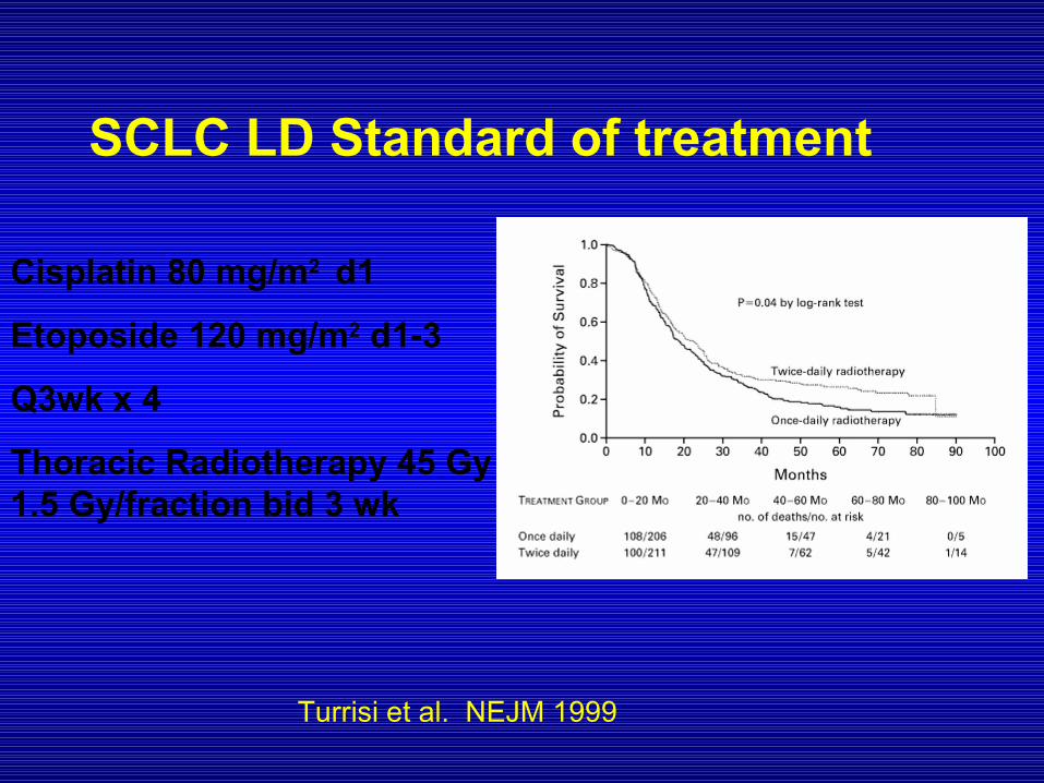
SCLC LD Standard of treatment
Cisplatin 80 mg/m2 d1
Etoposide 120 mg/m2 d1-3
Q3wk x 4
Thoracic Radiotherapy 45 Gy 1.5 Gy/fraction bid 3 wk
Turrisi et al. NEJM 1999

Approach to SCLC ED
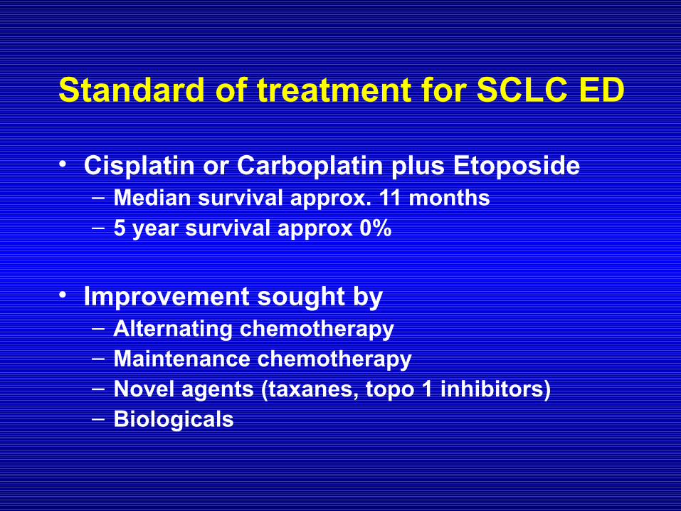
Standard of treatment for SCLC ED
• Cisplatin or Carboplatin plus Etoposide – Median survival approx. 11 months– 5 year survival approx 0%
• Improvement sought by– Alternating chemotherapy– Maintenance chemotherapy– Novel agents (taxanes, topo 1 inhibitors)– Biologicals
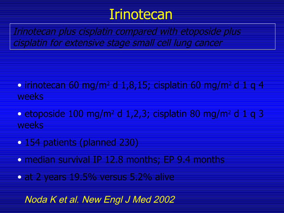
Irinotecan plus cisplatin compared with etoposide plus cisplatin for extensive stage small cell lung cancer
• irinotecan 60 mg/m2 d 1,8,15; cisplatin 60 mg/m2 d 1 q 4 weeks
• etoposide 100 mg/m2 d 1,2,3; cisplatin 80 mg/m2 d 1 q 3 weeks
• 154 patients (planned 230)
• median survival IP 12.8 months; EP 9.4 months
• at 2 years 19.5% versus 5.2% alive
Irinotecan
Noda K et al. New Engl J Med 2002
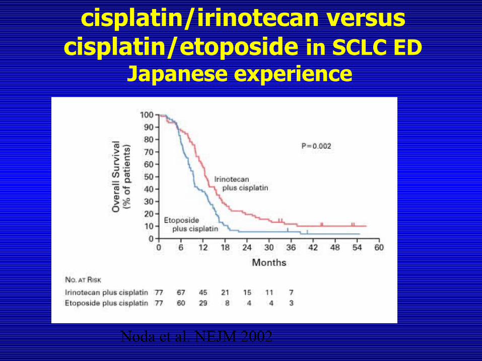
cisplatin/irinotecan versus cisplatin/etoposide in SCLC ED
Japanese experience
Noda et al. NEJM 2002
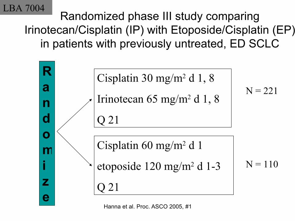
Hanna et al. Proc. ASCO 2005, #1094
Randomized phase III study comparingIrinotecan/Cisplatin (IP) with Etoposide/Cisplatin (EP)
in patients with previously untreated, ED SCLC
Randomize
Cisplatin 30 mg/m2 d 1, 8
Irinotecan 65 mg/m2 d 1, 8
Q 21
Cisplatin 60 mg/m2 d 1
etoposide 120 mg/m2 d 1-3
Q 21
LBA 7004
N = 221
N = 110
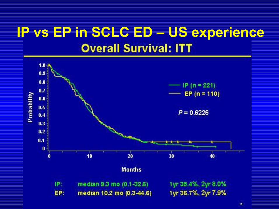
IP vs EP in SCLC ED – US experience
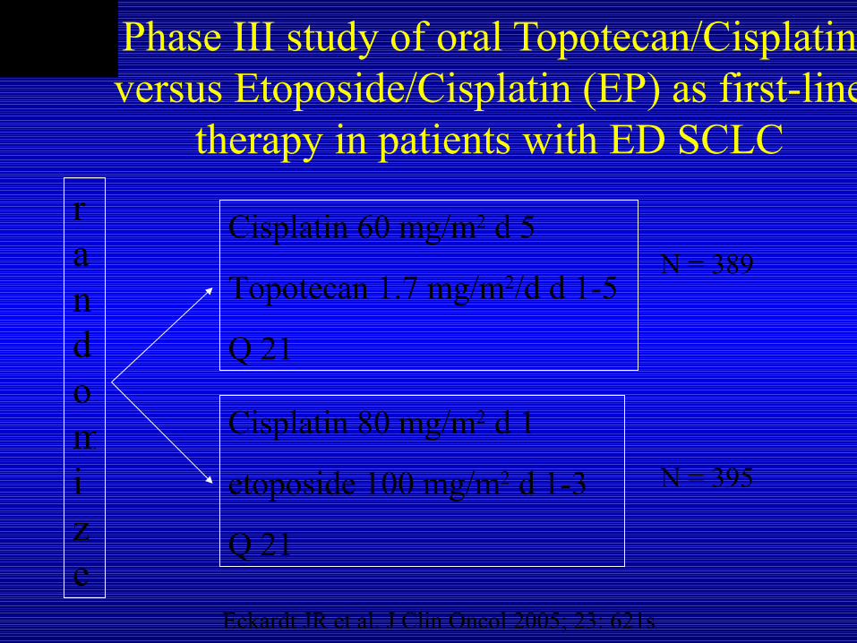
Phase III study of oral Topotecan/Cisplatin versus Etoposide/Cisplatin (EP) as first-line
therapy in patients with ED SCLC
randomize
Cisplatin 60 mg/m2 d 5
Topotecan 1.7 mg/m2/d d 1-5
Q 21
Cisplatin 80 mg/m2 d 1
etoposide 100 mg/m2 d 1-3
Q 21
abstract 7003
Eckardt JR et al. J Clin Oncol 2005; 23: 621s
N = 389
N = 395

Eckardt JR et al. J Clin Oncol 2005; 23: 621s
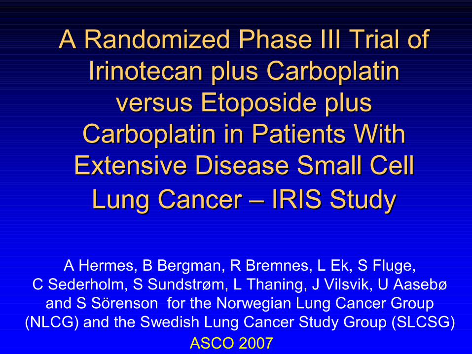
A A RandomizedRandomized Phase III Trial of Phase III Trial of IrinotecanIrinotecan plus plus CarboplatinCarboplatin
versusversus EtoposideEtoposide plus plus CarboplatinCarboplatin in Patients With in Patients With
Extensive Disease Small Cell Extensive Disease Small Cell Lung Cancer Lung Cancer –– IRIS IRIS StudyStudy
A Hermes, B Bergman, R Bremnes, L Ek, S Fluge, C Sederholm, S Sundstrøm, L Thaning, J Vilsvik, U Aasebø
and S Sörenson for the Norwegian Lung Cancer Group (NLCG) and the Swedish Lung Cancer Study Group (SLCSG)
ASCO 2007
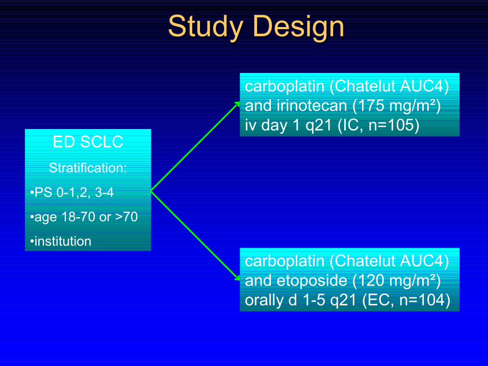
StudyStudy Design Design
ED SCLC
Stratification:
•PS 0-1,2, 3-4
•age 18-70 or >70
•institution
carboplatin (Chatelut AUC4) and irinotecan (175 mg/m²) iv day 1 q21 (IC, n=105)
carboplatin (Chatelut AUC4) and etoposide (120 mg/m²) orally d 1-5 q21 (EC, n=104)
Results Results
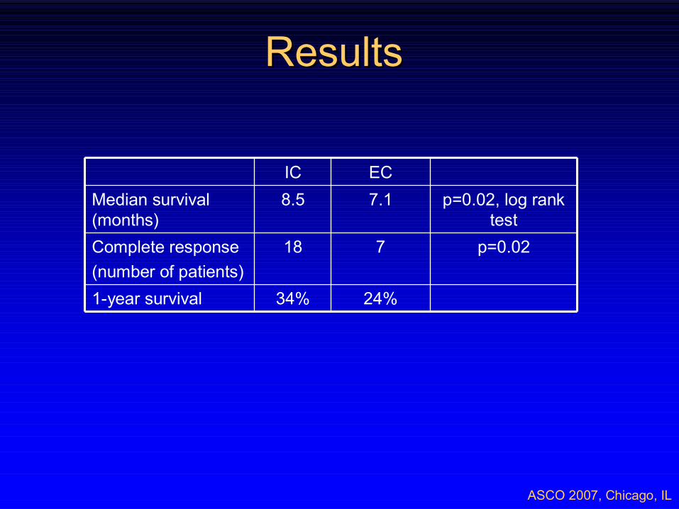
ASCO 2007, Chicago, IL
24%34%1-year survival
p=0.02718 Complete response
(number of patients)
p=0.02, log rank test
7.18.5Median survival (months)
ECIC
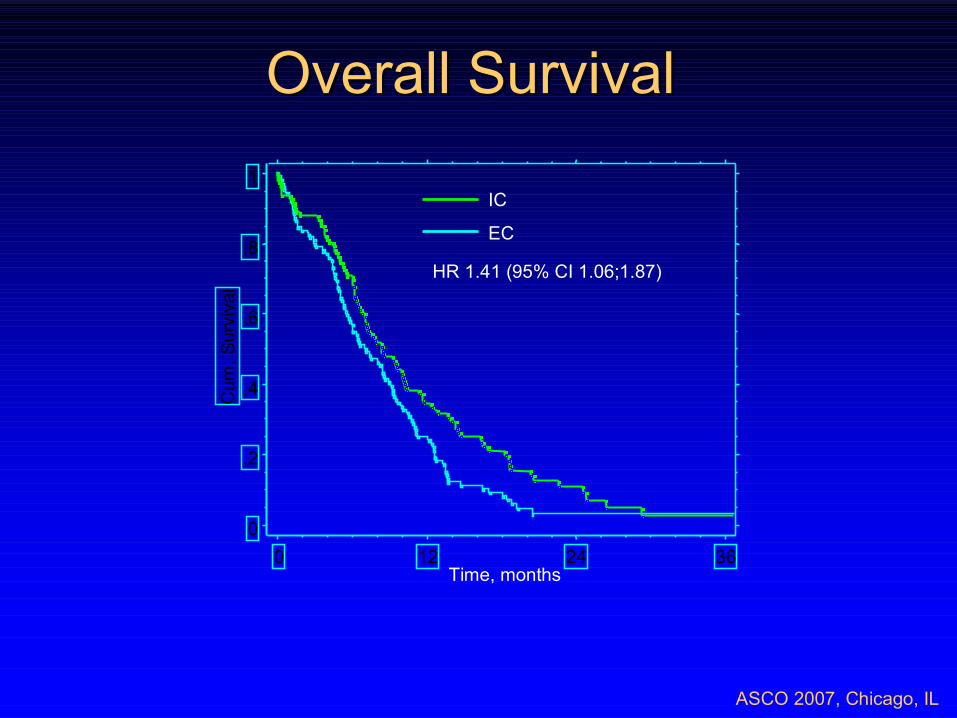
Overall Survival Overall Survival
ASCO 2007, Chicago, IL
IC
EC
HR 1.41 (95% CI 1.06;1.87)
Time, months
0
,2
,4
,6
,8
1
Cum
. S
urv
iva
l
0 12 24 36
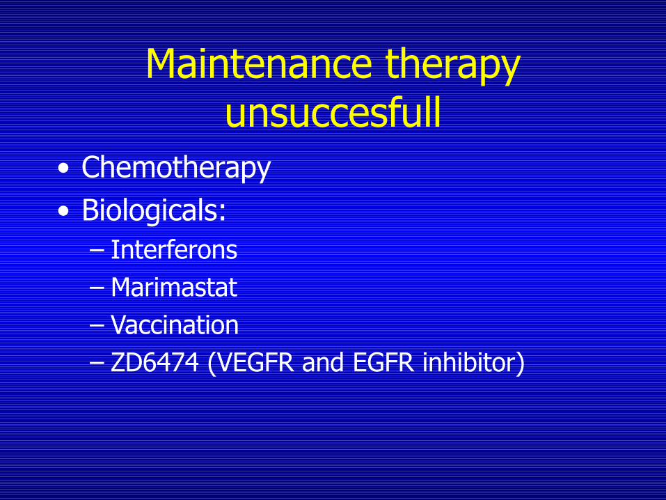
Maintenance therapyunsuccesfull
• Chemotherapy• Biologicals:
– Interferons– Marimastat– Vaccination – ZD6474 (VEGFR and EGFR inhibitor)
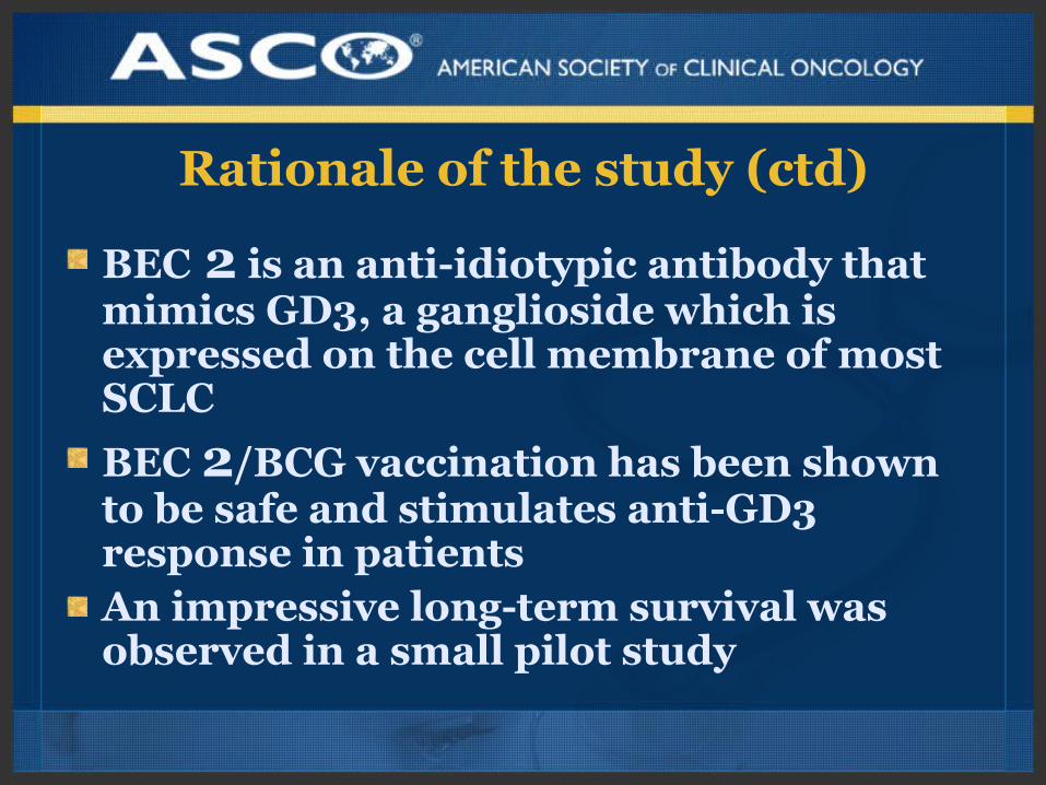
Rationale of the study (ctd)
BEC 2 is an anti-idiotypic antibody that mimics GD3, a ganglioside which is expressed on the cell membrane of most SCLC
BEC 2/BCG vaccination has been shown to be safe and stimulates anti-GD3 response in patientsAn impressive long-term survival was observed in a small pilot study
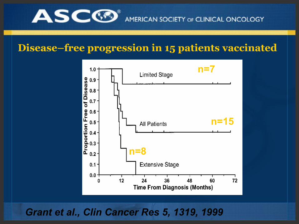
n=8
n=7
n=15
Disease–free progression in 15 patients vaccinated
Grant et al., Clin Cancer Res 5, 1319, 1999
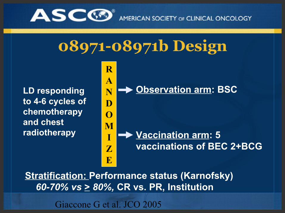
08971-08971b DesignRANDOMIZE
Observation arm: BSC
Vaccination arm: 5 vaccinations of BEC 2+BCG
Stratification: Performance status (Karnofsky) 60-70% vs > 80%, CR vs. PR, Institution
LD responding to 4-6 cycles of chemotherapy and chest radiotherapy
Giaccone G et al. JCO 2005
(months)
0 7 14 21 28 35 42 49 56 63 70
0
10
20
30
40
50
60
70
80
90
100
O N Number of patients at risk : Treatment
180 258 196 148 93 55 33 19 8 4 1
190 257 191 129 86 56 29 18 6 4 0
Observation
Vaccination
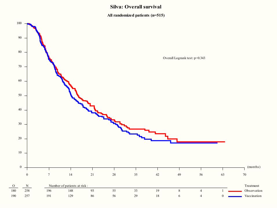
Silva: Overall survival
All randomized patients (n=515)
Overall Logrank test: p=0.343
(months)
0 7 14 21 28 35 42 49 56 63 70
0
10
20
30
40
50
60
70
80
90
100
O N Number of patients at risk : Treatment
196 258 120 82 54 39 28 17 8 4 1
205 257 110 77 59 35 20 14 6 4 0
Observation
Vaccination
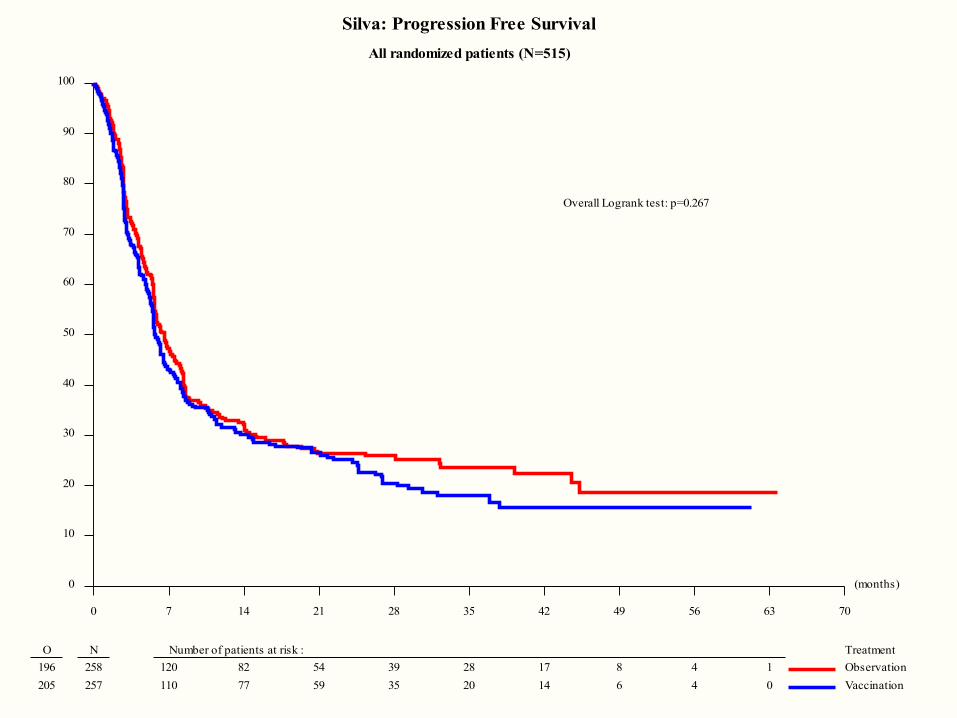
Silva: Progression Free Survival
All randomized patients (N=515)
Overall Logrank test: p=0.267
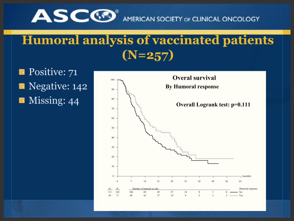
Humoral analysis of vaccinated patients (N=257)
Positive: 71
Negative: 142
Missing: 44
(months)
0 7 14 21 28 35 42 49 56 63
0
10
20
30
40
50
60
70
80
90
100
O N Number of patients at risk : Humoral response111 142 106 69 45 27 14 8 1 0
49 71 60 42 27 19 9 5 3 2
No
Yes
Overal survivalBy Humoral response
Overall Logrank test: p=0.111
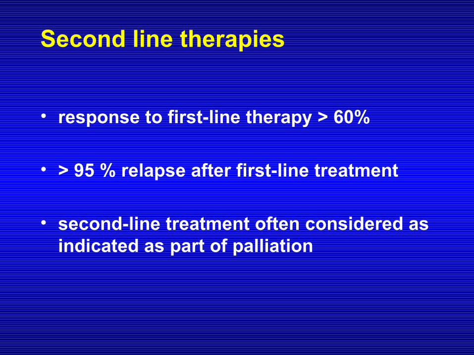
Second line therapies
• response to first-line therapy > 60%
• > 95 % relapse after first-line treatment
• second-line treatment often considered as indicated as part of palliation
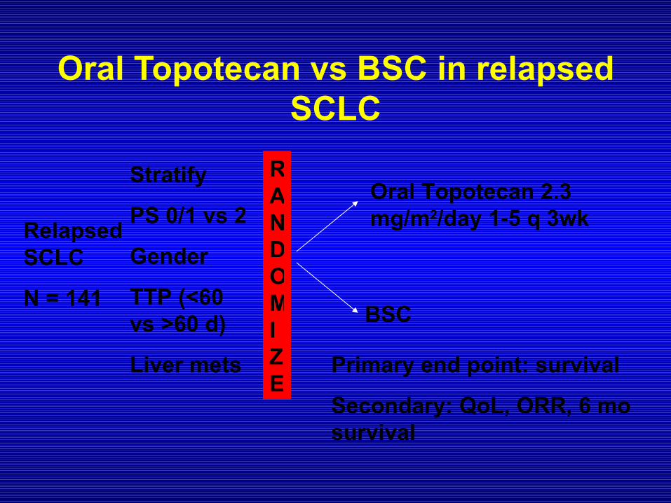
Oral Topotecan vs BSC in relapsed SCLC
Relapsed SCLC
N = 141
Stratify
PS 0/1 vs 2
Gender
TTP (<60 vs >60 d)
Liver mets
RANDOMIZE
Oral Topotecan 2.3 mg/m2/day 1-5 q 3wk
BSC
Primary end point: survival
Secondary: QoL, ORR, 6 mo survival
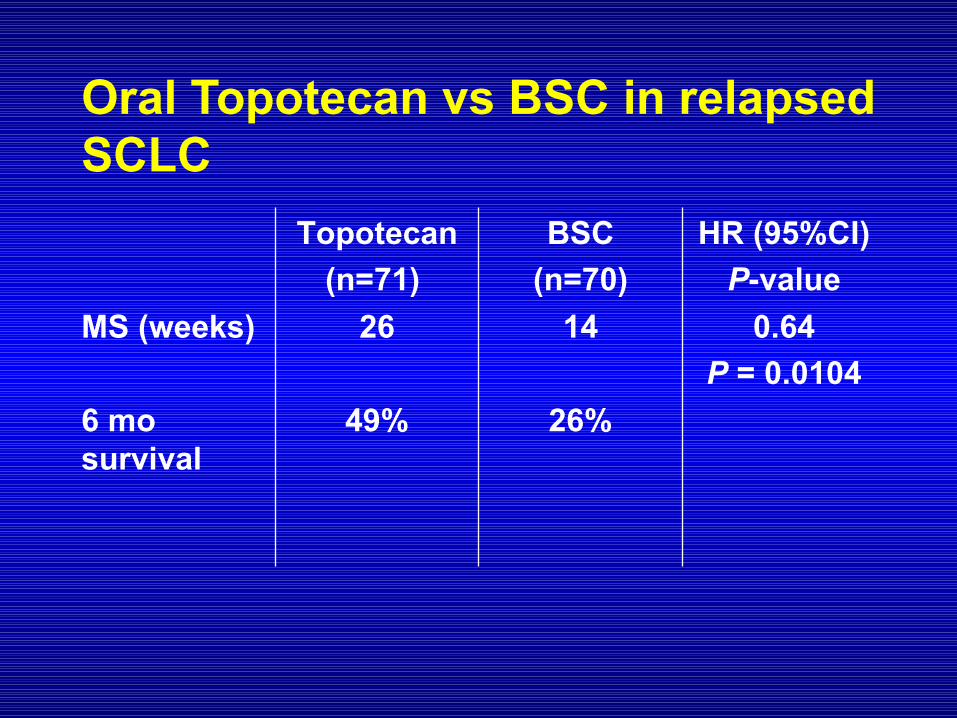
Oral Topotecan vs BSC in relapsed SCLC
26%49%6 mo survival
0.64
P = 0.0104
1426MS (weeks)
HR (95%CI)
P-value
BSC
(n=70)
Topotecan
(n=71)
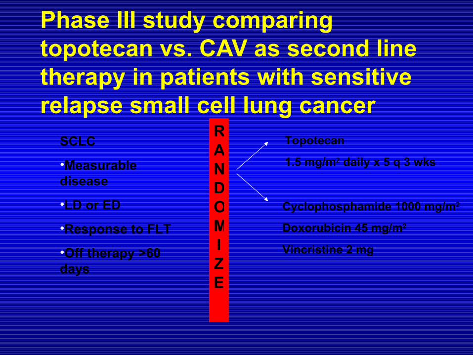
Phase III study comparing topotecan vs. CAV as second line therapy in patients with sensitive relapse small cell lung cancer
SCLC
•Measurable disease
•LD or ED
•Response to FLT
•Off therapy >60 days
RANDOMIZE
Topotecan
1.5 mg/m2 daily x 5 q 3 wks
Cyclophosphamide 1000 mg/m2
Doxorubicin 45 mg/m2
Vincristine 2 mg
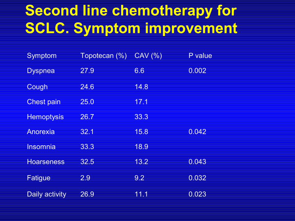
Second line chemotherapy for SCLC. Symptom improvement
Symptom Topotecan (%) CAV (%) P value
Dyspnea 27.9 6.6 0.002
Cough 24.6 14.8
Chest pain 25.0 17.1
Hemoptysis 26.7 33.3
Anorexia 32.1 15.8 0.042
Insomnia 33.3 18.9
Hoarseness 32.5 13.2 0.043
Fatigue 2.9 9.2 0.032
Daily activity 26.9 11.1 0.023
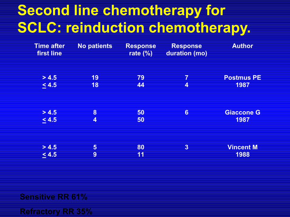
Second line chemotherapy for SCLC: reinduction chemotherapy.
Time after first line
No patients Response rate (%)
Response duration (mo)
Author
> 4.5 < 4.5
19 18
79 44
7 4
Postmus PE 1987
> 4.5 < 4.5
8 4
50 50
6 Giaccone G 1987
> 4.5 < 4.5
5 9
80 11
3 Vincent M 1988
Sensitive RR 61%
Refractory RR 35%
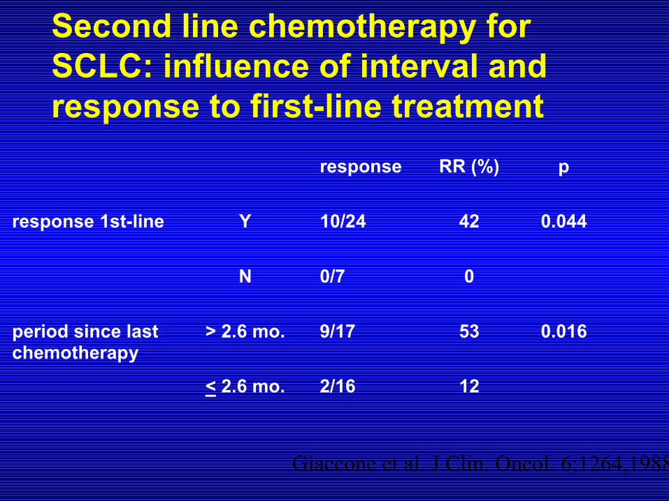
Second line chemotherapy for SCLC: influence of interval and response to first-line treatment
response RR (%) p
response 1st-line Y 10/24 42 0.044
N 0/7 0
period since lastchemotherapy
> 2.6 mo. 9/17 53 0.016
< 2.6 mo. 2/16 12
Giaccone et al. J.Clin. Oncol. 6;1264,1988
Prophylactic cranial irradiation in Prophylactic cranial irradiation in extensive disease small cell lung cancerextensive disease small cell lung cancer
(EORTC 08993-22993)(EORTC 08993-22993)
Ben Slotman, Corinne Faivre-Finn, Gijs Kramer†, Elaine Rankin,
Michael Snee, Matthew Hatton, Pieter Postmus, Laurence Collette, Murielle Mauer, Suresh Senan,
on behalf of the EORTC Radiation Oncology and Lung Cancer Groups
Slotman et al. NEJM 2007
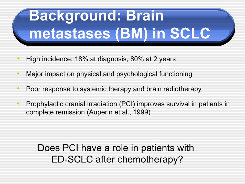
Background: Brain metastases (BM) in SCLC
• High incidence: 18% at diagnosis; 80% at 2 years
• Major impact on physical and psychological functioning
• Poor response to systemic therapy and brain radiotherapy
• Prophylactic cranial irradiation (PCI) improves survival in patients in complete remission (Auperin et al., 1999)
Does PCI have a role in patients with ED-SCLC after chemotherapy?
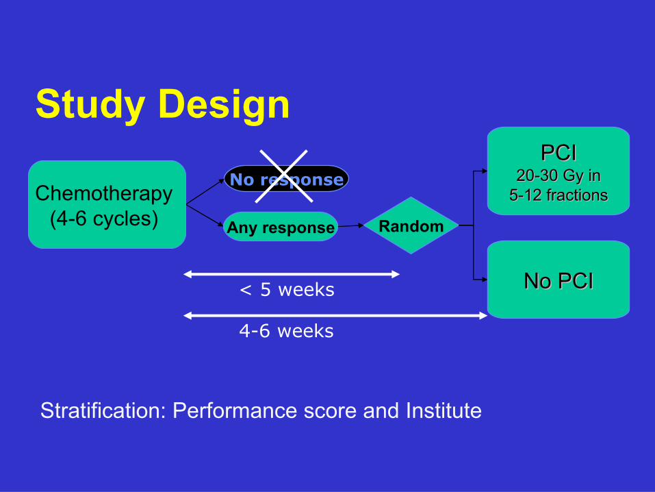
Study DesignPCIPCI
20-30 Gy in20-30 Gy in5-12 fractions5-12 fractions
No PCINo PCI
RandomAny response
Stratification: Performance score and Institute
< 5 weeks
4-6 weeks
No responseChemotherapy
(4-6 cycles)
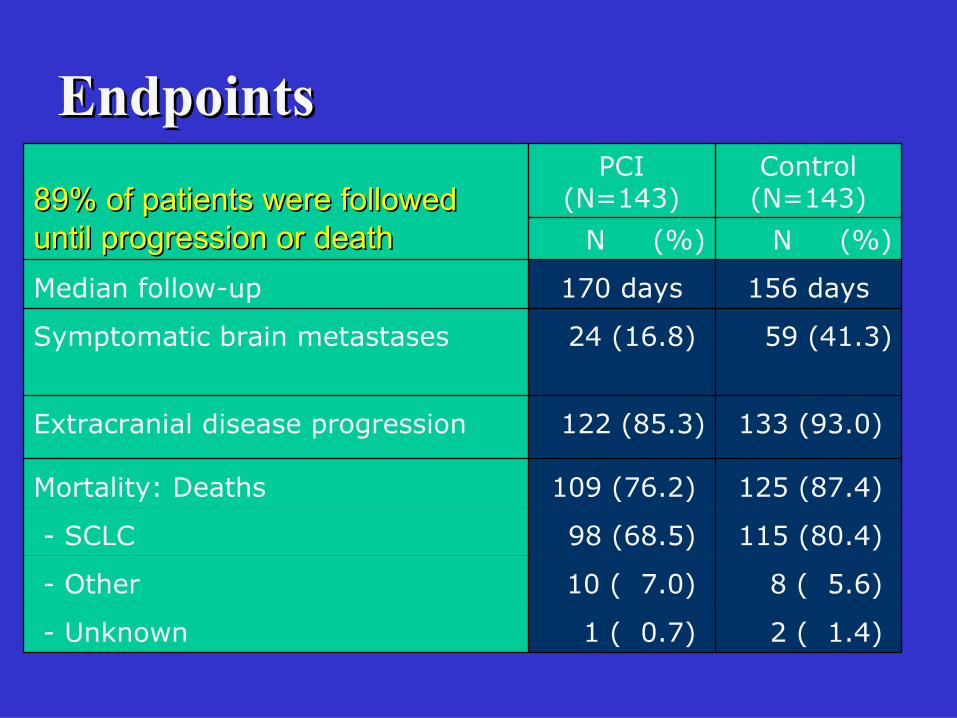
EndpointsEndpoints
156 days170 daysMedian follow-up
2 ( 1.4) 1 ( 0.7) - Unknown
8 ( 5.6) 10 ( 7.0) - Other
115 (80.4) 98 (68.5) - SCLC
125 (87.4) 109 (76.2) Mortality: Deaths
133 (93.0) 122 (85.3)Extracranial disease progression
59 (41.3) 24 (16.8) Symptomatic brain metastases
N (%)N (%)
Control(N=143)
PCI(N=143)89% of patients were followed 89% of patients were followed
until progression or deathuntil progression or death
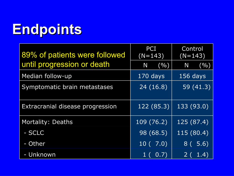
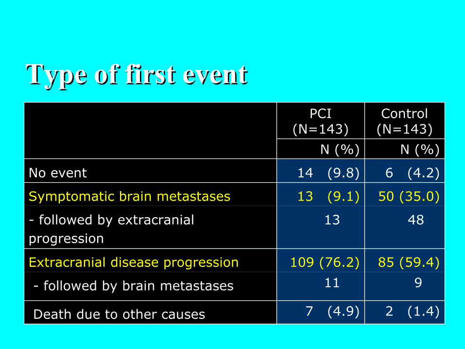
Type of first eventType of first event
50 (35.0)13 (9.1)Symptomatic brain metastases
48 13- followed by extracranial progression
85 (59.4) 109 (76.2)Extracranial disease progression
2 (1.4) 7 (4.9) Death due to other causes
9 11 - followed by brain metastases
6 (4.2)14 (9.8)No event
N (%)N (%)
Control(N=143)
PCI(N=143)
(months)
0 4 8 12 16 20 24 28 32 36
0
10
20
30
40
50
60
70
80
90
100
PCI
Control
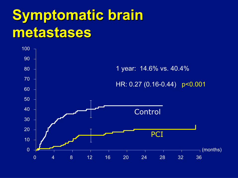
1 year: 14.6% vs. 40.4%
HR: 0.27 (0.16-0.44) p<0.001
Symptomatic brain Symptomatic brain metastasesmetastases
(months)
0 4 8 12 16 20 24 28 32 36
0
10
20
30
40
50
60
70
80
90
100
P=0.2699
Control
PCI
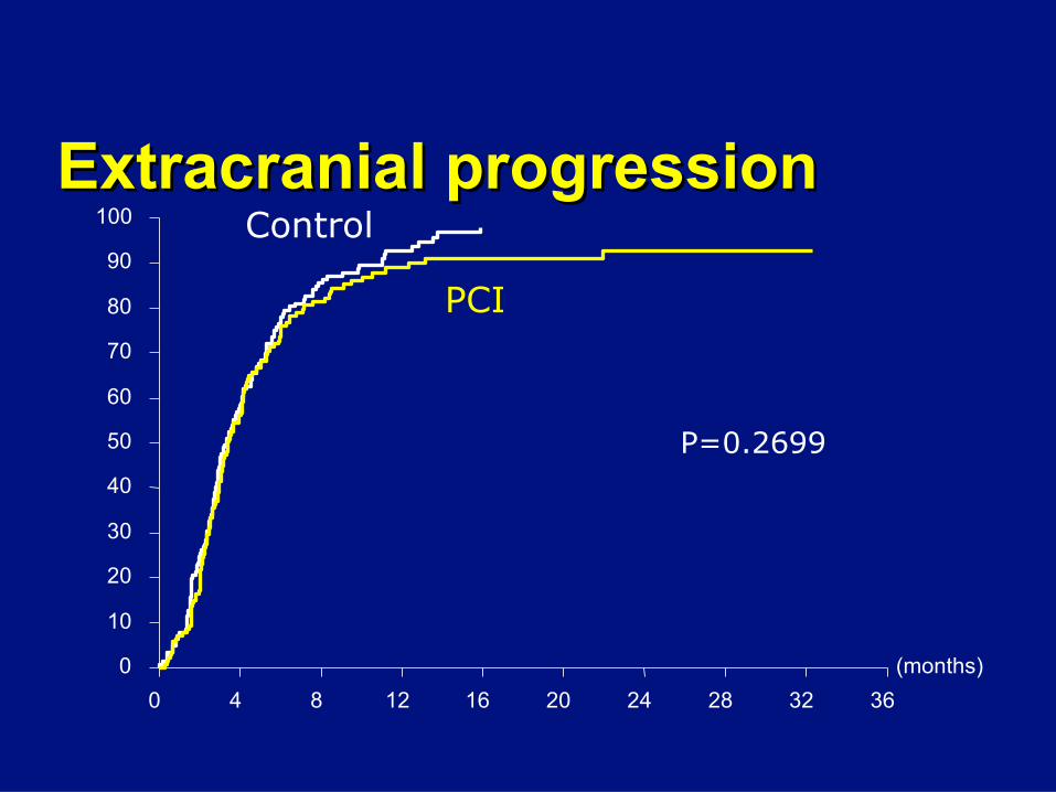
Extracranial progressionExtracranial progression
(months)
0 3 6 9 12 15 18 21 24 27
0
10
20
30
40
50
60
70
80
90
100
PCI
Control
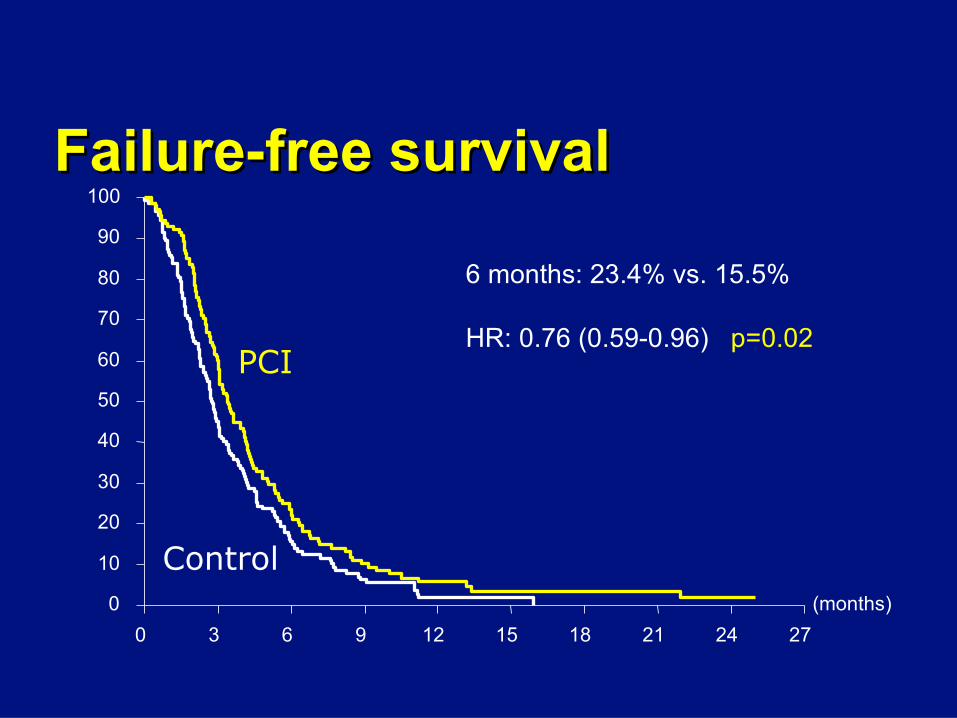
6 months: 23.4% vs. 15.5%
HR: 0.76 (0.59-0.96) p=0.02
Failure-free survivalFailure-free survival
(months)
0 4 8 12 16 20 24 28 32 36
0
10
20
30
40
50
60
70
80
90
100
PCIControl
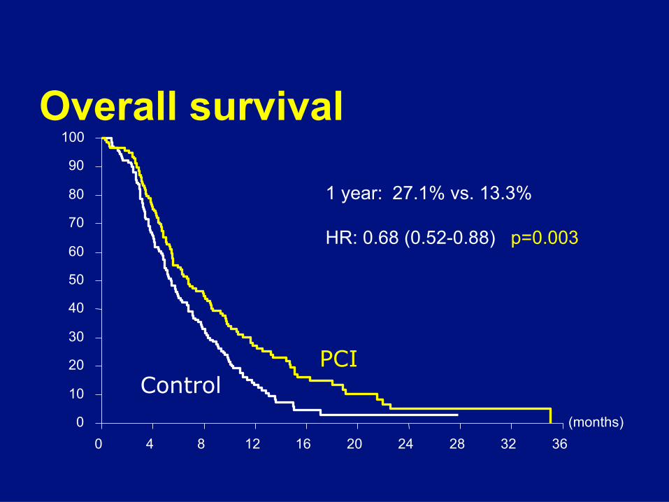
1 year: 27.1% vs. 13.3%
HR: 0.68 (0.52-0.88) p=0.003
Overall survival
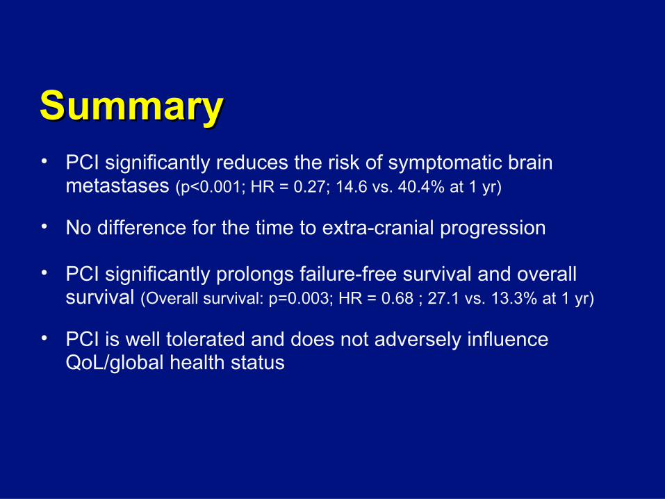
Summary Summary • PCI significantly reduces the risk of symptomatic brain
metastases (p<0.001; HR = 0.27; 14.6 vs. 40.4% at 1 yr)
• No difference for the time to extra-cranial progression
• PCI significantly prolongs failure-free survival and overall survival (Overall survival: p=0.003; HR = 0.68 ; 27.1 vs. 13.3% at 1 yr)
• PCI is well tolerated and does not adversely influence QoL/global health status
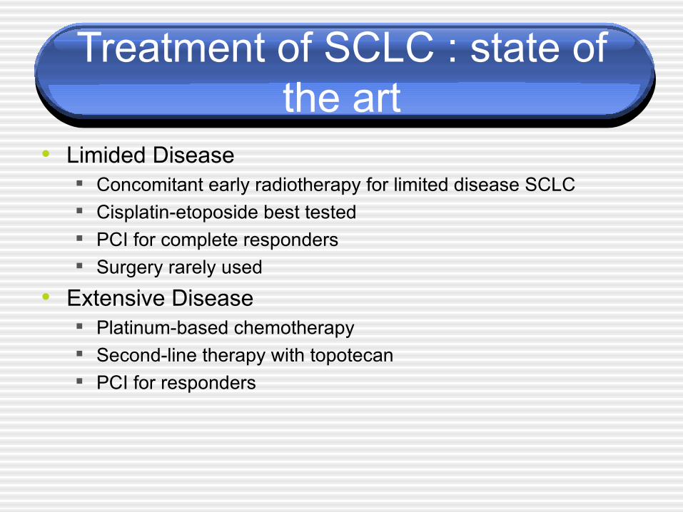
Treatment of SCLC : state of the art
• Limided Disease Concomitant early radiotherapy for limited disease SCLC Cisplatin-etoposide best tested PCI for complete responders Surgery rarely used
• Extensive Disease Platinum-based chemotherapy Second-line therapy with topotecan PCI for responders