Embed Size (px)
Citation preview

Oncology and surgical practice
By Dr. Thaere JasimGeneral surgeon
Tikrit University college of medicine

oncology is becoming a larger portion of
surgical practice. The surgeon often is
responsible for the initial diagnosis and
management of solid tumors. Knowledge of
cancer epidemiology, etiology, staging, and
natural history is required for initial patient
assessment, as well as to determination of the
optimal surgical therapy.

Cancer cells are psychopaths.
They have no respect for the rights of other
cells. They violate the democratic principles
of normal cellular organisation.
Their proliferation is uncontrolled; their
ability to spread is unbounded. Their
inexorable, relentless progress destroys first
the tissue and then the host.

Tumour Cells have to acquire a number of characteristics before they are fully malignant.
Malignant transformation
■ Establish an autonomous lineage
Resist signals that inhibit growth
Acquire independence from signals stimulating growth
■ Obtain immortality
■ Evade apoptosis
■ Acquire angiogenic competence
■ Acquire the ability to invade
■ Acquire the ability to disseminate and implant
■ Evade detection/elimination
■ Genomic instability
■ Jettison excess baggage
■ Subvert communication to and from the
environment/milieu

Hallmarks of Cancer

Establish an autonomous lineage:
This involves developing independence from the normal signals that control supply and demand.
Cancer cells escape from this normal system of checks and balances: they grow and proliferate in the absence of external stimuli; they proliferate and grow despite signals telling them not to. Their division is inappropriate and remorseless.

Obtain immortality
normal cells are permitted to undergo only a finite number of divisions.
For humans, this number is between 40 and 60. The limitation is imposed by the progressive shortening of the end of the chromosome, the telomere,
that occurs each time a cell divides.
Telomeric shortening is like a molecular clock and, when its time is up, it is time for that lineage to die out.
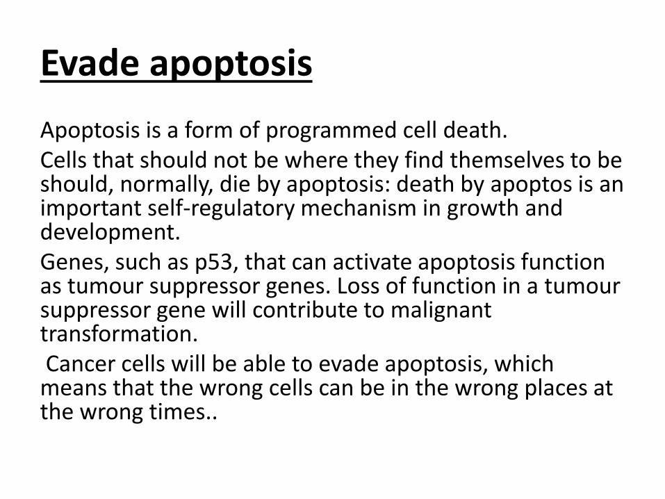
Evade apoptosis
Apoptosis is a form of programmed cell death.Cells that should not be where they find themselves to be should, normally, die by apoptosis: death by apoptos is an important self-regulatory mechanism in growth and development.Genes, such as p53, that can activate apoptosis function as tumour suppressor genes. Loss of function in a tumoursuppressor gene will contribute to malignant transformation.Cancer cells will be able to evade apoptosis, which
means that the wrong cells can be in the wrong places at the wrong times..

:Acquire angiogenic competence
A mass of tumour cells cannot, in the absence of a blood supply, grow beyond a diameter of about 1mm.
This places a severe restriction on the capabilities of the tumour: it cannot grow much larger and it cannot spread widely within the body.
However, if the mass of tumour cells is able to attract or to construct a blood supply, then it is able to quit its dormant state and behave in a far more aggressive fashion.
The ability of a tumour to form blood vessels is termed angiogenic competence and is a key feature of malignant transformation.

Acquire the ability to invade
Cancer cells have no respect for the structure of normal tissues.
Cancer cells acquire the ability to breach the basement membrane and thus gain direct access to blood and lymph vessels.
Cancer cells use three main mechanisms to facilitate invasion: they cause a rise in the interstitial pressure within atissue; they secrete enzymes that dissolve extracellular matrix; and they acquire motility.

Acquire the ability to disseminate and implantAs soon as motile cancer cells gain access to vascular and lymphovascular spaces, they have acquired the potential to use the body’s natural transport mechanisms to distribute themselves throughout the body. Distribution is not, of itself, sufficient to cause tumours to develop at distant sites. The cells also need to acquire the ability to implant.
Cancer cells probably implant themselves in distant tissues by exploiting, and subverting, the normal inflammatory response.
. Cancer cells simply subvert this physiological mechanism.

Evade detection/eliminationCancer cells are simultaneously both ‘self’ and ‘not self’.
Cancer cells, or at least those that give rise to clinical disease, appear to gain the ability to escape detection by the immune system.
This may be through suppressing the expression of tumour-associated antigens, a stealth approach, or it may be through actively coopting one part of the immune system to connive in helping the tumour to escape detection by other parts of the immune surveillance system

Genomic instability
A cancer is a genetic ferment. Cells are dividing without proper checks and balances.
DNA is being copied, and the proofreaders have been retired or ignored. Mutations are arising all the time
within tumours, and some of these mutations, particularly those in tumour suppressor genes, may have the ability to encourage the development and persistence of further mutations

Jettison excess baggageCancer cells are geared to excessive and remorseless proliferation.
They do not need to develop or retain those specialised functions that make them good cellular citizens.
They can therefore afford to repress or permanently lose those genes that control such functions.

Subvert communication to and from the environment/milieu
Providing false information is a classic military strategy. Degrading the command and control systems of the enemy is an essential component of modern warfare.
Cancer cells almost certainly use similar tactics in their battle for control over their host.
Given the complexity of communication between and within cells, this is not an easy statement either to disprove or to prove. Nor does it offer
any easy targets for therapeutic manipulation.

THE CAUSES OF CANCERBoth inheritance and environment are important determinants of whether or not an individual develops cancer. enviromental factors have been implicated in more than 80% of cases of cancer, this would still leave plenty of scope for the role of genetic inheritance: not just the 20% of tumours for which there is no clear environmental contribution but also, as environment alone can rarely cause cancer, the genetic contribution to the 80% oftumours to whose occurrence environmental factors contribute.As a plain example: not all smokers develop lung cancer; lung cancer can occur in people who have never smoked.Although environ-

Genetic
Chemical Carcinogens
Physical Carcinogens
Viral Carcinogens
Bacteria, fungi, parasite
Hormones
Others agents

Cancer Genetics
One widely held opinion is that cancer is a genetic disease that arises from an accumulation of mutations that leads to the selection of cells with increasingly aggressive behavior. These mutations may lead either to a gain of function by oncogenes or to a loss of function by tumor-suppressor genes. Most mutations in cancer are somatic and are found only in the cancer cells.
A few of these hereditary cancer genes are oncogenes, but most are tumor-suppressor genes. Although hereditary cancer syndromes are rare, somatic mutations that occur in sporadic cancer have been found to disrupt the cellular pathways altered in hereditary cancer syndromes.

Cancer siteLocation Gene
Colorectal adenomas and carcinomas, duodenal and gastric tumors, desmoids, medulloblastomas, osteomas
17q21APC
Breast cancer, soft tissue sarcoma, osteosarcoma, brain tumors, adrenocortical carcinoma, Wilms' tumor, phyllodestumor of the breast, pancreatic cancer, leukemia
17p13p53
Breast cancer, ovarian cancer, colon cancer, prostate cancer17q21BRCA1
Breast cancer, ovarian cancer, colon cancer, prostate cancer, , pancreatic cancer, gastric cancer, melanoma
13q12.3BRCA2
Basal cell carcinoma9q22.3PTC
Retinoblastoma, sarcomas, melanoma, and malignant neoplasms of brain and meninges
13q14rb
Medullary thyroid cancer, pheochromocytoma, parathyroid hyperplasia
10q11.2RET
Pancreatic islet cell cancer, parathyroid hyperplasia, pituitary adenomas
11q13MEN1

Chemical CarcinogensCurrently, approximately 60 to 90% of cancers are thought to be due to environmental factors. Chemicals are classified into three groups based on how they contribute to tumor formation. The first group of chemical agents, the genotoxins, can initiate carcinogenesis by causing a mutation. The second group, the cocarcinogens, by themselves cannot cause cancer but potentiate carcinogenesis by enhancing the potency of genotoxins. The third group, tumor promoters, enhances tumor formation when given after exposure to genotoxins

Predominant Tumor Chemical
Lung cancer, oral cancer, pharyngeal cancer, laryngeal cancer, esophageal cancer (squamous cell), pancreatic cancer, bladder cancer, liver cancer, renal cell carcinoma, cervical cancer, leukemia
Tobacco smoke
Liver cancerAflatoxins
LeukemiaBenzene
Bladder cancerBenzidine
Skin cancer, scrotal cancerCoal tar
Leukemia, lymphomaEthylene oxide
Lung cancer, nasal cancerNickel
Angiosarcoma of the liver, hepatocellular carcinoma, brain tumors,
Vinyl chloride
Oral cancerTobacco products, smokeless

Physical & enviromental CarcinogensPhysical carcinogenesis can occur through induction of inflammation and cell proliferation over a period of time or through exposure to physical agents that induce DNA damage. Foreign bodies can cause chronic irritation that can expose cells to carcinogenesis due to other environmental agents. In animal models, for example, subcutaneous implantation of a foreign body can lead to the development of tumors that have been attributed to chronic irritation from the foreign objects. In humans, clinical scenarios associated with chronic irritation and inflammation such as chronic nonhealing wounds, burns, and inflammatory bowel syndrome have all been associated with an increased risk of cancer.

Associated tumourPhysical agents
Skin tumourUV exposure
Leukaemia Breast Lymphoma thyroidIonisingradiation
Breast, Endometrium Kidney ColonOesophagus
Obesity/lack ofphysical exercise

infectionViruses may cause or increase the risk of malignancy through several mechanisms, including direct transformation, expression of oncogenes that interfere with cell-cycle checkpoints or DNA repair, expression of cytokines or other growth factors, and alteration of the immune system. Oncogenic viruses may be RNA or DNA viruses. Oncogenic RNA viruses are retroviruses and contain a reverse transcriptase.

H. pylori infection is associated with gastritis and gastric cancer, and thus its carcinogenicity may be considered physical carcinogenesis. Infection with the liver fluke Opisthorchisviverrini similarly leads to local inflammation and cholangiocarcinoma.

Associated tumorInfectious agents
Stomach cancerHelicobacter pylori
Bladder cancerBilharzia
Burkitt's lymphoma Hodgkin's disease Sinonasalangiocentric T-cell lymphoma Nasopharyngeal carcinoma
Epstein-Barr virus
HepatomaHepatitis B
Kaposi's sarcoma Non-Hodgkin's lymphoma
HIV
Cervical cancer Anal cancer
Human papillomavirus 16 and 18
Adult T-cell leukemia/lymphomaHuman T-cell lymphotropic viruses

Classification of tumuors
1- Behavioral classification: benign or malignant
2- Histogentic classification: cell of origin

Behavioral classificationmalignantBenign
Rapidly growingSlow growing
High mitotic activityLow mitotic activity
Differs from parent tissuesResemble parent tissue
InfilteratingNon-infilterating
Abnormal cellsCells normal
Frequently metastatasizedNever metastasizes
Often poorly defined or irregularOften encapsulated
Ulcerating skin or mucosaRarely ulcerated
Necrosis is commenRarely undergoes necrosis
Fatal if intreatedOnly fatal when damaging vital organs

Histogentic Classification:Classification by cell of origin: epithelial, connective tissues or lymphoid or haemopeotic.
Histologically determined : well differentiated, poorly differentiated or moderately differentiated.
Loss of differentiation, disorder of growth pattern,variability of cell size, variability in nuclear size, high nuclear/cytoplasmic ratio, increazed mitotic activity, abnormal chromatin, and abnormal nucleoli or multiple.

Hyperplasia
Hypertrophy
Metaplasia
Dysplasia
Carcinoma in-situ

Behavior of TumorsInvasion : is the most criteria of malignancy spread in direct continuation.
Metastasises : is the process of malignant tumor to spread from primary site to secondary site in indirect continuation. By:
Haematogenous route
Lymphatic route
Trans-ceolomic
Or direct implantation in surgical incision

Clinical effects of tumours
Local
Systemic
Para neoplastic
Metabolic
others

Local
Mass
Bleeding
Pain
Obstruction
Pressure on adjacent structure

Systemic
Enlarged lymph nodes
Hepatosplenomegaly
Jaundice
Ascites
Pathological fructure
Anemia
Fit or confusion
Pleural effusion

Paraneoplastic effects
These are not related to presence of tumor or metastasises. They can divided into:
1- humoral: like Cushing’s syndrome, inappropriate secretion of ADH, hypercalcemia, carsinoid syndrome.
2- immunological: autoimmune disease triggered by malignancy like membranous glomerulonephritis and dermatocytis.

Metabolic effects
These are caused by hormones directly secreted by tumour like thyrotoxicosis from thyroid adenoma insulin by insulinoma, hyperparathyroidism by parathyroid adenoma.

Others
Cachaxia
Pyrexia
Thrombophlebitis migrans

Tumor MarkersTumor markers are substances that can be detected in higher than normal amounts in the serum, urine, or tissues of patients with certain types of cancer. Tumors markers are produced either by the cancer cells themselves or by the body in a response to the cancer.
Over the past decade, there has been an especially high interest in identifying tissue tumor markers that can be used as prognostic or predictive markers. Although the terms prognostic marker and predictive marker are sometimes used interchangeably, the term prognostic marker generally is used to describe molecular markers that predict disease-free survival, disease-specific survival, and overall survival, whereas the term predictive marker often is used in the context of predicting response to certain therapies

CancerMarker
ProstateProstate-specific antigen (4 g/L)
Colon breast recurrent disease
Carcinoembryonic antigen
HepatocellularAlpha-fetoprotein
PancreaticCancer antigen 19-9
breastCancer antigen 27-29
breastCancer antigen 15-3

THE MANAGEMENT OF CANCER
Management is more than treatmentThe traditional approach to cancer concentrates on diagnosis and active treatment. This is a very limited view and one that, in terms of the public health, may not have served society particularly Well.
The management of cancer can be considered as taking place along two axes: one is an axis of scale, from the individual to the world population; the other is an axis based on the unnatural history of the disease, from prevention through to rehabilitation or palliative care.

Prevention:
Screening:
Diagnosis:
Staging:
Treatment:
Follow up:
Rehabilitation :(palliation and terminal care):

:ScreeningScreening involves the detection of disease in an asymptomatic population in order to improve outcomes by early diagnosis.
It follows that a successful screening programme must achieve early diagnosis, and that the disease in question has a better outcome when treated at an early stage.

Criteria for screening
The disease
■ Recognisable early stage
■ Treatment at an early stage more effective than at a later stage
■ Sufficiently common to warrant screening
The test
■ Sensitive and specific
■ Acceptable to the screened population
■ Safe
■ Inexpensive
The programme
■ Adequate diagnostic facilities for those with a positive test
■ High-quality treatment for screen-detected disease to minimisemorbidity and mortality
■ Screening repeated at intervals if the disease is of insidious onset
■ Benefit must outweigh physical and psychological harm

frequencyprocedurepopulationCancer Site
Monthly, starting at age 20
Breast self-examination
Women aged ≥20 y
Breast
Every 3 y, ages 20–39
Clinical breast examination
Annual, starting at age 40
Annual, starting at age 40
Mammography
Offer PSA test and DRE annually, starting at age 50, for men who have life expectancy of at least 10 y
Digital rectal examination (DRE) and prostate-specific antigen (PSA) test
Men aged ≥50 yprostate

Diagnosis and classificationAccurate diagnosis is the key to the successful management of cancer. The definitive diagnosis of solid tumors usually is obtained by performing a biopsy of the lesion. Biopsy findings determine the tumor histology and grade and thus assist in definitive therapeutic planning. When a biopsy has been performed at an outside institution, the slides should be reviewed to confirm the outside diagnosis.
Biopsy specimens of mucosal lesions usually are obtained endoscopically (e.g., via colonoscope, bronchoscope, or cystoscope). Lesions that are easily palpable, such as those of the skin, can either be excised or sampled by punch biopsy. Deep-seated lesions can be localized with computed tomographic (CT) scan or ultrasound guidance for biopsy.
.

Different tumours are classified in different ways: most squamous epithelial tumours are simply classed as well (G1), moderate (G2) or poorly (G3) differentiated . Adenocarcinomas are also often classified as G1, 2 or 3,

Investigation and stagingIt is not sufficient simply to know what a cancer is; it is imperative to know its site and extent. Staging is the process whereby the extent of disease is mapped out. Formerly, staging was a fairly crude process based on clinical examination and chest X-ray and the occasional ultrasound; nowadays, it is a highly sophisticated process, heavily reliant on the technology of modern imaging. The International Union against Cancer (UICC) is responsible
for the TNM (tumour, nodes, metastases) staging system for
cancer. This system is compatible with, and relates to, the
American Cancer Society (AJCC) system for stage grouping of cancer.

Staging of colorectal cancer
TNMTX Primary tumour cannot be assessedT0 No evidence of primary tumourTis Intraepithelial or intramucosal carcinomaT1 Tumour invades submucosaT2 Tumour invades muscularis propriaT3 Tumour invades through the muscularis propriainto the subserosa or into retroperitoneal (pericolicor perirectal) tissues.T4 Tumour directly invades beyond bowel.

NX Regional lymph nodes cannot be assessed
N0 No metastases in regional nodes
N1 Metastases in 1–3 regional lymph nodes
N2 Metastases in ≥ 4 regional lymph nodes
MX Not possible to assess the presence of distant metastases
M0 No distant metastases
M1 Distant metastases present

Therapeutic decision making and themultidisciplinary team
As the management of cancer becomes more complex, it becomes impossible for any individual clinician to have the intellectual and technical competence that is necessary to manage all the patients presenting with a particular type of tumour.
The era of feigned omniscience is past. The formation of multidisciplinary teams represents an attempt to make certain that each and every patient with a particular type of cancer is managed appropriately.
Teams should not only be multidisciplinary, they should be multiprofessional.-

Members of the multiprofessional team■ Site-specialist surgeon■ Surgical oncologist■ Plastic and reconstructive surgeon■ Clinical oncologist/radiotherapist■ Medical oncologist■ Diagnostic radiologist■ Pathologist■ Speech therapist■ Physiotherapist■ Prosthetist■ Clinical nurse specialist (rehabilitation, supportive care)■ Palliative care nurse (symptom control, palliation)■ Social worker/counsellor■ Medical secretary/administrator■ Audit and information coordinator

Principles of cancer surgery
For most solid tumours, surgery remains the definitive treatment and the only realistic hope of cure.
However, surgery has several roles in cancer treatment including diagnosis, removal of primary disease, removal of metastatic disease, palliation, prevention and reconstruction.

Diagnosis and staging
Removal of primary diseaseRadical surgery for cancer involves removal of the primary tumour and as much of the surrounding tissue and lymph node drainage as possible in order not only to ensure local control but also to prevent spread of the tumour through the lymphatics.,

Removal of metastatic disease
In certain circumstances, surgery for metastatic disease may be appropriate. This is particularly true for liver metastases arising from colorectal cancer where successful resection of all detectable disease can lead to long-term survival in about one-third of patients.

Palliation
In many cases, surgery is not appropriate for cure but may be
extremely valuable for palliation. A good example of this is the
patient with a symptomatic primary tumour who also has distant
metastases. In this case, removal of the primary may increase the patient’s quality of life but will have little effect on the ultimate outcome.
Other examples include bypass procedures such as an
ileotransverse anastomosis to alleviate symptoms of obstruction
caused by an inoperable caecal cancer or bypassing an unresectable carcinoma at the head of the pancreas by cholecysto- or choledochojejunostomy to alleviate jaundice.

Principles of the non-surgical treatment of cancer
Chemotherapy
Hormonal Therapy
Targeted Therapy
Immunotherapy
Gene Therapy
Radiation Therapy

ChemotherapyIn patients with documented distant metastatic disease, chemotherapy is usually the primary modality of therapy. The goal of therapy in this setting is to decrease the tumor burden, thus prolonging survival. It is rare to achieve cure with chemotherapy for metastatic disease for most solid tumors. Chemotherapy administered to a patient who is at high risk for distant recurrence but has no evidence of distant disease is referred to as adjuvant chemotherapy.
The goal of adjuvant chemotherapy is eradication of micrometastatic disease, with the intent of decreasing relapse rates and improving survival rates.
Adjuvant therapy can be administered after surgery (postoperative chemotherapy) or before surgery (preoperative chemotherapy, neoadjuvant chemotherapy, or induction therapy).

Hormonal Therapy
Some tumors, most notably breast and prostate cancers, originate from tissues whose growth is under hormonal control. The first attempts at hormonal therapy were through surgical ablation of the organ producing the hormones involved, such as oophorectomy for breast cancer. Currently, hormonal anticancer agents include androgens, antiandrogens, antiestrogens, estrogens, glucocorticoids, gonadotropin inhibitors, progestins, aromatase inhibitors, and somatostatin analogues. Hormones or hormone-like agents can be administered to inhibit tumor growth by blocking or antagonizing the naturally occurring substance, such as with the estrogen antagonist tamoxifen. Other substances that block the synthesis of the natural hormone can be administered as alternatives.

Targeted Therapy
Over the past decade, increased understanding of cancer biology has fostered the emerging field of molecular therapeutics. The basic principle of molecular therapeutics is to exploit the molecular differences between normal cells and cancer cells to develop targeted therapies. Thus targeted therapies usually are directed at the processes involved in tumor growth rather than directly targeting the tumor cells.

Immunotherapy
The aim of immunotherapy is to induce or potentiate inherent antitumor immunity that can destroy cancer cells. Central to the process of antitumor immunity is the ability of the immune system to recognize tumor-associated antigens present on human cancers and to direct cytotoxic responses through humoral or T-cell–mediated immunity. Overall, T-cell–mediated immunity appears to have the greater potential of the two for eradicating tumor cells. T cells recognize antigens on the surfaces of target cells as small peptides presented by class I and class II MHC molecules

Gene Therapy
Gene therapy is being pursued as a possible approach to modifying the genetic program of cancer cells as well as treating metabolic diseases. The field of cancer gene therapy uses a variety of strategies, ranging from replacement of mutated or deleted tumor-suppressor genes to enhancement of immune responses to cancer cells. Indeed, in preclinical models, approaches such as replacement of tumor-suppressor genes leads to growth arrest or apoptosis. However, the translation of these findings into clinically useful tools presents special challenges.

Radiation TherapyIonizing radiation is energy strong enough to remove an orbital electron from an atom.
Radiation deposition results in DNA damage manifested by single- and double-strand breaks in the sugar phosphate backbone of the DNA molecule.
Cross-linking between the DNA strands and chromosomal proteins also occurs. The mechanism of DNA damage differs by the type of radiation delivered. Electromagnetic radiation is indirectly ionizing through short-lived hydroxyl radicals produced primarily by the ionization of cellular hydrogen peroxide (H2O2). Protons and other heavy particles are directly ionizing and directly damage DNA.


Thank you