Embed Size (px)
Citation preview

Granuloma
Dr Mohammad Manzoor Mashwani
Collection of modified macrophages
EPI-THE-LI-OID Cells
Immune Granuloma
1. Epithelioid Cells 2. Rim of lymphocytes 3. Giant cells 4. Caseous Necrosis 5. Fibrous Cup


Tubercle/Granuloma- a type of ch. Inf.Granuloma is defined as a circumscribed, tiny lesion, about
1 mm in diameter, composed predominantly of collection of modified macrophages called epithelioid cells, and rimmed at the periphery by lymphoid cells.
The word ‘granuloma’ is derived from granule meaning circumscribed granule-like lesion, and -oma which is a suffix commonly used for true tumours but here it indicates a localised inflammatory mass or collection of macrophages.
Granuloma: Collection of epithelioid cells rimed by
lymphocytes.
EPITHELIOID
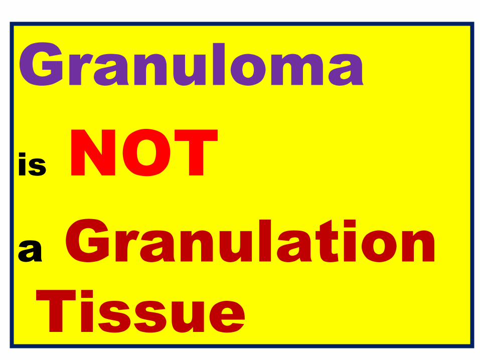
Granuloma is NOT a Granulation Tissue

• Granulation tissue is a new connective tissue and microscopic blood vessels that form on the surfaces of a wound during the healing process (Repair).
• Granulation tissue typically grows from the base of a wound and is able to fill wounds of almost any size.
Granulation tissue is highly vascularized connective tissue composed of
1.Newly formed capillaries,
2.Proliferating fibroblasts and
3.Residual inflammatory cells.
A Young Scar = Granulation tissue
Healing: 1. Regeneration 2. RepairRepair: 1. Granulation tissue formation 2. Contraction of wounds

Granulation tissue AppeArAnceDuring the migratory phase of wound healing, granulation tissue is: light red or dark pink in color, being perfused with new capillary loops or "buds"; soft to the touch; moist; and
bumpy (granular) in appearance, due to punctate hemorrhages pulseful on palpation painless when healthy.


Types of granulomas:1. Foreign body granuloma 2.Immune granuloma .
1. Foreign body granulomas – form when material such as talc, sutures, or other fibers are large enough to preclude (prevent) phagocytosis by a single macrophage.
1. Foreign Body Granuloma
2. Immune Granuloma

Immune granulomas2. Immune granulomas - caused by insoluble particles that are
capable of inducing a cell-mediated response. This type of immune response produces granulomas when the inciting agent is poorly soluble.
TuberculosisTubercle= GRANULOMA

Immune Granuloma• Macrophages engulf the foreign material and
process present some of it to appropriate • T lymphocytes, causing them to become activated,
responding T cells produce cytokines, such as IL-2 which activates other T cells and IFN-γ which is important in transforming macrophages into epithelioid cells and multinucleate giant cells.
• MaMacrophages Epithelioid cells + Giant cells

• The classic example for the immune granuloma is that caused by the bacillus of tuberculosis. In this disease, the granuloma is referred to as a
tubercle and is classically characterized
by the presence of central caseous necrosis. • Caseating necrosis is rare in other granulomatous
diseases.
TUBERCLE = GRANULOMA

• Granuloma formation is a strategy that has evolved to deal with those pathogens that have learned to evade (escape) the host immune system by various means like resisting phagocytosis and killing within the macrophages.
• Granulomas try to wall off these organisms and prevent their further growth and spread.
Mechanism of Granuloma Formation
Wall off Organisms

Granulomas try to wall off these organisms and prevent their further growth and spread.

Granuloma: bacilli are inhaled by droplets
Bacteria are phagocytosed by alveolar macrophages
After amassing (collect or gather) substances that they cannot digest, macrophages lose their motility, accumulate at the site of injury and transform themselves into nodular collections; the Granuloma
A localized inflammatory response recruits more mononuclear cells
The granuloma consists of a kernel (inner) of infected macrophages surrounded by foamy macrophages and a ring of lymphocytes and a fibrous cuff (containment phase)
Containment usually fails when the immune status of the patient changes; the granuloma caseates, ruptures and spills into the airway
Containment Phase

Main causes of granulomatous inflammation:
• Mildly irritant ‘foreign’ material
• Mycobacteria: Tuberculosis, leprosy• Syphilis• Other rare infections e.g. some fungi• Unknown causes: Sarcoid
Wegener’s granulomatosisCrohn’s disease

Granuloma comprises:1. Epithelioid Cells2. Caseous Necrosis 3. Giant cells4. Rim of lymphocytes 5. Fibrous Cup
1.Caseous Necrosis2.Epithelioid cells3.Giant cells4.Fibrous cup5.Rim of Lymphocytes
Infected Macrophages

Epithelioid cells• Epithelioid histiocytes (Epithelioid cells) are
activated macrophages resembling epithelial cells:• elongated, with finely granular, pale eosinophilic (pink)
cytoplasm and • central, ovoid nucleus (oval or elongate), which is
less dense than that of a lymphocyte. • They have indistinct shape contour, often appear
to merge into one another and can form aggregates
known as giant cells.


Giant cell• A giant cell is a mass formed by the union of several
distinct cells (usually macrophages), often forming a granuloma.
• It can arise in response to an infection, such as from tuberculosis, herpes, or HIV, or foreign body.

Types of Giant Cells1. Langhans giant cell
2. Foreign-body giant cell
3. Touton giant cells
4. Giant-cell arteritis
5. Reed–sternberg cell
Also as in subependymal giant cell astrocytoma

1. Langhans giant cell• This particular form of giant cell was named after a German
pathologist, Theodor Langhans. • Epithelioid cells fuse to form giant cells containing 20 or more nuclei.
• The nuclei are arranged peripherally and form a circle or semicircle related to the shape of a horseshoe.
• Langhans giant cell is said to be related to tuberculosis and it occurs in many types of granulomatous diseases.
Horseshoe

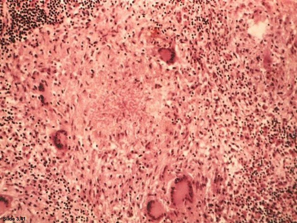
Slide 3.41


Langhans type giant cell - Tuberculosis

2. Foreign-body giant cell
• Foreign-body giant cells form when a subject is exposed to a foreign substance. Exogenous substances include talc or sutures.
• In this form of giant cell, the nuclei are arranged in an overlapping manner.
• This giant cell is often found in tissue because of medical devices, prostheses, and biomaterials.

Foreign body type giant cells


3.Touton giant cells
Touton giant cells are a type of multinucleated giant cell seen in lesions with high lipid content such as
fat necrosis, xanthoma, and xanthogranulomas. They are also found in dermatofibroma.

• Touton giant cells are named for Karl Touton, a German botanist and dermatologist. Karl Touton first observed these cells in 1885 and named them "xanthelasmatic giant cells", a name which has since fallen out of favor.

• They contain a ring of nuclei surrounding a central homogeneous cytoplasm, while foamy cytoplasm surrounds the nuclei.
Two cytoplasms:1. Central homogeneous
cytoplasm2. Peripheral foamy
cytoplasm
Touton: T for Two cytoplasms

Touton Giant cell

Necrosis: A series of morphological changes in a lethally injured cell (irreversible cell injury).
Coagulative Necrosis + Liquefactive Necrosis Soft, friable, whitish-grey/Yellowish debrisResembling dry cheese

• Fibrous connective tissue often surrounds granulomas (remodeling of tissue)
• Areas within the granuloma can undergo necrosis (prototype: caseous necrosis in tuberculosis).
• Necrosis can lead to calcification or liquefaction and formation of a cavern
(cave)if drained.
Fibrous Cup
Dystrophic Calcification(Laminated)
1. Caseous Necrosis 2. Epithelioid Cells 3. Giant Cells 4. Lymphocytes 5. Fibrous cup
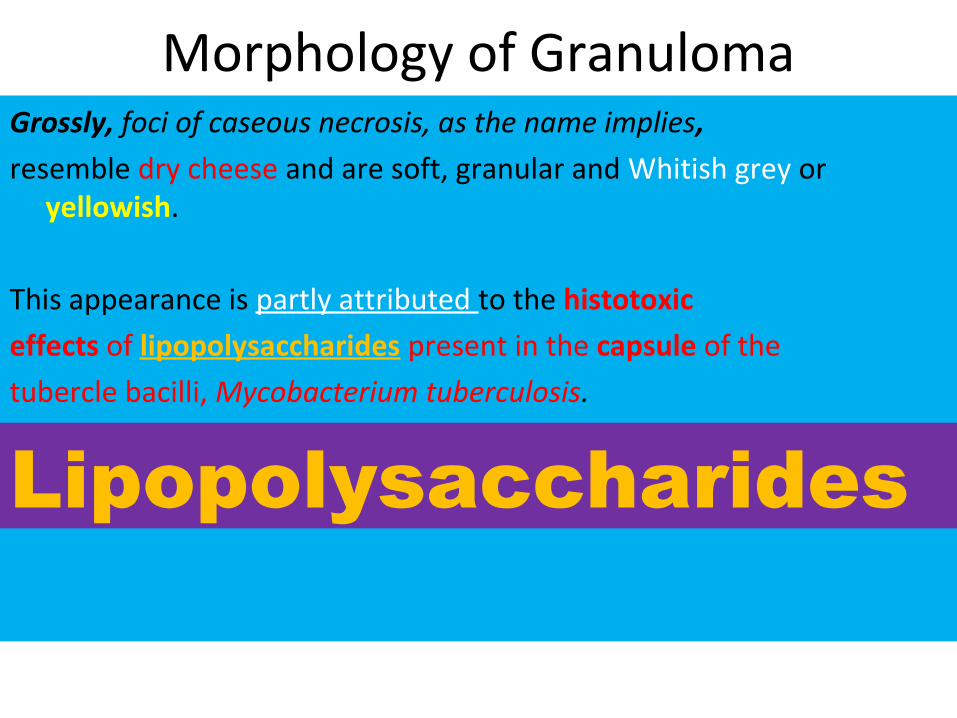
Morphology of GranulomaGrossly, foci of caseous necrosis, as the name implies,
resemble dry cheese and are soft, granular and Whitish grey or yellowish.
This appearance is partly attributed to the histotoxic
effects of lipopolysaccharides present in the capsule of the
tubercle bacilli, Mycobacterium tuberculosis.
Lipopolysaccharides

1. Caseous Necrosis 2. Epithelioid Cells 3. Giant Cells 4. Lymphocytes 5. Fibrous cup
Microscopically, 1. the necrosed foci are structureless, eosinophilic, and contain
granular debris.
2. The surrounding tissue shows characteristic granulomatous inflammatory reaction consisting of epithelioid cells with
3. interspersed giant cells of Langhans’ or foreign body type and
4. peripheral mantle (layer, covering, Ring, collar) of lymphocytes.
5. Fibrous cup.


Langhans Giant Cell
Caseous Necrosis
Epithelioid Macrophage
Lymphocytic Rim


Caseating granulomas

THANKS