Embed Size (px)
Citation preview
MEDICAL ACADEMY NAMED AFTER S.I. GEORGIEVSKY OF VERNADSKY CFU
DEFICIENCY ANEMIASDepartment of pathomorphology
Presented by,
Al auf jalaludeen
Group 308
DEFINITION
• Anaemia is defined as reduced haemoglobin concentration in blood below the lower
limit of the normal range for the age and sex of the individual. In adults, the lower
extreme of the normal haemoglobin is taken as 13.0 g/dl for males and 11.5 g/dl for
females. Newborn infants have higher haemoglobin level and, therefore, 15 g/dl is taken
as the lower limit at birth.
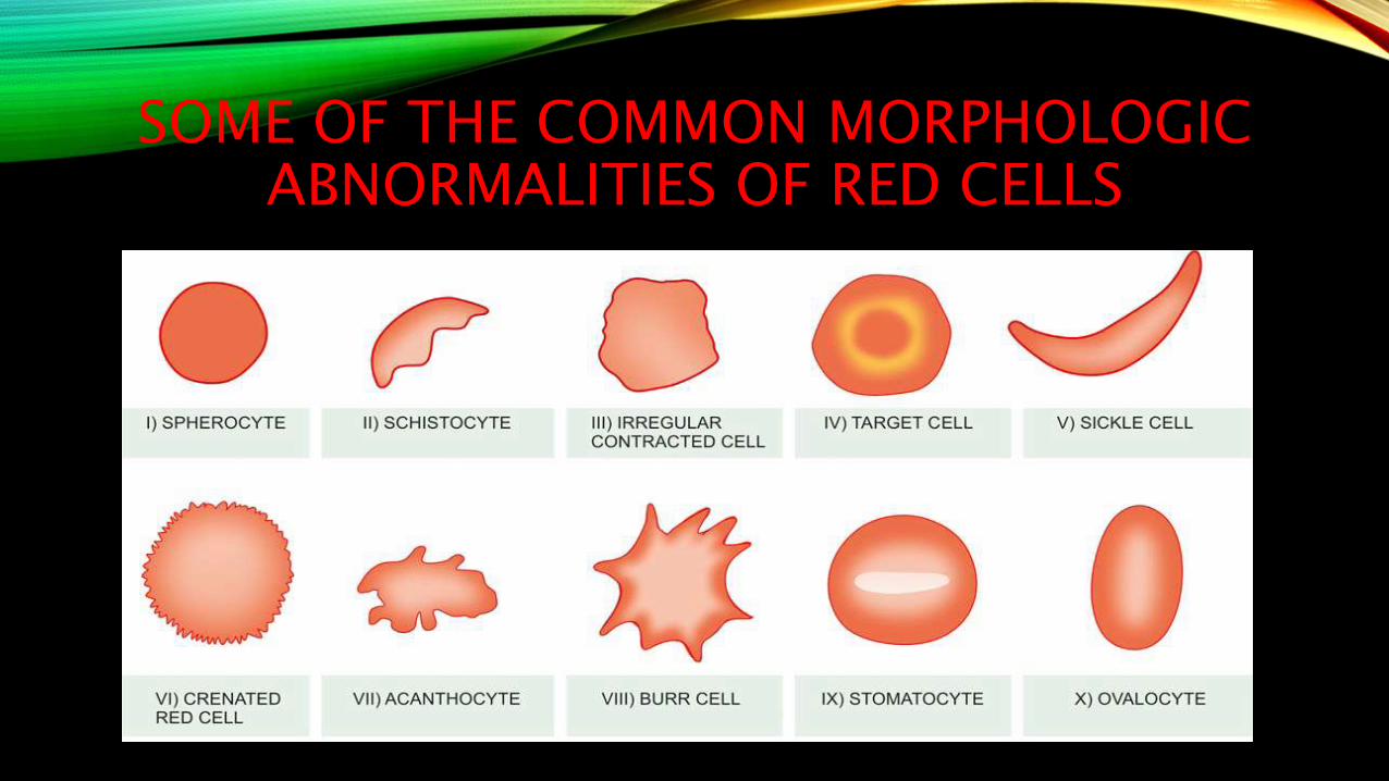
SOME OF THE COMMON MORPHOLOGIC ABNORMALITIES OF RED CELLS
CLASSIFICATION OF ANAEMIAS
• Several types of classifications of anaemias have been proposed. Two of the widely
accepted classifications are based on the pathophysiology and morphology.
PATHOPHYSIOLOGIC CLASSIFICATION
Depending Upon the pathophysiologic mechanism, anaemias are classified into 3 groups:
I. Anaemia due to blood loss
II. Anaemia due to impaired red cell formation
III. Anaemia due to increased red cell destruction (haemolytic anaemias)
ANAEMIA DUE TO BLOOD LOSS
This is further of 2 types:
A. Acute post-haemorrhagic anaemia
B. Anaemia of chronic blood loss

ANAEMIA DUE TO IMPAIRED RED CELL FORMATION
A disturbance due to impaired red cell production from various causes may produce anaemia. These are as under:
A. Cytoplasmicmaturation defects
1. Deficient haem synthesis: iron deficiency anaemia
2. Deficient globin synthesis: thalassaemic syndromes
B. Nuclear maturation defects
• Vitamin B12 and/or folic acid deficiency: megaloblastic anaemia
C. Haematopoietic stem cell proliferation and differentiation abnormality e.g.
1. Aplastic anaemia
2. Pure red cell aplasia
D. Bone marrow failure due to systemic diseases (anaemia of chronic disorders) e.g.
1. Anaemia of inflammation/infections, disseminated malignancy
2. Anaemia in renal disease
3. Anaemia due to endocrine and nutritional deficiencies (hypometabolic states)
4. Anaemia in liver disease
E. Bone marrow infiltration e.g.
1. Leukaemias
2. Lymphomas
3. Myelosclerosis
4. Multiple myeloma
F. Congenital anaemia e.g.
1. Sideroblastic anaemia
2. Congenital dyserythropoietic anaemia.
ANAEMIA DUE TO INCREASED RED CELL DESTRUCTION
This is further divided into 2 groups:
A. Intracorpuscular defect (hereditary and acquired).
B. Extracorpuscular defect (acquired haemolytic anaemias).
MORPHOLOGIC CLASSIFICATION
• Based on the red cell size, haemoglobin content and red cell indices, anaemias areclassified into 3 types:
1. Microcytic, hypochromic
2. Normocytic, normochromic
3. Macrocytic

MICROCYTIC, HYPOCHROMIC
• MCV, MCH, MCHC are all reduced e.g. in iron deficiency anaemia and in certain noniron
deficient anaemias (sideroblastic anaemia, thalassaemia, anaemia of chronic disorders).
NORMOCYTIC, NORMOCHROMIC
• MCV, MCH, MCHC are all normal e.g. after acute blood loss, haemolytic anaemias, bone
marrow failure, anaemia of chronic disorders.
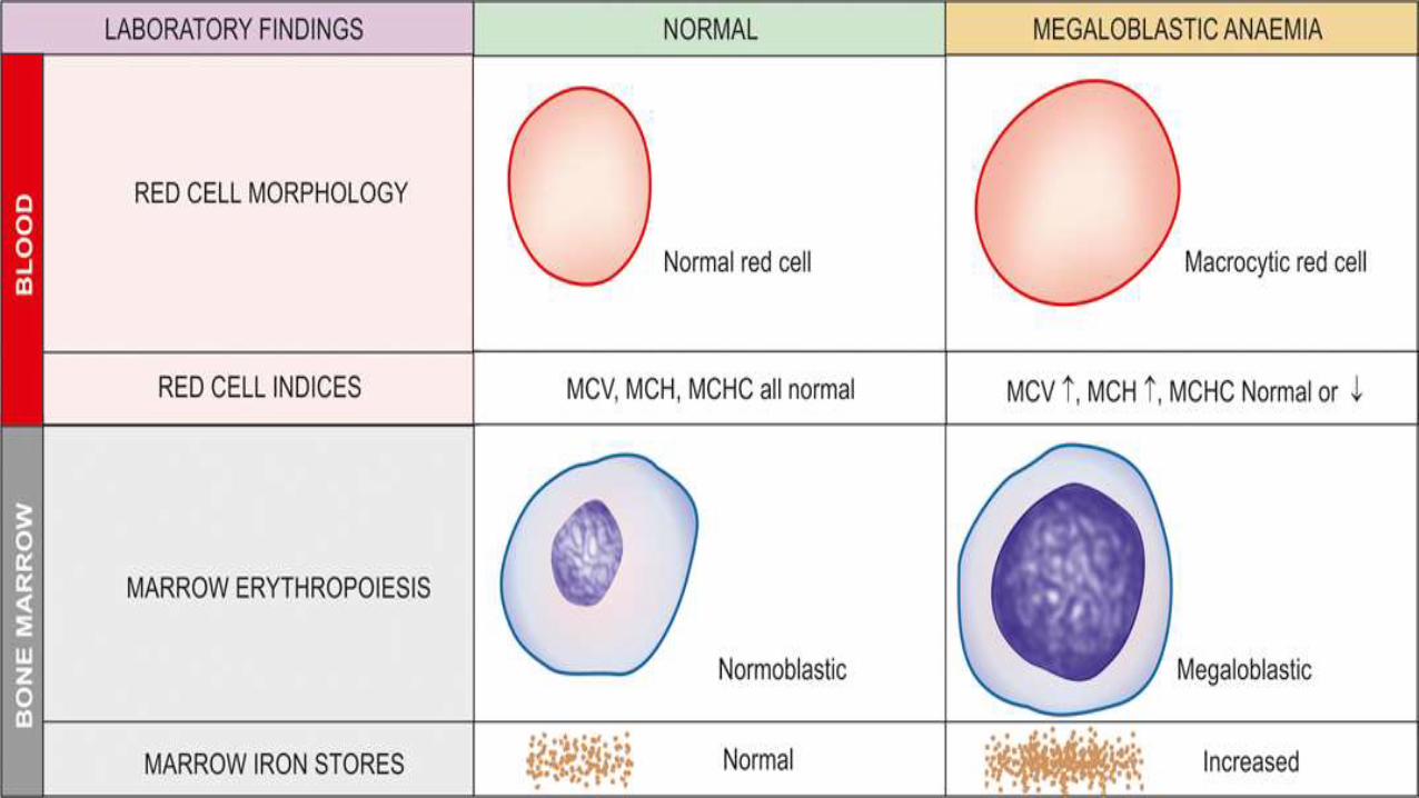
MACROCYTIC
• MCV is raised e.g. in megaloblastic anaemia due to deficiency of vitamin B12 or folic acid.
ANAEMIA DUE TO IMPAIRED RED CELL FORMATION(DEFICIENCY ANEMIAS)
• The anemia appearing as a result of breach of hemopoesis is called by three ways:
1. Deficient anemia
2. Impaired red cell production
3. Anemia of diminished erythropoiesis
Diminished erythropoiesis may be the result of deficiency of some vital substrate necessary for red
cell formation.
Some of the groups are discussed further……
IRON DEFICIENCY ANAEMIA
• Iron deficiency anaemia develops when the supply of iron is inadequate for the
requirement of haemoglobin synthesis.
• Initially, negative iron balance is covered by mobilisation from the tissue stores so as to
maintain haemoglobin synthesis. It is only after the tissue stores of iron are exhausted
that the supply of iron to the marrow becomes insufficient for haemoglobin formation
and thus a state of iron deficiency anaemia develops.
ETIOLOGY
I. INCREASED BLOOD LOSS
1. Uterine e.g. excessive menstruation in reproductive years, post-menopausal uterine bleeding
2. Gastrointestinal e.g. peptic ulcer, haemorrhoids hookworm infestation, cancer of stomach and
large bowel, oesophageal varices, hiatus hernia, chronic aspirin ingestion, ulcerative colitis,
diverticulosis
3. Renal tract e.g. haematuria, haemoglobinuria
4. Nose e.g. repeated epistaxis
5. Lungs e.g. haemoptysis
II. INCREASED REQUIREMENTS
1. Spurts of growth in infancy, childhood and adolescence
2. Prematurity
3. Pregnancy and lactation
III. INADEQUATE DIETARY INTAKE
1. Poor economic status
2. Anorexia e.g. in pregnancy
3. Elderly individuals due to poor dentition, apathy and financial constraints
IV. DECREASED ABSORPTION
1. Partial or total gastrectomy
2. Achlorhydria
3. Intestinal malabsorption such as in coeliac disease
CLINICAL FEATURES
• The onset of iron deficiency anaemia is generally slow. The usual symptoms areweakness, fatigue, dyspnoea on exertion, palpitations and pallor of the skin, mucousmembranes and sclerae. Older patients may develop angina and congestive cardiacfailure. Menorrhagia is a common symptom in iron deficient women.
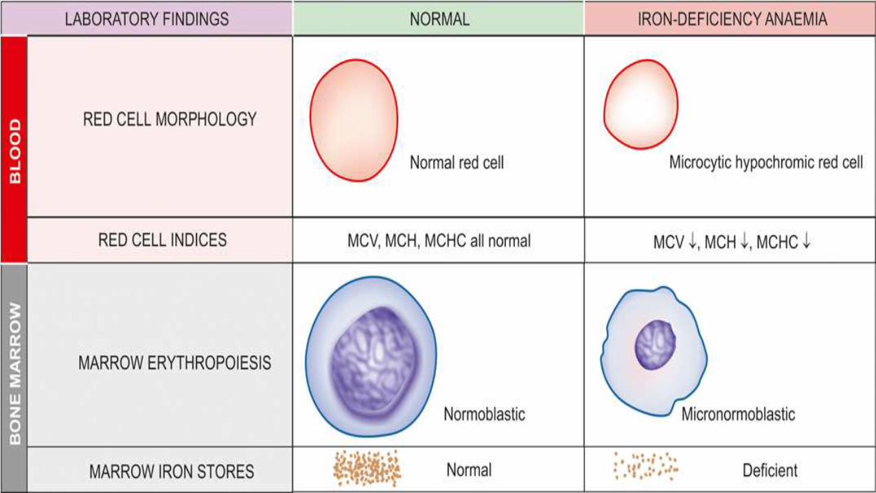
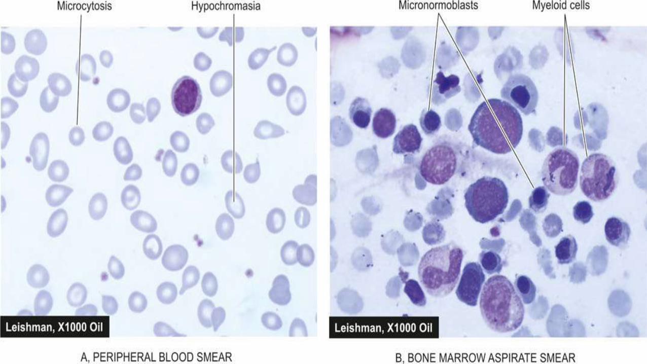
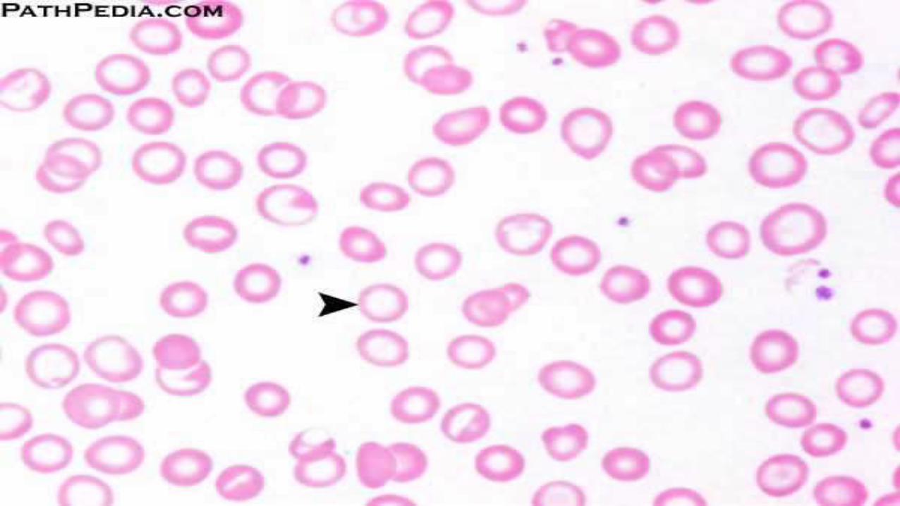
In peripheral blood:
• Red cells are pale(hypochromic) and small(microcytic)
In marrow:
• Hyperplasia of normoblasts, associated with loss of sideroblastsand absence of stainableiron in the reticuloendothelial cells.
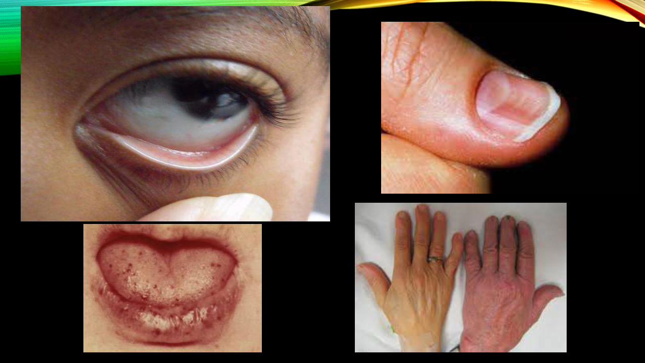
EPITHELIAL TISSUE CHANGES:
• Long-standing chronic iron deficiency anaemia causes epithelial tissue changes in some
patients.
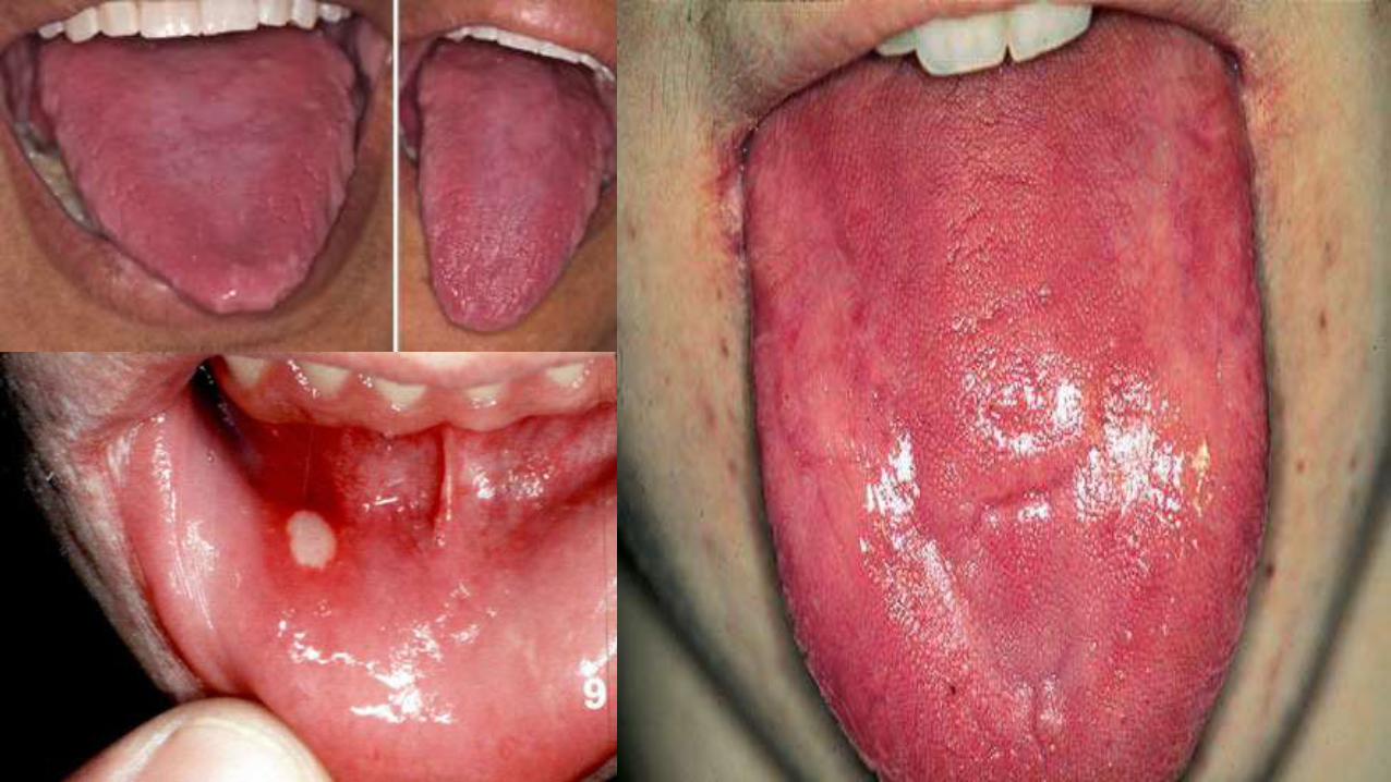
• The changes occur in the nails (koilonychias or spoon-shaped nails), tongue (atrophic
glossitis), mouth (angular stomatitis), and oesophagus causing dysphagia from
development of thin, membranous webs at the postcricoid area (Plummer-Vinson
syndrome).
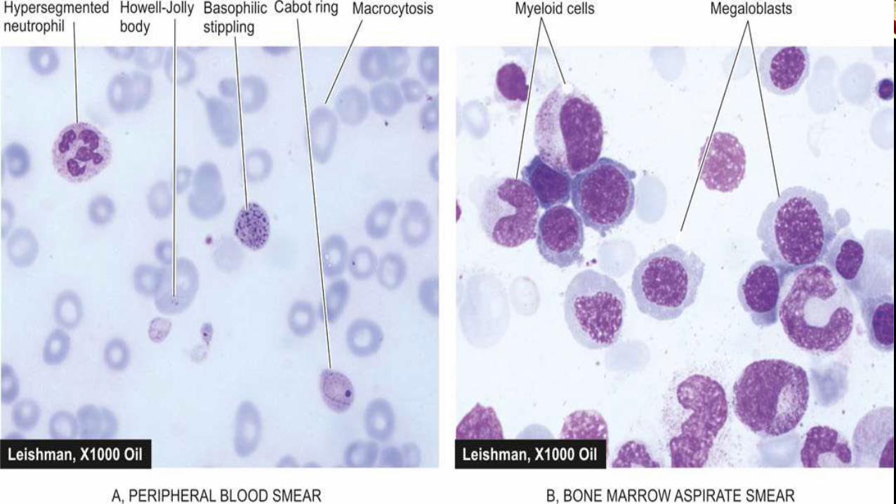
MEGALOBLASTIC ANAEMIAS
• Megaloblastic anaemias are associated with macrocytic blood picture and megaloblastic
marrow erythropoiesis. This group is due to deficiency of vitamin B12 and/or folate and
includes megaloblastic picture from the following two types of etiologies:
• 1. Nutritional deficiency of vitamin B12 or folate, or combined deficiency, most common
in developing countries.
• 2. Deficiency of intrinsic factor, causing impaired absorption of vitamin B12 called
pernicious anaemia
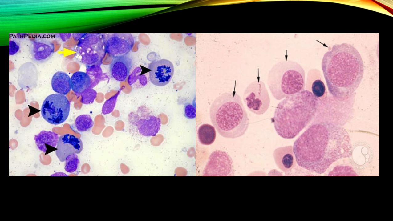
• The megaloblastic anaemias are disorders caused by impaired DNA synthesis and are
characterised by a distinctive abnormality in the haematopoietic precursors in the bone
marrow in which the maturation of the nucleus is delayed relative to that of the
cytoplasm.
ETIOLOGY
I. VITAMIN B12 DEFICIENCY
A. Inadequate dietary intake e.g. strict vegetarians, breast-fed infants.
B. Malabsorption
1. Gastric causes: pernicious anaemia, gastrectomy, congenital lack of intrinsic factor.
2. Intestinal causes: tropical sprue, ileal resection, Crohn’s disease, intestinal blind loop
syndrome, fish-tapeworm infestation.
II. FOLATE DEFICIENCY
A. Inadequate dietary intake e.g. in alcoholics, teenagers, infants, old age, poverty.
poverty.
B. Malabsorption e.g. in tropical sprue, coeliac disease, partial gastrectomy, jejunal
PERNICIOUS ANAEMIA
• Pernicious anemia (PA) is an autoimmune disorder in which the body fails to make
enough healthy red blood cells (RBCs). The body requires vitamin B-12 and a type of
protein called intrinsic factor (IF) to make red blood cells. Vitamin B-12, or cobalamin, is
found in certain foods and medications. IF is a protein made by the stomach’s mucosal
(mucus-secreting) cells, called parietal cells. When vitamin B-12 enters the body, it binds
with IF. The two are then absorbed in the last part of the small intestine.
• In the majority of cases of PA, the body’s immune system attacks and destroys the
stomach’smucosal cells. IF can no longer be made, and vitamin B-12 cannot be absorbed.
ETIOLOGY
• Vitamin B12 deficiency caused by a lack of intrinsic factor in stomach secretions.
• Vitamin B12 deficiency caused by surgery that removes or bypasses the end of the small
intestine where vitamin B12 is absorbed
• The inability to make intrinsic factor may be the result of several factors, such as chronic
gastritis, gastrectomy (removal of all or part of the stomach), or an autoimmune
condition (the body attacks its own tissues)
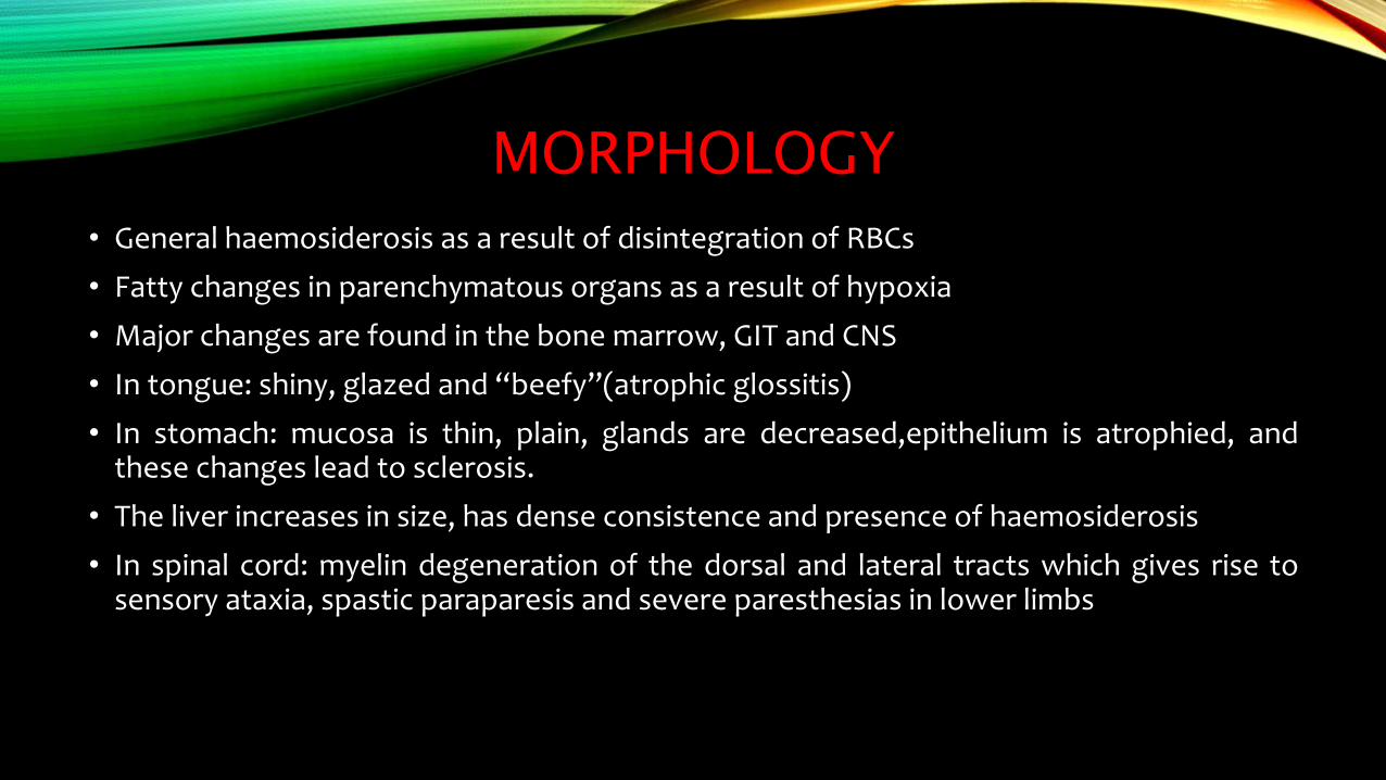
MORPHOLOGY
• General haemosiderosis as a result of disintegration of RBCs
• Fatty changes in parenchymatous organs as a result of hypoxia
• Major changes are found in the bone marrow, GIT and CNS
• In tongue: shiny, glazed and “beefy”(atrophic glossitis)
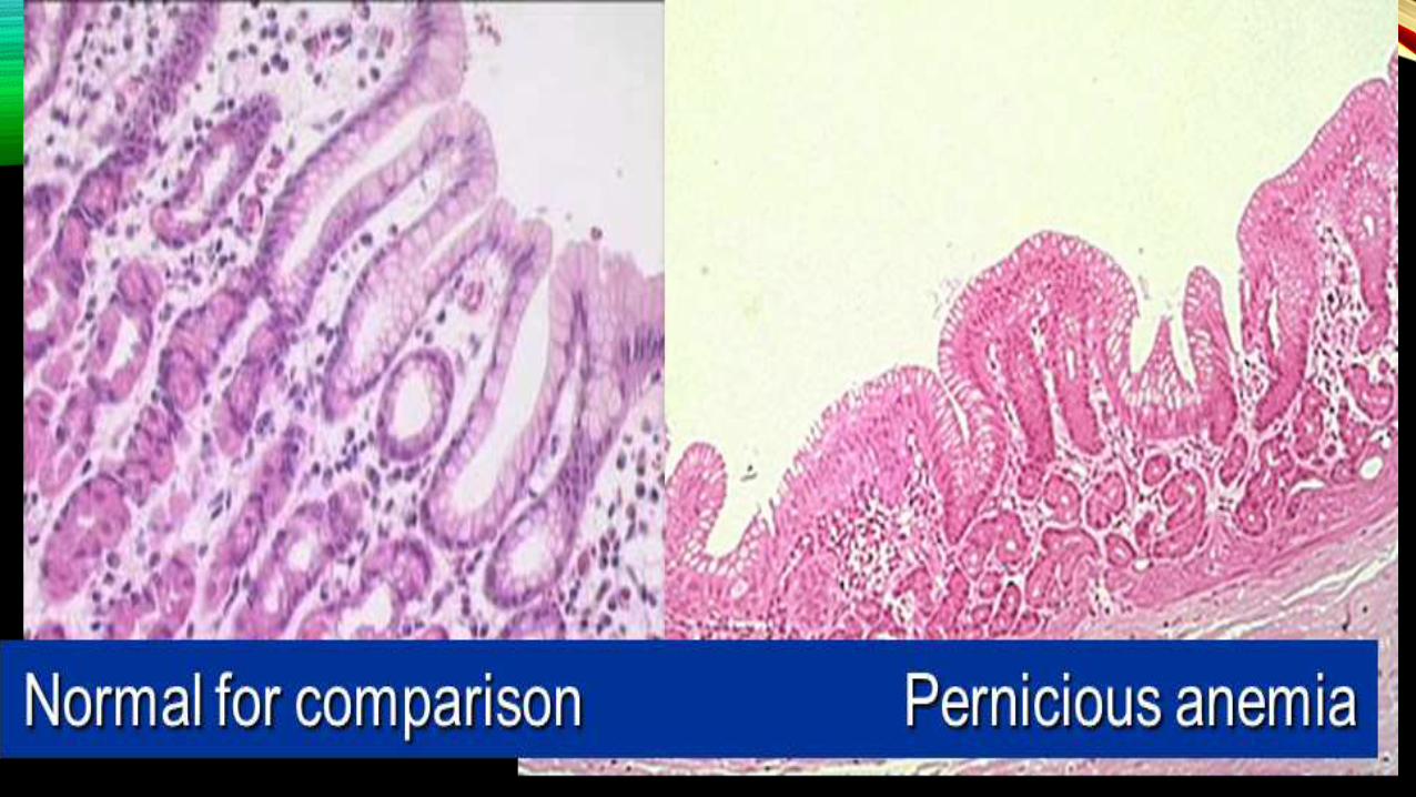
• In stomach: mucosa is thin, plain, glands are decreased,epithelium is atrophied, andthese changes lead to sclerosis.
• The liver increases in size, has dense consistence and presence of haemosiderosis
• In spinal cord: myelin degeneration of the dorsal and lateral tracts which gives rise tosensory ataxia, spastic paraparesis and severe paresthesias in lower limbs
FOLIC ACID DEFICIENCY ANAEMIA
• Folic acid is the synthetic version of the vitamin folate, also called B9
• They induces a megaloblastic anemia
• Neurologic changes and gastric atrophy is absent
CLINICAL FEATURES
Symptoms of folic acid deficiency include:
• fatigue
• mouth sores
• gray hair
• swollen tongue
• poor growth (also among the chief symptoms of malnutrition)
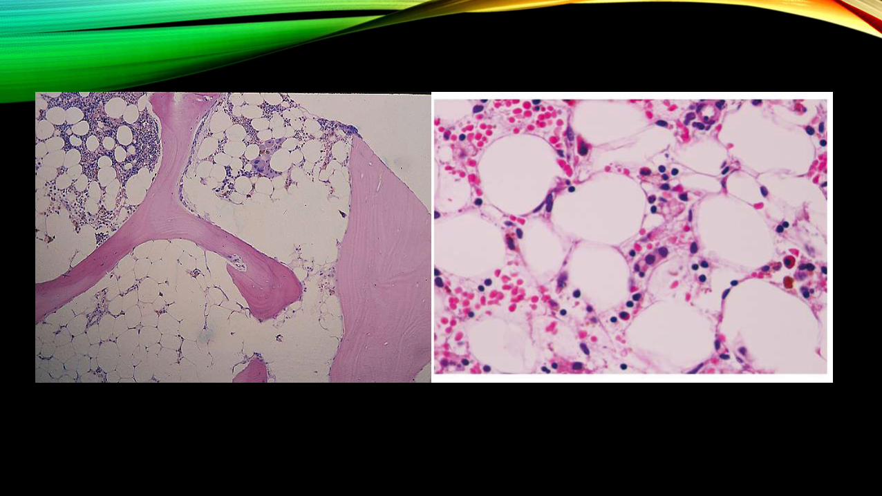
APLASTIC ANAEMIA
• Aplastic anaemia is defined as pancytopenia (i.e. simultaneous presence of anaemia,
leucopenia and thrombocytopenia) resulting from aplasia of the bone marrow.
• The underlying defect in all cases appears to be sufficient reduction in the number of
haematopoietic pluripotent stem cells which results in decreased or total absence of
these cells for division and differentiation.
ETIOLOGY
A. PRIMARY APLASTIC ANAEMIA
1. Fanconi’s anaemia (congenital)
2. Immunologically-mediated (acquired)
B. SECONDARY APLASTIC ANAEMIA
1. Drugs
2. Toxic chemicals e.g. benzene derivatives, insecticides, arsenicals.
3. Infections e.g. infectious hepatitis, EB virus infection, AIDS, other viral illnesses.
4. Miscellaneous e.g. association with SLE and therapeutic X-rays
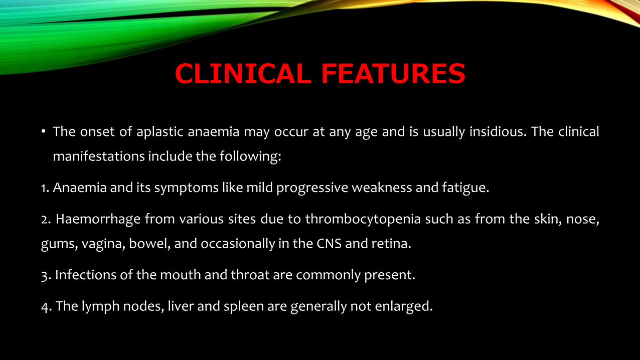
CLINICAL FEATURES
• The onset of aplastic anaemia may occur at any age and is usually insidious. The clinical
manifestations include the following:
1. Anaemia and its symptoms like mild progressive weakness and fatigue.
2. Haemorrhage from various sites due to thrombocytopenia such as from the skin, nose,
gums, vagina, bowel, and occasionally in the CNS and retina.
3. Infections of the mouth and throat are commonly present.
4. The lymph nodes, liver and spleen are generally not enlarged.
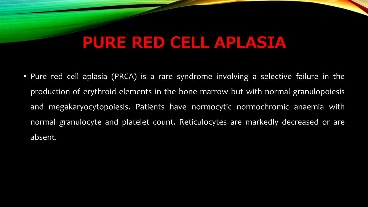
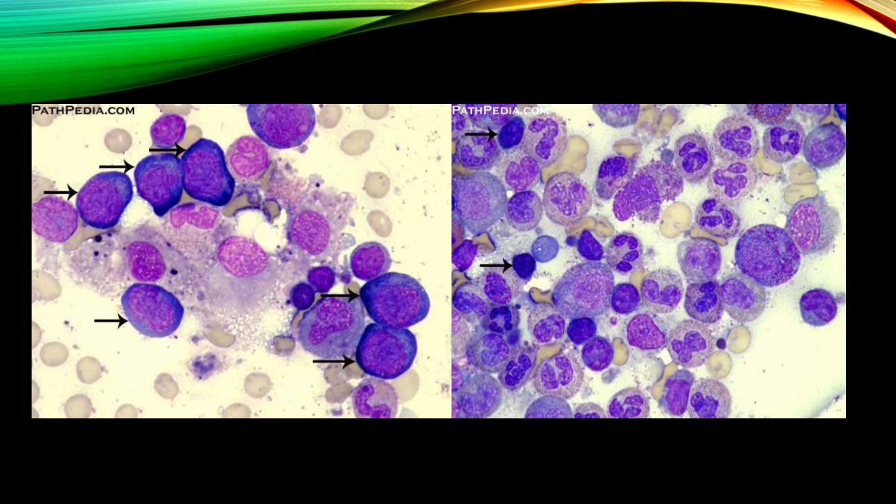
PURE RED CELL APLASIA
• Pure red cell aplasia (PRCA) is a rare syndrome involving a selective failure in the
production of erythroid elements in the bone marrow but with normal granulopoiesis
and megakaryocytopoiesis. Patients have normocytic normochromic anaemia with
normal granulocyte and platelet count. Reticulocytes are markedly decreased or are
absent.


![An Overview of the Anemias[1].ppt - School of Medicine · AO i fth A iAn Overview of the Anemias Iron Deficiency MegaloblasticIron Deficiency, Megaloblastic, ... {Malabsorption: Pernicious](https://img.dokumen.tips/doc/110x75/5ae1da537f8b9a5d648bed5f/an-overview-of-the-anemias1ppt-school-of-i-fth-a-ian-overview-of-the-anemias.jpg)