Embed Size (px)
Citation preview

Keseimbangan Cairan Tubuh & Elektrolit
Departemen Fisiologi
Fak. Kedokteran USU

Total Cairan Tubuh Infant mempunyai kandungan lemak, tulang
lebih rendah & sedangkan kandungan air sebesar 73% atau lebih
Total cairan tubuh berkurang seiring pertambahan usia
Pria sekitar 60% berat badan; Wanita sekitar 50% berat badan
Hal ini disebabkan karena pada wanita: Kandungan lemak lebih tinggi Otot rangka lebih sedikit
Pada usia tua, kandungan air sekitar 45%

Fluid Compartments Water occupies two main fluid compartments Intracellular fluid (ICF) – about two thirds by
volume, contained in cells Extracellular fluid (ECF) – consists of two major
subdivisions Plasma – the fluid portion of the blood Interstitial fluid (IF) – fluid in spaces between cells
Other ECF – lymph, cerebrospinal fluid, eye humors, synovial fluid, serous fluid, and gastrointestinal secretions


Copyright © 2004 Pearson Education, Inc., publishing as Benjamin Cummings
Electrolyte Composition of Body Fluids
Figure 26.2

Beberapa mekanisme sehingga suatu zat dapat melewati membran
1. Difusi
2. Difusi yang dipermudah
3. Osmosis
4. Endositosis dan eksositosis, dan
5. Epithelial transport


Difusi Yang Dipermudah
Transport Aktif Transport Aktif Primer Pompa
Natrium-Kalium Transport Aktif Primer Pompa
Kalsium Transport Aktif Sekunder:
Kotransport Natrium pada Glukosa dan Asam Amino

Transport Aktif
Saat membran sel menggerakkan molekul zat untuk mendaki melawan gradien konsentrasi (atau mendaki melawan arus listrik atau melawan gradien tekanan,) maka proses ini disebut transport aktif.
Memerlukan energi Transport aktif primer: dalam bentuk ATP Transport aktif sekunder: gradien
konsentrasi ion natrium

Osmosis
Keadaan pergerakan netto air yang disebabkan karena adanya pebedaan konsentrasi disebut osmosis

Endositosis dan Eksositosis
Endositosis merupakan suatu mekanisme dimana membran meliputi (membungkus) bahan – bahan yang penting atau cairan ekstraseluler dan isinya.
Dua bentuk dasar dari endositosis adalah pinositosis dan fagositosis.
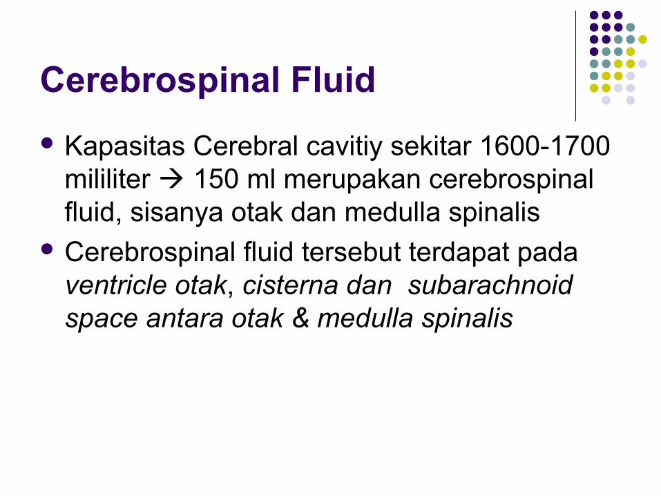
Cerebrospinal Fluid
Kapasitas Cerebral cavitiy sekitar 1600-1700 mililiter 150 ml merupakan cerebrospinal fluid, sisanya otak dan medulla spinalis
Cerebrospinal fluid tersebut terdapat pada ventricle otak, cisterna dan subarachnoid space antara otak & medulla spinalis

Fungsi utama CSF sebagai bantalan otak, keduanya memiliki gravitasi yg hampir sama

Blood–Cerebrospinal Fluid andBlood-Brain Barriers
Terdapat antara darah & CSF & cairan otak Barriers antara choroid plexus dan membran
kapiler jaringan semua area parenkhim otak kecuali sebagian hipothalamus, kelenjar pineal, area postrema yang mana pada daerah ini bahan-bahan (substans) jaringan lebih mudah berdifusi ke jaringan

Pembentukan
CSF dibentuk dengan kecepatan 500ml per hari. 2/3 dari CSF berasal dari plexus choroideus
pada ventrikel 4, terutama pada 2 ventrikel bagian lateral
Sebagian CSF merupakan hasil sekresi ependymal seluruh permukaan ventrikel dan membran arachnoid, dan sebagian berasal dari otak sendiri melalui bagian perivascular sekeliling pembuluh darah yang melewati otak

Aliran CSF Melalui choroid plexuses dan kemudian melalui
sistem cerebrospinal. Cairan yang disekresikan di lateral ventricles mula-mula menuju third ventricle; kemudian setelah bertambah mengalir kebawah menuju aqueduct of Sylvi sampai fourth ventricle, tempat cairan bertambah.
Akhirnya cairan keluar melalui 3 lubang kecil ; 2 foramen Luschka sebelah lateral dan sebuah midline foramen Magendie, memasuki cisterna magna, (ruangan cairan yang berada di belakang medulla dan antar acerebellum). Cisterna magna merupakan lanjutan ruangan subarachnoid yang berada di sekeliling otak dan medulla spinalis

Hampir seluruh CSF kemudian mengalir dari cisterna magna melalui ruang subarachnoid sekeliling cerebrum. CSF mengalir melalui arachnoidal villi menuju sinus venous sagittal dan sinuses venous yang lain. Dan CSF dikosongkan menuju aliran vena melalui pori-pori vili ini


Teori Pembentukan CSF
Sekresi CSF oleh plexus choroideus merupakan active transport sodium ions melalui epithelial cells yang berada di bagian luar plexus.
Ion sodium menarik ion chlorida. Kedua ion in meningkatkan konsentrasi sodium chloride di CSF yang mana kemudian segera menyebabkan osmosis air

Glukosa memasuki CSF dan ion potassium dan bicarbonate keluar CSF menuju kapiler

Susunan CSF
Karakteristik CSF sebagai berikut: Tekanan osmotik, kira-kira sama dengan plasma, konsentrasi ion , juga kira-kira sama dengan plasma; ion chlorida sekitar 15 per cent Lebih besar dari plasma; ion potassium, kira-kira 40 per cent lebih sedikit; dan glucosa, sekitar 30 per cent lebih sedikit

Copyright © 2004 Pearson Education, Inc., publishing as Benjamin Cummings
Water Intake and Output
Figure 26.4

Copyright © 2004 Pearson Education, Inc., publishing as Benjamin Cummings
Acid-Base Balance
Normal pH of body fluids
Arterial blood is 7.4
Venous blood and interstitial fluid is 7.35
Intracellular fluid is 7.0
Alkalosis or alkalemia – arterial blood pH rises above 7.45
Acidosis or acidemia – arterial pH drops below 7.35 (physiological acidosis)

Copyright © 2004 Pearson Education, Inc., publishing as Benjamin Cummings
Regulation of Water Intake: Thirst Mechanism
Figure 26.5

Copyright © 2004 Pearson Education, Inc., publishing as Benjamin Cummings
Figure 26.6
Mechanisms and Consequences of ADH Release

Copyright © 2004 Pearson Education, Inc., publishing as Benjamin Cummings
Regulation of Sodium Balance: Aldosterone
Figure 26.8

Copyright © 2004 Pearson Education, Inc., publishing as Benjamin Cummings
Figure 26.10
Mechanisms and Consequences of ANP Release

Copyright © 2004 Pearson Education, Inc., publishing as Benjamin Cummings
Disorders of Water Balance: Dehydration
Water loss exceeds water intake and the body is in negative fluid balance
Causes include: hemorrhage, severe burns, prolonged vomiting or diarrhea, profuse sweating, water deprivation, and diuretic abuse
Signs and symptoms: cottonmouth, thirst, dry flushed skin, and oliguria
Prolonged dehydration may lead to weight loss, fever, and mental confusion
Other consequences include hypovolemic shock and loss of electrolytes

Copyright © 2004 Pearson Education, Inc., publishing as Benjamin Cummings
Figure 26.7a
Disorders of Water Balance: Dehydration
Excessive loss of H2O from ECF
1 2 3ECF osmotic pressure rises
Cells lose H2O to ECF by osmosis; cells shrink
(a) Mechanism of dehydration

Copyright © 2004 Pearson Education, Inc., publishing as Benjamin Cummings
Renal insufficiency or an extraordinary amount of water ingested quickly can lead to cellular overhydration, or water intoxication
ECF is diluted – sodium content is normal but excess water is present
The resulting hyponatremia promotes net osmosis into tissue cells, causing swelling
These events must be quickly reversed to prevent severe metabolic disturbances, particularly in neurons
Disorders of Water Balance: Hypotonic Hydration

Copyright © 2004 Pearson Education, Inc., publishing as Benjamin Cummings
Figure 26.7b
Disorders of Water Balance: Hypotonic Hydration
Excessive H2O enters the ECF
1 2 ECF osmotic pressure falls
3 H2O moves into cells by osmosis; cells swell
(b) Mechanism of hypotonic hydration

Copyright © 2004 Pearson Education, Inc., publishing as Benjamin Cummings
Disorders of Water Balance: Edema
Atypical accumulation of fluid in the interstitial space, leading to tissue swelling
Caused by anything that increases flow of fluids out of the bloodstream or hinders their return
Factors that accelerate fluid loss include:
Increased blood pressure, capillary permeability
Incompetent venous valves, localized blood vessel blockage
Congestive heart failure, hypertension, high blood volume

Copyright © 2004 Pearson Education, Inc., publishing as Benjamin Cummings
Edema
Hindered fluid return usually reflects an imbalance in colloid osmotic pressures
Hypoproteinemia – low levels of plasma proteins
Forces fluids out of capillary beds at the arterial ends
Fluids fail to return at the venous ends
Results from protein malnutrition, liver disease, or glomerulonephritis, sindroma nefrotik

Copyright © 2004 Pearson Education, Inc., publishing as Benjamin Cummings
Edema
Blocked (or surgically removed) lymph vessels:
Cause leaked proteins to accumulate in interstitial fluid
Exert increasing colloid osmotic pressure, which draws fluid from the blood
Interstitial fluid accumulation results in low blood pressure and severely impaired circulation

Occur s with net loss of sodium or net water excess
Kidney disease with salt wasting, adrenal insufficiency, GI losses, increased sweating, diuretics, SIADHS&S: personality change, postural hypotension, postural dizziness, abd cramping, diarrhea, tachycardia, convulsions and coma
Caused by excess water loss or overall sodium excess
Excess salt intake, hypertonic solutions, excess aldosterone,diabetes insipidus, increased water loss, water deprivation S&S: thirst, dry, flushed skin, dry, stick tongue and mucous
membranes
Hypernatremia (Na > 145, sp gravity < 1.010)
Hyponatremia
(Na < 135,
sp gravi ty > 1.030

Copyright © 2004 Pearson Education, Inc., publishing as Benjamin Cummings
Regulation of Potassium Balance
Hyperkalemia and hypokalemia can:
Disrupt electrical conduction in the heart
Lead to sudden death
Hydrogen ions shift in and out of cells
Leads to corresponding shifts in potassium in the opposite direction
Interferes with activity of excitable cells

Copyright © 2004 Pearson Education, Inc., publishing as Benjamin Cummings
Regulation of Calcium
Ionic calcium in ECF is important for:
Blood clotting
Cell membrane permeability
Secretory behavior
Hypocalcemia:
Increases excitability
Causes muscle tetany

Copyright © 2004 Pearson Education, Inc., publishing as Benjamin Cummings
Regulation of Calcium
Hypercalcemia:
Inhibits neurons and muscle cells
May cause heart arrhythmias
Calcium balance is controlled by parathyroid hormone (PTH) and calcitonin

Copyright © 2004 Pearson Education, Inc., publishing as Benjamin Cummings
Regulation of Calcium and Phosphate
PTH promotes increase in calcium levels by targeting:
Bones – PTH activates osteoclasts to break down bone matrix
Small intestine – PTH enhances intestinal absorption of calcium
Kidneys – PTH enhances calcium reabsorption and decreases phosphate reabsorption
Calcium reabsorption and phosphate excretion go hand in hand

Copyright © 2004 Pearson Education, Inc., publishing as Benjamin Cummings
Influence of Calcitonin
Released in response to rising blood calcium levels
Calcitonin is a PTH antagonist, but its contribution to calcium and phosphate homeostasis is minor to negligible
InterActive Physiology®: Fluid, Electrolyte, and Acid/Base Balance: Electrolyte HomeostasisPLAYPLAY

Copyright © 2004 Pearson Education, Inc., publishing as Benjamin Cummings
Acid-Base Balance
Normal pH of body fluids
Arterial blood is 7.4
Venous blood and interstitial fluid is 7.35
Intracellular fluid is 7.0
Alkalosis or alkalemia – arterial blood pH rises above 7.45
Acidosis or acidemia – arterial pH drops below 7.35 (physiological acidosis)

Copyright © 2004 Pearson Education, Inc., publishing as Benjamin Cummings
Hydrogen Ion Regulation
Concentration of hydrogen ions is regulated sequentially by:
Chemical buffer systems – act within seconds
The respiratory center in the brain stem – acts within 1-3 minutes
Renal mechanisms – require hours to days to effect pH changes

Copyright © 2004 Pearson Education, Inc., publishing as Benjamin Cummings
Respiratory Acidosis and Alkalosis
Result from failure of the respiratory system to balance pH
PCO2 is the single most important indicator of respiratory inadequacy
PCO2 levels
Normal PCO2 fluctuates between 35 and 45 mm Hg
Values above 45 mm Hg signal respiratory acidosis
Values below 35 mm Hg indicate respiratory alkalosis

Copyright © 2004 Pearson Education, Inc., publishing as Benjamin Cummings
Respiratory Acidosis and Alkalosis
Respiratory acidosis is the most common cause of acid-base imbalance
Occurs when a person breathes shallowly, or gas exchange is hampered by diseases such as pneumonia, cystic fibrosis, or emphysema, impaired activity of diaphragm muscle and impaired respiratory control in the brain stem
Respiratory alkalosis is a common result of hyperventilation and is caused by low level of oxygen in the plasma, meningitis, head injury and anxiety

Copyright © 2004 Pearson Education, Inc., publishing as Benjamin Cummings
Metabolic Acidosis
All pH imbalances except those caused by abnormal blood carbon dioxide levels
Metabolic acid-base imbalance – bicarbonate ion levels above or below normal (22-26 mEq/L)
Metabolic acidosis is the second most common cause of acid-base imbalance
Typical causes are ingestion of too much alcohol and excessive loss of bicarbonate ions,such as diarrhea, and excessive vomitting
Other causes include accumulation of lactic acid, shock, ketosis in diabetic crisis, starvation, and kidney failure,high extracellular K+

Copyright © 2004 Pearson Education, Inc., publishing as Benjamin Cummings
Metabolic Alkalosis
Rising blood pH and bicarbonate levels indicate metabolic alkalosis
Typical causes are:
Vomiting of the acid contents of the stomach
Hypokalemia
Intake of excess base (e.g., from antacids)
Constipation, in which excessive bicarbonate is reabsorbed

Copyright © 2004 Pearson Education, Inc., publishing as Benjamin Cummings
Respiratory and Renal Compensations
Acid-base imbalance due to inadequacy of a physiological buffer system is compensated for by the other system
The respiratory system will attempt to correct metabolic acid-base imbalances
The kidneys will work to correct imbalances caused by respiratory disease

Copyright © 2004 Pearson Education, Inc., publishing as Benjamin Cummings
Respiratory Compensation
In metabolic acidosis:
The rate and depth of breathing are elevated
Blood pH is below 7.35 and bicarbonate level is low
As carbon dioxide is eliminated by the respiratory system, PCO2 falls below normal
In respiratory acidosis, the respiratory rate is often depressed and is the immediate cause of the acidosis

Copyright © 2004 Pearson Education, Inc., publishing as Benjamin Cummings
Respiratory Compensation
In metabolic alkalosis:
Compensation exhibits slow, shallow breathing, allowing carbon dioxide to accumulate in the blood
Correction is revealed by:
High pH (over 7.45) and elevated bicarbonate ion levels
Rising PCO2

Copyright © 2004 Pearson Education, Inc., publishing as Benjamin Cummings
Renal Compensation
To correct respiratory acid-base imbalance, renal mechanisms are stepped up
Acidosis has high PCO2 and high bicarbonate levels
The high PCO2 is the cause of acidosis
The high bicarbonate levels indicate the kidneys are retaining bicarbonate to offset the acidosis

Copyright © 2004 Pearson Education, Inc., publishing as Benjamin Cummings
Renal Compensation
Alkalosis has Low PCO2 and high pH
The kidneys eliminate bicarbonate from the body by failing to reclaim it or by actively secreting it
InterActive Physiology®: Fluid, Electrolyte, and Acid/Base Balance: Acid/Base HomeostasisPLAYPLAY