Embed Size (px)
Citation preview

1
RADIOLOGICAL FEATURES OF
BRONCHOGENIC CARCINOMA
BYDR. MAIMUNA ABDULKARIM HALLIRU
RADIOLOGY DEPARTMENTAMINU KANO TEACHING HOSPITAL
KANO.13th, June 2013

2
SYNOPSIS
Introduction
Radiological Features
Radiology Of Complications
Differential Diagnoses

3
INTRODUCTION
Definition: Bronchogenic carcinoma is a malignant neoplasm of the lung arising from the epithelium of the bronchus or bronchiole.

4
Epidemiology:
Carcinoma of the bronchus is the commonest fatal malignancy in adult males in the western world (35% of all cancer deaths).
Commoner in males but incidence in women is rising(21% of all cancer deaths).
Most cases occur between 40-70 years of age and peaks in the 50-60 age range; it is unusual below the age of 30 years.

5
Epidemiology: The country with the highest incidence of
lung cancer among males is the United Kingdom .
In general, the incidence of lung cancer in industrialized western countries is increased compared to third world countries

6
Epidemiology:
In a 30 month prospective study conducted by N. Ezemba et al at the University of Nigeria Teaching Hospital, Enugu from Jan 2003-June 2005; 51 new cases were identified during the study period.
The ages ranged from 30-81 years, mean of 56 ± 21years with a male: female ratio of 2:1.
In 42% of the males there was a history of cigarette smoking. No history of smoking found among the females.
Challenges of lung cancer in a developing country by Ndubueze Ezemba,
Eyo Ekpe & John Eze Nigerian Journal of Medicine 2012 Apr-Jun;21(2):214-7.

7
Risk factors: Cigarette Smoking: The single most
important aetiological factor is cigarette smoking.This is dose related, the risk being proportional to number of cigarettes smoked.
Second hand smoke is also bad. Non-smoking women married to smokers had a 1.2x risk of developing cancer.

8
Risk Factors:
Concomitant Disease: It is also reported that lung scarring ( tuberculosis, scleroderma, infarction, bronchiectasis ) is associated with the incidence of lung cancer especially adenocarcinoma.

9
Risk Factors:
Industrial Exposure: radiation exposure, asbestos, workers exposed to nickel/ chromate/ arsenic/ and newspaper industry workers.
Air pollution: both indoor and outdoor especially radon gas which may be the second leading cause for lung cancer with up to 20,000 deaths per year.
Combined risk factors approach 100% risk.
10
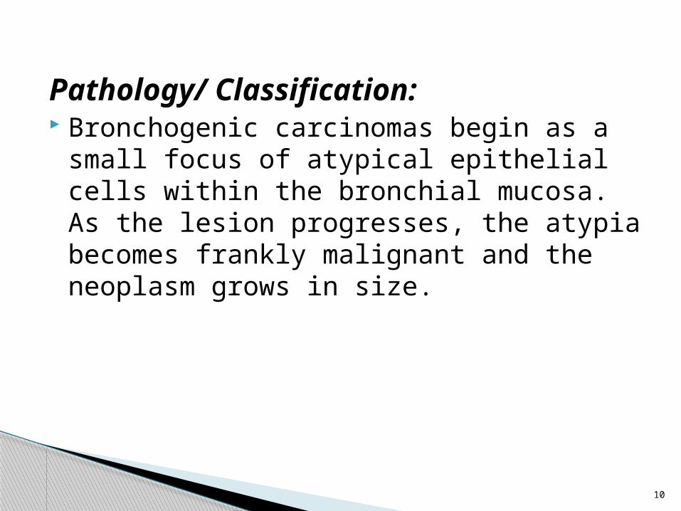
Pathology/ Classification: Bronchogenic carcinomas begin as a small
focus of atypical epithelial cells within the bronchial mucosa. As the lesion progresses, the atypia becomes frankly malignant and the neoplasm grows in size.

11
According to anatomy: (1)Central lung cancer,mostly is squamous
cell carcinoma and small cell carcinoma. (2) peripheral lung cancer, mostly is
adenocarcinoma and large cell carcinoma.

12
According to histologic classification: (1) Small cell lung cancer(SCLC) 20%
(2 ) Non-small cell lung cancer(NSCLC) includes ;
Adenocarcinoma 30-40%
Squamous cell carcinoma 30-40% Large cell Undifferentiated carcinoma 10%
13
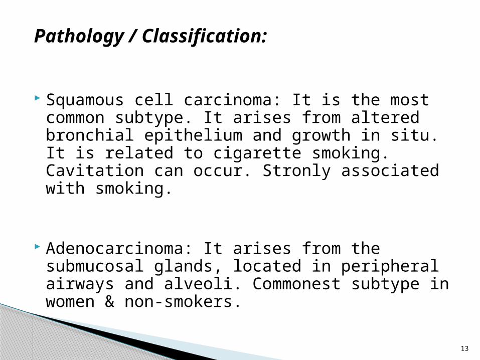
Pathology / Classification:
Squamous cell carcinoma: It is the most common subtype. It arises from altered bronchial epithelium and growth in situ. It is related to cigarette smoking. Cavitation can occur. Stronly associated with smoking.
Adenocarcinoma: It arises from the submucosal glands, located in peripheral airways and alveoli. Commonest subtype in women & non-smokers.

14
Pathology / Classification:
Large-cell carcinoma: are usually located peripherally. They can be quite large. Strongly associated with smoking.

15
Small Cell Lung Cancer belongs in a group of tumors derived from neuroendocrine cells that are responsible for the production and secretion of specific peptide products. They may be related to paraneoplastic syndromes such as syndrome of inappropriate ADH secretion, Cushing’s syndrome etc.
16
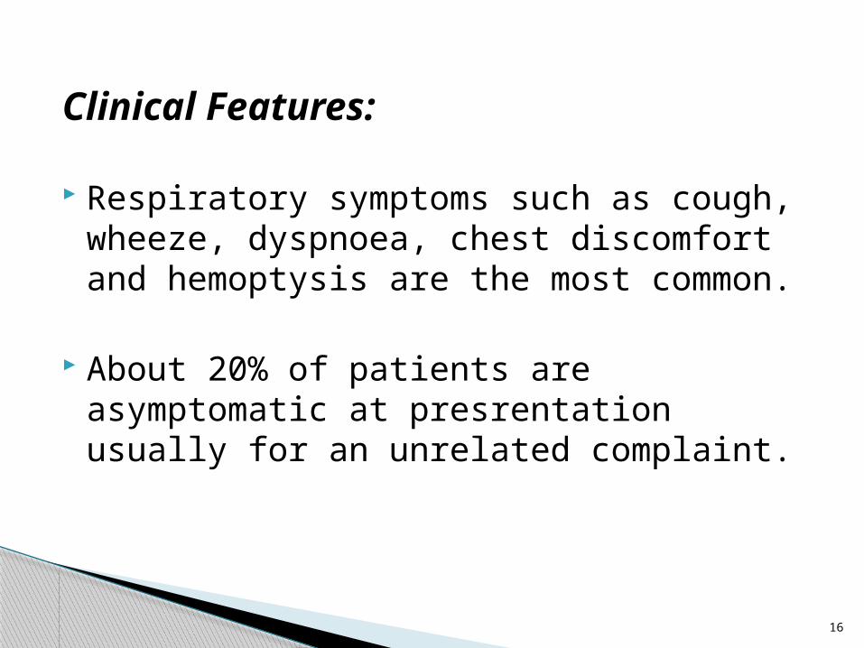
Clinical Features:
Respiratory symptoms such as cough, wheeze, dyspnoea, chest discomfort and hemoptysis are the most common.
About 20% of patients are asymptomatic at presrentation usually for an unrelated complaint.

17
Clinical Features:
Other presentations include superior vena caval obstruction, Horner’s syndrome, dysphagia and signs of pericardial tamponade.
Pneumonia particularly if it does not respond to treatment may be due to an underlying neoplasm.

18
Clinical Features:
A small number of patients present with paraneoplastic syndromes such as hypertrophic osteoarthropathy, endocrine disturbances e.g Cushing’s syndrome, syndrome of inappropriate ADH secretion, hypercalcaemia.
19
RADIOLOGICAL FEATURES
The radiological features of bronchogenic carcinoma are to be discussed under the different imaging modalities.

20
Imaging Modalities:
PLAIN CHEST RADIOGRAPH
BRONCHOGRAPHY
COMPUTED TOMOGRAPHY
MAGNETIC RESONANCE IMAGING

21
Imaging Modalities:
BARIUM STUDIES
ULTRASONOGRAPHY
POSITRON EMMISION TOMOGRAPHY
ANGIOGRAPHY

22
PLAIN CHEST RADIOGRAPH
The detection and diagnosis of lung cancer usually begins with a chest radiograph.
Either in a symptomatic patient or in a patient undergoing a chest radiograph for an unrelated reason.
23
Central tumours may be visible on the chest radiograph as an abnormal convexity or density in the hilar region.

24
Chest X-ray shows a dense left hilum, but no definite mass.

25
Chest Xray shows the primary tumour is at the left hilum.

26
In many cases, however, the major radiographic abnormality is abnormal parenchymal opacification due to atelectasis or postobstructive pneumonitis, which may obscure the central tumour.
The distribution of parenchymal findings depends on the tumour location, and can range from subsegmental atelectasis to the collapse of an entire lobe or lung.

27
Chest X-ray shows collapse and consolidation of right lower lobe.
28
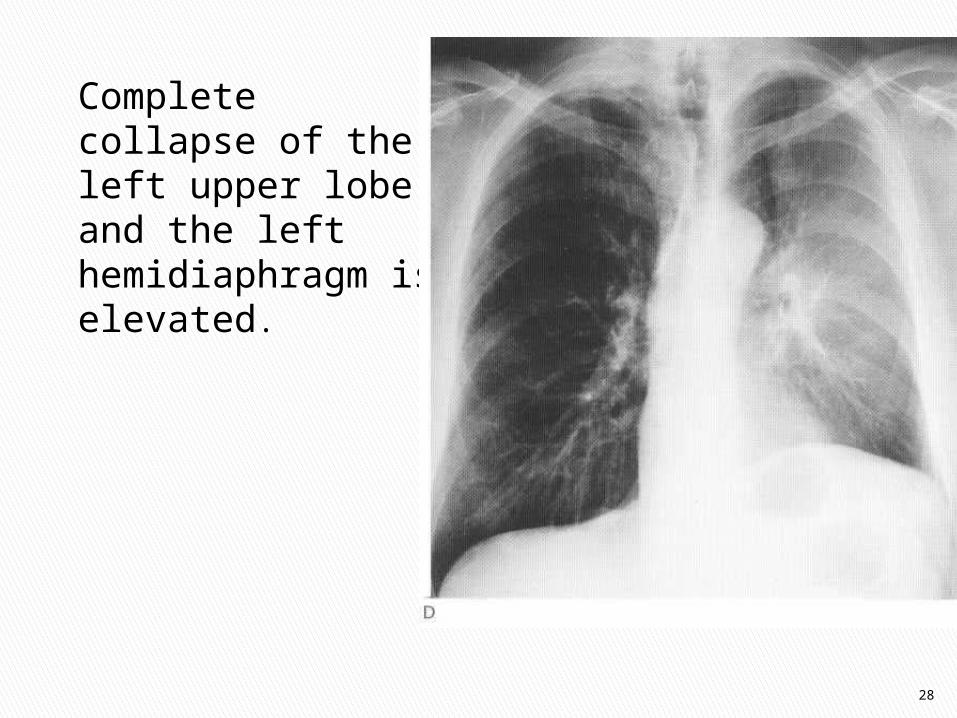
Complete collapse of the left upper lobe, and the left hemidiaphragm is elevated.

29
Occasionally, the cancer remains identifiable as a central contour bulge, and if it obstructs the right upper lobe bronchus, it may result in the S-sign of Golden.

30
‘Golden S sign.‘ Collapsed right upper lobe with mass at right hilum.

31
Other, less frequently seen manifestations of a central tumour include mucoid impaction, air trapping, and pulmonary vascular occlusion or reflex vasoconstriction leading to oligemia or infarction

32
Bronchocele with typical gloved-finger branching pattern

33
Often, the first indication that a cancer exists is the finding of a solitary pulmonary nodule (SPN) on a chest radiograph.
This is the commonest presentation of peripheral tumours on a chest radiograph.
The SPN is usually defined as a single round or oval opacity in the pulmonary parenchyma, measuring <3 cm in diameter.
34
With studies of good quality, a SPN larger than 1 to 2 cm is usually not difficult to detect, but can be overlooked easily in certain locations, i.e. the hidden areas of the lung.
Bronchogenic carcinoma is most often located in the upper lobes, particularly the right upper lobe, and most missed cancers are in the right upper lobe.

A large, round soft-tissue mass is present at the right apex. Blunting of the right costophrenic angle is due toa small pleural effusion.
35

36
A 1991 study of 93 patients with SPNs found 63% to be in the upper lobes, with the right lower lobe being the next most common site.
Lung Cancer: A radiologic overview Applied Radiology Journals> Volume 31, Number 8, Aug.2002
Edward W. Bouchard, MD; Steven Falen, MD; PhD; Paul L. Molina, MD
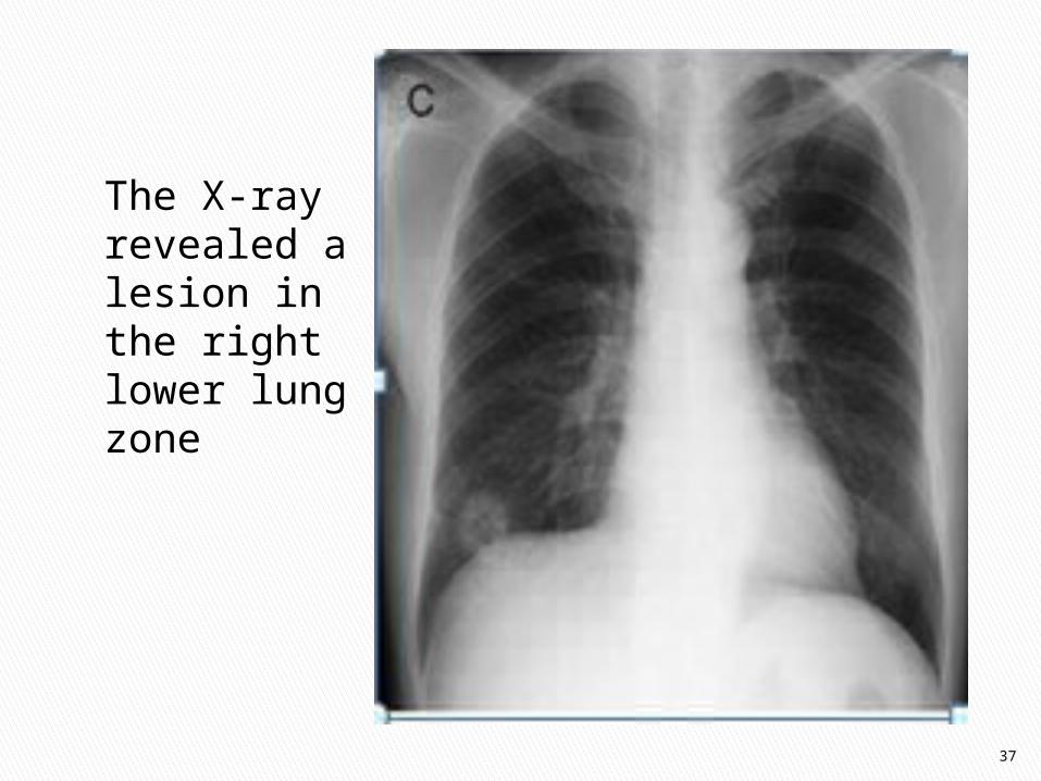
37
The X-ray revealed a lesion in the right lower lung zone

38
Once discovered, certain characteristics of an SPN, such as size, calcification, shape, edge characteristics, cavitation, and growth rate can help differentiate between a benign and malignant lesion.
Once a nodule reaches a size >3 cm, it is more likely to be malignant

39
However, the incidence of primary malignancy in smaller lesions, even in those <1.5 cm, is substantial enough that size alone is insufficient for differentiation.

40
Certain types of radiographically visible calcification, such as lamellated or central calcification in granulomas, and the popcorn pattern in hamartomas, are highly specific for benignity.
Caution must be exercised, however, as a growing lung cancer may surround a calcified granuloma.

41
The margin of a lesion can also provide useful information.
Lobulation of a nodule is a worrisome feature that suggests uneven growth, and supports malignancy.

42
Cavitation is seen in a minority of lung cancer, mostly squamous cell carcinoma, but also occasionally in adenocarcinoma or large cell types.
Usually, the cavity wall is thick (>5 mm) and may demonstrate a nodular internal margin.

43
A maximum wall thickness <4 mm is unlikely to be malignant, but rare cases do exist with thin walls simulating bullae.

44
Cavitating mass in the left mid-zone and there is bulging of the aortopulmonary window, indicating lymph node enlargement.

45
Irregular opacity in left mid-zone with central air density due to cavitation and inferior horizontal margin due to air-fluid level.

46
Spiculations, defined as linear strands extending from the nodule into the lung parenchyma, are of even greater concern, and are thought to represent a desmoplastic response to local tumor extension.
This is called the ‘Corona Radiata’ Sign.

47
Oat cell carcinoma. (A) Peripheral mass adjacent to the ribs. (B) Oblique tomogram shows an irregular mass with thin strands extending intothe surrounding lung
A B

48
Cancers arising in the lung apex, known as superior sulcus or Pancoast tumors (usually squamous cell carcinomas), are a distinct subgroup because of their characteristic location and constellation of symptoms.
Radiographic findings can be quite subtle and are frequently obscured by, or misinterpreted as, overlying musculoskeletal structures, brachiocephalic vessels, or benign pleural thickening.

49
Findings suggestive of malignancy include an apical cap >5 mm, asymmetry of apical caps >5 mm, an apical mass, and adjacent bone destruction.
Clinical symptoms of arm pain and a Horner's syndrome are classically associated with a Pancoast tumor.

50
Pancoast tumour. Chest X-ray shows a left apical mass.

51
Pancoast tumour. Chest X-ray shows asymmetrical right apical pleural thickening.

52
Lung cancer occasionally takes the form of focal or multifocal consolidation, typically with bronchioalveolar carcinoma (BAC).
Although the most common appearance of BAC is as a SPN (43%), consolidation is the second most common radiographic pattern (30%).

53
Chest radiograph obtained in a patient with bronchoalveolar cell carcinoma shows an area of consolidation in the right lower lobe with air bronchograms.

54
This pattern is caused by tumour growth along the framework of peripheral airways and alveoli, combined with mucoid secretions.
Air bronchograms and air alveolograms are characteristic, but not specific, features.
55
A pattern of focal or multifocal nodularity can result from involvement of one or more acini, and when confluent, can resemble non-neoplastic conditions, such as pneumonia, aspiration, or edema.
The consolidative pattern has a poorer prognosis than the solitary nodular pattern.

56
Alveolar cell carcinoma. (A) Chest X-ray shows solitary right upper zone mass
suggesting focal disease

57
Alveolar Cell Carcinoma(B) Eight months later, the disease has rapidly
progressed to the diffuse pattern with widespread nodules and consolidation

58
Hilar and/or mediastinal adenopathy is sometimes the sole manifestation of lung cancer.
Small-cell carcinoma tends to have bulky, central adenopathy with a relatively inconspicuous separate primary lung parenchymal site, but all cell types can have metastatic spread centrally.

59
Careful inspection of the normal contours, lines, and stripes that classically define the mediastinum may reveal enlargement of the aortopulmonary window, right paratracheal thickening, a double density adjacent to the aortic knob, all of which are frequent findings of mediastinal metastasis of lung carcinoma.

60
Small cell carcinoma of bronchus. (A) Chest X-ray shows right upper lobe
masses and extensive right paratracheal and right hilar lymphadenopathy.

61
The lateral film can be especially helpful in the evaluation of suspicious increased hilar or mediastinal density.
Intrathoracic spread of lung carcinoma is not limited solely to mediastinal and hilar adenopathy.

62
The pleura, chest wall, heart, great vessels, diaphragm, and nerves are additional structures that can be involved secondarily.
Such involvement significantly impacts tumour staging, treatment, and prognosis.

63
Small Cell Carcinoma Of the Bronchus 2 months after a tumour
was diagnosed, enlargement of the heart shadow was noted due to pericardial effusion
(confirmed by echocardiography).

64
Pleural involvement usually manifests as a pleural effusion, with or without pleural masses.
Pleural effusion (either free-flowing or loculated) implies seeding by tumour, but a non-malignant effusion can result from central lymphatic obstruction, or a coincidental benign cause, such as pneumonia, congestive heart failure, or pulmonary embolus.

65
Moderate sized Pleural fluid collection obscuring a central bronchogenic carcinoma in
this 56 year old woman. The fluid collection shows typical concave upper margin and is
tracking along the horizontal fissure (arrows).

66
Bone involvement is common, and can be due to direct extension or metastatic spread.
Pancoast tumours are typically associated with direct extension to ribs or vertebral bodies, but this can also occur with other peripheral cancers.
67
Metastatic disease may also involve other bones on the chest radiograph, as evidenced by bony destruction or lytic lesions in the humerus, sternum, clavicle, and scapula.

68
Elevation of the diaphragm may indicate phrenic nerve involvement by tumour, or be mimicked by a subpulmonic effusion.

69
Complete collapse of the left upper lobe, and the left hemidiaphragm is elevated
due to phrenic nerve involvement.

70
However, once the suspected tumour is identified, additional important information is often necessary that cannot be provided by the chest radiograph.
Therefore, the next step in the diagnostic work-up of lung cancer is computed tomography.

71
BRONCHOGRAPHY
This is now an obsolete investigation in the diagnosis of bronchogenic carcinoma.
It has being replaced by CT which is now the imaging modality of choice.

72
Bronchial alterations which are found in pulmonary malignancy include abrupt bronchial obstruction, localized bronchial displacement, concentric bronchial narrowing, "thumb-print" impression and abrupt bronchial narrowing without termination.

73
Malignant tracheobronchial disease. Stenoses of central airways are a common complication of endoluminal tumor growth (arrows).

74
COMPUTED TOMOGRAPHY Thoracic CT scanning plays several vital
roles in the evaluation of patients with known or suspected lung cancer.
One is to further characterize a suspicious abnormality seen on a chest radiograph, and to provide a more complete evaluation of a primary neoplasm.

75
A second and indispensable role is that of pre-treatment or pre- operative staging, for which CT is the primary imaging modality.
Additionally, chest CT helps provide a roadmap for other staging procedures such as bronchoscopy, mediastinoscopy, transthoracic needle biopsy, and video-assisted thorocoscopy.

76
Most, if not all, of the various manifestations of lung cancer described for chest radiography can be better evaluated with CT.
Cross-sectional imaging can help further clarify a tumuor's location, whether in a central or peripheral location, and delineate its relationship to pleura, chest wall, and mediastinal structures.
77
The level and degree of obstruction by central tumours leading to atelectasis and postobstructive pneumonitis can be visualized easily with cross-sectional imaging.
Trapped secretions distal to an obstructing lesion can produce the so-called mucous bronchogram.

78
Contrast-enhanced CT on lung window shows collapsed left lung and demonstrates tumour
extending into the left main bronchus.

79
Bronchocele due to carcinoma of the bronchus. CT shows dilated, fluid-filled
bronchi in the lingula, secondary to carcinoma at the left hilum.

80
Imaging features used to characterize an SPN on a chest radiograph are equally as useful on CT, including size and growth rate, calcification, shape and margins, and cavitation, along with the additional characteristics of density and contrast enhancement.

81
As with chest radiography, increasing size, especially >3 cm, correlates with an increasing chance of malignancy.

82
Right middle lobe peripheral carcinoma, 3.5 cm in diameter.

83
Contrast enhanced CT on lung window shows left lower lobe mass,
which proved to be an adenocarcinoma.

84
CT can better detect and evaluate calcifications within a nodule.
The distribution of calcium, rather than its presence alone, is a more important diagnostic consideration.

85
Thin layers of calcium in a lamellar pattern are indicative of a granuloma, and popcorn calcifications with associated fat density, are associated with a benign hamartoma.

86
Carcinoma of the lung incorporating calcification (arrow) from previous tuberculous granuloma. The tumour is extending into the mediastinum with vascular encasement (arrowheads).

87
A smooth peripheral margin on CT is associated more frequently with benign lesions.
As with chest radiographs, lobulations and spiculations are worrisome findings.
88
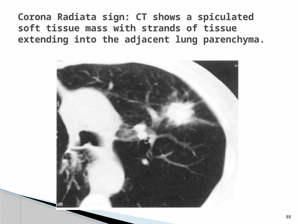
Corona Radiata sign: CT shows a spiculated soft tissue mass with strands of tissue extending into the adjacent lung parenchyma.

89
Corona Radiata sign: A malignant lesion with spiculations at its margin.
90
The wall thickness of a cavitary lesion can be measured more accurately with CT.
One study found that the majority (94%) of cavitary solitary pulmonary nodules with a wall thickness ≤4 mm were benign, and the majority (95%) with a wall thickness ≥15 mm were malignant.
Lung Cancer: A radiologic overview Applied Radiology Journals> Volume 31, Number 8, Aug.2002
Edward W. Bouchard, MD; Steven Falen, MD; PhD; Paul L. Molina, MD

91
Lesions with wall thicknesses between 5 and 15 mm were almost equally divided between benign and malignant.
CT also has the added advantage of better evaluating the contour of a cavity's wall. A smooth inner wall is more commonly associated with a benign aetiology, while a nodular internal margin reflects focal tumour nodules.

92
Squamous cell carcinoma of the bronchus. CT shows a thick-walled cavitating mass with a nodular inner surface.

93
CT showing a cavitating squamous cell carcinoma in the basal segment of the right lung.

94
Several other characteristics of an SPN can be evaluated with CT, such as attenuation and contrast enhancement.
Homogeneous attenuation has been found to be associated more often with a benign, rather than a malignant lesion.

95
A newer technique for the assessment of the SPN is based on differential nodule enhancement with IV contrast material, as measured with thin-slice CT.
It relies on qualitative and quantitative differences in the blood supply to benign and malignant nodules.

96
Results from a 1992 study suggest that malignant nodules tend to enhance significantly more (20 HU increase) than benign nodules, with the most diagnostically important measurement made at 2 minutes post-injection.
Lung Cancer: A radiologic overview Applied Radiology Journals> Volume 31, Number 8, Aug.2002
Edward W. Bouchard, MD; Steven Falen, MD; PhD; Paul L. Molina, MD

97
One additional CT finding that may be helpful in the evaluation of lobar consolidation and the clinical suspicion of bronchioalveolar cell carcinoma is the CT angiogram sign.
CT angiogram sign :This is defined as branching pulmonary vessels extending >3 cm into completely consolidated pulmonary parenchyma that is of diffusely homogeneous lower attenuation than that of muscle.

98
Although initially thought to be specific for bronchoalveolar cell carcinoma, it has now been recognised as a generic appearance provided the density of consolidation is relatively low.
This sign has been associated with : pulmonary lymphoma and infectious/post obstructive pneumonia.

99
CT angiogram sign—that is, a low- attenuating consolidation of the lung parenchyma with enhancing pulmonary vessels (arrow) extending into it.

100
Thin slice contrast enhanced CT scan showing normal enhancing pulmonary vessels (white arrows) within an area of consolidation produced by BAC.

CT is the preferred imaging technique for evaluating adenopathy.
The accurate localization of abnormal lymph nodes, whether peribronchial, hilar, mediastinal, scalene, or supraclavicular, is important.
101

Lymph node involvement is usually florid with small cell carcinomas.
Mediastinal invasion may involve the phrenic nerve causing elevation of a hemidiaphragm or the recurrent laryngeal nerve leading to hoarseness of the voice.
102

Chest CT on mediastinal windows showing (A) left hilar adenopathy and (B) extending into the sub-carinal region.
A B
103

Carcinoma of the bronchus. Contrast enhanced CT scan at the level of the carina showing retrocaval adenopathy.
104

Approximately 5% of all lung cancers invade the parietal pleura and chest wall.
CT has demonstrated a wide range of results when assessing for chest wall invasion by tumour. Sensitivity ranges from 38% to 87%, and specificity ranges from 40% to 90%, depending on the study.
105

The best criterion for diagnosing chest-wall invasion with CT is bony destruction, with or without tumour extension into the chest wall.
Other, less reliable signs of chest-wall invasion include pleural thickening, loss of the extra-pleural fat plane, and an obtuse angle between the mass and the chest wall.
106

CT scan showing large, right apical soft tissue mass extending through the chest wall into the apex of right axilla.
107

CT is also very useful in accurately identifying the involvement of adjacent structures such as the pleura (pleural effusion) and the heart (pericardial effusion).
108

109
Contrast enhanced computed tomography: Necrotic mass in the right lower lobe (short arrow) with pleural (p) and pericardial (pc) effusions which were confirmed to be malignant.

MAGNETIC RESONANCE IMAGING
MRI plays a complementary role to CT because of its superior soft tissue contrast, multiplanar imaging capability, and superb delineation of thoracic vessels.
There are areas of the chest where the geometry of the structures of interest are better imaged with MRI.
110
Perhaps the best example is the evaluation of Pancoast tumours, in which direct coronal and sagittal imaging with MRI facilitates assessment of invasion of the chest wall, brachial plexus, subclavian vessels, vertebral bodies, and neural foramina.
111

Pancoast Tumour. (A)T1- weighted coronal and (B) parasagittal MRI showing right apical mass with obliteration of the extrapleural fat where the mass invades the chest wall and enters the root of the neck.
A B 112

MRI has been shown to be superior to CT in detecting mediastinal extension when there is associated vessel involvement.
MRI is also believed to be more accurate in establishing superior vena caval patency or obstruction, which may be due to thrombus, compression by soft-tissue mass, or direct invasion.
113

Carcinoma of the bronchus. T1-weighted axial MRI of the chest at the level of the carina showing retrocaval lymphadenopathy.
114

A significant disadvantage of MRI is its poorer spatial resolution, which can lead to adjacent nodes on CT appearing as an enlarged mass on MRI, resulting in the mistaken diagnosis of abnormal nodal enlargement.
115

BARIUM STUDIES
Enlarged mediastinal lymph nodes may compress or invade the esophagus.
Barium swallow may therefore be used to evaluate the mediastinum, and is essential in patients with dysphagia.
In these patients, esophageal compression or invasion may be demonstrated.
116

117
Carcinoma of the bronchus. Barium Swallow shows extrinsic compression of the mid-esophagus by enlarged subcarinal lymph nodes.

ULTRASONOGRAPHY Ultrasound scanning provides a radiation-
free access to certain types of bronchogenic carcinoma.
This is particularly true for Pancoast tumours which occur at the lung apex and can be viewed from the supraclavicular fossa.
Ultrasonography can also be used to evaluate large pleural effusions in cases where a mass is suspected.
118

119
Ultrasound scan from the right supraclavicular fossa shows an apical pulmonary mass of relatively low echogenicity, and demonstrates the easiest route of access for percutaneous biopsy.

120
Large left pleural effusion due to carcinoma of bronchus.There is a large echo-free effusion above the left hemidiaphragm (arrowheads) and spleen (s).

POSITRON EMISSION TOMOGRAPHY
CT and MR imaging of the chest provides valuable information about the morphology of a lesion.
However, morphologic information alone may not offer all the information necessary to direct proper clinical management.
121

Many lesions are indeterminate as to whether they are benign or malignant by morphologic imaging techniques, such as CT and MRI, and further investigation is warranted.
122

One of the more recent advances in oncologic imaging that has generated a renewed interest in diagnosis, staging, and response to therapy is positron emission tomography (PET).
PET imaging with [2-18F]fluoro-2-deoxy-D-glucose (F-18 FDG) allows for the evaluation of the relative level of metabolic activity of a lesion compared with other tissues.
123

F-18 FDG PET imaging has been shown to be an accurate, non-invasive imaging test for the assessment of pulmonary nodules and larger mass lesions
124

A comprehensive meta-analysis by Gould et al of 40 eligible studies, including 1,474 focal pulmonary lesions of any size, found the mean sensitivity and specificity for detecting malignancy were 96.0% and 73.5%, respectively.
However, in this analysis, there was little data for nodules <1 cm in diameter.
125
Lung Cancer: A radiologic overview Applied Radiology Journals> Volume 31, Number 8, Aug.2002
Edward W. Bouchard, MD; Steven Falen, MD; PhD; Paul L. Molina, MD

When a lung mass is shown to be malignant, it is important to stage the extent of disease accurately.
Several studies have shown that PET is more accurate than CT for the staging of NSCLC.
PET appears to be more accurate than CT in detecting metastatic mediastinal lymphadenopathy.
126

Valk et al conducted a prospective study in 76 patients of PET imaging for staging of NSCLC in which mediastinal PET and CT findings were compared with the results of surgical staging.
They reported the sensitivity and specificity for the diagnosis of mediastinal nodal disease were 83% and 94% for PET and 63% and 73% for CT, respectively.
127
Lung Cancer: A radiologic overview Applied Radiology Journals> Volume 31, Number 8, Aug.2002
Edward W. Bouchard, MD; Steven Falen, MD; PhD; Paul L. Molina, MD

Detection of unsuspected metastatic disease by PET may permit reduction in the number of thoracotomies performed for non-resectable disease.
128

PET scan showing abnormal uptake of FDG in a tumour nodule in the right upper lobe(arrow) & in two superior mediastinal lymph nodes(arrowheads).
129

Unresectable lung cancer. FDG-PET scan shows large primary tumour with metastases in lymph nodes, bone, & right adrenal.
130

PET is also very useful in clarifying those cases in which occurence of benign nodal enlargement coexists with a malignant lung lesion.
131
132
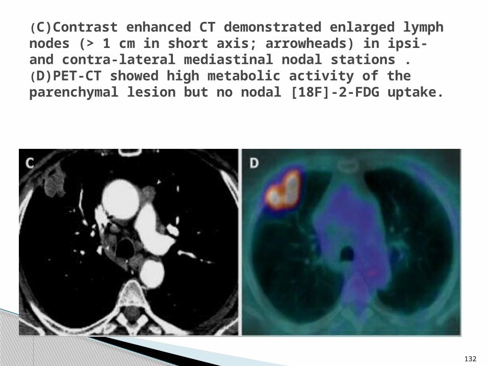
(C)Contrast enhanced CT demonstrated enlarged lymph nodes (> 1 cm in short axis; arrowheads) in ipsi- and contra-lateral mediastinal nodal stations .(D)PET-CT showed high metabolic activity of the parenchymal lesion but no nodal [18F]-2-FDG uptake.

ANGIOGRAPHY
This is mainly carried out to assess the vascularity of a diagnosed tumour and also for pre-operative embolisation to reduce tumour bulk/ reduce intra-op bleeding.
133

134
A left inferior phrenic arteriogram shows the vessel accounting for systemic arterial supply to the tumour through hypertrophied pleural collateral channels. Polyvinyl alcohol embolisation was carried out with good clinical results

135
STAGING OF BRONCHOGENIC CARCINOMA
Staging is done using the lung cancer TNM staging system.
T= Tumour size
N= Level of nodal involvement
M= Presence or absence of metastases.

136
TNM STAGING
T1 <3cm in diameter, sorrounded by lung/visceral pleura
T2 >3cm in diameter/invasion of visceral pleura/lobar atelectasis/obstructive pneumonitis/at least 2cm from the carina.
T3 Tumour of any size; less than 2cm from the carina/ invasion of parietal pleura, chest wall, diaphragm, mediastinal pleura, pericardium.
T4 Invasion of the heart, great vessels, trachea, esophagus, vertebral body, carina/ malignant effusion
N1 Peribronchial / ipsilateral hilar nodes
N2 Ipsilateral mediastinal nodes.
N3 Contralateral hilar/ mediastinal nodes
M0 No metastases
M1 Distant metastases present.

1.LOCAL COMPLICATIONS: Superior Vena Cava Syndrome Intractable Hemoptysis
2.DISTANT COMPLICATIONS: Metastases
3. PARANEOPLASTIC SYNDROMES: Hypertrophic Osteoarthropathy
RADIOLOGY OF COMPLICATIONS
137

1.SVC SYNDROME
SVC (Superior Vena Cava) Syndrome is a set of symptoms that result when blood flow through the superior vena cava is obstructed by extrinsic compression or by tumour invasion.
138

Lung cancer is the leading malignant cause of SVC syndrome, with non–small cell lung cancer accounting for about 50% of the cases and SCLC accounting for about 25% of cases occurring in malignancy.
This syndrome is a complication that occurs in 2% to 4% of people living with lung cancer, and in some cases is the first symptom that leads to the diagnosis.
139

Clinical features include:
Swelling of the face, arms, or chest wall
Difficulty breathing (dyspnoea)
Widening of the veins in the neck and chest
140

Axial and coronal images of the CT demonstrating extensive mediastinal mass with compression of the SVC. The mass also was compressing the trachea and proximal airways.
141

Stenting of superior vena cava is a well-known but not so commonly used technique to alleviate this syndrome.
142

The catheter wire is placed in the vena cava stenotic segment. The stent is delivered and the stenosis is solved.
143

2.INTRACTABLE HEMOPTYSIS
Bronchial artery angiography with embolization has become a mainstay in the treatment of intractable hemoptysis in some patients with lung cancer.
Major complications are rare and immediate clinical success defined as cessation of hemorrhage ranges in most series from 85% to 100%, although recurrence of hemorrhage ranges from 10% to 33%.
144

Reports of neurological damage following bronchial angiography indicate care in avoiding obstruction of the artery of Adamkiewicz.
145

Angiographic image showing blood ejecting from a ruptured bronchial artery branch (arrow)
Selective embolization of the feeding artery obtained with gel foam.
146

3.HYPERTROPHIC OSTEOARTHROPATHY
A.k.a Bamberger-Marie syndrome
Hypertrophic osteoarthropathy is a paraneoplastic syndrome most often found in non-small cell lung cancer.
147
It is a medical condition combining clubbing and periostitis of the long bones of the upper and lower extremities.
Distal expansion of the long bones as well as painful, swollen joints and synovial villous proliferation are often seen.
148

Diagnosis is confirmed by the characteristic bone changes on plain radiograph and periostitis on bone scintigram.
The syndrome generally resolves dramatically with treatment of the underlying malignancy.
149

150
Radiograph showing thickened, columnar diaphyses and erosion of the terminal phalangeal tufts in Hypertrophic Osteoarthropathy

151
Bone scintigraphy showing periosteal proliferation along the margins of the shafts of the tibias, radii, ulnae and pelvic bones.

4.DISTANT METASTASES
Small cell> Adeno > Large> Squamous
Lung cancer spread (metastatases) is sadly too common.
Nearly 40% of people with lung cancer have metastases to a distant region of the body at the time of diagnosis.
152

Lung cancer can spread to any region of the body, but most commonly spreads to the liver, the lymph nodes, the brain, the bones, and the adrenal glands.
153

LIVER METASTASES
The staging CT scan of the thorax is usually extended to include the liver and adrenal glands.
CT scanning has a sensitivity of about 85% in the detection of liver metastases. Similar rates may be obtained with MRI and ultrasonography performed by experienced imagers.
154

155
A computed tomographic (CT) scan of the abdomen showed multiple hepatic metastases (arrows).
156
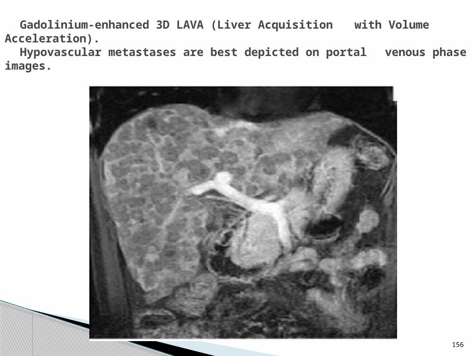
Gadolinium-enhanced 3D LAVA (Liver Acquisition with Volume Acceleration).
Hypovascular metastases are best depicted on portal venous phase images.

ADRENAL METASTASES
Adrenal metastases are common and often solitary.
They must be differentiated from adrenal adenomas, which occur in 1% of the adult population.
157

Lesions smaller than 1 cm are usually benign.
Metastases are usually larger than 3 cm; on non-enhanced CT scans, they have an attenuation coefficient of 10 HU or higher.
Adenomas and metastases can also be distinguished by using MRI and PET scanning.
158

159
Sonogram showing a 6-cm right adrenal metastasis of lung cancer.

Adrenal metastasis from small cell lung cancer
160

161
Coronal PET/CT image demonstrating intense FDG uptake in the primary left upper lobe lung carcinoma (curved arrow) and in the adrenal metastases (arrows)
BONE METASTASES Osteolytic (70%) Osteoblastic (30%)
Technetium-99m (99m Tc) radionuclide bone scanning is indicated in patients with bone pain or local tenderness.
The test has a 95% sensitivity for the detection of metastases but a high false-positive rate because of degenerative disease and trauma.
162

The assessment of these metastases requires comparison of the bone scans with plain radiographs.
Vertebrae(70%), Pelvis(40%), Femora(25%)
Plain radiographs typically show destructive lytic lesions ± pathological fractures.
Similar features are seen on CT scans.
163

164
Bone Metastasis from Primary Lung Cancer :Lytic lesion of humerus with a pathological fracture.

165
Bone Metastasis from Primary Lung Cancer Expansile lytic rib lesions (arrows).

166
FDG PET images demonstrate bone metastases (arrows).

167
Isotope bone scan. Hot spots due to bony metastases.

BRAIN METASTASES
SCLC and adenocarcinoma are the most common sources of cerebral metastases.
MRI is superior to CT, especially in the depiction of the posterior fossa and the area adjacent to the skull base.
168

However, the brain is not routinely imaged in asymptomatic patients with NSCLC, because the incidence of silent cerebral metastases is only 2-4%.
Brain metastases are typically hemorrhagic and occur at the grey-white mater junction of the brain.
169

170
Non small cell lung cancer with hemorrhagic brain metastasis (A) Pre-operative non-contrast enhanced computed tomography (CT). (B) Pre-operative contrast-enhanced CT.

Contrast-enhanced CT scans of the same patient showing multiple enhancing cerebral metastases of lung cancer in the left cerebral hemisphere.
171

DIFFERENTIAL DIAGNOSES
Pulmonary metastasesPulmonary AV malformation
172

DIFFERENTIAL DIAGNOSES
Pulmonary tuberculosis Pulmonary hamartoma
173

CONCLUSION / SUMMARY
Lung cancer is an extremely prevalent disease that most radiologists will encounter on a frequent basis.
Familiarity with the various manifestations of lung cancer on the different imaging modalities may help suggest the initial diagnosis, especially in an older patient with a history of cigarette smoking.
174

175
CHEST RADIOGRAPHY 1st line investigation; cheap and readily available; can depict most of the features of overt lung cancer and its complications.
COMPUTED TOMOGRAPHY
The gold standard in diagnosis and staging of lung cancer; gives cross-sectional imaging with better representation of anatomy; clearly depicts mediastinal adenopathy and involvement of adjacent structures.

MAGNETIC RESONANCE IMAGING
Excellent soft tissue resolution; clearly depicts vascular invasion better than CT; imaging modality of choice for assessing Pancoast tumours; of importance in cases where CT findings are indeterminate or equivocal.
POSITRON EMISSION TOMOGRAPHY
Provides excellent depiction of functional status of suspicious lung masses; helps to sort out status of nodal enlargement coexisting with lung cancer. 176

177
THANK YOU FORLISTENING!!!


![Transbronchial Needle Aspiration Staging of Bronchogenic ...downloads.hindawi.com/journals/dte/1996/237680.pdfChest, 80,48-50. [18] Transbronchialneedle bronchogenic carcinoma, In:](https://img.dokumen.tips/doc/110x75/5fef28f6c0cad34ae7313439/transbronchial-needle-aspiration-staging-of-bronchogenic-chest-8048-50-18.jpg)