Embed Size (px)
Citation preview

SUPRVENTRICULAR TACHYARRHYTHMIAS
Mohammad Hassan Khedr MD Cardiology

Case #1
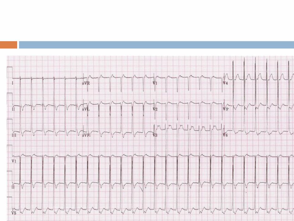
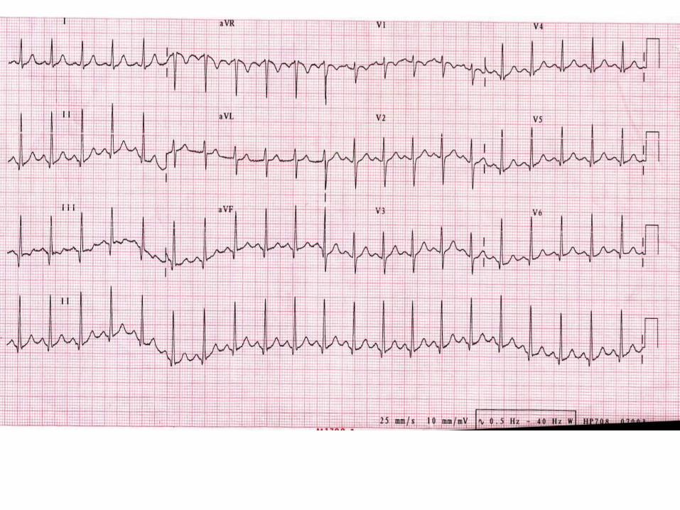
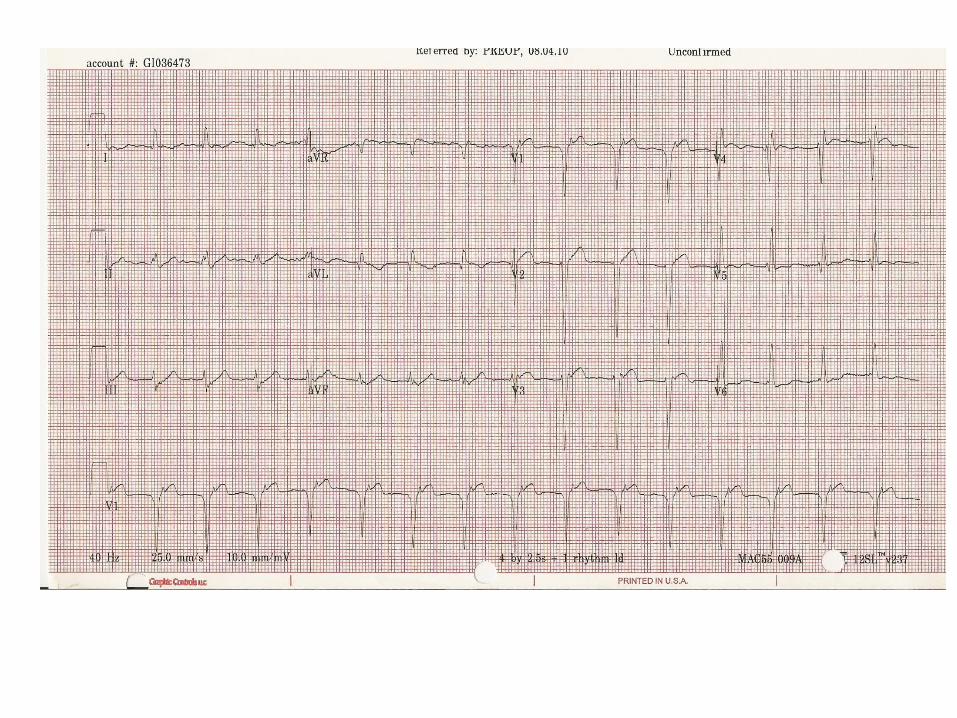
Q. A 32 year old female is treated in the emergency room for palpitations. The first ECG is tachycardia and the second is after adensosine.What is the arrhythmia?
A. AVNRT B. ORT C. Atrial tachycardia D. Atrial fibrillation
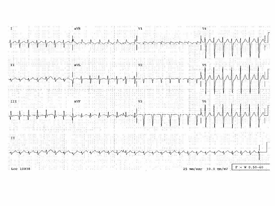
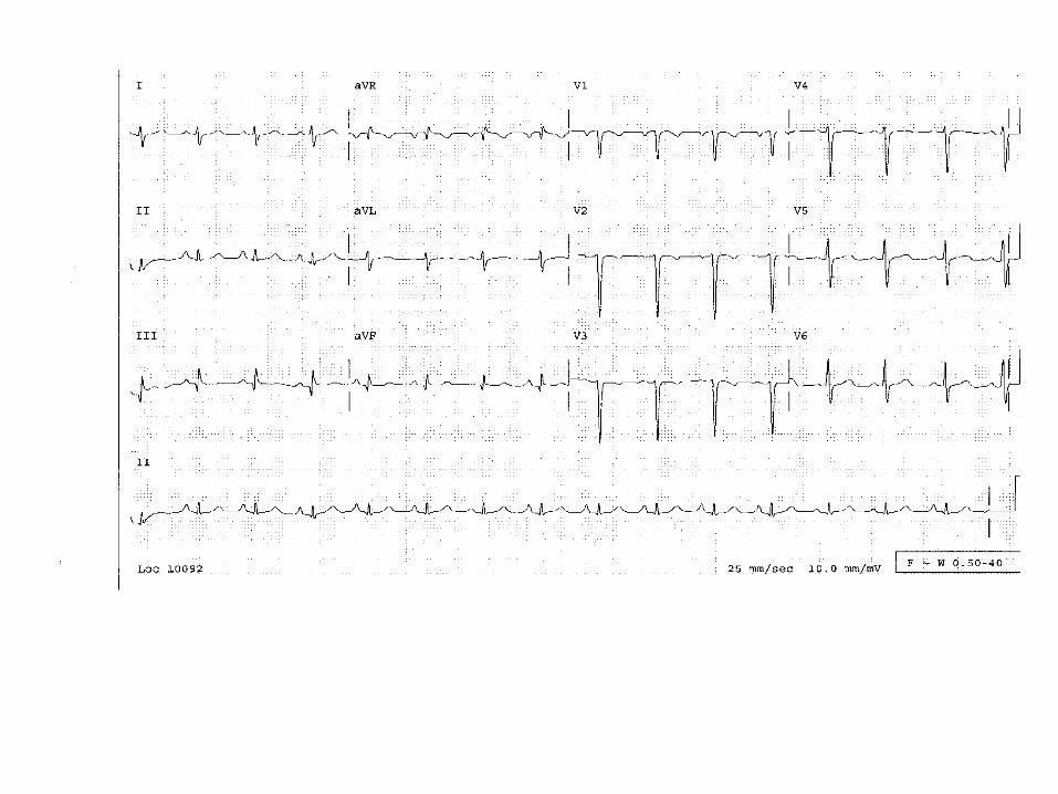
Answer: AVNRT (A)
A small R’ is seen is lead V1 with pseudo-S waves in the inferior leads that are absent after termination of the arrhythmia. These represent retrograde atrial activation with a very short RP interval.
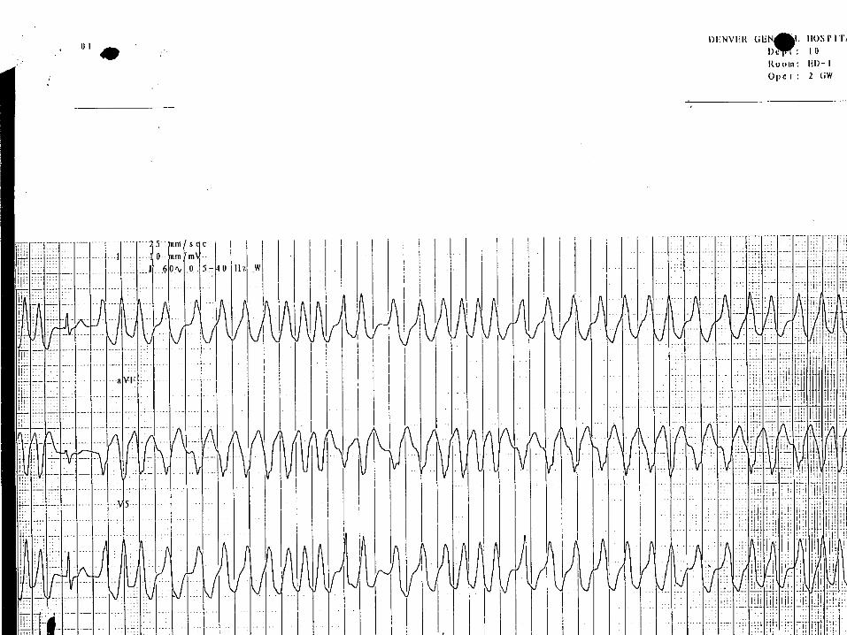
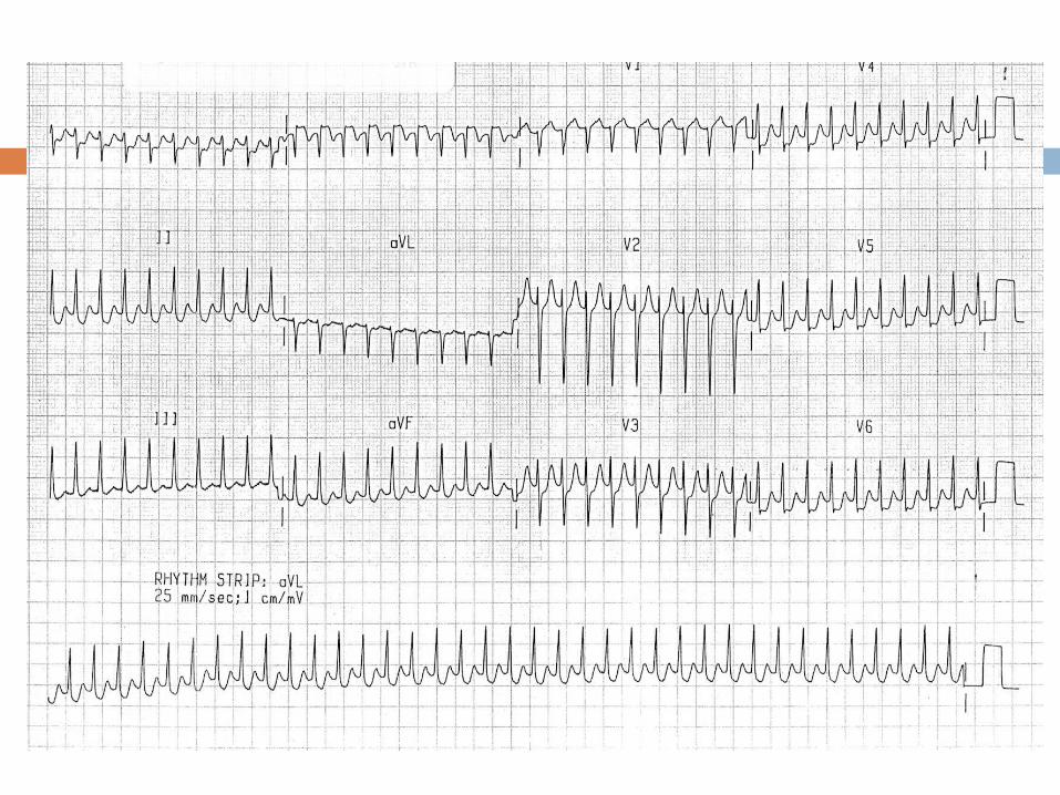
Q. A 42 year old smoker presents to the ED with palpitations. His blood pressure is 110/60. The following rhythm strip is obtained . What is the next appropriate step?
Emergent cardioversion for polymorphic VT. B. I.V. procainamide C. I.V. lidocaine D. diltiazem drip to obtain rate control.

Approach to classification of SVT1) Clinical behavior (ie. Paroxysmal, persistent,
permanent, incessant, sustained, nonsustained, chronic, and repetitive)
2) Mechanism (ie, ectopic, automatic, reenterant, orthodromic, antidromic)
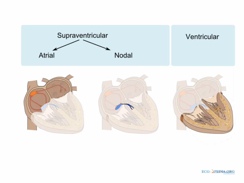
3) ECG appearance (narrow or wide)4) Location (sinus, atrial, AV nodal/ juntional)

Mechanism
All cardiac tachyarrhythmias are produced by one or more mechanisms including:
1) Disorders of impulse initiation 2) Abnormalities of impulse conduction.

Mechanisms of Arrhythmia
Abnormal automaticity automatic impulse generation from unusual site
or overtakes sinus node Triggered activity
secondary depolarization during or after repolarization
Dig toxicity, Torsades de Pointes Reentry
90 % of arrhythmiasythmias
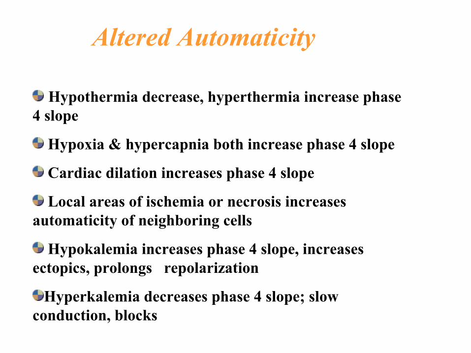
Hypothermia decrease, hyperthermia increase phase 4 slope
Hypoxia & hypercapnia both increase phase 4 slope
Cardiac dilation increases phase 4 slope
Local areas of ischemia or necrosis increases automaticity of neighboring cells
Hypokalemia increases phase 4 slope, increases ectopics, prolongs repolarization
Hyperkalemia decreases phase 4 slope; slow conduction, blocks
Altered Automaticity

Reentry
Most common mechanism
Requires two separate paths of conduction
Requires an area of slow conduction
Requires unidirectional block

Symptoms
palpitations fatigue lightheadedness Chest discomfort dyspnea Presyncope Polyurea (release of atrial natriuretic peptide in
response to increased atrial pressures from contraction of atria against a closed AV valve)
Syncope (rare)
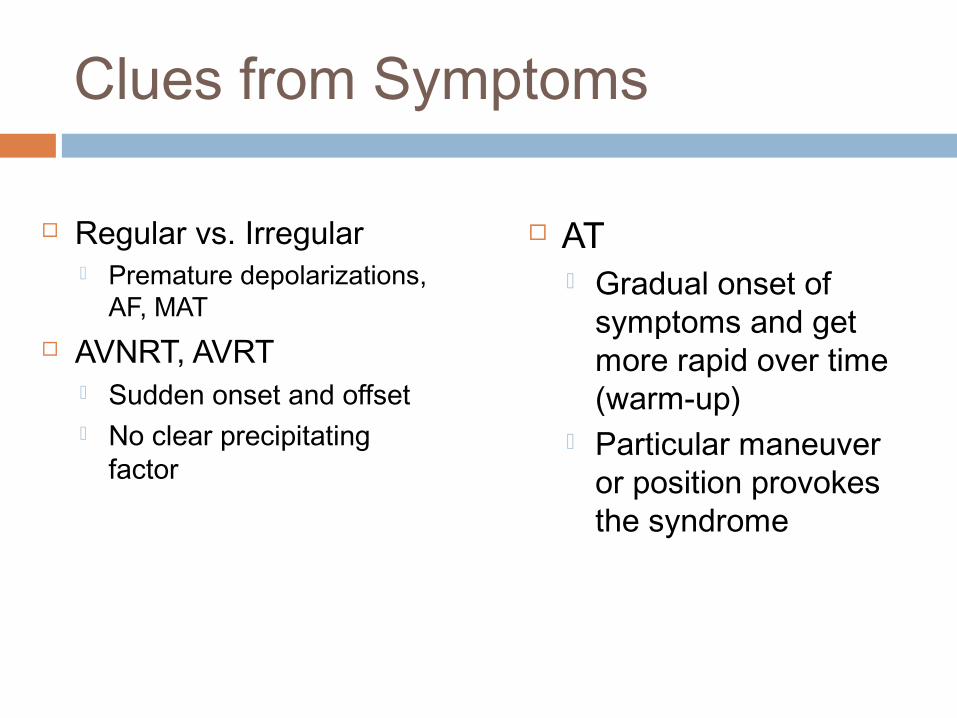
Clues from Symptoms
Regular vs. Irregular Premature depolarizations,
AF, MAT AVNRT, AVRT
Sudden onset and offset No clear precipitating
factor
AT Gradual onset of
symptoms and get more rapid over time (warm-up)
Particular maneuver or position provokes the syndrome
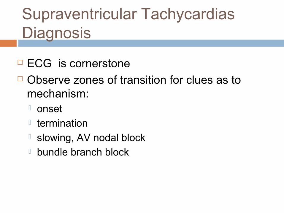
Supraventricular TachycardiasDiagnosis
ECG is cornerstone Observe zones of transition for clues as to
mechanism: onset termination slowing, AV nodal block bundle branch block
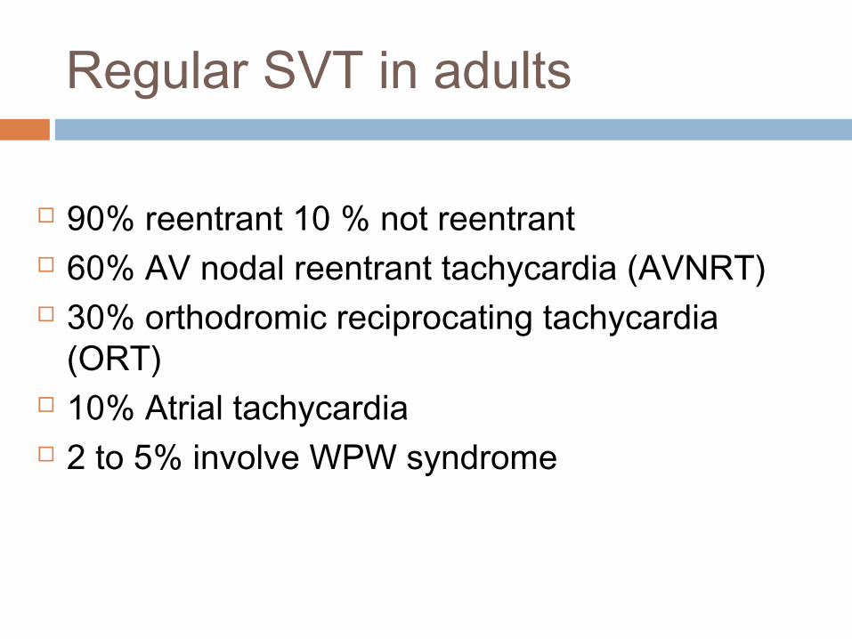
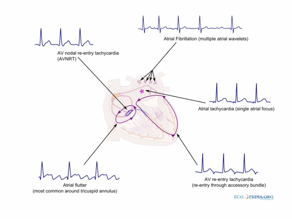
Regular SVT in adults
90% reentrant 10 % not reentrant 60% AV nodal reentrant tachycardia (AVNRT) 30% orthodromic reciprocating tachycardia
(ORT) 10% Atrial tachycardia 2 to 5% involve WPW syndrome
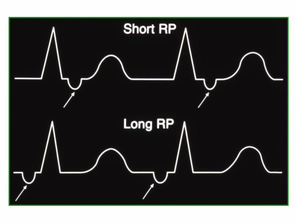
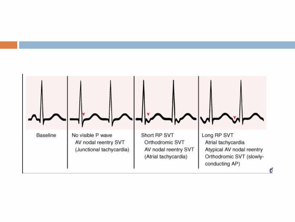
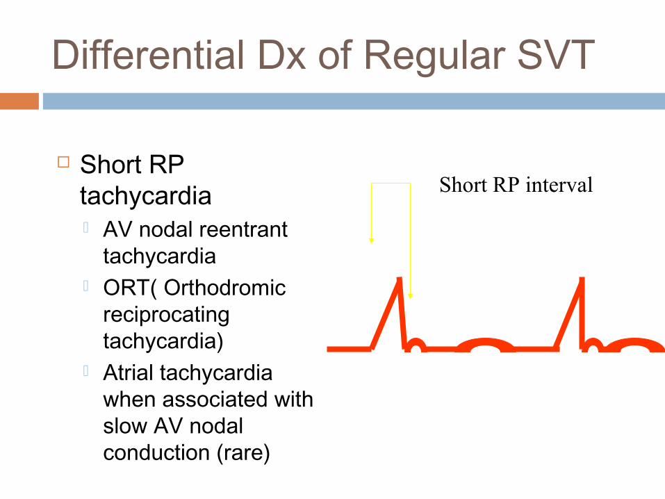
Differential Dx of Regular SVT
Short RP tachycardia AV nodal reentrant
tachycardia ORT( Orthodromic
reciprocating tachycardia)
Atrial tachycardia when associated with slow AV nodal conduction (rare)
Short RP interval

Differential Dx of Regular SVT
Long RP tachycardia Atrial tachycardia Sinus node reentry Sinus tachycardia Atypical AV nodal
reentrant tachycardia
Long RP interval

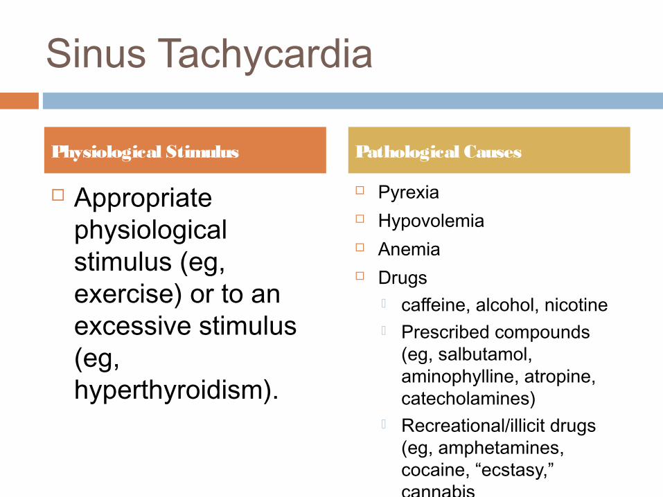
Sinus Tachycardia
Appropriate physiological stimulus (eg, exercise) or to an excessive stimulus (eg, hyperthyroidism).
Pyrexia Hypovolemia Anemia Drugs
caffeine, alcohol, nicotine Prescribed compounds
(eg, salbutamol, aminophylline, atropine, catecholamines)
Recreational/illicit drugs (eg, amphetamines, cocaine, “ecstasy,” cannabis
Anticancer treatments
Physiological Stimulus Pathological Causes
Management
Treat underlying mechanism Beta blockade for physiological symptomatic
sinus tachycardia triggered by emotional stress and other anxiety related disorders

Other Long RP tachycardias
Sinus node reentrant abrupt onset and
offset P wave complex
same as sinus Amenable to calcium
channel blockers, much less responsive to beta blockers
Amenable to catheter ablation
Syndrome of inappropriate sinus tachycardia typical sinus
tachycardia with lowest rate on Holter of 130 bpm
Treated with high dose beta blockers
Poor results with catheter ablation

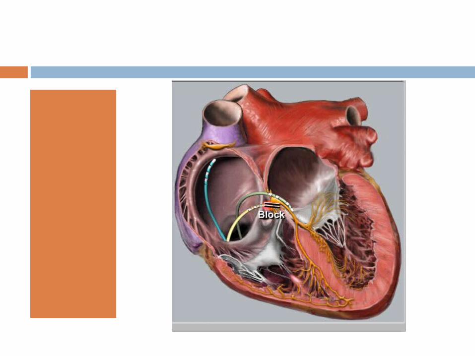
AV Nodal Reentrant Tachycardia
2 pathways within or limited to perinodal tissue anterograde
conduction down fast pathway blocks with conduction down slow pathway, with retrograde conduction up fast pathway.
Slow pathway
Fast pathway
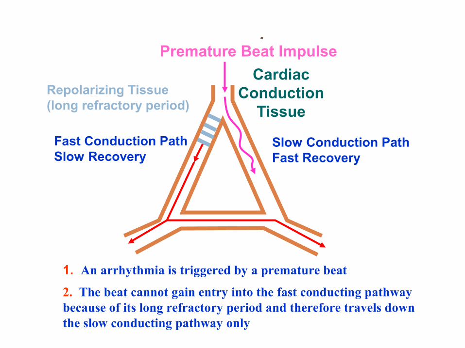
Fast Conduction PathSlow Recovery
Slow Conduction PathFast Recovery
Premature Beat Impulse
Cardiac Conduction
Tissue
1. An arrhythmia is triggered by a premature beat
2. The beat cannot gain entry into the fast conducting pathway because of its long refractory period and therefore travels down the slow conducting pathway only
Repolarizing Tissue (long refractory period)
The “Re-Entry” Mechanism of Ectopic Beats & Rhythms.

3. The wave of excitation from the premature beat arrives at the distal end of the fast conducting pathway, which has now recovered and therefore travels retrogradely (backwards) up the fast pathway
Fast Conduction PathSlow Recovery
Slow Conduction PathFast Recovery
Cardiac Conduction
Tissue
The “Re-Entry” Mechanism of Ectopic Beats & Rhythms.
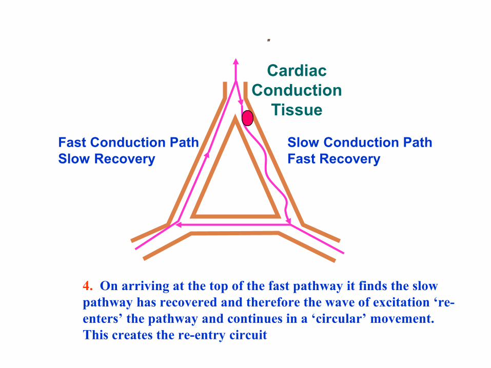
4. On arriving at the top of the fast pathway it finds the slow pathway has recovered and therefore the wave of excitation ‘re-enters’ the pathway and continues in a ‘circular’ movement. This creates the re-entry circuit
Fast Conduction PathSlow Recovery
Slow Conduction PathFast Recovery
Cardiac Conduction
Tissue
The “Re-Entry” Mechanism of Ectopic Beats & Rhythms.
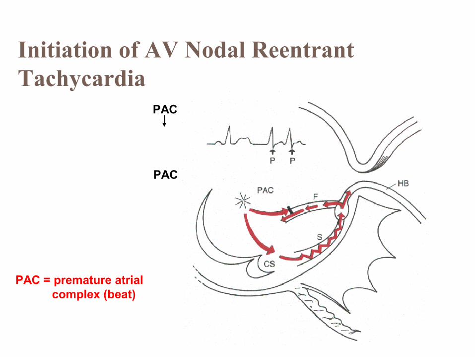
Initiation of AV Nodal Reentrant Tachycardia
PAC = premature atrial complex (beat)
PAC
PAC

Sustainment of AV Nodal Reentrant Tachycardia
Rate 150-250beats per min
P waves generatedretrogradely(AV node→ atria) andfall within orat tail of QRS
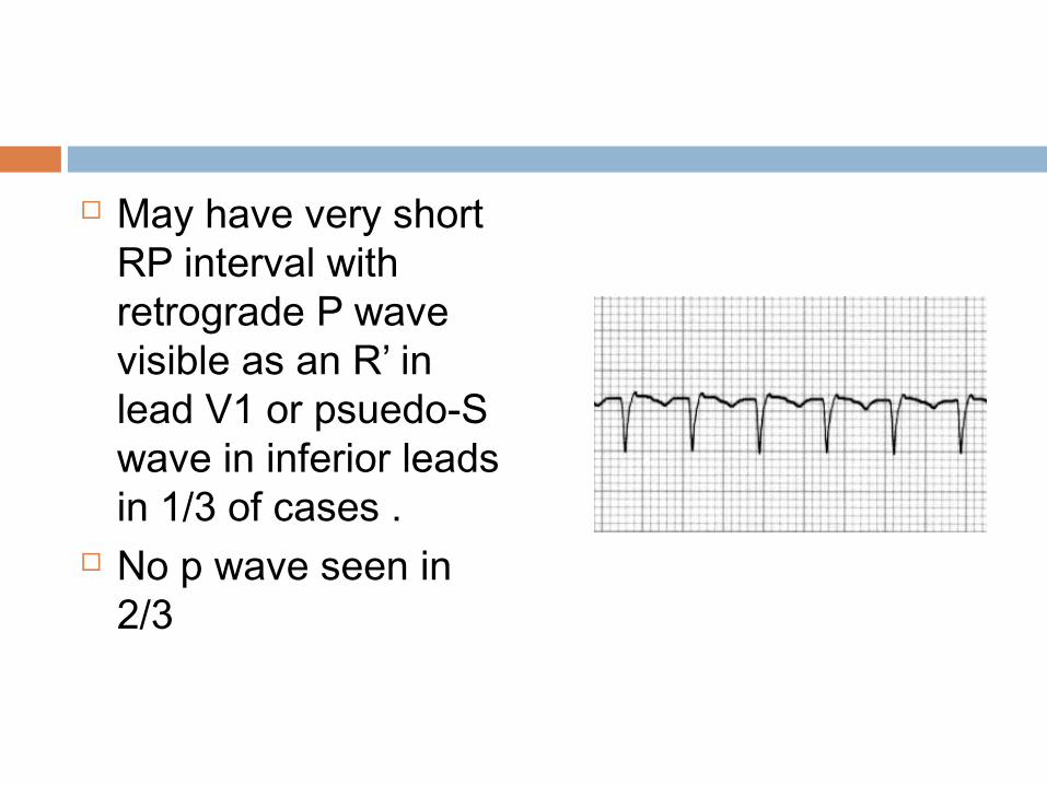
May have very short RP interval with retrograde P wave visible as an R’ in lead V1 or psuedo-S wave in inferior leads in 1/3 of cases .
No p wave seen in 2/3

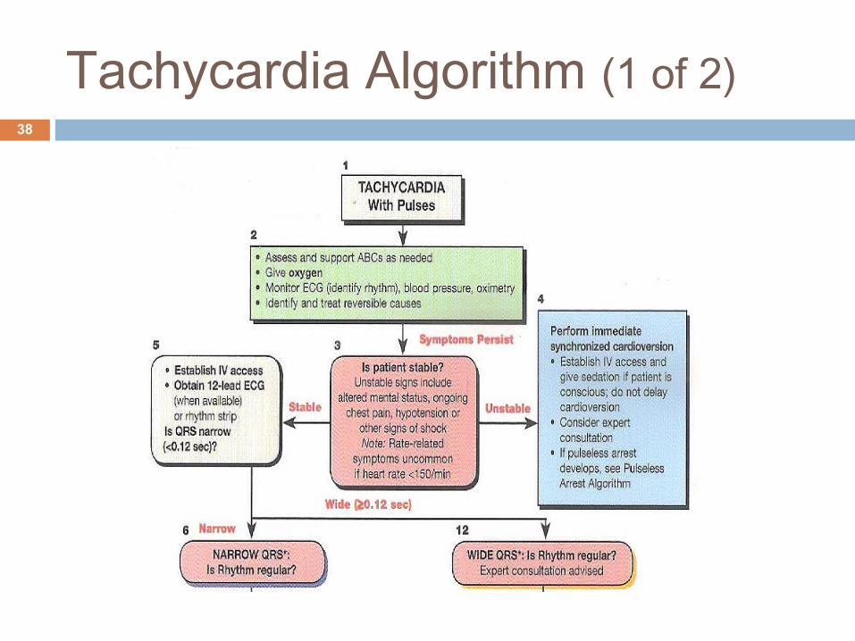
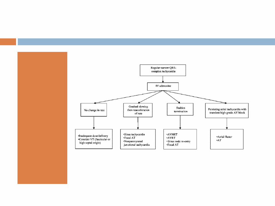
Tachycardia Algorithm (1 of 2)38
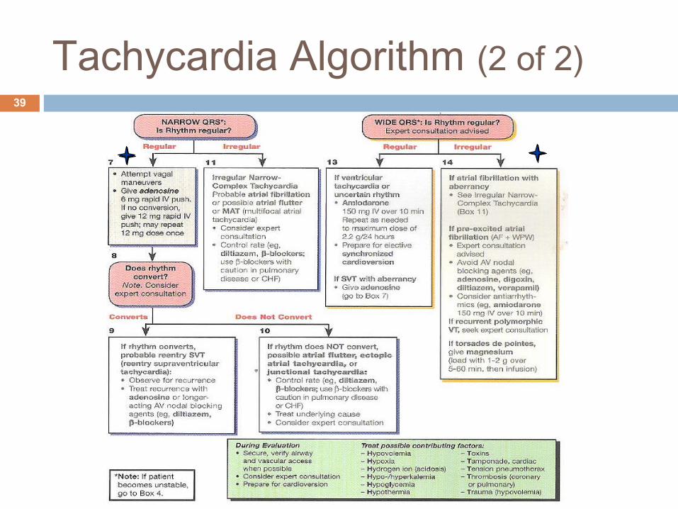
Tachycardia Algorithm (2 of 2)39

Narrow QRS Tachycardia40
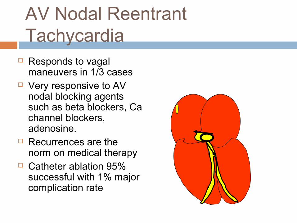
AV Nodal Reentrant Tachycardia
Responds to vagal maneuvers in 1/3 cases
Very responsive to AV nodal blocking agents such as beta blockers, Ca channel blockers, adenosine.
Recurrences are the norm on medical therapy
Catheter ablation 95% successful with 1% major complication rate

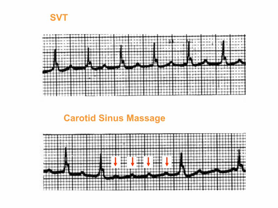
Determining AV Nodal Participation in SVT by Transiently Depressing AV Nodal Conduction
Vagotonic Maneuvers Carotid sinus massage Valsalva maneuver (bearing down) Facial ice pack (“diving reflex;” for kids)
Adenosine (6-12 mg I.V.) If SVT “breaks,” a reentrant mechanism
involving the AV node is likely If atrial rate unchanged, but ventricular rate
slows (#P’s > #QRS’s), SVT is atrial in origin
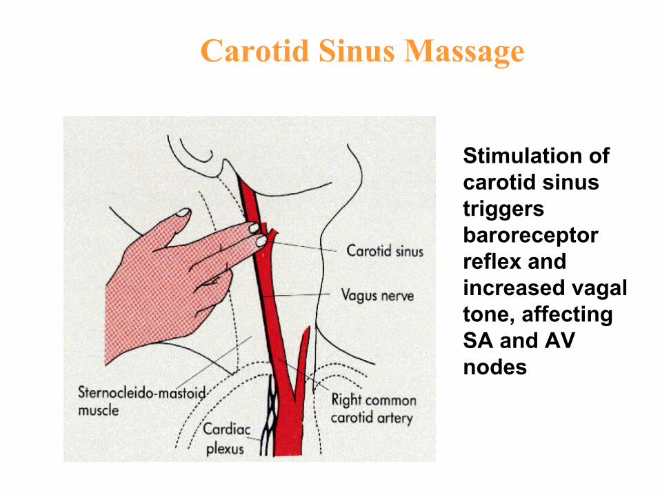
Carotid Sinus Massage
Stimulation of carotid sinus triggers baroreceptorreflex and increased vagaltone, affectingSA and AV nodes
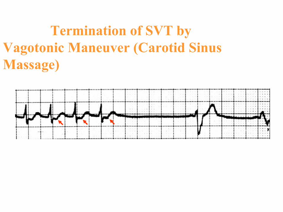
Termination of SVT by Vagotonic Maneuver (Carotid Sinus Massage)
SVT
Carotid Sinus Massage


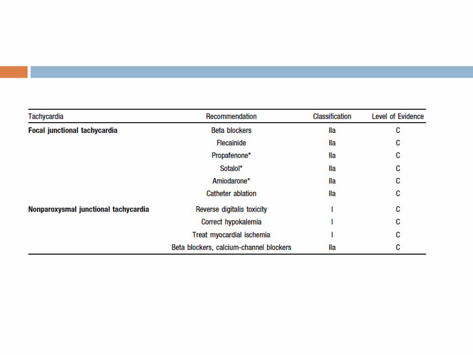
Junctional Tachycardias
1) Focal Junctional Tachycardia (automatic orparoxysmal junctional tachycardia)
2) Non paroxysmal Junctional Tachycardia
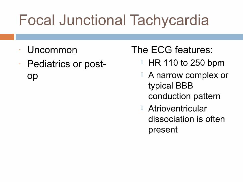
Focal Junctional Tachycardia
- Uncommon- Pediatrics or post-
op
The ECG features: HR 110 to 250 bpm A narrow complex or
typical BBB conduction pattern
Atrioventricular dissociation is often present

Nonparoxysmal Junctional Tachycardia Narrow complex
tachycardia with rates of 70 to 120 bpm
A typical “warm-up” and “cool-down” pattern
The arrhythmia mechanism enhanced automaticity
arising from a high junctional focus or
in response to a triggered mechanism
it may be a marker for a serious underlying condition, such as digitalis toxicity, postcardiac surgery, hypokalemia, or myocardial ischemia

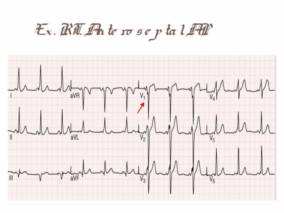
Ex . RT. Ante ro s e p ta l AP

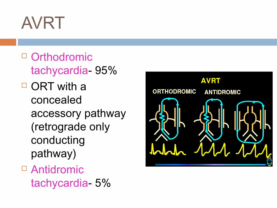
AVRT
Orthodromic tachycardia- 95%
ORT with a concealed accessory pathway (retrograde only conducting pathway)
Antidromic tachycardia- 5%
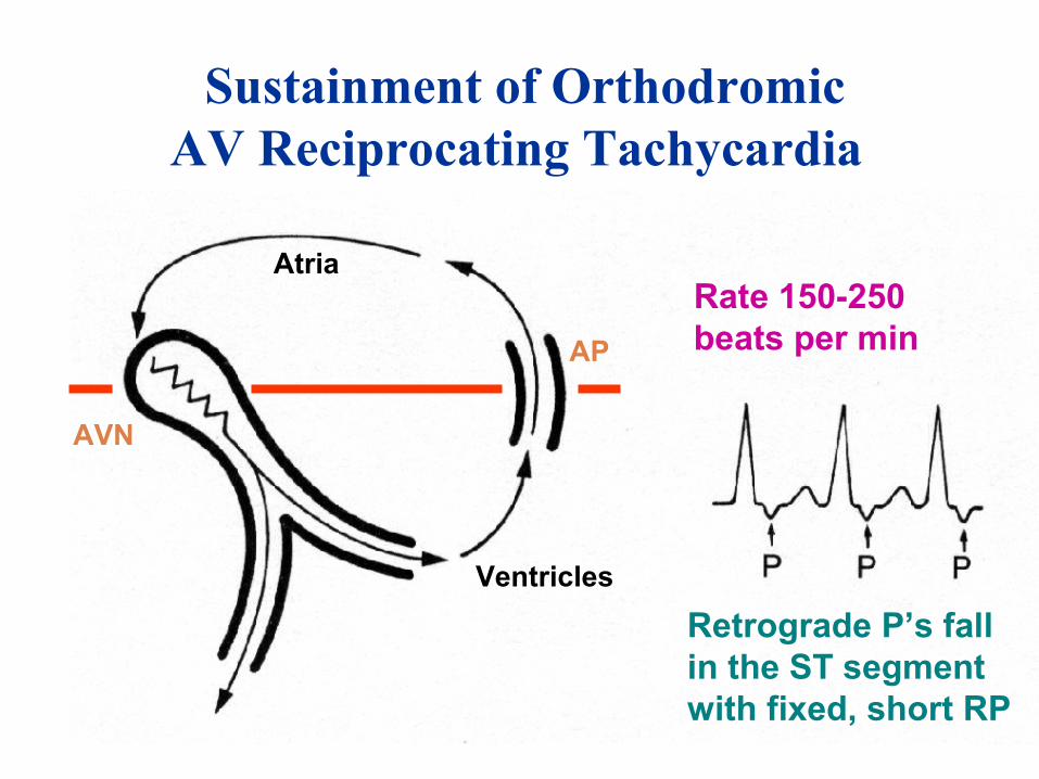
Sustainment of Orthodromic AV Reciprocating Tachycardia
Atria
AP
AVN
Ventricles
Retrograde P’s fall in the ST segmentwith fixed, short RP
Rate 150-250beats per min

Accessory Pathway with Ventricular Preexcitation(Wolff-Parkinson-White Syndrome)
“Delta” Wave
APPR < .12 s
QRS ≥ .12 s
Sinusbeat
Hybrid QRS shape
In sinus rhythm, every ventricular activation is a fusion between accessory pathway and AV nodal conduction
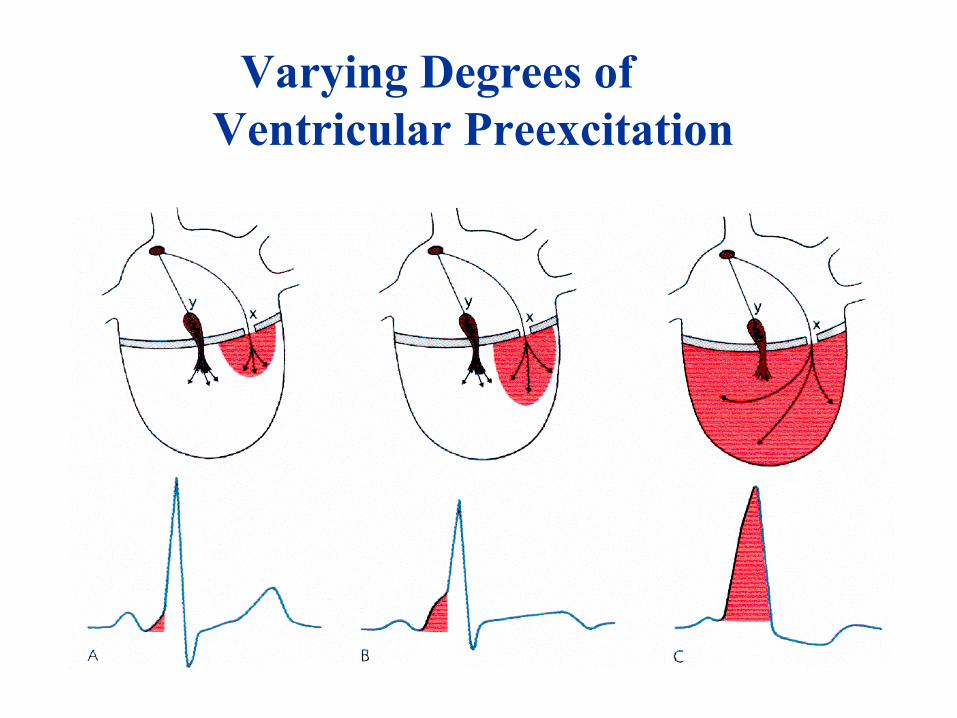
Varying Degrees of Ventricular Preexcitation
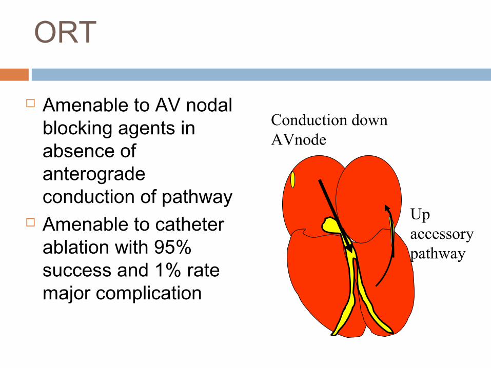
ORT
Amenable to AV nodal blocking agents in absence of anterograde conduction of pathway
Amenable to catheter ablation with 95% success and 1% rate major complication
Conduction down AVnode
Up accessory pathway

WPW syndrome The diagnosis of WPW
syndrome is reserved for patients who have both pre-excitation and tachyarrhythmias
AVRT is the most common arrhythmia, accounting for 95% of re-entrant tachycardias that occur in patients with an accessory pathway
Louis Wolff, Sir John Parkinson and Paul Dudley

Classic ECG pattern
Accelerated AV conduction PR <120 msec
Prolonged QRS > 120 msec Abnormal slurred upstroke of QRS ( delta
wave) Abnormal depolarization and
repolarization may lead to pseudoinfarction pattern
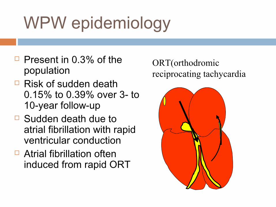
WPW epidemiology
Present in 0.3% of the population
Risk of sudden death 0.15% to 0.39% over 3- to 10-year follow-up
Sudden death due to atrial fibrillation with rapid ventricular conduction
Atrial fibrillation often induced from rapid ORT
ORT(orthodromic reciprocating tachycardia
Markers associated with increased sudded cardiac death1) a shortest pre-excited R-R interval less than 250 ms during spontaneous or induced AF2) a history of symptomatic tachycardia3) multiple accessory pathways4) Ebstein’s anomaly

The detection of intermittent preexcitation, which is characterized by an abrupt loss of the delta wave and normalization of the QRS complex, is evidence that an accessory pathway has a relatively long refractory period and is unlikely to precipitate VF.
The loss of pre-excitation after administration of the antiarrhythmic drug procainamide has also been used to indicate a low-risk subgroup.

Antiarrhythmic drugs that primarily modify conduction through the AV node include: digoxin, verapamil, beta blockers, adenosine, and diltiazem
Antiarrhythmic drugs that depress conduction across the accessory pathway include: Class I drugs, such as procainamide, disopyramide,
propafenone, and flecainide, as well as class III antiarrhythmicdrugs, such as ibutilide, sotalol, and amiodarone.

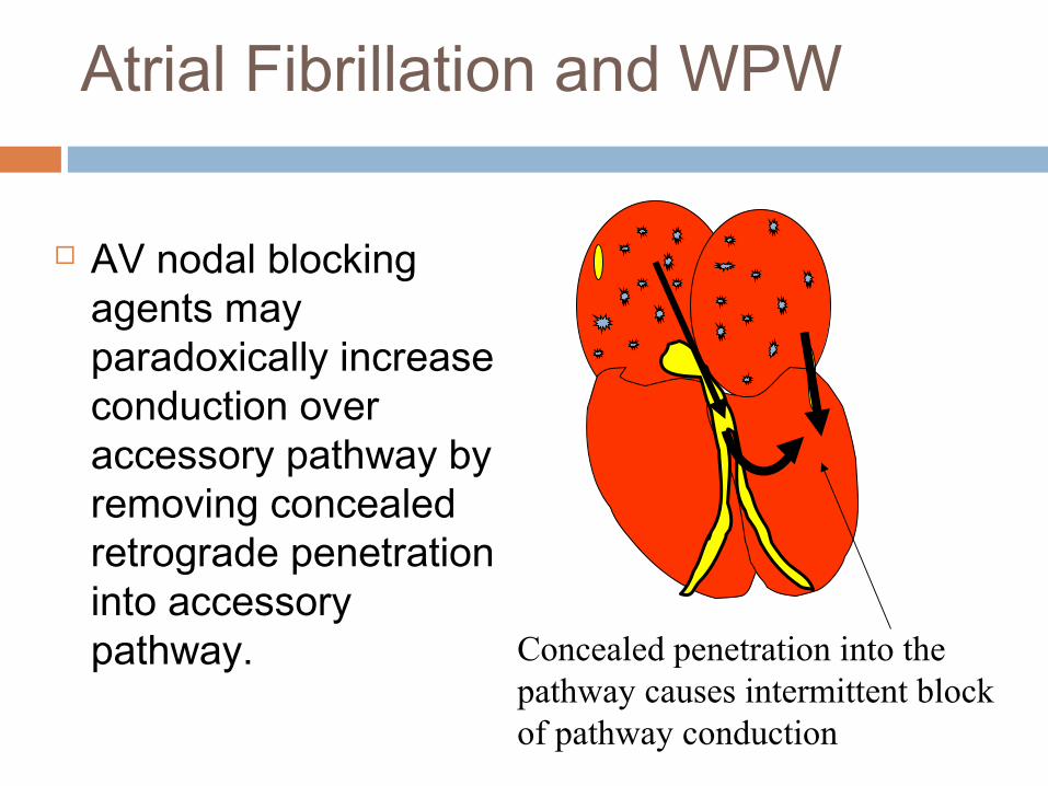
Atrial Fibrillation and WPW
Atrial fibrillation is a potentially life-threatening arrhythmia in patients with WPW syndrome.
If an accessory pathway has a short anterograde refractory period, then rapid repetitive conduction to the ventricles during AF can result in a rapid ventricular response with subsequent degeneration to VF.
Atrial Fibrillation and WPW
AV nodal blocking agents may paradoxically increase conduction over accessory pathway by removing concealed retrograde penetration into accessory pathway. Concealed penetration into the
pathway causes intermittent block of pathway conduction

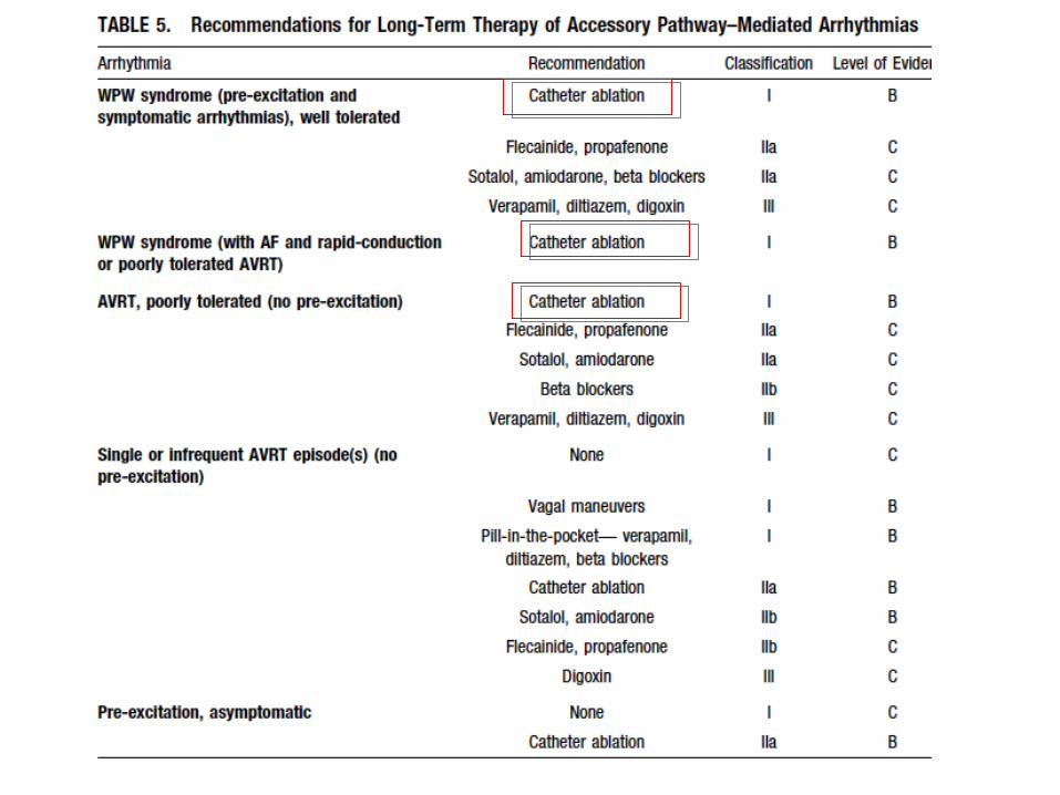
Management of Atrial Fibrillation with WPW
Avoid AV nodal blockers IV procainamide to slow accessory pathway
conduction Amiodarone if decreased LVEF DC cardioversion if symptomatic with
hypotension
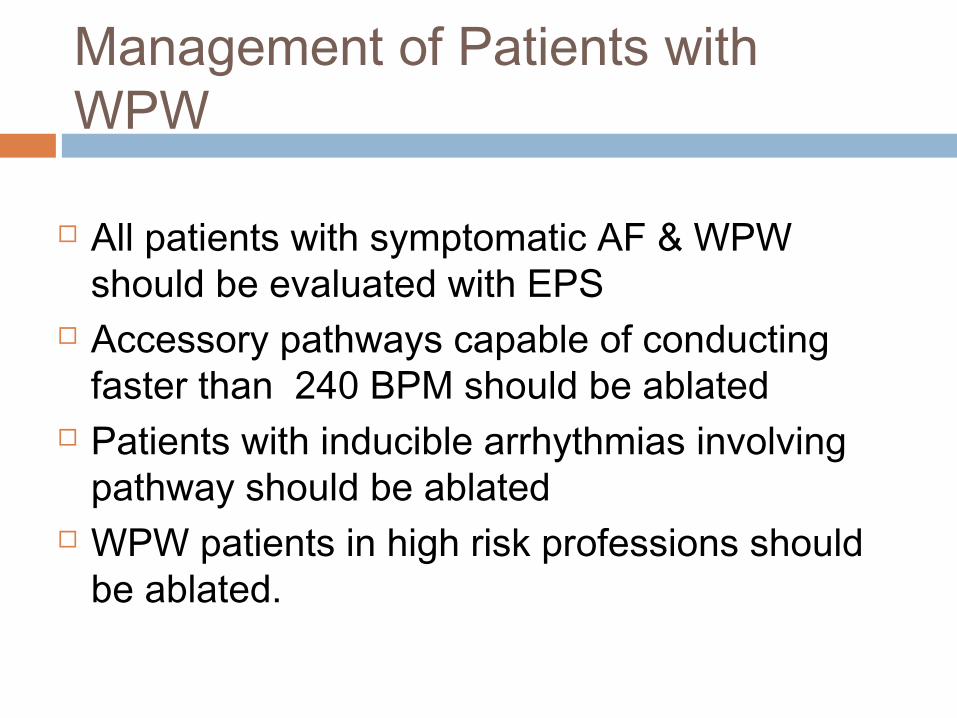
Management of Patients with WPW
All patients with symptomatic AF & WPW should be evaluated with EPS
Accessory pathways capable of conducting faster than 240 BPM should be ablated
Patients with inducible arrhythmias involving pathway should be ablated
WPW patients in high risk professions should be ablated.

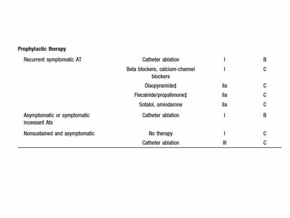
Atrial Tachycardia
Atrial rate between 100 and 250 bpm Does not require AV nodal or infranodal
conduction P wave morphology different than sinus P-R interval > 120 msec differentiating from
junctional tachycardia
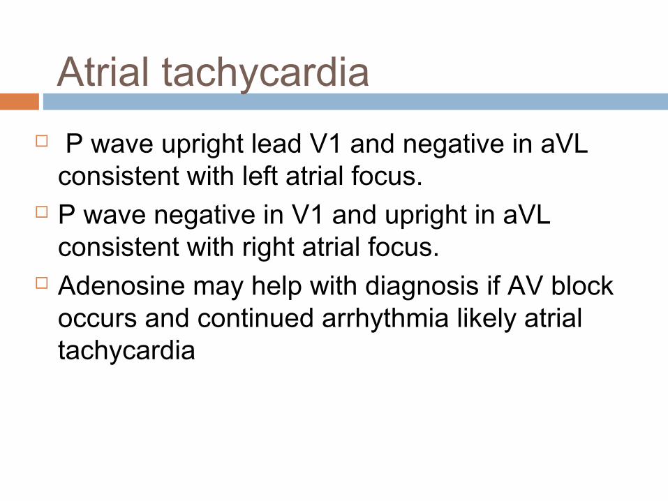
Atrial tachycardia P wave upright lead V1 and negative in aVL
consistent with left atrial focus. P wave negative in V1 and upright in aVL
consistent with right atrial focus. Adenosine may help with diagnosis if AV block
occurs and continued arrhythmia likely atrial tachycardia

Atrial Tachycardia
Most are due to abnormal automaticity and have right atrial focus
May be reentry particularly in patients with previous atriotomy scar, such as CABG or congenital repair patients
it is often associated with
underlying cardiac abnormalities