Embed Size (px)
Citation preview

ANTERIOR ABDOMINAL MUSCLES(Complete Info. & Presentation)(Complete Info. & Presentation)
By- Dr. Armaan SinghBy- Dr. Armaan Singh
Anterior Abdominal Wall• The muscles of the anterior
abdominal wall play a major role in movements of the trunk
• Protecting the abdominal organs
• Increase the intra-abdominal pressure, aid in expiration and all straining activities such as micturition, coughing and vomiting
• Supplied by lower five intercostal and subcostal nerves
Anterior Abdominal Muscles• Strong abdominals are important in
helping to stabilise the trunk
• Support the spine
• They flex and rotate the trunk
• Acting with the adductors and abductors of the hip
• They help to stabilise the pelvis during walking and running

Anterior Abdominal Wall• Superficial fatty layer
• Membranous layer of superficial fascia
• Below umbilicus
• Continuous with Colles’ fascia in the perineum

Skin of Anterior Abdominal Wall
• Lower five intercostal nerves
• Subcostal nerve T12
• 10th intercostal nerve is at the level of the umbilicus
• Iliohypogastric nerve L1
• Ilioinguinal nerve L1
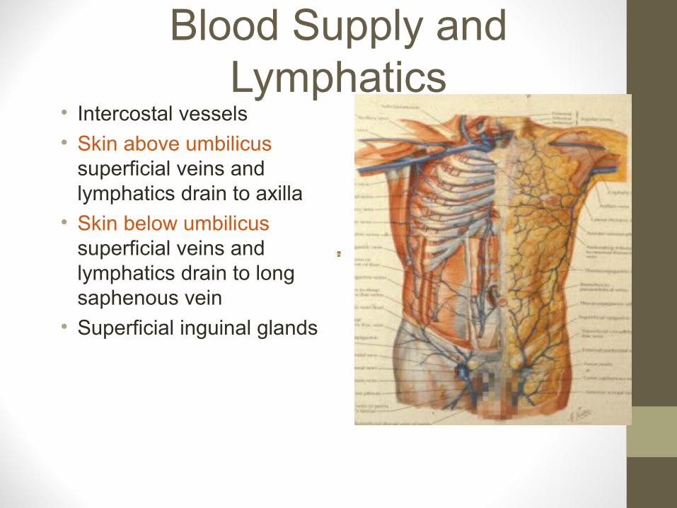
Blood Supply and Lymphatics
• Intercostal vessels
• Skin above umbilicus superficial veins and lymphatics drain to axilla
• Skin below umbilicus superficial veins and lymphatics drain to long saphenous vein
• Superficial inguinal glands

Inguinal Glands• Proximal group parallel to
inguinal ligament
• Enlarged tender inguinal glands
• Part of a generalised lymphadenopathy
• Secondaries

Inguinal Glands• Proximal group
• Lesions in local structures
• Skin of lower anterior abdominal wall
• Gluteal region
• Skin of scrotum or labia Distal superficial glands
• Skin of leg area drained by long saphenous vein
• All drain to deep inguinal glands along femoral vein

Abdominal Muscles• External oblique
• Internal oblique
• Transversus
• Rectus abdominus
• Pyramidalis
• Nerves and vessels
• Lie between internal oblique and transversus

External Oblique• Origin
• Outer surfaces lower borders lower eight ribs
• Interdigitating with serratus anterior and latissimus dorsi
• Fibres pass medially and inferiorly
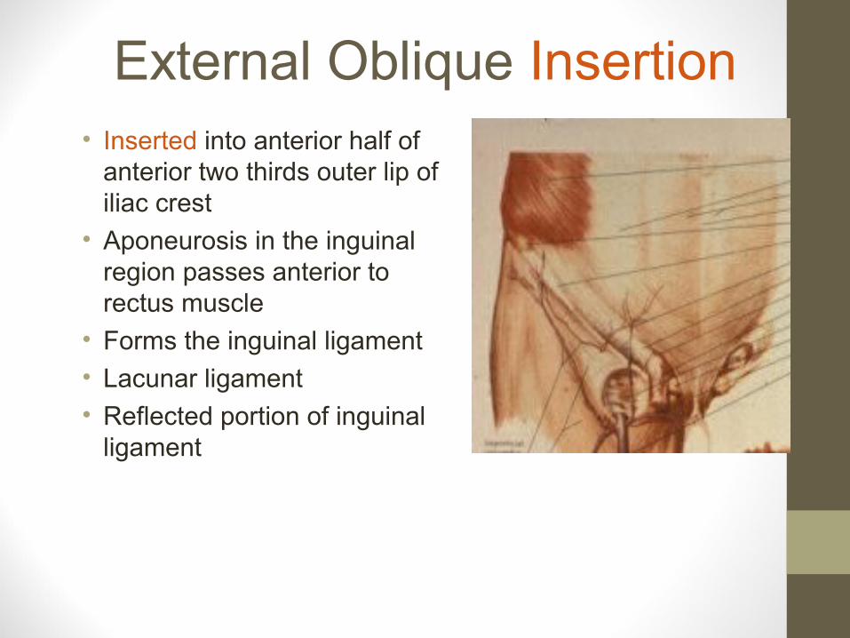
External Oblique Insertion• Inserted into anterior half of
anterior two thirds outer lip of iliac crest
• Aponeurosis in the inguinal region passes anterior to rectus muscle
• Forms the inguinal ligament
• Lacunar ligament
• Reflected portion of inguinal ligament
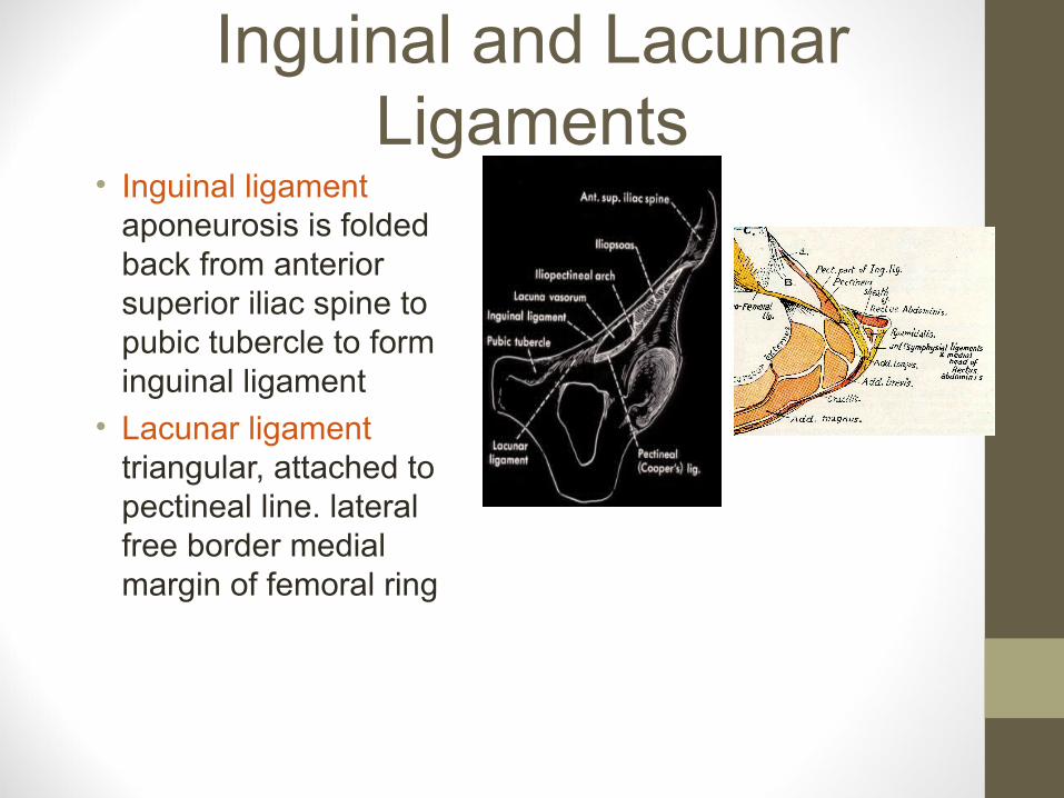
Inguinal and Lacunar Ligaments
• Inguinal ligament aponeurosis is folded back from anterior superior iliac spine to pubic tubercle to form inguinal ligament
• Lacunar ligament triangular, attached to pectineal line. lateral free border medial margin of femoral ring
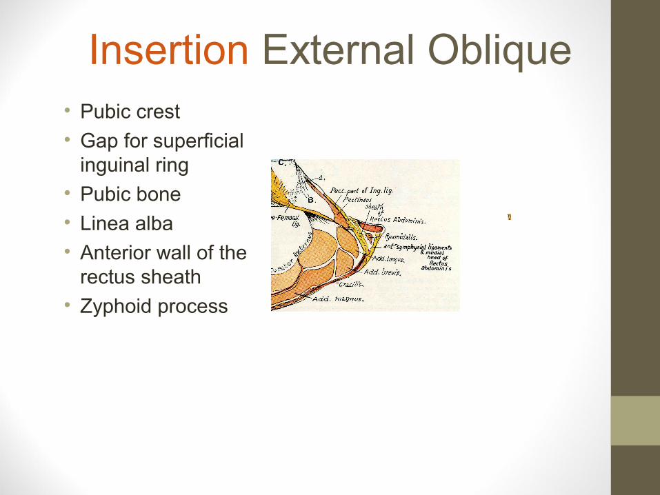
Insertion External Oblique• Pubic crest
• Gap for superficial inguinal ring
• Pubic bone
• Linea alba
• Anterior wall of the rectus sheath
• Zyphoid process
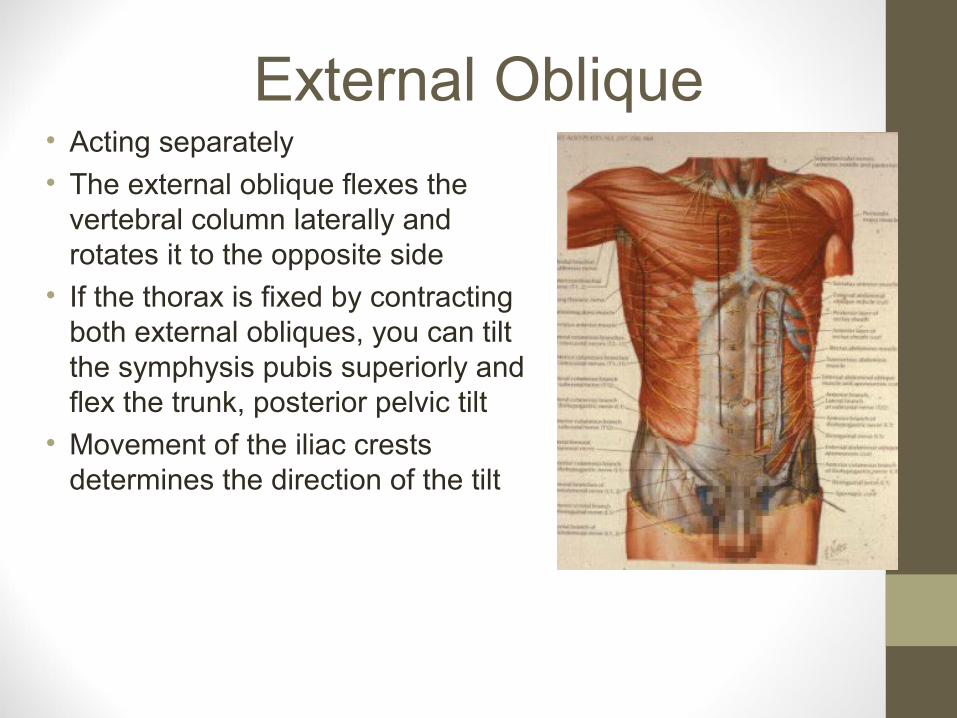
External Oblique• Acting separately
• The external oblique flexes the vertebral column laterally and rotates it to the opposite side
• If the thorax is fixed by contracting both external obliques, you can tilt the symphysis pubis superiorly and flex the trunk, posterior pelvic tilt
• Movement of the iliac crests determines the direction of the tilt

Internal Oblique• Muscular origin lateral two
thirds of inguinal ligament
• Anterior two thirds intermediate lip of iliac crest
• Lumbar fascia
• Muscular fibres arch over contents of inguinal canal anterior to rectus muscle
• Fibres pass medially and superiorly
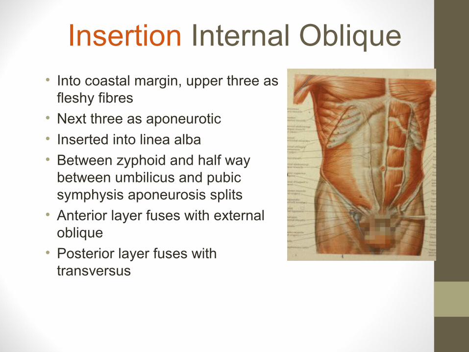
Insertion Internal Oblique• Into coastal margin, upper three as
fleshy fibres
• Next three as aponeurotic
• Inserted into linea alba
• Between zyphoid and half way between umbilicus and pubic symphysis aponeurosis splits
• Anterior layer fuses with external oblique
• Posterior layer fuses with transversus

Internal Oblique Conjoint Tendon
• Half way between umbilicus and pubic symphysis
• Aponeurosis of the internal oblique and transversus fuse to form conjoint tendon
• Anterior portion of rectus sheath
• Inserted into pectineal line behind superficial inguinal ring

Internal Oblique
• The right side of the muscle twists to the right and the left side twists to the left
• The lower six intercostals nerve
• Subcostal nerve
• Iliohypogastric nerves
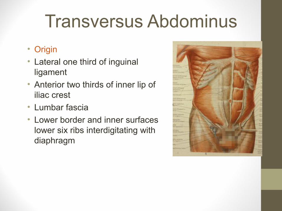
Transversus Abdominus• Origin
• Lateral one third of inguinal ligament
• Anterior two thirds of inner lip of iliac crest
• Lumbar fascia
• Lower border and inner surfaces lower six ribs interdigitating with diaphragm

Insertion Transversus Abdominus
• Into zyphoid, linea alba
• Half way between umbilicus and pubic symphysis
• Fuses with posterior lamella of internal oblique
• Below forms conjoint tendon
• Inserted into pectineal line behind superficial inguinal ring

Transversus Abdominus• The transversus abdominus helps
to support the abdominal viscera
• Maintain intra-abdominal pressure
• Stabilises the lumbar spine
• It is supplied by the lower six intercostals nerves
• Subcostal nerves
• Iliohypogastric nerves
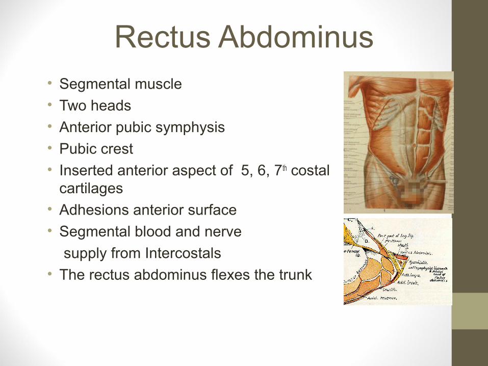
Rectus Abdominus• Segmental muscle
• Two heads
• Anterior pubic symphysis
• Pubic crest
• Inserted anterior aspect of 5, 6, 7th costal cartilages
• Adhesions anterior surface
• Segmental blood and nerve
supply from Intercostals
• The rectus abdominus flexes the trunk
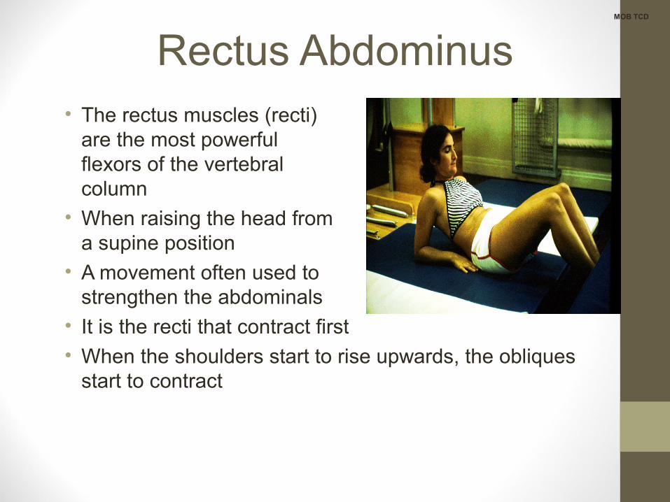
Rectus Abdominus• The rectus muscles (recti)
are the most powerful flexors of the vertebral column
• When raising the head from a supine position
• A movement often used to strengthen the abdominals
• It is the recti that contract first
• When the shoulders start to rise upwards, the obliques start to contract
MOB TCD
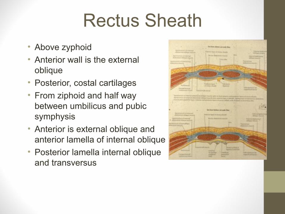
Rectus Sheath• Above zyphoid
• Anterior wall is the external oblique
• Posterior, costal cartilages
• From ziphoid and half way between umbilicus and pubic symphysis
• Anterior is external oblique and anterior lamella of internal oblique
• Posterior lamella internal oblique and transversus

Rectus Sheath• Below half way between
umbilicus and pubic symphysis
• The aponeurosis of the external oblique, internal oblique and transversus (conjoint tendon) pass anterior to the rectus
• Posterior lies the transversalis fascia

Rectus Sheath• Contents
• Rectus muscle
• Pyramidalis
• Superior and inferior epigastric vessels
• Lower five intercostal vessels and nerves
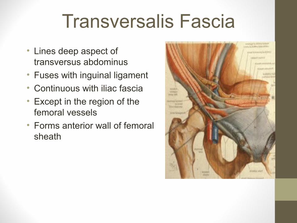
Transversalis Fascia• Lines deep aspect of
transversus abdominus
• Fuses with inguinal ligament
• Continuous with iliac fascia
• Except in the region of the femoral vessels
• Forms anterior wall of femoral sheath

Extraperitoneal Tissue• Extraperitoneal connective
tissue
• If fatty, it separates the
transversalis fascia from
the peritoneum
• If thin, they are in close contact with one another
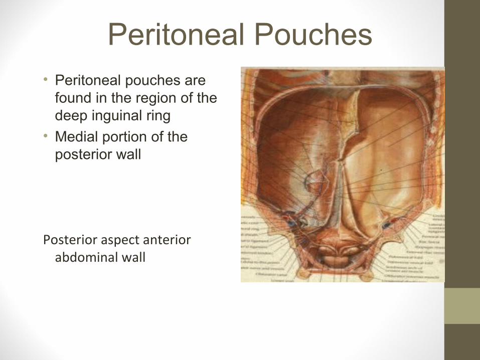
Peritoneal Pouches• Peritoneal pouches are
found in the region of the deep inguinal ring
• Medial portion of the posterior wall
Posterior aspect anterior abdominal wall
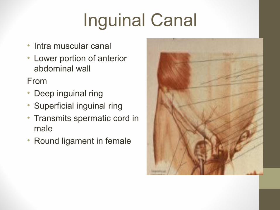
Inguinal Canal• Intra muscular canal
• Lower portion of anterior abdominal wall
From
• Deep inguinal ring
• Superficial inguinal ring
• Transmits spermatic cord in male
• Round ligament in female
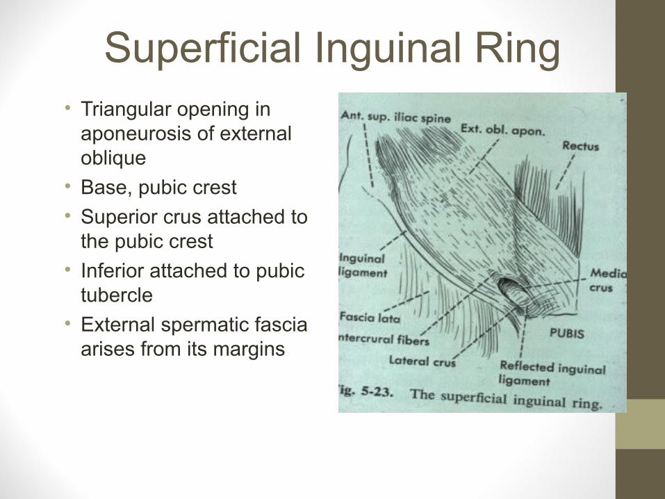
Superficial Inguinal Ring• Triangular opening in
aponeurosis of external oblique
• Base, pubic crest
• Superior crus attached to the pubic crest
• Inferior attached to pubic tubercle
• External spermatic fascia arises from its margins

Deep Inguinal Ring• Oval opening 2.5 cm
• Above the middle of inguinal ligament
• Inferior epigastric artery passes medial to the deep ring
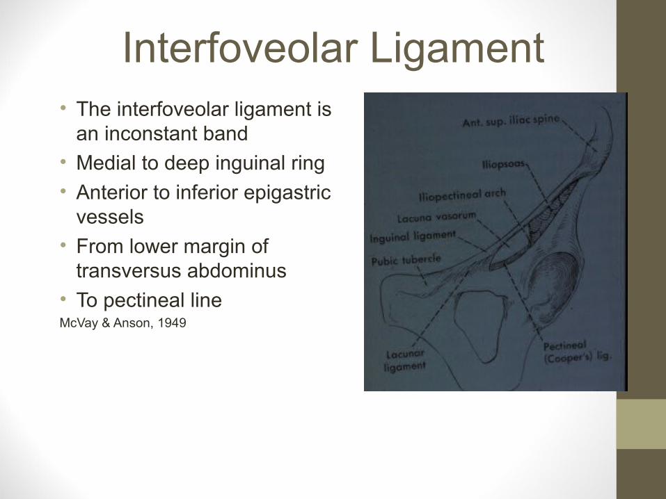
Interfoveolar Ligament• The interfoveolar ligament is
an inconstant band
• Medial to deep inguinal ring
• Anterior to inferior epigastric vessels
• From lower margin of transversus abdominus
• To pectineal lineMcVay & Anson, 1949

Inguinal Canal• Anterior Wall
• External oblique forms
• Whole anterior wall
• Internal oblique forms
• Lateral half only

Inguinal Canal• Posterior Wall
• Transversalis fascia
• Whole of wall
• Medial half conjoint tendon
• Medial quarter reflected portion of inguinal ligament

Roof of Inguinal Canal
• Roof
• Arching fibres of internal oblique
• Transversus as they both arise from the inguinal ligament
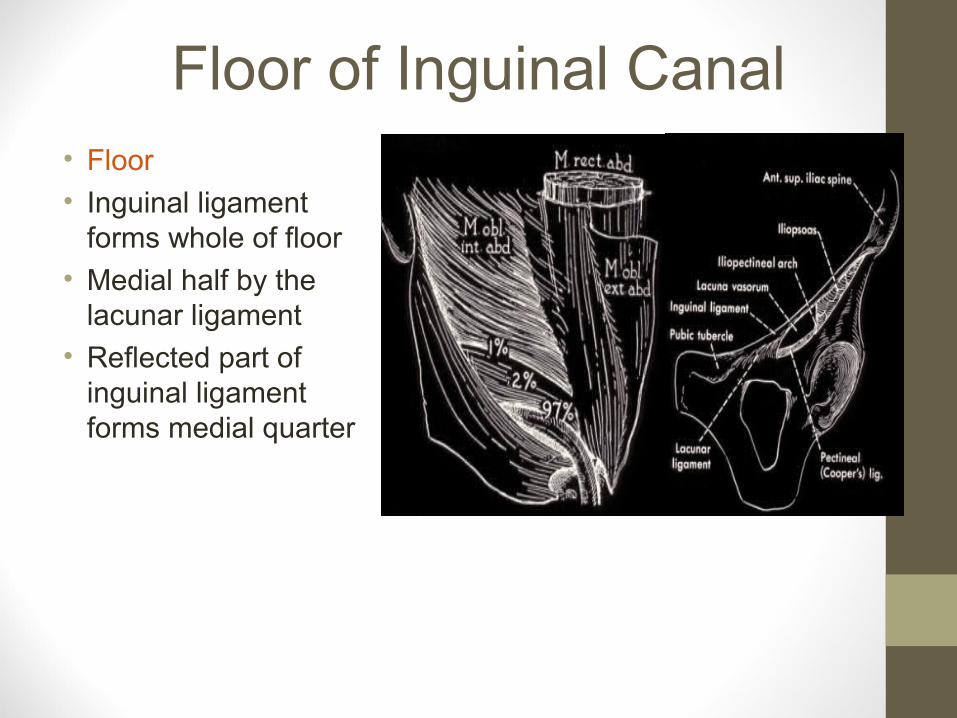
Floor of Inguinal Canal• Floor
• Inguinal ligament forms whole of floor
• Medial half by the lacunar ligament
• Reflected part of inguinal ligament forms medial quarter

Passing Through Deep Ring Male
• Vas Deferens
• Testicular artery
• Pampiniform plexus of veins
• Remains of processus vaginalis
• Genital branch of genitofemoral nerve
• Lymphatics from testes
• Cremaster artery

Passing through Superficial Ring Male
• Everything that went through deep ring
• Plus
• Ilioinguinal nerve
• Internal spermatic fascia from margins of the deep ring
• Cremaster muscle and fascia
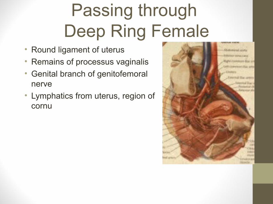
Passing through Deep Ring Female
• Round ligament of uterus
• Remains of processus vaginalis
• Genital branch of genitofemoral nerve
• Lymphatics from uterus, region of cornu

Passing through Superficial Ring Female
• Everything that went through deep ring
• Plus ilioinguinal nerve

Inguinal Canal• Contraction of the
abdominal muscles increases the obliquity of the inguinal canal
• Protecting the two ringsLytle, 1945

Increase in Intra-Abdominal Pressure
• Pain aggravated by an increase in intra- abdominal pressure
• Hernia
• Inguinal or femoral hernia
• Entrapment of the ilioinguinal nerve
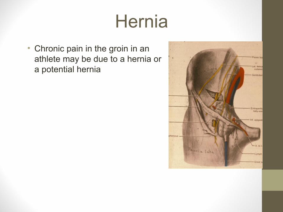
Hernia• Chronic pain in the groin in an
athlete may be due to a hernia or a potential hernia

Inguinal Hernia• Sudden severe pain in
lower abdomen
• Associated with lifting a heavy object
• Common history of a direct inguinal hernia

Indirect Inguinal Hernia• Passes through
• Deep inguinal ring
• May extend to pass through the superficial ring into the scrotum
• Congenital or acquired
• Congenital inside the tunica vaginalis (serous membrane, covers part of testes)
• Acquired outside
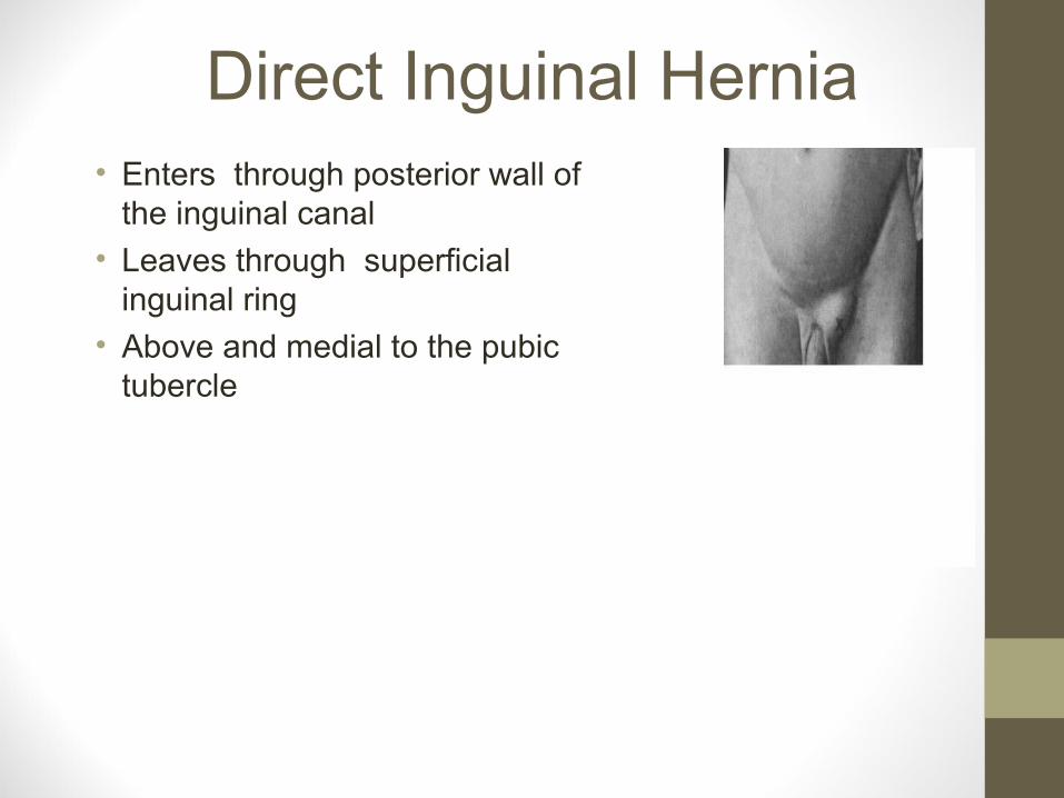
Direct Inguinal Hernia• Enters through posterior wall of
the inguinal canal
• Leaves through superficial inguinal ring
• Above and medial to the pubic tubercle
Inguinal Hernia

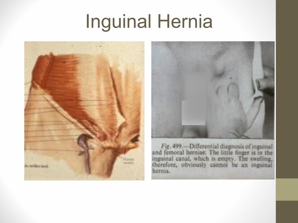
Inguinal Versus Femoral Hernia• Inguinal hernia above and
medial to pubic tubercle
• Femoral hernia below and lateral to the tubercle
• More common in females and more likely to strangulate

Femoral Ring

Femoral Hernia• Enters through femoral ring
• Enters femoral canal
• Medial compartment of femoral sheath
• More common in women

Femoral Hernia
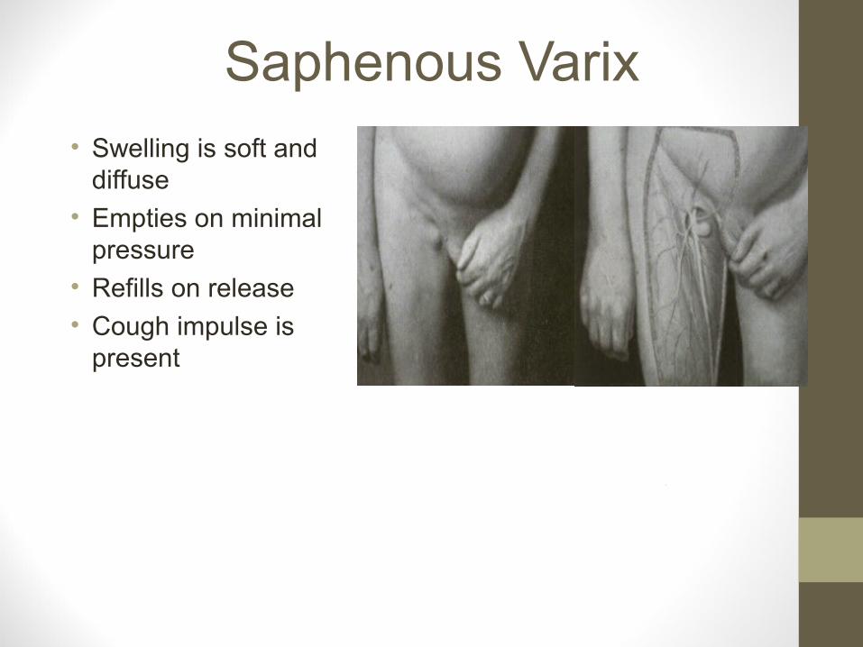
Saphenous Varix• Swelling is soft and
diffuse
• Empties on minimal pressure
• Refills on release
• Cough impulse is present

Gilmore’s Groin• Common cause of chronic groin
pain in field sports
• Particularly soccer players
• Pain on any sudden change of movement, sneezing, coughing

Gilmore’s Groin• Trying to sprint
• Will increase the pain
• Pain is worse getting out of bed
• The day after a match or a training session
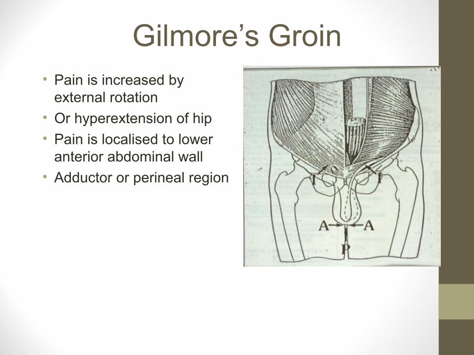
Gilmore’s Groin• Pain is increased by
external rotation
• Or hyperextension of hip
• Pain is localised to lower anterior abdominal wall
• Adductor or perineal region
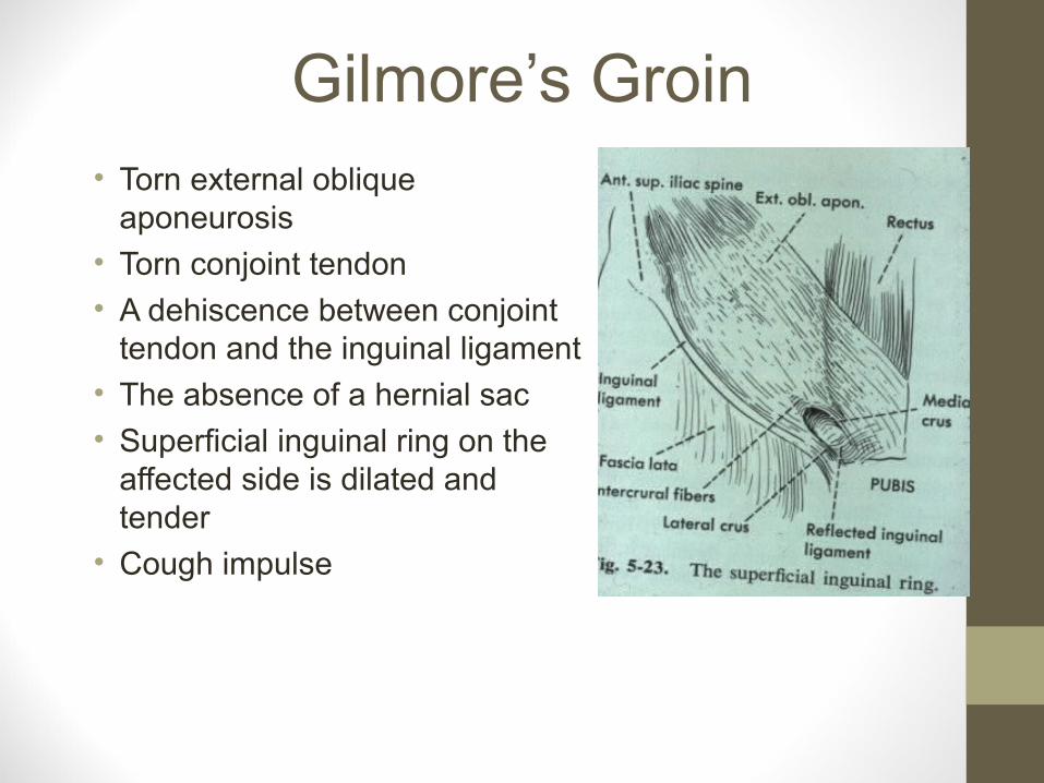
Gilmore’s Groin• Torn external oblique
aponeurosis
• Torn conjoint tendon
• A dehiscence between conjoint tendon and the inguinal ligament
• The absence of a hernial sac
• Superficial inguinal ring on the affected side is dilated and tender
• Cough impulse

Gilmore’s Groin Surgery• Treatment is surgical
• 90% return to sport
• Strengthen lower abdominal muscles
1.Plication of the transversalis fascia in “Shouldice hernia repair”
2.Repair of torn conjoint tendon
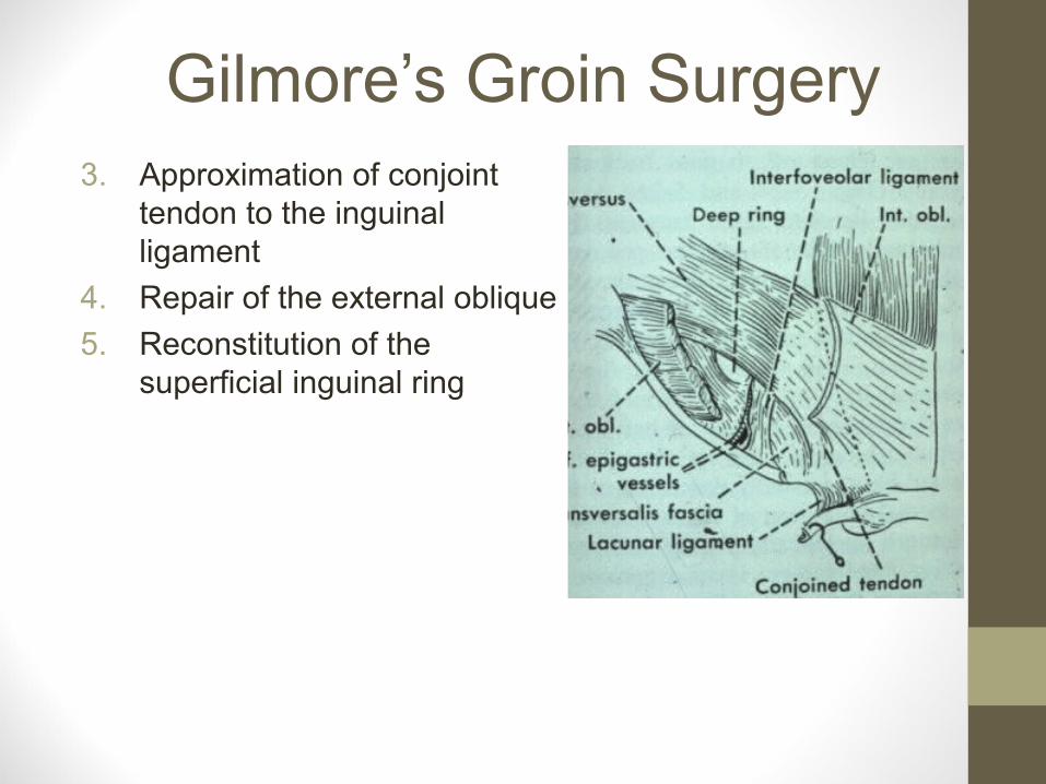
Gilmore’s Groin Surgery3. Approximation of conjoint
tendon to the inguinal ligament
4. Repair of the external oblique
5. Reconstitution of the superficial inguinal ring