Embed Size (px)
Citation preview
4
CHAPTER I
INTRODUCTION
Gram staining has been widely used in medicine to diagnose certain diseases by
identifying their causative agents. Crystal violet and Safranin, both dyes used in the Gram
staining method, are synthetic dyes. They give purple and red color, dividing bacteria to Gram
positive and Gram negative making identification easy. Through this, doctors can diagnose
correctly and can provide an efficient treatment. However, both of these synthetic dyes have
toxic effects to people who are constantly in contact with them. Most of these people at risk are
medical technologists.
According to Brit (2008), synthetic dyes contain the chemical Aniline which is the basis
for Azo dyes and is considered as a deadly poison, giving off carcinogenic amines. Crystal violet
and Safranin are both classified as IARC Group 1: carcinogenic to humans ( International
Agency for Research on Cancer of the World Health Organization, 2016). Crystal violet is
considered a hazardous substance in the statement of hazardous nature of the Occupational
Safety and Health Administration 29 Code of Federal Regulation 1910.1200 (Chemwatch, 2010).
Safranin red has also been found to be carcinogenic and can cause malformations in embryo or
fetuses of pregnant women (Summers, Texley, & Kwan, 2006).
Because of this, a lot of people are now leaning towards using natural dyes as they are
less toxic, less health hazardous, non-carcinogenic, and non-poisonous (Roberts, 2015). Plants
have anthocyanins that are responsible for the bright attractive red, orange, purple and blue
colors of most fruits and vegetables (Giusti, 2008). In the Philippines, plants like Alugbati

5
(Basella rubra) and Gumamela (Hibiscus rosa sinensis) have been found out to be potential dyes
for biological staining as they both contain anthocyanins.
Deshmukh and Gaikwad (2013) used Alugbati berries as a biological stain for plant
nuclei and organelles. Alugbati berries extract turned out to be a favorable substitute for crystal
violet (Enerva, Utilization of an Indigenous dyestuff from Basella rubra (alugbati) as
microbiological stain, 2006; Stuart, 2015). On the other hand, Gumamela flower extract has been
found out to be an alternative dye for eosin (Ampana, et al., 2013). Another similar study by
Castor, Fabile, Jimenez, San Jose, Sumarago et al (2014) used Alugbati extract as primary stain,
kalamansi extract as mordant and Gumamela flower extracts as counter stain as alternative for
Gram staining and established a conclusion that these were comparable to Gram stain.
With these new developments, the proponents of this study aim to find safer, alternative
stains for the Gram staining using Alugbati berries and Gumamela flowers as primary and
secondary stains, respectively and to find locally-made dyes from natural and organic materials.
The researchers will perform this experiment with the recommendation of increasing
concentrations of both extracts to improve staining and color.
6
Review of Related Literature
Gram Stain
Nowadays, Gram staining is the principal stain used for the microscopic examination of
clinically important bacteria. This was first devised by Hans Christian Gram in the late
nineteenth century, and has been used until now to divide most bacterial species into two large
groups: gram-positive, – those that take up the basic dye, crystal violet and gram-negative, –
those that allow the crystal violet dye to be washed out easily with the decolorizer alcohol such
as acetone and take up the red dye, safranin red. Its principle lies on the thickness and
composition of the bacterial cell walls. Gram-positive cell walls contain thick peptidoglycan with
numerous teichoic acid cross-linkages, while gram-negative cell walls contain a thinner layer of
peptidoglycan. The teichoic acid cross-links contribute to the ability of gram-positive organisms
to resist alcohol decolorization. Gram reaction coupled with the determination of cell size,
morphology, and arrangement are key aspects in accurately identifying bacteria (Brooks, 2013;
Forbes, Sahm, & Weissfeld, 2007).
Stains used in grams staining are both basic dyes, therefore carrying positive ions. The
positive ion in a basic dye is attracted to the slightly negative charged bacterial cell (Tortora,
Funke, & Case, 2002).
Crystal Violet as Primary Stain
Crystal Violet is a triarylmethane dye which can be used as a primary dye in Gram
staining to facilitate classification of bacteria or for simple histological staining procedure.
Furthermore, Crystal Violet also has antibacterial, antifungal and anti-helminthic properties and

7
was used in the past as a topical antiseptic. The medical use of the dye has been superseded by
more modern drugs, although it is still listed by the World Health Organization (Crystal Violet,
2013).
Consequently, various studies have reinforced that Crystal Violet has a hazardous effect
to humans and the environment. It has been reported as a recalcitrant dye molecule that persists
in environment for a long period of time and can pose toxic effects. It acts as mitotic poison,
potent carcinogen and a potent clastogene promoting tumor growth in some species of fish.
Crystal Violet is thus regarded as a biohazard substance (Mani & Bharagava, 2016). It is also
considered a hazardous substance in the statement of hazardous nature of the Occupational
Safety and Health Administration 29 Code of Federal Regulation 1910.1200 (Chemwatch, 2010).
In one study, it demonstrated dose-related carcinogenic potential at several different organ sites
in mice (The Carcinogenic Potency Project, 2007; Littlefield, Blackwell, Hewitt, & Gaylor,
1985).
Safranin as Secondary Stain
Safranin (also Safranin O or basic red 2) is a biological stain employed in microbiology,
histology and cytology. It is used as a counter stain in some staining protocols, imparting color to
all cell nuclei red. This is the classic counter stain in both Gram stains, and endospore stains. It
can also be used for the detection of cartilage, mucin and mast cell granules. Safranin typically
has the chemical structure sometimes described as Dimethyl safranin. There is also
Trimethylsafranin, which has an added methyl group in the ortho- position of the lower ring.
Both compounds behave essentially similar in biological staining applications, and most
manufacturers of safranin do not distinguish between the two. Commercial safranin preparations
often contain a blend of both types. Safranin is also used as redox indicator in analytical
8
chemistry (Rosenberg, 1971).
Safranin is also known by the names of safranin O, safranin Y, safranin T, safranin A,
basic red 2, gossypimine and cotton red. It consists of dark red crystals available as a dimethyl or
trimethyl derivatives; both show the same staining properties. Commercial safranin stains may
contain either of these two or a combination of both derivatives. Futhermore, it also used in
histological and cytological staining procedures to stain cellular nuclei red and used as a
component in the Flemming triple stain to stain chromosomes. Safranin has also been used in the
staining of cartilage, where the nuclei appear red against a pink background (Stewart, 2006).
The stain can cause both skin and eye irritation, and may be harmful if swallowed,
absorbed through the skin, or inhaled. Safranin O is also considered an irritant of mucous
membranes and the upper respiratory tract, but is not flammable or explosive under typical
laboratory conditions (Davidson, 2015). It is also found to be carcinogenic and causing
malformations in embryo or fetuses of pregnant women (Summers, Texley, & Kwan, 2006). In
addition, exposure to these chemicals can be irritating to the skin, harmful to the respiratory
system when inhaled, and to the digestive tract when ingested (Mohammed, Ibrahim, & Shitu,
2014).
Natural Dyes
Plants containing anthocyanins are responsible for the bright attractive red, orange,
purple and blue colors of most fruits and vegetables (Giusti, 2008). Anthocyanins are versatile
flavonoid pigments found in red/purplish fruits and vegetables, hence, have a potential of
becoming a natural dye for staining. This pigment occurs naturally in plants in the form of
glycosides, in which an anthocyanidin molecule is paired with a sugar. The part of the pigment

9
that exists free of sugar (generically known as aglycone) is called an anthocyanidin (Webb,
2014). The most common sugars present in these natural pigments are glucose, fructose,
galactose, xylose, arabinose and rhamnose (Tazzini, 2014).
Anthocyanin changes color, ranging from red, under very acidic conditions to purple-
blue, in intermediate pH conditions until yellow-green, in alkaline conditions. In addition to the
pH, the color of these flavonoids can be affected by the degree of hydroxylation or methylation
pattern of the A and B rings, and by glycosylation pattern (Tazzini, 2014). At pH 8, anthocyanin
remaind purple color and then changed to violet, blue respectively when pH rose from 9-11 due
to the presence of quinoidal ion (Nghia, 2014). To obtain the best yield of anthocyanin
extraction, weak organic acids such as acetic acid and citric acid and low concentration of strong
acids such as hydrochloric acids at less than <1% (Dai & Mumper, 2010), are used.
Anthocyanin is rapidly degraded in the presence of light and high temperature
(Bakowska-Barczak, 2005). Therefore, crude extracts which will be obtained in this study will be
stored in amber bottles to prevent interference of light, and refrigerated at 4˚C so as to prevent
anthocyanin degradation (Zozio, Pallet, & Dornier, 2011).
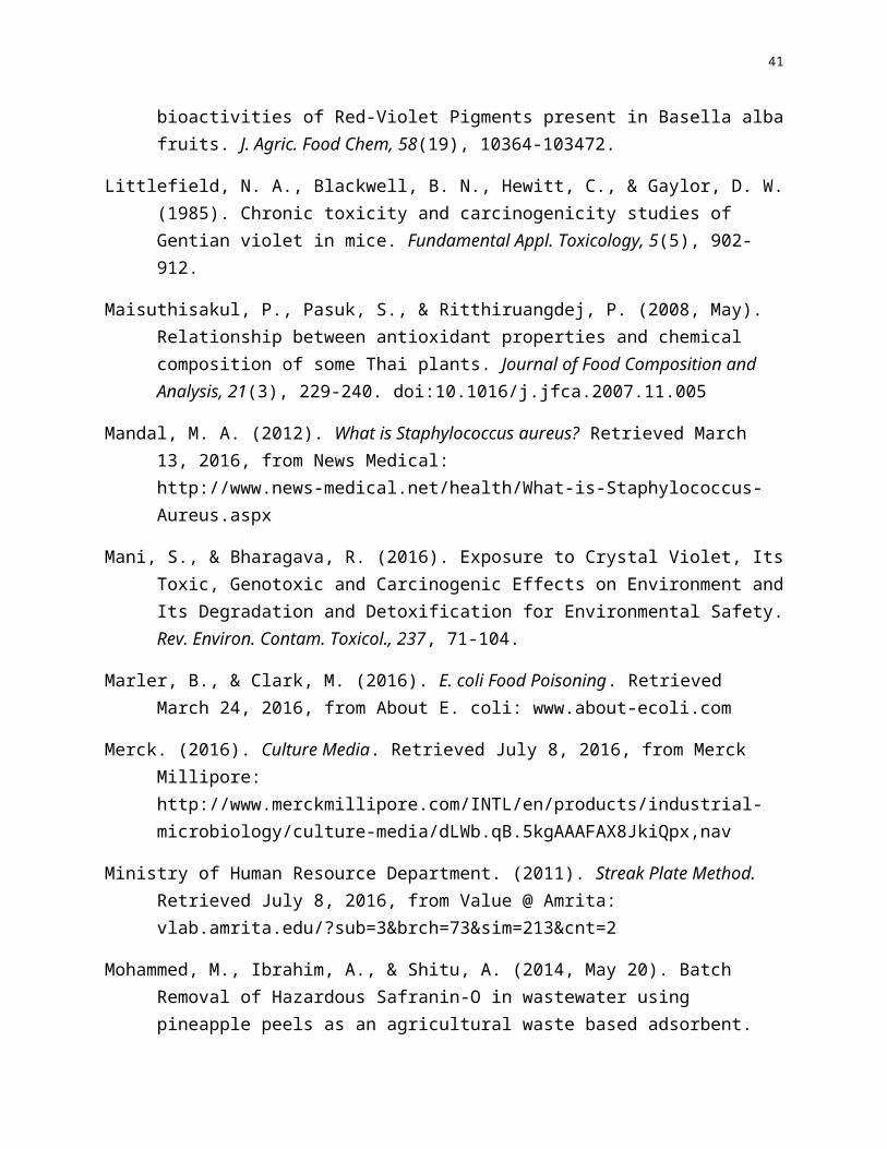
Alugbati (Basella alba) berries
Figure 1. Basella alba (Alugbati) BerriesRetrieved: January 26, 2016 from https://en.wikipedia.org/wiki/Basella_alba
10
Local Name: Alugbati
Kingdom- Plantae
Phylum- Magnoliophyta
Class- Magnoliopsida
Order- Caryophyllales
Family- Basellaceae
Genus- Basella
Species- alba
(Adhikari, Kumar, & Shruthi, 2012)
Alugbati is a succulent, branched, smooth, twining herbaceous vine, several meters in
length. The fruit is fleshy, stalkless, ovoid or nearly spherical, 5 to 6 millimeters long, and purple
when mature. Phytochemical screening of various extracts yielded chemicals, specifically
anthocyanin. This chemical is a water-soluble vacuolar pigment that may appear red, purple, or
blue depending on the pH (Stuart, 2015).
There are four varieties of alugbati. One, Basella rubra has purple pigment on the stem,
leaves and as well as the petioles. The leaf is ovate with a cordate base. Two, Basella alba has
green stems, leaves and petioles. The leaf is ovate and cordate at the base. Three, Basella
cordifolia has a green stem, leaves and petiole with elongated heart shaped or cordate leaves.
This type has pigmentation on the node as well as the base of the petioles and the first two
internodes above the soil level. Four, Basella alba variety has green stem, leaves and petioles
with oval to almost round leaves (T.A. & Mabel, 2015).

11
It is considered an important green leafy vegetable found commonly in the tropical
regions of the world and also has great ethno-medicinal importance. The plant is rich in Vitamin
A and Vitamin C along with flavonoids, saponins, carotenoids, many amino acids and organic
acids. It is known to be androgenic, anti-diabetic, anti-inflammatory, antimicrobial, antioxidant,
antiulcer, antiviral, central nervous system depressant, hepatoprotective and can heal wounds.
Also, the plant possesses a valuable ethno-medicinal importance and is used to cure
digestivedisorders, skin diseases, bleeding piles, diarrhea and many more (Deshmukh &
Gaikwad, 2013).
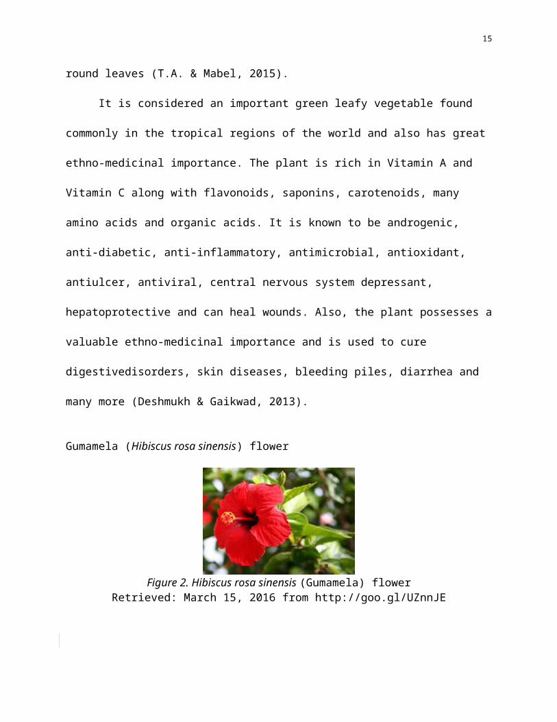
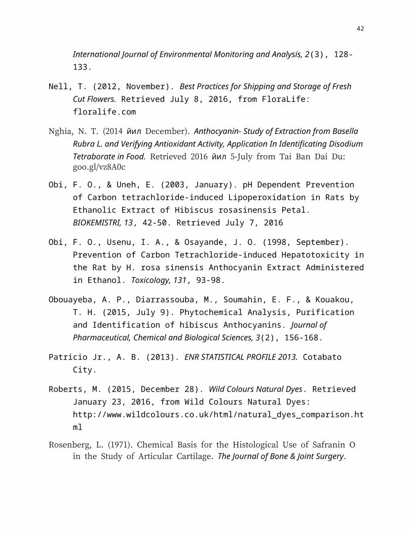
Gumamela (Hibiscus rosa sinensis) flower
Figure 2. Hibiscus rosa sinensis (Gumamela) flowerRetrieved: March 15, 2016 from http://goo.gl/UZnnJE
Scientific Name: Hibiscus rosa-sinensis
Local Name: Gumamela
Kingdom: Plantae
Division: Magnoliophyta
Class: Magnoliopsida
Order: Malvales
Family: Malvaceae

12
Genus: Hibiscus
Species: rosa-sinensis
(Chua, 2010)Gumamela is a shrub that grows from 1 meter up to 4 meters high. It is also known as
Hibiscus, China Rose, and Shoeflower. In the Philippines, it is cultivated as an ornamental plant
and it comes in many colors: red, yellow, orange, white, purple, pink and other color
combinations (Philippine Herbal Medicine, 2). Its flowers are solitary, axillary, very large about
10 cm long and 12 cm in diameter. Petals are obovate, entire, rounded tip and imbricate. Flowers
are best collected from May to August (Stuart, 2015).
Gumamela contains taraxeryl acetate, beta-sitosterol, campesterol, stigmasterol,
ergosterol, lipids. Arachidic acid, behenic acid, oxalic acid, palmitic acid, octanoic acid, stearic
acid, sterculic acid, tricosanoic acid, tridecanoic acid, undercanoic acid, citric acid, tartaric,
fructose, glucose, sucrose, flavonoids and flavonoid glycosides, hibiscetin, alkanes,
hentriacontanecyanidin, cyanidin chloride and cyaninglucosides (Lim, 2014).
Test Organisms
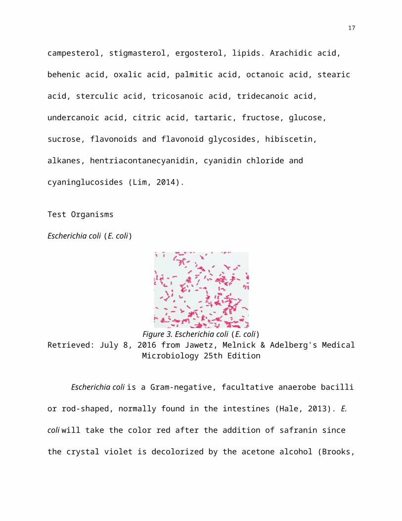
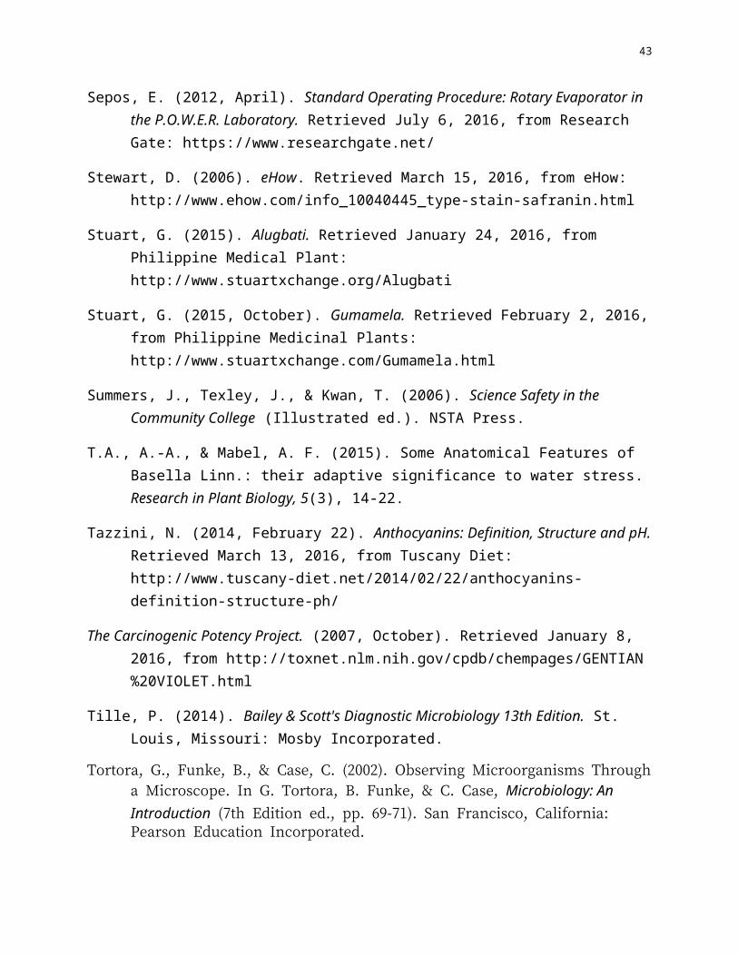
Escherichia coli (E. coli)
Figure 3. Escherichia coli (E. coli)Retrieved: July 8, 2016 from Jawetz, Melnick & Adelberg's Medical Microbiology 25th Edition

13
Escherichia coli is a Gram-negative, facultative anaerobe bacilli or rod-shaped, normally
found in the intestines (Hale, 2013). E. coli will take the color red after the addition of safranin
since the crystal violet is decolorized by the acetone alcohol (Brooks, 2013). E. coli bacteria
were discovered in the human colon in 1885 by German bacteriologist Theodor Escherich. He
showed that certain strains of the bacterium were responsible for infant diarrhea and
gastroenteritis (Marler & Clark, 2016).
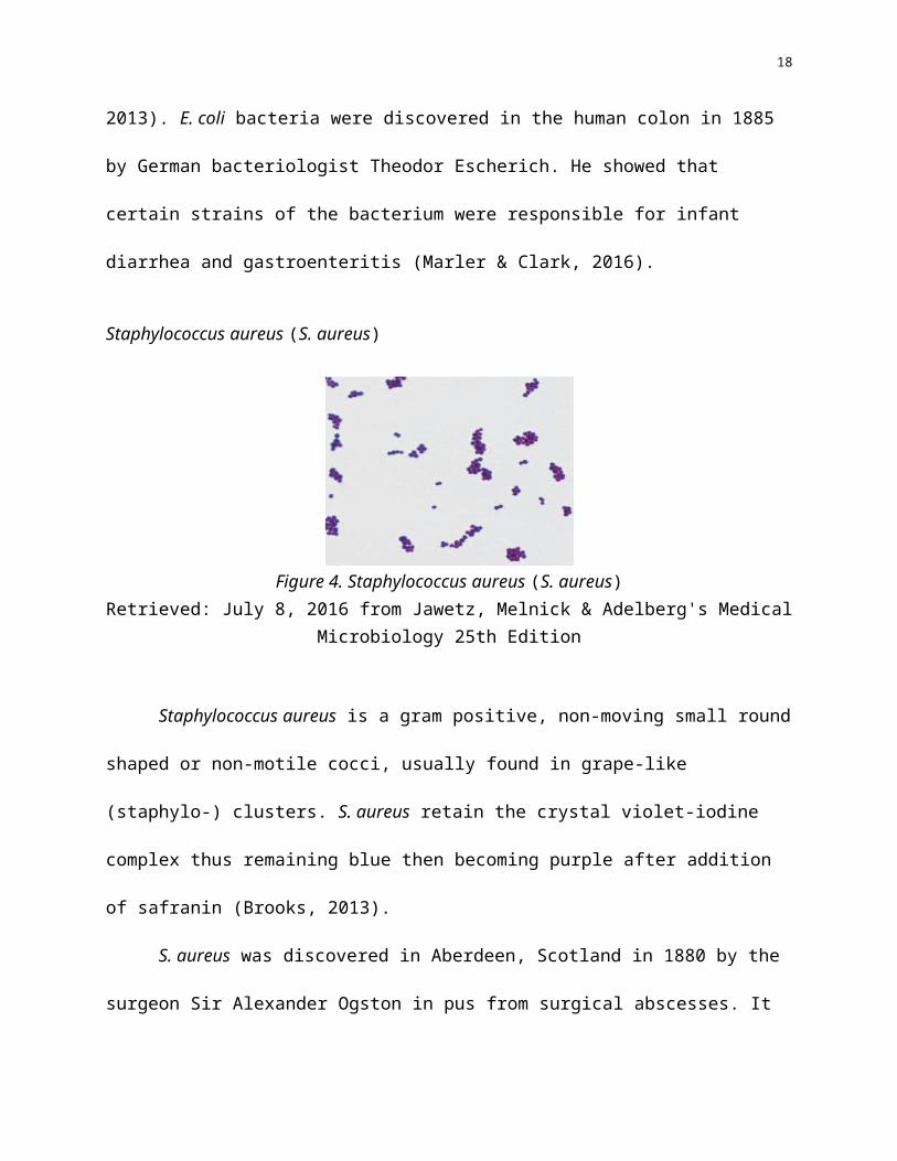
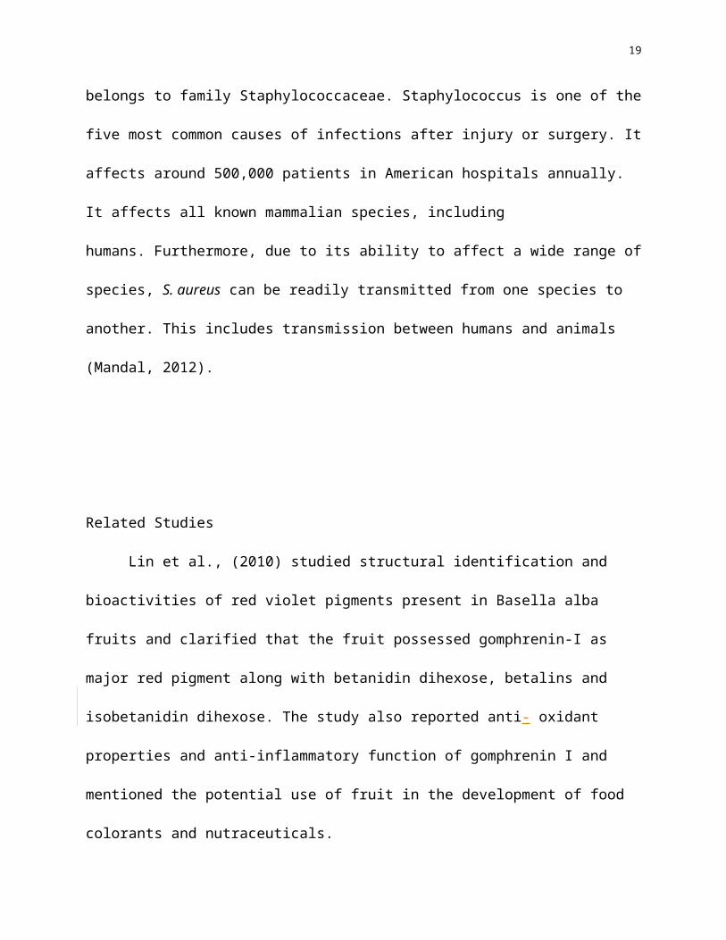
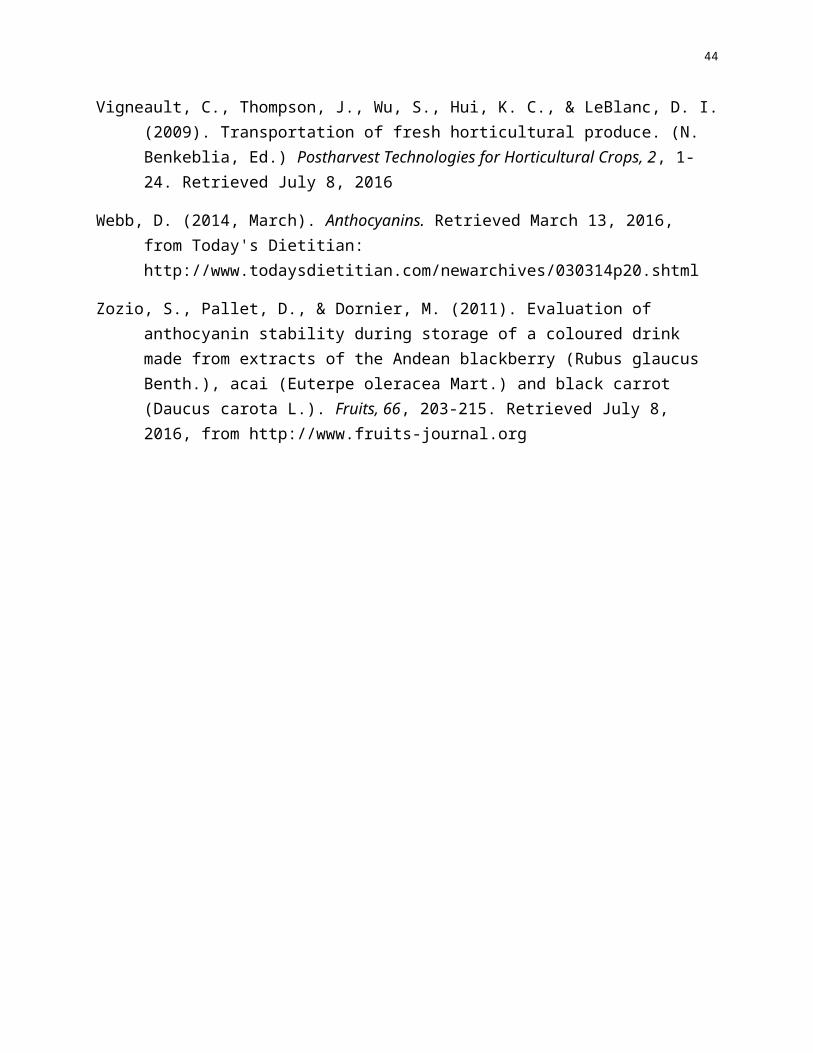
Staphylococcus aureus (S. aureus)
Figure 4. Staphylococcus aureus (S. aureus) Retrieved: July 8, 2016 from Jawetz, Melnick & Adelberg's Medical Microbiology 25th Edition
Staphylococcus aureus is a gram positive, non-moving small round shaped or non-motile
cocci, usually found in grape-like (staphylo-) clusters. S. aureus retain the crystal violet-iodine
complex thus remaining blue then becoming purple after addition of safranin (Brooks, 2013).
S. aureus was discovered in Aberdeen, Scotland in 1880 by the surgeon Sir Alexander
Ogston in pus from surgical abscesses. It belongs to family Staphylococcaceae. Staphylococcus
is one of the five most common causes of infections after injury or surgery. It affects around
500,000 patients in American hospitals annually. It affects all known mammalian species,
including humans. Furthermore, due to its ability to affect a wide range of species, S. aureus can
14
be readily transmitted from one species to another. This includes transmission between humans
and animals (Mandal, 2012).
Related Studies
Lin et al., (2010) studied structural identification and bioactivities of red violet pigments
present in Basella alba fruits and clarified that the fruit possessed gomphrenin-I as major red
pigment along with betanidin dihexose, betalins and isobetanidin dihexose. The study also
reported anti- oxidant properties and anti-inflammatory function of gomphrenin I and mentioned
the potential use of fruit in the development of food colorants and nutraceuticals.
Amon & Pladio (2012) studied Basella rubra fruit extracts for their potentiality as a food
colorant and that the extract showed the presence of anthocyanin and also exhibited DPPH
radical scavenging activity. The extract was proved to be non toxic and can also be implied in
food coloring industries to impart reddish colour.
In a study by Obi and Uneh (2003) and Obi, Osenu and Osayande (1998), gumamela
petals were treated with formic acid, ethanol and water as extracting agents for anthocyanin.
Futhermore, in the study conducted by De Leon, Latoza, Nues, Pilac, Soliman, Sistoza (2016),
alugbati was extracted by adding 5ml of methanol to 5grams of berries, macerated and then
centrifuged at 3000 rpm for 3-5 minutes. Another study used 80% ethanol to extract air-dried
Rhus coriaria L. berries (Abu-Shanab, Adwan, Abu-Safiya, Adwan, & Abu-Shanab, 2005) and
air-dried Moringa pterygosperma Gaertn flowers (Bargah, 2015) using 95% ethanol
(Maisuthisakul, Pasuk, & Ritthiruangdej, 2008).
15
Alugbati extract turned out to be comparable with synthetic stains in a study conducted
by Enerva (2006) where alugbati berries were macerated in a blender and extracted with 1% HCl
in 95% methanol and then filtered, the crude extract was used as to stain for S. aureus and E.coli.
Ampaña et al (2013) found out that Gumamela could be an alternative dye for eosin. Castor et al
(2014) used Alugbati extract as primary stain, kalamansi extract as mordant and Gumamela
flower extracts as counter stain as alternative for Gram staining using 100 grams of berries and
gumamela petals and extracted it using 50ml of 95% methanol and 50ml of 1% Hydrochloric
Acid. Results of their study were comparable to Gram stain but increasing concentration was
recommended to prolong the stain as that of the gram stain.
16
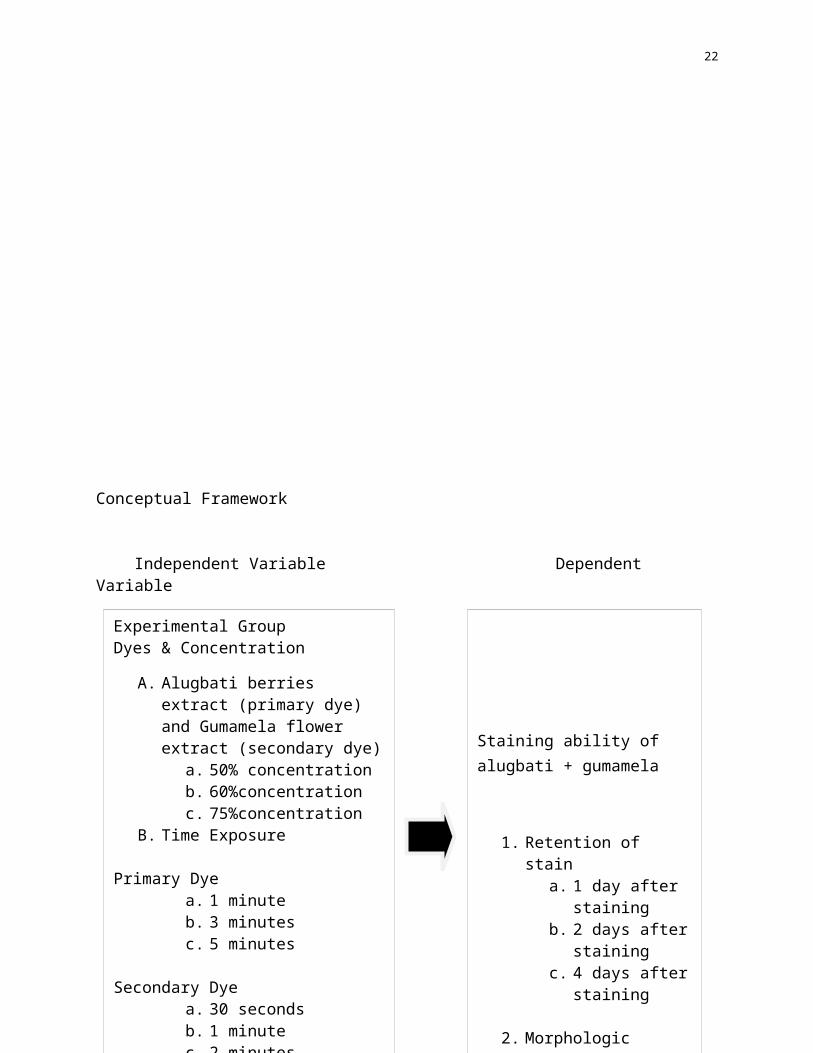
Conceptual Framework
Independent Variable Dependent Variable
Staining ability of alugbati + gumamela
1. Retention of staina. 1 day after
stainingb. 2 days after
stainingc. 4 days after
staining
2. Morphologic characteristic of the stained organisms in terms of:a. colorb. shape
Experimental GroupDyes & Concentration
A. Alugbati berries extract (primary dye) and Gumamela flower extract (secondary dye)
a. 50% concentrationb. 60%concentrationc. 75%concentration
B. Time Exposure
Primary Dye a. 1 minuteb. 3 minutesc. 5 minutes
Secondary Dye a. 30 secondsb. 1 minutec. 2 minutes
Test OrganismsA. Staphylococcus aureusB. Escherichia coli
Positive ControlOriginal Gram Stain
Negative controlA. No primary and / or secondary
stain
17
Figure 5. Relationship of Variables
Figure 3 shows the relationship of the variables that will be used in the study. Alugbati
berries and Gumamela flowers crude extracts as the independent variable while the standard
Gram stain will serve as the positive control and no application of primary and secondary dye
will serve as the negative control. The dependent variable for this study is the staining ability of
alugbati and gumamela extracts in terms of stain retention and morphological characteristics of
the test organisms.
Statement of the Problem
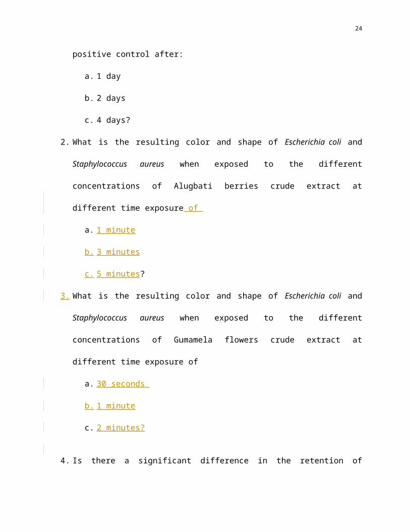
1. What is the retention capacity of Alugbati berries crude extract and Gumamela flower
crude extract as compared to the positive control after:
a. 1 day
b. 2 days
c. 4 days?
2. What is the resulting color and shape of Escherichia coli and Staphylococcus aureus
when exposed to the different concentrations of Alugbati berries crude extract at different
time exposure of
a. 1 minute
b. 3 minutes
c. 5 minutes?
3. What is the resulting color and shape of Escherichia coli and Staphylococcus aureus
when exposed to the different concentrations of Gumamela flowers crude extract at
different time exposure of
18
a. 30 seconds
b. 1 minute
c.[b.] 2 minutes?
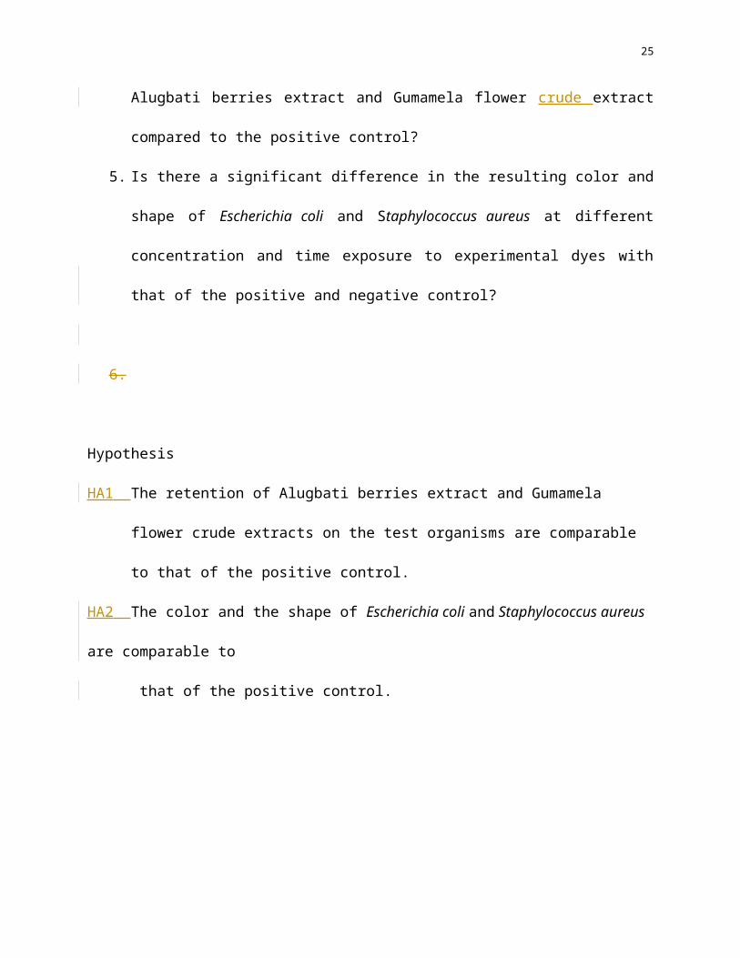
4.[3.] Is there a significant difference in the retention of Alugbati berries extract and
Gumamela flower crude extract compared to the positive control?
5.[4.] Is there a significant difference in the resulting color and shape of Escherichia coli and
Staphylococcus aureus at different concentration and time exposure to experimental dyes
with that of the positive and negative control?
[5.]
Hypothesis
HA1 The retention of Alugbati berries extract and Gumamela flower crude extracts on the test
organisms are comparable to that of the positive control.
HA2 The color and the shape of Escherichia coli and Staphylococcus aureus are comparable to
that of the positive control.
19
CHAPTER II
METHODS
Presented in this chapter are the research design, locale, subjects, measures and
procedures that will be used in the study.
Research Design
This study utilizes an experimental design to determine the efficiency of Alugbati
(Basella alba) berries and Gumamela (Hibiscus rosa sinensis) flower crude extracts dyes as
alternative primary and secondary stain, respectively, for Gram staining in the context of its
characteristic morphology in terms of color and shape. A positive and negative control will be
used and outside variables like temperature will be controlled as not to affect the result of this
study. Making sure that exposure of the experimental treatments, positive and negative control
will be the same throughout the duration of the experiment.
20
Setting
The preparation and extraction of plant will be executed in a Dominican Learning
Community founded by Dominican Sisters at Clovis Thibault building, room 306 of San Pedro
College located at C. Guzman Street, Davao City.
Test Organisms
The test organisms that will be used in the study are Staphylococcus aureus as the gram
positive bacteria and Escherichia coli as the gram negative bacteria. The organisms will be
purchased from the Department of Science and Technology, Davao City.
Plant Subjects
Alugbati berries should be ripe and purple. Gumamela flowers should be in bloom and
petals are red. Plants will be purchased in an Alugbati garden in Barangay Mua-an, Kidapawan
City and a Gumamela Farm in Tugbok, Davao City. The requirements needed for Alugbati
cultivation are the following: adequate water supply; soil rich in organic matter; and hot, humid
climate (Holmer, et al., 2008). Barangay Mua-an, Kidapawan City is a tropical environment with
mild climate change and the garden soil is filled with organic matter from decaying sawdust and
is near from a water source which is suitable for the growth and development of the Alugbati
plant (Patricio Jr., 2013). Meanwhile, the requirements needed for cultivation of Gumamela
flowers are the following: full sun, loamy soil, and adequate water supply and water should not
be in excess to prevent rotting of roots (Forsling, 2016). Tugbok, Davao City has a humid
atmosphere and provides an adequate water supply. The area has an open field that ensures the
plant’s sustenance of light which is highly recommended for the plant growth and development
(Diansay, 2003).

21
Measures
The test would be measured through grading the degree of retention of the stain and
similarity on the cell morphology, in terms of shape and cell color which is microscopically
determined with the positive control. Tool or criteria in grading seen in Appendix D.
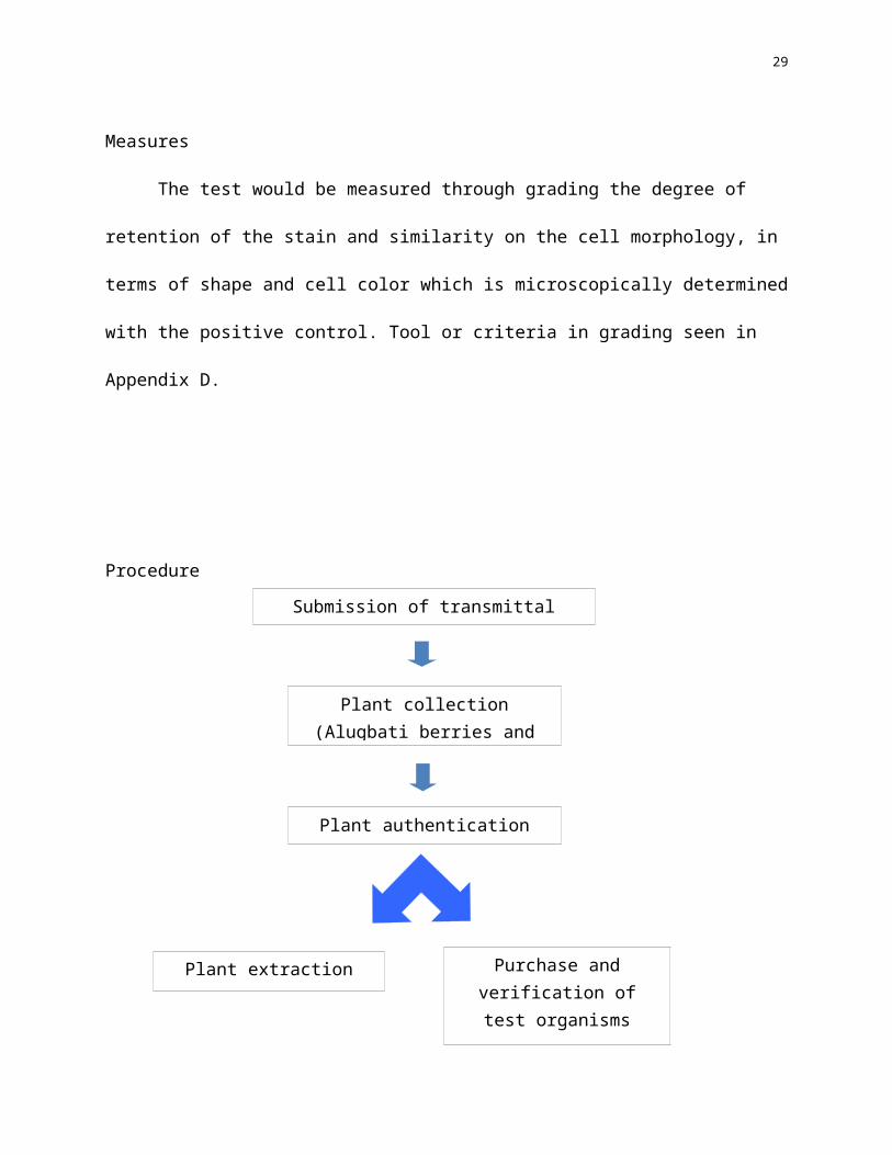
Procedure
Plant collection (Alugbati berries and Gumamela flowers)
Purchase and verification of test organisms
(Staphylococcus aureus and Escherichia coli)
Plant extraction
Plant authentication
Submission of transmittal letters
Experimentation Proper
SmeSmearingSubculture Staining
22
Figure 6. Schematic Diagram showing the protocol of the study
Pertinent letters will be submitted to the laboratory stockroom head Helen J. Ancla, and
to the BMLS Department Dean, Dr. Josephine Bandalan, by the researches to ask permission for
the conduct of the study in this institution. A letter of request will also be given to the
Department of Science and Technology for the purchase of the test organisms and to the farms
where the plants will be purchased (See Appendix A).
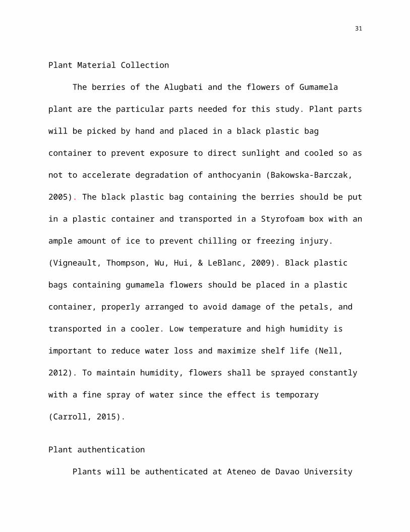
Plant Material Collection
The berries of the Alugbati and the flowers of Gumamela plant are the particular parts
needed for this study. Plant parts will be picked by hand and placed in a black plastic bag
container to prevent exposure to direct sunlight and cooled so as not to accelerate degradation of
anthocyanin (Bakowska-Barczak, 2005). The black plastic bag containing the berries should be
put in a plastic container and transported in a Styrofoam box with an ample amount of ice to
prevent chilling or freezing injury. (Vigneault, Thompson, Wu, Hui, & LeBlanc, 2009). Black
plastic bags containing gumamela flowers should be placed in a plastic container, properly
arranged to avoid damage of the petals, and transported in a cooler. Low temperature and high
humidity is important to reduce water loss and maximize shelf life (Nell, 2012). To maintain
Analysis of Data Gathered
Gathering and Evaluation of Data
Waste Disposal and Management
23
humidity, flowers shall be sprayed constantly with a fine spray of water since the effect is
temporary (Carroll, 2015).
Plant authentication
Plants will be authenticated at Ateneo de Davao University Biology Department, Davao
City. (See Appendix B)
Plant extraction
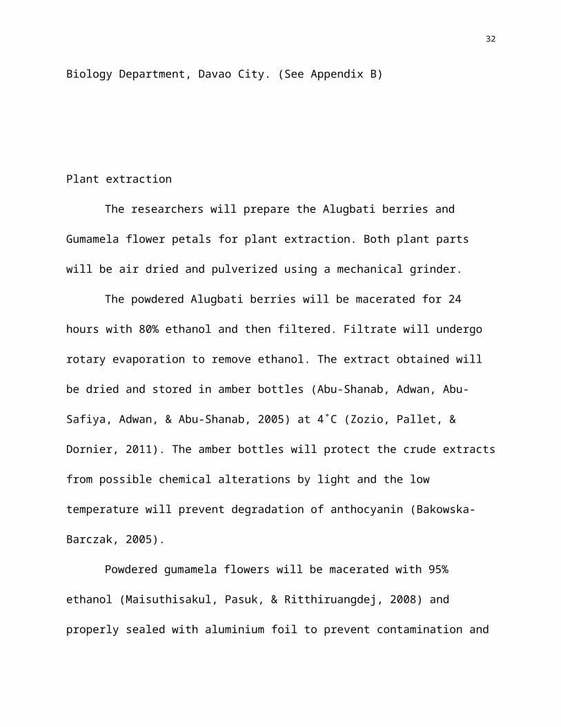
The researchers will prepare the Alugbati berries and Gumamela flower petals for plant
extraction. Both plant parts will be air dried and pulverized using a mechanical grinder.
The powdered Alugbati berries will be macerated for 24 hours with 80% ethanol and
then filtered. Filtrate will undergo rotary evaporation to remove ethanol. The extract obtained
will be dried and stored in amber bottles (Abu-Shanab, Adwan, Abu-Safiya, Adwan, & Abu-
Shanab, 2005) at 4˚C (Zozio, Pallet, & Dornier, 2011). The amber bottles will protect the crude
extracts from possible chemical alterations by light and the low temperature will prevent
degradation of anthocyanin (Bakowska-Barczak, 2005).
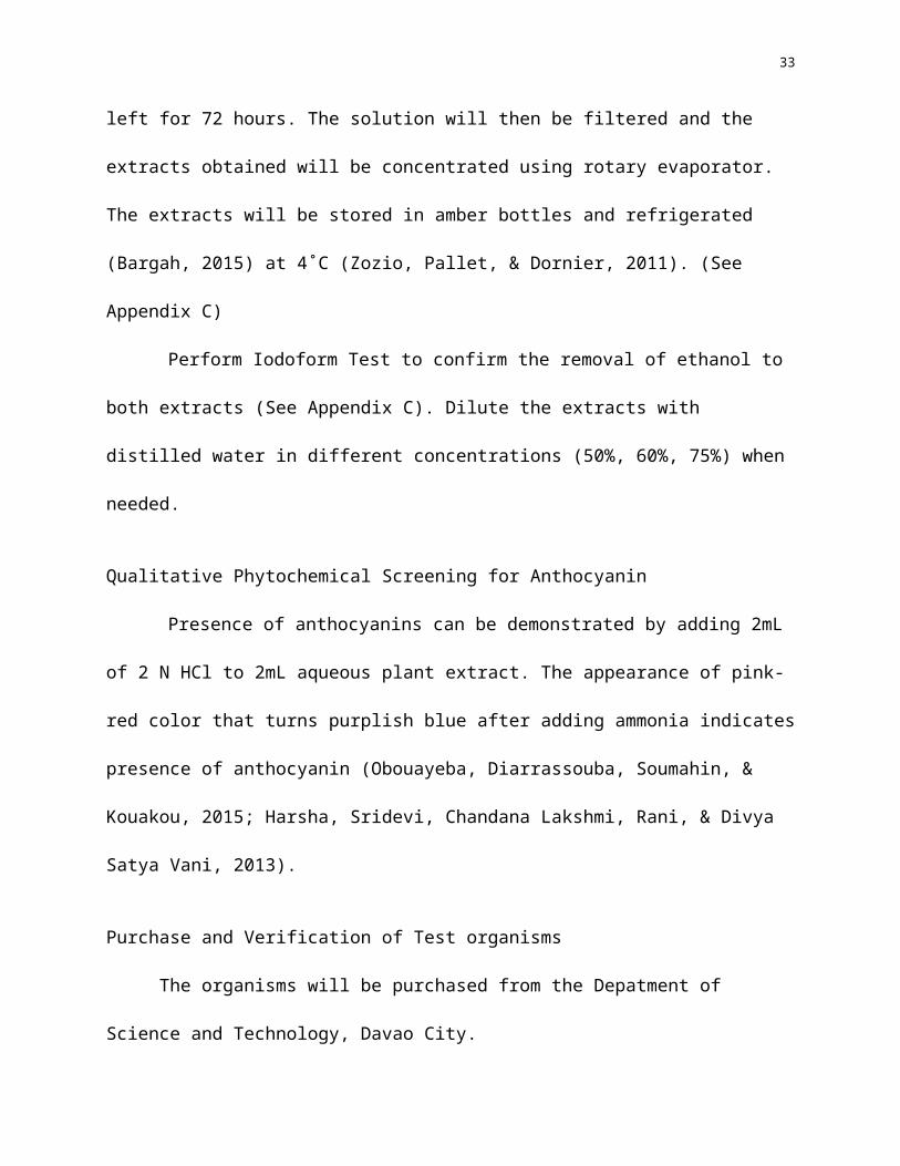
Powdered gumamela flowers will be macerated with 95% ethanol (Maisuthisakul,
Pasuk, & Ritthiruangdej, 2008) and properly sealed with aluminium foil to prevent
contamination and left for 72 hours. The solution will then be filtered and the extracts obtained
will be concentrated using rotary evaporator. The extracts will be stored in amber bottles and
refrigerated (Bargah, 2015) at 4˚C (Zozio, Pallet, & Dornier, 2011). (See Appendix C)
Perform Iodoform Test to confirm the removal of ethanol to both extracts (See Appendix
24
C). Dilute the extracts with distilled water in different concentrations (50%, 60%, 75%) when
needed.
Qualitative Phytochemical Screening for Anthocyanin
Presence of anthocyanins can be demonstrated by adding 2mL of 2 N HCl to 2mL
aqueous plant extract. The appearance of pink-red color that turns purplish blue after adding
ammonia indicates presence of anthocyanin (Obouayeba, Diarrassouba, Soumahin, & Kouakou,
2015; Harsha, Sridevi, Chandana Lakshmi, Rani, & Divya Satya Vani, 2013).
Purchase and Verification of Test organisms
The organisms will be purchased from the Depatment of Science and Technology, Davao
City.
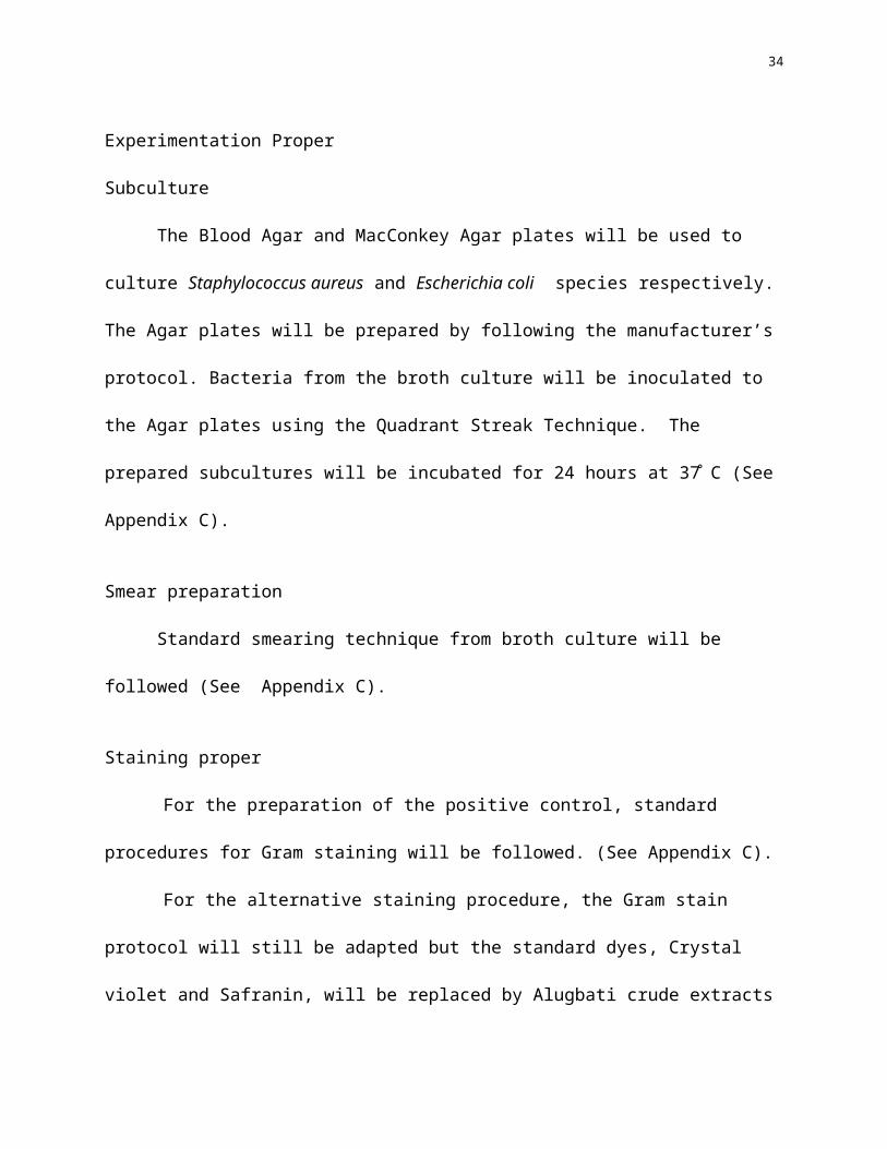
Experimentation Proper
Subculture
The Blood Agar and MacConkey Agar plates will be used to culture Staphylococcus
aureus and Escherichia coli species respectively. The Agar plates will be prepared by following
the manufacturer’s protocol. Bacteria from the broth culture will be inoculated to the Agar plates
using the Quadrant Streak Technique. The prepared subcultures will be incubated for 24 hours at
37̊ C (See Appendix C).
Smear preparation
Standard smearing technique from broth culture will be followed (See Appendix C).
Staining proper
For the preparation of the positive control, standard procedures for Gram staining will be
25
followed. (See Appendix C).
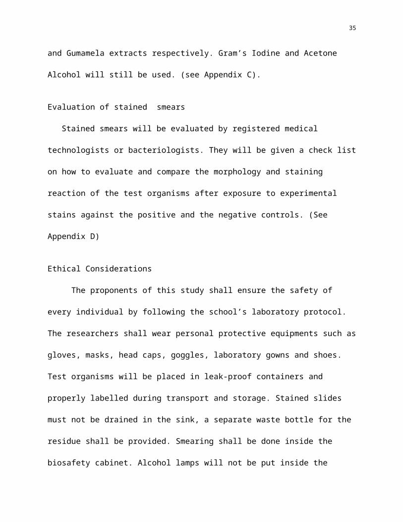
For the alternative staining procedure, the Gram stain protocol will still be adapted but
the standard dyes, Crystal violet and Safranin, will be replaced by Alugbati crude extracts and
Gumamela extracts respectively. Gram’s Iodine and Acetone Alcohol will still be used. (see
Appendix C).
Evaluation of stained smears
Stained smears will be evaluated by registered medical technologists or bacteriologists. They
will be given a check list on how to evaluate and compare the morphology and staining reaction
of the test organisms after exposure to experimental stains against the positive and the negative
controls. (See Appendix D)
Ethical Considerations
The proponents of this study shall ensure the safety of every individual by following the
school’s laboratory protocol. The researchers shall wear personal protective equipments such as
gloves, masks, head caps, goggles, laboratory gowns and shoes. Test organisms will be placed in
leak-proof containers and properly labelled during transport and storage. Stained slides must not
be drained in the sink, a separate waste bottle for the residue shall be provided. Smearing shall be
done inside the biosafety cabinet. Alcohol lamps will not be put inside the biosafety cabinet.
Heat fixing of the smear will be done outside of the biosafety cabinet. In case of accidents and
other unexpected outcomes such as catching fire of laboratory gowns, spilling of hazardous
reagents to skin or other body parts, the researchers shall immediately notify the mentor or any
laboratory stockroom personnel present to address the problem. Working tables and equipments
should be disinfected using Lysol or bleach and then alcohol. Inoculating loops should be heated
26
before and after using as a form of sterilization process. Agar plates and test tubes must be sealed
and will be given to the laboratory stockroom for autoclaving and disposal as stated in the San
Pedro College Laboratory Stockroom Protocol (See Appendix E).
References
International Agency for Research on Cancer of the World Health Organization. (2016). IARC Monographs on the Evaluation of Carcinogenic Risks to Humans. Retrieved February 2, 2016, from http://monographs.iarc.fr/ENG/Classification/latest_classif.php
Abu-Shanab, B., Adwan, G., Abu-Safiya, D., Adwan, K., & Abu-Shanab, M. (2005). Antibacterial Activity of Rhus coriaria. L Extracts growing in Palestine. Journal of the Islamic University of Gaza, (Natural Sciences Series), 13(2), 147-153. Retrieved July 8, 2016, from http://www.iugaza.edu/ara/research/
Adhikari, R., Kumar, H. N., & Shruthi, S. (2012). A Review on Medicinal Importance of Basella alba L. International Journal of Pharmaceutical Sciences and Drug Research, 4(2), 110-114.
Amon, M. F., & Pladio, L. P. (2012). Potential Food Colorant from the Extracts of Alugbati (Basella rubra L.). Retrieved February 4, 2016, from http://goo.gl/KjsjD2
Ampana, R. G., Atienza, M. K., Gote, N., Salamat, R. A., Ubana, J. J., & Villareal, C. G. (2013, September 30). The Efficacy of Ipomoea tricolor (Morning Glory) and Hibiscus rosa sinensis (Gumamela) Dye as an Alternative to Hematoxylin and Eosin in staining Epithelial Tissues.
Bakowska-Barczak, A. (2005). Acylated Anthocyanins as Stable, Natural Food Colorants-Review. Polish Journal of Food and Nutrition Sciences, 14/55, 107-116. Retrieved July 8, 2016
27
Bargah, R. K. (2015). Preliminary test of phytochemical screening of crude ethanolic. Retrieved July 8, 2016, from Journal of Pahrmacognosy and Phytochemistry: http://www.phytojournal.com/vol4Issue1/Issue_may_2015/6.1.pdf
Brit. (2008, June 18). Synthetic Dyes: A look at Environmental & Human Risks. Retrieved February 1, 2016, from Green Cotton: https://greencotton.wordpress.com/2008/06/18/synthetic-dyes-a-look-at-the-good-the-bad-and-the-ugly/
Brooks, G. F. (2013). In Jawetz, Melnick & Adelberg's Medical Microbiology 25th Edition (pp. 185-187, 189, 190-191, 213-214, 217-220).
Carroll, J. (2015, February 17). Raising Humidity: How to Increase Humidity for House Plants. Retrieved July 8, 2016, from Gardening Know How: gardeningknowhow.com
Castor, J. K., Fabile, S. F., Jimenez, V. A., San Jose, M. U., Sumarago, N. G., & Tapel, K. J. (2014). Synergistic Bacteriological Staining Effect of Alugbati (Basella rubra Linn), Kalamansi (Citrus microcarpa Bunge) and Gumamela (Hibiscus rosasinensis Linn) Extracts.
Chemwatch. (2010, October 8). Crystal Violet Material Safety Data Sheet (MSDS No. sc-207460). Retrieved January 24, 2016, from http://datasheets.scbt.com/sc-207460.pdf
Chua, R. T. (2010). Taxonomy of Vascular Plants in Botanical Garden, Baguio City. Retrieved March 23, 2016, from Academia: https://www.academia.edu/3876692/Taxonomy_of_Vascular_Plants_in_Botanical_Garden_Baguio_City
Crystal Violet. (2013). Retrieved January 24, 2016, from National Biochemicals, LLC. USA: www.nationalbiochem.com/pdf/pis/MC3886%20PS.pdf
Dai, J., & Mumper, R. J. (2010 йил 21-October). Plant Phenolics: Exraction, Analysis and Their Antioxidant and Antcaner Properties. Molecules, 15, 7313-7352. Retrieved 2016 йил 5-July from www.mdpi.com/journal/molecules
Davidson, M. W. (2015). Molecular Expression Optical Microscopy Primer. Retrieved March 15, 2016, from Molecular Expression Optical Microscopy Primer: http://micro.magnet.fsu.edu/primer/techniques/polarized/gallery/pages/safraninosmall.html
De Leon, M., Latoza, A. G., Nues, A., Pilac, M. R.-G., Sistoza, C. J., Soliman, D. G., . . . Mortel, F. (2016). Methanolic Fruit Extract of Basella rubra: Organic Stain for Hematologic Blood smear. Retrieved March 24, 2016, from Academia:

28
https://www.academia.edu/4323993/Methanolic_Fruit_Extract_of_Basella_rubra_Organic_Stain_for_Hematologic_Blood_Smear
Deshmukh, S. A., & Gaikwad, D. K. (2013, January). A review of the Taxonomy, Ethnobotany, Phytochemistry and Pharmacology of Basella alba (Basellaceae). Retrieved January 26, 2016, from Journal of Applied Pharmaceutical Science: http://imsear.li.mahidol.ac.th/handle/123456789/151999
Diansay, D. P. (2003). Cateel Centennial Book. Davao City.
Enerva, L. T. (2006). Utilization of an indigenous dyestuff from Basella rubra (alugbati) as microbiologial stain. Transactions of the National Academy of Science and Technology, 28(1). Retrieved May 8, 2016, from http://scinet.dost.gov.ph
Enerva, L. T. (2006). Utilization of an Indigenous dyestuff from Basella rubra (alugbati) as microbiological stain. Retrieved February 2, 2016, from The Science and Technology Information Network of the Philippines: http://scinet.dost.gov.ph
Forbes, P. D., Sahm, P. D., & Weissfeld, P. D. (2007). Bailey & Scott's Diagnostic Microbiology (International Edition, 12th Edition ed.). St. Louis, Missouri: Mosby, Inc., an affiliate of Elsevier Inc.
Forsling, Y. (2016). Hibiscus Care. Retrieved July 07, 2016, from hibiscus-sinensis: hibiscus-sinensis.com
Giusti, M. M. (2008, July 31). Expanding the Market of Anthocyanin-Rich Commodities through the characterization of components with Biological Activity. Retrieved February 1, 2016, from http://reeis.usda.gov
Hale, M. C. (2013, September 22). Microbiology Gram negative Escherichia coli. Retrieved March 24, 2016, from Pathology Outlines.com: http://www.pathologyoutlines.com/topic/microbiologyescherichiacoli.html
Harsha, N., Sridevi, V., Chandana Lakshmi, M. V., Rani, K., & Divya Satya Vani, N. (2013, November). Phytochemical Analysis of Some Selected Spices. International Journal of Innovative Research in Science, Engineering and Technology, 2(11), 6618-6621.
HiMedia Laboratories. (2012). Blood Agar Base (Infusion Agar). Retrieved July 8, 2016, from HiMedia: http://www.himedialabs.com/intl/en/products/Microbiology/Dehydrated-Culture-Media-Diagnostic-Animal-based-Media-Bacterial/Blood-Agar-Base-Infusion-Agar-M073
Holmer, R. J., Santos Jr., C. A., Sol, G. Y., Lee, S. O., Elorde Jr., E., Aquino, A. A., . . . Montes, A. A. (2008). Philippine Allotment Garden Manual with an Introduction to Ecological Sanitation. Cagayan de Oro City.
29
James, D. E. (2008). Microorganisms Safety Guide, Nine Safe Practices for the Microbiology Laboratory. Retrieved April 24, 2016, from Science Buddies: http://www.sciencebuddies.org/science-fair-projects/project_ideas/Micro_Safety.shtml
Ladziata, V. (2007, June 19). Iodoform Test Procedure. Retrieved July 8, 2016, from University of Minnesota Duluth: http://www.d.umn.edu/chemistry/faculty/carlson/downloads/Handouts/Chem2544_W3a_Ald&ket.doc.
Lim, T. K. (2014). Hibiscus rosa-sinensis. In T. K. Lim, Edible Medicinal and Non Medicinal Plants (Vol. 8, pp. 306-323).
Lin, S., Lin, B., Hsieh, W., Ko, H., Liu, C., Chen, L., & Chiou, R. (2010, October 13). Structural identification and bioactivities of Red-Violet Pigments present in Basella alba fruits. J. Agric. Food Chem, 58(19), 10364-103472.
Littlefield, N. A., Blackwell, B. N., Hewitt, C., & Gaylor, D. W. (1985). Chronic toxicity and carcinogenicity studies of Gentian violet in mice. Fundamental Appl. Toxicology, 5(5), 902-912.
Maisuthisakul, P., Pasuk, S., & Ritthiruangdej, P. (2008, May). Relationship between antioxidant properties and chemical composition of some Thai plants. Journal of Food Composition and Analysis, 21(3), 229-240. doi:10.1016/j.jfca.2007.11.005
Mandal, M. A. (2012). What is Staphylococcus aureus? Retrieved March 13, 2016, from News Medical: http://www.news-medical.net/health/What-is-Staphylococcus-Aureus.aspx
Mani, S., & Bharagava, R. (2016). Exposure to Crystal Violet, Its Toxic, Genotoxic and Carcinogenic Effects on Environment and Its Degradation and Detoxification for Environmental Safety. Rev. Environ. Contam. Toxicol., 237, 71-104.
Marler, B., & Clark, M. (2016). E. coli Food Poisoning. Retrieved March 24, 2016, from About E. coli: www.about-ecoli.com
Merck. (2016). Culture Media. Retrieved July 8, 2016, from Merck Millipore: http://www.merckmillipore.com/INTL/en/products/industrial-microbiology/culture-media/dLWb.qB.5kgAAAFAX8JkiQpx,nav
Ministry of Human Resource Department. (2011). Streak Plate Method. Retrieved July 8, 2016, from Value @ Amrita: vlab.amrita.edu/?sub=3&brch=73&sim=213&cnt=2
Mohammed, M., Ibrahim, A., & Shitu, A. (2014, May 20). Batch Removal of Hazardous Safranin-O in wastewater using pineapple peels as an agricultural waste based adsorbent. International Journal of Environmental Monitoring and Analysis, 2(3), 128-133.

30
Nell, T. (2012, November). Best Practices for Shipping and Storage of Fresh Cut Flowers. Retrieved July 8, 2016, from FloraLife: floralife.com
Nghia, N. T. (2014 йил December). Anthocyanin- Study of Extraction from Basella Rubra L. and Verifying Antioxidant Activity, Application In Identificating Disodium Tetraborate in Food. Retrieved 2016 йил 5-July from Tai Ban Dai Du: goo.gl/vz8A0c
Obi, F. O., & Uneh, E. (2003, January). pH Dependent Prevention of Carbon tetrachloride-induced Lipoperoxidation in Rats by Ethanolic Extract of Hibiscus rosasinensis Petal. BIOKEMISTRI, 13, 42-50. Retrieved July 7, 2016
Obi, F. O., Usenu, I. A., & Osayande, J. O. (1998, September). Prevention of Carbon Tetrachloride-induced Hepatotoxicity in the Rat by H. rosa sinensis Anthocyanin Extract Administered in Ethanol. Toxicology, 131, 93-98.
Obouayeba, A. P., Diarrassouba, M., Soumahin, E. F., & Kouakou, T. H. (2015, July 9). Phytochemical Analysis, Purification and Identification of hibiscus Anthocyanins. Journal of Pharmaceutical, Chemical and Biological Sciences, 3(2), 156-168.
Patricio Jr., A. B. (2013). ENR STATISTICAL PROFILE 2013. Cotabato City.
Roberts, M. (2015, December 28). Wild Colours Natural Dyes. Retrieved January 23, 2016, from Wild Colours Natural Dyes: http://www.wildcolours.co.uk/html/natural_dyes_comparison.html
Rosenberg, L. (1971). Chemical Basis for the Histological Use of Safranin O in the Study of Articular Cartilage. The Journal of Bone & Joint Surgery.
Sepos, E. (2012, April). Standard Operating Procedure: Rotary Evaporator in the P.O.W.E.R. Laboratory. Retrieved July 6, 2016, from Research Gate: https://www.researchgate.net/
Stewart, D. (2006). eHow. Retrieved March 15, 2016, from eHow: http://www.ehow.com/info_10040445_type-stain-safranin.html
Stuart, G. (2015). Alugbati. Retrieved January 24, 2016, from Philippine Medical Plant: http://www.stuartxchange.org/Alugbati
Stuart, G. (2015, October). Gumamela. Retrieved February 2, 2016, from Philippine Medicinal Plants: http://www.stuartxchange.com/Gumamela.html
Summers, J., Texley, J., & Kwan, T. (2006). Science Safety in the Community College (Illustrated ed.). NSTA Press.
T.A., A.-A., & Mabel, A. F. (2015). Some Anatomical Features of Basella Linn.: their adaptive significance to water stress. Research in Plant Biology, 5(3), 14-22.
31
Tazzini, N. (2014, February 22). Anthocyanins: Definition, Structure and pH. Retrieved March 13, 2016, from Tuscany Diet: http://www.tuscany-diet.net/2014/02/22/anthocyanins-definition-structure-ph/
The Carcinogenic Potency Project. (2007, October). Retrieved January 8, 2016, from http://toxnet.nlm.nih.gov/cpdb/chempages/GENTIAN%20VIOLET.html
Tille, P. (2014). Bailey & Scott's Diagnostic Microbiology 13th Edition. St. Louis, Missouri: Mosby Incorporated.
Tortora, G., Funke, B., & Case, C. (2002). Observing Microorganisms Through a Microscope. In G. Tortora, B. Funke, & C. Case, Microbiology: An Introduction (7th Edition ed., pp. 69-71). San Francisco, California: Pearson Education Incorporated.
Vigneault, C., Thompson, J., Wu, S., Hui, K. C., & LeBlanc, D. I. (2009). Transportation of fresh horticultural produce. (N. Benkeblia, Ed.) Postharvest Technologies for Horticultural Crops, 2, 1-24. Retrieved July 8, 2016
Webb, D. (2014, March). Anthocyanins. Retrieved March 13, 2016, from Today's Dietitian: http://www.todaysdietitian.com/newarchives/030314p20.shtml
Zozio, S., Pallet, D., & Dornier, M. (2011). Evaluation of anthocyanin stability during storage of a coloured drink made from extracts of the Andean blackberry (Rubus glaucus Benth.), acai (Euterpe oleracea Mart.) and black carrot (Daucus carota L.). Fruits, 66, 203-215. Retrieved July 8, 2016, from http://www.fruits-journal.org
32
APPENDIX ASan Pedro College
Davao City
July 11, 2016
DR. JOSEPHINE BANDALANDean of the Department of Medical Laboratory Science
Dear Dr. Bandalan:
The following undersigned are 4th year students of San Pedro College, Guzman St., Obrero, Davao City as course requirements for Bachelor in Medical Laboratory Science and presently conducting a research entitled: “Basella alba (Alugbati) Berries and Hibiscus rosa sinensis (Gumamela) Flower Crude Extracts as Alternative Dyes for Gram Staining”. The objectives of the study are:
a) To determine the efficiency of Basella alba (Alugbati) Berries and Hibiscus rosa sinensis (Gumamela) flower crude extracts as alternative primary and secondary stain, respectively, for Gram Staining in the context of its characteristic morphology in terms of color and shape.
b) To find locally-made dyes from natural and organic materials that is not carcinogenic.
The proponents of this research study are hereby seeking your consent to allow us to conduct our research in this institution. With your expertise, we are humbly asking your permission to validate the following attachments to this letter to assist you in reaching a decision.
(a) A copy of an ethical clearance certificate issued by the Institution(b) A copy of the research instruments which I intend using in my research
Should you require any further information, please do not hesitate to contact us. Our contact details are as follows:
E-mail: [email protected]
33
Mobile number: 09329740053
Upon completion of the study, the proponents of this research study shall undertake to provide you with a bound copy of the dissertation.
Your permission to conduct this study will be greatly appreciated. Thank you and God Bless!
Respectfully yours,
Aballe, Loreyleine M.Ara, Janessa M.Boctoto, Rejoice D.Dimacuta, Shaquillah S.Estrada, Ian Mark R.Galido, Marie Remcy Kyle B.Millan, Maria Monica C.Mokamad, Aira Ann A.Pacete, Shaira Leigh V.Questo, Irel Anne R.Villegas, Hazel Mae P.BMLS – 4D Research students
Conforme:
Aileen Grace L. Ang, RMT, MAST-Bio, MSMTResearch Adviser/Mentor
34
San Pedro CollegeDavao City
July 11, 2016
MS. HELEN J. ANCLALaboratory Stockroom Head
Dear Ma’am Gustillo:
The following undersigned are 4th year students of San Pedro College, Guzman St., Obrero, Davao City as course requirements for Bachelor in Medical Laboratory Science and presently conducting a research entitled: “Basella alba (Alugbati) Berries and Hibiscus rosa sinensis (Gumamela) Flower Crude Extracts as Alternative Dyes for Gram Staining”.
In connection with this, we would like to ask your help to provide the necessary list of things needed for conducting our experiment which is attached to this letter. We would like to appreciate your assistance and support in this particular research endeavor.
We are hoping for your positive response. Thank you and God bless!
Respectfully yours,
Aballe, Loreyleine M.Ara, Janessa M.Boctoto, Rejoice D.Dimacuta, Shaquillah S.Estrada, Ian Mark R.Galido, Marie Remcy Kyle B.Millan, Maria Monica C.Mokamad, Aira Ann A.Pacete, Shaira Leigh V.Questo, Irel Anne R.
35
Villegas, Hazel Mae P.BMLS – 4D Research students
Conforme:
Aileen Grace L. Ang, RMT, MAST-Bio, MSMTResearch Adviser/Mentor
San Pedro CollegeDavao City
July 11, 2016
DR. ANTHONY C. SALES, CESO IIIRegional DirectorDepartment of Science and Technology
Dr. Sales:
Greetings!
We, the 4th year Medical Laboratory Science students of San Pedro College are currently embarking a study on the entitled “Basella alba (Alugbati) Berries and Hibiscus rosa sinensis (Gumamela) Flower Crude Extracts as Alternative Dyes for Gram Staining”. This is in partial fulfilment of the requirement of the abovementioned course
In this regard, the group would like to seek your permission to inquire and purchase for the availability of the following microorganisms to be used in the study.
Pure culture of Staphylococcus aureus Pure culture of Escherichia coli
We assure you to perform the proper protocol in handling, processing and disposal of such infectious organisms.
We are hoping for your positive response. Thank you and God Bless!
Respectfully yours,
Loreyleine AballeBMLS 4D Group Representative
Noted by:
36
Aileen Grace L. Ang, RMT, MAST-Bio, MSMTResearch Adviser/Mentor
Approved by:
Dr. Anthony C. Sales, CESO IIIRegional Director-Department of Science and Technology
San Pedro CollegeDavao City
July 12, 2016
DR. OSCAR P. GRAGEDA, MD, PSP, PSMIDChief Pathologist, San Pedro Hospital
Dr. Grageda:
Greetings!
We, the 4th year Medical Laboratory Science students of San Pedro College are currently embarking a study on the entitled “Basella alba (Alugbati) Berries and Hibiscus rosa sinensis (Gumamela) Flower Crude Extracts as Alternative Dyes for Gram Staining”. This is in partial fulfilment of the requirement of the abovementioned course
In this regard, the group would like to seek your permission to inquire and purchase for the availability of the following microorganisms to be used in the study.
Culture of Staphylococcus aureus Culture of Escherichia coli
We assure you to perform the proper protocol in handling, processing and disposal of such infectious organisms.
We are hoping for your positive response. Thank you and God Bless!
Respectfully yours,
Loreyleine AballeBMLS 4D Group Representative
Noted by:
Aileen Grace L. Ang, RMT, MAST-Bio, MSMT
37
Research Adviser/Mentor
Approved by:
Dr. Anthony C. Sales, CESO IIIRegional Director-Department of Science and Technology
San Pedro CollegeDavao City
July 11, 2016
To whom it may concern:
Greetings!
We, the 4th year Medical Laboratory Science students of San Pedro College are currently embarking a study on the entitled “Basella alba (Alugbati) Berries and Hibiscus rosa sinensis (Gumamela) Flower Crude Extracts as Alternative Dyes for Gram Staining”. This is in partial fulfilment of the requirement of the above mentioned course.
In this regard, the group would like to seek your permission to inquire and purchase for the availability of the following microorganisms to be used in the study.
Alugbati Berries Gumamela flowers
We assure you to perform the proper protocol in handling, transport, processing and disposal of such plants.
We are hoping for your positive response. Thank you and God Bless!
Respectfully yours,
Loreyleine AballeGroup Representative
Noted by:
Aileen Grace L. Ang, RMT, MAST-Bio, MSMTResearch Adviser/Mentor

38
APPENDIX B
39
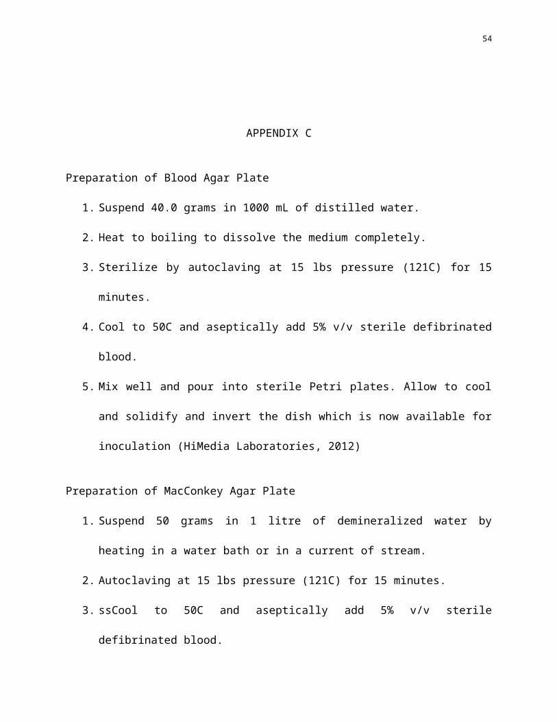
APPENDIX C
Preparation of Blood Agar Plate
1. Suspend 40.0 grams in 1000 mL of distilled water.
2. Heat to boiling to dissolve the medium completely.
3. Sterilize by autoclaving at 15 lbs pressure (121C) for 15 minutes.
4. Cool to 50C and aseptically add 5% v/v sterile defibrinated blood.
5. Mix well and pour into sterile Petri plates. Allow to cool and solidify and invert the dish
which is now available for inoculation (HiMedia Laboratories, 2012)
Preparation of MacConkey Agar Plate
1. Suspend 50 grams in 1 litre of demineralized water by heating in a water bath or in a
current of stream.
2. Autoclaving at 15 lbs pressure (121C) for 15 minutes.
3. ssCool to 50C and aseptically add 5% v/v sterile defibrinated blood.
4. Mix well and pour into sterile Petri plates. Allow to cool and solididy and invert the dish
which is now available for inoculation (Merck, 2016)
40
Subculture
1. Select an agar (Blood Agar Plate for Staphylococcus aureus and MacConkey Agar Plate
for Escherichia coli). Label the plate with the group number and section, test organism
and date.
2. Sterilize the loop by heating it to redness.
3. Obtain a loopful of Staphylococcus aureus and Escherichia coli colony from their
respective broth cultures and perform Quadrant Streaking. Avoid air contamination by
lifting the lid just enough to permit entry of the loop.
4. Replace the lid then sterilize the loop to destroy remaining bacteria.
5. Pick another sterilized loop. Pass the loop one time across the previous streak to pick up
some bacteria and continue streaking into the second quadrant. Distance of the streak
from the previous should be a bit farther. Replace the lid and sterilize the loop.
6. Pick another sterilized loop. Pass the loop one time across the previous streak and streak
to the next area. Streaks should be far now to obtain isolated and separated colonies.
Replace the lid and sterilize the loop.
7. Invert the plate and incubate for 24 hours at 37°C.
41
Quadrant streak
1. Loosen the cap of the bottle containing inoculums.
2. Hold an inoculation loop in your right hand.
3. Flame the loop and allow it to cool.
4. Lift the test tube containing inoculums with your left hand.
5. Remove the cap/ cotton wool plug of the test tube with the little finger of your right
hand.
6. Flame the neck of the test tube.
7. Insert the loop into the culture broth and withdraw. At all times hold the loop as still
as possible.
8. Flame the neck of the test tube again.
9. Replace the cap/cotton wool plug of the test tube using little finger of your right
hand. Place the test tube in a rack.
10. Partially lift the lid of the perti dish containing solid medium. Touch an unused part
of the agar surface close to the periphery of the plate and then drag it several times
across the first quadrant.
11. Close the petridish and reflame and cool the loop.
12. Turn the dish 90 degrees clockwise then the touch the loop on a corner of the culture
in quadrant 1 and drag it several times across the agar in quadrant 2, hitting the
42
original streak a few times.
13. Close the petridish and reflame and cool the loop.
14. Turn the dish 90 degrees clockwise then the touch the loop on a corner of the culture
in quadrant 2 and drag it several times across the agar in quadrant 3, hitting the
original streak a few times.
15. Close the petridish and reflame and cool the loop.
16. Turn the dish 90 degrees clockwise then the touch the loop on a corner of the culture
in quadrant 3 and drag it several times across the agar in quadrant 4, hitting the
original streak three times.
17. Close the petridish and reflame and cool the loop.
18. Incubate the plate in inverted position in an incubator for 24-48 hours (Ministry of
Human Resource Department, 2011).
43
Plant Extraction
Alugbati berries extraction
1. Prepare 1 kilogram of the Alugbati berries. Wash thoroughly to remove debris. Air dry the
berries under a shade.
2. Pulverize the dried berries using a mechanical grinder. Deposit the pulverized berries in a
large 4-liter capacity liquid container. Note: Transparent container should be wrapped in
black cellophane to prevent possible degradation of the extract by light.
3. Macerate powdered berries in 4 liters of Ethanol. This acts as the extracting agent. Store
mixture away from light at 4̊ C for 24 hours.
4. Carefully filter the contents through a Whatman No. 2 filter paper under suction using a
Buchner funnel.
5. Perform Rotary Evaporation to the filtrate.
6. Measure the resulting extract t in a weighing scale.
7. Store in Amber-colored bottles at 4̊ C away from light. (Abu-Shanab, Adwan, Abu-Safiya,
Adwan, & Abu-Shanab, 2005)
Gumamela flowers extraction
1. Gumamela flowers will be air dried at room temperature followed by pulverization to
powder using a mechanical grinder.
44
2. Weigh 500 grams of the powdered gumamela flowers and mix with 1500 mL of ethanol
then filter.
3. Cover the container with aluminium foil to prevent contamination and macerate for 72
hours at 4˚C.
4. The solution was then filtered using funnel and filter paper.
5. The extracts obtained will be concentrated using rotary evaporator.
6. Extracts will be stored in amber bottles and stored at 4˚C (Bargah, 2015).
45
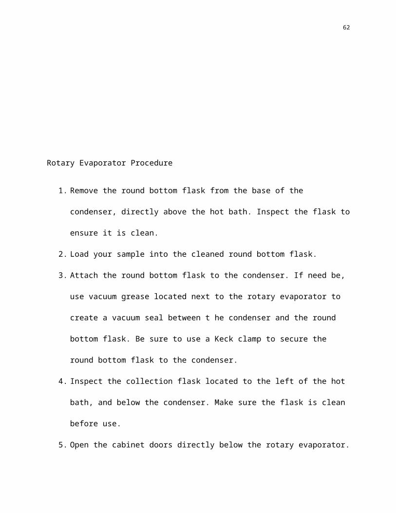
Rotary Evaporator Procedure
1. Remove the round bottom flask from the base of the condenser, directly above the hot
bath. Inspect the flask to ensure it is clean.
2. Load your sample into the cleaned round bottom flask.
3. Attach the round bottom flask to the condenser. If need be, use vacuum grease located
next to the rotary evaporator to create a vacuum seal between t he condenser and the
round bottom flask. Be sure to use a Keck clamp to secure the round bottom flask to the
condenser.
4. Inspect the collection flask located to the left of the hot bath, and below the condenser.
Make sure the flask is clean before use.
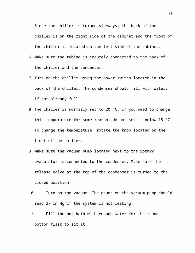
5. Open the cabinet doors directly below the rotary evaporator. Since the chiller is turned
sideways, the back of the chiller is on the right side of the cabinet and the front of the
chiller is located on the left side of the cabinet.
6. Make sure the tubing is securely connected to the back of the chiller and the condenser.
7. Turn on the chiller using the power switch located in the back of the chiller. The
condenser should fill with water, if not already full.
8. The chiller is normally set to 20 °C. If you need to change this temperature for some
reason, do not set it below 15 °C. To change the temperature, rotate the knob located on
the front of the chiller.
46
9. Make sure the vacuum pump located next to the rotary evaporator is connected to the
condenser. Make sure the release valve at the top of the condenser is turned to the closed
position.
10. Turn on the vacuum. The gauge on the vacuum pump should read 27 in Hg if the system
is not leaking.
11. Fill the hot bath with enough water for the round bottom flask to sit it.
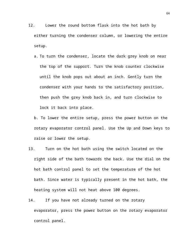
12. Lower the round bottom flask into the hot bath by either turning the condenser column, or
lowering the entire setup.
a. To turn the condenser, locate the dark grey knob on near the top of the support. Turn
the knob counter clockwise until the knob pops out about an inch. Gently turn the
condenser with your hands to the satisfactory position, then push the grey knob back
in, and turn clockwise to lock it back into place.
b. To lower the entire setup, press the power button on the rotary evaporator control
panel. Use the Up and Down keys to raise or lower the setup.
13. Turn on the hot bath using the switch located on the right side of the bath towards the
back. Use the dial on the hot bath control panel to set the temperature of the hot bath.
Since water is typically present in the hot bath, the heating system will not heat above
100 degrees.
14. If you have not already turned on the rotary evaporator, press the power button on the
rotary evaporator control panel.
15. Use the dial on the rotary evaporator control panel to set the desired spin speed of the
condenser.
16. If the desired evaporation should be timed, press the timer button on the rotary evaporator
47
control panel and use the dial to set a time in minutes, or press the into button and use the
dial to set a time in seconds.
17. When all the specifications have been made, press the dial on the rotary evaporator
control panel in. The rotary evaporator will begin to rotate.
18. To stop the rotary evaporator, push the dial on the rotary evaporator control panel in
again, or wait until the specified length of time passes. The rotary evaporator will stop
spinning then.
19. If the separation was successful, the higher boiling point substance will be left in the
original round bottom flask, and the lower boiling point substance will be collected in the
collection flask.
20. Press the power button to turn the rotary evaporator off. If the entire system was lowered
in step 12b, the system will raise to its original height.
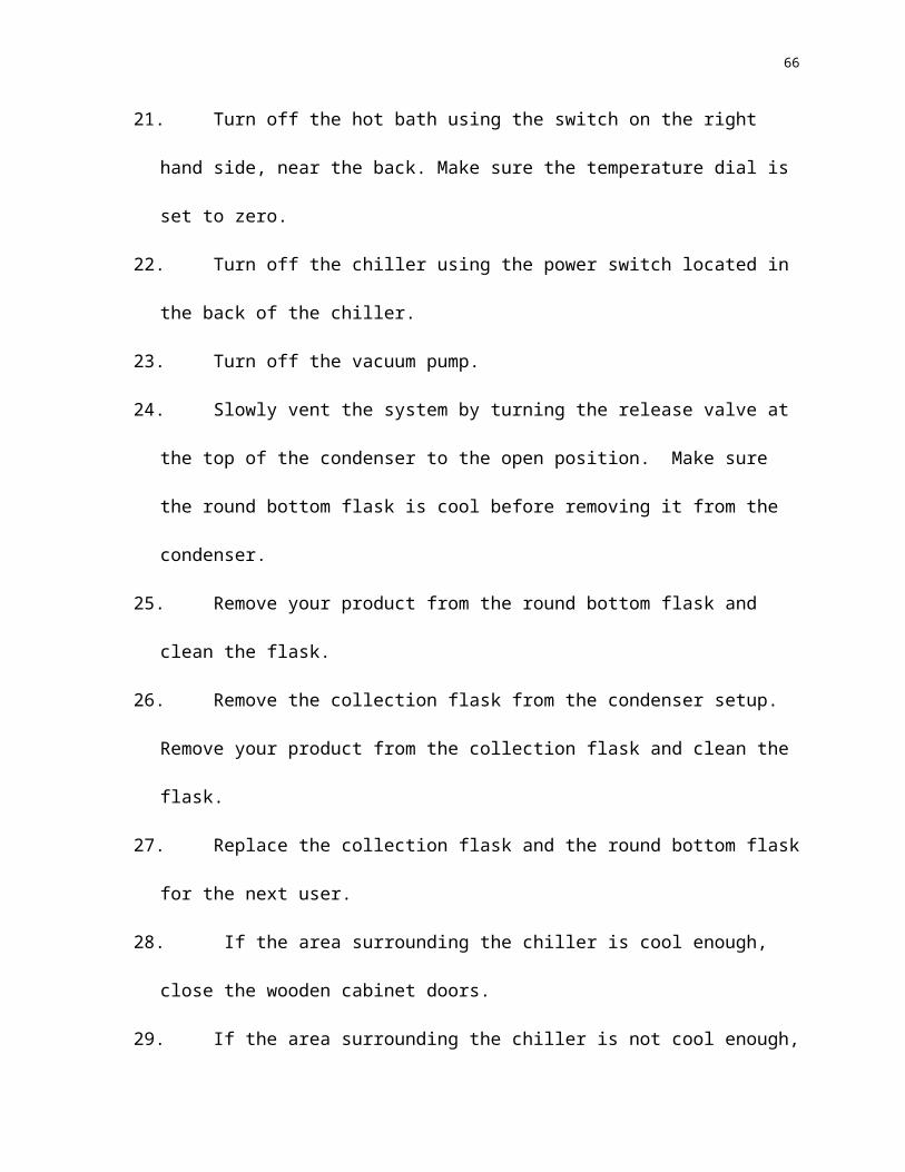
21. Turn off the hot bath using the switch on the right hand side, near the back. Make sure the
temperature dial is set to zero.
22. Turn off the chiller using the power switch located in the back of the chiller.
23. Turn off the vacuum pump.
24. Slowly vent the system by turning the release valve at the top of the condenser to the
open position. Make sure the round bottom flask is cool before removing it from the
condenser.
25. Remove your product from the round bottom flask and clean the flask.
26. Remove the collection flask from the condenser setup. Remove your product from the
collection flask and clean the flask.
27. Replace the collection flask and the round bottom flask for the next user.
48
28. If the area surrounding the chiller is cool enough, close the wooden cabinet doors.
29. If the area surrounding the chiller is not cool enough, leave the doors open, and someone
will close them (Sepos, 2012).
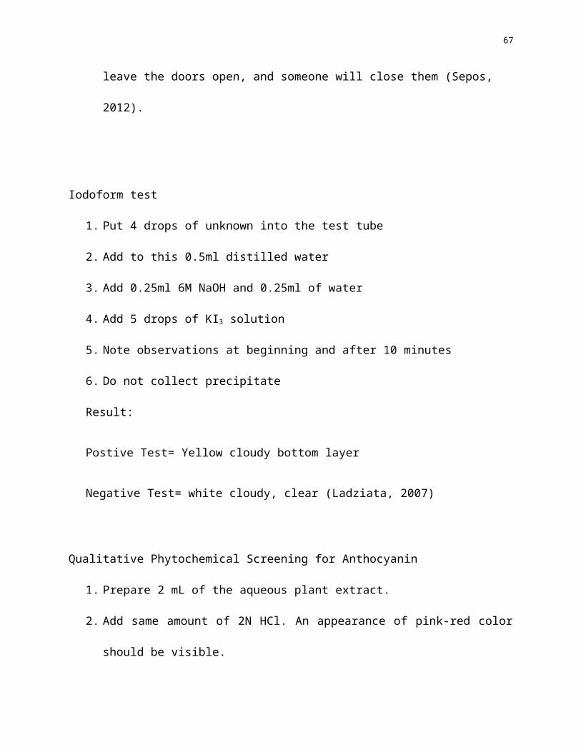
Iodoform test
1. Put 4 drops of unknown into the test tube
2. Add to this 0.5ml distilled water
3. Add 0.25ml 6M NaOH and 0.25ml of water
4. Add 5 drops of KI3 solution
5. Note observations at beginning and after 10 minutes
6. Do not collect precipitate
Result:
Postive Test= Yellow cloudy bottom layer
Negative Test= white cloudy, clear (Ladziata, 2007)
Qualitative Phytochemical Screening for Anthocyanin
1. Prepare 2 mL of the aqueous plant extract.
2. Add same amount of 2N HCl. An appearance of pink-red color should be visible.
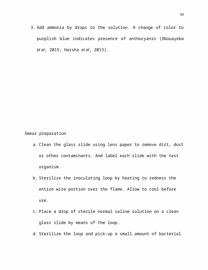
3. Add ammonia by drops to the solution. A change of color to purplish blue indicates
presence of anthocyanin (Obouayeba et al, 2015; Harsha et al, 2013).
49
Smear preparation
a. Clean the glass slide using lens paper to remove dirt, dust or other contaminants. And
label each slide with the test organism.
b. Sterilize the inoculating loop by heating to redness the entire wire portion over the
flame. Allow to cool before use.
c. Place a drop of sterile normal saline solution on a clean glass slide by means of the loop.
d. Sterilize the loop and pick-up a small amount of bacterial growth by touching lightly
upon the surface colony and transfer to the slide.
e. Emulsify and spread to about 1 square centimetre area. Re-sterilize the inoculating loop.
f. Allow the smear to dry.
g. Fix the dried smear by passing the slide rapidly just over the flame, film side up, 2 or 3
times. This prevents the film from being washed away and also kills the bacteria. Avoid
excessive heating since it will alter the composition of the organisms.

50
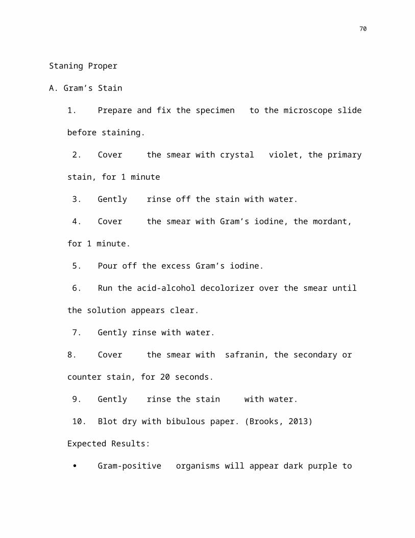
Staning Proper
A. Gram’s Stain
1. Prepare and fix the specimen to the microscope slide before staining.
2. Cover the smear with crystal violet, the primary stain, for 1 minute
3. Gently rinse off the stain with water.
4. Cover the smear with Gram’s iodine, the mordant, for 1 minute.
5. Pour off the excess Gram’s iodine.
6. Run the acid-alcohol decolorizer over the smear until the solution appears clear.
7. Gently rinse with water.
8. Cover the smear with safranin, the secondary or counter stain, for 20 seconds.
9. Gently rinse the stain with water.
10. Blot dry with bibulous paper. (Brooks, 2013)
Expected Results:
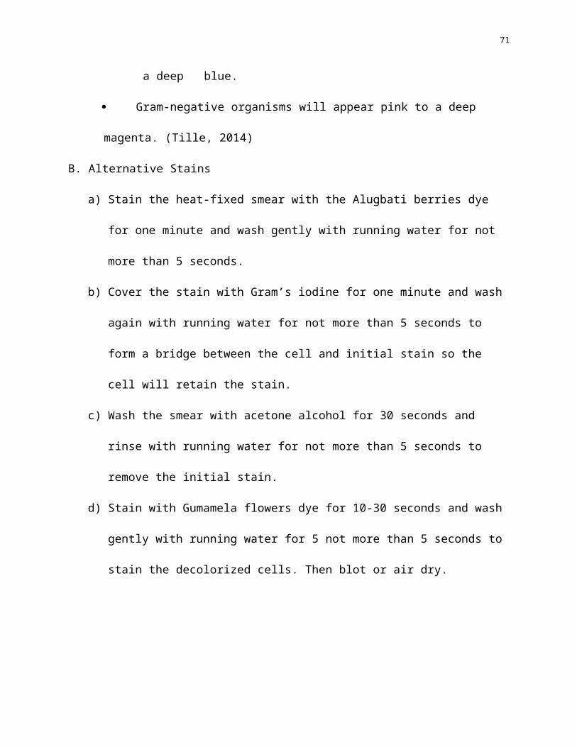
Gram-positive organisms will appear dark purple to a deep blue.
Gram-negative organisms will appear pink to a deep magenta. (Tille, 2014)
B. Alternative Stains
a) Stain the heat-fixed smear with the Alugbati berries dye for one minute and wash gently
with running water for not more than 5 seconds.
51
b) Cover the stain with Gram’s iodine for one minute and wash again with running water
for not more than 5 seconds to form a bridge between the cell and initial stain so the cell
will retain the stain.
c) Wash the smear with acetone alcohol for 30 seconds and rinse with running water for
not more than 5 seconds to remove the initial stain.
d) Stain with Gumamela flowers dye for 10-30 seconds and wash gently with running
water for 5 not more than 5 seconds to stain the decolorized cells. Then blot or air dry.
52
APPENDIX DMicroscopic Observations of Gram Stained Bacteria
Name of Evaluator: ________________________________Date of Evaluation: ________________________________CHECKLIST:
A. Organism: Staphylococcus aureus
Alugbati berries extract (primary dye)
andGumamela flower extract
(secondary dye)
Concentration Time exposure to experimental dye
50% 60% 75% Primary Dye Secondary Dye1 minute 3 minutes 5 minutes 30
seconds1 minute 2 minutes
Results: 5 3 0 5 3 0 5 3 0 5 3 0 5 3 0 5 3 0 5 3 0 5 3 0 5 3 0Cell morphology
ShapeCoccus:Bacillus:
Cell color:Red:Purple:

53
B. Organism: Escherichia coli
Alugbati berries extract (primary dye)
andGumamela flower extract
(secondary dye)
Concentration Time exposure to experimental dye
50% 60% 75% Primary Dye Secondary Dye1 minute 3 minutes 5 minutes 30
seconds1 minute 2 minutes
Results: 5 3 0 5 3 0 5 3 0 5 3 0 5 3 0 5 3 0 5 3 0 5 3 0 5 3 0Cell morphology
ShapeCoccus:Bacillus:
Cell color:Red:Purple:
Criteria:5 – Strongly comparable3 – Moderately comparable0 – Not comparable
Legend:
Shape E.coli S.aureus5 Rod shape Oval shape3 Irregular rod shape Irregular oval shape0 Unidentifiable Unidentifiable
Color E.coli S.aureus5 Red Purple3 Light pink Light blue0 No color No color
54
Retention of StainA. Organism: Staphylococcus aureus
1 day after staining 2 days after staining 4 days after stainingRetention of stain 5 3 0 5 3 0 5 3 0
a. Clarity/ visibility of stained organism
b. Intensity of color
c. Quality of shape
1 day after staining 2 days after staining 4 days after stainingRetention of stain 5 3 0 5 3 0 5 3 0
d. Clarity/ visibility of stained organism
e. Intensity of color
f. Quality of shape
B. Organism: Escherichia coli

55
Criteria:5 – Strongly comparable3 – Moderately comparable0 – Not comparable
Legend:
Clarity/ visibility of stained organism
E.coli S.aureus
5 Very visible Very visible3 Slightly visible Slightly visible0 Not visible Not visible
Intensity of color E.coli S.aureus5 Complete retention of color as
comparable to the gram stained specimen
Complete retention of color as comparable to the gram stained specimen
3 Partial retention of color as comparable to the gram stained
Partial retention of color as comparable to the gram
56
specimen stained specimen0 No retention of color No retention of color
Quality of shape E.coli S.aureus5 Complete retention of shape as
comparable to the gram stained specimen
Complete retention of shape as comparable to the gram stained specimen
3 Partial retention of shape as comparable to the gram stained specimen
Partial retention of shape as comparable to the gram stained specimen
0 No retention of shape No retention of shape

57
APPENDIX E
Ethical considerations
a. Wearing personal protective equipments such as gloves, masks, head caps, goggles,
laboratory gowns and shoes.
b. Placing test organisms in leak-proof containers and properly labelled during transport
and storage.
c. Rinsing stained slides must not be drained in the sink. Provide a separate waste bottle
for the residue.
d. Smearing inside the biosafety cabinet. Alcohol lamps will not be put inside the biosafety
cabinet. Heat fixing of the smear will be done outside of the biosafety cabinet.
e. Disinfecting working tables and equipments using Lysol or bleach and then alcohol.
Inoculating loops should be heated before and after using as a form of sterilization
process.
f. Sealing and autoclaving at 121°C for 30 minutes agar plates and test tubes containing
bacteria before disposal and will be given to the laboratory stockroom for autoclaving
and disposal.
58
Laboratory stockroom protocol in disposal
a. Place items to be discarded such as culture tubes, culture plates, swabs, toothpicks, wipes,
disposable transfer needles, and gloves, in a biohazard autoclave bag.
b. Submit contaminated agars to the stockroom.
c. Autoclave 30 to 40 minutes at 121° C at 20 pounds of pressure (James, 2008).
Wrap autoclaved agars in a yellow bag.
d. Dispose into a black trash can.
e. CENRO personnel collect waste for disposal.

59
APPENDIX F
Time Table
Month ActivityJuly (week)
1-2
3-4
July 12, Tuesday; Submission of proposal to the ethics committee
Locate possible sources of Alugbati berries and Gumamela flowers in the vicinity
August (week)1-2
3-4
Retrieval of results from the Ethics Committee. Later revisioning.
Plant preparation and extraction (Rotavap) and purchase of organisms, Subculture, Experimentation proper
September (week)1-3
4
Experimentaation proper (continuation)
Gathering and Evaluation of data, Analysis of dataOctober (week)
1-2
3
4
Analysis of data (continuation)
Preparation for defense
Final DefenseNovember
Submission of hardbound copy of Thesis
60
Curriculum Vitae
Personal Information
Name: Loreyleine Moncal Aballe
Age: 19
Gender: Female
Date of Birth: December 22, 1996
Citizenship: Filipino
Marital Status: Single
Father’s Name: Reynaldo L. Aballe
Mother’s Name: Lorellie M. Aballe
Contact Information
Address: St. Michael Village Daliao Toril, Davao City
Telephone: N/A
Mobile: 09329740053
E-mail: [email protected]
Education
Pre-School: Little Sunbeam Pre-School of Toril
Primary: Don Juan dela Cruz Central Elementary School (2009)
Secondary: Davao Central College (2013)
Tertiary: San Pedro College
Awards and Honors: 1st Honorable Mention (2009 & 2013)
61
Personal Information:
Name: Irel Anne Rebuta Questo
Age: 19
Gender: Female
Date of Birth: September 11, 1996
Citizenship: Filipino
Marital Status: Single
Father’s Name: Irwin R. Questo
Mother’s Name: Eloisa R. Questo
Contact Information:
Address: B11 L11 Mt. Makiling Street, Dinaville Subdivision, Ma-a Davao City
Telephone: N/A
Mobile: 09997244503
E-mail: [email protected]
Education:
Pre-School: Rotary Anns Pre-School of Davao
Primary: Kapitan Tomas Monteverde Senior Central Elementary School (2009)
Secondary: Davao City National High School (2013)
Tertiary: San Pedro College
62
Personal Information:
Name: Shaquillah Salic Dimacuta
Age: 19
Gender: Female
Date of Birth: August 23, 1996
Citizenship: Filipino
Marital Status: Single
Father’s Name: Tagoranao L. Dimacuta
Mother’s Name: Sittie S. Dimacuta
Contact Information:
Address: Sto. Domingo Village 2, Davao City
Telephone: N/A
Mobile: 09233057284
E-mail: [email protected]
Education:
Pre-School: Project Hope Learning School
Primary: Doña Asuncion Elementary School (2009)
Secondary: Philippine Nikkei Jin Kai International School (2013)
Tertiary: San Pedro College
63
Personal Information:
Name: Rejoice Dela Cruz Boctoto
Age: 19
Gender: Female
Date of Birth: June 5, 1996
Citizenship: Filipino
Marital Status: Single
Father’s Name: Rolando P. Boctoto
Mother’s Name: Grace D. Boctoto
Contact Information:
Address: Blk 17 Lot 16, Pearl St., Country Homes, Cabantian, Davao City
Telephone: N/A
Mobile: 09053040016
E-mail: [email protected]
Education:
Pre-School: Project Hope- 2003
Primary: Buhangin Central Elementary School- 2009
Secondary: Cabantian National High School- 2013
Tertiary: San Pedro College
64
Personal Information:
Name: Ian Mark Ricamara Estrada
Age: 19
Gender: Male
Date of Birth: September 25, 1996
Citizenship: Filipino
Marital Status: Single
Father’s Name: Winebaldo O. Estrada
Mother’s Name: Maria Dolores R. Estrada
Contact Information:
Address: Km. 18, Tibungco, Davao City
Telephone: N/A
Mobile: 09223935458
E-mail: [email protected]
Education:
Pre-School: Immanuel School of Davao Inc.
Primary: Immanuel School of Davao Inc. (2009)
Secondary: Immanuel School of Davao Inc. (2013)
Tertiary: San Pedro College
Awards and Honors: Drummer of the Year Award (2009); Loyalty Award (2013)

65
Personal Information:
Name: Janessa Mala Ara
Age: 20
Gender: Female
Date of Birth: August 26, 1995
Citizenship: Filipino
Marital Status: Single
Father’s Name: Abdulnasser Alongan Ara
Mother’s Name: Noria Mala Ara
Contact Information:
Address: Blk 12 Lot 12 Don Isidro Village, Madapo Hills, Davao City
Telephone: 082-222-9030
Mobile: +639162993567
E-mail: [email protected]
Education:
Pre-School: Lugay-lugay, Cotabato City
Primary: Cotabato City Central Pilot School (2009)
Secondary: Notre Dame-RVM College of Cotabato (2013)
Tertiary: San Pedro College
66
Personal Information:
Name: Aira Ann Alan Mokamad
Age: 19 years old
Gender: Female
Date of Birth: May 30, 1996
Citizenship: Filipino
Marital Status: Single
Father’s Name: Abdulrakman A. Mokamad
Mother’s Name: Melinda A. Mokamad
Contact Information:
Address: D11 Lacson St. Extension, Bo. Obrero, Davao City
Telephone: (082) 327 2697
Mobile: 09065208798
E-mail: [email protected]
Education:
Pre-School: Notre Dame-RVM College of Cotabato
Primary: Notre Dame-RVM College of Cotabato (2009)
Secondary: Notre Dame-RVM College of Cotabato (2013)
Tertiary: San Pedro College
67
Personal Information:
Name: Maria Monica Castillon Millan
Age: 19
Gender: Female
Date of Birth: January 7, 1997
Citizenship: Filipino
Marital Status: Single
Father’s Name: Rafael Mejos Millan
Mother’s Name: Josephine Castillon Millan
Contact Information:
Address: #47 Mercedez Street, Sarphil Village, Davao City; Aguinaldo Street, Poblacion,
Lupon, Davao Oriental
Telephone: N/A
Mobile: 09998031721
E-mail: [email protected]
Education:
Pre-school: Lupon Christian Learning Center (2003)
Primary: Maryknoll School of Lupon (2009)
Secondary: Maryknoll School of Lupon (2013)
Tertiary: San Pedro College
68
Personal Information:
Name: Hazel Mae Palmes Villegas
Age: 19
Gender: Female
Date of Birth: October 25, 1996
Citizenship: Filipino
Marital Status: Single
Father’s Name: Herminio Villegas
Mother’s Name: Ma. Victoria Villegas
Contact Information:
Address: 980 Purok 28 Ma-a Davao City
Telephone: 082-244-1752
Mobile: +639996795452
E-mail: [email protected]
Education:
Pre-School: San Isidro (Batangas City) Elementary School
Primary: Maa Central Elementary School (2009)
Secondary: Davao City National High School (2013)
Tertiary: San Pedro College

69
Personal Information
Name: Shaira Leigh Vallejos Pacete
Age: 19
Gender: Female
Date of Birth: November 20, 1996
Citizenship: Filipino
Marital Status: Single
Father’s Name: Enrico C. Pacete
Mother’s Name: Estrella V. Pacete
Contact Information
Address: 12 D. Guzman Street, Davao City
Telephone: N/A
Mobile: 09101843842
E-mail: [email protected]
Education
Pre-School: St. Mary’s Academy of Kidapawan
Primary: Kidapawan City Pilot Elementary School (2009)
Secondary: Kidapawan City National High School (2013)
Tertiary: San Pedro College
Awards and Honors: First Honor (Pre School); Top 10 (2009); COMELEC President (2013)
70
Personal Information
Name: Marue Remcy Kyle Buenaflor Galido
Age: 19
Gender: Female
Date of Birth: November 20, 1996
Citizenship: Filipino
Marital Status: Single
Father’s Name: Ricky C. Galido
Mother’s Name: Dinah Marie B. Galido
Contact Information
Address: NHA Bangkal, Phase 1, Block 1, Lot 18
Telephone: N/A
Mobile: 09126865660
E-mail: [email protected]
Education
Pre-School: Patrick Ryan School
Primary: Salaman Central Elementary School
Secondary: Notre Dame of Salaman College
Tertiary: San Pedro College
71